
Document 6476
CONTENTS
Topic
Page No.
Multimodal Cerebral Monitoring After Severe Brain Injury
Dr Arun Kumar Gupta
1
Fluid management in the neurosurgical patient
Concezione Tommasino
9
Transcranial Doppler Ultrasonography in Neuroanaesthesiology
Deepak Sharma
Protocol Based Management in Neurotrauma
Prof. Dilip Kumar Kulkarni
19
Outcome of Neurological Patients – A Neuroanaesthesiology Perspective
G.S. Umamaheswara Rao
41
Slow but Great Wave of ASNACC
Haekyu Kim
57
EMERGENCE AFTER NEUROSURGERY
Hemant Bhagat
58
Anaesthesia for Neurosurgery in Pregnancy
Indranil Ghosh
62
Anesthesia for Deep Brain Stimulation
Dr J N Monteiro
68
When Will We Have General-Purpose Neuroprotection?
John Hartung
75
Utility of transesophageal echocardiography in neuro-anaesthesia and neurosurgical
intensive care unit
Kathirvel Subramaniam
29
76
Anaesthesia for intraoperative MRI
Kyeong Tae Min
90
Postoperative pain management following Neurosurgical procedures
Dr. M.Radhakrishnan
94
Cardiopulmonary Complications of Brain Injury
Martin Smith
105
Intravenous Magnesium Sulphate for Aneurysmal Subarachnoid Hemorrhage: the iMASH Trial
Matthew TV CHAN
122
Neuroanaesthesia for Intra-operative Magnetic Resonance Imaging Operating Theatre
Mohamad Takrouri, Khalid M al Shuaibi, Omar Ahmad Talab, Ishfak Hussein Ketaba
129
Pediatric Neurotrauma
Monica S. Vavilala
134
1
Topic
Page No.
Glycaemic control in Neurosurgical Patients
Nidhi Bidyut Panda
139
An Update of Safety Issues for Anaesthesia in MRI
Peter Farling
143
Patient Safety: A Paradigm Shift
Prof. Ravi P Mahajan
149
Need for Intensive care after Neurosurgery
Dr S Manikandan
155
Anaesthesia for endoscopic neurosurgical procedures
Dr. Shashi Srivastava
164
Post Operative Blindness Following Surgery on the Spine
Dr Smita Sharma
167
Magnesium: Old drug new concepts
Dr. Sri Rahardjo
172
Neuroanesthesia – History and the Future
Takefumi Sakabe
173
Does exogenous lactate infusion improve cognitive impairment in post traumatic
brain injury patients?
Tatang Bisri
179
The Application of Improvement Science in Neuroanaesthesia and Critical Care
Thomas WK Lew
185
The role of beta-adrenoreceptor antagonists for neuroprotection
Toru Goyagi
190
Cerebral Blood Flow Measurement Techniques & Neurosurgery
Venkatesh HK
191
Nitrous Oxide in Neuroanaesthesia
Virendra Jain, Kavita Sandhu
201
Application of blood salvage in neurosurgery
Wang Bao-Guo and Liang Hui
203
Abscracts
208
List of Sponsors
2
MULTIMODAL CEREBRAL MONITORING AFTER
SEVERE BRAIN INJURY
Dr Arun Kumar Gupta, M.B.B.S, M.A., F.R.C.A., PhD
Neuroscience Critical Care Unit and Department of Anaesthesia,
Cambridge University Hospitals,Cambridge, United Kingdom
Email : [email protected]
Introduction
The management of patients with traumatic brain injury is driven by protocolised management
algorithms in many neurointensive care centres, with evidence of improvement in outcome [1] which
may, in part be due to the prevention of secondary brain damage. Monitoring the injured brain is an
integral part of the management of severely brain injured patients in intensive care and can be classified
into systemic and brain specific methods. Systemic monitoring including invasive arterial blood pressure,
end tidal carbon dioxide, oxygen saturation of blood and temperature enables detection of gross global
changes in physiology. Brain specific monitoring enables more focused assessment of secondary insults
to the brain and may help the intensivist in making appropriate interventions guided by the various
monitoring techniques.
Intracranial pressure (ICP) monitoring, which has become standard in many units, is supplemented
with the measurement of cerebral blood flow, brain tissue oxygenation and metabolism. Analysis of the
data collected from the different modalities of neuromonitoring gives us a better understanding of brain
injury pathophysiology, modifies treatment and leads to development of new monitoring tools
INTRACRANIAL PRESSURE (ICP) MONITORING
Whilst routine ICP monitoring is widely accepted as a mandatory monitoring technique for
management of patients with severe head injury and is a guideline suggested by the European Brain
Injury Consortium [2], there is some debate over it’s efficacy in improving outcome from severe traumatic
brain injury. A survey of Canadian neurosurgeons revealed that only 20.4% of the respondents had a high
level of confidence in ICP monitoring [3], and a survey of neurointensive care units in the United Kingdom
showing that only 75% of centres monitor ICP may reflect some of the drawbacks of ICP monitoring [4].
A recently published trial in survivors beyond 24 hours following severe brain injury that compared ICP
targeted intensive care against management based on clinical observations and CT findings reported no
improvement in outcome with ICP monitoring [5]. However a review of neurocritical care and outcome
from traumatic brain injury (TBI) suggested that ICP/cerebral perfusion pressure (CPP) guided therapy
may benefit patients with severe head injury including those presenting with raised ICP in the absence of
a mass lesion and also in patients requiring complex interventions [1].
Cerebral Perfusion Pressure (CPP) is calculated from mean arterial pressure (MAP) – ICP, and is a
key determinant of targeted therapy in acute brain injury. The optimal level of CPP is still under some
debate, with the most recent Brain Trauma Foundation guidelines recommending values between 5070mmHg [6].
However, there is significant interindividual variation of the optimal CPP within this range, and
further work is underway to identify optimal CPP for each individual patient. One potential method of
targeting CPP more accurately is by using the Pressure Reactivity Index (PRx) as an adjunct to CPP. PRx
is calculated as a linear correlation coefficient between averaged arterial blood pressure and ICP from a
1
time window of 3-4 minutes. Good cerebrovascular reactivity is associated with negative PRx and a poor
reactivity with a positive PRx value. Balestreri et al.[7] performed retrospective analysis of ICP waveforms
in 96 patients with intracranial hypertension in relation to outcome. The latter was intentionally
dichotomised into favourable or death to increase the sensitivity of the analysis. In patients who died
cerebrovascular pressure reactivity (PRx index) was compromised (p<0.024) and ICP was higher.
ASSESSMENT OF CEREBRAL BLOOD FLOW (CBF)
Cerebral blood flow can be estimated non-invasively using transcranial Doppler ultrasonography or
measured invasively using thermodilution flowmetry- a method which is still in its infancy.
TRANSCRANIAL DÖPPLER (TCD) ULTRASONOGRAPHY
TCD measures red cell Flow Velocity (FV) in real time using the Döppler shift principle, thereby
deriving an indirect measure of CBF. Data are generally derived from the Middle Cerebral Artery (MCA)
as it is easy to insonate (using a probe which transmits ultrasound waves at 2MHz), carries a large
proportion of supratentorial blood and its location allows easy fixation of the probe (to keep the angle of
insonation constant) for prolonged monitoring. The transtemporal window through the thin bone above
the zygomatic arch is commonly used to insonate the proximal segment (M1) of the MCA.
As the volume of blood flowing through a vessel depends on the velocity of the moving cells and
the diameter of the vessel concerned, then for a given blood flow, the velocity will increase with decreases
in vessel diameter. Mean FV (FVmean) is the weighted mean velocity that takes into account the different
velocities of the formed elements in the blood vessel which are insonated and is normally around 55 ±
12cmsec-1. This represents the most physiological correlate with the actual CBF. The time-mean FV
refers to the mean velocity of FVmax and is determined from the area under the spectral curve.
Applications of TCD
There are many advantages of using TCD. It is noninvasive, relatively inexpensive and provides
real time information with high temporal resolution.
The most common use of TCD is in the assessment of flow velocity in patients who have suffered
subarachnoid haemorrhage (SAH). An MCA FVmean above 120 cm s-1 is regarded as being significant [8]
and may indicate either hyperaemia or vasospasm. Although it is generally regarded that vasospasm is
likely if the ratio of MCA FV to extracranial ICA FV (Lindegaard ratio) is greater than 3 [9] and hyperaemia
is present if MCA FV > 120 cm s-1 with a Lindegaard ratio less than 3, the distinction is not well defined
especially when commonly used TCD indices for diagnosis of vasospasm are compared to cerebral
perfusion findings using Positron Emission Tomography.
TCD is also used in assessment of cerebral autoregulation and vasoreactivity to carbon dioxide
(CO2) as loss of these mechanisms in patients with brain injury may indicate a poor prognosis.
Autoregulation may be tested at the bedside using the Transient Hyperaemic Response Test (THRT)
[10], which assesses the hyperaemic response in the TCD waveform following 5-9 seconds of digital
carotid artery compression. Lam et al found that following an aneurysmal subarachnoid haemorrhage,
patients with an initial impairment of the response to THRT were more likely to develop delayed ischaemic
neurological deficits (DIDs) compared to patients with a normal response [11].
TCD continues to draw attention as a potential method for non-invasive ICP and CPP measurement
using flow velocity (FV), critical closing pressure of cerebral circulation or ultrasound propagation speed.
Pulsatility index (PI or Gosling index), which reflects cerebrovascular resistance, can be calculated using
2
flow velocity (PI = (FVsys-FVdias)/FVmean). A rise in the PI in the absence of cerebral blood vessel narrowing
may represent raised intracranial pressure. Bellner [12] performed prospective evaluation of PI in 81
patients with various intracranial pathology and found a strong correlation between PI and ICP measured
from intraventricular catheters, which seemed to be independent of the nature of intracranial pathology.
The difference between the observed ICP and the ICP predicted from the PI was within the ±4.2mmHg.
Dominguez-Roldan.[13,14] support the use of transcranial doppler in confirming the diagnosis of
brain death. Their study demonstrates high sensitivity of TCD in establishing cessation of cerebral
circulation.
The limitations of using TCD are that it requires a certain degree of technical expertise, is operator
dependent and the skull thickness, which varies with age, gender and race, may cause problems with
transmission of ultrasound.
Thermal Diffusion Flowmetry
Thermal conductivity of cerebral cortical tissues varies proportionally with CBF and measurement
of thermal diffusion at the cortical surface can be used for CBF determination [15]. A monitor that
measures thermal diffusion flowmetry consists of two small metal plates, which are thermistors, one of
which is heated. Insertion of a thermal diffusion (TD) probe on surface of brain at a cortical region of
interest allows CBF to be calculated from the temperature difference between the plates. An
intraparenchymal TD probe has also been evaluated and the results are encouraging [16].
Thermal diffusion flowmetry has the potential for bedside monitoring of cerebral perfusion at the
tissue level but it is invasive and relatively expensive. Although an intraparenchymal probe is now
commercially available, further studies are required to determine whether data from these sensors are
robust enough to confer clinical benefit.
CEREBRAL OXYGENATION
Maintaining the balance between cerebral metabolic supply and demand underpins much of
neurocritical care and neuroanaesthetic practice. Cerebral perfusion pressure and metabolic rate can be
manipulated by a number of means but these are not benign interventions and do not guarantee the
absence of regional oxygenation deficits. A bedside technique for the quantitative, continuous measurement
of cerebral oxygen supply and demand is clearly highly desirable.
Currently the methods available to monitor brain oxygenation include jugular bulb oximetry, direct
brain tissue oxygen measurement and near infrared spectroscopy (NIRS).
Jugular venous saturation (SjO2), is the most common method of assessing global brain oxygenation.
Assuming constant arterial oxygenation, the SjO2 represents the balance between oxygen supply and
demand of the brain. In health, flow-metabolism coupling maintains SjO2 between about 55 and 75%
[17, 18]. These values are somewhat lower than mixed venous saturations, reflecting the brain’s relatively
high metabolic requirements.
Jugular venous saturation below 50% suggests a relative failure of oxygen supply compared to
demand and such episodes have been demonstrated to predict poor outcome after head injury [19] in a
dose-dependent way. Low SjO2 is often seen in comatose patients with head injuries or subarachnoid
haemorrhage; even when therapy is guided by invasive haemodynamic and intracranial pressure monitoring
[17].
3
One of SjO2 derived parameters, which can indicate adequacy of oxygen delivery to the brain is
arterio-jugular difference of oxygen content (AJDO2). Stocchetti [20] re-evaluated its value in a large
cohort (n=229) of consecutive patients with severe head injury. They found that higher mean AJDO2 is
associated with better neurological outcome. In a logistical regression model AJDO2 was a significant
and independent predictor of outcome (p=0.0125). This is an easy bed-side method, which can supplement
patient assessment. Brain lactate level (measured from jugular venous blood) was shown to correlate
with severity of injury and adverse outcome, although the exact role of lactate in brain metabolism is still
unclear [21]. Artru et al.[22] retrospectively examined the role of jugular lactate and derived indices in
diagnosis of cerebral ischemia. Higher jugulo-arterial difference in lactate was found to have a predictive
value for worse neurological outcome.
However, although SjO2 is a considered to be a good global monitor of brain oxygenation, its main
deficiency is the lack of sensitivity to focal or regional brain ischaemia. This is supported by a study by
Coles et al.[23] who demonstrated that SjO2 level only falls to 50% when the volume of ischemic brain
tissue reaches 13±5%.
Continuous monitoring of SjO2 also has a number of practical problems, including a high proportion
of artefactual readings for a variety of reasons
Near Infrared Spectroscopy
Near infrared spectroscopy (NIRS) is a non-invasive method of estimating regional cerebral
oxygenation, in the areas of the brain targeted by probe placement. Biological tissues contain a number
of light-absorbing pigments known as chromophores such as haemoglobin, myoglobin and cytochrome
oxidase which have absorption spectra which depend on their redox state. These changes are reasonably
pronounced in the near infrared part of the electromagnetic spectrum (700-1000 nm) where absorption
from skin and bone is low. Such light can thus penetrate several centimetres through scalp and skull to
non-invasively probe brain tissue. By measuring optical absorption at several wavelengths, relative tissue
chromophore concentrations can be inferred in real time.
NIRS as a clinical brain oxygenation monitor was initially used in neonates as the skull thickness is
thin, thereby allowing near infrared light to ‘transmit’ through the skull. Unfortunately as the neonate
grows and the skull thickness and soft tissue increases in size and amount, this method becomes increasingly
less possible. NIRS measurements in older children and adults use the principle of reflectance spectroscopy.
The probes or optodes are placed 4-7cm apart on the same side of the forehead and thence track changes
in cerebral oxygenation. The optodes are kept away from the midline, cerebral cavernous sinus and
temporalis muscles and a relatively acute angle to each other. The normal range for readings is 60-80%
where 47% is considered the ischaemic threshold.
One of the most useful clinical applications in adults has been in the intraoperative monitoring of
carotid endarterectomy, with cerebral desaturation demonstrated in 50% of patients after internal carotid
artery cross-clamping [24]. NIRS brain oximetry has also been evaluated as a method of preventing
cerebral injury during cardiac surgery, and monitoring patients with traumatic brain injury in ICU.
Extracerebral signal contamination, changes in the optical properties of injured tissue and the local
nature of the technique are all limitations of the NIRS as a mature monitoring technology. However, its
completely non-invasive nature, portability, real-time temporal resolution and relatively low cost make
the technique promising.
4
Brain tissue oxygen tension.
Recent advancement in technology over the last decade has allowed the measurement of extracellular
cerebral tissue oxygen tension (PbtO2) by introducing a Clark electrode inserted into a microsensor directly
into the cerebral parenchyma either through a bolt or tunnelled from a craniotomy site. Oxygen from
brain tissue within a few millimetres from the device diffuses into the sensor cell where it accepts electrons
on a charged catalytic surface generating an electric current in proportion to the dissolved oxygen tension.
Clinical experience to date with direct tissue oxygen sensors has mainly been accrued from patients with
either severe traumatic brain injuries or undergoing cerebrovascular surgery. Measured values of PbtO2
are accurate to within about 10% and values of around 22 mmHg are typical in health [25]. This is much
lower than the oxygen tension of arterial blood reflecting the high metabolic requirements of cerebral
tissue. Thresholds for ischaemia are not yet clearly defined but a PbtO2 of less than 8-10 mmHg seems to
indicate a high risk of ischaemia in patients with subarachnoid haemorrhage [26]. Low values of PbtO2,
particularly if they are sustained, are associated with poor outcome after traumatic brain injury [27] and
there is some evidence that brain tissue oxygen directed therapy may improve outcome in such patients
[28].
Recent evidence has suggested that PbtO2 levels of 20mmHg as being a threshold of outcome [29] although
the Brain Trauma Foundation (2007) recommends a treatment threshold of <15mmHg
Monitoring of cerebral metabolism
Microdialysis is a tool for sampling the brain extracellular fluid for a range of molecules including
fundamental substrates and metabolites, cytokines and drugs. It enables monitoring of the chemical milieu
within the brain providing information on the underlying physiological and pathological processes that
follow neuronal injury by inserting a microdialysis catheter into the brain parenchyma. Following an
international consensus meeting, recommendations for use of microdialysis in neurointensive care were
published[30], in an attempt to unify indications, techniques, and interpretation of values. In summary,
the guidelines recommend the use of cerebral microdialysis in severe cases of traumatic brain injury,
which require monitoring of ICP and CPP. If a focal mass lesion is present one catheter should be placed
in the pericontusional area and a second catheter may be inserted in tissue considered normal. Placement
of catheter directly into the contusion is not recommended. In patients with diffuse injury there is no
preference to the location, but a catheter is commonly placed in the right frontal region. Due to insertion
artefacts the period of unreliable values extends for at least an hour after insertion. Lactate-pyruvate (L/
P) ratio, glucose, glycerol and glutamate remain the most useful markers of cerebral ischemia. The
relationship of microdialysis values accepted as indicators of tissue ischemia with other monitoring
parameters remains an area of ongoing research. Hlatky et al.[31] studied changes in microdialysis
parameters during episodes of brain tissue hypoxia (defined as PbtO2 d”10mmHg). They found that
dialysate glucose closely followed PbtO2 values. Other metabolites exhibited different metabolic patterns,
depending on the degree and temporal profile of ischemia. In early ischemia pyruvate was present or
elevated (possibly representing continued glycolysis exceeding the ability of hypoxic tissue to utilize
pyruvate), becoming depleted as ischemia progressed. Lactate was elevated throughout the ischemic
period and glutamate, in keeping with previous observations, increased only very late, when PbtO2 values
reached extremely low levels. The important implication of the findings is that the lactate-pyruvate (L/P)
ratio could therefore be normal or even low in early ischemia and this may lead to misinterpretations of
the tissue state.
5
Microdialysis continues to be used to evaluate the effects of therapeutic interventions on cerebral
metabolism. Good example is from the study by Tolias et al.[32] which demonstrates significant
improvement in cerebral tissue metabolism following a period of 100% FiO2 and supports the use of
normobaric hyperoxia. Hlatky et al.[33] evaluated the role of tissue monitoring in predicting unfavourable
post-operative course following evacuation of acute subdural haematoma. They demonstrated significant
difference in neuromonitoring parameters between the groups of patients who had complicated (intractable
intracranial hypertension, deleayed traumatic injury) or uneventful post-operative period. There was no
difference in initial radiological appearance, but dramatic difference in mortality between the groups.
Thus tissue monitoring may serve as an indicator of evolving secondary injury and trigger further
therapeutic activity.
References
1.
Patel HC, Menon DK, Tebbs S, Hawker R, Hutchinson PJ, Kirkpatrick PJ. Specialist neurocritical
care and outcome from head injury. Intensive Care Med 2002;28:547-53
2.
Maas AI, Dearden M, Servadei F, Stocchetti N, Unterberg A. Current recommendations for
neurotrauma. Curr Opin Crit Care 2000;6:281-92.
3.
Sahjpaul R, Girotti M. Intracranial pressure monitoring in severe traumatic brain injury – results of
a Canadian survey. Can J Neurol Sci 2000;27:143-7
4.
Wilkins IA, Menon DK, Matta BF. Management of comatose head-injured patients: are we getting
any better? Anaesthesia 2001;56:350-2
5.
Cremer OL, van Dijk GW, van Wensen E, Brekelmans GJ, Moons KG, Leenen LP, Kalkman CJ.
Effect of intracranial pressure monitoring and targeted intensive care on functional outcome after
severe head injury. Crit Care Med 2005;33:2207-13.
6.
The Brain Trauma Foundation. The American Association of Neurological Surgeons. The Joint
Section on Neurotrauma and Critical Care. Recommendations for intracranial pressure monitoring
technology. J Neurotrauma 2007;24: S37-44
7.
Balestreri M, Czosnyka M, Steiner LA et al. Intracranial hypertension: what additional information
can be derived from ICP waveform after head injury? Acta Neurochir (Wien) 2004;146:131-41.
8.
Bhatia A, Gupta AK. Neuromonitoring in the Intensive Care Unit 1. ICP and transcranial doppler.
Intensive Care Medicine 2007;33:1263-71
9.
Lindegaard KF, Nornes H, Bakke SJ, Sorteberg W, Nakstad P. Cerebral vasospasm after subarachnoid
haemorrhage investigated by means of transcranial Döppler ultrasound. Acta Neurochir (Wein)
1998;42:81-4.
10. Smielewski P, Czosnyka M, Iyer V, Piechneik S, Whitehouse H, Pickard JD. Computerised transient
hyperaemic response test-a method for assessing autoregulation. Ultrasound Med Biol 1995;21:599611.
11. Lam JM, Smielweski P, Czosnyka M, Pickard JD, Kirkpatrick PJ. Predicting delayed ischaemic
deficits after aneurysmal subarachnoid haemorrhage using a transient hyperaemic response test of
cerebral autoregulation. Neurosurgery 2000;47:819-25.
12.
Bellner J, Romner B, Reinstrup P et al. Transcranial Doppler sonography pulsatility index (PI)
reflects intracranial pressure (ICP). Surg Neurol 2004; 62:45-51.
6
13. Dominguez-Roldan JM, Garcia-Alfaro C, Jimenez-Gonzalez PI. Brain death due to supratentorial
masses: diagnosis using transcranial Doppler sonography. Transplant Proc 2004; 36:2898-900.
14. Dominguez-Roldan JM, Jimenez-Gonzalez PI, Garcia-Alfaro C. Diagnosis of brain death by
transcranial Doppler sonography: solutions for cases of difficult sonic windows. Transplant Proc
2004; 36:2896-7.
15. Carter LP. Surface monitoring of cerebral cortical blood flow. Cerebrovasc Brain Metab Rev
1991;3:246-61.
16. Vajkoczy P, Roth H, Horn P, Lucke T, Thome C, Hubner U, Martin GT, Zappletal C, Klar E, Schilling
L, Schmiedek P. Continuous monitoring of regional cerebral blood flow : experimental and clinical
validation of a normal thermal diffusion microprobe. J Neurosurg 2000;93:265-74.
17. Sheinberg, GM, Kanter MJ, Robertson CS, Constant CF, Narayan RK, Grossman RG. Continuous
monitoring of jugular venous oxygen saturation in head-injured patients. J Neurosurg 1992;76:2127.
18. Jones PA, Andrews PJ, Midgley S, Anderson SI, Piper IR, Tocher JL, Housley AM, Corrie JA,
Slattery J, Dearden NM. Measuring the burden of secondary insults in head-injured patients during
intensive care. J Neurosurg Anesthesiol 1994;6:4-14.
19. Robertson, C. Desaturation episodes after severe head injury: Influence on outcome. Acta Neurochir
Suppl 1993;59,98-101.
20. Stocchetti N, Canavesi K, Magnoni S. Arterio-jugular difference of oxygen content and outcome
after head injury. Anesth Analg 2004; 99:230-4.
21. Schurr A. Lactate, glucose and energy metabolism in the ischemic brain (Review). Int J Mol Med
2002; 10:131-6.
22. Artru F, Dailler F, Burel E et al. Assessment of jugular blood oxygen and lactate indices for detection
of cerebral ischemia and prognosis. J Neurosurg Anesthesiol 2004;16:226-31.
23. Coles JP, Fryer TD, Smielewski P et al. Incidence and mechanisms of cerebral ischemia in early
clinical head injury. J Cereb Blood Flow Metab 2004;24:202-11.
24. Kirkpatrick PJ, Smielewski P, Whitfield P, Czosnyka M, Menon D, Pickard JD. An observational
study of near infrared spectroscopy during carotid endarterectomy. J Neurosurgery, 1995;82:75663.
25. Pennings FA, Schuurman PR, van den Munckhof P, Bouma GJ. Brain tissue oxygen pressure
monitoring in awake patients during functional neurosurgery: The assessment of normal values. J
Neurotrauma 2008;25:1173-7.
26. Kett-White R, Hutchinson PJ, Al-Rawi PG, Gupta AK, Pickard JD, Kirkpatrick PJ. Adverse cerebral
events detected after sub-arachnoid hemorrhage using brain oxygen and microdialysis probes.
Neurosurgery 2002;50,1213-21.
27. van den Brink W, van Santbrink H, Steyerberg E, Avezaat C, Suazo J, Hogesteeger C, Jansen WJ,
Kloos LM, Vermeulen J, Maas AI. Brain oxygen tension in severe head injury. Neurosurgery
2000;46,868-76.
28. Stiefel MF, Spiotta A, Gracias VH, Garuffe AM, Guillamondegui O, Maloney-Wilensky E, Bloom
7
S, Grady MS, LeRoux PD. Reduced mortality rate in patients with severe traumatic brain injury
treated with brain tissue oxygen monitoring. J Neurosurgery 2005;103:805-11.
29. Chang JJ, Youn TS, Benson D, Mattick H, Andrade N, Harper CR, Moore CB, Madden CJ, DiazArrastia RR. Physiologic and functional outcome correlates of brain tissue hypoxia in traumatic
brain injury. Crit Care Med. 2009;37:283-90.
30. Bellander BM, Cantais E, Enblad P et al. Consensus meeting on microdialysis in neurointensive
care. Intensive Care Med 2004;30:2166-9.
31. Hlatky R, Valadka AB, Goodman JC. Patterns of energy substrates during ischemia measured in the
brain by microdialysis. J Neurotrauma 2004;21:894-906.
32. Tolias CM, Reinert M, Seiler R et al. Normobaric hyperoxia—induced improvement in cerebral
metabolism and reduction in intracranial pressure in patients with severe head injury: a prospective
historical cohort-matched study. J Neurosurg 2004;101:435-44.
33. Hlatky R, Valadka AB, Goodman JC, Robertson CS. Evolution of brain tissue injury after evacuation
of acute traumatic subdural hematomas. Neurosurgery 2004;55:1318-23.
8
FLUID MANAGEMENT IN THE NEUROSURGICAL PATIENT
Concezione Tommasino, MD
Associate Professor of Anesthesiology and Intensive Care
Department of Anesthesiology and Intensive Care, University of Milano
Department of Anesthesia, San Paolo University Hospital, Milano, Italy
E-mail: [email protected]
Fluid management and the brain
Few substantial human data exist con-cerning the impact of fluids on the brain that can guide rational
fluid management in neurosurgical patients, and the optimal fluid choice for prevention of secondary
brain damage after neurologic insult is unknown. The lack of abundant data makes it difficult to state a
confident and clear fluid choice.
It is pos-sible, however, to examine those factors that influence water movement into the brain and
to make some reasonable recommendations.
Physiologic Principles
Osmotic pressure is the hydrostatic force acting to equalize the concentration of water on both
sides of a membrane impermeable to substances dissolved in that water. Water will move along its
concentration gradient until both osmolalities equalize.
Osmolarity quantifies the number of particles in a solution, expressed as milliosmoles per liter of
solution (mOsm/L). This value is cal-culated by adding up the milliequivalent (mEq) con-centrations of
the various ions in the solution (Tab. 1). Osmolality describes the molar number of osmotically active
particles per kilogram of solvent (mOsm/kg). This value is measured by determining either the freezing
point or the vapor pressure of the solution. For most dilute salt solutions, osmolality is equal to or slightly
less than osmolarity.
Table 1: How to calculate the osmolarities of crystalloid solutions
a)
0.9% NaCl = 9 g of NaCl (MW 58.43 g/mol) in 1000 ml of solution
Calculate the molarity: divide 9 g/L by 58.43 g/mol = 0.154 mol/L or 154 mmol/L
Calculate Osmolarity: In water NaCl dissociates in Na+ and Cl- ions = moltiply
154 x 2 = 308 mOsm/L
b)
7.5% NaCl = 75 g of NaCl in 1000 ml of solution
Calculate the molarity: divide 75 g/L by 58.43 g/mol = 1.283 mol/L or 1283 mmol/L
Calculate Osmolarity: moltilpy 1283 x 2 = 2566 mOsm/L
c)
20% Mannitol = 20 g of M (MW 182 g/mol) in 1000 ml of solution
Calculate the molarity: divide 20 g/L by 182 g/mol = 0.1098 mol/L or 1098 mmol/L
Calculate the osmolarity: 1098 mOsm/L
9
Note that os-motic activity of a solution demands that particles be “independent”; as NaCl dissociates
into Na+ and Cl-, two osmotically active particles are created. If electro-static forces act to prevent
dissociation of the two charged particles, osmolality will be reduced.
Colloid oncotic pressure (COP) is the osmotic pressure generated by large molecules (eg. albumin,
hydroxyethyl starch, dextran). In biological systems, where vascular membranes are often permeable to
small ions but not to large molecules, proteins are the only osmoti-cally active particles. Normal COP is
approximately 20 mm Hg (or H” 1 mOsm/kg).
Fluid movement between capillaries and tissues, Starling equation
The major factors that control fluid movement between the intravascular and extravascular spaces
are the transcapillary hydrostatic gradient, the osmotic and oncotic gradients, and the relative permeability
of the capillary membranes that separate these spaces. The Starling equation describes the forces driving
water across capillary endothelium:
FM = k (Pc + ði – Pi – ðc)
where FM is fluid movement, k is the filtration coef-ficient of the capillary wall (i.e., how leaky it
is), Pc is the hydrostatic pressure in the capillaries, Pi is the hy-drostatic pressure (usally negative) in the
interstitial (extravascular) space, and pi and pc are interstitial and capillary os-motic pressures, respectively.
Fluid movement is thus proportional to the hydrostatic pressure gradient minus the osmotic pressure
gradient across a capillary wall. The magnitude of the osmotic gradient will depend on the relative
perme-ability of the vessels to solutes.
In the periphery (mus-cle, bowel, lung, etc.), the capillary endothelium is freely permeable to small
molecules and ions but not to large mole-cules, such as plasma proteins and synthetic colloids (Fig. 1).
As a result, ð is defined only by colloids and the Starling equation can be simplified: Fluid will move into
a tissue whenever the hydrostatic gradient increases (either intravascular pressure rises or interstitial
pressure falls) or the osmotic gradi-ent decreases.
Figure 1: Schematic diagram of a peripheral capillary. The capillary endothelium is freely permeable to
both water (H2O) and small ions, but not to large molecules, such as proteins or synthetic colloids (P).
10
The intravascular COP is higher than in the interstitium, and draws water back into the vascular
space. If COP is reduced, e.g. by infusion of large volumes of crystalloids, fluid will begin to accumulate
in the interstitium. This is familiar to all anesthesiologists who have seen marked peripheral edema in
patients given many li-ters of crystalloids during surgery or resuscitation.
By contrast, in the brain, where the blood–brain barrier (BBB) is normally impermeable to large
molecules (plasma proteins and synthetic colloids), and relatively im-permeable to many small polar
solutes (Na+, K+, Cl-) (Fig. 2), os-motic pressure is determined by the total osmotic gra-dient, of which
COP contributes only a tiny fraction (COP H” 20 mmHg H” 1 mOsm/kg). Administration of large volumes
of iso-osmolar crystalloids will dilute plasma protein concentrations and result in peripheral edema, but
will not generally increase brain water content or ICP.
Figure 2: Schematic diagram of a cerebral capillary. The Blood-Brain Barrier is impermeable to large
molecules (P=plasma proteins and synthetic colloids) and to many polar solutes (Na+ and small ions).
Fluids for intravenous administration
Crystalloid is the term commonly applied to solutions that contain water and low molecular weight
(MW) solutes, which may be charged (e.g. Na+, Cl-, Mg++, K+) or uncharged (e.g. glucose or mannitol)
(Tab. 2).
Crystalloid solutions have a COP of zero and may be hypo-osmolar, iso-osmolar or hyper-osmolar.
Crystalloids can be made hyperosmolar by the inclusion of electrolytes (e.g., Na+ and Cl-, as in hypertonic
saline - HS), low MW solutes, such as mannitol (MW 182) or glucose (MW 180).
Hypo-osmolar crystalloids
Large amounts of hypo-osmolar fluids reduce plasma osmolality, drive water across the BBB, and
increase cerebral water content and ICP. Five per cent dextrose (D5W) is essentially water (the sugar is
metabolized very quickly), provides “free water” which disperses throughout the intracellular and
extracellular compartments. As a consequence, hypo-osmolar crystalloids (0.45% NaCl or D5W) should
be avoided in neurosurgical patients.
Iso-osmolar crystalloids
Iso-osmolar solution, with an osmolality H” 300 mOsm/L, such as normal saline, Ringer’s solution
or Plasmalyte, do not change plasma osmolality and do not increase brain water content. A note for
caution should be addressed concerning commercial lactated Ringer’s solution because it is not truly iso11
Table 2: Composition of commonly used intravenous fluids: Crystalloids
Fluids
5% dextrose in H2O
(D5W)
5% dextrose in
0.45% NaCl
5% dextrose in 0.9%
NaCl
5% dextrose in
Ringer’s solution
Ringer’s solution
Lacteted Ringer’s
solution
5% Dextrose in
Lactated Ringer’s
solution
Plasmalyte**
0.45% NaCl
0.9% NaCl
3% NaCl
5.0% NaCl
7.5% NaCl
10% NaCl
23.4% NaCl
29.2% NaCl
20% Mannitol
Osmolarity
Na
Cl
mOsm/l
278
K
Ca
Mg
Lactate
mEq/l
Dextrose
g/l
50
405
77
77
50
561
154
154
50
525
130
109
4
3
309
275
147
130
156
109
4
4
4-4.5
3
28
525
130
109
4
3
28
298
154
308
1026
1710
2566
3424
8008
10000
1098
140
77
154
513
855
1283
1712
4004
5000
-
98
77
154
513
855
1283
1712
4004
5000
-
5
50
50
3
* osmolarity = calculated value; ** acetate 27 mEq/L and gluconate 23 mEq/L
osmolar with respect to plasma. Its “measured” osmolality is H” 254 mOsmol/kg, and explain why
administration of large volumes can reduce plasma osmolality, and increase brain water content and ICP.
Lactated Ringer’s solution, as well as Plasmalyte, contain bicarbonate precursors. These anions (e.g.
lactate) are the conjugate base to the corresponding acid (e.g. lactic acid) and do not contribute to
development of acidosis, as they are administered with Na+ rather than H+, as the cation. The metabolism
of lactate in the liver results in production of an equivalent amount of bicarbonate.
Hyper-osmolar crystalloids: Mannitol and Hypertonic Saline
In presence of a normal BBB, these solutions increase the osmotic gradient between the intravascular
and cellular/interstitial compartments (brain to blood), leading to reduction of brain water content, brain
volume, and ICP.
Mannitol is the primary agent used for therapeutic brain dehydration. It is effective for control of
raised ICP at doses of 0.25 to 1 g/kg, and the smallest possible dose is infused over 10 to 15 minutes.
Mannitol is recommended as the osmotic drug of choice by both the Brain Trauma Foundation and the
European Brain Injury Consortium.
12
We still do not know how exactely mannitol works. Possibly through two distinct effects: a rheological
effect (immediate plasma expanding effect, which reduces the Hct and blood viscosity, increases CBF,
and cerebral oxygen delivery) and an osmotic gradient (established between plasma and cells). Mannitol
has several limitations: hyperosmolality is a common problem and a serum osmolarity >320 mOsmol/L
is associated with adverse renal and CNS effects. The osmotic diuresis may lead to hypotension, especially
in hypovolemic patients, and, although controversial, accumulation of mannitol in cerebral tissue may
lead to a rebound phenomenon and increased ICP.
Hypertonic saline (HS)
The permeability of the BBB to sodium is low, and the reflection coefficient (selectivity of the BBB
to a particular substance) of NaCl is more than that of mannitol, making HS potentially a more effective
osmotic drug. Additional benefit may derive from a reported reduction in cerebrospinal fluid production.
The use of HS for ICP control was discovered from studies on “small volume resuscitation”. In
patients with traumatic head injury (TBI) and haemorrhagic shock, HS showed the greatest benefit in
terms of survival and haemodynamics. The findings that HS may benefit patients with TBI, while preserving
or even improving haemodynamic parameters, stimulated further research in patients with SAH, or stroke.
The principal disadvantage of HS is the danger of hypernatremia. During elective supratentorial
procedures, we have shown that equal volumes of 20% mannitol and 7.5% HS reduce brain bulk and
CSF pressure to the same extent. However, serum sodium increased during the administration of HS and
peaked at over 150 mEq/L at the end of the infusion.
HS infusion bears the risk of central pontine myelinolysis in patients with preexisting chronic
hyponatremia. In conditions of normonatremia, it has never been reported, and RMI or postmortem
studies failed to demonstrate central pontine myelinolysis, despite maximum sodium level of 182
mmol/L.
HS has beneficial effects on vascular tone, and in animal models of TBI, improves systemic
haemodynamics, CBF and may enhance cerebral microcirculation, by reducing the adhesion of
polymorphonuclear cells and by stimulating local release of nitric oxide.
HS may have a place in treating refractory intracranial hypertension. In patients with severe TBI and
elevated ICP, unresponsive to mannitol treatment, 7.5% hypertonic saline, administered as second tier
therapy, is associated with a significant increase in brain oxygenation, and improved cerebral and systemic
haemodynamics.
Colloids
Colloid solutions contain high MW molecules, and have an oncotic pressure similar to that of plasma.
Colloids used in clinical practice are divided into the naturally occurring human plasma derivatives
(5% or 25% albumin solutions, plasma protein fraction, fresh frozen plasma, and immunoglobulin solution)
and the semisynthetic colloids (gelatins, dextrans (MW 40 and 70), and hydroxyethyl starches (HES)
(Tab. 3).
Human albumin is not considered a suitable alternative for volume. Human albumin versus crystalloid
for fluid resuscitation did not improve outcome in 6,997 critically ill patients; moreover, in the subgroup
of patients with TBI, it actually increased mortality.
HES products can be divided into three classes by their weight-averaged MW: high MW (450–480
kDa), medium MW (~ 200 kDa), and low MW (70–130 kDa). Examples of commercially available
13
Table 3: Composition of commonly used intravenous fluids: Colloids
* osmolarity = calculated value
starches are 6% high-MW hetastarch in saline (Hespan™), 6% high-MW hetastarch in balanced electrolytes
(Hextend™), medium-MW pentastarch in saline (Pentaspan™, EloHAES™, HAES-steril™), and lowMW tetrastarch in saline (Voluven™).
Dextran and hetastarch are dissolved in normal saline, so that the os-molarity of the solution is
approximately 290 to 310 mOs-mol/L.
Colloids remain in the intravascular space for longer, and are used for volume expansion and to
sustain blood pressure, without complications from fluid overload. Volume effects of colloids have been
demonstrated to be context sensitive, that is the effects of colloids depend on the “context,” i.e., the
volume and hydration state of the patient. It seems to be more reasonable infusing colloids not before but
when relative hypovolemia occurs. The administration of iso-oncotic colloids during acute bleeding,
carefully maintaining intravascular normovolemia, led to volume effects of more than 90%.
Colloids are well known to induce severe side effects including pruritus, pleiotropic effects on the
coagulation system, including reductions in coagulation factor levels, a decrease in number and function
of platelets, and increased fibrinolysis. These effects, however, are clearly related to cumulative dose,
mean molecular size, and substitution degree of the respective preparation. Small doses of hetastarch
(1000 ml/day), however, do not affect coagulation, and it is recommended not to exceed this dosage in
TBI patients.
Volume replacement with third-generation HES preparation (130/0.4) has been shown to have almost
no effects on coagulation, and to reduce the inflammatory response in patients undergoing major surgery
compared with a crystalloid-based volume therapy. This can be due to an improved microcirculation,
with reduced endothelial activation and less endothelial damage.
Fluid choice and volume expansion (PVE)
“We should use the right kind of fluid in appropriate amounts at the right time to reduce collateral
damage”. Ninety minutes after the administration of 1 L of fluids, crystalloids produce a PVE of
approximately 0.25 L, while dextran and HES produce a PVE of 0.7– 0.8 L (Tab. 4).
To obtain high PVE with crystalloids, we must infuse large amounts of these fluids, that will cause:
a reduction of the intravascular COP, a significant expansion of the extracellular volume, and tissue
edema in the periphery. Intestinal edema has been associated with impaired gastrointestinal function,
tolerance for enteral nutrition, bacterial translocation, and multiple organ dysfunction syndrome. Evidence
14
Table 4: Intravascular volume after infusion of replacement fluid (volume effect)
Fluid infused
Intravascular volume increase
1 liter isotonic crystalloid
H• 250 ml
1 liter 5% albumin
H• 500 ml
1 liter hetastarch
He 750 ml
suggests that colloid resuscitation may result in less edema and better quality of recovery in the
postoperative period.
Hypertonic/Hyperoncotic solutions
Recently, attention has been directed at hypertonic/hyperoncotic solutions (typically hypertonic
hetastarch or dextran solutions). Because of the powerful hemodynamic properties of these fluids in
hypovolemic shock, administration in patients with TBI might be advantageous for preventing secondary
ischemic brain damage. Small volumes of such solutions restore normovolemia, without increasing ICP.
They have been successfully used to treat intracranial hypertension in TBI patients, in patients with SAH,
and in patients with stroke.
Implications for Care of Patients with Intracranial Pathology
Clinically acceptable fluid re-striction has little effect on brain edema formation. The first human
study on fluid therapy demonstrated that reducing the “standard” maintainance volume in neurosurgical
patients (from 2.000 to 1.000 mL/day of 0.45 N saline in 5% dextrose) increases serum osmolality over
about a week. Thus the “old concept” of the beneficial effects of fluid restriction was simply a consequence
of an increased serum osmolality over time.
Volume replace-ment and expansion will have no effect on cerebral edema as long as normal serum
osmolality is main-tained, and as long as cerebral hydrostatic pressures are not markedly increased (e.g.,
due to true volume overload and elevated right heart pressures). Wheth-er this is achieved with crystalloid
or colloid seems irrelevant, although the osmolality of the selected fluid is crucial. Serum osmolality
should be checked repeatedly, with the goal being to maintain this value or to increase it slightly
In the clinical setting: Calculated versus measured serum osmolality
The reference method to measure serum osmolality is the delta-cryoscopic technique. If the technology
is not available, osmolality can be calculated from osmoles routinely measured, such as sodium (mmol/
l), urea (mg/dl) and glucose (mg/dl):
Calculated Serum Osmolality = 2x (Na+) + urea/2.8 + glucose/18
Be advice that calculation of osmolality introduces a bias, overestimating osmolality in the lower
ranges and underestimating it in the higher ranges.
Fluid administration that reduces os-molality should be avoided. Small volumes of lactated Ringer’s
(1-3 L) are unlikely to be detrimental and can be safely used. If large volumes are needed, a change to a
more isotonic fluid is probably advisable, such as nor-mal saline (NS), taking into consideration that
large and rapid infusion of NS can induce a dose-dependent hyperchloremic metabolic acidosis. Whether
this acid-base abnormality is, in fact, harmful remains unclear. Usually, hyperchloremic metabolic acidosis
15
requires no treatment, but does require differentiation from other causes of metabolic acidosis
(hyperchloremic metabolic acidosis has a normal anion gap).
If large volumes are needed, a combination of isotonic crystalloids and colloids may be the best
choice. High-volume crystalloid resuscitation reduces COP and may predispose to peripheral and
pulmonary edema, which interferes with tissue oxygen exchange. Hetastarch should be used with caution
due to coagulation effects, however third-generation HES preparation (130/0.4) seems to be promising.
Dextran-40 interferes with normal platelet function and is therefore not advisable for patients with
intracranial pathology, other than to improve rheology, such as in ischemic brain diseases.
These recommendations should not be interpreted as “give all the isotonic fluid you like.” Volume
overload can have detrimental ef-fects on ICP, by increasing cerebral blood volume or by hydrostatically
driven edema for-mation.
Glucose: there is solid evidence that excessive glucose exacerbates neu-rologic damage and can
worsen outcome, from both focal and global ischemia. It has not yet entirely elucidated why hyperglycemia
worsens the outcome, however, glucose me-tabolism enhances tissue acidosis in ischemic areas, and
increased intracellular lactate can result in neuronal death. Several clinical studies have indicated a negative
relationship between plasma glucose on admission and outcome in patients after cardiac arrest, stroke,
SAH, and TBI, and that hyperglycemia is an independent predictor of poorer outcomes, particularly
mortality. Consensus is lacking on the target range at which blood glucose levels should be maintained,
especially in patients with ongoing cerebral damage, thus, it is prudent to withhold glucose-containing
fluids from acutely TBI and elective surgical patients. This caveat does not apply to the use of alimentation,
perhaps because such hyperglycemic solutions are typically started several days after the primary insult,
and insulin is usually administered. While several studies have reported that controlling stress
hyperglycemia with intensive insulin therapy improves outcome, a recent multicenter, prospective trial
(NICE-SUGAR) suggests that aggressive control of hyperglycemia by insuline therapy may increase
mortality. After brain surgery, it has been recently demonstrated that strict glycemic control carries a
definite risk for hypoglycemia. Moreover, in SAH patients, intensive glycemic control had no effect on
overall in-hospital mortality; however it increased the incidence of hypoglycemia, which was powerfully
associated with mortality. In neurosurgical patients, blood glucose should be controlled carefully, the
goal being to avoid both hypo- and hyperglycemia, and maintain glu-cose between 120 and 150 mg/dL.
Haemoglobin, haematocrit and haemodilution
One common accompaniment of fluid infusion is a reduction in haemoglobin (Hb) and haematocrit
(Hct). Haemodilution might be bene-ficial during and immediately after a focal cerebral ischemic event.
Haemodilution reduces blood viscosity and is tipically accompanied by an increase in CBF and, in the
normal brain, it represents an active compensatory response to a decrease in arterial oxygen content,
identical to that seen with hypoxia. Several animal studies have shown that regional oxygen delivery may
be increased (or better maintained) at modest haemodi-lution (Hct ~ 30%), with improvement in CBF
and reduction in infarct volume. In spite of this, several clinical trials have failed to demonstrate any
benefit (survival and/or functional outcome) from haemodilution in stroke patients, except in those who
were polycythemic to begin with.
In TBI patients, the normal CBF responses to hypoxia and haemodilution are attenuated. A Hct level
of 30-33% gives the optimal combination of viscosity and O2 carrying capacity and may improve
neurologic outcome, while marked haemodilution (Hct < 30%) reduces oxygen delivery capacity, and
can exacerbate brain injury.
16
Subarachnoid Haemorrhage: When treating patients with SAH two problems should be considered:
hyponatremia and hypovolemia. Patients with SAH develop hypovolemia, haemodynamic depression,
and increased RBC aggregability. The cause of relative hypovolemia is multifactorial and hyponatremia
results from an increased release of a brain natriuretic factor. The triple H therapy, a combination of
induced hypertension, hypervolemia, and haemodilution, is a strategy widely accepted to prevent/treat
cerebral vasospasm after aneurismal SAH. Hypervolemic haemodilution decreases Hct level and RBC
aggregability, while increasing cardiac output. This type of hyperdynamic management and manipulation
of blood viscosity counteracts the reduction in cerebral and circulating blood volume that generally
occurs with cerebral vasospasm. Triple H therapy theoretically improves CBF to regions of hypoperfusion,
however, although this paradigm has gained widespread acceptance over the last 20 years, it has never
been extensively and carefully studied, and it is not clear whether hypertension and/or hypervolemia is
the critical factor. Volume loading is usually performed with colloids and great care is required to avoid
reduction in serum osmolality, because this will increase brain water content in ischemic as well as
normal cerebral regions.
Conclusion: As neuro-anesthesiologists/ intensivists we should always remember that we treat
patients and not only brains: the old dogma “run them dry” must be replaced with “run them isovolemic,
isotonic, and isooncotic.”
SUGGESTED READINGS
1.
Bilotta F, Caramia R, Paoloni FP, Delfini R, Rosa G. Safety and efficacy of intensive insulin therapy
in critical neurosurgical patients. Anesthesiology 2009; 110: 611-9
2.
Chappell D, Jacob M, Hofmann-Kiefer K, Conzen P, Rehm M. A Rational Approach to Perioperative
Fluid Management. Anesthesiology 2008; 109:723–40
3.
Clifton GL, Miller ER, Choi SC, Levin HS. Fluid thresholds and outcome from severe brain injury.
Crit Care Med 2002; 30:739–45
4.
Drummond JC, Patel PM, Cole DJ, Kelly PJ. The effect of the reduction of colloid oncotic pressure,
with and without reduction of osmolality, on post-traumatic cerebral edema. Anesthesiology 1998;
88:993–1002.
5.
Gemma M, Cozzi S, Tommasino C, et al. 7.5% hypertonic saline versus 20% mannitol during
elective neurosurgical supratentorial procedures. J Neurosurg Anesthesiol 1997;9:329–34.
6.
Guidelines for the Management of Severe Traumatic Brain Injury 3rd Edition, J Neurotrauma 2007;
24, Suppl 1: 4-117
7.
Rahman M, Friedman Wa. Hyponatremia In Neurosurgical Patients: Clinical Guidelines
Development. Neurosurgery 2009;65:925–36
8.
Robertson CS: Management of cerebral perfusion pressure after traumatic brain injury. Anesthesiology
2001; 95: 1513-7
9.
Rudehill A, Gordon E, Ohman G, Lindqvist C, Andersson P. Pharmacokinetics and effects of mannitol
on hemodynamics, blood and cerebrospinal fluid electrolytes, and osmolality during intracranial
surgery. J Neurosurg Anesthesiol 1993;5:4–12.
10. Schlenk F, Vajkoczy P, Sarrafzadeh A. Inpatient hyperglycemia following aneurysmal subarachnoid
hemorrhage: relation to cerebral metabolism and outcome. Neurocrit Care 2009; 11:56-63
17
11. Shenkin HA, Benzier HO, Bouzarth W. Restricted fluid intake: rational management of the
neurosurgical patient. J Neurosurg 1976;45:432–6.
12. Todd MM, Tommasino C, Moore S. Cerebral effects of isovolemic hemodilution with a hypertonic
saline solution. J Neurosurg 1985; 63:944-8
13. Tommasino C, Moore S, Todd MM. Cerebral effects of isovolemic hemodilution with crystalloid or
colloid solution. Crit Care Med 1988; 16:862-8
14. Tommasino C. Fluids and the neurosurgical patient. Anesthesiol Clin North Am 2002;20:329–46.
15. Vialet R, Léone M, Albanése J, Martin C. Calculated serum osmolality can lead to systematic bias
compared to direct measurement. J Neurosurg Anesthesiol 2005;17:106–9.
16. Zornow MH, Scheller MS, Todd MM, Moore S. Acute cerebral effects of isotonic crystalloid and
colloid solutions following cryogenic brain injury in the rabbit. Anesthesiology 1988;69:180–4.
18
TRANSCRANIAL DOPPLER ULTRASONOGRAPHY IN
NEUROANAESTHESIOLOGY
Deepak Sharma MD, DM
Assistant Professor of Anesthesiology & Pain Medicine
Adjunct Assistant Professor of Neurosurgery
University of Washington, Seattle, WA. USA
Email: [email protected]
Monitoring of physiologic functions is a vital aspect of patient care during anesthesia and intensive
care. Despite the fact that virtually all interventions in neuroanaesthesia and neurointensive care are
directed towards maintaining adequacy of cerebral perfusion, perioperative monitoring of the same is
limited because of technical and logistic issues. Introduced by Aaslid, Markwalder and Nornes, Transcranial
Doppler (TCD) ultrasonography is a non-invasive, non-radioactive, inexpensive and portable technique
that provides continuous real-time information regarding cerebral circulation.
Technology
TCD uses a range-gated, pulsed-Doppler ultrasound beam of 2 MHz frequency. Ultrasound technology
is based on the Doppler principle. The TCD probe (transducer) is a piezoelectric crystal, which acts as
both emitter as well as receiver of ultrasound signals reflected by moving erythrocytes in cerebral
vasculature. The Doppler shift in frequency (Fd) is determined by the Doppler equation:
Fd = 2f v cos¸ / c
The erythrocytes moving towards the probe cause an increase in the reflected frequency and those
moving away from the probe cause a decrease in reflected frequency. The probe acting as receiver, translates
the reflected frequencies back into electrical signals and the computer performs fast Fourier transformation
of this signal, the information being displayed in a moving graphical manner with time represented on
‘X’ axis and CBFV on ‘Y’ axis. If flow in the vessel being insonated is directed towards the probe, it is
displayed as ‘positive’ waveform (above the baseline) and if it is directed away from the probe, it is
displayed as ‘negative’ waveform (below the baseline). The intensity of reflected signal is indicated by
the brightness of the displayed velocity. Since within the insonated vessel different erythrocytes move
with different velocities, the Doppler signal obtained is actually a mixture of different corresponding
frequency components. The continuous moving display on the monitor represents the changes in CBFV
during various phases of cardiac cycle.
For calculating the CBFV using the Doppler equation where ‘f’ and ‘c’ are known and ‘Fd’ is
calculated, the computer assumes AOI (¸) to be either 0 or 180Ú since at these values of ¸, the frequency
shift is maximal (cos0 = 1 and cos180 = -1). The AOI should not change during examination and hence,
it is desirable to fix the probe at same position using any of the commercially available fixation devices.
For calculating CBFV, a ‘spectral envelope’ corresponding to the maximum flow velocity throughout the
cardiac cycle is created and then the various parameters are calculated from this ‘envelope’.
Examination Technique
TCD examination is performed through the cranial acoustic ‘windows’- naturally occurring foramina
or fissures or relatively thin regions in otherwise thick skull bones, which allow penetration of ultrasound
beam.
Transtemporal window: Area just above the zygomatic arch (line joining tragus to lateral canthus of
eye), which allows insonation of terminal internal carotid artery (ICA) before it’s bifurcation, M1 portion
19
of MCA (Middle Cerebral Artery), A1 portion of ACA (Anterior Cerebral Artery), and P1 and P2 portions
of PCA (Posterior Cerebral Artery).
Suboccipital window: Area just below posterior aspect of skull, allowing insonation of distal portions
of VA (Vertebral Artery) and BA (Basilar Artery) through the foramen magnum.
Transorbital window: Over the eyes, which allows insonation of OA (Ophthalmic Artery) and the
cavernous ICA (carotid siphon) through the orbit.
Submandibular window: Area below the angle of mandible, allowing insonation of distal portion of
extracranial ICA.
A major limitation of TCD is the absence of adequate acoustic ‘windows’ in about 8% individuals,
more often in elderly and in women.
Table 1. Criteria for Vessel Identification on TCD ultrasonography
Artery
Acoustic
Window
Depth of
Insonation (mm)
Transducer
Orientation
Direction of
Flow
Mean Flow
Velocity (cm sec-1)
Effect of
Ipsilateral
Carotid
Compression
MCA (M1)
Transtemporal
30-60
Perpendicular
Towards
55 ± 12
Ob / Dim
TICA
Transtemporal
55-65
Perpendicular
Towards
39 ± 9
Ob / Rev
ICA Bifurcation
Transtemporal
55-65
Perpendicular
Bidirectional
-
Same as MCA/ACA
ACA
Transtemporal
60-80
Anterior
Away
50 ± 11
Ob / Dim / Rev
PCA (P1)
Transtemporal
60-70
Posterior
Towards
39 ± 10
No change / Aug
PCA (P2)
Transtemporal
60-70
Posterior
Away
40 ± 10
No change
OA
Transorbital
40-60
Slightly medial
Towards
21 ± 5
Ob
Carotid Siphon
Transorbital
60-80
Slightly medial
Away / Birectional /
41 ± 11
Ob / Rev
Towards
VA
Suboccipital
60-90
Superior, oblique
Away
38 ± 10
-
BA
Suboccipital
80-120
Superior
Away
41 ± 10
-
Submandibular
30-60
Superior
Away
37 ± 9
-
ECICA
Data Analysis:
The TCD waveform is usually described in terms of peak systolic (FVs), end diastolic (FVd), and
time averaged mean maximal velocities (FVm). Additionally, following calculated indices are frequently
analysed:
Indices of cerebrovascular resistance (CVR):
(a) Pulsatility Index (PI) of Gosling and King:
PI = (FVs – FVd) / FVm
(b) Resistance Index of Pourcelot:
RI = (FVs – FVd) / FVs
20
Both these indices are measures of resistance to flow encountered by blood flowing in cerebral
vessels, with higher value indicating more resistance and lower value indicating lower resistance in the
vascular bed. Normal PI ranges between 0.6 and 1.0. Within the ICP ranges of 5-40 mmHg, correlation
between ICP and PI is strong, though it is less strong between CPP and PI. The chief advantage of PI is
that being a ratio it is not influenced by angle of insonation. However, PI and RI are influenced by
physiological parameters including MAP and changes in PaCO2 in addition to CVR and thus have limited
role in clinical or research settings.
Lindegaard ratio:
Since both cerebral vasospasm and hyperemia lead to increased CBFV, differentiating between
them is difficult based on CBFV alone. Lindegaard ratio is the ratio of FV in MCA to that in extracranial
ICA. In the event of elevated MCA FV, Lindegaard ratio less than 3 indicates hyperemia, a ratio of 3-6
indicates mild vasospasm and a ratio more than 6 indicates severe vasospasm.
Factors affecting CBFV
Normal values of CBFV are affected by a number of physiological and demographic factors.
1.
2.
3.
4.
5.
6.
7.
8.
9.
Age: At birth MCA FV is about 24 cm sec-1 which increases to a maximum at the age of 4-6 years
(100 cm sec-1) and decreases thereafter to be about 40 cm sec-1 in seventh decade of life.
Gender: Women have FV 10-15% higher than males.
Pregnancy: FV decreases in third trimester of pregnancy.
Arousal state: FV during sleep is 15% lower than awake state.
PCO2: Increase in PCO2 causes to dilatation of resistance vessels leading to increased CBFV. Similarly,
decrease in PCO2 causes constriction of resistance vessels leading to decreased CBFV.
Haematocrit: Decrease in haematocrit causes compensatory increase in CBFV by reducing CVR.
PO2: Reduction in PO2 can lead to increased CBFV but unless there is significant hypoxia, this
effect is much less important than that of PCO2.
Blood viscosity: Increased blood viscosity associated with significant dehydration and
hyperproteinemia cause reduction of CBFV.
Cerebral Perfusion Pressure (CPP): CBFV remains fairly constant over changing CPP in
autoregulatory range (60-160 mmHg), but beyond this range CBFV becomes CPP dependent.
Table 2. Causes of changes in CBFV on TCD examination
Increased CBFV
Hyperdynamic circulation
Hypercarbia
Hyperaemia
Vasospasm
Arterial stenosis
Volatile anaesthetics
Arteriovenous malformation
Becterial meningitis
Sickle cell anaemia
Pre-eclampsia
Decreased CBFV
Hypotension (below autoregulatory limits)
Hypocarbia
Hypothermia
Raised ICP
Brain death
Intravenous anaesthetics
Fulminant hepatic failure
21
Assessment of Cerebral autoregulation using TCD:
TCD ultrasonography is a valuable technology for evaluation in CBFV changes in response to
changes in CPP. Following methods of autoregulatory testing are frequently used:
1.
Static autoregulation: MCA FV is measured using TCD under normal physiological conditions and
then after achieving steady state following induction of 20-30 mmHg rise in MAP using phenylephrine
infusion. Autoregulatory index (ARI) defined as percent change in CVR per percent change in MAP
is then calculated and an ARI < 0.4 indicates impaired autoregulation.
2.
Dynamic autoregulation: MCA FV is measured continuously while the MAP is lowered transiently
by rapid deflation of bilateral thigh tourniquets to assess the speed of autoregulation. Normally
there is an initial fall in MCA FV along with MAP, but FV normalizes rapidly despite MAP taking
longer time to recover owing to autoregulatory phenomena. FV recovery following passively the
recovery of MAP indicates impaired autoregulation. An autoregulatory index is calculated on the
basis of goodness of fit between observed and predicted FV changes, the normal value of which is
5.
3.
Transient Hyperemic Response (THR) Test: Changes in MCA FV following the release of a brief
(3-10 seconds) compression of ipsilateral common carotid artery are measured. Occurrence of
transient hyperaemia attributable to vasodilation in vascular beds distal to MCA due to reduction in
perfusion pressure caused by carotid compression indicates intact autoregulatory mechanisms.
Figure1. Transient Hyperemic Response to release of carotid compression in ipsilateral
MCA indicating intact autoregulation
4.
Other methods: Other methods of assessment of autoregulation include changes in CBFV with
spontaneous fluctuations in MAP over a period of time, phase shifts with respiration, head-tilt,
standing etc.
22
Assessment of Cerebrovascular reactivity to Carbon dioxide (CRCO2):
TCD can be used to assess cerebrovascular reactivity to carbon dioxide by inducing changes in
PCO2 by any of the methods – inspiring CO2, altering ventilatory pattern (hypo / hyper ventilation), or
administration of acetazolamide. Normal value of CRCO2 is 2.5-5% change in MCA FV per mmHg
change in PCO2. It is reduced with advanced age, hypertension and COPD and increased in patients with
sleep apnea.
Figure2, Reduction in MCA FV with hyperventilation indicating normal cerebrovascular reactivity to carbon dioxide
Non-invasive estimation of Cerebral Perfusion Pressure (CPP):
Aaslid first suggested non-invasive estimation of CPP using TCD. Thereafter, a number of other
estimates of CPP have been proposed, including the Belfort’s formula which has been used in many
studies. Non-invasive assessment of CPP has enormous value in patients with head injury and intracranial
hypertension when invasive monitoring techniques are either not possible (e.g. due to coagulopathy) or
not available. However, it must be noted that none of the currently known methods of estimation of CPP
using TCD is fully validated and more work is needed in this field.
Clinical Applications of TCD Ultrasonography
The Therapeutics and Technology Assessment Subcommittee of the American Academy of Neurology
has reviewed the evidence to support the use of TCD and published its’ recommendations. The main
results of this review are: TCD is of established value in the screening of children aged 2 to 16 years with
sickle cell disease for stroke risk (Type A, Class I) and the detection and monitoring of angiographic
vasospasm after spontaneous subarachnoid hemorrhage (Type A, Class I to II). TCD provides important
information and may have value for detection of intracranial steno-occlusive disease (Type B, Class II to
III), vasomotor reactivity testing (Type B, Class II to III), detection of cerebral circulatory arrest / brain
death (Type A, Class II), monitoring carotid endarterectomy (Type B, Class II to III), monitoring cerebral
thrombolysis (Type B, Class II to III), and monitoring coronary artery bypass graft operations (Type B to
C, Class II to III). Contrast-enhanced TCD/TCCS can also provide useful information in right-to-left
cardiac / extracardiac shunts (Type A, Class II), intracranial occlusive disease (Type B, Class II to IV),
and hemorrhagic cerebrovascular disease (Type B, Class II to IV), although other techniques may be
preferable in these settings.
23
Table 3. Applications of Transcranial Doppler ultrasonography
Detection of intracranial vascular stenosis / spasm
Cerebral vasospasm (post anurysmal / traumatic subarachnoid haemorrhage)
Detection of intracranial vaso-occlusive disease (resolving embolus / arteritis)
Detection of cerebral hypaeremia
Following intracranial arteriovenous malformation resection / embolization
Following carotid endarterectomy
Monitoring for adequacy of cerebral blood flow
Extracranial arterial stenosis / occlusion (e.g. neck trauma)
Following extracranial-intracranial bypass surgery
Carotid endarterectomy
Coronary artery bypass grafting
Head injured patients undergoing extracranial surgery
Sitting position surgery
Assessment of presence and evaluation of adequacy of collateral circulation
Non-invasive assessment of ICP
Diagnosis of brain death
Diagnosis of right-to-left cardiac shunts (‘bubble test’)
Emboli monitoring
Stroke
Carotid endarterectomy
Coronary artery bypass grafting
Intracranial arteriovenous malformations
Detection
Evaluation following treatment (resection / embolization)
Assessment of cerebral autoregulation
Assessment of cerebrovascular reactivity to carbon dioxide
Screening of children aged 2 to 16 years with sickle cell disease for stroke risk
24
TCD ultrasonography as intraoperative monitor
Carotid Endarterectomy (CEA):
Neurological complications associated with CEA are attributable to intraoperative ischaemia,
intraoperative and postoperative embolism, and postoperative thrombosis / hyperperfusion. TCD
ultrasonography has the ability to provide useful information to prevent all these complications. It can
also detect malfunctioning of shunts due to kinking or thrombosis, if they are used. Although TCD has
been reported to be as sensitive and specific as EEG or NIRS (Near Infra Red Spectroscopy) in detection
of flow ischaemia, it is not as sensitive as neurological evaluation of awake patient. A recent trial has
reported similar accuracy for detection of cerebral ischaemia with percent change in TCD FVs, percent
change in NIRS values and stump pressure measurement. But importantly, the authors found TCD to be
the ‘least practical’ of the investigated monitoring devices because of the high rate of technical difficulties
(21%); though the study had a small sample population. Although the incidence of ‘technical difficulties’
was much lower (9%) in another recently published large experience of 500 CEAs, the investigators did
not find TCD monitoring to be a reliable indicator of ischemia. According to the Therapeutics and
Technology Assessment Subcommittee of the American Academy of Neurology, Type B, Class II to III
evidence exists to recommend the use of TCD for monitoring CEA. According to the generally accepted
criteria, ischaemia developing during cross clamping of carotid artery is severe if mean MCA FV after
cross clamping is 0-15% of pre-clamping value, mild if it is 16-40% and absent if > 40%. Previously, a
large multicentric study had suggested that selective shunting based on above TCD criteria leads to best
results, particularly in terms of reducing shunt insertion related embolic strokes, but recent research
indicates that TCD guided use of shunts does not appear to influence morbidity. However, the use of
TCD for detection of emboli does allow the surgeon to improve his technique. Additionally, reduction in
postoperative stroke rates has been observed when TCD was used as a guide to administer Dextran-40 as
anti-embolic therapy. TCD also has important value in differentiating postoperative stroke due to carotid
occlusion from that due to cerebral haemorrhage resulting from hyperperfusion syndrome. Thus, it can
guide early re-exploration as well as early institution of tight blood pressure control. To summarize,
although TCD has not been conclusively proven to improve outcome after CEA, it does provide important
real-time information regarding cerebral circulation at risk and helps guide perioperative management.
Resection / Embolization of Arteriovenous Malformations (AVMs):
AVM ‘feeders’ are characterized on TCD evaluation by high flow velocity, low pulsitality and reduced
cerebrovascular reactivity to carbon dioxide. Rapid normalization of these indices occurs following surgical
resection / embolization of the AVM. Main clinical applications of TCD in patients with AVMs are –
detection of residual AVM and, diagnosis and treatment of hyperperfusion syndrome. Although angiography
remains the gold standard for assessment of residual AVM, simultaneous monitoring of bilateral intracranial
vessels to detect progressive disappearance of side-to-side difference in TCD indices can help detect
significant residual AVM and select patients for need of intraoperative angiography. Hyperperfusion
syndrome is indicated by increase in ipsilateral FV following resection / embolization of AVMs. Moreover,
development of vasomotor paralysis in postoperative period has been observed to be associated with
development of normal perfusion pressure breakthrough phenomenon.
Head injured patients undergoing extracranial surgery:
Cerebral haemodynamics are constantly changing in head injured patients. Moreover, cerebral
autoregulation can be frequently impaired in this patient population. These patients are susceptible to
secondary brain insults while they undergo extracranial surgery. TCD ultrasonography offers the
25
opportunity to continuously monitor CBF and autoregulation perioperatively. The release of tourniquets
commonly used for lower limb surgeries may lead to increased CBF secondary to increased PCO2. In
patients with coexisting head injury, this increase in CBF may translate to significant increase in ICP.
Monitoring with TCD can help ventilatory adjustments to prevent and manage this situation.
Sitting position:
Sitting / semi-sitting position is sometimes used for posterior fossa surgeries, cervical laminectomies,
and shoulder surgeries. Presence of right-to-left cardiac shunts is a relative contraindication to such
positioning and it is desirable to rule out such lesions before these positions are used for surgery. TCD
ultrasonography is known to be as sensitive as Transesophageal Echocardiography for this purpose, and
has the advantage of being non-invasive. Hence, it can be used conveniently to screen patients for surgery
in sitting position.
Once sitting position has been decided, cerebral hypoperfusion secondary to fall in systemic blood
pressure is a definite risk, particularly in patients with impaired cerebral autoregulation (e.g uncontrolled
hypertensives, diabetics). In this scenario, TCD is a useful tool to assess lower limit of autoregulation in
individual patient for intraoperative blood pressure measurement. At the same time, it can be used for
continuous monitoring of adequacy of CBF while the patient is seated.
TCD in neurointensive care
Vasospasm following aneurysmal SAH:
Cerebral vasospasm remains the major cause of morbidity and mortality in patients with aneurysmal
SAH. Although angiography is considered to be the gold standard for diagnosing vasospasm, majority of
vasospasms can be detected at TCD. Being non-invasive, non-radioactive and bedside technique, daily
TCD monitoring in patients with aneurysmal SAH has now become a standard of care for monitoring
vasospasm and response to therapeutic interventions in many neurointensive care units. Aaslid et al
defined mean TCD flow velocity higher than 120 cm sec-1 as indicative of mild and those higher than 200
cm sec-1 of severe vasospasm of MCA. The positive predictive value of MCA FV of e•200 cm sec-1 for
moderate / severe vasospasm has been reported to be 87% and negative predictive value of MCA FV of
d•120 cm sec-1 to be 94%. However, since high FV can represent hyperaemia as well as vasospasm,
Lindegaard ratio (vide above) is frequently used to diagnose MCA vasospasm. Similar to this ratio, a
ratio of FV in basilar artery to that in extracranial vertebral arteries is often used to diagnose posterior
circulation spasm with a ratio > 2 indicating basilar spasm.
The value of TCD for reliably diagnosing ACA vasospasm is limited. Meta-analyses have shown
the sensitivity and specificity of TCD for MCA vasospasm are 67% and 99% respectively, but for ACA
the sensitivity and specificity are only 42% and 76% respectively.
Interpretation of TCD in patients with SAH must take into account the fact that hypervolemic
haemodilution frequently used to treat vasospasm can increase FV. At the same time low FV may be
observed if ICP is high or if the patient is hyperventilating. Intermittent TCD exam is also likely to miss
significant peaks and troughs resulting from moment-to-moment variability in FV, notwithstanding
interobserver variability.
Traumatic Brain Injury:
TCD ultrasonography has been employed in head injured patients to derive information about CBF,
detection of vasospasm, non-invasive assessment of ICP and evaluation of autoregulation. It has been
26
observed that FV decreases during initial 48 hours following head trauma and thereafter increases in FV
are observed which may be attributable to hyperaemia / vasospasm. The ability of TCD to distinguish
between these two conditions may be useful to guide clinical management. Vasospasm may occur in as
many as 40% head injured patients and is attributable to traumatic SAH. Indeed basilar artery vasospasm
has been reported to occur more commonly after traumatic SAH rather than spontaneous SAH.
Non-invasive estimation of ICP and CPP using TCD (vide above) has clinical value in patients with
coagulopathy in whom standard invasive monitoring is not possible or if such monitoring is not available.
Loss of autoregulation frequently occurs in head injured patients and is associated with increased mortality.
It has been argued that knowledge of autoregulatory status allows individualization of therapy in these
patients. Finally, TCD may be a useful adjunct to establish brain death in severely head injured patients.
Brain Death:
Diagnosis of brain death is primarily a clinical one and American Academy of Neurology has published
practice parameters for diagnosis. However, there are certain clinical circumstances where tests of brain
stem function cannot be performed reliably. These conditions include severe facial trauma, preexisting
abnormalities of pupil function, elevated levels of sedative drugs, aminoglycosides, tricyclic
antidepressants, anticholinergics, antiepileptics, chemotherapeutic agents or neuromuscular blocking
agents, and conditions like sleep apnea or severe lung disease. Under such circumstances, confirmatory
tests are required – these include cerebral angiography, Transcranial Doppler (TCD) ultrasonography, or
isotopic perfusion scan. While cerebral angiography is a sensitive technique, it involves exposure to
radiation, administration of radioactive contrast agent and need to transport the critically ill patient to a
specialized neuroradiology suite. On the other hand, TCD ultrasonography is a non-invasive, nonradioactive and relatively inexpensive bedside technique, which has been validated against cerebral
angiography and radionuclide scanning in the setting of brain death.
TCD diagnostic criteria for cerebral circulatory arrest in brain death have been published. With
increasing ICP, TCD waveforms exhibit characteristic high-resistance profiles first with low, then zero
and subsequently reversed diastolic flow. The characteristic patterns include the oscillating flow
(reverberating or biphasic flow), short systolic spikes or no flow signals in an individual in whom flow
signals were observed previously. The specificity of TCD for diagnosing brain death is 100%, but the
sensitivity is only 91-99%.
Limitations of TCD:
1.
TCD measures flow velocity and not cerebral blood flow itself. The CBF is linearly related to FV as
long as vessel caliber remains constant. A number of studies have compared CBF measurement by
Kety Schmidt method, Fick principle, xenon etc. with TCD estimates. Although correlation between
absolute flow velocity and CBF in any given population is poor, good correlation between relative
changes in FV and CBF has been demonstrated.
2.
As discussed above, angle of insonation more than 15° can significantly underestimate FV, and for
all comparisons AOI must remain constant.
3.
Adequate acoustic window is absent in about 8% people. Increasing power of Doppler signal, using
1MHz probe, and contrast injection may help sometimes.
4.
There is considerable inter-observer variability.
27
Conclusion:
TCD ultrasonography provides real-time information about cerebral circulation. It has significant
advantages in being a non-invasive, non-radioactive, inexpensive and portable tool and its’ applications
are diverse. However, there are few randomized controlled studies to prove improved outcome attributable
to use of TCD. While strong evidence for its intraoperative and perioperative utility is still awaited, the
role of TCD in neuroanaesthesia and neurointensive care in increasing and neuroanaesthesiologists need
to increase their expertise in this technology.
28
PROTOCOL BASED MANAGEMENT IN NEUROTRAUMA
Prof. Dilip Kumar Kulkarni
Dept.of Anaesthesiology & Intensive Care
NIMS, Hyderabad, India
Email: [email protected]
Neurotrauma (acquired brain or spinal cord injury) is a devastating and costly occurrence in the
lives of world wide population that is largely predictable and preventable.
Traumatic brain injury (TBI):
Traumatic brain injury continues to be an enormous public health problem, even with modern
medicine in the 21st century. Most patients with TBI (75-80%) have mild head injuries; the remaining
injuries are divided equally between the moderate and severe categories.
Definition : Head injury can be defined as any alteration in mental or physical functioning related to
a blow to the head. Loss of consciousness does not need to occur.
The damage can be focal - confined to one area of the brain - or diffuse - involving more than one
area of the brain. TBI can result from a closed head injury or a penetrating head injury. A closed injury
occurs when the head suddenly and violently hits an object but the object does not break through the
skull. A penetrating injury occurs when an object pierces the skull and enters brain tissue.
Frequency: The annual incidence of TBI in the United States has been estimated to be 180-220
cases per 100,000 population.
Etiology : While various mechanisms may cause TBI, the most common causes include motor
vehicle accidents (eg, collisions between vehicles, pedestrians struck by motor vehicles, bicycle accidents),
falls, assaults, sports-related injuries, and penetrating trauma. The male-to-female ratio for TBI is nearly
2:1, and TBI is much more common in persons younger than 35 years.
Pathophysiology : TBI may be divided into 2 categories, primary brain injury and secondary brain
injury.
Primary brain injury is defined as the initial injury to the brain as a direct result of the trauma. (Both
direct impact and countercoup injuries). This is the initial structural injury caused by the impact on the
brain, and , patients recover poorly. Head trauma patients may experience one or a combination of primary
injuries, depending on the degree and mechanism of trauma. Specific types of primary injury include
scalp injury, skull fracture, basilar skull fracture, concussion, contusion, intracranial hemorrhage,
subarachnoid hemorrhage, epidural hematoma, subdural hematoma, intraventricular hemorrhage,
subarachnoid hemorrhage, penetrating injuries, and diffuse axonal injury.
Secondary brain injury is defined as any subsequent injury to the brain after the initial insult. Secondary
brain injury can result from systemic hypotension, hypoxia, elevated ICP, or as the biochemical result of
a series of physiologic changes initiated by the original trauma.
TBI combines mechanical stress to brain tissue with an imbalance between CBF and metabolism,
excitotoxicity, oedema formation, and inflammatory and apoptotic processes. The treatment of head
injury is directed at either preventing or minimizing secondary brain injury.
29
Quality of science is often improved when study objectives and methods are clearly thought through
and described. A written protocol facilitates high quality science and is an invaluable tool to investigators
as they develop and conduct studies.
Evidence-based practice is linked with clinical guidelines and care protocols and these should be
developed from an evaluation of the current best evidence of clinical efficacy and cost-effectiveness.
Evidence-based practice involves using the evidence from across a range of sources to inform the care of
individual patients, taking into account their specific preferences, needs and circumstances. It therefore
requires the practical application of evidence to be clear and for practitioners to make decisions regarding
how far general recommendations apply in specific cases.
The Brain trauma Foundation ( BTF) maintains and revises several TBI Guidelines on an annual
basis resulting in a 5-year cycle. These Guidelines are developed and maintained in a collaborative
agreement with the American Association of Neurological Surgeons (AANS) and the Congress of
Neurological Surgeons (CNS), and in collaboration with the AANS/CNS Joint Section on Neurotrauma
and Critical Care, European Brain Injury Consortium, other stakeholders in TBI patient outcome.
These TBI Guidelines can be classified as :
I.
Guidelines for Prehospital Management of Traumatic Brain Injury
II.
Guidelines for the Management of Severe Traumatic Brain Injury
III. Prognosis of Severe Traumatic Brain Injury
After literature search and retrieval of the evidence tables (both new studies and those maintained
from the previous edition) these guidelines can be classified as Class I, II, or III, based on the design and
quality criteria .
Class I Evidence is derived from randomized controlled trials (RCTs).
Class II Evidence is derived from clinical studies in which data were collected prospectively, and
retrospective analyses that were based on reliable data. Comparison of two or more groups must be
clearly distinguished. Types of studies include observational, cohort, prevalence, and case control. Class
II evidence may also be derived from flawed RCTs.
Class III Evidence is derived from prospectively collected data that is observational, and
retrospectively collected data. Types of studies include case series, databases or registries, case reports,
and expert opinion. Class III evidence may also be derived from flawed RCTs, cohort, or case-control
studies.
Levels of recommendation are Level I, II, and III, derived from Class I, II, and III evidence,
respectively. Level I recommendations are based on the strongest evidence for effectiveness, and represent
principles of patient management that reflect a high degree of clinical certainty. Level II recommendations
reflect a moderate degree of clinical certainty. For Level III recommendations, the degree of clinical
certainty is not established.
30
I.
Prehospital managenment guidelines
Topic
Evidence
1.Oxygenetion & Blood Pressure
Class III studies and indirect evidence:A. Patients with suspected severe traumatic
brain injury (TBI) should be monitored in the prehospital setting for hypoxemia
(<90% arterial hemoglobin oxygen saturation) or hypotension (<90 mmHg systolic
blood pressure [SBP]).B. Percentage of blood oxygen saturation should be
measured continuously in the field with a pulse oximeter.C. Systolic (SBP) and
diastolic blood pressure (DBP) should be measured using the most accurate method
available under the circumstances.D. Oxygenation and blood pressure should be
measured as often as possible, and should be monitored continuously if possible.
2. Glasgow Coma Scale (GCS)
Class III studies and indirect evidence:A. Prehospital measurement of the
Glasgow Coma Scale (GCS) is a significant and reliable indicator of the severity
of traumatic brain injury (TBI), and should be used repeatedly to identify
improvement or deterioration over time.B. The GCS must be obtained through
interaction with the patient (i.e., by giving verbal directions or, for patients unable
to follow commands, by applying a painful stimulus such as nail bed pressure or
axillary pinch).C. The GCS should be measured after airway, breathing, and
circulation are assessed, after a clear airway is established, and after necessary
ventilatory or circulatory resuscitation has been performed.D. The GCS should be
measured preferably prior to administering sedative or paralytic agents, or after
these drugs have been metabolized.E. The GCS should be measured by prehospital
providers who are appropriately trained in how to administer the GCS.
3. Pupil Examination
Class III studies and indirect evidence: A. Pupils should be assessed in the field
for use in diagnosis, treatment, and prognosis.B. When assessing pupils?o
Evidence of orbital trauma should be noted.o Pupils should be measured after the
patient has been resuscitated and stabilized.o Left and right pupillary findings
should be identifiedo Unilateral or bilateral dilated pupil(s).o Fixed and dilated
pupil(s).Asymmetry is defined as > 1mm difference in diameter. A fixed pupil is
defined as < 1mm response to bright light.
4. Airway, Ventilation, and
Oxygenation
Class III:A. In ground transported patients in urban environments, the routine use
of paralytics to assist endotracheal intubation in patients who are spontaneously
breathing, and maintaining an SpO2 above 90% on supplemental oxygen, is not
recommended.B. Hypoxemia (oxygen saturation [SpO2] < 90%) should be
avoided, and corrected immediately upon identification.C. An airway should be
established, by the most appropriate means available, in patients who have severe
traumatic brain injury (GCS < 9), the inability to maintain an adequate airway, or
hypoxemia not corrected by supplemental oxygen.D. Emergency Medical Service
(EMS) systems implementing endotracheal intubation protocols including the use
of rapid sequence intubation (RSI) protocols should monitor blood pressure,
oxygenation, and when feasible, ETCO2.E. When endotracheal intubation is used
to establish an airway, confirmation of placement of the tube in the trachea should
include lung auscultation and ETCO2 determination.F. Patients should be
maintained with normal breathing rates (ETCO2 35-40 mmHg), and
hyperventilation (ETCO2 < 35 mmHg) should be avoided unless the patient shows
signs of cerebral herniation.
5. Fluid Resuscitation
Class III or Class II:A. Hypotensive patients should be treated with isotonic
fluids.B. Hypertonic resuscitation is a treatment option for TBI patients with a
GCS < 8.
31
6. Cerebral Herniation
Class III:A. Mild or prophylactic hyperventilation (PaCO2 < 35 mmHg) should be
avoided. Hyperventilation therapy titrated to clinical effect may be necessary for
brief periods in cases of cerebral herniation or acute neurologic deterioration.B.
Patients should be assessed frequently for clinical signs of cerebral herniation. The
clinical signs of cerebral herniation include dilated and unreactive pupils,
asymmetric pupils, a motor exam that identifies either extensor posturing or no
response, or progressive neurologic deterioration (decrease in the GCS Score of
more than 2 points from the patient’s prior best score in patients with an initial
GCS < 9).C. In patients who are normoventilated, well oxygenated, and
normotensive - and still have signs of cerebral herniation - hyperventilation should
be used as a temporizing measure, and discontinued when clinical signs of
herniation resolve.
7. Transportation,and Destination
Class III:A. All regions should have an organized trauma care system.B. Protocols
are recommended to direct Emergency Medical Service (EMS) personnel
regarding destination decisions for patients with severe TBI.C. Patients with severe
TBI should be transported directly to a facility with immediately available CT
scanning, prompt neurosurgical care, and the ability to monitor intracranial
pressure (ICP) and treat intracranial hypertension.D. The mode of transport should
be selected so as to minimize total prehospital time for the patient with TBI.
II. Guidelines for the management of severe traumatic brain injury.
1. Blood Pressure and
Oxygenation
Level II
Blood pressure should be monitored and hypotension (systolic blood
pressure <90 mm Hg) avoided. Level IIIOxygenation should be monitored and
hypoxia (PaO2 < 60 mm Hg or O2 saturation < 90%) avoided.
2.Hyperosmolar Therapy
Level II
Mannitol is effective for control of raised intracranial pressure (ICP) at
doses of 0.25 gm/kg to 1 g/kg body weight. Arterial hypotension (systolic blood
pressure <90 mm Hg) should be avoided.Level IIIRestrict mannitol use prior to
ICP monitoring to patients with signs of transtentorial herniation or progressive
neurological deterioration not attributable to extracranial causes.
3. Prophylactic Hypothermia
Level III
Pooled data indicate that prophylactic hypothermia is not significantly
associated with decreased mortality when compared with normothermic controls.
However, preliminary findings suggest that a greater decrease inmortality risk is
observed when target temperatures are maintained for more than 48 h.
Prophylactic hypothermia is associated with significantlyhigher Glasgow Outcome
Scale (GOS) scores when compared to scores for normothermic controls.
4. Infection Prophylaxis
Level II
Periprocedural antibiotics for intubation should be administered to reduce
the incidence of pneumonia. However, it does not change length of stay or
mortality. Early tracheostomy should be performed to reduce
mechanicalventilation days. However, it does not alter mortality or the rate of
nosocomial pneumonia. Level IIIRoutine ventricular catheter exchange or
prophylactic antibiotic use for ventricular catheter placement is not recommended
to reduce infection.Early extubation in qualified patients can be done without
increased risk of pneumonia.
32
5. Deep Vein Thrombosis
Level III
Graduated compression stockings or intermittent pneumatic compression
Prophylaxis (IPC) stockings are recommended, unless lower extremity injuries
prevent their use. Use should be continued until patients are ambulatory. Low
molecular weight heparin (LMWH) or low dose unfractionated heparin should be
used in combination with mechanical prophylaxis. However, there is an increased
risk for expansion of intracranial hemorrhage. There is insufficient evidence to
support recommendations regarding the preferred agent, dose, or timing of
pharmacologic prophylaxis for deep vein thrombosis (DVT).
6. Indications for Intracranial
Pressure Monitoring
Level II
Intracranial pressure (ICP) should be monitored in all salvageable patients
with a severe traumatic brain injury (TBI; Glasgow Coma Scale [GCS] score of
3–8 after resuscitation) and an abnormal computed tomography (CT) scan.
Level III
ICP monitoring is indicated in patients with severe TBI with a normal CT scan if
two or more of the following features are noted at admission: age over 40 years,
unilateral or bilateral motor posturing, or systolic blood pressure (BP) <90 mm Hg.
7. Intracranial Pressure
Monitoring Technology
In the current state of technology, the ventricular catheter connected to an external
strain gauge is the most accurate, low-cost, and reliable method of monitoring
intracranial pressure (ICP). It also can be recalibrated insitu. ICP transduction via
fiberoptic or micro strain gauge devices placed in ventricular catheters provide
similar benefits, but at a higher cost.Subarachnoid, subdural, and epidural monitors
(fluid coupled or pneumatic) are less accurate.
8. Intracranial Pressure Thresholds Level II
Treatment should be initiated with intracranial pressure (ICP) thresholds above 20
mm Hg.
Level III
A combination of ICP values, and clinical and brain CT findings, should be used to
determine the need for treatment.
9. Cerebral Perfusion Thresholds
Level II
Aggressive attempts to maintain cerebral perfusion pressure (CPP) above 70 mm
Hg with fluids and pressors should be avoided because of the risk of adult
respiratory distress syndrome (ARDS).
Level III
CPP of <50 mm Hg should be avoided. The CPP value to target lies within the
range of 50–70 mm Hg. Patients with intact pressure autoregulation tolerate higher
CPP values. Ancillary monitoring of cerebral parameters that include blood flow,
oxygenation, or metabolism facilitates CPP management.
10. Brain Oxygen Monitoring
and Thresholds
Level III
Jugular venous saturation (<50%) or brain tissue oxygen tension (<15 mm Hg) are
treatment thresholds. Jugular venous saturation or brain tissue oxygen monitoring
measure cerebral oxygenation.
11. Anesthetics, Analgesics,
and Sedatives
Level II
Prophylactic administration of barbiturates to induce burst suppression EEG is not
recommended. High-dose barbiturate administration is recommended to control
elevated ICP refractory to maximum standardmedical and surgical treatment.
Hemodynamic stability is essential before and during barbiturate therapy. Propofol
is recommended for the control of ICP, but not for improvement in mortality or 6
month outcome.High-dose propofol can produce significant morbidity.
33
12.Nutrition
Level II
Patients should be fed to attain full caloric replacement by day 7 post-injury.
13. Antiseizure Prophylaxis
Level II
Prophylactic use of phenytoin or valproate is not recommended for preventing late
posttraumatic seizures (PTS).Anticonvulsants are indicated to decrease the
incidence of early PTS (within 7 days of injury). However, early PTS is not
associated with worse outcomes.
14.Hyperventilation
Level II
Prophylactic hyperventilation (PaCO2 of 25 mm Hg or less) is not recommended.
C. Level III
Hyperventilation is recommended as a temporizing measure for the reduction of
elevated intracranial pressure (ICP).Hyperventilation should be avoided during the
first 24 hours after injury when cerebral blood flow (CBF) is often critically
reduced.If hyperventilation is used, jugular venous oxygen saturation (SjO2) or
brain tissue oxygen tension (PbrO2) measurements are recommended to monitor
oxygen delivery.
15. Steroids
Level I
The use of steroids is not recommended for improving outcome or reducing
intracranial pressure (ICP). In patients with moderate or severe traumatic brain
injury (TBI), high-dose methylprednisolone is associated with increased mortality
and is contraindicated.
III. Evidence for Prognostic parameters guidelines
1. Glasgow coma scale score
A. Class I evidence and a 70% positive predictive value? There is an
increasing probability of poor outcome with a decreasing Glasgow Coma
Scale (GCS) score in a continuous, stepwise manner. The GCS can be fairly
reliably measured by trained medical personal.
2. Age
A. Class I evidence and a 70% positive predictive value? There is an increasing
probability of poor outcome with increasing age, in a stepwise manner.
3. Pupillary diameter
And light reflex
A. Class I evidence and a 70% positive predictive value? Bilaterally absent
pupillary light reflex.B. Recommendations for parameter measurement for
prognosis:
1.
How should it be measured?
o A measurement difference of 1mm or more is defined as
asymmetry.
o A fixed pupil shows no response (< 1mm) to bright light.
o A pupillary size of > 4mm is recommended as the measure for a
dilated pupil.C. Who should measure it?
o Trained medical personnel
4. Hypotension
A. Class I evidence and a 70% positive predictive value (PPV)? A systolic blood
pressure less than 90 mm Hg was found to have a 67% PPV for poor outcome
and, when combined with hypoxia, a 79% PPV by direct blood pressure
values.
B. Who should measure it? Blood pressure should be measured by trained
medical personnel.
5. Ct scan features
A. Class I and strong Class II evidence and a 70% positive predictive value
(PPV) in severe head injury?
34
a.
b.
c.
d.
Presence of abnormalities on initial computed
tomography (CT) examination
CT classification
Compressed or absent basal cisterns
Traumatic subarachnoid hemorrhage (tSAH):
o Blood in the basal cisterns
o Extensive tSAH
B. Parameter measurement:
1. How should it be measured?
o
Compressed or absent basal cisterns measured at the midbrain
level.
o
tSAH should be noted in the basal cisterns or over the convexity.
o
Midline shift should be measured at the level of the septum
pellucidum.
2. When should it be measured?
o
Within 12 hours of injury
o
The full extent of intracranial pathology, however, may not be
disclosed on earlyCT examination.
3. Who should measure it? A neuroradiologist or other qualified physician,
experienced in reading CT-scans of the brain
Spinal cord injuries (SCI)
Spinal cord injuries occur approximately 14,000 times per year in North America. The majority
involves the cervical spinal region. Most patients, although not all, will have cervical spinal fracturedislocation injuries as well. Patients who sustain cervical spinal cord injuries usually have lasting, often
devastating neurological deficits and disability. Cervical cord injuries are associated with TBI in 2-6%
of cases.
Traumatic spinal cord injury is classified into five categories by the American Spinal Injury
Association and the International Spinal Cord Injury Classification System:
•
A : indicates a “complete” spinal cord injury where no motor or sensory function is preserved in the
sacral segments S4-S5.
•
B: indicates an “incomplete” spinal cord injury where sensory but not motor function is preserved
below the neurological level and includes the sacral segments S4-S5. This is typically a transient
phase and if the person recovers any motor function below the neurological level, that person
essentially becomes a motor incomplete, i.e. ASIA C or D.
•
C: indicates an “incomplete” spinal cord injury where motor function is preserved below the
neurological level and more than half of key muscles below the neurological level have a muscle
grade of less than 3, which indicates active movement with full range of motion against gravity.
•
D: indicates an “incomplete” spinal cord injury where motor function is preserved below the
neurological level and at least half of the key muscles below the neurological level have a muscle
grade of 3 or more.
•
E: indicates “normal” where motor and sensory scores are normal. Note that it is possible to have
spinal cord injury and neurological deficits with completely normal motor and sensory scores.
In addition, there are several clinical syndromes associated with incomplete spinal cord injuries.
35
•
The Central cord syndrome is associated with greater loss of upper limb function compared to lower
limbs.
•
The Brown-Séquard syndrome results from injury to one side with the spinal cord, causing weakness
and loss of proprioception on the side of the injury and loss of pain and thermal sensation of the
other side.
•
The Anterior cord syndrome results from injury to the anterior part of the spinal cord, causing
weakness and loss of pain and thermal sensations below the injury site but preservation of
proprioception that is usually carried in the posterior part of the spinal cord.
•
Tabes Dorsalis results from injury to the posterior part of the spinal cord, usually from infection
diseases such as syphilis, causing loss of touch and proprioceptive sensation.
•
Conus medullaris syndrome results from injury to the tip of the spinal cord, located at L1 vertebra.
•
Cauda equina syndrome is, strictly speaking, not really spinal cord injury but injury to the spinal
roots below the L1 vertebra.
The American Association of Neurological Surgeons and the Congress of Neurological Surgeons
are associated with identification of “best care” strategies is desired for all aspects of the care of acute
cervical injury patients including pre-hospital care and transport, neurological and radiographic assessment,
medical management of spinal cord injury, closed reduction of cervical fracture-dislocations and specific
treatment options, both operative and non-operative, for each specific cervical injury type known to
occur from the occiput through thoracic level one.
The Guidelines for the Management of Acute Cervical Spine and Spinal Cord Injuries were developed
using the evidence-based approach with literature search and retrieval.
In the paradigm for therapeutic maneuvers, evidence is classified into class I,class II and class III as
in TBI Guidelines development.
Spinal cord injuries Guidelines
1.
Pre-hospital cervical
spinal immobilization
following trauma
Guidelines: There is insufficient evidence .Options:
o It is suggested that all trauma patients with a cervical spinal column injury or
with a mechanism of injury having the potential to cause cervical spinal injury
should be immobilized at the scene and during transport using one of several
available methods.
o A combination of a rigid cervical collar and supportive blocks on a backboard
with straps is very effective in limiting motion of the cervical spine and is
recommended. The longstanding practice of attempted cervical spinal
immobilization using sandbags and tape alone is not recommended.
2.
Transportation of patients
with acute traumatic
cervical spineInjuries
Guidelines: There is insufficient evidence.Options: Expeditious and careful
transport of patients with acute cervical spine or spinal cord injuries is
recommended from the site of injury using the most appropriate mode of
transportation available to the nearest capable definitive care medical facility.
3.
Radiographic assessment
of the cervical spine in
asymptomaticTrauma
patients
Standards: Radiographic assessment of the cervical spine is not recommended in
trauma patients who are awake, alert, and not intoxicated, who are without neck
pain or tenderness, and who do not have significant associated injuries that detract
from their general evaluation.
Guidelines: None
36
4.
Radiographic assessment
of the cervical spine in
symptomaticTrauma
patients
Guidelines: There is insufficient evidence .Options:
• It is recommended that cervical spine immobilization in awake patients with
neck pain or tenderness and normal cervical spine x-rays (including
supplemental CT as necessary) be discontinued following either:
a) Normal and adequate dynamic flexion/extension radiographs; or
b) Normal MRI study obtained within 48 hours of injury
• Cervical spine immobilization of obtunded patients with normal cervical
spine x-rays (including supplemental CT as necessary) may be
discontinued:
a) Following dynamic flexion/extension studies performed under fluoroscopic
guidance; or
b) Following a normal MRI study obtained within 48 hours of injury; or
c) At the discretion of the treating physician.
5.
Management of acute
spinal cord injuriesIn an
intensive care unit or
other monitored setting
Guidelines: There is insufficient evidence.Options:
• Management of patients with acute SCI, particularly patients with severe
cervical level injuries, in an intensive care unit or similar monitored setting is
recommended.
• Use of cardiac, hemodynamic, and respiratory monitoring devices to detect
cardiovascular dysfunction and respiratory insufficiency in patients following
acute cervical spinal cord injury is recommended.
• Airway: careful orotracheal intubation with inline spinal immobilization.
• Hypotension (systolic blood pressure < 90 mm Hg) should be avoided if
possible or corrected as soon as possible following acute SCI.
• Maintenance of mean arterial blood pressure at 85 – 90 mm Hg for the first
seven days following acute SCI to improve spinal cord perfusion is
recommended.
6.
Therapy afterAcute
cervical spinal cord injury
Corticosteroids: Guidelines: There is insufficient evidence.Options: Treatment
with There is insufficient evidence to support treatment standards.
Methylprednisolone for either 24 or 48 hours is recommended as an option in the
treatment of patients with acute spinal cord injuries that should be undertaken only
with the knowledge that the evidence suggesting harmful side effects is more
consistent than any suggestion of clinical benefit.
The implementation of TBI Guidelines produces improved outcomes beginning with the pre-hospital
phase and extending throughout long-term care.
In-hospital guidelines application leads to: a reduction in ICU days & costs, 30%-50% decrease in
deaths & disabilities and improved neurological outcome upon discharge. In addition, it is projected that
if all US trauma centers adopt the BTF guidelines, there would be $3.8 billion in cost savings. Also,
medical personnel and families are educated on long-term patient prognosis.5
In conclusion the Protocols based management of both TBI and SCI reduces the morbidity ,
mortality and cost of treatment.
37
References
1.
https://www.braintrauma.org/
2.
http://www.spineuniverse.com/professional/acute-cervical-spine-injury-guide.
3.
http://emedicine.medscape.com/article/793582-overview
4.
http://www.thescizone.com/info/7565/first-ever-guidelines-for-spinal-cord-injuries
5. Hesdorffer DC, Ghajar J. Marked improvement in adherence to Traumatic brain injury Guidelines
in United States trauma centres. J Trauma.2007;63:841-8.
6.
Werner C, Engelhard K. Pathophysiology of traumatic brain injury. Br J Anaesth 2007;99:4–9.
7.
Rosen’s Emergency Medicine 5th. ed. 2002.
8.
Guidelines for the management of acute cervical spine and SCI. Neurosurg 2002;50 (suppl) :1-200.
9. Bracken M. Steroid for acute spinal cord injury. Cochrane Database Systemic Review
2002;3:CD001046.
10. Minute Emergency Medicine Consult 2nd ed. 2002.
Professor Harwir Singh Memorial Oration at ASNACC 2011
38
39
References
1.
https://www.braintrauma.org/
2.
http://www.spineuniverse.com/professional/acute-cervical-spine-injury-guide.
3.
http://emedicine.medscape.com/article/793582-overview
4.
http://www.thescizone.com/info/7565/first-ever-guidelines-for-spinal-cord-injuries
5.
Hesdorffer DC, Ghajar J. Marked improvement in adherence to Traumatic brain injury Guidelines
in United States trauma centres. J Trauma.2007;63:841-8.
6. Werner C, Engelhard K. Pathophysiology of traumatic brain injury. Br J Anaesth 2007;99:4–9.
7. Rosen’s Emergency Medicine 5th. ed. 2002.
8. Guidelines for the management of acute cervical spine and SCI. Neurosurg 2002;50 (suppl) :1-200.
9. Bracken M. Steroid for acute spinal cord injury. Cochrane Database Systemic Review
2002;3:CD001046.
10. Minute Emergency Medicine Consult 2nd ed. 2002.
40
OUTCOME OF NEUROLOGICAL PATIENTS –
A NEUROANAESTHESIOLOGY PERSPECTIVE
G.S. Umamaheswara Rao M.D.
Professor of Neuroanaesthesia
National Institute of Mental Health and Neurosceinces (NIMHANS)
Bangalore, India
Email : [email protected]
Henry Beecher’s words in his classic paper “A study of the deaths associated with anesthesia and
surgery” in 1954 [1] reflect the general attitude of anaesthesiologists towards the effect of anaesthesia on
the outcome of surgical patients. “Anesthesia is an adjunct to the care of the patient; hardly ever is it an
end in itself, except where it is the principal physical tool used in the study and treatment of mental
illness or where, for another example, nerve blocks are used in the treatment of paresthesias or circulatory
disorders. In such limited cases, anesthesia is perhaps an end in itself.” In saying this, Beecher
acknowledged that, to a limited extent, what we do in anaesthesia practice may have an impact on the
long term outcome of the patient. More than five decades after Beecher’s statement, with much wider
range of clinical care activities that we perform in neuroanaesthesia, many of which may have a direct
effect on the outcome, it is logical to introspect into the possible impact of neuroanaesthetic practice on
the outcome of the patients. We may be able to show a positive impact; if not, at least, it is necessary to
reflect (as Beecher himself admits) on “the extent of the responsibility which must be borne by anesthetic
for failure in the total care of the surgical patient.”
Neurological patients receive the care of neuroanaesthesia services in the operation theatres and
outside. Questions related to the impact of the various components of this clinical care on patient outcome
crop up from time to time and attempts have been made to answer a few of them, but unequivocal
evidence is lacking. There are questions yet to be raised, leave alone, finding the answers. Even if there
are answers, are they applicable in the context of India or for that matter, the developing world? In this
presentation, while keeping the international perspective on the background, effort has been made to
focus on our institutional data, where available.
INTRAOPERATIVE PRACTICES AND OUTCOME OF NEUROSURGICAL PATIENTS
Anaesthetic technique and neurological outcome
Studies evaluating the merits of individual anaesthetic techniques examined their effects on
intracranial pressure (ICP), and cerebral flow/metabolism (CBF/CMRO2). The outcome measures sought
for are immediate postoperative parameters such as time taken for awakening, postoperative nausea and
vomiting etc. Studies evaluating the effects of the anaesthetic technique on long-term neurological outcome
have been scarce. The most-quoted study by Todd et al [2] compared three anesthetic techniques in 121
adults undergoing elective surgical removal of supratentorial mass lesions. In group 1 (n = 40), anesthesia
was induced with propofol and maintained with fentanyl (approximately 10 µg/kg load, 2-3 µg/kg/h
infusion) and propofol (50-300 µg/kg/min). In group 2 (n = 40), anaesthesia was induced with thiopental
sodium and maintained with isoflurane and nitrous oxide (N2O). Up to 2 µg/kg fentanyl was given after
replacement of the bone flap. In group 3 (n = 41), anaesthesia was induced with thiopental sodium and
maintained with fentanyl (approximately 10 µg/kg load, 2-3 micrograms/kg/h infusion), nitrous oxide,
41
and low-dose isoflurane, if required. Epidural ICP was measured via the first burr hole, brain swelling
was rated at the time of dural opening, and emergence was monitored closely. Preoperative computed
tomography or magnetic resonance imaging scans were evaluated and pre- and postoperative neurologic
examinations were performed by a neurosurgeon unaware of group assignments. As for the results, there
were few inter-group haemodynamic differences, and no clinically important inter-group differences in
mean ICP. Emergence was, overall, more rapid with fentanyl/nitrous oxide. There were no differences in
the incidence of new postoperative deficits, total hospital stay, or cost. The study concluded that although
there are modest differences among the three anaesthetics, short-term outcome was not affected. Despite
their respective cerebrovascular effects, all of the anaesthetic regimens used were acceptable in these
patients undergoing elective surgery.
Thus, though different anaesthetic agents have different effects on cerebral and extracerebral variables
in patients undergoing neurosurgical procedures, with a properly conducted anaesthetic, these differences
do not seem to have any major impact on the immediate technical success of a surgical procedure.
Anaesthetic state per se, immaterial of the agents used, seems to offer enhanced tolerance to ischemia.
There is no substantial evidence at present to show that the neurological outcome of intraoperative cerebral
insults is greatly different under different anaesthetic techniques.
Nitrous oxide and neurosurgical outcome
In recent years, evidence on complications associated with N2O has raised arguments against the
use of N2O in anaesthesia, particularly for patients with cerebral pathology. Arguments for avoiding N2O
in anaesthesia, in general, are based on its adverse effects: (1) Potential to interfere with vitamin B12 and
folate metabolism, leading to megaloblastic anaemia; (2) expansion of the volume of gas-filled cavities
(e.g., pneumocephalus); and (3) post-operative nausea and vomiting (PONV). The ENIGMA trial [3],
comprising 2,050 patients, showed that avoidance of N2O during anaesthesia decreased the incidence of
major post-operative complications such as severe PONV, wound infection, pneumonia, and atelectasis.
The hospital stay was not significantly affected by N2O; however, among the patients admitted to the
intensive care unit (ICU), the probability of discharge from ICU, on any given day, was higher in the
N2O-free group.
The additional concerns for using N2O in neurosurgery are increase in CBF [4], cerebral blood
volume (CBV), and CMRO2 [5]. However, intravenous anaesthetic agents like propofol [6] and
hyperventilation [7] limit the increase in CBF and CBV. N2O-induced PONV may confound post-operative
vomiting caused by raised ICP (e.g., post-operative haematoma). Venous air embolism during surgery in
a sitting position and post-operative tension pneumocephalus after a craniotomy may be accentuated by
N2O [8]. N2O can decrease the amplitude of cerebral electrophysiological signals [9-10], dissuading its
use when crucial electrophysiological monitoring is required. It has been implicated in the reversal of
neuronal protection offered by thiopentone in experimental cerebral injury [11]. N2O interferes with
homocysteine metabolism by oxidising the cobalt atom in vitamin B12, which, in turn, leads to defects of
neuronal myelination [12-13]. N2O has also been implicated as a cause of post-operative dementia and
cognitive deficits [14-15]. By increasing serum homocysteine concentration, N2O may increase platelet
aggregation, cause vasoconstriction, and impair endothelial function [16].
Do all the aforementioned effects of N2O affect the outcome of neurosurgery? Two recent publications
address this issue, both of which are posthoc analyses of the Intra-operative Hypothermia for Aneurysm
Surgery Trial (IHAST) data [17]. The first study [18] analysed the effect of N2O on neurologic and
neuropsychological function after intracranial aneurysm surgery. Of the 1,000 patients studied, 373 received
N2O. N2O did not influence the development of delayed ischemic neurological deficit (DIND). Outcomes
42
at three months, as assessed by Glasgow Outcome Scale (GOS), National Institutes of Health (NIH)
stroke scale, Rankin Disability Score, Barthel Index and neuropsychological testing, were similar between
the groups. The authors speculate on the cause for the lack of difference between the groups: First, the
analysis is posthoc in nature. Second, many of the patients may not have suffered ischemia that could
have been influenced by N2O. In a subsequent study [19], where only patients subjected to temporary
vascular occlusion intraoperatively were included, N2O increased the risk of DIND [odds ratio (OR) =
1.78; 95% confidence interval (CI) = 1.08-2.95, P = 0.025). However, at three months, there was no
difference in the neurological outcome scores. The data suggest that N2O may influence the short-term, if
not the long-term neurological morbidity. An adequately powered outcome trial, where raised ICP or
cerebral ischemia is objectively quantified, is required to address the issue.
Intraoperative hypothermia and outcome of aneurysmal subarachnoid haemorrahge (SAH)
There is an endless list of drugs and physiological interventions that have been tried for their cerebral
protective potentials in various forms of cerebral injury – traumatic, ischemic etc. Familiarity with routine
intraoperative temperature management prompted anaesthesiologists to take an initiative to investigate
the potential of hypothermia to protect the ischemic brain. In a pilot study of 100 patients of cerebral
aneurysmal subarachnoid haemorrhage, Hindman et al showed a trend towards better functional outcome
in patients subjected to mild intraoperative hypothermia compared to those managed by normothermia
(p = 0.07) [20]. In an observational study of spontaneous intraoperative hypothermia in 50 patients
undergoing surgery for ruptured cerebral aneurysms, we found that patients who had no neurological
deterioration within 24 h after surgery had a significantly lower intraoperative nasopharyngeal temperature
than patients who had neurological deterioration. Mean temperature for 2 h starting from the time of
temporary vascular occlusion was 35.3 ± 1.50C in the deteriorated patients as against 34.2 ± 1.50C in the
non-deteriorated patients [21]. A major randomized clinical trial of patients with aneurysmal subarachnoid
haemorrhage including about 1000 patients (a sample size derived from Hindman’s study) – the IHAST
study - could not show any improvement in the long term neurological or neuropsychological outcome of
the patients subjected to intraoperative moderate hypothermia [22]. The negative results of the study may
have multiple explanations. Firstly, the majority of patients were in Hunt and Hess grade I and II; the risk
of cerebral ischemia being low in this class of patients, the need for a protective strategy is doubtful.
Chouhan et al, in a publication in 2006 also did not find a statistically significant improvement with mild
intraoperative hypothermia [23]. Some recent publications have reported a change in the attitudes and
practices of neuroanaesthesiologists with regard to hypothermia, after the publication of the IHAST
results [24].
Intraoperative blood glucose and long term neurological outcome
Many small series in TBI and SAH documented association between intraoperative hyperglycemia
and poor neurological outcome. Valuable information comes from a posthoc analysis of the IHAST trial
patients [25]. Blood glucose concentrations at the time of aneurysm clipping ranged from 59 to 331 mg/
dL. At 3 months after surgery, those with blood glucose concentrations of 129 mg/dL or more (upper 2
quartiles) were more likely to have impaired cognition (P=.03). Those with glucose concentrations of
152 mg/dL or more (upper quartile) were more likely to experience deficits in gross neurologic function
as assessed by the NIH Stroke Scale (P<.05). Length of stay in intensive care units was longer in those
with glucose concentrations of 129 mg/dL or more, but there was no difference among glucose groups in
the duration of overall hospital stay or the fraction of patients discharged to home. The study concluded
that in patients at high risk for ischemic brain injury, intraoperative hyperglycemia, of a magnitude
commonly encountered clinically, may be associated with long-term changes in cognition and gross
43
neurologic function. Does this suggest that a tight-control regimen is to be preferred much against some
of the major recently published studies, against such an approach?
Mortality prediction model for elective craniotomies
There are few studies for prediction of elective neurosurgical mortality. Copeland et al developed an
outcome predictive scoring system called POSSUM (Physiological and Operative Severity Score for the
Enumeration of Mortality and morbidity) scoring system for general surgical patients using twelve
physiological variables (Age, Cardiac signs, Respiratory history, Systolic blood pressure, Pulse, Glasgow
coma score, Haemoglobin, White cell count, Urea, Sodium, Potassium, Electrocardiogram) and six
operative variables (Operative severity, Multiple procedures, Total blood loss, Peritoneal soiling, Presence
of malignancy, Mode of surgery). This scoring system was later modified for better performance and
termed as P-POSSUM (Portsmouth POSSUM) scoring system where the regression equation constants
were changed to derive better predicted mortality score. POSSUM and P-POSSUM scoring systems
were recently evaluated in elective neurosurgical patients in our hospital [26]. We compared the observed
in-hospital mortality with that predicted by POSSUM and P- POSSUM scores. We calculated the observed
to expected mortality ratios where near unity represents best prediction. The correlation between expected
and observed frequencies was good with P- POSSUM score (P=0.424) not POSSUM score (P=0.001).
Thus, we concluded that P- POSSUM, and not POSSUM scoring system, may be used for accurate
prediction of the mortality in elective neurosurgical patients undergoing craniotomy. This type of predictive
model may be helpful to assess the performance of the individual neurosurgical services.
Outcome of operated head injuries
Patients requiring surgical intervention merit special consideration in the analysis of the outcome
because of the possible effects of various perioperative interventions on the injured brain. We conducted
a study, aimed at identifying the factors affecting the short term outcome of operated head injuries [27].
Data was collected prospectively from 127 patients operated upon in 2002 at our institution. Clinical
parameters were collected in the emergency room, immediate preoperative period, intraoperative period,
and at the time of discharge from the hospital. Outcome was classified into ‘good outcome’ and ‘poor
outcome’ based on the difference in Glasgow Coma Score (GCS) from admission to discharge.
In conformity with many other series, 48%, 32% and 20% of the patients had mild, moderate and
severe head injury as assessed by GCS. Mortality for the entire group was 8% (10 out of 127). Seventy
six percent (n=97) were discharged with a GCS of 13-15, ten percent (n=13) with a GCS of 9-12 and six
percent (n=7) with a GCS of < 9. Seventy one patients (56%) were discharged with a GCS of 15. On
univariate analysis, age > 40 years (p=0.007), preoperative GCS < 9 (p=0.004), bilateral absence of
pupillary reactivity (p=0.002), subdural haematoma as a diagnostic entity (p=0.001), tense brain at the
end of surgery (p=0.025) and large volume of intraoperative fluid administration (p = 0.005) were the
factors that had significant correlation with the outcome of the patient. Power analysis suggested the
need for larger sample size in future studies. Factors that had an association with the outcome of the
patients are shown in Table 1.
OUTCOMES OF PATIENTS IN NEUROLOGICAL INTENSIVE CARE UNIT
Traumatic brain injury
Traumatic brain injury (TBI) remains the biggest challenge for neurosurgeons and
neuroanaesthesiologists. Practices have changed over decades, but it remains doubtful if the functional
outcomes have changed for the better. Quantification of the severity of head injury by Glasgow Coma
Scale in 1970’s has made it feasible for clinicians to stratify the patients and compare the outcomes of
44
45
different institutions and evaluate new therapies. Some years ago, outcomes of TBI at AIIMS, New Delhi
and Richmond, Virginia were reported not to differ significantly despite more aggressive neurological
monitoring management at Virginia [28]. A major stride in the management of TBI in recent years is the
development of evidence based guidelines on various aspects of clinical management by the Brain Trauma
Foundation (BTF). These guidelines are based on very little class I evidence and abundant Class II evidence.
Some recent reports have suggested that the guidelines have changed the management practices. Whether
such a change has bettered the outcome significantly remains uncertain.
As an institution dealing with about 6000 cases of TBI every year and operating on about 800 of
them, our interest in the outcome of TBI dates back by about 25 years. The first systematic study of
outcome, based on ICP monitoring, was conducted in 1985. Thirty patients were monitored for ICP by
using an extradural device. The study established the relationship between intracranial hypertension and
poor outcome. A good correlation was also established, during osmotherapy, between serum osmolality
and ICP. Overall mortality, in this cohort, was 40%, with patients having severe injury contributing to the
majority of mortality [29]. In late1990’s, when an intense debated was going on, worldwide, on optimal
CPP in TBI, we conducted a study on CPP-based approach. The study methodology aimed to maintain a
CPP > 70 mmHg in patients with severe TBI. We found that a CPP of 60-80 mmHg was associated with
the lowest ICP. We could also demonstrate the importance of monitoring intracranial compliance in this
study; a progressive decline in compliance characterized patients with unfavorable outcome. Also, patients
whose CPP could be maintained above 70 mmHg tended to have better neurological outcomes as assessed
by GOS [30]. Very recently, we investigated the possible effect of hypertonic saline on the outcome of
severe TBI. Mannitol 20% and 3% hypertonic saline were used to control episodes of ICP > 25 mmHg in
38 patients. The patients were managed as per the BTF guidelines and the hypertonic agents were used in
equi-osmolar quantities. Some of the earlier studies failed to control this factor. Mean ICP, mean CPP,
ICP change after a single bolus, GCS at hospital discharge were not different between the groups. The
slope of ICP response (baseline ICP vs change in ICP after a single bolus) was steeper with hypertonic
saline than with mannitol. In-hospital mortality was lower with hypertonic saline, but mortality at 3
months was not significantly different [31].
One issue that confronted us when we analysed different series at different times is, “Has the outcome
got any better over years?” A cursory evaluation of the series shows that the answer is “no” (Table 2).
46
One possible explanation for this is that the series are not exactly comparable with regards to the case
mix – Shibu’s study (30) comprised patients with non-operated diffuse injuries. Santhanam’s study [32]
had patients already admitted to ICU and comprised of a mix of diffuse and focal injuries. Anirudh’s
series [31] consisted of a mix with predominantly operated focal lesions. Prusty’s study [29] had only
diffuse injuries. Secondly, Shibu’s series with lowest mortality at 19%, though not statistically significant,
suggests that mortality of diffuse injuries has improved by following BTF guidelines and paying attention
to CPP.
Prediction of survival in neurosurgical intensive care patients
There are many models of outcome prediction of critically ill patients in general ICU’s e.g., APACHE
models (Acute Physiology and Chronic Health Evaluation), MPM (Mortality Probability Model), SAPS
(Simplified Acute Physiology Score) etc. At present, there are no major predictive models to prognosticate
the outcome of neurosurgical intensive care patients, even though some of the routinely used ICU outcome
prediction models have included neurosurgical patients in the development of these models.
Predictive models for mortality and morbidity are available for individual diagnostic entities such
as TBI, SAH etc. A typical neurosurgical ICU (NSICU) however, has a mix of patients with a variety of
diagnoses. Models that predict the outcome of these patients (to be able to offer prognosis to the families
and to ensure rational use of the beds) are practically non-existent. Recently, we developed a predictive
model for survival of patients in an NSICU [33]. We collected data prospectively over a period of two
years from 406 patients admitted to our NSICU. All patients were admitted to the NSICU based on the
perceived need for ICU admission by the treating surgeon. The data was collected within 24 h of the
patient’s admission to the NSICU. The parameters collected were as follows: Demographic variables :
age, sex, date of admission to ICU, date of admission to hospital, diagnosis and primary reason for
admission to NSICU; Clinical and laboratory parameters of organ function: systolic blood pressure
(SBP), respiratory rate (RR), arterial oxygen tension after resuscitation (PaO2), GCS, pupillary signs,
blood urea, creatinine, albumin, glucose, sodium, potassium, SGOT, SGPT, alkaline phosphatase, bilirubin,
hemoglobin concentration, leucocyte count, platelet count, and temperature; Evidence of infections at the
time of admission to the NSICU. Multivariate analysis identified age, diagnosis, GCS, pupillary status,
serum albumin and serum sodium as independent predictors of survival in NSICU patients (Table 3). The
accuracy of the model as assessed by cross validation technique was 80.9%. The area under the ROC
curve was 0.796 (Figure 1). Both of these indicate a good predictive value of the model to discriminate
survivors from non-survivors.
A search of the existing literature showed only two retrospective studies that have been published
from a polyvalent neurological ICU. In one, which had a case-mix like ours, the performance of APACHE
II and SAPS II in 672 neurosurgical patients was assessed [34]. In this retrospective study, both the
models over-predicted the actual mortality (24.8% Vs 37.7% and 38.4% respectively). The discrimination
(area under ROC curve) was 0.79 with APACHE II and 0.81 with SAPS II. Unlike the above study which
tried to evaluate the performance of an existing general model of ICU outcome prediction, our study
prospectively attempted to develop an independent predictive model in neurosurgical patients. The results
suggest that in addition to the neurological variables, non-neurological variables also play a role in
predicting the outcome. A more recent retrospective investigation analyzed 796 consecutive patients
admitted to a non-surgical neurologic intensive care unit over a period of two years (2006 and 2007).
Age, length of ventilation and therapy-intensity score on Day 1 were strongly predictive of the outcome.
The diagnoses of hemorrhagic stroke and cerebral neoplasm leading to neurocritical care predisposed for
functional dependence or death, whereas patients with Guillain Barre Syndrome and myasthenia gravis
47
Figure 1: Receiver operating characteristic curve for the predictive model of survival
of the patients in Neurosurgical ICU
48
recovered well after neurocritical care. Overall in-hospital mortality was 22.5% of all patients, and a
good long-term functional outcome was achieved in 28.4%. The mortality in this series was similar to
ours [35].
Effect of fluid resuscitation and blood transfusion on neurological outcome
Colloid vs crystalloid controversy
There has been an ongoing controversy for many decades on the issue of colloids vs crystalloids for
resuscitation of a hypotensive head injured patient. Studies from Todd’s group in 80’s and 90’s have
suggested the need to focus on the osmolality changes caused by the fluid than be concerned about the
oncotic pressure changes [36]. This led to the concept of avoiding hypotonic solutions during resuscitation
of a patient with TBI. Occasional publications showed the benefits of maintaining colloid oncotic pressure
over and above sustaining the total osmotic pressure. But recent evidence from a posthoc analysis of
SAFE trial data [37] seems to argue against colloids in acute resuscitation of TBI. The trial compared
saline and albumin for ICU resuscitation of patients with TBI. At 24 months after the resuscitation, 71 of
214 patients in the albumin group (33.2%) had died, as compared with 42 of 206 in the saline group
(20.4%) (Relative risk, 1.63; 95% confidence interval [CI], 1.17 to 2.26; P = 0.003). Among patients with
severe brain injury, 61 of 146 patients in the albumin group (41.8%) died, as compared with 32 of 144 in
the saline group (22.2%) (Relative risk, 1.88; 95% CI, 1.31 to 2.70; P<0.001). These results contradict
the Lund concept of managing TBI, where colloids have a major role. In the SAFE study, the patients
who were randomized to albumin group also received more units of packed red cells as compared to the
saline group, which remains significant even at the end of 4th day. Is it possible that the multiple units of
blood transfusion rather than albumin per se led to organ failure and poor outcome? In other words,
albumin may not have had any adverse effect on the intracranial or extracranial dynamics that would
have led to increased mortality, which persisted at the end of 24 months [38]. Despite such concerns
raised about the Lund protocol of TBI management (which emphasizes liberal use of albumin for controlling
the serum oncotic pressure), the originators of the Lund concept continue to be supportive of the use of
albumin [39].
Blood transfusion and outcome of operated head injuries
Anemia in the setting of TBI is relatively common and is recognized as a possible cause of secondary
injury following TBI. Laboratory studies and human data suggest that anemia below a haemoglobin
concentration of 7 g/dL results in impaired brain function and values below 10 g/dL may be detrimental
to recovery from TBI [40]. However, clinical studies that have evaluated the association of anemia with
clinical outcomes have not yielded consistent results. Since the evidence is inconclusive, there is
considerable variation in transfusion preferences in TBI and the preferred transfusion threshold remains
controversial. Studies from general intensive care units have documented association between blood
transfusion and poor outcome. The relevance of these conclusions to specific groups such as TBI has not
been investigated. In a retrospective review of 139 patients of TBI who met the criteria for moderate
anemia, half of whom received blood/blood product transfusions, univariate analysis showed significant
correlations between outcome and number of days with a haematocrit level < 30%, number of patients
receiving a transfusion, and transfusion volume, in addition to many other variables. On multivariate
analysis, admission GCS score, receiving a transfusion, and transfusion volume were the only variables
associated with outcome (F = 2.458, p = 0.007; F = 11.694, p = 0.001; and F = 1.991, p = 0.020,
respectively). There was no association between transfusion and death [41].
49
In our centre, we tried to examine the influence of blood transfusion on outcome of operated TBI
patients in a retrospective review of 315 operated TBI patients from December 2008 to May 2009
(Unpublished data). Of the 315 patients studied, 172 received blood/blood products transfusion in the
operation theatre and/or the ICU. On univariate analysis, transfused patients had a significantly longer
hospital stay (6.5 ± 3.8 vs. 4.7 ± 4.7 days), higher mortality (14/172 vs. 0/142; p<0.001), and lower GCS
at discharge (12 ± 2.6 vs. 13.6 ± 2.3; p<0.01) compared to non-transfused patients. Contusions and
subdural haematomas received more blood than depressed fractures. On logistic regression analysis,
volume of blood loss and requirement for transfusion were independent predictors of mortality (p<0.001)
along with preoperative platelet count (p=0.007), need for colloid transfusion (p<0.001) and immediate
postoperative GCS (p<0.001). This study establishes an association between blood transfusion and poor
outcome in patients undergoing surgery for TBI. The cause-effect relation needs to be established in
future studies.
Outcomes of patients with reversible neuromuscular diseases
Guillain Barre Syndrome (GBS) and myasthenia gravis constitute two important reversible
neuromuscular diseases in the neurological intensive care units. Despite the fact that the primary disease
itself is reversible, ICU-related complications contribute to the majority of mortality. Better concepts of
mechanical ventilation and other ICU care have made a difference to the outcomes of these patients in
the recent years.
Guillain Barre Syndrome requiring mechanical ventilation
GBS patients requiring mechanical ventilation constitute the sickest of the patients with this disease.
Mortality in a cohort of 114 patients from 1976 to 1996 reported from Mayo Clinic in 2000 was substantial
at 20%. Although recovery was prolonged, most survivors regained independent ambulation [42].
In our own series published in 1994, mortality was as high as 38% (18 out of 47 patients) in GBS
requiring mechanical ventilation [43]. In a recent review of ten years’ (1997-2007) data comprising 173
patients, 18 patients died giving a mortality rate of 10.3% [44]. Availability of definitive therapies such
as plasma exchange and IVIG has been responsible for decreasing the ICU stay of the patients and a
reduction in the complications associated with ICU. In our former series, the incidence of respiratory
complications was 30%; it decreased to 20.8% in the recent series. The duration of mechanical ventilation
was 29.9 ± 26.3 days and the duration of ICU stay, 34.2 ± 28.9 days in the recent series.
We also undertook a comparative evaluation of the effectiveness of various immune-modulatory
therapies in patients with GBS requiring mechanical ventilation. The effect of three modes of intervention
- immunoglobulin (IVIG), small volume plasmapheresis (SVP) and large volume plasmapheresis (LVP),
was studied in 173 patients of GBS requiring mechanical ventilation. Of the 173 (M: F: 118:55) patients
who required mechanical ventilation during the study period, 106 patients received single mode of treatment
(IVIG - 31, LVPP – 45 and SVPP - 30), based on availability, affordability and feasibility. The mean
duration of mechanical ventilation (p=0.61) and hospital stay (p=0.44) and Hughes scale at discharge
(SVP vs LVP; p = 0.45, SVP vs IVIG; p = 0.22, LVP vs IVIG; p = 0.68) did not differ among the three
groups. Various ICU complications were also similar except for hypoalbuminemia being more frequent
in LVP group. Ten patients died (IVIG-2, LVP-2, SVP-6), mortality being higher in SVP group (SVP v/s
LVP; p = 0.04, SVP v/s IVIG; p = 0.12). Thus SVP does not seem to be effective while between LVP and
IVIG, there is no significant difference [45].
50
Seasonal variations in the disease severity of GBS
Another important observation that we made was that there was a seasonal variation in the outcome
of the patients (Unpublished data). Patients admitted during early summer months (March-May) fared
better on various variables of recovery viz; onset of recovery, duration of mechanical ventilation, duration
of ICU stay and time to walk (Table 4). It would be interesting to study if these differences are explained
by a variable causative mechanism of the disease and the corresponding intensity of immune response.
Critically-ill myasthenia gravis
Myasthenic and cholinergic crises necessitate myasthenic patients to be admitted to ICU. Though
recovery may be expected with spontaneous remission or remission induced by immuno-modulatory
therapy (e.g., plasma exchange, steroids, immunosprressant drugs etc.), extubation failure is relatively
common. Older age and pulmonary complications like atelectasis are strong predictors of this complication
[46-47]. Extubation failure is associated with significant in-hospital morbidity. Adequate expiratory force
and cough strength correlate significantly with extubation success [48].
We reviewed the clinical courses of 44 patients with myasthenia gravis admitted to ICU for 73
crises (Unpublished data). There were 36 male and 18 female patients. All except 2 patients had ocular
and/or bulbar symptoms at the time of admission to the hospital. Six patients had episodes of infection
that preceded the onset of symptoms. Symptoms of myasthenia were present for a median (inter quartile
range) duration of 30 (7-90) days. Median (inter quartile range) power in the upper limbs was 4/5 (3-4
proximal and 4-5 distal) and 4/5 (3-5) in the proximal muscles of the lower limbs and 5/5 (4-5) in the
distal muscles of the lower limbs. Four patients were admitted to ICU for observation of impending
respiratory failure, 11 patients were admitted following a thymectomy. All except the 4 patients admitted
for observation required mechanical ventilation at entry into the ICU. One patient had haemodynamic
instability. Severe hypertension was seen in two patients and acute left ventricular failure in one. One
patient was admitted following resuscitation of a cardiac arrest caused by radiocontrast dye injection for
a thoracic computed tomographic scanning. Invasive mechanical ventilation was used in all except one
51
patient who could be managed by non-invasive ventilation. Five patients required tracheostomy.
Twenty-nine patients were treated with large volume plasmapharesis (LVP); 22 patients received
small volume plasmapharesis (SVP). The median number of sessions were 5 (IQR 4-14) for LVP and
10.5 (IQR 3.5-8) for SVP. Steroid therapy was used in 36 patients. Of these 11 received pulse dose
methyl prednisolone in a dose of 1.0 g / day for 5 days. Immunosuppressant drugs were used in 19
patients (Azathioprine 14 and cyclophosphamide 5).
Pulmonary complications occurred in 10 patients (pneumonia – 7, lung collapse – 3 and pleural
effusion – 2). Significant cardiac complications occurred in five patients (acute coronary syndromes
including MI – 2, pericardial effusion – 1, heart block 1, hypotension –1). Two patients developed features
of Cushing’s syndrome; one had osteoporosis and one patient developed subconjunctival haemorrhage.
Median (IQR) duration of ICU stay in these patients was 11 (5-23) days (mean ± SD = 26 ± 44 days).
Median (IQR) duration of hospital stay was 35 (22-62) days (mean ± SD = 53 ± 53 days).
Eight patients died giving a mortality rate of 11% of myasthenia admissions of to ICU. Four of these
deaths resulted from uncorrectable hypoxia due to acute lung injury. Three deaths occurred due to
cardiovascular causes including coronary artery disease, and complete heart block. One death was related
to intracranial bleed due to malignant hypertension.
Refractory status epilepticus
Refractory status epilepticus (RSE) is a challenge in a neuro-ICU. Control of the seizures may be
difficult. Even after successful control, there exists a big risk of long term neurological impairment due
to the hypoxic injury caused by prolonged seizures. In a series of 177 patients, ninety days after intensive
care unit admission, half the survivors had severe functional impairments. Longer seizure duration, cerebral
insult, and refractory convulsive status epilepticus were strongly associated with poor outcomes, suggesting
a role for early neuroprotective strategies [49].
We carried out a retrospective analysis of 98 adult patients with RSE [50]. All had received a
combination of parenteral benzodiazepine and phenytoin or phenobarbitone followed by other antiepileptic
drugs (AEDs) before the seizures were labeled refractory. Seventy six patients had de novo RSE for the
first time in life. The mean duration of RSE, before and during NICU admission was 3.4 ± 3.2 days and
2.9 ± 2.4 days respectively. The mean duration of NICU stay and mechanical ventilation were 17.4 ± 14.5
and 14.4 ± 12.8 days respectively. The main precipitating factors included viral fever and AED
discontinuation. EEG was abnormal in 81.5% of these patients. CT and MRI were abnormal in 63.4%
and 82.3% respectively. Thirty-four patients died. Compared to survivors, non-survivors were older, had
shorter duration of NICU stay and elevated CSF protein. Dependence for activities of daily living (ADL)
score at discharge was: Recovered—29, mild to moderate—13 and severe—22. Seizure outcome in 64
patients after 43.5 ± 58.2 weeks were — seizure-free: 65.6%, one seizure: 21.8%, 1 seizure/month:
14.1%, and seizure recurrence requiring admission: 1.5%. After six and twelve months of follow up, the
long-term seizure outcome were: seizure-free: 48.3% and 28.6%; one seizure: 27.6% and 38.1%; > 1
seizure/month: 20.7% and 28.6%; and seizure recurrence requiring admission: 3.4% and 4.7% respectively.
Among the survivors, about one-third developed post-SE symptomatic seizures after 30.1 ± 54.4 weeks.
Thus, with current medications and ICU care, seizures could be controlled in two-thirds of patients with
convulsive RSE and about 30% of patients achieved long-term seizure freedom too. Another recent
publication also has shown that despite high mortality rate, survival with meaningful functional and
cognitive recovery is possible after RSE. Prolonged duration of status epilepticus alone should not be
considered a reason to discontinue treatment [51].
52
Anaesthesiology has been one of the earliest medical specialties to pay its attention to morbidity and
mortality issues in clinical practice. Having established a high degree of safety in day–to–day clinical
practice in the operation theatres and having extended their services to many critical areas outside the
operation theatres, time has come for neuroanaesthesiologists not to shy away from exploring the impact
– beneficial or otherwise – of the routine anaesthetic and other interventions on neurological outcome.
They should also investigate potential therapies/interventions that positively contribute to the outcomes
of neuro-surgical and neuro-critical care patients.
REFERENCES
1.
Beecher HK, Todd DP. A study of the deaths associated waith anesthesia and surgery. Ann Surg
1954;140:2-32.
2.
Todd MM, Warner DS, Sokoll MD, et al. A prospective, comparative trial of three anesthetics for
elective supratentorial craniotomy. Propofol/fentanyl, isoflurane/nitrous oxide, and fentanyl/nitrous
oxide. Anesthesiology 1993;78:1005-20.
3.
Myles PS, Leslie K, Chan MT, et al. Avoidance of nitrous oxide for patients undergoing major
surgery: A randomized controlled trial. Anesthesiology 2007;107:221-31.
4.
Umamaheswara Rao GS, Girish V. The effects of nitrous oxide on cerebral blood flow velocities in
the absence of extraneous modifiers in healthy volunteers. Indian J Anaesth 2001;45:104-7.
5.
Kolbitsch C, Lorenz IH, Hörmann C, et al. Sevoflurane and nitrous oxide increase regional cerebral
blood flow (rCBF) and regional cerebral blood volume (rCBV) in a drug-specific manner in human
volunteers. Magn Reson Imaging 2001;19:1253-60.
6.
Fox J, Gelb AW, Enns J, et al. The responsiveness of cerebral blood flow to changes in arterial
carbon dioxide is maintained during propofol-nitrous oxide anesthesia in humans. Anesthesiology
1992;77:453-6
7.
Reinstrup P, Ryding E, Ohlsson T, Dahm PL, Uski T. Cerebral blood volume (CBV) in humans
during normo- and hypocapnia: Influence of nitrous oxide (N2O). Anesthesiology 2001;95:107982.
8.
Biyani N, Silbiger A, Ben-Ari J, Constantini S. Postoperative brain stem tension pneumocephalus
causing transient locked-in syndrome. Pediatr Neurosurg 2007;43:414-7.
9.
da Costa VV, Saraiva RA, de Almeida AC, et al. The effect of nitrous oxide on the inhibition of
somatosensory evoked potentials by sevoflurane in children. Anaesthesia 2001;56:202-7.
10. Kunisawa T, Nagata O, Nomura M, et al. A comparison of the absolute amplitude of motor evoked
potentials among groups of patients with various concentrations of nitrous oxide. J Anesth
2004;18:181-4.
11. Hartung J, Cottrell JE. Nitrous oxide reduces thiopental-induced prolongation of survival in hypoxic
and anoxic mice. Anesth Analg 1987;66:47-52.
12. Badner NH, Drader K, Freeman D, et al. The use of intraoperative nitrous oxide leads to postoperative
increases in plasma homocysteine. Anesth Analg 1998;87:711-3.
13. Felmet K, Robins B, Tilford D, Hayflicks SJ. Acute neurologic decompensation in an infant with
cobalamin deficiency exposed to nitrous oxide. J Pediatr 2000;137:427-8.
53
14. Culley DJ, Raghavan SV, Waly M, et al. Nitrous oxide decreases cortical methionine synthase
transiently but produces lasting memory impairment in aged rats. Anesth Analg 2007;105:83-8.
15. El Otmani H, El Moutawakil B, Moutaouakil F, et al. Postoperative dementia: Toxicity of nitrous
oxide. Encephale 2007;33:95-7.
16. Myles PS, Chan MT, Kaye DM, et al. Effect of nitrous oxide anesthesia on plasma homocysteine
and endothelial function. Anesthesiology 2008;109:657-63.
17. Todd MM, Hindman BJ, Clarke WR, Torner JC; Intraoperative Hypothermia for Aneurysm Surgery
Trial (IHAST) Investigators. Mild intraoperative hypothermia during surgery for intracranial
aneurysm. N Engl J Med 2005;352:135-45.
18. McGregor DG, Lanier WL, Pasternak JJ, et al. Effect of nitrous oxide on neurologic and
neuropsychological function after intracranial aneurysm surgery. Anesthesiology 2008;108:568-79.
19. Pasternak JJ, McGregor DG, Lanier WL, et al. Effect of nitrous oxide use on long-term neurologic
and neuropsychological outcome in patients who received temporary proximal artery occlusion
during cerebral aneurysm clipping surgery. Anesthesiology 2009;110:563-73.
20. Hindman BJ, Todd MM, Gelb AW, et al. Mild hypothermia as a protective therapy during intracranial
aneurysm surgery: a randomized prospective pilot trial. Neurosurgery. 1999; 44:23-32.
21. Murthy HS, Chidanandaswamy MN, Umamaheswara Rao GS, Kolluri S. Spontaneous intraoperative
hypothermia and cerebral protection in aneurysmal subarachnoid hemorrhage. Middle East J
Anesthesiol 2005;18:313-32.
22. Todd MM, Hindman BJ, Clarke WR, et al. Mild intraoperative hypothermia during surgery for
intracranial aneurysm. N Engl J Med. 2005;352:135-45.
23. Chouhan RS, Dash HH, Bithal PK, et al. Intraoperative Mild Hypothermia For Brain Protection
During Intracranial Aneurysm Surgery. J Anaesth Clin Pharmacol 2006; 22 : 21-8.
24. Costello TG, Thomas RD, Hong L. IHAST II and the response of neuroanaesthetists. J Clin Neurosc
2007;14: 322–7.
25. Pasternak JJ, McGregor DG, Schroeder DR, et al. Hyperglycemia in patients undergoing cerebral
aneurysm surgery: its association with long-term gross neurologic and neuropsychological function.
Mayo Clin Proc 2008;83:406-17.
26. Ramesh VJ, Umamaheswara Rao GS, Guha A, Thennarasu K. Evaluation of POSSUM and PPOSSUM scoring systems for predicting the mortality in elective neurosurgical patients. Br J
Neurosurg. 2008;22:275-8.
27. Basavaraj KG, Venkatesh HK, Umamaheswara Rao GS. A prospective study of demography and
outcome in operated head injuries. Indian J Anaesth 2005;49:24-30.
28. Colohan AR, Alves WM, Gross CR, et al. Head injury mortality in two centers with different
emergency medical services and intensive care. J Neurosurg. 1989;71:202-7
29. Prusty GK. Outcome of severe head injury with antioedema measures and intracranial pressure
monitoring. Dissertation submitted to Bangalore University for M.Ch. (Neurosurgery) in 1986
30. Pillai S, Praharaj SS, Rao GS, Kolluri VR. Cerebral perfusion pressure management of severe diffuse
head injury: effect on brain compliance and intracranial pressure. Neurol India 2004;52:67-71
54
31. Anirudha TJ. A comparative study of mannitol and hypertonic saline in severe TBI. Dissertation
submitted to NIMHANS University for M.Ch. (Neurosurgery) in 2010
32. Santhanam R, Pillai SV, Kolluri SV, Umamaheswara Rao GS. Intensive care management of head
injury patients without routine intracranial pressure monitoring.Neurol India 2007;55:349-54
33. Ramesh VJ, Umamaheswara Rao GS. A Predictive model for survival among neurosurgical intensive
care patients. J Clin Neurosci (In press)
34. Park SK, Chun HJ, Kim DW, et al. Acute Physiology and Chronic Health Evaluation II and Simplified
Acute Physiology Score II in predicting hospital mortality of neurosurgical intensive care unit patients.
J Korean Med Sci 2009;24:420-6.
35. Kiphuth, Schellinger PD, Köhrmann M, et al. Predictors for good functional outcome after
neurocritical care. Critical Care 2010;14:R136.
36. Kaieda R, Todd MM, Cook LN, Warner DS. Acute effects of changing plasma osmolality and colloid
oncotic pressure on the formation of brain edema after cryogenic injury. Neurosurgery 1989;24:6718.
37. The SAFE Study Investigators. Saline or Albumin for fluid resuscitation in patients with traumatic
brain injury. N Engl J Med 2007;357:874-84.
38. Jaganath L, Umamaheswara Rao GS. Traumatic brain injury, albumin and the Lund protocol. Acta
Anaesthesiol Scand. 2008;52:864.
39. Grände PO. Time out for albumin or a valuable therapeutic component in severe head injury? Acta
Anaesthesiol Scand 2008;52:738-41.
40. Utter G, Shahlaie K, Zwienenberg-Lee M, Muizelaar JP. Anemia in the setting of traumatic brain
injury: The arguments for and against liberal transfusion. J Neurotrauma 2010 [Epub ahead of print]
41. Warner MA, O’Keeffe T, Bhavsar P, et al. Transfusions and long-term functional outcomes in
traumatic brain injury. J Neurosurg 2010;113:539-46.
42. Fletcher DD, Lawn ND, Wolter TD, Wijdicks EF. Long-term outcome in patients with GuillainBarré syndrome requiring mechanical ventilation. Neurology 2000;54:2311-5.
43. Taly AB, Gupta SK, Vasanth A, et al. Critically ill Guillain Barre’ syndrome. J Assoc Phys India
1994;42:871-4.
44. Taly AB, Archana NB, Kulkarni GB, Umamaheswara Rao GS. Prognosis of patients with Guillain
Barre Syndrome requiring mechanical ventilation. Neurology. (In press)
45. Archana NB, Taly AB, Kulkarni GB, Umamaheshwara Rao GS, et al. Effect of various
immunomodulatory therapies in mechanically ventilated patients of Guillain Barré Syndrome.
Neurology (In press)
46. Rabinstein AA, Mueller-Kronast N. Risk of extubation failure in patients with myasthenic crisis.
Neurocrit Care 2005;3:213-5.
47. Seneviratne J, Mandrekar J, Wijdicks EF, Rabinstein AA. Predictors of extubation failure in
myasthenic crisis. Arch Neurol 2008;65:929-33.
48. Wu JY, Kuo PH, Fan PC, et al. The role of non-invasive ventilation and factors predicting extubation
outcome in myasthenic crisis. Neurocrit Care 2009;10:35-42.
55
49. Legriel S, Azoulay E, Resche-Rigon M, et al. Functional outcome after convulsive status epilepticus.
Crit Care Med 2010 [Epub ahead of print]
50. Sinha S, Prashantha DK, Thennarasu K, Umamaheshwara Rao GS, Satishchandra P. Refractory
status epilepticus: a developing country perspective. J Neurol Sci. 2010; 290:60-5.
51. Cooper AD, Britton JW, Rabinstein AA. Functional and cognitive outcome in prolonged refractory
status epilepticus. Arch Neurol 2009;66:1505-9.
56
SLOW BUT GREAT WAVE OF ASNACC
Haekyu Kim, MD, PhD
President, The Asian Society for Neuroanesthesia and Critical Care
Professor, Department of Anesthesia and Pain Medicine, Pusan National University School of
Medicine, Busan, Korea
Email: [email protected]
Asia is the largest of the continents, having a geographic area of about 17,000,000 square miles, or
about one-third of the whole of the dry land. It is also the oldest known portion of the globe, the earliest
known seat of civilization, and, in all probability, the cradle of the human race.
We live in such a land with a wonderful scientific background even though we could not ride on the
back of recent scientific development during last several centuries. In the past century, we could not give
our interesting to any special field especially neuroanesthesia and critical care in anaesthesia because it
was so hard for us to follow up the development in western countries at that time unfortunately. Although
such a condition was true, we never forgot that Asia had the most important scientific things, paper,
gunpowder & printing that affected to the development of science in the world.
In the morning of new millennium I thought we needed an organization to talk our interesting and
knowledge each others in Asia on the beach near my home. There already were two big organizations in
America and Europe at that time. Two years before that, I asked professor Cottrell to organize PanPacific Association of Neuroanesthesia and Critical Care at the annual meeting of ASA. He would like to
know the number of neuroanaesthesiologists in Korea. There was no such a condition to do neuroanesthesia
only in Korea at that time frankly. We did general anaesthesia in whole day but we had a passion for
neuroanesthesia.
I made a plan to organize the Asian Society but I had to know other’s thought before making my
plan. I asked professor Oh of Seoul National University in Korea, professor Sakabe of Yamaguchi
University and professor Sumigawa of Nagasaki University in Japan. I also asked professor Wang of
Capital University in China. After then I decided to organize the Korea-Japan-China Joint Symposium of
Neuroanesthesia before establishing the Asian Society.
The first joint symposium was held in Jeju, Korea. All process was going well at first but there was
a big barrier. Sever Acute Respiratory Syndrome (SARS) attacked China and some other neighbor countries.
We could not have our joint symposium in 2003. It gave me a big economic problem if The Korean
Society of Neuroanesthesia did not decide to support me financially. Even though SARS gave us a big
problem we tried to reorganize our first meeting step by step and finally did The first Korean-JapanChina Joint Symposium in 2004. I appreciated all participants on this successful meeting.
After the first joint symposium, I had a trip to present my idea to other countries and asked to join
with us. Professor Bisri in Indonesia gave me a time to present my idea and I could have a big help at
there. I thought that Indonesia, Singapore, and Malaysia were also important countries in Asia. In 2006,
the second joint meeting was held in Japan. We had an establishing meeting of the Asian Society for
Neuroanesthesia and Critical Care (ASNACC) at there. Professor Sakabe was elected as the first president
of ASNACC and we decided the first congress of ASNACC was held on 2008 in Beijing, China. Professor
Dash from India joined with us.
The first congress of ASNACC was held on November 29th, 2008. We elected Professor Haekyu
Kim as the 2nd president of ASNACC and decided the second congress of ASNACC will be held in India
on February, 2011.
57
EMERGENCE AFTER NEUROSURGERY
Hemant Bhagat MD, DM
Assistant Professor
Department of Anaesthesia and Intensive Care
Postgraduate Institute of Medical Education and Research
Chandigarh, India
Email: [email protected]
INTRODUCTION
The word emergence comes from greek ethymology “i-mur-jens” which carries a literal meaning
“the act of coming out; becoming apparent”. Similarly emergence in neurosurgical patients is an act of
waking up the patient following neurosurgery and anaesthesia.
CONCERNS FOR EMERGENCE
Early and smooth emergence from anesthesia is a major concern in neurosurgical patients. A feared
complication after intracranial surgery is the development of postoperative hematoma, the incidence
being 0.8 to 2.2% [1]. Rapid emergence following craniotomy is a common practice, the goal of which is
to permit early neurological examination [2]. Smooth emergence with minimal reaction to endotracheal
tube prevents sympathetic stimulation and increase in cerebral venous pressure, which theoretically may
prevents disruption of venous hemostasis and formation of postoperative hematoma.
TYPES OF EMERGENCE
The types of emergence can be broadly classified depending either on the time to clinical response
of patient after cessation of anaesthesia or based on how smooth the patient has been tracked out of
anaesthesia. Based on the time to extubation/response to verbal commands it can be arbitrarily classified
as - 1) Early emergence – emergence within fifteen minutes of cessation of anaesthetics, 2) Delayed
emergence- emergence after 15 minutes of cessation of anaesthetics and 3) Deferred emergence- the
emergence of patient is deliberately postponed in the need for stabilization of systemic and/or cerebral
physiology [3]. Second type of classification that can be proposed is based on the smoothness of emergence
– 1) Smooth (no fighting /coughing /bucking on tube, minimal emergence hypertension and the patient is
calm) and 2) Turbulent (fighting/coughing on tube, uncontrolled emergence hypertension and emergence
agitaion).
PHYSIOLOGICAL AND CLINICAL CONSEQUENCES OF EMERGENCE
The increases in oxygen consumption (VO2), sympathetic activation and cathecholamine release
are the most relevant metabolic changes during recovery [1]. In an event of incomplete recovery from
anaesthesia, increase in VO2 may predispose to ventilatory insufficiency. Sympathetic activation and
increased noradrenaline levels may account for emergence hypertension. Intracranial hypertension occurs
in nearly 20% of patients after craniotomy [4]. There is cerebral hyperemia during emergence from
neurosurgical anaesthesia as evident by the increase in cerebral blood flow velocities and jugular venous
bulb oxygen saturation [5]. It has been postulated that cerebral hyperemia can lead to intracranial hematoma
formation despite blood pressure control during recovery period. Further it is speculated that cerebral
hyperemia is a non-specific stress response occurring during emergence and is independent of anaesthetic
or ventilatory changes [5].
58
FACTORS AFFECTING EMERGENCE
There are multiple factors that have to be taken into cognizance to plan for a smooth and predictable
emergence. They can be broadly categorized as patient factors, anesthetic factors and surgical factors.
The preoperative level of consciousness may be an important factor which will govern the postoperative
emergence of the patient. The anesthetic agents, opioids and muscle relaxants must be judiciously used
to facilitate smooth and early emergence [3]. The surgery near or on the vital brain structures, vascular
damage as well as hemostasis and laxity of brain at the end of surgery are important issues that will
dictate the emergence of the patient.
PLANNING FOR EMERGENCE IN NEUROSURGICAL PATIENTS
The awakening of neurosurgical patients from anaesthesia has been from long considered an enigma.
Traditionally it is believed amongst the general population and even the medical community that these
patients are usually non-responders at the end of surgery. Though this belief may be true in cases of
surgeries done under emergency situations where the cerebral dynamics have been grossly compromised,
the same may not hold good for surgeries done as elective procedures [3].
The basic goals of emergence in neurosurgical patients consists of triad of i) early awakening, ii)
smooth awakening and iii) minimizing the emergence hypertension. Besides these the patient must be
calm and free of nausea/vomiting, seizures, shivering and pain. Many of the scientific research have
looked into these aspects of emergence either in isolation or in variable combination.
Early emergence
Facilitating early emergence requires meticulous planning. If the drugs during neuroanaesthesia are
judiciously used then there is sufficient evidence to suggest that anaesthetic agents do not come on the
way of emergence [3,6,7]. Similarly the various fentanyl congeners including fentanyl, sufentanyl and
remifentanil do not have gross issues related to emergence [8,9]. However the comparison of morphine
with fentanyl congeners needs to be looked into. Our own experience dictates that use of low dose
anaesthetics or opioids during craniotomy closure may not make a clinical difference in time to emergence
[3].
Smooth Emergence
As described earlier coughing and bucking on the tube is a clinically worrisome entity related to
emergence. It is an unpleasant experience for the patient. What exactly is the cause leading to this
response is yet to be elucidated. Perhaps there is unmasking of the airway reflexes following cessation of
anaesthesia. One study in a general surgical population has found out that maintaining the remifentanil
effect-site concentration at 1.5 ng.ml (-1) during emergence from anaesthesia reduces the incidence and
severity of coughing without serious adverse events and may provide haemodynamic stability. However,
awakening may be delayed [10]. To achieve a smooth emergence without a delay in awakening will need
a very meticulous emergence plan with pharmacologically accurate anaesthetic delivery.
Emergence Hypertension
The most important issue during emergence of neurosurgical patient is striking a balance between
rapid awakening and emergence hypertension. Fast-track emergence is frequently associated with systemic
hypertension which may predispose to formation of intracranial hematoma [11].The occurrence of
hypertension during emergence has been reported to be over 90% in neurosurgical patients [6,12]. Various
non-anesthetic pharmacological agents, beta-blockers, combined alpha-beta blockers, calcium channel
59
blockers and lidocaine have been tried successfully to blunt the hemodynamic response to emergence
and extubation [12,13,14]. Such pharmacological agents have disadvantages of their own [1]. It is probably
more convenient and easier for the anesthetist to titrate low dose anesthetics at the end of surgery to
control hemodynamic changes. Among the anesthetic agents propofol has been shown to be effective in
attenuating the sympathetic response to extubation in cardiac patients [15,16]. A narcotic infusion seems
a logical choice because several studies have demonstrated that opioids could blunt the hemodynamic
response to extubation [3, 17, 18]. Early emergence with good hemodynamic control requires meticulous
planning. Though an awakening sequence has been suggested in neurosurgical patients [1], there has
been only one trial to address this issue [3]. The study concludes that use of low doses of fentanyl during
craniotomy closure facilitates early emergence and is advantageous over propofol and isoflurane for
limiting the early postoperative hypertension. Pain during surgical closure may be an important cause for
sympathetic stimulation leading to emergence hypertension [3]. Significant midline shift in the preoperative
cerebral imaging scans has been found to be an independent risk factor for emergence hypertension [3].
CONCLUSIONS
The conduct of emergence in a neurosurgical patient is an art. Predictable and smooth awakening
should be guided by an emergence plan which has to be skillfully designed based on the understanding of
the cerebral and systemic physiology in relation to neuropathology, neuroanaesthesia and neurosurgery.
REFERENCES
1.
Bruder N, Ravussin P. Recovery from anesthesia and postoperative extubation of neurosurgical
patients: a review. J Neurosurg Anesthesiol 1999; 11: 282-93.
2.
Bruder NJ. Awakening management after neurosurgery for intracranial tumors. Curr Opin Anesthesiol
2002; 15: 477-82.
3.
Bhagat H, Dash HH, Bithal PK, Chouhan RS, Pandia MP. Planning for early emergence in
neurosurgical patients: a prospective randomized trial of low dose anesthetics. Anesth Analg 2008;
107: 1348-55.
4.
Constantini S, Covet S, Rappaport ZH, Pomeranz S, Shahit MN. Intracranial pressure monitoring
after elective craniotomy: a retrospective study of 514 consecutive patients. J Neurosurg 1988; 69:
540-4.
5.
Bruder N, Pellicier D, Grillot P, Gouin F. Cerebral hyperemia during recovery from general anaesthesia
in neurosurgical patients. Anesth Analg 2002; 94: 650-4.
6.
Todd MM, Warner DS, Sokoll MD, Maktabi MA, Hindman BJ, Scamman FL, Kirschner J. A
prospective, comparative trial of three anesthetics for elective supratentorial craniotomy: Propofol /
fentanyl, isoflurane / nitrous oxide and fentanyl / nitrous oxide. Anesthesiology 1993; 78: 1005-20.
7.
Talke P, Caldwell JE, Brown R, Dodson B, Howley J, Richardson CA. A comparison of three
anesthetic techniques in patients undergoing craniotomy for supratentorial intracranial surgery. Anesth
Analg 2002; 95: 430-5.
8.
Guy J, Hindman BJ, Baker KZ, Borel CO, Maktabi M, Ostapkovich N, Kirchner J, Todd MM,
Fogarty-Mack P, Yancy V, Sokoll MD, McAllister A, Roland C, Young WL, Warner DS. Comparison
of remifentanil and fentanyl in patients undergoing craniotomy for supratentorial space-occupying
lesions. Anesthesiology 1997; 86: 514-24.
60
9.
Bilotta F, Caramia R, Paoloni FP, Favaro R, Araimo F, Pinto G, Rosa G. Early postoperative cognitive
recovery after remifentanil-propofol or sufentanil-propofol anaesthesia for supratentorial craniotomy:
a randomized trial. Eur J Anaesthesiol 2007; 24:122-7.
10. Jun NH, Lee JW, Song JW, Koh JC, Park WS, Shim YH. Optimal effect-site concentration of
remifentanil for preventing cough during emergence from sevoflurane-remifentanil anaesthesia.
Anaesthesia 2010 Jul 13. [Epub ahead of print].
11. Basali A, Mascha EJ, Kalfas I, Schubert A. Relation between perioperative hypertension and
intracranial hemorrhage after craniotomy. Anesthesiology 2000; 93: 48-54.
12. Muzzi DA, Black S, Losasso TJ, Cucchiara RF. Labetalol and esmolol in the control of hypertension
after intracranial surgery. Anesth Analg 1990; 70: 68-71.
13. Kross RA, Ferri E, Leung D, Pratila M, Broad C, Veronesi M, Melendez JA. A comparative study
between a calcium channel blocker (Nicardipidine) and a combined alpha-beta? blocker (Labetalol)
for the control of emergence hypertension during craniotomy for tumor surgery. Anesth Analg 2000;
91: 904-9.
14. Fujii Y, Saitoh Y, Takahashi S, Toyooka H. Combined diltiazem and lidocaine reduces cardiovascular
response to tracheal extubation and anesthesia emergence in hypertensive patients. Can J Anesth
1999; 46: 952-6.
15. Conti J, Smith D. Haemodynamic responses to extubation after cardiac surgery with and without
continued sedation. Br J Anaesth 1998; 834-6.
16. Kulkarni A, Price G, Saxena M, Skowronski G. Difficult extubation: calming the sympathetic storm.
Anaesth Int Care 2004; 32: 413-6.
17. Inagaki Y, Shindo H, Mashimo T, Yoshiya I. The effects of epidural fentanyl on hemodynamic
responses during emergence from isoflurane anesthesia and tracheal extubation: a comparison with
intravenous fentanyl. Anesth Analg 1997; 85: 328-35.
18. Nishina K, Mikawa K, Maekawa N, Obara H. Fentanyl attenuates cardiovascular responses to tracheal
extubation. Acta Anesthesiol Scand 1995; 39: 85-9.
61
ANAESTHESIA FOR NEUROSURGERY IN PREGNANCY
Indranil Ghosh, MD, DM
AMRI Hospitals, Kolkata, India
Email: [email protected]
Recently, maternal mortality depending on the obstetric causes has declined but in contrast, there is
a relative increase in the maternal mortality and morbidity rate due to nonobstetric causes. Neurologic
pathologies are among the leading causes of these nonobstetric ones. The most common neurologic
problems encountered during pregnancy are: low back pain, cerebrovascular accidents including
subarachnoid hemorrhage (SAH) due to aneurysms or arteriovenous malformations, intracranial tumors
and neurotrauma.
Due to significant lack of class 1 and even, class 2 evidence complex decisions need to be made
through constant communication and discussion between medical team and the patient. Evidently, the
care of a pregnant woman with a neurosurgical condition requires coherent teamwork between the
obstetrician, neurosurgeon and neuroanaesthetist.
Spontaneous SAH occurs in 10–20 women per 100 000 pregnancies, the third most common cause
of ‘‘indirect death’’ in pregnant women, accounting for approximately 5% of all maternal deaths3 and
has a substantial maternal mortality of 35–83%. There is some indication that the incidence of SAH
increases during pregnancy [1] and with maternal age and parity [2]. Increased plasma volumes and
pregnancy induced hypertension are hypothesized to account for this substantial overlap with pre-eclampsia
and eclampsia exist. The distinction is further complicated as in SAH a reactive hypertension may occur
and proteinuria is present in up to 30% of cases.
Accepting that certain tumours, such as choriocarcinoma, are specifically associated with pregnancy,
in general the pregnant woman is as susceptible to developing an intracranial tumour as her non-pregnant
counterpart [3]. Pregnancy seems to aggravate the natural istory of an intracranial tumour, and may even
unmask a previously unknown diagnosis. Factors hypothesised to contribute to this exacerbation include
immunological tolerance and steroid mediated growth [4], with the haemodynamic changes inherent to
pregnancy mediating a common pathway to increasing intracranial mass effect.
Low back pain is common during pregnancy, reported in over half of pregnant women.48 However,
symptomatic lumbar disc herniation is extremely rare, affecting approximately 1 in 10 000 pregnancies
[5]. There seems to be little difference in the prevalence of back pain between pregnant and non-pregnant
women [6,7]. However, the backache experienced during pregnancy is more severe, disabling and present
for a greater proportion of the time, 52 and is attributed to sacroiliac dysfunction.
Traumatic brain injury in the pregnant patient may be associated with other trauma.
The fetus may be compromised indirectly by maternal hypotension, uterine artery vasoconstriction,
maternal hypoxemia, and acid–base changes, indeed any change in maternal physiology that reduces
uteroplacental perfusion or compromises fetal gas exchange [8]. Recommendations in relation to radiation
exposure of the pregnant patient, suggest a maximum acceptable dose of 1 rem (roentgen equivalent man
_ 10mSivert) [9]. and a safe maximum fetal dose of 0.5 rem [10]. The decision to use fetal heart rate
(FHR) monitoring perioperatively should be individualized and based on consultation with obstetricians.
It will only be of clinical utility if the woman is willing to accept intervention in the event of significant
and uncorrected fetal compromise, if a person capable of interpreting the findings is present to avoid
unnecessary intervention, [11, 12] and if immediate delivery is feasible (staff and facilities).
62
If neurosurgical intervention has been performed in early pregnancy (at <24 wk), the decision about
subsequent fetal management can be based on obstetric considerations [13].
If the fetus is viable at the time of planned neurosurgery, a decision must also be made whether
delivery is appropriate.
The anesthesiologist may face one of three scenarios:
1.
Neurosurgery performed with a view to maintaining the fetus in utero in early pregnancy. General
principles of neurosurgical and obstetric anesthesia apply. Previous neurosurgical procedures and
current neuropathology may have implications for anesthetic management for later cesarean delivery.
2.
Cesarean delivery before the neurosurgical procedure
Obstetric and neurosurgical anesthesia principles may need to be modified.
3.
Cesarean delivery followed by later neurosurgery.
The anesthesiologist must understand the physiological changes of pregnancy, their implications,
and the specific risks of anesthesia during pregnancy, [14, 15]. When time permits, a multidisciplinary
and cooperative approach involving neurosurgeon, neuroradiologist, anesthesiologist, obstetrician,
midwife, and neonatologist is recommended [14,15]
Anticonvulsant therapy may need to be implemented or continued in the preoperative phase, and
pregnancy-induced changes occur in the clearance, unbound fractions and half-lives of some anticonvulsant
drugs [16].
Aspiration prophylaxis is considered to be important before anesthesia during pregnancy, because
pregnant women are more likely to experience both symptomatic and silent regurgitation. Inhibitors of
gastric acid secretion, such as ranitidine 150–300 mg, may be given orally 1 h before anesthesia or as a
50mg IV dose once a decision to proceed with operative delivery has been made [17]. Thirty milliliter of
0.3M sodium citrate is recommended within 30 min of induction of general anesthesia.
During pregnancy, oxygen requirements increase and respiratory mechanics change due to the effects
of the gravid uterus and weight gain, thorough administration of oxygen is essential [18].
Careful airway assessment and management planning is necessary and awake fiberoptic intubation
should be considered when significant difficulty is anticipated. Although LMAs have been successfully
used for airway management during cesarean delivery in a large series of pregnant women, [19] their use
in pregnant neurosurgical patients should not extend beyond emergency use as a rescue device for the
unanticipated difficult intubation [20]. To avoid caval venous compression, after 20 wk gestation, left
lateral tilt of the whole body should be applied through “log-rolling,” because a wedge under the right hip
may result in undesirable vertebral column rotation. Rapid sequence induction is advisable early within
the second trimester to reduce the risk of aspiration.
Intraarterial BP monitoring is recommended before induction of anesthesia, so that hemodynamic
changes are quickly observed and treated. Central venous access may be considered for administration of
concentrated vasoactive drugs, central venous pressure monitoring, or aspiration of air emboli.
BP should be regulated within narrow limits, close to baseline values Ephedrine is no longer
considered the vasopressor of choice for obstetric anesthesia, because good levels of evidence support
advantages such as better maternal cardiovascular stability and improved neonatal acid–base status when
an α-receptor agonist, such phenylephrine, is administered.
63
As a result of increased ventilation during pregnancy, the normal arterial carbon dioxide tension
(PAco2) at steady-state is 30–32 mm Hg. Controlled hyperventilation to reduce the ICP remains an
option in the case of acutely increased ICP. Providing an adequate depth of anesthesia will reduce the risk
of awareness [21]. Bispectral index or an alternative monitor of conscious state may be useful if electrode
placement does not interfere with surgical access [21]. Preservation of normal body temperature of the
pregnant patient undergoing neurosurgery may be achieved with a forced air warmer and the body
temperature monitored with a urinary bladder or esophageal temperature probe. Mannitol given to the
pregnant woman slowly accumulates in the fetus, and fetal hyperosmolality leads to physiological changes
such as reduced fetal lung fluid production, reduced urinary blood flow, and increased plasma sodium
concentration [22, 23]. IV fluid therapy during cerebral and spinal neurosurgery should consist of
isonatremic, isotonic, and glucose-free solutions to reduce the risk of cerebral edema and hyperglycemia.
A single dose of dexamethasone is not teratogenic or carcinogenic in animals and appears safe,
having been used in limited numbers of pregnant women without evidence of harm. Most antiemetic
drugs appear to be safe to use during pregnancy, with the best risk categorizations and widest clinical
experience supporting drugs such as metoclopramide, antihistamines, and droperidol [24]. To reduce
fluctuations in ICP and cerebral blood flow secondary to the intubation-induced hypertensive response or
anesthesia-induced hypotension, a smooth rapid sequence induction with pharmacological ablation of
the response to laryngoscopy is required Thiopental is still most frequently used as the IV induction drug
for general anesthesia during pregnancy because in several countries propofol is stated by the manufacturer
to be contraindicated during pregnancy. Volatile anesthetics suitable for anesthesia during pregnancy
include isoflurane and sevoflurane
These maintain a suitable depth of anesthesia, a degree of uterine relaxation because of their tocolytic
effect and preserve cerebral autoregulation. Postoperative prophylactic tocolysis with drugs such as
nifedipine and nonsteroidal antiinflammatory drugs is generally only used to prevent premature labor if
the risk of fetal loss is high. Some authors recommend tocodynamometric monitoring during the
postoperative period [25]. Few neurosurgical procedures are indicated urgently during pregnancy
Postpartum hemorrhage from uterine atony remains a risk during the subsequent neurosurgery. Despite
infusion of an oxytocic drug, some authors suggest a change from a volatile-based anesthetic for cesarean
delivery to an IV technique for the intracranial procedure to further reduce uterine blood loss [26].
For general anesthesia, either total IV anesthesia with propofol or balanced IV or volatile anesthesia
are reasonable choices. When adequate doses of thiopental (4–5 mg/kg) or propofol (2–2.5 mg/kg) are
followed by succinylcholine (1–1.5 mg/kg), there may be a transient, but clinically unimportant, increase
in ICP IV magnesium sulfate 30–60 mg/kg given as a bolus immediately after induction is effective and
a good choice for patients with eclampsia or SAH [27]. Esmolol 0.5–1 mg/kg may cause fetal bradycardia
and lidocaine 1 mg/kg is less effective than remifentanil.
The effect of oxytocic drugs on ICP and cerebral blood flow has not been well studied, but the use
of synthetic oxytocin without adverse effect has been described in patients with intracranial tumors [28].
The use of ergometrine in the presence of intracranial disease in pregnancy should be discussed
with the neurosurgical team.
Interventional neuroradiology for the pregnant patient should be considered a major procedure and
the anaesthetic planned accordingly. Heparin is administered for interventional neuroradiology and may
need reversal in the presence of emergency cesarean delivery or obstetric hemorrhage. If fetal compromise
is detected, the neuroradiologic procedure may have to be halted until the baby is delivered [29].
64
In the late second and third trimesters, if neurosurgery is undertaken and the fetus remains well, the
pregnancy can be allowed to continue. There are several considerations if subsequent cesarean delivery
is planned. Regional anesthesia may be appropriate to use when cesarean delivery is performed subsequent
to recent successful and uncomplicated neurosurgery. Regional anesthesia (spinal or combined
spinalepidural) has been successfully used for cesarean delivery in patients with paraplegia, autonomic
hyperreflexia [30], cervical AVM [31], and ventriculoperitoneal shunt [32]. Epidural anesthesia has been
used for cesarean delivery in patients with pseudotumor cerebri and a lumbar-peritoneal shunt in situ
[33]).
Although generally less painful than extracranial surgery, craniotomy pain is moderate to severe in
50% of patients [34]. Good postoperative analgesia should be provided for maternal comfort and mobility
and to reduce undesirable hemodynamic disturbances. Analgesia is best obtained using a multimodal
approach combining local anesthetic infiltration or scalp blocks, opioids, and paracetamol. The safe use
of drugs during lactation must also be considered. Pregnancy is a hypercoagulable state and confers a
substantially increased risk of thromboembolism after surgery, and so nonpharmacological prophylaxis
(antithromboembolic [TED] stockings, calf stimulation, calf compressors, or pedal pumps) should be
used perioperatively.
The pregnant woman has an increased plasma volume and to a lesser extent red cell mass, so is
relatively hypervolemic and hemodiluted compared with the nonpregnant state. In theory, these changes
should be beneficial in the prevention of cerebral vasospasm after SAH Nimodipine, which is commonly
used to reduce the incidence of intracranial vasospasm, is potentially teratogenic and embryotoxic in
animals [15], but it has been used without apparent adverse fetal or neonatal effect in the management of
preeclampsia. It may cause maternal hypotension and treatment of pregnant patients with SAH-induced
vasospasm with nimodipine and “Triple-H” has not been reported [10].
A pregnant woman is no more susceptible to a neurosurgical condition than a non- pregnant one.
Neurosurgical intervention, either diagnostic or therapeutic, does not preclude vaginal delivery in the
neurologically intact infant and mother. Modification of neuroanesthetic and obstetric practices to
accommodate the safety requirements of the mother and fetus may be required.
References
1.
Dias MS, Sekhar LN. Intracranial hemorrhage from aneurysms and arteriovenous malformations
during pregnancy and the puerperium. Neurosurgery 1990;27:855–65
2.
Longstreth WT Jr, Nelson LM, Koepsell TD, et al. Clinical course of spontaneous subarachnoid
hemorrhage: a population-based study in King County, Washington. Neurology 1993;43:712–18.
3.
Roelvink NC, Kamphorst W, van Alphen HA, et al. Pregnancy-related primary brain
tumors. Arch Neurol 1987;44:209–15.
4.
Goldberg M, Rappaport ZH. Neurosurgical, obstetric and endocrine aspects of meningioma during
pregnancy. Isr J Med Sci 1987;23:825–8.
5.
LaBan MM, Perrin JC, Latimer FR. Pregnancy and the herniated lumbar disc. Arch
Rehabil 1983;64:319–21.
6.
Ostgaard HC, Andersson GB, Karlsson K. Prevalence of back pain in pregnancy.
1991;16:549–52.
65
and spinal
Phys Med
Spine
7.
Svensson HO, Andersson GB, Hagstad A, et al. The relationship of low-back pain to pregnancy and
gynecologic factors. Spine 1990;15:371–5.
8.
Bassell GM, Marx GF. Optimization of fetal oxygenation. Int J Obstet Anesth
9.
Shah AJ, Kilcline BA. Trauma in pregnancy. Emerg Med Clin N Am 2003;21:615–29
1995;4:238–43
10. Harrigan MR, Thompson BG Pregnancy and treatment of vascular disease. In: Winn
Youmans neurological surgery. 5th ed. Philadelphia: Saunders, 2004:2421–31
HR, ed.
11. Shaver SM, Shaver DC. Perioperative assessment of the obstetric patient undergoing abdominal
surgery. J PeriAnesth Nurs 2005;20: 160–6
12. Balki M, Manninen PH. Craniotomy for suprasellar meningeoma in a 28-week pregnant woman
without fetal heart rate monitoring. Can J Anaesth 2004;51:573–6
13. Laidler JA, Jackson IJ, Redfern N. The management of Caesarean section a patient with an intracranial
arteriovenous malformation. Anaesthesia 1989;44:490–1
14. Isert PR. Intracranial disease during pregnancy: anesthetic management. In: Birnbach DJ, Gatt SP,
Datta S, eds. Textbook of obstetric anesthesia. New York: WB Saunders, 2000, Chapter 47
15. Selo-Ojeme DO, Marshman LAG, Ikomi A, Ojutiku D, Aspoas RA, Chawda SJ, Gursharan PSB,
Manjit SR. Aneurysmal subarachnoid haemorrhage in pregnancy. Euro J Obstet Gynecol Repro
Biol 2004;116:131–43
16. Anderson GD. Pregnancy-induced changes in pharmacokinetics. Clin Pharmacokinet 2005;44:989–
1008
17. Rout CC, Rocke A, Gouws E. Intravenous ranitidine reduces the risk of acid aspiration of gastric
contents at emergency Caesarean section. Anesth Analg 1993;76:156–61
18. Archer GW, Marx GF. Arterial oxygen tension during apnoea in parturient women. Br J Anaesth
1974;46:358–60
19. Han HT, Brimacombe J, Lee EJ, Yang HS. The laryngeal mask airway is effective (and probably
safe) in selected healthy parturients for elective Caesarean section: a prospective study of 1067
cases. Can J Anaesth 2001;48:1117–21
20. Preston R. The evolving role of the laryngeal mask in obstetrics (Editorial). Can J Anaesth 2001;48:
1061–5
21. Myles PS, Leslie K, McNeil J, Forbes A, Chan MTV; for the B-Aware Trial Group. Bispectral
index monitoring to prevent awareness during anaesthesia: the B-Aware randomised controlled
trial. Lancet 2004;363:1757–63
22. Ross MG, Leake RD, Ervin MG, Fisher DA. Fetal lung fluid response to maternal hyperosmolality.
Pediatr Pulmonol 1986;2:40–3
23. Lumbers ER, Stevens AD. Changes in fetal renal function in response to infusions of a hyperosmotic
solution of mannitol to the ewe. J Physiol 1983;343:439–46
24. Australian Drug Evaluation Committee. Prescribing Meds in Pregnancy. An Australian categorisation
of risk of drug use in pregnancy. 4th Ed. Publications Unit, Therapeutic Goods Administration,
Commonwealth of Australia. Available at: www.tga.gov.au/docs/html/medpreg.htm
66
25. Glosten B Anesthesia for obstetrics. In: Miller RD, ed. Anesthesia. 5th ed. Philadelphia: Churchill
Livingstone, 2000:2024–68
26. Boker A, Ong BY. Anesthesia for Caesarean section and posterior fossa craniotomy in a patient with
von Hippel-Lindau disease. Can J Anaesth 2001;48:387–90
27. Ashton WB, James MFM, Janicki P, Uys PC. Attenuation of the pressor response to tracheal intubation
by magnesium sulphate with and without alfentanil in hypertensive proteinuric patients undergoing
caesarean section. Br J Anaesth 1991;67:741–7
28. Chang L, Looi-Lyons L, Bartosik L, Tindal S. Anesthesia for cesarean section in two patients with
brain tumours. Can J Anaesth 1999;46:61–5
29. Meyers PM, Halbach VV, Malek AM, Phatouros CC, Dowd CF, Lawton MT, Lempert TE, Higashida
RT. Endovascular treatment of cerebral artery aneurysms during pregnancy: report of three cases.
Am J Neuroradiol 2000;21:1306–11
30. Pastor MPR, Vanarase M. Peripartum anaesthetic management of a parturient with
injury and autonomic hyperreflexia. Anaesthesia 2004;59:94
spinal cord
31. Ong BY, Littleford J, Segstro R, Paetkau D, Sutton I. Spinal anaesthesia for Caesarean section in a
patient with a cervical arteriovenous malformation. Can J Anaesth 1996;43:1052–8
32. Littleford JA, Brockhurst NJ, Bernstein EP, Georgoussis SE. Obstetrical anesthesia for a parturient
with a ventriculoperitoneal shunt and third ventriculostomy. Can J Anaesth 1999;46:1057–63
33. Kim K, Orbegozo M. Epidural anesthesia for Caesarean section in a parturient with pseudotumor
cerebri and lumboperitoneal shunt. J Clin Anesth 2000;12:213–5
34. Leslie K, Williams DL. Postoperative pain, nausea and vomiting in neurosurgical patients. Curr
Opin Anaesthesiol 2005;18:461–5
67
ANESTHESIA FOR DEEP BRAIN STIMULATION
Dr J N Monteiro, MD
Senior Consultant Anesthesiologist
P D Hinduja National hospital, Mumbai, India
Email: [email protected]
Introduction
Wertheimer a disciple of Rene Leriche first compiled a book on movement disorders while Otfrid
Foerster has been recognized as the pioneer of movement disorder surgery. The movement disorder
program is a multidisciplinary program staffed by a neurosurgeon, neurologist, neuropsychologist,
neuroanesthesiologist, nurse and physical therapist with specialization in the care and rehabilitation of
patients with movement disorders.
Deep brain stimulation
is considered only after all attempts at conservative management have failed or have provided
suboptimal results. Deep Brain Stimulation (DBS) is indicated for Parkinsonism, Dystonia and for essential
or familial tremor. Pallidotomy for the most part has been replaced by (STN) DBS in Parkinson’s disease.
The FDA has approved pallidal DBS for treatment of certain dystonias. Thalamotomy is rarely used
since the introduction of DBS.
Deep brain stimulation is a safe, effective, reversible, therapy without permanent damage. Currently,
DBS is most commonly performed on drug-resistant Parkinson’s disease. It must be performed
stereotactically. Precise placement of the electrodes is the most important criteria. The exact technique
depends on the skill, experience, technical expertise, habits of the team that produces satisfactory results.
It is effective in alleviating the rigidity, slowness of movement, on stage dyskinesias, and tremor in about
80% of cases. It has also been effective in improving gait and gait freezing, but this usually requires
bilateral (i.e., right and left) implants. It is more effective in younger and less disabled patients. It is not
effective in cases that have not shown a good response to L-DOPA. In general, it improves signs and
symptoms most that are present when a patient is in the “off-state” (the wearing off state of L-DOPA,
nearing the time when the next dose is due). The exception to this is the “on-stage” dyskinesias which
also improve after STN DBS. DBS patients reduce their dose of L-DOPA an average of 50%.
It involves three surgical procedures and two MRI sessions.
Step1: Frame fixation / ventricular implantation: of titanium skull screws for repositioning. After
sedation, a scalp infiltration at the pin sites is performed and a stereotaxic base-ring together with a
localizer is fixed to the patient’s head
Step2: Targeting MRI: High resolution images of the target area are obtained and transferred via
ethernet to the computer workstation, which is used to derive the target coordinates and pathway or
trajectory for the recording and stimulating electrodes and DBS electrode.
Step3 : Implantation of electrodes in target: After the scalp nerve block, a transverse frontal incision
is made and right and left frontal bur holes (about 1/2 inch diameter) are made The aiming arc is then
placed on the base ring and the microelectrode guide is attached to it. An array of four microelectrodes is
advanced to the right (and subsequently the left) targets with continuous recording of electrical activity.
Typically, high frequency and amplitude electrical activity is recorded at the target point and this activity
68
is further amplified with passive movement of the opposite arm. During the recording the patient is
usually awake so that recordings from the target site are optimized. After completion of the recordings,
the DBS electrode is introduced to the target area. Proper positioning is confirmed with intraoperative
fluoroscopy .Stimulation is performed after the implant to assure that no unwanted motor or speech
responses are obtained. None of this is felt by the patient since the brain is not pain-sensitive. The DBS
electrode is then locked in place with a silastic ring, and fluoroscopy is repeated to assure proper electrode
position the other side is implanted similarly.
Step4: Post-implant control MRI: to confirm the position of the electrodes
Step5: Implantation of programmable stimulator: after the test stimulation has been found to be
satisfactory, the permanent stimulator is inserted in the subclavicular region in a subcutaneous pouch.
Anesthetic Considerations
The essential components of success outlined are establishing rapport with the patient, careful
patient positioning and coordinated teamwork. Attention should be directed toward maintenance of perioperative drug therapy, associated physiological disturbances and potential adverse drug interactions.
Emotional stress, which is unavoidable and difficult to address can exacerbate the disease. Parkinson
related disturbances of systemic physiology include respiratory dysfunction, dysphagia, autonomic
dysfunction, and sleep related ventilatory abnormalities.
Respiratory dysfunction: is particularly a prominent feature. Parkinson’s disease can produce
restrictive lung disease secondary to chest wall rigidity but pulmonary function tests often reveal an
obstructive pattern with a “saw tooth” pattern on flow volume loops. Upper airway abnormalities may
occur. Involuntary movements of the glottis and supra glottic structures cause intermittent airway
obstruction a condition that can be exacerbated by levodopa withdrawal. Upper airway obstruction,
laryngospasm, and respiratory arrest are documented complications of Parkinson’s disease and may occur
outside the setting of anesthesia and surgery. Laryngopasm has been reported in awake patient’s hours
after surgery. Direct visualization of the larynx during such episodes reveals complete apposition of the
vocal cords requiring succinylcholine administration to provide relief. Although some cases occur despite
maintenance of anti parkinsonism drugs, most followed withdrawal of drug therapy of Parkinsonism
medication. Not only should interruption in drug therapy be avoided but the dosage may need to be
increased if airway problems persist despite otherwise adequate therapy.
Aspiration: Parkinsonism patients have a propensity to aspiration because they often have severe
though asymptomatic, dysphagia and dysmotility that combined with upper airway abnormalities present
an especially troublesome situation. In fact pulmonary aspiration is a common cause of death in patients
with Parkinson’s disease. Administration of antacids and prokinetic drugs should be considered, but
whether anesthesia increases the risk of aspiration in these patients is unknown.Metocolpramide should
be avoided, however, because it is a dopamine receptor antagonist and could exacerbate the disease. In
contrast prokinetic drugs like cisapride or domperidone have no effect on doapminergic balance and are
reasonable alternatives.
Autonomic dysfunction: can be an issue as it affects the ability of Parkinsonism patients to respond
to hypovolemia and vasodilatation sometimes associated with anesthesia and surgery. Orthostatic
hypotension or thermoregulatory or genitourinary dysfunction suggests preexisting autonomic insufficiency
and should heighten the awareness of the potential for peri-operative hemodynamic stability and altered
responses to vasopressors.
69
Anesthetic agents: and a number of other agents used in the peri-operative period may affect the disease
process. Volatile anesthetic agents may alter the doapminergic balance in the brain, but whether they
exacerbate Parkinson’s disease is unknown.Propofol produces both dyskinesias and ablation of resting
tremor, suggesting that it may have both excitatory as well as inhibitory effects in this patient population.
Propofol is known to decrease neuronal firing rates in the locus coeruleus and neocortical areas confounding
the assessments of neuronal firing rates in the dystonic palladium.
Dexmedetomidine a selective á2 agonist, is a unique sedative medication as it can provide patient
comfort with respiratory and hemodynamic stability.It does not impair the intensity of the movement
disorder or interfere with micro electrode recordings which are significant favourable features of
dexmedetomidine use in DBS surgery.
Ketamine should be used avoided because of interactions with levodopa and its sympathomimetic
properties. However in one single case report ketamine stopped temporarily the motor symptoms of the
disease. Butyrophrenones and phenothiazines, which block dopamine receptors, exacerbate Parkinson’s
disease. In one case dropridol actually induced parkinsonism is a normal patient. Ondansetron, a serotonin
antagonist, appears to be a safe alternative for the prevention and treatment of emesis in these patients.
Opioids are considerably more likely to produce muscular rigidity in Parkinsonism patients but
acute dystonia has been observed in only a single patient of untreated disease. Meperidine should be
avoided in patients taking mono amine oxidase (MAO) inhibitors because of the potential for developing
stupor, rigidity agitation and hyperthermia.
Muscle relaxants: responses to depolarizing and non depolarizing muscle relaxants are normal in
Parkinson’s disease though a single case of succinylcholine induced hyperkalemia ahs been reported.
Confusion is seen more commonly in such patients than that in the rest of the population.
Anesthetic technique
General anesthesia is administered in the pediatric population as well as some uncooperative adults;
while local anesthesia as a scalp nerve block with conscious sedation is an option in some adults.
Pre-anesthetic preparation: a detailed anesthetic assessment is performed with a special emphasis
on the basic pathology, maintenance of peri-operative drug therapy, establishing a rapport with the patient,
explaining the risks involved and formulating the anesthetic plan. The importance of pre-procedure fasting
is explained. Any contraindications to MRI are also determined at this stage. In adults, patients on
antiplatelet medication care should be taken to stop antiplatelet medication 72 hours prior to surgery and
the coagulation profile should be tested surgery may be delayed if the INR >1.5 since frame fixation and
implantation of the electrodes may be complicated by intra and post operative bleeding.
Monitoring: in the operation theatre involves the continuous electrocardiogram (ECG), pulse
oximetry (SPO2), End-tidal carbon dioxide (ETCO2) and the Noninvasive blood pressure (NIBP). This
monitoring also is required while the patient is transported to the MRI suite and back to the operating
room. Monitoring in the MRI suite is a challenge, since the equipment has to be MR safe as well as MR
compatible. MR safe indicates that when the device is used in the suite it presents no additional risk to
the patient; while MR compatible indicates that the device is MR safe as well as has been demonstrated
to neither affect the diagnostic quality of the imaging procedure. The availability of commercial systems
for safe monitoring makes “practical” MRI incompatible/ unsafe solutions unacceptable. In addition to
the ECG, End-tidal carbon dioxide (ETCO2) Noninvasive / Invasive blood pressures, pulse oximetry,
respiratory rate and a slave monitor in the console room along with closed circuit television monitoring
70
which can zoom in on the patient in the gantry. The anesthesiologist must be familiar with the MR
installation, particularly the extent of the fringe fields and the location of the resuscitation equipment.
The use of ear plugs or other auditory protection can reduce the stimulation associated with imaging and
permit the use of lower doses of sedatives or anesthetic agents. Anesthetic equipment may be within the
MRI suite or outside it. The former will ensure that the anesthesiologist will be able to leave the room
without interrupting the scan the latter will limit access to the patient to inter-scan intervals, except in an
emergency.
Local anesthesia: is a practical and effective technique with a low complication rate and provides
an excellent alternative to general anesthesia because it allows the opportunity for neurological testing
during surgery. The preliminary portions of the operation, which do not require patient cooperation like
frame fixation and the scalp nerve block, are performed with patient receiving a rapid-acting intravenous
sedative (Midazolam 0.03mg/kg) and a narcotic (Fentanyl citrate1µg/kg) in order to maximize comfort
A technique consisting of a regional field nerve block of the scalp with 0.5% bupivacaine with epinephrine
as well as a judicious titration of drugs providing conscious sedation (Propofol 1-2mg/kg/hr) in an
atmosphere of monitored anesthesia care.
Local scalp nerve block: The scalp is innervated by the supraorbital, supratrochlear, auriculotemporal,
posterior auricular, greater and lesser occipital nerves. A solution of 0.25% bupivacaine through a 23
gauge needle under aseptic conditions for the scalp nerve block is used. The supraorbital and the
supratrochlear nerves are anesthetized as they exit from the orbit above the eyebrow by injecting 2 ml of
bupivacaine at each site. The auriculotemporal nerve is anesthetized with 5 ml of bupivacaine 2 cm
anterior to the tragus, the postauricular nerve with 2 ml of the local anesthetic solution 2cm posterior to
the pinna at the level of the tragus The greater and lesser occipital nerves are injected using 5 ml in a fan
shaped injection commencing midway between the inion and the mastoid process crossing over to the
opposite side at the level of the inion. The block is similarly replicated on the contra lateral side.
Conscious sedation: Intravenous Midazolam (0.005 mg/kg) may be administered as soon as
monitoring is established. This followed by Intravenous Fentanyl (1µg/kg) just prior to the scalp nerve
block. After 15-20 minutes the frame is applied by the surgeon preceded by a bolus of Propofol (0.5-1mg/
kg) this dose is titrated to effect ensuring the patient is comfortable, pain free, breathing spontaneously
and maintained the airway independently. For the rest of the procedure the dose of propofol is titrated
(1mg/kg to 2 mg/kg) varying the dose depending on the comfort of the patient as well as ensuring the
patient cooperation and participation in the neurological assessment and monitoring during the procedure.
However since Propofol is known to decrease neuronal firing rates in the locus coeruleus and neocortical
areas confounding the assessments of neuronal firing rates in the dystonic palladium intravenous Propofol
may be avoided in favor of a technique involving Opioids (Fentanyl) boluses besides the scalp nerve
block or the dose of Propofol should be titrated to the bare minimum. During the procedure, intravenous
fluids are kept at minimum (50ml/hr). Anticipation of the phases of the surgery when the plane of anesthesia
has to be carefully altered and patient required to be sedated, anesthetized or awake titrating the appropriate
dose of Propofol necessitates for team work between the surgeon and the anesthesiologist. The initial
hour of the surgery starting from the infiltration, skin incision, infiltration of the skin and the drilling of
the burr holes necessitates deeper planes of anesthesia, later the surgery on the brain parenchyma are
painless and the plane of anesthesia may be lightened maintaining patient comfort. Skin infiltration and
dural infiltration with 1% lignocaine by the surgeon is performed to ensure an absolutely pain free
procedure. The risk of patient losing consciousness and becoming apnoeic is a paramount concern. Full
preparation for immediate control of the patient’s airway must be available during the procedure, in the
71
event that the operation does not go as planned. Oxygen is supplemented throughout the intraoperative
period with an oxygen mask ensuring oxygen delivery throughout the procedure thus avoiding the feeling
of claustrophobia under the drapes. The patient is made as comfortable as possible on the operation table.
Careful attention is paid to the protection of all the pressure points, which are padded an extra thick foam
mattress ensuring patient comfort. The back of the patient is provided with a firm support, and a pillow
below the legs. The ambient temperature of the operation theatre is kept at comfortable level and the
patient is provided with a warming mattress and forced warming air blanket. A definite attempt is made
to permit maximum exposure of the face and head to allow access to the patient and communication. The
position of the drapes is such as to avoid a feeling of suffocation. Freedom of some movements of hands
and legs is permitted to ensure the patient comfort. Constant reassurance and verbal contact helps in
allaying anxiety. Human traffic is restricted in the theatre and a calm operating environment without
disquieting conversation is provided. The other pharmacological interventions include antiemesis.
Intraoperative vomiting, regurgitation and aspiration are extremely hazardous since access to the airway
can be compromised. Ondansetron in the dose of 0.08 - 0.15 mg/kg administered prophylactically. Propofol
is also well known for its antiemetic action.
The advantages of Propofol include short elimination half life leading to rapid recovery profile thus
permitting, intraoperative and immediate postoperative neurological assessment, minimal PONV, no
psychomotor impairment, minimal inhibition of cortisol production and no hallucinations as compared
to other intravenous non narcotic sedatives drug to achieve the desired end point. Conversion to general
anesthesia may be required in case of inadequate analgesia, a restless, uncooperative patient or excessive
sedation leading to loss of airway. Hence preparations for general anesthesia with airway control is
mandatory for every case, as controlling the airway with the frame on is challenging right from mask
holding to securing the airway with an endotracheal tube. Removal of apart of the frame directly across
the mandible may be necessitated in an emergency at the risk of losing the alignment of the coordinates
however the airway must be given priority.
Dexmedetomidine a selective á2 agonist, is a unique sedative medication as it can provide patient
comfort with respiratory and hemodynamic stability. It has been used in cases of DBS and the numbers of
interventions to prevent hypertension during the procedure were found to be reduced as compared to
patients where no procedural sedation was used. The fact that it does not impair the intensity of the
movement disorder or interfere with micro electrode recordings is significant favourable features of
Dexmedetomidine use in DBS surgery. Dexmedetomidine is used as a bolus of 0.5-1µg/kg followed by
a titrated infusion of 0.1-0.4µg/kg/min during the stage of insertion of the electrodes, bradyarrhythmias
are prevented by intravenous Glycopyrrolate. The infusion of Dexmedetomidine is tapered down to ensure
complete emergence. We have commenced using Dexmedetomidine in our institution and have found it
a safe, effective method of conscious sedation in this patient population with the benefit of hemodynamic
and respiratory stability besides no effect on the microelectrode recordings. However due to lack of large
randomized controlled trials involving its use during DBS at present it is early to recommend it as the
standard of care.
Conscious sedation combined with a scalp block is an extremely effective, safe anesthetic technique
with a low complication rate, uniform patient tolerance and it facilitates awake neurological testing. It
carries low morbidity and mortality rates and minimizes ICU and total hospital stay.
General anesthesia
The patient is pre-oxygenated, prior to induction intravenous Fentanyl citrate (1µg/kg) followed by
Thiopentone Sodium (3-5 mg/kg) and Atracurium besylate (0.5mg/kg) the airway is secured with a cuffed
72
endotracheal tube that is taped securely after confirming its position. Anesthesia is maintained with the
patient either ventilated using a mixture of nitrous oxide: oxygen and volatile anesthetic agents like
Sevoflurane or Isoflurane or a mixture of oxygen: air with total intravenous anesthesia using Propofol
and muscle relaxation maintained with an infusion of Atracurium besylate (0.5mg/kg/hr) monitored by
(TOF) Train of Four Neuromuscular Monitoring. Anesthesia is prolonged since this involves all the
stages of the procedure following frame fixation, the patient is transported, monitored to the MRI suite
for the targeting MRI.
MRI Challenges: besides the monitoring issues described above there are also safety issues in the
MRI suite.
Projectile risks from ferromagnetic objects: Identification badges, scissors, coins, hair pins, paging
devices, mobile phones, credit cards, watches, oxygen cylinders constitute missile risks to patients and
all individuals should be screened for such object which should be left outside the suite. Ideally patients
and staff should be provided with lockers to keep such valuables safe and secure. Ferromagnetic objects
that are essential can be kept outside the 50Gauss line. Many implanted clinical devices are nonferromagnetic, movement of implanted ferromagnetic implants under the influence of the magnet can be
catastrophic .While product information supplied with some implants make statements regarding MR
compatibility or otherwise, repeated sterilization and handling could induce ferromagnetism in some
previously non magnetic alloys.
Implanted electrically magnetically and mechanically activated devices: MRI may interfere with
the function of such devices or result in image distortion or burns. Cardiac pacemakers are the most
common electrically activated devices found in patients referred for MRI the acceptable safe level for
exposure to magnetic fringe fields for cardiac patients with pacemakers is set at 5G. Above this the
pacemaker will go into the fixed rate mode and may trigger ventricular fibrillation. MRI is contraindicated
in any person with any implantable devise unless it is known that it will function with certainty.
Potential biological effects: Higher fields and rapidly switched gradient fields can cause altered
taste, dizziness and nausea. Exposure to gradient or radiofrequency fields can reach biologically relevant
levels and produce local heating effects. While available human data generally supports the safety of
exposure to low magnetic fields in clinical staff, there is currently an impetus to measure occupational
exposure to static, gradient and radio frequency fields and define safety limits for exposure.
Others: The contrast agent, gadopentate dimeglumine (Gd-DTPA Magnevist) can improve image
quality and has an excellent safety record in comparison to other contrast agents. Cryogenic magnets
with superconducting coils operate in liquid helium that boils (quenches) rapidly if the cryostat temperature
rises. The released helium dilutes room oxygen and the cold vapor caused cryogenic burns and frostbite.
Following the MRI the patient is transported monitored back to the operation theatre for the insertion
of the electrodes into the target areas. This is later followed by yet another post implant control MRI. The
patient is then transferred back again to the operating room for the implantation of programmable stimulator.
Reversal of the neuromuscular blockade is performed with Neostigmine (0.05mg/kg) and
Glycopyrrolate after the return of the train of four (TOF).
Complications
surgical complications described in a larger series are related to hemorrhage supraventricular
hematoma, microhematoma with transient minor permanent symptoms, asymptomatic microhematoma,
subdural hematoma, MRI hyper signals along electrode tracks, asymptomatic presence of blood in the
73
ventricles (trans ventricular approach), local late infections, local hematoma in subcutaneous
pocket,ruptures of external extension needing replacement Hence preoperative normal coagulation profile
must be ensured as these patients are on antiplatelet medication for their co morbidities. Other complications
reported are thrombophlebitis with thromboembolism, pulmonary embolism, and post operative confusion
related to the general condition of the patient, apraxia of the eyelid, transient psychological complications
mania, paranoia, and depression leading to suicide /suicide attempts. However no mortality has been
reported.
Post operative care
After surgery, patients spend the first postoperative night in the ICU (intensive care unit) for
precautionary purposes. They then transfer to a regular room, and usually are discharged home on the
third postoperative day. This later followed by the programming of the stimulators. Safe anesthesia depends
on team work, well designed protocols and systems not just individual competence and care.
74
WHEN WILL WE HAVE GENERAL-PURPOSE
NEUROPROTECTION?
John Hartung, PhD
Associate Editor, Journal of Neurosurgical Anesthesiology
Email : [email protected]
How could anyone know when we will have neuroprotection? Strangely enough, I think I know ...
not because I can see into the future ... but because a woman who died in 1965, Augustina Otero, gave me
a clue. So I want to tell ASNACCINDIA attendees ... first by discussing a problem that has delayed the
discovery of substantive, general-purpose neuroprotection (and most other long-sought-after medical
advances): bias in medical research. After reviewing the condition of our literature and detailing examples
that show how bias works its magic, I will discuss research that I think has overcome the problem of bias
... work that presents promising candidates for neuroprotection. In closing, I hope to explain an underlying
solution to the problem of bias that Augustina Otero would have understood.
75
UTILITY OF TRANSESOPHAGEAL ECHOCARDIOGRAPHY IN
NEURO-ANAESTHESIA AND NEUROSURGICAL INTENSIVE
CARE UNIT
Kathirvel Subramaniam MD
Clinical Assistant Professor, Dept of Anesthesiology, University of Pittsburgh Medical Center
Presbyterian Hospital, Pittsburgh PA, USA
Email: [email protected]
Introduction
The indications for TEE can be divided into diagnostic and therapeutic. Evaluation of intra and
postoperative hemodynamic instability is the main therapeutic indication for the use of TEE in the operating
room and ICU. TEE also has specific utility in the diagnosis and management of intraoperative vascular
air embolism (VAE) and paradoxical air embolism (PAE). Other diagnostic indications include evaluation
for hypoxemia, diagnosis of possible infective endocarditis (IE) in patients with sepsis and in patients
with metastatic brain abscesses, possible embolic source and aortic pathology in patients presenting with
stroke, prosthetic valve function, pericardial disease and heart and great vessel injuries in polytrauma
victims.
There can be two approaches; first, initial screening with transthoracic echocardiography (TTE)
followed by TEE in patients with suboptimal images or where more information is needed. However, the
usefulness of TTE is limited in mechanically ventilated patients and postoperative patients. TTE leads to
successful exam in 50% of attempts as opposed to 90% with TEE [2-4]. Second approach will be doing
a TEE exam without initial TTE where TTE is unlikely to provide the definite diagnosis (e.g., infective
endocarditis, mitral valve pathology).
Hutteman et al reviewed the studies of TEE in ICU and found that TEE had diagnostic impact 4499%, therapeutic impact in 10-69% and surgical impact in 2-29% of patients [1]. The superiority of TEE
over other monitoring techniques such as pulmonary artery catheterization (PAC) is that TEE provides
specific diagnosis and impacts patient care directly. Safety is an important consideration when a decision
is made to use a monitoring tool such as TEE.
SAFETY
Serious injuries such as esophageal perforation are uncommon with TEE. Proper screening for
esophageal disease, careful insertion of the probe and gentle manipulations will avoid serious injuries.
More common side effects are hemodynamic disturbances. Inadequate sedation may result in hypertensive
response which may result in increased bleeding or disruption of surgical repair (e.g., aortic sutures).
Drugs used for sedation (midazolam, fentanyl) may result in exaggerated hemodynamic responses (severe
hypotension) in already compromised patients. Titrated sedation and drugs like ketamine or etomidate
are preferred in such situations. Displacement of endotracheal tube, feeding tubes and nasogatric tubes,
transient hypoxemia from coughing and emesis leading to aspiration in unintubated patients are other
complications of the use of TEE in ICU patients.
EVALUATION OF HEMODYNAMIC INSTABILITY
Hypotension is a common problem in critically ill patients. Prolonged hypotension may lead to
organ ischemia, dysfunction and poor outcome [5]. Rapid diagnosis and intervention may prevent this
76
deterioration and improve eventual outcome. Intensivists routinely look for common, immediately
treatable problems. However clinical examination alone may not be sufficient to make these immediate
therapeutic decisions. In spite of years of controversy, PACs are commonly used to assess left ventricular
volume using pressure measurements. However, this relationship between filling pressures and filling
volumes may not be accurate in any particular patient and especially in a patient receiving positive
pressure ventilation. A number of methods have been proposed to assess left ventricular volume and
pressures using echocardiography. These are used both for one time snap shot assessment and for
continuous monitoring the response to fluid challenges and subsequent fluid responsiveness. Assessment
of hypotension is commonly approached echocardiograhically in terms of assessment of preload,
contractility and afterload.
Loading conditions
Transgastric short axis (TGSAX) view at mid papillary level is commonly used for assessment of
preload. Qualitative assessment of volume in the LV cavity is all that is required in most clinical conditions.
Kissing papillary muscle sign (end systolic cavity obliteration) is indicative of severe hypovolemia (Figure
1). This is a very sensitive predictor of decreases in the end-systolic area (100%) but the specificity for
predicting decrease in preload is low (30%) [6]. Quantitative assessment of preload and afterload should
include measurement of left-ventricular end systolic diameter (LVESD), left ventricular end-diastolic
diameters (LVEDD), left ventricular end-diastolic area (LVESA) and left ventricular end diastolic area (
LVEDA). All these measurements can be taken from TGSAX view. Decreased left ventricular end systolic
dimensions and increased LV end-diastolic dimensions indicate decreased afterload. Decrease in both
end systolic and end diastolic dimensions indicate decreased preload [7]. The criteria for the diagnosis of
hypovolemia include LVEDD < 25 mm and LVEDA < 55 cm2. Superior vena cava (SVC) collapsibility
[8,9] inferior vena cava (IVC) size and fluid responsiveness [10] are other parameters used for preload
assessment.
Figure 1. Kissing papillary muscles indicative of hypovolemia
Preexisting cardiac disease or poor compliance of the LV will alter the pressure-volume relationships
in specific patients and optimal LVEDA may even be greater than normal. This highlights the usefulness
of following trends coupled with stroke volume measurements for a given LVEDA. Rather then basing
decision making on single measurements, serial LVEDV measurements are ideal, but are time consuming
and may be difficult to implement in real-time practice. LVEDA measurements have been validated to
77
track fluid status changes and are calculated from tracings of still frames of LV at end-diastole in the
aforementioned view. This process can be simplified by using automated acoustic quantification border
detection systems [11].
Ventricular function/ Left ventricular function
Systolic function- Global systolic function
Assessment of global ventricular systolic function is important as many diseases of the critically ill
and in particular sepsis may lead to global rather then focal ventricular dysfunction. Ventricular function
depends on both preload and afterload. Estimates of systolic function should be done under different
preload and afterload in order to ascertain the true function. Once again this demonstrates the importance
of obtaining serial assessments over single snapshot views. Qualitative assessment of contractility of left
ventricle and visual estimation of ejection fraction are commonly used in clinical practice. This has been
shown to correlate well with quantitative measures of systolic function with experienced
echocardiographers [12]. Qualitative methods used for assessing global LV systolic function using
echocardiography are ejection fraction (EF) (using Simpson’s method), fractional shortening (FS),
fractional area change (FAC), mitral annular motion, dP/dt using mitral regurgitant jet and tissue Doppler
imaging.
Ejection Fraction:
EF is stroke volume (SV) divided by the end-diastolic volume (LVEDV). SV is the difference
between the LVEDV and LV end-systolic volume (LVESV). The American Society of Echocardiography
(ASE) recommends the modified Simpson’s method. This method calculates EF in two planes and averages
them. When using TEE, the mid-esophagus the 4-chamber and 2-chamber planes can be used for this
purpose. However, this method has some limitations. Endocardial borders need to be well visualized to
trace them out. Mitral annular calcifications commonly interfere with border detection. Lateral wall
dropouts can occur in the 4-chamber view, as the ultrasound beam is parallel to the lateral wall. The
trabeculations in the left ventricle can also interfere with border detection. In those situations, a contrast
solution can be used to improve detection of the borders.
Fractional Area Change:
Endocardial detection can be time consuming especially if the borders are not well seen. FAC can
be used to quantify LV function. FAC % = (LVEDA-LVESA)/LVEDA x 100% (Figure 2). This measure
Figure 2. Fractional area change calculated from LVEDA and LVESA
78
is heavily afterload dependent and slightly preload dependent. A mid-papillary TGSAX view is used to
calculate FAC has shown close correlation to randionuclide angiography and scintigraphy [13,14].
Systolic Index of contractility (dP/dt) (Figure 3):
While ejection phase indices of LV performance are easy to obtain, the load dependency of these
units may confound the accurate assessment of true ventricular performance. The rate of pressure increase
during the isovolumic contraction phase, dP/dt (max) is sensitive to changes in contractility, insensitive
to changes in afterload and wall motion abnormalities, and only mildly affected by changes in preload.
Continuous wave Doppler (CWD) is used to determine the velocity of the mitral regurgitation jet. A
value of more > 1200 mm Hg/sec, or a time of <27msec is considered normal and <1000 mm Hg/sec or
a time of > 32 msec is considered abnormal. The caveat to these measurements is that one has to have a
mitral regurgitant jet and there is no scale for the intermediate LV contractile states.
Figure 3. Systolic index of contractility dp/dt calculated from mitral regurgitant jet
Pressure-Volume (P-V) Loops:
The slopes of P-V loops indicate load independent true contractile states. With recent advances in
technology, continuous measurement of LV area as a surrogate for LV volume is possible.
Tissue Doppler Imaging:
This parameter provides quantitative measurement of global as well as segmental LV function.
Mitral annular descent as detected by tissue Doppler gives a global estimate of left ventricular contractile
function [15]. Myocardial tissue velocity is measured at septal, lateral, inferior, anterior, posterior and
anteroseptal mitral annulus. An equation was derived to predict ejection fraction from the average peak
mitral annular descent velocity derived from the aforementioned sites. This has the potential to evaluate
global LV function in cases where endocardial border definition is suboptimal for tracing. Myocardial
velocity imaging does not have the ability to differentiate tethered myocardial movement or translational
movement from true movements. These parameters can be obtained from segmental strain imaging
patterns. As of now, they are vigorously used as a research tool but have not found simple real time
widespread clinical use.
79
Systolic function/Regional systolic dysfunction
Regional wall motion abnormalities (RWMA) associated with myocardial ischemia are evaluated
by regional myocardial wall thickening and endocardial excursions. 17-segment model is used to locate
the RWMA and correlate with coronary artery supply distribution. It is important to differentiate tethering
(RWMA in adjacent normal segment) and non-ischemic RWMA (bundle branch block, pacing, ventricular
preexcitation, constrictive pericarditis and hypovolemia) from true ischemia induced RWMA. RWMA
appear earlier than EKG changes of ischemia. Early detection of RWMA after cardiac surgery is important
since restoration of flow within 60-120 minutes will avoid irreversible injury.
Left ventricular Diastolic function
Diastolic dysfunction in patients with ischemic or hypertensive heart disease can be a cause of heart
failure and hemodynamic instability. Mitral valve inflow waveforms and pulmonary vein waveforms are
used to grade diastolic function. Flow propagation velocity and tissue Doppler imaging are also utilized.
Diastolic dysfunction happens earlier than systolic dysfunction with myocardial ischemia [16]. Diastolic
dysfunction without systolic dysfunction was seen in 30% of patients with congestive heart failure [17].
Diastolic function can be graded into impaired relaxation, pseudonormal and restrictive pattern in order
of increasing severity. Adverse outcomes after myocardial infarction are seen in 21-65% of patients with
restrictive dysfunction compared to 13-24% of patients with impaired relaxation [18]. In the absence of
other obvious abnormalities, it is important to consider and evaluate for diastolic dysfunction.
Right ventricular (RV) function
RV dysfunction in critically ill patients can be caused by pressure overload from pulmonary
hypertension (from various causes including mitral valve (MV) disease, LV dysfunction, lung disease
and primary pulmonary hypertension), fluid overload, PE, primary cardiac dysfunction after heart
transplantation and RV infarction (proximal right coronary disease). RV function plays a key role in the
outcome of critically ill patients.
TEE examination of RV
RV ejection results from inward motion of the free wall and very little from the descent of the base
of heart. This can be described in terms of movements along major and minor axes and can help calculate
RV function indices. Tricuspid annular motion can be used to estimate RV systolic function [19]. Midesophageal four chamber view (ME4C), RV inflow-outflow view, transgastric mid short-axis view, and
transgastric RV inflow view are some of the useful echo views to evaluate RV function [20]. The ME4C
is the most useful view and allows assessment of RV volume, size, function of the base, free wall and the
apex. The inflow-outflow view gives added information by visualizing the entire RV free wall. RV
examination should have the following descriptions; dilatation, hypertrophy and wall motion abnormalities
(akinesis, hypokinesis and dyskinesis).
RV Dilatation and hypertrophy
RV size is assessed in relation to the entire heart. The RV apex should stop short of two-third of the
distance to the apex. If the RV contributes to the apex, then it is dilated (Figure 4). A quantitative
estimate of the RV volume can be done using Simpson’s method, the fractional area change, end-systolic
pressure volume detections and three-dimensional echocardiography. Because of the unique shape of
the RV, these estimates when used to calculate the volume are often inaccurate and are rarely used in
clinical practice. Diagnosis of the RV hypertrophy (RVH) is made when the septal wall is 5 mm or more
thick at end-diastole. Presence of prominent intracavitary trabecular pattern is another clue for significant
RV hypertrophy.
80
Figure 4. Dilated RV in a patient with severe
pulmonary hypertension
Interventricular septal paradoxical motion
Examination of ventricular septal motion is useful in differentiating volume overload from pressure
overload of the RV. In volume overload situations, maximal septal distortion occurs at end-diastole,
while its occurrence in end systole and early diastole it corresponds to the pressure overload situation
[21].
Other echocardiographic signs of RV dysfunction
Pulmonary artery (PA) dilatation, pulmonary and tricuspid regurgitation (TR), shift of interventricular
septum to the left, patent foramen ovale (PFO) (bubble study and color Doppler), right atrial (RA) and
SVC dilatation and dilated IVC with absence of changes in size with respiration are other signs which
should be looked for while evaluating RV dysfunction.
SPECIFIC CONDITIONS CAUSING HEMODYNAMIC INSTABILITY
Pericardiac tamponade
It is vital to recognize that pericardial tamponade is a clinical syndrome and requires clinical suspicion
and bedside evaluation. Echocardiographic confirmation should not delay treatment. Tamponade varies
with rapidity of fluid collection in the pericardial sac and the amount of fluid in the pericardial space
(Fig.5). Chambers collapse when pericardial pressure exceeds chamber pressure and this is seen in the
Figure 5. Pericardiac tamponade TGSAX view of the
ventricles
81
lower pressure atrium earlier than the ventricles. This occurs in diastole before systole and on the right
side before left side. Pulmonary hypertension, rapid volume expansion and RVH may be protective and
the typical echocardiographic features may not be seen [22,23]. The most sensitive 2-D echo finding is
RV collapse during diastole in a patient with a pericardial effusion. RA invagination can also be seen
occurring in late diastole. The EKG input and also the mitral valve (MV) opening may both be used to
obtain accurate timing of these phenomena. It is vital to recognize that presence of anterior fat pad can
easily be confused with a pericardial effusion. These fat pads may be as large as 1cm or more anteriorly.
In the inferior pericardium there is no fat pad and any finding is likely to be fluid.
Pulmonary embolism
In a haemodynamically unstable patient with increased alveolar arterial oxygen gradient and no
other obvious explanation, the diagnosis of PE should be considered. Post traumatic neurosurgical patients,
patients with major orthopedic surgery, major pelvic trauma and history of hypercoagulability disorders
are at a higher risk of developing a thromboembolic complication. The gold standard for the diagnosis of
PE is pulmonary angiography and currently spiral computerized tomography (CT). Both these diagnostic
modalities necessitate transport of a haemodynamically unstable patient, a difficult, time consuming and
often dangerous experience for the patient. TEE has 70% sensitivity and 81% specificity for confirmation
of PE [24]. It is very uncommon to visualize the thrombus per se. It may be seen in the main pulmonary
artery (PA) or the right PA. The left PA is usually not well visualized with TEE. The echocardiographic
diagnosis of PE is made from indirect signs of RV overload and dysfunction. In the absence of these
findings the diagnosis of PE is unlikely [25]. If the diagnosis is found to be unlikely, a difficult and
potentially dangerous patient transport is avoided and an alternative diagnosis sought. If the diagnosis of
PE is suggested by TEE, prompt initiation of a thrombolytic therapy should be followed up with another
TEE exam to evaluate the efficacy of therapy. Commes et al [26] studied the incidence of PE in unexplained
sudden cardiac arrest with pulseless electrical activity (PEA). The diagnosis of PE is established more
often in patients with sudden cardiac arrest and PEA (9/25, 36%) than in patients with asystole or ventricular
fibrillation (0%). The diagnostic accuracy of TEE was high (8/9 patients) and survival to discharge was
possible in 2 of 8 patients.
Left ventricular outflow obstruction
Left ventricular outflow tract obstruction (LVOTO) is an under- recognized entity in the ICU. The
typical patient has long-standing, poorly controlled hypertension or significant aortic stenosis (AS), which
leads to small, hypertrophic and hyperdynamic left ventricle. A small LVOT and a long anterior mitral
leaflet (AML) will predispose to systolic anterior motion (SAM) of the MV apparatus. In such a situation
there is a very high velocity ejection causing venturi effect in LVOT. AML is pulled into LVOT during
ejection resulting in a late peaking intra-cavitary gradient. The forward blood flow through aortic valve is
interrupted and there is unimpeded mitral regurgitant flow. The dynamic LVOT obstruction is exacerbated
by tachycardia and use of inotropic agents or when the preload and afterload are reduced.
The echocardiographic findings are SAM (subvalvular or valvular) and bidirectional turbulent jets
(LVOT and MR) and this is well appreciated in the mid-esophageal long-axis view (MELAX) of the
LVOT and MV. There is a variable amount of mitral regurgitation (MR), LVOTO and there is intracavitary
gradient with a midsystolic closure of AV. Decreasing or stopping the inotropic support altogether, initiation
of vasopressor and volume replacement therapy or if possible beta-blockade are the keys to the management
of SAM/ LVOTO [27].
82
Acute valve pathology
Patients with AS and mitral stenosis (MS) may present with acute haemodynamic instability and
pulmonary edema. Echocardiography is diagnostic. Acute MR may result from myocardial infarction
(MI) with papillary muscle rupture or dysfunction, myxomatous disease with chordal rupture or from IE.
TEE can be used to evaluate the etiology and severity of regurgitation in these patients.
Complications after myocardial infarction
Patients with MI can present with complications leading to haemodynamic instability on their
admission or during their ICU or hospital stay. Ventricular septal defect, reinfarction, left ventricular
rupture and MR are few of those complications which can be reliably diagnosed by TEE
DIAGNOSTIC INDICATIONS OF TEE IN ICU
Infective endocarditis
Endocardial involvement by echocardiography is one of the major criteria for the diagnosis of IE
(Positive blood culture is another major criteria). Any patient with fever and positive blood culture,
intravenous drug abuse, pre-existing valve disease or previous cardiac prosthesis should be evaluated by
TEE for IE. TEE is more sensitive and specific than TTE for the detection of vegetations, periannular
complications (abscess, cavitations, pseudoaneurysm and fistula formation (Figure 6)) and prosthetic
valve endocarditis (new onset of paravalvular leak, new onset of perivalvular thickening and rocking
motion of prosthesis). Patients with brain abscess should be investigated for the source of infection.
Figure 6. Periannular abscess of aortic valve endocarditis
Evaluation for source of embolism
As a part of work up for new onset neurological dysfunction (stroke or transient neurological defect),
TEE is done to rule out cardiac cause of embolism. Possible sources are atheromatous aorta, LV and LA
thrombus, intracardiac tumors and vegetations, atrial septal aneurysm and PFO.
In patients with new onset of atrial fibrillation, TEE is indicated to rule out atrial thrombi before
electrical cardioversion. TEE detected thrombus in 5% of these patients. If no thrombus is found by TEE,
cardioversion is performed followed by anticoagulation. If thrombus is detected, systemic anticoagulation
should be established before cardioversion. Repeat TEE is recommended to confirm resolution of thrombus
83
before cardioversion. Spontaneous echocardiographic contrast in the left atrium without evidence of
thrombus is not a contraindication for cardioversion [28].
Evaluation for hypoxemia
When shunt is suspected as the etiology for hypoxemia, then TEE for assessment of shunts is indicated
[29]. PFO has a 27% incidence as found in an adult autopsy study at the Mayo clinic. When the rightsided pressures are raised due to PEEP, acute PE or RV pressure overload due to pulmonary hypertension,
right to left shunting may occur and this shunting may be significant.
Acute aortic syndrome
AAS is a group of pathological conditions of aorta (acute dissection, intramural hematoma, penetrating
ulcers, traumatic aortic injury and ruptured aortic aneurysms), which may present with neurological
symptoms related to dissection involving head vessels or embolization. These conditions will need accurate
diagnosis and immediate intervention. TEE is as sensitive and specific as other imaging modalities such
as magnetic resonance imaging (MRI) and CT angiography [30]. In acutely ill patients at risk for
hemodynamic collapse with needs for hemodynamic monitoring and infusion of medications, a bedside
TEE is preferable to other imaging methods. TEE also eliminates the need for contrast agent and x-ray
exposure required for CT and aortography and avoids the time delays and practical difficulties associated
with MRI. TEE can confirm the diagnosis of AAS, locate the disease (type A aortic dissection requires
immediate surgery while type B can be medically managed), classify the pathology (dissection, intramural
haematoma), rule out complications such as rupture (pericardial , left pleural effusion and mediastinal
haematoma), coronary artery and aortic valve involvement [31]. The key is visualization of an intimal
flap in two image planes and being cognizant of the presence of artifacts (Figure 7).
Figure 7. Intimal flap of aortic arch dissection
Traumatic chest and aortic injury
In polytrauma victims who can present to the neurosurgery, it is important to rule out blunt injuries
to the heart and aorta. TEE is safe and effective in diagnosing acute cardiac injuries following blunt chest
trauma [32]. RV injury is common due to its anterior location although valvular injuries are more common
in the left side due to the higher pressures in the left. Echocardiographic manifestations of myocardial
contusion include an increase in wall thickness of the LV in diastole, impaired regional wall systolic
84
function, increased brightness of the ventricular walls. In the LV usually apical and septal injuries are
common whereas with the RV it is the right apical wall [33]. Severe deceleration injuries may cause
aortic injury and aortic isthmus is involved in 90% of the cases. TEE is 91% sensitive and 100% specific
in diagnosing aortic injury. Early diagnosis is important since endovascular stenting may be life saving in
these unstable aortic lesions.
OTHER USES OF TEE IN ICU
Hand held ultrasound guided procedures
Handheld portable devices are small, simple and convenient. They can provide a focused qualitative
assessment [34]. Hand-held devices can be of immense help in ultrasound guided thoracentesis,
paracentesis and central venous cannulation. New generations of battery powered devices are now
available. The role and utility of these devices in the assessment of the haemodynamically unstable ICU
patient still remains to be worked out.
Evaluation of donor heart
TEE provides information about structural and functional abnormalities of donor heart and has been
validated for use in organ donors by studies which demonstrated the feasibility of the technique [35].
Several limitations of TEE should be understood. Global decrease in systolic function may be caused by
metabolic abnormalities and loading conditions. Repeat examination after correction of these abnormalities
showed improved systolic function in donor hearts. Neuro-cardiogenic injury (after subarachnoid
haemorrhage) may result in RWMA which affect only the base of the septum and the anterior wall of the
LV in spite of normal coronary vasculature. Reversal of these abnormalities after transplantation has
been described. In patients with poor image quality, contrast echocardiography can be used to accurately
measure LVEF and intravenous injection of agitated saline contrast will improve RV visualization and
enhance the tricuspid regurgitation signal, thus facilitating measurement of pulmonary arterial systolic
pressure. Availability of proper echo equipment and experienced echocardiographers in donor hospitals
is a serious limitation. TEE is routinely used in United States to screen potential cardiac donors and is the
best imaging technique available for this use.
Intraoperative detection of vascular and paradoxical air embolism
VAE occurs frequently (>25%) during sitting position craniotomy, posterior fossa surgery and
craniosynostosis repair. VAE has also been reported (5-25%) during other neurosurgical procedures like
spinal fusion, cervical laminectomy and deep brain stimulator placement. VAE is also reported to occur
after nonoperative procedures which are common in neurosurgical patients such as central venous
catheterization and removal, interventional and diagnostic neuroradiology procedures, epidural/spinal
catheter placement and rapid blood infusion systems use [36]. Transesophageal echocardiography,
precordial echocardiography and precordial Doppler all can be used to detect VAE intraoperatively during
neurosurgery. TEE is highly sensitive and can detect even 0.02 ml/kg of air injected into the vascular
system. TEE requires training and expertise, semi-invasive and more expensive compared to precordial
Doppler. It is used by 38% of patients undergoing intracranial procedures by a survey.
Precordial Doppler is used commonly by neuroanaesthesiologists compared to TEE, easy to interpret
and noninvasive. This can detect air as little as 0.05 ml/kg by change in the character and intensity of
emitted sound. Precordial echocardiography can be substituted for TEE in the detection of VAE/PAE
intraoperatively. The use of precordial methods is limited in various positions required for surgery, obese
and lung disease patients. Precordial Doppler is also prone for artifacts.
85
Preoperative screening for atrial defects and foramen ovale are recommended in patients planned to
undergo craniotomy in sitting position. If no PFO is detected, sitting craniotomy can be allowed. If PFO
is detected, the surgery can be done in other positions with appropriate monitoring (TEE) or in sitting
position after PFO closure (surgical or catheter based closure) [37]. The sensitivity of precordial Doppler
with valsalva maneuver to detect PFO preoperatively has been shown not to detect all the shunts. VAE
and PAE have been reported even in patients with preoperative negative study [38]. Intraoperative TEE
can be used to diagnose PFO and right to left shunt. Color Doppler, Two dimensional examination and
bubble contrast study (valsalva maneuver) all should be used to diagnose PFO intraoperatively.
The atrial septum should be assessed by 2D, color flow mapping across the septum and bubble
filled saline contrast in the RA with and without valsalva release. The inter-atrial septum is best visualized
with the probe in the mid esophagus (ME4C view and ME bicaval view). Negative bubble study may
occur in patients with left to right shunt. Injection of bubble contrast through femoral venous line to IVC
will produce better result because of alignment of IVC to the interatrial septum. Bubbles appearing in the
LA after 5 cardiac cycles may indicate extracardiac pulmonary shunting. Presence of an atrial septal
aneurysm (15 mm excursion of interatrial septum) has been shown to be closely associated with coexisting
PFO (90% incidence).
ECHOCARDIOGRAPHY EDUCATION FOR NON CARDIAC ANESTHESIOLOGISTS AND
INTENSIVISTS
WINFOCUS (World interactive Network Focused on Critical Ultrasound) ECHO-ICU group
developed guidelines and requirements for training in echocardiography in the ICU setting for intensivists
[39]. Emergency echo level (basic level) involves training in basic TTE examination in emergency settings
such as cardiac arrest (FEEL- Focused Echocardiographic Evaluation in Life Support, FATE- Focused
Assessment with Transthoracic Echocardiography). Three levels structure for echocardiography training
is proposed with increasing expertise required with increasing levels.
References:
1.
Huttemann E, Schelenz C, Kara F et al. The use and safety of TEE in the general ICU- a minireview.
Acta Anesthesiol Scand 2004;48:827-36
2.
Cheitlin MD, Armstrong WF, Aurigemma GP et al. ACC/AHA/ASE 2003 guideline update for the
clinical application of echocardiography: summary article: a report of the American College of
Cardiology/American Heart Association Task Force on Practice Guidelines (ACC/AHA/ASE
Committee to Update the 1997 Guidelines for the Clinical Application of Echocardiography).
Circulation 2003; 108:1146-62.
3.
Cook CH, Praba AC, Beery PR, Martin LC. Transthoracic echocardiography is not cost-effective in
critically ill surgical patients. J Trauma 2002; 52: 280-4.
4.
Pearson AC. Noninvasive evaluation of the hemodynamically unstable patient: the advantages of
seeing clearly. Mayo Clin Proc 1995; 70: 1012-4.
5.
Rivers E, Nguyen B, Havstad S et al. Early goal-directed therapy in the treatment of severe sepsis
and septic shock. N Engl J Med 2001; 345: 1368-77.
6.
Leung JM, Levine EH. Left ventricular end-systolic cavity obliteration as an estimate of intraoperative
hypovolemia. Anesthesiology 1994; 81:1102-9.
86
7.
Subramaniam B, Talmor D. Echocardiography for management of hypotension in the intensive care
unit. Crit Care Med 2007; 35:S401-7
8.
Vieillard-Baron A, Chergui K, Rabiller A et al. Superior vena caval collapsibility as a gauge of
volume status in ventilated septic patients. Intensive Care Med 2004; 30: 1734-9.
9.
Jardin F, Vieillard-Baron A. Ultrasonographic examination of the venae cavae. Intensive Care Med
2006; 32: 203-6.
10. Barbier C, Loubieres Y, Schmit C et al. Respiratory changes in inferior vena cava diameter are
helpful in predicting fluid responsiveness in ventilated septic patients. Intensive Care Med 2004;
30: 1740-6.
11. Perrino AC, Jr., Luther MA, O’Connor TZ, Cohen IS. Automated echocardiographic analysis.
Examination of serial intraoperative measurements. Anesthesiology 1995; 83:285-92.
12. Folland ED, Parisi AF, Moynihan PF, et al. Assessment of left ventricular ejection fraction and
volumes by real-time, two-dimension echocardiography. A comparison of cineangiographic and
radionuclide techniques. Circulation 1979; 60: 760-6.
13. Clements FM, Harpole DH, Quill T et al. Estimation of left ventricular volume and ejection fraction
by two-dimensional transoesophageal echocardiography: comparison of short axis imaging and
simultaneous radionuclide angiography. Br J Anaesth 1990; 64: 331-6.
14. Urbanowicz JH, Shaaban MJ, Cohen NH et al. Comparison of transesophageal echocardiographic
and scintigraphic estimates of left ventricular end-diastolic volume index and ejection fraction in
patients following coronary artery bypass grafting. Anesthesiology 1990; 72: 607-12.
15. Gulati VK, Katz WE, Follansbee WP, Gorcsan J, 3rd. Mitral annular descent velocity by tissue
Doppler echocardiography as an index of global left ventricular function. Am J Cardiol 1996; 77:
979-84.
16. Higashita R, Sugawara M, Kondoh Y, et al. Changes in diastolic regional stiffness of the left ventricle
before and after coronary artery bypass grafting. Heart Vessels 1996; 11:145–51.
17. Rusconi C. [Diastolic function of the left ventricle and congestive heart failure with normal systolic
function]. Ital Heart J Suppl. 2000; 1:1273-80.
18. Thune JJ, Solomon SD. Left ventricular diastolic function following myocardial infarction. Curr
Heart Fail Rep. 2006; 3:170-4.
19. Hammarstrom E, Wranne B, Pinto FJ et al. Tricuspid annular motion. J Am Soc Echocardiogr 1991;
4: 131-9.
20. Kaul S, Tei C, Hopkins JM, Shah PM. Assessment of right ventricular function using two-dimensional
echocardiography. Am Heart J 1984; 107: 526-31.
21. Louie EK, Rich S, Levitsky S, Brundage BH. Doppler echocardiographic demonstration of the
differential effects of right ventricular pressure and volume overload on left ventricular geometry
and filling. J Am Coll Cardiol 1992; 19: 84-90.
22. Tsang TS, Oh JK, Seward JB. Diagnosis and management of cardiac tamponade in the era of
echocardiography. Clin Cardiol 1999; 22: 446-52.
87
23. Merce J, Sagrista-Sauleda J, Permanyer-Miralda G et al. Correlation between clinical and Doppler
echocardiographic findings in patients with moderate and large pericardial effusion: implications
for the diagnosis of cardiac tamponade. Am Heart J 1999; 138: 759-64.
24. Kline JA, Johns KL, Colucciello SA, Israel EG. New diagnostic tests for pulmonary embolism. Ann
Emerg Med 2000; 35: 168-80.
25. Pruszczyk P, Torbicki A, Kuch-Wocial A et al. Diagnostic value of transoesophageal echocardiography
in suspected haemodynamically significant pulmonary embolism. Heart 2001; 85: 628-34.
26. Commes KA, DeRook FA, Russell ML, Tognazzi-Evans TA, Beach KW. The incidence of pulmonary
embolism in unexplained sudden cardiac arrest with pulseless electrical activity. Am J Med 2000;
109: 351-6.
27. Brown JM, Murtha W, Fraser J, Khoury V. Dynamic left ventricular outflow tract obstruction in
critically ill patients. Crit Care Resusc 2002; 4:170-2.
28. Singer DE, Albers GW, Dalen JE, Fang MC, Go AS, Halperin JL, Lip GY, Manning WJ; American
College of Chest Physicians. Antithrombotic therapy in atrial fibrillation: American College of Chest
Physicians Evidence-Based Clinical Practice Guidelines (8th Edition). Chest. 2008;133(6
Suppl):546S-592S.
29. Nacht A, Kronzon I. Intracardiac shunts. Crit Care Clin 1996; 12:295-319.
30. Shiga T, Wajima Z, Apfel CC, Inoue T, Ohe Y. Diagnostic accuracy of transesophageal
echocardiography, helical computed tomography, and magnetic resonance imaging for suspected
thoracic aortic dissection: systematic review and meta-analysis. Arch Intern Med. 2006; 166:13506.
31. Meredith EL, Masani ND. Echocardiography in the emergency assessment of acute aortic syndromes.
Eur J Echocardiography 2009; 10:i31-i39.
32. Garcia-Fernandez MA, Lopez-Perez JM, Perez-Castellano N et al. Role of transesophageal
echocardiography in the assessment of patients with blunt chest trauma: correlation of
echocardiographic findings with the electrocardiogram and creatine kinase monoclonal antibody
measurements. Am Heart J 1998; 135:476-481.
33. O’Connor C. Chest trauma: the role of transesophageal echocardiography. J Clin Anesth 1996; 8:
605-613.
34. Oh JK, Seward JB, Khandheria BK et al. Transesophageal echocardiography in critically ill patients.
Am J Cardiol 1990; 66: 1492- 1495.
35. Zaroff J. Echocardiograhic evaluation of the potential cardiac donor. J Heart Lung Transplant
2004; 23:1339-44.
36. Mirski MA, Lele AV, Fitzsimmons L, Toung TJK. Diagnosis and treatment of vascular air embolism.
Anesthesiology 2007; 106:164-77.
37. Fathi AR, Eshtehardi P, Meier B. Patent foramen ovale and neurosurgery in sitting position: a
systematic review.Br J Anaesth. 2009;102:588-96.
38. Cucchiara RF, Seward JB, Nishimura RA, Nugent M, Faust RJ.Identification of patent foramen
ovale during sitting position craniotomy by transesophageal echocardiography with positive airway
pressure. Anesthesiology. 1985 ;63:107-9.
88
39. Price S, Via G, Sloth E, Guarracino F, Breitkreutz R, Catena E, Talmor D; World Interactive Network
Focused On Critical UltraSound ECHO-ICU Group. Echocardiography practice, training and
accreditation in the intensive care: document for the World Interactive Network Focused on Critical
Ultrasound (WINFOCUS). Cardiovasc Ultrasound. 2008; 6:49.
89
ANAESTHESIA FOR INTRAOPERATIVE MRI
Kyeong Tae Min, M.D., Ph.D.
Department of Anesthesiology and Pain Medicine
Yonsei University Health System, Seoul, Korea
Email : [email protected]
Diagnostic magnetic resonance imaging (MRI) has been introduced into clinical field since 1977.
But at the introductory era of MRI, the scanning was possible only outside of operating suite with a
specialized construction. Anaesthesiologists already have been working in the MR suite outside of operating
theater for diagnostic procedures. Recently, with advancement of computer and imaging system, MRI
has come to the operating theater and a new anaesthetic field has emerged. Whereas most diagnostic MR
procedures can be performed under sedation, intraoperative MRI (iMRI) needs the surgical anaesthetic
depth. Intraoperative MRI performed in operating room raises new challenges to anaesthesiologists. This
new environment makes anaesthesiologists consider some important issues; the safety of personals
including patient and medical staffs working in the vicinity of MR and quality of image as well as proper
anaesthetic care. As there have been already several tragedies reported in associated with MRI,
anaesthesiologists should be aware of basic physics of magnetic resonance and the multi-mutual effects
among MR facilities and surgical and anaesthetic equipments and human beings. Thus the strict screening,
regulation and education on MRI must be applied to both medical staffs and patient engaged in iMRI
suite. A well trained anaesthesiologist is needed because he or she cannot be helped during scanning
from outside of iMRI suite and should be able to keep every equipment to function properly and take care
of the patient without affecting the quality of image. Since 2005, our institute has installed Polestar N-20
iMRI, 0.15 Tesla of low magnetic field strength, in specially radiofrequency (RF) insulated room, and
more than 300 neurosurgical procedures have been performed. Unless used, it is stored in storage cabinet
and conventional surgeries are performed in this room.
iMRI Systems and operation room design
iMRI provides almost real time and multi-planar images as well as highly resoluted and threedimensional image during surgical procedures. Any brain shift during operation can be detected. For
example, brain shift after dural opening can be visualized. Intraoperative navigation is also allowed.
Therefore, iMRI improves tumor resection and reduces the chance of the repeat surgery and the risk of
tumor recurrence. Currently 0.12 to 3 Tesla of static magnetic field strength is used for iMRI system. Of
course, there are different iMRI systems designed; one is portable magnet in which surgical procedure is
allowed to undergo in the limited scanning space; another system uses a rotating operation table to bring
the patient into the magnetic core for scanning and surgery is performed outside of the 5 Gauss line. For
high field magnetic strength, anaesthetized patient in operating room can be moved into near-by MRI
suite for scanning.
MR and safety for anaesthetic challenge
In general, it is believed that MRI is a safe imaging tool because there is no evidence for biological
harms from even 7 Tesla static magnetic fields. Will it be also applicable to the patient under general
anaesthesia? Three variants of magnetic fields can affect patient being scanned or medical staff in the
vicinity of the MR equipment; static magnetic field (B0), time-varying magnetic field gradients (dB/dt),
and radiofrequency (RF) magnetic fields (B1). Even though there is no correlation between the morbidity
90
or mortality and procedural duration, it takes more than 30 minutes per scanning and at least more than 2
scanning are usually performed.
1.
Static Magnetic field
The static magnetic field can induce projectile missile-like force and torque on magnetic materials
which can result in physical injuries to human beings as well as mechanical and functional damage
to the implantable medical device. Radiofrequency waves under strong magnetic field can affect
any kinds of metallic tissue implants or cardiac pacemakers.
2.
Time-varying magnetic field gradients (dB/dt)
Time-varying magnetic fields gradient induces electric currents that potentially interfere with the
normal function of nerve cells and muscle fibers. The speed of the imaging acquisition is proportional
to the rate of change of the gradient fields, and the current density can be induced in the tissue. For
example, peripheral nerve is stimulated or ventricular fibrillation might occur in severe case by the
electric currents flowing through the body.
3.
Radiofrequency magnetic field
Under the strong magnetic field, magnetization of hydrogen atoms in the body aligns, and if RF
wave once applied in magnetic field, the settled alignment of this magnetization is altered and
electrical currents are induced. During MR scanning, the majority of the radiofrequency power is
transformed into heat within the patient’s tissue and can lead to either type of burn injuries; contact
burn with metallic materials or induced current burns from ferromagnetic cable. Burn injury is risky
especially in the patients of obesity or with thermoregulatory disorder.
Newly changed MR related terminology regarding device
In 2007, Medicine and Healthcare products Regulatory Agency (MHRA) suggested safety guidelines
for MRI equipment in clinical use, in which MR related terminologies on medical devices were changed.
By previous terminology, ‘MR Safe’ device indicated that the device demonstrated to present no additional
risk to the patient but might affect the quality of the diagnostic information and ‘MR Compatible’ indicated
that the device was ‘MR Safe’ and demonstrated to neither significantly affect the quality of the diagnostic
information nor its function. But in updated version, ‘MR Safe’ indicates an item that poses no known
hazards in all MRI environments and is non-conducting, non-metallic, and non-magnetic item. ‘MR
Conditional’ indicates an item that has been demonstrated to pose no known hazards in a specified MR
environment with specified conditions of use. Therefore, “Field” conditions that define the MR
environment include static magnetic field strength, spatial gradient, dB/dt (time rate of change of the
magnetic field), radio frequency (RF) fields, and specific absorption rate (SAR). ‘MR Unsafe’ indicates
an item that is known to pose hazards in all MRI environments. New terminology is being supported by
many international standards. Aanaesthetists of Great Britain and Ireland (AAGBI) released safety
guidelines in MR units for anaesthetic care in 2010.
Screening of human beings for iMRI vicinity and education for MR
Every individual including the patient and personnels working around the MR vicinity should be
screened for their any implants of materials that is contraindicated for MR. Any human beings who have
following materials cannot be allowed to enter into MRI vicinity. These materials include cardiac
pacemaker, aneurysm clips, implanted cardiac defibrillator, any type of implanted electrode, metallic
cervical fixation, denture, tattooed eyeliner and so on. In simple, iron, nickel and cobalt are magnetic,
91
and aluminum, titanium, copper, silver and gold are non-magnetic. Recently, ‘MR conditional’ implantable
materials have come on market. The patients with implanted cardiac pacermaker are successfully tried in
some MR conditions.
As well as screening the human beings, personnel working in the vicinity of the MRI needs to be educated
continually on the safety. Teaching hospital needs to operate an educational program for trainee
anaesthesiologists because they can be involved in iMRI environment by rotating schedule or happen to
be involved in emergent cases of surgery.
Anaesthetic technique
Anaesthetic technique for iMRI is not quite different from the conventional neuroanaesthetic
techniques like awake craniotomy, monitored anaesthetic care, and general anaesthetic technique. But
special anaesthetic equipments including anaesthetic machine and monitors are required. They are
commercially available for iMRI. Our institute uses Aestiva/5MR incorporated with TEC 5 and TEC 7
vaporizers. In recent, syringe pumps which can be used in MR environment are also commercially available,
but MR safe or MR conditional computer-driven target controlled infusion system is not found on the
market yet. So, for target controlled infusion technique of total intravenous anaesthesia, we put TCI
pumps in radiofrequency shielded enclosure and use long i.v. tubings. Wire reinforced endotracheal tube
or metallic bite block should not be used.
Monitorings
Standard monitors of general anaesthesia with visual alarms are essential. Conventional monitors
and their belongings that contain ferromagnetic components can be attracted by strong static magnetic
field, and electromagnetic fields can affect the function of monitors. During scanning, electrical currents
generated in the conductive components of monitor can cause heating and thermal injury to the patient.
RF from conventional monitors can produce the electromagnetic interference to alter MR images.
Therefore, RF shielded or fiber-optic cables, RF filters or special outer RF-shielded enclosures should be
used. Monitor could be operable with DC chargeable battery. Many MR safe or conditional monitors are
available commercially for these purposes. A remote monitor outside of iMRI facility should be available
for other anaesthesiologists to monitor the situation of MR suite during MR scanning.
Electrocardiogram
Blood acts as a conductive fluid in the vasculature and its flow induces current to cause distortion of
ECG waveform under MR environment. Eventually, this effect can be shown as augmented T-wave
amplitude, although other non-specific waveform-changes are also apparent. So, detection of altered Twaves or ST segments under MR environment can be problematic. For this reason, a baseline recording
of the ECG prior to placing the patient inside the MR system is important along with a recording obtained
immediately after the MRI procedure to determine the cardiac status of the patient. Additional artifacts
induced by the static, gradient, and RF electromagnetic fields of the MR system can severely cause
morphologic changes of ECG and makes the recognition of arrhythmias difficult.
Contrast agents
Gadolinium-based contrast agents (GBCAs) have been commonly used for iMRI with the 1:100,000
incidence of anaphylaxis. The adverse effects are mild and transient. But recent postmarketing survey
reported the risk of a rare, life threatening complication of nephrogenic systemic fibrosis (NFS) in the
patients with kidney disease. FDA released new warning on use of GBCAs. Glomerular filteration rate
(GFR) should be assessed in the patients with a history of kidney disease. Unless GFR excesses 30 ml/
92
1.73 m2, balance between the benefit and risk of GBCAs should be assessed and minimal dose should be
considered if necessary.
References
1.
Archer DP, McTaggart Cowan RA, Falkenstein RJ, Sutherland GR. Intraoperative mobile magnetic
resonance imaging for craniotomy lengthens the procedure but does not increase morbidity. Can J
Anaesth 2002;49:420-6.
2.
Bergese SD, Puente EG. Anesthesia in the intraoperative MRI environment. Neurosurg Clin N Am
2009;20:155-62.
3.
Farling PA, Flynn PA, Darwent G, De Wilde J, Grainger D, King S, et al. Safety in magnetic resonance
units: an update. Anaesthesia 2010;65:766-70.
4.
Mislow JM, Golby AJ, Black PM. Origins of intraoperative MRI. Neurosurg Clin N Am 2009;20:13746.
5.
Parney IF, Goerss SJ, McGee K, Huston J, 3rd, Perkins WJ, Meyer FB. Awake craniotomy,
electrophysiologic mapping, and tumor resection with high-field intraoperative MRI. World
Neurosurg 2010;73:547-51.
6.
Tan TK, Goh J. The anaesthetist’s role in the setting up of an intraoperative MR imaging facility.
Singapore Med J 2009;50:4-10.
93
POSTOPERATIVE PAIN MANAGEMENT FOLLOWING
NEUROSURGICAL PROCEDURES
Dr. M.Radhakrishnan, MD, DM
Assistant professor, Department of Neuroanaesthesia
National Institute of Mental Health and Neuro Sciences
Bangalore, India
Email: [email protected]
Introduction:
Pain following neurosurgical procedures, especially craniotomy, is frequently observed, but commonly
under-treated because of the fear of the risks caused by the use of conventional analgesic agents like
opioids and non steroidal anti-inflammatory drugs (NSAID) [1]. In a postal survey conducted among
neuroanaesthetists, 97% used only codeine for postoperative pain control and many felt that pain control
was inadequate but showed refusal to use opioids for fear of respiratory depression [2]. Opioids, which
are commonly used to control postoperative pain, can result in miosis and sedation, thereby interfering
with the neurologic assessment especially in the immediate postoperative period. Moreover, the respiratory
depression induced by these agents can result in hypercarbia and cerebral vasodilatation leading to raised
intracranial pressure. This can lead to catastrophic events in patients with reduced intracranial compliance
[3]. Postoperative nausea and vomiting induced by opioids and tramadol can also lead to elevated
intracranial pressure. NSAIDs can interfere with the platelet function and might lead to bleeding which
can be catastrophic if it occurs within the intracranial compartment [4]. The other reason for the undertreatment of postcraniotomy pain is the belief that pain following craniotomy is minimal and does not
require potent analgesics including opioids. In a retrospective study, Dunbar et al noted that pain scores
following craniotomy were less compared to lumbar surgeries. They also noted that consumption of
opiods in the intraoperative and in the postoperative period were significantly less [5]. Even though it has
been observed that pain following craniotomy is less as compared to spine surgeries [6]. Postcraniotomy
patients do experience moderate to severe pain. Quiney et al found that only 12% of their study patients
were pain-free at the end of 24 hours following craniotomy and the majority of their patients had severe
pain at the end of 2 hours following surgery [7]. In a previous study it has been found that 50-70% of
postcraniotomy patients experienced moderate to severe pain [8]. Patients undergoing spine surgeries
especially for degenerative pathology already suffer from severe pain (neuropathic) preoperatively and
many might be on NSIDs and opioids. Tackling postoperative pain in these patients might be difficult.
Moreover, these patients experience severe pain during movement and this limits their ambulation leading
to morbidity.
Some patients remain either drowsy or aphasic in the postoperative period and are not able to express
their pain sensation. This can be one other reason for inadequate pain control.
On the other hand, pain, if not adequately treated, causes sympathetic activation. This increases
arterial blood pressure and intracranial pressure leading to cerebral edema and intracranial bleed [9].
Thus, it becomes clear that neurosurgical patients do require effective postoperative pain management
and at the same time, efforts to minimize the side effects associated with such treatment [10].
94
Pain mechanisms and pathways:
Pain perception is a protective mechanism aimed at preventing tissue damage by noxious stimuli
and includes both peripheral and central mechanisms. Pain being a subjective experience is influenced
by psychological and environmental factors.
Nociceptors are end organs situated in skin, muscle, joints, viscera and meninges and convert noxious
stimuli into action potentials for conduction to the central nervous system. The receptors which sense
noxious stimuli are transient receptor potentials (TRP) channels, Na v1.7 channels, acid sensing ion
channels, purine channels, bradykinin and potassium receptors. Myelinated A-delta and unmyelinated Cpolymodal fibres are the primary sensory afferents transmitting impulses from the end organs to the
spinal cord and have their cell bodies in the dorsal root ganglia (first order neuron).
Tissue injury due to skin incision and muscle dissection during surgery leads to accumulation of
inflammatory cells, degranulation of mast cells, induction of cyclooxygenase -2 (COX 2) enzymes and
release of inflammatory cytokines and chemokines, substance P and calcitonin gene related polypeptide.
All these substances sensitise the nociceptors and facilitate propagation of action potentials along the
sensory afferents. This is called primary hyperalgesia.
The A-delta fibres synapse in lamina I, III, IV and V of the dorsal horn of the spinal cord, while cfibres synapse in the lamina I-II. From here, second order neurons, namely nociceptive specific (from
laminae I,II) and wide dynamic range of neurons (from laminae III to V) emerge. At this synapse, spinal
modulation occurs and it is called neuronal plasticity. Because of neuronal plasticity, there is increased
response from noxious stimulus and the receptive field also increases, termed wind up phenomenon. The
wind up phenomenon results in alteration in spinal processing leading to long term potentiation. This is
called central sensitization [11]. The receptor participating in the pain pathway at the dorsal horn is the
neurokinin-I, AMDA and NMDA, glutamate being the neurotransmitter. Other receptors at the dorsal
horn modulating pain include opioid receptors (mu, kappa and delta), alpha-2 adrenergic receptors,
GABAnergic and glycinergic receptors. Except for the NMDA receptors, all other receptors inhibit pain
pathways facilitating analgesia. Memory of the pain responses leads to chronic headache which can be
seen even up to 6 months after surgery [12].
From the spinal cord, pain sensations are carried by the second neurons along the spinothalamic,
spinoreticular and spinomesencephalic pathways. After synapsing in the thalamus, third order neurons
relay these sensations to the frontal cortex. Some fibres before synapsing in the thalamus branch out and
relay to the amygdale, limbic system, periaqueductal gray matter, hypothalamus, and magnus raphe nuclei.
This results in manifestations of emotional, autonomic and affective responses to pain stimuli.
Pain mechanisms are modulated by descending serotoninergic pathways. Inter neurons in the spinal
cord acting via gamma amino butyric acid (GABA) and glycine receptors also modulate the pain perception.
Scalp is innervated anteriorly by the branches of the trigeminal nerve (supraorbital, supratrochlear,
zygomaticotemporal and auriculo temporal) up to the vertex and posteriorly by the branches of the upper
cervical nerves arising from the superficial cervical plexus (greater auricular, lesser and greater occipital).
Meninges are innervated by the plexus running along the meningeal arteries. Primary afferents reach the
spinal trigeminal nuclei and synapse in the brainstem trigeminal nuclei. In cases of spinal surgeries, pain
sensations from the skin, muscles, joint, vertebrae, facet and discs travel along the spinal nerves and
reach the dorsal horn of the spinal cord via the dorsal root ganglia.
95
Pain following craniotomy can arise due to handling of the skin, soft issues, pericranial muscles and
dura mater. Postoperative pain experienced by patients depends on the presence of preoperative pain, site
and extent of surgeries, amount of muscle dissection involved, gender and psychological status of the
patients [13]. As age increases, the requirement of morphine decreases by two to four folds because of
changes in pharmacodynamics and blood brain barrier penetrability [14]. Infratentorial procedures seem
to be associated with greater intensity of pain as compared to supratentorial surgeries, but there are
studies contradicting this [15,16]. Endoscopic procedures are usually associated with less pain as compared
to open surgeries [17].
Pain management following craniotomy:
Parenteral opioids:
These agents are very effective in controlling postoperative pain which is moderate to severe in
nature. The most commonly used agents are codeine, morphine, and fentanyl. The other drugs used are
oxycodone and nalbuphine [18]. They act both pre-synaptically and post-synaptically in the dorsal horn
of the spinal cord and reduce neuronal excitability. They have both peripheral and central mechanisms of
action mediated via mu (µ) and kappa (ê) receptors. They also act at higher centres and facilitate descending
inhibitory pathways.
Opioids are commonly administered intermittently either by intravenous or by intramuscular route,
which results in periods of excessive sedation followed by periods of inadequate pain relief. To overcome
this, the drugs are administered by patient controlled analgesia (PCA) pumps. Morphine either given by
intramuscular route or PCA mode has been found to be superior to codeine and tramadol [19, 20].
Similarly, PCA fentanyl was found to be superior to intravenous fentanyl [20]. The adverse effects
of opioids include pruritis (more commonly seen with PCA), postoperative nausea and vomiting (PONV),
urinary retention, impaired gastrointestinal motility and respiratory depression. Sedation often precedes
respiratory depression. When carefully titrated these complications can be reduced.
Intraoperative use of potent opioids like remifentanil can produce pronociception and induce
hyperalgesia with exacerbated pain in the postoperative period. These patients must receive morphine
before turning off remifentanil infusion to prevent this phenomenon [22].
Nonsteroidal anti inflammatory agents (NSAID):
These drugs act by reversible nonselective inhibition of cyclo oxygenase (COX)-1 and 2 enzymes
which synthesize prostaglandins from arachidonic acid. At the site of inflammation, COX-2 enzyme is
induced to a greater extent and is responsible for amplification of nociception. NSAIDs prevent both
central and peripheral synthesis of prostaglandins and thromboxanes. Blockade of prostaglandin and
thromboxane synthesis results in gastric irritation, sodium retention, renal dysfunction, and bleeding
problems. These drugs should be used with caution in patients prone for bleeding postoperatively, ex:
AVM resections, hematoma evacuation [23,24]. CoX-2 inhibitors are devoid of these complications.
After few studies have quoted increased cardiovascular risks following long term use of COX-2 inhibitors.
The same may not hold true for short term use in the immediate postoperative period. A study conducted
by Jones with parecoxib found that there was wide variability in the analgesic requirements and parecoxib
had limited efficacy in improving pain scores [25]. Cox-2 enzymes are also present in the brain and
spinal cord and inhibition of this enzyme has antihyperalgesic effect [26].
96
Paracetamol:
This drug acts by central inhibition of cyclo oxygenase enzyme and has minimal peripheral antiinflammatory action. It is devoid of side effects commonly seen with NSAIDs. It also inhibits supstance
P and NMDA receptors in the spinal cord [27]. Given alone, it has poor analgesic effect [28]. When
combined with drugs like tramadol, codeine, it has analgesic effect and opioid sparing effect. Paracetamol
given by intravenous route is more efficacious as compared to other routes of administration. When
given orally in the immediate postoperative period, it has been found to have sub therapeutic blood levels
in the blood [29]. In children, it is a usual practice to place paracetamol rectally. This route has erroneous
absorption, delayed onset and most often results in sub therapeutic levels. So it may not be ideal to
depend on rectal paracetamol to relieve postoperative pain, especially when placed at the end of surgery.
Tramadol:
This is a synthetic derivative having predominant action at the mu and kappa receptors. It also
inhibits reuptake of serotonin and noradrenalin in the nerve terminals. Its main advantage is the lack of
respiratory depression. However it does produce nausea and vomiting [30]. As this has the potential to
reduce seizure threshold, it has to be used with caution, as many of the neurosurgical patients present
with seizure either preoperatively or develop postoperatively. But studies have shown that the incidence
of seizures associated with tramadol is low. This drug can be administrated orally, rectally, intramuscularly,
intravenously or by PCA pump and has opioid sparing effect. The efficacy of tramadol is decreased by
the concurrent administration of ondansetron which is used to control vomiting in the postoperative
period [31].
Wound infiltration:
Preincisional local anaesthetic infiltration suppresses the hemodynamic response to incision but
does not provide adequate postoperative pain relief [32]. Wound infiltration after skin closure reduces
opioid requirement briefly in the postoperativer period with insignificant changes in the pain scores [33].
Bloomfield et al in their study observed lower pain scores postoperatively in patients who received
bupivacaine wound infiltration administered before incision as well as after skin closure [34]. Local
anesthetics have potent anti-inflammatory properties by reducing the leucocyte activation and inhibiting
the release of chemical mediators which sensitize sensory afferents. In this way it can reduce peripheral
sensitization. This warrants its use before surgery as well as at the end of surgery. Batoz et al noted that
patients who received postsurgical ropivacaine scalp infiltration experienced significantly less pain at
the end of two months after craniotomy (persistent postoperative pain) when compared with saline group
[35].
Scalp block:
Supraorbital, supratrochlear, zygamatico temporal and auriculo temporal nerves can be blocked and
provides effective analgesia for patients undergoing supratentorial craniotomy (figure 1). Similarly for
infratentorial procedures, branches of the superficial cervical plexus- greater auricular, lesser and greater
occipital nerves can be blocked or superficial cervical plexus block can be performed. Nerve block does
not prevent inflammation induced sensitization of nerve terminals even though it blocks conduction.
Scalp blocks provide transitional analgesia, decrease opioid requirements, reduce pain scores [36]. When
repeated at the end of surgery, provide good analgesia, reduces opioid consumption and does not interfere
with the neurologic assessment [37,38]. In a study by Pinosky et al, it was found that scalp block at the
end of surgery produced analgesia with lower pain scores even up 48 hours after surgery [39]. For sublabial
procedures, performing bilateral infraorbital nerve blocks both before and after surgery effectively produces
good analgesia [40].
97
Figure 1:
Figure 1: Innervation of the face and scalp.
Alpha 2 agonists:
This group consists of drugs like clonidine and dexmedetomidine. They act presynaptically at the
spinal cord dorsal horn as well as centrally at the locus cereleus. They provide sedation, anxiolysis and
analgesia without respiratory depression. The patient can be aroused rapidly even with the background
infusions of these agents which facilitate neurologic assessment. It can be useful as a transitional analgesia.
These drugs also reduce shivering. But it has been found that this drug does not reduce pain scores [41].
N-methyl D-aspartate (NMDA) antagonists:
Clinically useful drugs in this category are ketamine and dextromethorphan [42]. It acts at the spinal
cord preventing central sensitization and prevents neuropathic pain and chronic pain. But ketamine is
seldom used in neuroanaesthsia practice as it increases intracranial pressure and reduces seizure threshold.
But it has been used effectively to suppress hemodynamic response to skull pin placement [42]. Low
dose infusion effectively prevents chronic pain after surgery [44].
Pain management following spine surgeries:
Patients undergo spine surgeries for trauma for stabilization or degenerative disorder. Pain sensation
can arise from vertebra, intervertebral disc, facet joint capsule or compression/ irritation/ inflammation
of the spinal nerve roots. These patients commonly suffer from neuropathic pain because of the involvement
of nerve roots. If these patients are on medications for neuropathic pain (antidepressants, gabapentin,
pregabalin), it is better to continue them in the perioperative period. There is no difference in pain intensity
in patients undergoing surgeries at cervical, thoracic or lumbar spine. The severity of pain depends upon
the number of vertebral levels operated upon [45].
98
Opioids:
Opioids are excellent agents to control postoperative pain, but its use is limited by unwanted side
effects. IV PCA provides superior analgesia as compared to intermittent IM doses. It would be better to
combine opioids with other drugs which have opioid sparing effects, to minimize opioid induced side
effects.
Opioids administered by intrathecal route during lumbar spine surgeries provide excellent analgesia,
the duration of effect being longer for morphine (preservative free) as compared to other synthetic opioids
[46]. Doses in excess of 0.3 mg tend to produce respiratory depression. Doses in ranges of 0.002 – 0.003
mg/kg seem to work well with acceptable side effects. These patients have to be monitored for at least
24hrs after surgery as there are chances for development of delayed respiratory depression and are not
ideal for ambulatory procedures [47,48]. Drugs like clonidine and neostigmine have been used as adjuncts
to intrathecal morphine but further studies are warranted before being recommended for routine clinical
practice.
Opioids administered by epidural route have less potential to cause respiratory depression as compared
to intrathecal route, provide long lasting analgesia and are superior to intravenous route [49]. They can
be administered either as boluses or via PCA pump, the latter producing better analgesia with minimal
side effects as compared to the former. It also facilitates assessment of motor and sensory functions
which may not be possible if only local anesthetics are used [50]. A combination of the two however,
produces superior analgesia with minimal opioid-induced side effects and reduces movement associated
pain. Some studies observed that intravenous PCA is as efficacious s epidural PCA [51,52]. Buprenorphine
can be used as an alternative to morphine for epidural route. A study on lumbar laminectomy patients
showed that gelfoam soaked with buprenorphine, when place in the epidural space, provided longer
duration analgesia and also more side effects in the form of sedation and nausea [53].
Epidural clonidine has been found to reduce postoperative pain especially the neuropathic pain for
atleast 4-5 hours without the risk of respiratory depression [54]. It can be used alone or in combination
with local anaesthetics/opioids. The chances for side effects like bradycardia, hypotension are commonly
seen with doses exceeding 0.15mg.
Local anesthetics:
Wound infiltration with local anesthetics at the end of surgery reduces pain. For lumbar spine
procedures, injection into the paraspinal muscles provides pain relief during movement.
Administration through the epidural route provides effective pain relief especially when given by
infusion. It can be combined with opioids to provide analgesia. Lower concentrations of these agents do
not interfere with either neurologic assessment or neurophysiologic monitoring [55].
NSAIDs:
These drugs per se does not control severe acute postoperative pain but remain useful adjuncts to
opioids [56]. Among NSAIDs. ketorolac has been extensively studied. High doses have been found to
interfere with bone healing in fusion surgeries [57].
COX-2 inhibitors:
A previous study has found that these drugs reduce osteogenesis [58]. But short term administration
produced effective pain relief, reduced opioid consumption and did not affect the bone healing [59].
99
Paracetamol:
It is a useful adjuvant to opioids and can be used where NSAIDs are contraindicated and where
postoperative hemostasis is a concern.
Steroids:
These drugs have potent anti-inflammatory properties and are useful in reducing postoperative pain.
They also have antihyperalgesic action and reduce pain when compared with placebo [60].
Moreover, patients undergoing lumbar spine surgeries have persistent radicular pain because of
inflammation of nerve roots. Dexamethasone administered in doses ranging from 10 – 40 mg has been
found to reduce postoperative [62,62]. It can also be applied locally or epidurally to relieve postoperative
neuropathic pain. Epidural methylprednislone has been found to reduce postoperative pain following
lumbar spine procedures [63-65].
Conclusions:
Pain following craniotomy inspite of being moderate to severe in nature, is being under-treated for
fear of opioid sedative side effects and risk of bleeding with NSAIDs. Morphine in titrated doses produces
effective analgesia with acceptable side effects. Tramadol, paracetamol and NSAIDs, given alone, are
not effective to control postoperative pain, but given in combination, have analgesic effects and opioid
sparing properties. Wound infiltration and scalp blocks are effective in producing analgesia for a limited
duration. For lumbar spine surgeries, neuraxial morphine supplemented by local anesthetic agents is
effective in controlling postoperative pain.
References:
1.
Roberts GC. Post-craniotomy analgesia: current practices in British neurosurgical centres—a survey
of post-craniotomy analgesic practices. Eur J Anaesthesiol 2005;22:328–32.
2.
Stoneham MD, Walters FJ. Post-operative analgesia for craniotomy patients: current attitudes among
neuroanaesthetists. Eur J Anaesthesiol 1995;12:571–5.
3.
Bruder N, Ravussin P. Recovery from anaesthesia and postoperative extubation of neurosurgical
patients: a review. Journal of Neurosurg Anaesthesiol 1999;11: 282–93.
4.
Palmer JD, Sparrow OC, Iannotti F. Postoperative hematoma: a 5-year survey and identification of
avoidable risk factors. Neurosurgery 1994;35:1061-4.
5.
Dunbar PJ, Visco E, Lam AL. Craniotomy procedures are associated with less analgesic requirements
than other surgical procedures. Anesth Analg 1999;88:335-40.
6.
Klimek M, Ubben JF, Ammann J, et al. Pain in neurosurgically treated patients: a prospective
observational study. J Neurosurg 2006;104:350–9.
7.
Quiney N, Cooper R, Stoneham M, et al. Pain after craniotomy: a time for reappraisal? Br J Neurosurg
1996;10:295–9.
8.
Gottschalk A, Berkow LC, Stevens RD, et al. Prospective evaluation of pain and analgesic use
following major elective intracranial surgery. J Neurosurg 2007;106:210–6.
9.
Basali A, Mascha EJ, Kalfas I, Schubert A. Relation between Perioperative hypertension and
intracranial hemorrhage after craniotomy. Anesthesiology 2000;93:48-54.
100
10. Rahimi SY, Vender JR,Macomson SD, et al. Postoperative pain management after craniotomy:
evaluation and cost analysis. Neurosurgery 2006;59:852–7.
11. Sandkuhler J. Models and mechanisms of hyperalgesia and allodynia. Physiol Rev 2009;89:707–
58.
12. Rocha-Filho PA, Gherpelli JL, de Siqueira JT, et al. Post-craniotomy headache: characteristics,
behaviour and effect on quality of life in patients operated for treatment of supratentorial intracranial
aneurysms. Cephalalgia 2008;28:41–8.
13. Fillingim RB, King CD, Ribeiro-Dasilva MC, et al. Sex, gender, and pain: a review of recent clinical
and experimental findings. J Pain 2009;10:447–85.
14. Gagliese L, Gauthier LR, Macpherson AK, et al. Correlates of postoperative pain and intravenous
patient-controlled analgesia use in younger and older surgical patients. Pain Med 2008;9:299–314.
15. Thibault M, Girard F, Moumdjian R, et al. Craniotomy site influences postoperative pain following
neurosurgical procedures: a retrospective study. Can J Anaesth 2007;54:544–8.
16. Irefin SA, Schubert A, Bloomfield EL, et al. The effect of craniotomy location on postoperative pain
and nausea. J Anesth 2003;17:227–31.
17. Flynn BC, Nemergut EC. Postoperative nausea and vomiting and pain after transsphenoidal surgery:
a review of 877 patients. Anesth Analg 2006;103:162–7.
18. Verchere E, Grenier B, Mesli A, et al. Postoperative pain management after supratentorial craniotomy.
J Neurosurg Anesthesiol 2002;14:96–101.
19. Jellish WS, Leonetti JP, Sawicki K, et al. Morphine / ondansetron PCA for postoperative pain,
nausea, and vomiting after skull base surgery. Otolaryngol Head Neck Surg 2006;135:175–81.
20. Sudheer PS, Logan SW, Terblanche C, et al. Comparison of the analgesic efficacy and respiratory
effects of morphine, tramadol and codeine after craniotomy. Anaesthesia 2007;62:555–60.
21. Morad AH, Winters BD, Yaster M, et al. Efficacy of intravenous patient-controlled analgesia after
supratentorial intracranial surgery: a prospective randomized controlled trial. J Neurosurg
2009;111:343-50.
22. Gelb AW, Salevsky F, Chung F, et al. Remifentanil with morphine transitional analgesia shortens
neurological recovery compared to fentanyl for supratentorial craniotomy. Canadian Journal of
Anaesthesia 2003;50:946–52.
23. Sebastian J, Hunt K. United Kingdom and Ireland survey of the use of nonsteroidal anti-inûammatory
drugs after intracranial surgery. J Neurosurg Anaesthesiol 2006;18:267.
24. Palmer JD, Sparrow OC, Iannotti F. Postoperative hematoma: a 5-year survey and identiûcation of
avoidable risk factors. Neurosurgery 1994;35:1061–4.
25. Jones SJ, Cormack J, Murphy MA, et al. Parecoxib for analgesia after craniotomy. Br J Anaesth
2009;102:76–9.
26. Yaksh TL, Dirig DM, Conway CM, Svensson C, Luo ZD, Isakson PC. The acute antihyperalgesic
action of nonsteroidal, anti-inûammatory drugs and release of spinal prostaglandin E2 is mediated
by the inhibition of constitutive spinal cyclooxygenase-2 (COX-2) but not COX-1. J Neurosci.
2001;21:5847–53.
101
27. Gottschalk A, Yaster M. The Perioperative management of pain from intracranial surgery. Neurocrit
care 2009;10:387-402.
28. Verchere E, Grenier B, Mesli A, et al. Postoperative pain management after supratentorial craniotomy.
J Neurosurg Anesthesiol 2002;14:96–101.
29. Oscier CD, Milner QJ. Peri-operative use of paracetamol. Anaesthesia 2009;64:65–72.
30. Jeffrey HM, Charlton P, Mellor DJ, et al. Analgesia after intracranial surgery: a double-blind,
prospective comparison of codeine and tramadol. Br J Anaesth 1999;83:245–9.
31. De Witte JL, Schoenmaekers B, Sessler DI, et al. The analgesic efficacy of tramadol is impaired by
concurrent administration of ondansetron. Anesth Analg 2001;92:1319–21.
32. Biswas BK, Bithal PK. Preincision 0.25% bupivacaine scalp infiltration and postcraniotomy pain: a
randomized double-blind, placebo-controlled study. J Neurosurg Anesthesiol 2003;15:234–9.
33. Law-Koune JD, Szekely B, Fermanian C, et al. Scalp inûltration with bupivacaine plus epinephrine
or plain ropivacaine reduces postoperative pain after supratentorial craniotomy. J Neurosurg
Anesthesiol 2005;17:139–43
34. Bloomûeld EL, Schubert A, Secic M, et al. The inûuence of scalp inûltration with bupivacaine on
hemodynamics and postoperative pain in adult patients undergoing craniotomy. Anesth Analg.
1998;87:579–82.
35. Batoz H, Verdonck O, Pellerin C, et al. The analgesic properties of scalp inûltrations with ropivacaine
after intracranial tumoral resection. Anesth Analg. 2009;109:240–4.
36. Ayoub C, Girard F, Boudreault D, et al. A comparison between scalp nerve block and morphine for
transitional analgesia after remifentanil-based anesthesia in neurosurgery. Anesth Analg
2006;103:1237–40.
37. Nguyen A, Girard F, Boudreault D, et al. Scalp nerve blocks decrease the severity of pain after
craniotomy. Anesth Analg 2001;93:1272–6.
38. Bala I, Gupta B, Bhardwaj N, et al. Eûect of scalp block on postoperative pain relief in craniotomy
patients. Anaesth Intensive Care 2006;34:224–7.
39. Pinosky ML, Fishman RL, Reeves ST, et al. The eûect of bupivacaine skull block on the hemodynamic
response to craniotomy. Anesth Analg 1996;83:1256–61.
40. McAdam D, Muro K, Suresh S. The use of infraorbital nerve block for postoperative pain control
after transsphenoidal hypophysectomy. Reg Anesth Pain Med 2005;30:572-3.
41. Stapelfeldt C, Lobo EP, Brown R, et al. Intraoperative clonidine administration to neurosurgical
patients. Anesth Analg 2005;100:226–32.
42. Helmy SA. The eûect of the preemptive use of the NMDA receptor antagonist dextromethorphan
on postoperative analgesic requirements. Anesth Analg 2001;92:739–44.
43. Agarwal A, Sinha PK, Pandey CK, et al. Effect of a subanaesthetic dose of intravenous ketamine
and/or local anaesthetic inûltration on hemodynamic responses to skull-pin placement: a prospective,
placebo-controlled, randomized, double-blind study. J Neurosurg Anaesthesiol 2001;13:189–94.
44. McCartney CJ, Sinha A, Katz J. A qualitative systematic review of the role of N-methyl-D-aspartate
receptor antagonists in preventive analgesia. Anesth Analg 2004;98:1385–400.
102
45. Bernard JM, SurbledM, Lagarde D, et al. Analgesia after surgery of the spine in adults and adolescents.
Can J Anesth 1995;43:557–64.
46. Meylan N, Elia N, Lysakowski C, et al.Benefit and risk of intrathecal morphine without local
anaesthetic in patients undergoing major surgery: meta-analysis of randomized trials. Br J Anaesth
2009;102:156–67.
47. UrbanMK, Jules-Elysee K, Urquhart B, et al. Reduction in postoperative pain after spinal fusion
with instrumentation using intrathecal morphine. Spine 2002;27:535–7.
48. France JC. The use of intrathecal morphine for analgesia after posterolateral lumbar fusion: a
prospective, double-blind, randomized study. Spine 1997;22:2272–7.
49. WheatleyRG, Schug SA,WatsonD. Safety and eûcacy of postoperative epidural analgesia. Br J
Anaesth 2001;87:47–61.
50. Schenk MR, Putzier M, Kugler B, et al. Postoperative analgesia after major spine surgery: patientcontrolled epidural analgesia versus patient-controlled intravenous analgesia. Anesth Analg
2006;103:1311–7.
51. Fisher CG, Belanger L, Gofton EG. Prospective randomized clinical trial comparing patient-controlled
intravenous analgesia with patient-controlled epidural analgesia after lumbar spinal fusion. Spine
2003;28:739–43.
52. O’Hara JF. The eûect of epidural vs intravenous analgesia for posterior spinal fusion surgery. Paediatr
Anaesth 2004;14:1009–15.
53. Mishra L, Nath SS, Gairola RL, et al. Buprenorphine-soaked absorbable gelatin sponge: an alternative
method for postlaminectomy pain relief. J Neurosurg Anesthesiol 2004;16:115–21.
54. Jellish WS, Abodeely A, Fluder EM, et al. The eûect of spinal bupivacaine in combination with
either epidural clonidine and/or 0.5% bupivacaine administered at the incision site on postoperative
outcome in patients undergoing lumbar laminectomy. Anesth Analg 2003;96:874–80.
55. Pham Dang C, Delecrin J, Pereon Y, et al. Epidural analgesia after scoliosis surgery: electrophysiologic
and clinical assessment of the effects of bupivacaine 0.125% plus morphine versus ropivacaine
0.2% plus morphine. J Clin Anesth 2008;20:17–24.
56. LeRoux PD. Postoperative pain after lumbar disc surgery: a comparison between parenteral ketorolac
and narcotics. Acta Neurochir (Wien) 1999;141:261–7.
57. Reuben SS. High dose nonsteroidal anti-inûammatory drugs compromise spinal fusion. Can J Anaesth
2005;52:506–12.
58. Daluiski A. Cyclooxygenase-2 inhibitors in human skeletal fracture healing. Orthopedics
2006;29:259–61.
59. Reuben SS. The eûect of cyclooxygenase-2 inhibition on analgesia and spinal fusion. J Bone Joint
Surg Am 2005;87:536–42.
60. Kehlet H. Glucocorticoids for peri-operative analgesia: how far are we from general
recommendations? Acta Anaesthesiol Scand 2007;51:1133–5.
61. Aminmansour B, Khalili HA, Ahmadi J, et al. Eûect of high-dose intravenous dexamethasone on
postlumbar discectomy pain. Spine 2006;31:2415–7.
103
62. King JS. Dexamethasone: a helpful adjunct in management after lumbar discectomy. Neurosurgery
1984;14:697–700.
63. Mirzai H, Tekin I, Alincak H. Perioperative use of corticosteroid and bupivacaine combination in
lumbar disc surgery: a randomized controlled trial. Spine 2002;27:343–6.
64. Jirarattanaphochai K, Jung S, Thienthong S, et al. Peridural methylprednisolone and wound infiltration
with bupivacaine for postoperative pain control after posterior lumbar spine surgery: a randomized
double-blinded placebo-controlled trial. Spine 2007;32:609–16.
65. Loffinia I, Khallaghi E, Meshkini A, et al. Interaoperative use of epidural methylprednisolone or
bupivacaine for postsurgical lumbar discectomy pain relief: a randomized, placebo-controlled trial.
Annals of Saudi Medicine 2007;27:279–83.
104
CARDIOPULMONARY COMPLICATIONS OF BRAIN INJURY
Martin Smith
Consultant & Honorary Professor in Neurocritical Care
The National Hospital for Neurology and Neurosurgery
University College London Hospitals, London, UK
Email: [email protected]
Cadiopulmonary complications are common after brain injury and are independent contributors to
poor outcome. They arise from neurogenic causes, such brain injury-related catecholamine and
neuroinflammatory responses, as a complication of brain-directed therapies or because of co-incidental
injuries in brain trauma patients. This lecture will review the aietiology of cardiopulmonary complications
in critically ill neurological patients, identify options for their prevention and treatment and consider
their effects on outcome.
INCIDENCE
More than 80% of patients suffer dysfunction of at least one non-neurological organ system after
traumatic brain injury (TBI), with multiple organ dysfunction occurring in 60% [1]. Systemic organ
system failure develops in around 35% of patients. Cardiovascular and respiratory dysfunction and failure
are most common, with renal and hepatic failure occurring rarely (figure 1). Non-neurological organ
dysfunction is independently associated with higher hospital mortality and worsened neurological outcome
in survivors.
Dysfunction and failure of at least one systemic organ system occurs in around 80% and 26% of
patients respectively after aneurysmal subarachnoid haemorrhage (SAH) [2]. The most common
complications and their incidence are shown in table 1. Around 23% of deaths after SAH are directly
Figure 1
The incidence of non-neurological organ dysfunction and failure after severe head injury
105
related to systemic complications which is similar to the mortality associated with vasospasm (23%) and
re-bleeding (22%) [3]. Mortality rate increases from 31% for single systemic system failure to over 90%
when two or more systems fail.
NEUROGENIC CAUSES OF CARDIOPULMONARY DYSFUNCTION
Several central nervous system (CNS) changes, including endogenous catecholamine release,
activation of adrenoceptors and neuroinflammation, contribute to cardiopulmonary dysfunction and failure
after brain injury [4]. The catecholamine surge is driven by the central neuroendocrine axis which massively
increases sympathetic outflow and activates the adrenal glands. Although this might be a ‘protective’
mechanism designed to maintain cerebral perfusion in the presence of intracranial hypertension, it also
has an adverse impact on many organ systems, particularly the cardiovascular and respiratory systems
(Figure 2).
Figure 2
Cardiopulmonary effects of the brain injury-related catecholamine surge
(From Lim and Smith, Anaesthesia, 62:474-482)
Neurogenic stunned myocardium syndrome
It was previously assumed that the cardiac abnormalities after brain injury were related to coronary
artery spasm or myocardial ischemia secondary to hypertension & tachycardia. However, normal
angiographic findings in patients with significant ECG and echocardiography abnormalities indicated
that this was not the case [5]. It is now known that brain injury related cardiac abnormalities are caused
by excessive norepinephrine release from myocardial sympathetic nerve terminals, leading to a
physiological myocardial dennervation in the presence of normal coronary perfusion [6]. The clinical
syndrome that results from these changes is called the neurogenic stunned myocardium (NSM) syndrome
106
and is characterised by ECG changes, reversible left ventricular dysfunction and release of biomarkers of
cardiac injury in the absence of a defect in coronary perfusion [7]. It may cause minimal clinical effects
but, in severe cases, can lead to cardiogenic shock and pulmonary oedema [8].
NSM is associated with a typical histological picture (myocardial contraction band necrosis) that is
characterised by focal myocytolysis, myofibrillar degeneration, hypercontracted sarcomeres and irregular
cross-band formation [9]. Although classically associated with SAH, the histological changes of NSM
have also been demonstrated in patients with TBI. The pattern of the histological injury corresponds with
ventricular regional wall motion abnormalties (RWMA) and is most dense in sub-endocardial regions of
the heart, with relative apical sparing.
Systemic catecholamine effects
The catecholamine surge after brain injury causes an initial hyperdynamic response of hypertension
and tachycardia. Intense systemic arterial vasoconstriction increases cardiac afterload which results in
increased myocardial workload and oxygen demand. Sympathetic over stimulation within the myocardium
leads to coronary vasoconstriction and, because there is no simultaneous increase in myocardial oxygen
delivery, sub-endocardial ischaemia may ensue. This can lead to impaired ventricular function, hypotension
and cardiogenic pulmonary oedema.
Although neurogenic hypotension is uncommon in adults, it is associated with higher mortality than
haemorrhagic hypotension [10]. The exact aetiology is unclear but it is likely to be related to disruption
of brain stem centres for haemodynamic control since it is particularly common in diffuse axonal injury.
Neurogenic pulmonary oedema
Neurogenic pulmonary oedema (NPO) is brain injury-related pulmonary oedema that develops in
the absence of primary cardiac failure or significant volume overload. It is associated with TBI and SAH
as well as other neurological problems including epileptic seizures or an abrupt rise in ICP from any
cause. Clinically, it is characterised by interstitial and alveolar oedema, decreased lung compliance,
increased pulmonary shunt and profound hypoxaemia. NPO is usually bilateral, although unilateral oedema
has been described. Onset is generally acute, occurring within seconds or minutes after brain injury,
although NPO can develop anytime during the first 14 days after injury [11].
The mechanism of NPO is controversial but is likely to be related to neurogenic mediated
catecholamine release that leads to both hydrostatic and permeability oedema [12]. α- and β-adrenoceptor
activation causes systemic vasoconstriction and a fluid shift to the relatively lower resistance pulmonary
beds. Thus, the development of NPO requires the presence of a normal circulating volume. Pulmonary
vasoconstriction results in increased pulmonary intravascular hydrostatic pressure and transudation of
plasma fluid into the extravascular space. The increased extra vascular lung water leads to reduced
pulmonary compliance and an increase in the alveolar-arterial oxygen difference. The sympathetic surge
may also damage pulmonary capillary endothelium because of direct injury from sustained increases in
hydrostatic pressure as well as via central mechanisms, including release of brain cytokines and adhesion
molecules that alter its barrier function [12]. This leads to the development of permeability oedema with
high protein content oedema fluid in some patients. NSM-induced ventricular dysfunction is also likely
to contribute to brain injury associated pulmonary oedema and, because the two are frequently associated,
the cardiac contribution to NPO is probably under-recognised.
Neurogenic ventilation-perfusion mismatch
Respiratory failure can occur in the absences of interstitial or alveolar oedema because of a neurogenic
107
ventilation-perfusion mismatch causing moderate to severe hypoxaemia in the absence radiographic
evidence of pulmonary oedema [13]. The aietiology is unknown but might include hypothalamic-mediated
redistribution of pulmonary blood flow, increase in dead-space secondary to pulmonary microembolism
and depletion of lung surfactant because of excessive sympathetic stimulation.
NEURO-INFLAMMATION
Although the brain was previously believed to be immunologically inert, recent evidence confirms
that brain injury, particularly TBI and SAH, activates an intense inflammatory response [14]. Cells of the
CNS are an abundant source of inflammatory mediators and brain injury induces the production and
activation of complements, cytokines, adhesion molecules and other multifunctional peptides [15]. CNS
expression of pro-inflammatory cytokines and complement components leads to recruitment of peripheral
inflammatory cells across a modified blood brain barrier which further enhances the established
neuroinflammation. The passage of central inflammatory mediators into the systemic circulation results
in a systemic inflammatory response syndrome which is an important mediator of non-neurological organ
dysfunction and failure after brain injury [12;13]. This complex interaction between the brain and immune
system, including the systemic effects of neuroinflammation, is mediated via neuroendocrine pathways
including the hypothalamic-pituitary-adrenal axis and autonomic nervous system.
The overall immunosuppressive effect of TBI is multi-factorial [16]. Endogenous catecholamines
cause a selective suppression of cellular immunity through immunoinhibitory cytokines, and the stress
induced hypermetabolic response may also contribute to the immunocompromised state. This early
immunosuppression correlates with the high rate of infection, particularly pneumonia, in the acute phase
after TBI.
COMPLICATIONS OF BRAIN-DIRECTED TREATMENT
Therapies directed toward minimising secondary brain injury may adversely affect non-neurological
organ function. The balance between these potentially opposing interests can be a major challenge during
the neurointensive care management of brain injury.
Cardiovascular support
Induced hypertension with fluid and inotropes has been advocated to maintain cerebral perfusion
pressure (CPP) after TBI but, in one study, this was associated with a five-fold increase in the occurrence
of acute lung injury (ALI) in patients managed with a higher CPP threshold (70 vs. 50 mmHg) [17].
Subsequent analysis of these data strongly indicated a link between excessive fluid resuscitation and
administration of exogenous catecholamines and the development of symptomatic ALI [18]. Triple-H
therapy is widely used after SAH but it can also exacerbate the cardiopulmonary complications of SAH
as well as cause intracranial complications such as worsening of cerebral oedema, increased ICP and
intracranial haemorrhage [19].
Therapeutic hypothermia
Moderate hypothermia is an effective means of reducing ICP and is widely incorporated into ICP
management protocols [20]. Hypothermia suppresses both cellular and humoral immunity and is associated
with an increased risk of infection, particularly pneumonia, and other systemic complications including
hypokalaemia, hypomagnesaemia, coagulopathy and cardiac arrhythmias [21].
Propofol
Propofol is widely used as a sedative agent after brain injury because it reduces ICP, has profound cerebral
metabolic suppressive effects and a pharmacological profile that allows easy control of sedation levels
108
and rapid wake-up. Propofol has well-recognised adverse effects on blood pressure and cardiac output
but these can generally be managed with fluid and vasopressor support. More severe complications in
brain injured patients include sudden cardiovascular collapse and the propofol infusion syndrome (PRIS)
[22]. The exact aetiology of PRIS is unclear, but impaired utilisation of fatty acids within the mitochondria
and subsequent cellular energy failure is likely. Cardiac and peripheral myocytolysis, rhabdomyolysis
and acute renal failure follow.
PRIS is characterised by unexplained metabolic acidosis and elevated creatinine kinase, lipaemia,
rhabdomyolysis and widespread ECG changes, including broad complex bradycardia. It is generally
associated with high-dose or long term propofol use but can also occur as an idiosyncratic response [23].
It is more common when high-dose propofol is administered simultaneously with a vasopressor, a common
association in brain injury. To minimize the risk of PRIS it is recommended that a propofol infusion rate
of 4 mg/kg/h should not be exceeded. If PRIS is suspected, propofol should be discontinued immediately.
Barbiturates
Barbiturates are reserved for treatment of refractory intracranial hypertension. Hypotension is a frequent
complication of treatment and associated with an increased requirement for inotropic/vasopressor support
to maintain CPP. Barbiturates are also immunosuppressive [24] and associated with increased risk of
pneumonia and other infections after brain injury [25].
CARDIOVASCULAR COMPLICATIONS
Cardiovascular dysfunction often resolves spontaneously after a variable period and the importance of a
proactive role, including treatment of the underlying brain injury and general supportive critical care,
during episodes of instability cannot be overestimated.
ECG changes
ECG abnormalities have been recognised after SAH for more than 50 years but they also occur after TBI
and other intracranial pathologies. In SAH, the overall incidence of ECG abnormalities ranges from 49%
to 100% (Table 2). ST segment changes and QTc prolongation are the most common and can be difficult
to distinguish from an acute coronary event (Figure 3) [26]. Prolongation of the QTc interval is particularly
associated with TBI and the degree of prolongation with the severity of the injury [27]. Both ST
abnormalities and QTc wave changes occur within the first few days after the injury and, although they
are usually transient, may persist for up to eight weeks in some patients. ECG abnormalities are related to
the severity of the neurological injury but in themselves do not appear to contribute significantly to
mortality or morbidity [28].
As well as changes in ECG morphology, rhythm disturbances can be also associated with brain injury.
Most are benign and include sinus tachycardia and premature atrial and ventricular contractions, although
clinically significant arrhythmias, such as atrial fibrillation and AV dissociation, may also occur. Serious
arrhythmias, such as torsades de point and ventricular fibrillation, are rare. Although standard therapies,
including correction of electrolyte disturbances, should be provided, management of the underlying
intracranial pathology is the most effective way to prevent and treat ECG abnormalities.
Most neurogenic arrhythmias are innocuous but a small proportion carries a poor prognosis and may
progress to sudden cardiac death. The exact mechanism is unclear but severe QTc prolongation, driven
by abnormalities in the insula, is presumed to be responsible [29]. Drugs that prolong the QTc interval
should therefore be avoided after brain injury.
109
Table 1
Non-neurological complications after subarachnoid haemorrhage
Complication
Percentage of total SAH
population affected
Percentage of SAH patients with poor
outcome (dead or severely disabled) affected
Fever
54%
75%
Anaemia
36%
48%
Hyperglycaemia
30%
48%
Hypertension
27%
35%
Pneumonia
20%
33%
Hypotension
18%
36%
Hypernatraemia
18%
34%
Pulmonary oedema
15%
25%
Hyponatraemia
15%
17%
From Wartenberg et al, Crit Care Med 2006; 34: 617–623
Table 2
ECG changes after subarachnoid haemorrhage
ECG abnormality
Reported incidence
ST segment changes
15-51%
Inverted or isoelectric T waves
12-92%
QTc prolongation
11-66%
Prominent U waves
4-47%
Sinus bradycardia
16%
Sinus tachycardia
8.5%
Biomarkers of cardiac injury
Cardiac troponin I (cTnI) is elevated in 20-40% of patients after SAH, with peak levels usually
below the threshold for diagnosis of myocardial infarction [30]. cTnI is superior to CK-MB for the
prediction of cardiac dysfunction after brain injury, being 100% sensitive and 91% specific after SAH
[31]. It usually peaks in the first 24 to 36 hours after injury and resolves within 5 to 10 days. The degree
of elevation in cTnI is associated with a significantly increased risk of poor functional outcome and death
after SAH [32], and a highly positive cTnI response (> 1.0µg/L) with the development of RWMAs and
LV dysfunction [33]. Pulmonary oedema occurs in 79% of patients with a highly positive response but in
only 29% of those with no cTnI rise.
110
Figure 3
Typical ECG changes after subarachnoid haemorrhage. Note the deep T wave inversion and prolonged
QTc interval.
B-type natriuretic peptide (BNP) levels also increase soon after brain injury and return to baseline in
1 to 2 weeks [34]. The aietiology of brain injury-related release of BNP is controversial but may be
related to hypothalamic hypoxic injury as well as catecholamine-induced myocardial damage.
Ventricular dysfunction
The typical echocardiographic findings in patients with NSM syndrome include decreased LV
contractility, hypokinesia and low ejection fractions. The majority of RWMAs involve the basal and
middle ventricular portions of the anteroseptal and anterior walls with relative apical sparing, i.e. correlated
with the sites of neurogenic injury rather than vascular territories. LV dysfunction occurs in around 18%
of patients after SAH, usually within 3 days of the ictus, although the degree of dysfunction is often mild
[28]. In a recent study, 17 of 103 patients had acute RWMA after SAH but only 4 had abnormal LV
ejection fractions (LVEF) of 27%, 30%, 40% and 41% [33]. The presence of LV dysfunction is related to
the degree of troponin rise. It occurs in 54% of patients with highly positive cTnI response (> 1.0µg/L),
in 10% of those with a mildly positive response (cTI 0.1-1.0µg/L) and in only 2% of those with a negative
response (cTI < 0.1µg/L) [33].
LV apical ballooning (LVAB), often referred to as Takotsubo cardiomyopathy, is characterised by
transient, virtually global LV dysfunction consisting of apical and mid-ventriclular akinesia with relative
sparing of the basal segment. It is a well-recognised response to sudden physical or emotional stress and
occurs predominantly (90% of reports) in females. In most conditions LVAB has a benign prognosis but,
although it is a rare cause of ventricular dysfunction after SAH and TBI, it is associated with increased
mortality in the context of brain injury [35].
111
Diastolic dysfunction is not well investigated after brain injury but may be more common than
previously believed. In one study, 71% of 223 patients had some degree of diastolic dysfunction and this
was an independent predictor for the development of pulmonary oedema [36].
Differentiation between neurogenic myocardium and acute coronary syndromes
It is important for the clinician to be able to differentiate between stunned myocardium and acute
coronary syndromes but this is not always straightforward because they have many clinical and diagnostic
features in common. Angiography is the definitive diagnostic tool but is rarely indicated in this high-risk
patient population. A neurogenic cause of cardiovascular dysfunction after brain injury is therefore usually
a diagnosis of exclusion. Factors favouring NSM include widespread ECG changes in isolation, modest
elevation in serum cTnI concentration, inconsistency between echocardiographic and ECG findings,
inconsistency between cTnI and LVEF (cTI < 2.8µg/L in association with EF <40%) and spontaneous
resolution of cardiac abnormalities [37].
Blood pressure control
Studies from the 1980s demonstrated that attenuation of the catecholamine surge with â-adrenergic
blockade reduces myocardial injury and improves neurological outcome following SAH [38]. Although
sympathetic blockade is generally considered to be impractical because of its potential adverse effects on
blood pressure and CPP, retrospective cohort studies have demonstrated that patients pre-exposed to âblockers have significantly reduced mortality after TBI despite being older and more severely injured
[39]. Further studies are required to identify the role of â-blockade after TBI, including the optimal type/
dose and safety. Other sympathomimetic therapies, such as magnesium sulphate and clonidine, have
been investigated but there is no clinical evidence to support their use after brain injury.
The initial hyperdynamic phase is often followed by hypotension because of unopposed peripheral
vasodilatation, ventricular dysfunction and cardiac arrhythmias. Hypotension usually responds to fluid
resuscitation and standard vasporesspor/inotropic support. Norepinephrine offers predictable augmentation
of blood pressure and CPP after TBI and is widely used [40]. Refractory hypotension may be treated with
vasopressin although there is some concern that it might cause cerebral vasoconstriction and worsen
ischaemic injury [41]. Dobutamine normalises cardiac index and improves cerebral oxygenation in the
presence NSM-related low cardiac output states after SAH [42].
MANAGEMENT OF PULMONARY COMPLICATIONS
Respiratory dysfunction and failure occurs in around 80% and 30% of patients respectively after
TBI [1]. Non-evacuated mass lesions are associated with a 5-fold increase in the incidence of ALI and
diffuse axonal injury with a 10-fold increase. The mortality rate in TBI patients with ALI is 38% compared
to around 20% in those without respiratory failure.
Pneumonia
Pneumonia is the commonest non-neurological complication of severe brain injury. It occurs in 40
to 65% of patients after TBI, with those with the severest injury being most at risk [25;43]. It aggravates
secondary brain injury because of its association with fever, hypotension, hypoxaemia and hypercapnea.
Pneumonia is associated with a significantly longer duration of mechanical ventilation, intensive care
unit and hospital lengths of stay, but not increased hospital mortality, after brain injury.
Early onset pneumonia (EOP), developing within the first 5 days after injury, is most common and
causative organisms include Staphylococcus aureus, Haemophilus influenzae and Streptococcus pneumonia
[25;43]. Risk factors for the development of EOP include bacterial colonisation of the upper airway,
112
pulmonary aspiration and barbiturate infusion. Late onset pneumonia (LOP), developing after 5 days, is
a typical ventilator associated pneumonia (VAP) associated with gram negative and multi-resistant bacteria.
In a single centre study, 45% of ventilated patients with severe TBI developed VAP [43]. The application
of a ventilator care bundle that includes head up positioning (also useful for lowering ICP), meticulous
oral hygiene, regular oropharyngeal suctioning and early enteral feeding reduces the incidence of VAP.
Since early tracheal colonisation is a risk factor for the development of pneumonia after brain injury,
there has been interest in the use of prophylactic antibiotics to reduce its incidence. A small single centre
study comparing treatment with 3-days intravenous ampicillin-sulbactam to standard (no antibiotic) care
in patients with TBI and SAH was terminated early because of a significantly reduced incidence of early
onset pneumonia in the treatment group (39.5% vs. 57.9%) [44]. However, there was a tendency towards
a greater likelihood of multi-resistant organisms as the causative agent of LOP in the patients who had
received prophylactic antibiotics. Many other studies have also demonstrated an increased risk of risk of
colonisation with resistant pathogens, as well as increased risk of opportunistic infections such clostridium
difficile colitis, following prophylactic antibiotics.
Neurogenic pulmonary oedema
General approaches to the management of pulmonary oedema, including fluid restriction and diuretic
therapy, are not appropriate in many patients with brain injury. In NPO, catecholamine-induced
redistribution of blood into the pulmonary circulation results in an under-filled systemic circulation and
diuretics may exacerbate this acutely hypovolaemic state and adversely affect cerebral perfusion. Positive
end expiratory pressure (PEEP) may be used to optimise oxygenation and reduce extravascular lung
water acutely after NPO, but pharmacological intervention is seldom required as the oedema tends to be
self-limiting. In resistant cases, the â-adrenergic agonist dobutamine can be effective in improving
oxygenation and reversing NPO because of beneficial effects on myocardial function and reduction of
systemic and pulmonary vascular resistance [42].
Mechanical ventilation in brain-injured patients
The link between high tidal volumes, barotrauma and the development of ALI/ARDS is as relevant
for brain-injured patients as for the general ICU population. In a prospective observational study in
patients with severe TBI, those ventilated with a ‘high’ tidal volume (average10.4 ml/kg) had a 5.4 times
greater likelihood of developing ALI/ARDS than those ventilated with a protective strategy (tidal volume
< 6 ml/kg) [45]. The patients who developed ALI/ARDS had a significantly greater length of ICU stay
(25 vs. 20 days) and higher mortality (28% vs. 22%) than those who did not.
However, the extension of lung-protective ventilation strategies to patients with brain injury is
challenging because some aspects conflict with brain-protective therapy [46]. Permissive hypoxaemia
and hypercapnea are key components of protective ventilation strategies but the ARDSnet PaO2 target of
7.5kPa - 10kPa is likely to be too low in the context of brain injury where consensus guidance recommends
that PaO2 should be maintained >13.0 kPa. Elevated PaCO2 may also cause worsening of intracranial
hypertension in patients with intact cerebrovascular reactivity.
Although the use of several standard ventilator strategies has been questioned in brain injured patients,
their potential disadvantages have probably been over-emphasised. High levels of PEEP may compromise
cerebral venous outflow by increasing intrathoracic pressure and this led to the classic teaching of no or
low level PEEP after brain injury to minimise rises in ICP. However, this is inappropriate because ventilation
without PEEP often fails to correct hypoxaemia. Clinical studies demonstrate that with adequate volume
resuscitation PEEP < 12-15 cmH2O has minimal effect on ICP [47]and when PEEP results in significant
113
alveolar recruitment, ICP is likely to be reduced because of improved systemic oxygenation [48]. 5
cmH2O PEEP should be applied in all mechanically ventilated brain injured patients and higher levels
considered in those in whom oxygenation is problematic.
Further research is needed to determine optimal ventilation strategies in brain injured patients. In
the meantime, a ventilation strategy that maximises oxygenation whilst balancing the risks to the lungs
and the injured brain should be identified for each patient individually [46].
CONCLUSIONS
Non-neurological organ dysfunction and failure is common after brain injury because of the dynamic
interaction between the injured brain and systemic organ systems. Catecholamine and neuroinflammatory
effects play key roles in the generation of systemic complications, which may also arise because of
complication of brain-directed treatments.
Non-neurological organ dysfunction and failure are independent contributors to morbidity and
mortality and therefore present potentially modifiable targets for treatment. However, the management
of non-neurological organ dysfunction presents a significant challenge because optimum treatment for
the failing systemic organ system can have adverse effects on the injured brain and vice versa. Large
scale studies are required to determine whether prevention, early recognition and treatment of nonneurological organ system dysfunction results in improved outcome after brain injury.
REFERENCES
1.
Zygun DA, Kortbeek JB, Fick GH, et al. Non-neurologic organ dysfunction in severe traumatic
brain injury. Crit Care Med 2005; 33: 654-60.
2.
Wartenberg KE, Schmidt JM, Claassen J, et al. Impact of medical complications on outcome after
subarachnoid hemorrhage. Crit Care Med 2006; 34: 617-23.
3.
Solenski NJ, Haley EC, Jr., Kassell NF, et al. Medical complications of aneurysmal subarachnoid
hemorrhage: a report of the multicenter, cooperative aneurysm study. Participants of the Multicenter
Cooperative Aneurysm Study. Crit Care Med 1995; 23: 1007-17.
4.
Lim HB, Smith M. Systemic complications after head injury: a clinical review. Anaesthesia 2007;
62: 474-82.
5.
Lee VH, Abdelmoneim SS, Daugherty WP, et al. Myocardial contrast echocardiography in
subarachnoid hemorrhage-induced cardiac dysfunction: case report. Neurosurgery 2008; 62: E2612.
6.
Banki NM, Kopelnik A, Dae MW, et al. Acute neurocardiogenic injury after subarachnoid hemorrhage.
Circulation 2005; 112: 3314-9.
7.
Mayer SA, Fink ME, Homma S, et al. Cardiac injury associated with neurogenic pulmonary edema
following subarachnoid hemorrhage. Neurology 1994; 44: 815-20.
8.
Wartenberg KE, Mayer SA. Medical complications after subarachnoid hemorrhage: new strategies
for prevention and management. Curr Opin Crit Care 2006; 12: 78-84.
9.
Samuels MA. The brain-heart connection. Circulation 2007; 116: 77-84.
10. Mahoney EJ, Biffl WL, Harrington DT, Cioffi WG. Isolated brain injury as a cause of hypotension
in the blunt trauma patient. J Trauma 2003; 55:1065-9.
114
11. Macmillan CS, Grant IS, Andrews PJ. Pulmonary and cardiac sequelae of subarachnoid haemorrhage:
time for active management? Intensive Care Med. 2002; 28:1012-23.
12. Mascia L. Acute lung injury in patients with severe brain injury: a double hit model. Neurocrit.Care
2009; [Epub ahead of print].
13. Pelosi P, Severgnini P, Chiaranda M. An integrated approach to prevent and treat respiratory failure
in brain-injured patients. Curr Opin Crit Care 2005; 11: 37-42.
14. Schmidt OI, Heyde CE, Ertel W, Stahel PF. Closed head injury—an inflammatory disease? Brain
Res Brain Res Rev 2005; 48: 388-99.
15. Barnum SR. Complement in central nervous system inflammation. Immunol Res 2002; 26: 7-13.
16. Boddie DE, Currie DG, Eremin O, Heys SD. Immune suppression and isolated severe head injury:
a significant clinical problem. Br J Neurosurg 2003; 17: 405-17.
17. Robertson CS, Valadka AB, Hannay HJ, et al. Prevention of secondary ischemic insults after severe
head injury. Crit Care Med 1999; 27: 2086-95.
18. Contant CF, Valadka AB, Gopinath SP, et al. Adult respiratory distress syndrome: a complication of
induced hypertension after severe head injury. J Neurosurg 2001; 95: 560-8.
19. Smith M. Intensive care management of patients with subarachnoid haemorrhage. Curr Opin
Anaesthesiol 2007; 20: 400-7.
20. Polderman KH, Tjong Tjin JR, et al. Effects of therapeutic hypothermia on intracranial pressure and
outcome in patients with severe head injury. Intensive Care Med 2002; 28: 1563-73.
21. Polderman KH. Application of therapeutic hypothermia in the intensive care unit. Opportunities
and pitfalls of a promising treatment modality - Part 2: Practical aspects and side effects. Intensive
Care Med 2004; 30: 757-69.
22. Otterspoor LC, Kalkman CJ, Cremer OL. Update on the propofol infusion syndrome in ICU
management of patients with head injury. Curr Opin Anaesthesiol 2008; 21: 544-51.
23. Cremer OL. The propofol infusion syndrome: more puzzling evidence on a complex and poorly
characterized disorder. Crit Care 2009; 13: 1012.
24. Loop T, Humar M, Pischke S, et al. Thiopental inhibits tumor necrosis factor alpha-induced activation
of nuclear factor kappaB through suppression of kappaB kinase activity. Anesthesiology 2003; 99:
360-7.
25. Bronchard R, Albaladejo P, Brezac G, et al. Early onset pneumonia: risk factors and consequences in
head trauma patients. Anesthesiology 2004; 100: 234-9.
26. Zaroff JG, Rordorf GA, Newell JB, et al. Cardiac outcome in patients with subarachnoid hemorrhage
and electrocardiographic abnormalities. Neurosurgery 1999; 44: 34-9.
27. Collier BR, Miller SL, Kramer GS, et al. Traumatic subarachnoid hemorrhage and QTc prolongation.
J Neurosurg Anesthesiol 2004; 16: 196-200.
28. Macrea LM, Tramer MR, Walder B. Spontaneous subarachnoid hemorrhage and serious
cardiopulmonary dysfunction—a systematic review. Resuscitation 2005; 65: 139-48.
115
29. Cheung RT, Hachinski V. The insula and cerebrogenic sudden death. Arch Neurol 2000; 57: 16858.
30. Tung P, Kopelnik A, Banki N, et al. Predictors of neurocardiogenic injury after subarachnoid
hemorrhage. Stroke 2004; 35: 548-51.
31. Parekh N, Venkatesh B, Cross D, et al. Cardiac troponin I predicts myocardial dysfunction in
aneurysmal subarachnoid hemorrhage. J Am Coll Cardiol 2000; 36: 1328-35.
32. Naidech AM, Kreiter KT, Janjua N, et al. Cardiac troponin elevation, cardiovascular morbidity, and
outcome after subarachnoid hemorrhage. Circulation 2005; 112: 2851-6.
33. Tanabe M, Crago EA, Suffoletto MS, et al. Relation of elevation in cardiac troponin I to clinical
severity, cardiac dysfunction, and pulmonary congestion in patients with subarachnoid hemorrhage.
Am J Cardiol 2008; 102: 1545-50.
34. Tung PP, Olmsted E, Kopelnik A, et al. Plasma B-type natriuretic peptide levels are associated with
early cardiac dysfunction after subarachnoid hemorrhage. Stroke 2005; 36: 1567-9.
35. Trio O, de Georgio C, Ando G. Myocardial dysfunction after subarachnoid haemorrhage and takotsubo cardiomyopathy: a differential diagnosis? Ther Adv Cardiovasc Dis 2010; 4: 105-7.
36. Kopelnik A, Fisher L, Miss JC, et al. Prevalence and implications of diastolic dysfunction after
subarachnoid hemorrhage. Neurocrit Care 2005; 3: 132-8.
37. Bulsara KR, McGirt MJ, Liao L, et al. Use of the peak troponin value to differentiate myocardial
infarction from reversible neurogenic left ventricular dysfunction associated with aneurysmal
subarachnoid hemorrhage. J Neurosurg 2003; 98: 524-8.
38. Neil-Dwyer G, Walter P, Cruickshank JM. Beta-blockade benefits patients following a subarachnoid
haemorrhage. Eur J Clin Pharmacol 1985; 28 Suppl: 25-9.
39. Cotton BA, Snodgrass KB, Fleming SB, et al. Beta-blocker exposure is associated with improved
survival after severe traumatic brain injury. J Trauma 2007; 62: 26-33.
40. Steiner LA, Johnston AJ, Czosnyka M, et al. Direct comparison of cerebrovascular effects of
norepinephrine and dopamine in head-injured patients. Crit Care Med 2004; 32: 1049-54.
41. Yeh CC, Wu CT, Lu CH, et al. Early use of small-dose vasopressin for unstable hemodynamics in an
acute brain injury patient refractory to catecholamine treatment: a case report. Anesth Analg 2003;
97: 577-9.
42. Deehan SC, Grant IS. Haemodynamic changes in neurogenic pulmonary oedema: effect of
dobutamine. Intensive Care Med 1996; 22: 672-6.
43. Zygun DA, Zuege DJ, Boiteau PJ, et al. Ventilator-associated pneumonia in severe traumatic brain
injury. Neurocrit Care 2006; 5: 108-14.
44. Acquarolo A, Urli T, Perone G, et al. Antibiotic prophylaxis of early onset pneumonia in critically ill
comatose patients. a randomized study. Intensive Care Med 2005; 31: 510-6.
45. Mascia L, Zavala E, Bosma K, et al. High tidal volume is associated with the development of acute
lung injury after severe brain injury: an international observational study. Crit Care Med 2007; 35:
1815-20.
116
46. Young N, Rhodes JK, Mascia L, Andrews PJ. Ventilatory strategies for patients with acute brain
injury. Curr Opin Crit Care 2010; 16: 45-52.
47. Muench E, Bauhuf C, Roth H, et al. Effects of positive end-expiratory pressure on regional cerebral
blood flow, intracranial pressure, and brain tissue oxygenation. Crit Care Med 2005; 33: 2367-72.
48. Huynh T, Messer M, Sing RF, et al. Positive end-expiratory pressure alters intracranial and cerebral
perfusion pressure in severe traumatic brain injury. J Trauma 2002; 53: 488-92.
117
INTRAOPERATIVE MONITORING OF MOTOR EVOKED
POTENTIALS DURING NEUROSURGICAL PROCEDURES
Masahiko Kawaguchi, MD
Department of Anesthesiology, Nara Medical University, Nara, Japan
Email: [email protected]
Despite the recent progress in neuroradiological and surgical techniques, post-operative neurological
dysfunction including motor deficits remained to be a critical issue. Patients undergoing craniotomy for
cerebral aneurysm and brain tumor, or spine/spinal surgery for trauma, neoplastic disease or scoliosis are
considered to have a risk for postoperative motor deficits. In such cases, intraoperative monitoring of
motor evoked potentials (MEPs) can provide a method for monitoring the functional integrity of descending
motor pathways during the operations [1]. In this lecture, I would focus on the following topics: 1) the
techniques of myogenic MEP monitoring under general anesthesia, 2) combined use of transcranial and
direct stimulation of motor cortex in patients undergoing craniotomy and 2) a recently developed “posttetanic MEP (p-MEP) techniques during the spine/spinal surgery.
Techniques of myogenic MEP monitoring
MEPs can be elicited by transcranial stimulation or by direct stimulation of the motor cortex. Motor
evoked responses are recorded from the spinal cord (spinal MEPs), or from muscles (myogenic MEPs).
In awake subjects, spinal MEPs contain a corticospinal “D wave” and then a series of “I waves” generated
indirectly by cortical synapses. These descending corticospinal volleys summate to depolarize spinal
motor neurons, producing muscle responses. However, during general anesthesia, anesthetic agents
suppress cortical and anterior horn synapses, and this suppression eliminates the I waves of spinal MEPs
and myogenic MEPs. Only the D wave is resistant to anesthesia and could be used to monitor corticospinal
tract integrity during general anesthesia [2]. For the recording of spinal MEPs (D wave), anesthetic
depression is less than that occurring with myogenic MEPs. However, epidural electrodes have to be
inserted invasively for recording spinal MEPs. Furthermore, because spinal MEPs assess only conduction
in the corticospinal tracts, spinal MEPs are resistant to spinal cord ischemia. Spinal MEPs may disapper
slowly after the interruption of spinal cord blood flow. In contrast, myogenic MEPs require no invasive
electrode placement and have been shown to be highly sensitive in predicting paraplegia. Although
myogenic MEPs are more sensitive to anesthetic depression than spinal MEPs and the use of neuromuscular
blockade is limited, the intraoperative recording of myogenic MEPs has become clinically feasible after
the introduction of the multipulse stimulation technique.
Myogenic MEPs elicited by single pulse stimulation have been shown to be very sensitive to
suppressionby most the anesthetic agents [1]. To overcome anesthetic-induced depression of myogenic
MEPs, multiple-stimulus setups, with paired pulses or a train of pulses for stimulation of the motor
cortex have recently come into use. When descending impulses are inhibited, the temporal accumulation
of several excitatory postsynaptic potentials (EPSPs) is required to bring motor neurons from the resting
state to the firing threshold [3,4]. A train of three to six pulses, with an interstimulus interval of 2ms (500
Hz), is the recommended setup for transcranial electrical stimulation under general anesthesia.
Combined use of transcranial and direct stimulation during craniotomy
Two methods, direct cortical stimulation and transcranial electrical stimulation, are currently used
for MEP monitoring. The latter can be performed without insertion of grid electrodes on the brain surface
of the motor area, and is therefore widely performed not only in brain surgery but also spine and aortic
118
surgery. However, some caution is needed when high-intensity transcranial electric stimulation is
performed. It may result in stimulation of sites far from the target area, since the resistance of the cranium
is high. Direct recording of MEPs from the spinal cord (D waves) after transcranial stimulation
demonstrated that high-intensity stimulation can activate the corticospinal tract as deep as the pyramidal
decussation [5]. If MEPs were elicited by stimulation of the medulla oblongata, they would not be useful
in intraoperative monitoring for surgery of intracranial aneurysms. To avoid false-negative findings,
adjustment of stimulation intensity is important. Generally in spine and aortic surgery the stimulus intensity
of transcranial stimulation is determined at the beginning of MEP recording and is set just supramaximal
to each stimulus (approximately 500 V). However, during the craniotomy, we search the threshold level,
at which waveform of MEP is elicited from only contralateral abductor pollicis brevis (APB). Then
stimulation intensity is set supra-minimally to stably elicit MEPs at least 50ìV in amplitude. Neuloh and
Schramm [6], who used the weakest electrical stimulation possible, reported that monitoring for blood
flow insufficiency in the internal capsule was possible using their procedure.
Direct cortical stimulation MEP has recently been performed with high sensitivity and reliability
[7,8]. However, installation of specially designed grid electrode strips is required in the subdural space to
elicit direct MEPs. This is interfered by subdural adhesions due to previous surgery and might result in
injury of bridging veins. In addition, MEPs elicited by direct cortical stimulation can only be recorded
during microsurgery, when the brain surface is exposed. On the other hand, transcranial MEP can be
performed without insertion of grid electrodes on the brain surface and throughout the course of surgery,
and is therefore widely used not only in brain surgery but spine and aortic surgery as well. To compensate
for the weaknesses of transcranial and direct MEP, we have been using transcranial electrical stimulation
combined with direct cortical stimulation to elicit MEPs for intraoperative monitoring during craniotomy.
P-MEP during spine/spinal surgery
Tetanic stimulation of peripheral nerves has been widely used as a method to potentiate muscle
response under neuromuscular blockade. During the administration of nondepolarizing neuromuscular
blocking agents, tetanic nerve stimulation at 50–100 Hz is followed by a post-tetanic increase in twitch
tension. The post-tetanic count after tetanic stimulation at 50 Hz for 5 s has therefore become an accepted
technique to quantify the degree of intense neuromuscular blockade under conditions in which responses
to single-twitch stimulation are no longer obtained. Considering this phenomenon, we speculated that
myogenic MEPs could also be augmented when tetanic stimulation was applied prior to transcranial
stimulation of the motor cortex. In 2005, we originally reported a new technique for MEP recording,
called “post-tetanic MEP” (p-MEP), in which the MEP amplitude can be enlarged by tetanic stimulation
of the peripheral nerve prior to transcranial stimulation, compared with the amplitude of conventional
MEP (c-MEP) [9]. Using this technique, we can successfully enlarge MEP amplitudes under general
anesthesia with partial neuromuscular blockade. In the study, we determined the appropriate conditions
of tetanic stimulation at 50 Hz for p-MEP augmentation as follows; duration of titanic stimulation, 2–5 s;
stimulus intensity of tetanic stimulation, 25–50 mA; and post-tetanic interval until transcranial stimulation,
1–5 s.
The exact mechanisms of augmentation in p-MEP are still unclear. However, possible explanations
are as follows. First, potentiation of neuromuscular transmission at neuromuscular junctions could be a
primary mechanism for p-MEP augmentation. Tetanic stimulation at 50 Hz has been widely recognized
to induce an augmentation of subsequent muscle response to peripheral nerve stimulation. The mobilization
and enhanced synthesis of acetylcholine can continue during and after cessation of tetanic stimulation, so
that, following the end of tetanic stimulation, there is an increase in the fractional release of acetylcholine
119
from the nerve ending. In addition to peripheral mechanisms at neuromuscular junctions, central
mechanisms at the levels of brain and spinal cord may be also be involved in p-MEP augmentation. The
results of a subsequent study from our laboratory showed some evidence of which central mechanisms
may be involved in p-MEP augmentation [10]. Originally, we proposed that MEP augmentation by tetanic
stimulation of peripheral nerves would be limited to those muscles innervated by nerves with tetanic
stimulation (TS muscles). For example, tetanic stimulation of the left tibial nerve was considered to
augment MEP only in the TS muscles innervated by the left tibial nerve, including the left abductor
hallucis muscle, but not other muscles (non-TS muscles) that are not innervated by the left tibial nerve.
However, Hayashi et al. [10] recently demonstrated that tetanic stimulation of the unilateral tibial nerve
prior to transcranial stimulation augmented MEP amplitudes from non-TS muscles in the bilateral upper
and lower limbs, as well as augmenting MEP amplitudes from TS muscles. These findings suggested that
central mechanisms at the levels of spine and brain may be also involved in p-MEP augmentation.
For the reliable monitoring of MEP, high success rates of MEP recording and low false-positive and
false negative rates are required. However, the success rates of baseline MEP recording during general
anesthesia have remained unsatisfactory, especially in patients with preoperative neurological dysfunction.
Recent reports have indicated that the success rates of baseline MEP recording during spinal surgery
were 89%–100% and 30%–50% in patients without and with preoperative motor weakness, respectively
[11,12]. In our recent study, the success rates of baseline MEP recording were significantly improved by
using p-MEP in patients without and with preoperative motor weakness (c-MEP, 74.5% and 51.7%,
respectively; p-MEP, 96.1% and 86.2%, respectively) [13]. A false-positive result of intraoperative MEP
was defined as persistent MEP loss or a signifi cant decrease with no new postoperative motor deficits. In
fact, false positive results of intraoperative MEPs have been reported repeatedly, at a range of 0% to
27%, because of the suppressive effects exerted by a variety of factors, including anesthetic agents,
neuromuscular blocking agents, hemodynamic changes, temperature, and failure of electrodes [14,15].
In our series of 80 patients undergoing spine and spinal cord surgery, the false-positive rate of MEP
monitoring was 4% when c-MEP was used for monitoring; however, when p-MEP was used for monitoring,
the false-positive rate was 0% [13]. This study also showed that there were no false-negative results of cMEP and p-MEP. These results indicate that the application of p-MEPs does not seems to interfere with
the accuracy of MEP monitoring, although, for a definitive result, further study with more patients would
be required.
References:
1.
Kawaguchi M, Furuya H. Intraoperative spinal cord monitoring of motor function with myogenic
motor evoked potentials: a consideration in anesthesia. J Anesth 2004;18:18-28.
2.
Deletis V, Isgum V, Amassian VE. Neurophysiological mechanisms underlying motor evoked
potentials in anesthetized humans. Part 1. Recovery time of corticospinal tract direct waves elicited
by pairs of transcranial electrical stimuli. Clin Neurophysiol 2001;112:438–44.
3.
Landgren S, Phillips CG, Porter R. Minimal synaptic actions of pyramidal impulses on some alpha
motoneurons of the baboon’s hand and forearm. J Physiol (Lond) 1962; 161:91–111.
4.
Day BL, Rothwell JC, Thompson PD, et al. Motor cortex stimulation in intact man. Multiple
descending volleys. Brain 1987; 110:1191–1209.
5.
Katayama Y, Tsubokawa T, Maejima S, et al. Corticospinal direct response in humans: identification
of the motor cortex during intracranial surgery under general anesthesia. J Neurol Neurosurg
120
Psychiatry 1988;51:50–9.
6.
Neuloh G, Schramm J. Monitoring of motor evoked potentials compared with somatosensory evoked
potentials and microvascular Doppler ultrasonography in cerebral aneurysm surgery. J Neurosurg
2004;100:389-99.
7.
Horiuchi K, Suzuki K, Sasaki T, et al. Intraoperative monitoring of blood flow insufficiency during
surgery of middle cerebral artery aneurysms. J Neurosurg 2005;103:275-83.
8.
Suzuki K, Kodama N, Sasaki T, et al. Intraoperative monitoring of blood flow insufficiency in the
anterior choroidal artery during aneurysm surgery. J Neurosurg 2003;98:507-14.
9.
Kakimoto M, Kawaguchi M, Yamamoto Y, et al. Tetanic stimulation of the peripheral nerve before
transcranial electrical stimulation can enlarge amplitudes of myogenic motor evoked potentials during
general anesthesia with neuromuscular blockade. Anesthesiology 2005;102:733–8.
10. Hayashi H, Kawaguchi M, Yamamoto Y, et al. The application of tetanic stimulation of the unilateral
tibial nerve before transcranial stimulation can augment the amplitudes of myogenic motor-evoked
potentials from the muscles in bilateral upper and lower limbs. Anesth Analg. 2008;107:215–20.
11. Sala F, Palandri G, Basso E, et al. Motor evoked potential monitoring improves outcome after surgery
for intramedullary spinal cord tumors: a historical control study. Neurosurgery 2006;58:1129–43.
12. El-Hawary R, Sucato DJ, Sparagana S, et al. Spinal cord monitoring in patients with spinal deformity
and neural axis abnormalities: a comparison with adolescent idiopathic scoliosis patients. Spine
2006;31:E698–706.
13. Hayashi H, Kawaguchi M, Yamamoto Y, et al. Evaluation of reliability of posttetanic motor evoked
potential monitoring during spinal surgery under general anesthesia. Spine 2008:33:E994-1000.
14. Costa P, Bruno A, Bonzanino M, et al. Somatosensory and motor evoked potential monitoring during
spine and spinal cord surgery. Spinal Cord 2007;45:86–91.
15. Kim DH, Zaremski J, Kwon B, et al. Risk factors for false positive transcranial motor evoked
potential monitoring alerts during surgical treatment of cervical myelopathy. Spine 2007;32:3041–
6.
121
INTRAVENOUS MAGNESIUM SULPHATE FOR ANEURYSMAL
SUBARACHNOID HEMORRHAGE: THE iMASH TRIAL
Matthew TV CHAN
Department of Anesthesia and Intensive Care, Prince of Wales Hospital,
The Chinese University of Hong Kong, Hong Kong.
Email: [email protected]
Aneurysmal subarachnoid hemorrhage (SAH) accounts for 3-5% of stroke (Sudlow and Warlow,
1997; Huang et al., 1990; Velthuis et al., 1998). Half of the patients died within one month, whereas
survivors are left with significant disability (Hijdra et al., 1987). Despite recent advances in microsurgical,
endovascular and intensive care treatment, complications such as early brain injury and delayed ischemic
neurological deficits remained a major cause of morbidity.
In experimental models of cerebral vasospasm, magnesium blocks voltage-dependent calcium
channels and reverses cerebral vasoconstriction (Perales et al., 1991; Alborch et al., 1992; Ram et al.,
1991). Furthermore, it blocks N-methyl-D-aspartate (NMDA) receptor in the brain and prevents glutamate
stimulation, calcium influx during ischemic injury (Nowak et al., 1984; Lin et al., 2002). Clinically, the
vasodilatory effect of magnesium has been proven beneficial in women with pre-eclampsia (Altman et
al., 2002; Belfort et al., 2003). It is plausible that magnesium may have a beneficial effect on SAH.
Subsequently, pilot studies have reported trends towards reduction in delayed cerebral ischemia and
improvement in clinical outcome after magnesium sulphate (MgSO4) infusion in SAH (Boet and Mee,
2000; Chia et al., 2002; Veyna et al., 2002; van den Bergh et al., 2005; Wong et al., 2006; Prevedello et
al., 2006; Schmid-Elsaesser et al., 2006; Muroi et al., 2008). With the background of these pilot clinical
trials, we have recently reported a randomized controlled trial to evaluate the efficacy and safety for
MgSO4 infusion to improve neurological outcome at six months after SAH (Wong et al., 2010).
This iMASH Trial was a multicentre, randomized controlled trial with blinded outcome assessment.
Patients were recruited from 10 centers in Australia, Hong Kong, China and Malaysia, between June
2002 and December 2008. All patients or their next-of-kins provided written informed consent.
Patients who were diagnosed with aneurysmal SAH by CT or catheter angiography were randomly
assigned to receive either intravenous MgSO4 or normal saline infusion. Patients were eligible if they
were 18 years or older; who had radiological diagnosis of aneurysmal SAH within 48 hours of the onset
of hemorrhage. Exclusion criteria included patients who were moribund on admission; pregnancy or
history major hepatic, pulmonary or cardiac disease. Patients with significant renal impairment, defined
as plasma creatinine concentration > 200 ìmol/L or patients with clinical indication or contraindication to
MgSO4 infusion were also excluded.
In patients allocated to the magnesium group, MgSO4 20 mmol was administered over 30 min; this
is followed by a continuous infusion of MgSO4 80 mmol/day for up to 14 days after hemorrhage. Plasma
magnesium concentration was measured regularly. Laboratory results were reviewed by a medical/nursing
staff independent to the study team. MgSO4 infusion rate was adjusted so that the plasma magnesium
concentration was raised to approximately twice the baseline value and < 2.5 mmol/L. Renal function
would also be monitored closely.
122
All patients were treated according to a standard protocol in the intensive care unit. Normotension,
defined as systolic blood pressure (SBP) between 90 and 160 mmHg, was maintained except during
episodes of vasospasm when hypertensive treatment were induced. All patients were given nimodipine
60 mg orally every 6 hours for 30 days if blood pressure tolerated. The intracranial aneurysm was either
occluded by endovascular coils or clipped microsurgically. The decision of either treatment was largely
depended on the configuration of the aneurysm, patient’s medical condition, and local expertise. Treatment
was usually performed within 48 hours after admission. We recorded patient demographics and medical
history. Disability of stroke was documented according to the modified National Institutes of Health
Stroke Scale (mNIHSS) scores (Meyer et al., 2002). Plasma magnesium concentrations of magnesium
were measured before and daily after study infusion were started. The severity of SAH was scored clinically
by the World Federation of Neurological Surgeons (WFNS) grading scale (Drake, 1988) and radiologically
by the Fisher’s scale (Fisher et al., 1980). At six months after randomization, Glasgow Outcome Scale
Extended (GOSE) (Wilson et al., 1998) and modified Rankin Scale (mRS) (Wilson et al., 2002) were
assessed by an assessor blinded to the treatment allocation.
Rebleed as confirmed by CT brain, seizure, hydrocephalus, cerebral infarction, post-treatment (coiling
or clipping) complications, adverse events (hypotension with SBP < 90 mmHg not related to sedation or
septic shock, weakness with respiratory compromise requiring ventilation support), and overall mortality
during treatment and follow-up were documented.
The primary outcome was GOSE at six months after randomization. We choose GOSE because it
showed consistent relationship with other outcome measures and it included subjective reports of health
outcome. Secondary outcomes included: clinical vasospasm during the initial two weeks and mRS at six
months. Clinical vasospasm was defined clinically as new focal neurological deficits (motor or speech
deficit) developed after SAH or a decrease of e” 2 points in Glasgow coma scale score for > 6 hours or
new cerebral infarction, not related to other complications. The diagnosis was based on clinical, radiological
and laboratory assessments by independent neurosurgeons.
The independent medical/nursing staff responsible for study drug infusion had knowledge of group
identity after randomization for safe drug administration. The patient, follow-up research personnel,
neurosurgical, other nursing and medical staffs were blinded to group identity. All staffs responsible for
decision regarding treatment were blinded to group identity.
We stratified GOSE as unfavorable outcome if the score was d” 4, and favorable outcome if the
score was between 5 and 8. Similarly, favorable outcome was defined if mRS was 0-2 and unfavorable
outcome was 3-6. Their associations with treatment group were determined using ÷2 tests. Odds ratios
(ORs) with 95% confidence intervals (CI) were calculated. All prespecified subgroup analyses were
done by intention to treat and compared the primary outcome across the subgroups. The prespecified
subgroups were: age (< 65, e” 65 years), pre-existing hypertension, admission WFNS grade (1-2, 3-5),
intracerebral hematoma, intraventricular hemorrhage, location of aneurysm (anterior, posterior circulation),
and treatment modalities (clipping, coiling).
A total of 387 patients were included in the iMASH Trial. The baseline characteristics did not differ
between groups (Table 1). The mean (± standard deviation, SD) time from initial hemorrhage to start of
study drug infusion was 31.7 ± 15.5 hours. Thirty four (10%) patients had start of study drug infusion
between 49 hours and 77 hours. Baseline mean plasma magnesium concentration was 0.79 ± 1.01 mmol/
L, and ranged from 0.53 to 1.08 mmol/L. The average plasma magnesium concentrations were significantly
higher in the MgSO4 group than that in the saline group (1.67 ± 0.27 mmol/L vs 0.91 ± 0.16 mmol/L, p <
123
Table 1: Baseline characteristics of patients in the magnesium sulphate infusion group and placebo
(saline) group
MgSO4 group (n = 169)
Saline group (n = 158)
57 (12.5)
57 (12.5)
Male
61(36)
58(37)
Female
108(64)
100(63)
5
20(12)
17(11)
4
42(25)
39(25)
3
17(10)
14(9)
2
40(24)
43(27)
1
50(30)
45(29)
1
1(1)
1(1)
2
8(5)
16(10)
3
141(83)
121(77)
4
19(11)
20(13)
ICA
59(35)
57(36)
ACA
53(31)
43(27)
MCA
31(20)
33(21)
PC
16(10)
15(10)
Coiling
85(50)
72(46)
Clipping
72(43)
69(44)
11(7)
17(11)
Age (years)
Gender
Admission WFNS
Fisher CT Grade
Aneurysm location
Aneurysm treatment
No treatment
Data are numbers (%), or mean ± SD.
124
Figure 1: Prespecified subgroup analysis.
n = number of favorable outcome (GOSE 5-8). N = number randomized in group. WFNS = World
Federation of Neurological Surgeons grade.
125
0.001). Two hundred and ninety-five (91%) patients completed at least 10 days of study drug infusion.
Four (1%) patients had their study drug infusions stopped because of severe limb weakness (1 in MgSO4
group), refractory hypernatremia (1 in saline group), and severe hypocalcemia (2 in MgSO4 group).
At 6 months, the proportions of patients with favorable outcome (GOSE 5-8) were similar in the
MgSO4 group (64%) compared with the saline group (63%, absolute risk reduction, AAR 1%; OR 1.0,
95%CI: 0.7-1.6). The proportions of patients with clinical vasospasm were similar between groups (MgSO4
25% vs Saline 18%; ARR 7%; OR 1.5, 95%CI: 0.9-2.5). The proportions of patients with favorable mRS
score (0-2) also did not differ between groups (MgSO4 57% vs Saline 58%; ARR 0%; OR 1.0, 95%CI:
0.6-1.5).
Hypotension was recorded in 15% of patients receiving MgSO4 infusion and 13% in the saline
group (p = 0.59). There was no difference in incidence of cardiac failure, acute renal failure, pneumonia,
sepsis, pulmonary embolism, myocardial infarction, or gastrointestinal bleeding between groups. There
was no mortality related to study drug infusion reported. Inpatient mortalities were similar, 10% in the
MgSO4 group and 12% in the saline group (ARR 2%; OR 0.8, 95%CI: 0.4-1.6). Results of the subgroup
analysis are shown in Figure 1. None of the subgroups showed a better outcome at six months with
MgSO4 infusion.
Our data showed that intravenous MgSO4 infusion did not improve clinical outcome 6 months after
SAH. There was no difference in outcome (GOSE, mRS), and clinical vasospasm between groups. One
possible explanation for the lack of benefit with MgSO4 infusion would be a low penetration of the
magnesium cations to the cerebrospinal fluid. Using our current regimen, on average cerebrospinal fluid
magnesium concentration was raised by 20% (Wong et al., 2009a; 2009b). In recent studies, intracisternal
infusion of MgSO4 infusion was required to elevate cerebrospinal fluid magnesium concentrations by
fourfold to reverse cerebral vasospasm (Mori et al., 2008; 2009a; 2009b). It is therefore possible that a
much higher concentration is required for cerebral vasodilatation.
In conclusion, the use of MgSO4 infusion to elevate the plasma magnesium concentration by 2 folds
did not improve neurological outcome.
Acknowledgement
The study was supported by the Research Grant Council in Hong Kong (CUHK4183/02M).
References
1.
Alborch E, Salom JB, Perales AJ, Torregrosa G, Miranda FJ, Alabadi JA, Jover T. Comparison of
the anticonstrictor action of dihydropyridines (nimodipine and nicardipine) and Mg2+ in isolated
human cerebral arteries. Eur J Pharmacol 1992; 229:83-9.
2.
Altman D, Carroli G, Duley L, Farrell B, Moodley J, Neilson J, Smith D; Magpie Trial Collaboration
Group. Do women with pre-eclampsia, and their babies, benefit from magnesium sulphate? The
Magpie Trial: a randomised placebo-controlled trial. Lancet 2002; 359:1877-90.
3.
Belfort MA, Anthony J, Saade GR, Allen JC Jr; Nimodipine Study Group. A comparison of
magnesium sulphate and nimodipine for the prevention of eclampsia. N Engl J Med 2003; 348:30411.
4.
Boet R, Mee E. Magnesium Sulphate in the Management of Patients with Fisher Grade 3
Subarachnoid Haemorrhage: A Pilot Study. Neurosurgery 2000; 47:602-7.
126
5.
Chia RY, Hughes RS, Morgan MK. Magnesium: a useful adjunct in the prevention of cerebral
vasospasm following aneurysmal subarachnoid haemorrhage. J Clin Neurosci 2002; 9;279-81.
6.
Drake CG. Report of World Federation of Neurological Surgeons committee on a universal
subarachnoid haemorrhage grading scale. J Neurosurg 1988; 68:985-6.
7.
Fisher CM, Kistler JP, Davis JM. Relation of cerebral vasospasm to subarachnoid haemorrhage
visualized by computerized tomographic scanning. Neurosurgery 1980; 6:1-9.
8.
Huang CY, Chan FL, Yu YL, Woo E, Chin D. Cerebrovascular disease in Hong Kong Chinese.
Stroke 1990; 21:230-5.
9.
Lin JY, Chung SY, Lin MC, Cheng FC. Effects of magnesium sulphate on energy metabolites and
glutamate in the cortex during focal cerebral ischemia and reperfusion in the gerbil monitored by a
dual-probe microdialysis technique. Life Sci 2002; 71:803-11.
10. Mori K, Miyazaki M, Iwata J, Yamamoto T, Nakao Y. Intracisternal infusion of magnesium sulphate
solution improved reduced cerebral blood flow induced by experimental subarachnoid haemorrhage.
Neurosurg Rev 2008; 31:197-203.
11. Mori K, Mikazaki M, Hara Y, Aiko Y, Yamamoto T, Nakao Y. Novel vasodilatory effect of
intracisternal injection of magnesium sulphate solution on spastic cerebral arteries in the canine two
haemorrhage model of subarachnoid haemorrhage. J Neurosurg 2009; 110:73-8.
12. Mori K, Yamamoto T, Nakao Y, Osada H, Hara Y, Oyama K, Esaki T. Initial clinical experience of
vasodilatory effect of intracisternal infusion of magnesium sulphate for the treatment of cerebral
vasospasm after aneurysmal subarachnoid haemorrhage. Neurol Med Chir (Tokyo) 2009; 49:13945.
13. Muroi C, Terzic A, Fortunati M, Yonekawa Y, Keller E. Magnesium sulphate in the management of
patients with aneurysmal subarachnoid haemorrhage: a randomized, placebo-controlled, dose-adapted
trial. Surg Neurol 2008; 69:33-9.
14. Nowak L, Bregestovski P, Ascher P, Herbet A, Prochiantz A. Magnesium gates glutamate-activated
channels in mouse central neurons. Nature 1984; 307:462-5.
15. Perales AJ, Torregrosa G, Salom JB, Miranda FJ, Alabadi JA, Monleon J, Alborch E. In vivo and in
vitro effects of magnesium sulphate in the cerebrovascular bed of the goat. Am J Obstet Gynecol
1991; 165:1534-8.
16. Ram Z, Sadeh M, Shacked I, Sahar A, Hadani M. Magnesium sulphate reverses experimental delayed
vasospasm after subarachnoid haemorrhage in rats. Stroke 1991; 22:922-927.
17. van den Bergh WM; on behalf of the MASH study group. Magnesium sulphate in aneurysmal
subarachnoid haemorrhage. Stroke 2005; 36:1011-5.
18. Velthuis BK, Rinkel GJ, Ramos LM, Witkamp TD, van der Sprenkel JW, Vandertop WP, van Leeuwen
MS. Subarachnoid haemorrhage: aneurysm detection and preoperative evaluation with CT
angiography. Radiology 1998; 208:423-30.
19. Veyna RS, Seyfried D, Burke DG, Zimmerman C, Mlynarek M, Nichols V, Marrocco A, Thomas
AJ, Mitsias PD. Magnesium sulphate therapy after aneurysmal subarachnoid haemorrhage. J
Neurosurg 2002; 96:510-4.
127
20. Wilson JTL, Pettigrew LEL, Teasdale GM. Structured interviews for the Glasgow outcome scale
and extended Glasgow outcome scale: guidelines for their use. J Neurotrauma 1998; 15:573-85.
21. Wilson JTL, Hareendran A, Grant M, Baird T, Schulz UG, Muir KW, Bone I. Improving the assessment
of outcomes in stroke: Use of a structural interview to assign grades on the modified Rankin Scale.
Stroke 2002; 33:2243-6.
22. Wong GK, Chan MTV, Boet R, Poon WS, Gin T. Intravenous magnesium sulphate after aneurysmal
subarachnoid haemorrhage: a prospective randomized pilot study. J Neurosurg Anesthesiol 2006;
18:142-8.
23. Wong GK, Lam CW, Chan MT, Gin T, Poon WS. The effect of hypermagnesemic treatment on
cerebrospinal fluid magnesium level in patients with aneurysmal subarachnoid haemorrhage. Mag
Res 2009; 22:60-5.
24. Wong GK, Kwok R, Tang K, Yeung D, Ahuja A, King AD, Poon WS. Effects of magnesium sulphate
infusion on cerebral perfusion in patients after aneurysmal SAH. Acta Neurochir Suppl 2010; 106:1335.
25. Wong GKC, Poon WS, Chan MTV, Boet R, Gin T, Ng SCP, Zee BCY, IMASH investigators.
Intravenous Magnesium Sulphate for Aneurysmal Subarachnoid Haemorrhage (IMASH): a
randomized double-blinded placebo-controlled multicenter phase III trial. Stroke 2010; 41:921-6.
26. Sudlow CL, Warlow CP. Comparable studies of the incidence of stroke and its pathological types:
results from an international collaboration. Stroke 1997; 28:491-9.
128
NEUROANAESTHESIA FOR INTRA-OPERATIVE MAGNETIC
RESONANCE IMAGING OPERATING THEATRE
Mohamad Takrouri, Khalid M al Shuaibi, Omar Ahmad Talab, Ishfak Hussein Ketaba
Department of Anaesthesia
King Fahad Medical City
Riyadh, Kingdom of Saudi Arabia
Email: [email protected]
Introduction
Neuroanaesthesia for intra-operative magnetic resonance imaging operating theatre (iMRI-OT) is
evolving. These facilities are state of art, founded on many experiences of operative imaging. The advantage
of taking intraoperative scanning help the neurosurgeon to do more precise surgery and compensate for
brain shift experienced during surgery. This type of theatre is spreading fast around the world. KFMC has
the fifth brain suite established world wide on Sept.26th 2007. It attracted attention of government officials
and public alike.
It was first launched at the American Association of Neurological Surgeons (AANS) trade show in
Chicago in April 2002. Around the same time, the first installation of BrainSuite iMRI was at Erlangen
University in Germany.
In this paper an account on anesthesia experience of the initial cases operated upon and usefulness
of newly established service ideas, features of the facility is described. As a new environment there
would be an account on promotion of patients and staff safety. The potentials creating new ground for
carrying out lengthy, challenging and complicated operations are tremendous. This undoubtedly adds
new professional demands, of this new technology, on neuro-anaesthesiologists
Methods
IMRI-OT (Brainsuite) was designed with one area dedicated to neurosurgery and the other to
performing MRI under surgical conditions (sterility and anesthesia). The operating table is motorized,
enabling transfer of the patient into the MRI system. This report is about anesthesia experiences from the
first 161 patients who had undergone scheduled neurosurgery in iMRI-OT.
Results
116 patients (M/F 73/43) underwent elective neurosurgery in iMRI suite. The age group of the
patients varied from less than 1 yr to 86 yrs [Figure 1]. 110 had intracranial tumor, 6 patients had spinal
tumors. MRI of the head for adults and children and spine for small children needed to be conducted. 480
iMRI examinations were performed. Each case required three MRI examinations: immediately before
surgical incision, during surgery while operative field covered “see pictures”, and on completion of the
surgery while operative field is closed but patient still anesthetized and draped. Minor technical
dysfunctions prolonged one iMRI procedures; however, no serious iMRI-related incidents occurred. Single
iMRI examinations took an average of 45 ± 20 minutes to perform was prolonged when contrast was
used. In these patients, iMRI led to no further tumour resection because removable residual tumour was
identified and a complete tumour resection was achieved The average anaesthesia duration was 11 hours
(range 8 - 34 h).
129
Figure 1: Demonstrates percentile age groups patients below 20 years old are relatively higher percentage than middle
aged and above 60 patients groups p<05
Discussion
The key components of the BrainSuite
iMRI are
1.
Navigation System. Automatic Image Registration Vector Vision software. It acts as a patient
positioning system. Images from the iMRI are transferred in real-time to the navigation system.
Brain tumor is localized in extreme accuracy. Surgeon can access the tumor avoiding highly functional
areas of the brain.
2.
Microscope Zeiss OPMI Neuro NC4 MultiVision. VectorVision sky navigation system guides the
neurosurgeon’s surgical instrument with an infrared laser. Surgeon simultaneously follows the
procedure under microscope, where full tumor mass can be seen.
3.
Data Billboard BrainSuite and Data Management System
4.
BrainSuite iMRI system: high-field MRI scanner (1.5 Tesla Siemens Magnetom Espree). It is a
functional-MRI (fMRI) enabling instant and continual updating of the tumor position and the
‘brainshift’. It is essential the co-ordinates of the tumor are continually updated to ensure precise
and accurate surgery. Some facility includes diffusion tensor magnetic resonance imaging (DTI)
with fiber tracking. e.g. the pyramidal tracts – motor nerve pathways in the brain and spinal cord
Figure 2: Operating room in brainsuite showing major component and demarcation line around iMRI
130
The anaesthesia team at KFMC initially dealt with a new experience of an expensive, complex and
inherently dangerous environment of neurosurgical iMRI OT facility. The planning and work team
included: Asaad al Asaad Mohamad Takrouri. Khalid al Shuaibi Mazen Al Souhaibani Assaf al Malki,
staff of anesthesia department and staff of anesthesia technicians. The management of the working staff
included a short initial period of fulltime members, rotation of anaesthesia staff members, teams allocated
specific work days involving a consultant, assistant consultant and anaesthesia technicians in shifts and
conduct of regular FINAS neuroanaesthesia symposiums.
The phases of neuroanaesthesia for neurosurgery in iMRI-OT involved an initial stage of setting the
patient and positioning. Induction, intubation and insertion of lines were carried out in an adjacent
anesthesia room using balanced anesthesia with short acting muscle relaxation for intubation. Subsequent
of maintenance was done on infusion pumps with or without inhalational agents or neuromuscular blockers
(NMB). The conduct of the entire procedure would involve a preoperative iMRI scan, planned surgery,
intraoperative check iMRI, surgical removal of the remaining tumour and following closure a follow up
iMRI scan. Finally, the stage of gradual shutting off anesthetics is followed by full recovery of the patients.
Figure 3: Anesthesia machine vaporizers and flowmeters compatible with MRI note the long light one use anesthetic
circuit inspected by the first author
Figure 4: Monitors and tower of infusion pumps for narcotics, intravenous agent and muscle relaxants if used or any other
drug needed all are compatible with MRI
131
Figure 5: Serial images demonstrate fixing and protecting the head before MRI with lengthy tubings and monitor cords
The most used anaesthesia technique was balanced anaesthesia modified by given inhalation agent
Sevoflurane or using either TIVA based or narcotics without muscle relaxants in certain surgeries where
physiological neuromonitoring was required. The technique of conscious Sedation for “Awake
Craniotomy” under scalp block was also used.
Patients scheduled for full recovery after surgery were subjected to a fast track technique using
drugs that are quickly eliminated i.e., propofol fentanyl and remifentanil and extubating the patient before
shifting to SICU. Some cases were sent to SICU unreversed. Propofol is an acceptable agent for continuous
infusion and can be easily manipulated. Fentanyl and later remifentanil was available at KFMC. These
drugs supported with sevoflurane are suitable for neurosurgery for their quick elimination properties. It
is essential to protect the head of the patients during iMRI manoeuvers and also the connections during
the movement of the patient from the operating room to the iMRI and back. To reduce the potential for
complications like disconnections and burn injuries certain modifications are needed like long tubings,
staged table movements, remotely operated pulse oximeter and ECG. Interference by the wirings and
equipment needs to be avoided. The extended hours of surgery, long hours of anaesthesia adds to the
stress of iMRI environment. The decision whether to take patient to ICU intubated or breathing
spontaneously? The other special concerns regarding intracranial tension, cerebral perfusion, blood loss,
fluid replacement and CVS stability needs to be constantly addressed. A special concern in conscious
sedation technique/ awake craniotomy is to maintain a patent airway throughout the procedure.
The intra-operative monitoring involves EtCO2, Pulse oximetry, patients’ body temp, ECG, NIBP,
ABP, ABGs, CVP, urine output, blood loss, ventilator parameters, anaesthetic Insp-Exp concentration
monitor. The MRI compatible equipment needed are the anaesthetic machine, infusion sets, suction
machine, and monitoring apparatus. These demands are difficult but should be followed as no incident
should put the patient at risk as access to airway and resuscitation would be difficult in the middle of
surgery while patient is in iMRI tube. We did not have major incidents but the airway in the presence of
evoked motor potential affecting the jaw may cut the ETT and produce leak since armoured tubes can not
be used in magnetic field. Free flying objects could happen if ferrous material and objects were forgotten
and enter the magnetic field accidentally.
132
The various anaesthetic considerations are tabulated as under.
Issues
Anaesthetic Considerations
Ferromagnetic Objects
Implant devices: Aneurismal clips. Prosthetic heart valves. Tissue expanders
with metallic ports. Cardiac pacemakersAlso antable defibrillators/ cardioverters
and implantable infusion pumps. Metallic based substances, pens, key chains,
scissors, stethoscope, non lithium batteries etc
Noise
It comes from scanner due to torque of wire have gradient currents induced in
them during RF pulses. This cause vibration and audible noise
Occupational Exposure
There are no reports of harmful tissue contact with the magnetic fields
Anaesthesia Machines Ventilators
Safety. Proper functioning in the magnetic field. No effect on MRI image quality
ECG Monitor
can cause image degradation of MRI scans from the wire leads acting as antennas
Pulse Oximetry
Burn to finger
Blood Pressure Monitoring
measurements transmission
Adjustment and MRI compatible measuring screens. Some irregular
Conclusion
The intraoperative MRI suite is thus a development in neurosurgical science which is redefining the
way conventional neurosurgery used to be carried out. Rapidly adapting to the new and challenging
environment we have been able to carry out more complex neurosurgeries.
The layout of the new complex allows open access to the 1.5-T iMRI system except when it is in use
under surgical conditions. The patients benefited from third iMRI examination to assure total or neartotal resection. No permanent complications were observed. Therefore, the 1.5-Tesla iMRI is feasible
and appears to be a safe tool for intra-operative surgical planning and assessment. Anaesthesia adaptation
to iMRI compatible equipment, using pharmacologically suitable drugs for neurosurgical brain
homeostasis, quickly eliminated anesthetic agents, and adopting non-muscle relaxants technique during
evoked potential studies has given us more insights in the conduct of this evolving field.
References:
1.
2.
3.
4.
5.
6.
7.
Takrouri M, Seif OS. Leap into future in image navigation neurosurgery: visit to the brain suite at
King Fahad Medical City in Riyadh, KSA. The Internet Journal of Health 2007
Delvi MB, Samarkandi A, Zahrani T, Faden A. Recovery profile for magnetic resonance imaging in
pediatric daycase—sevoflurane vs. isoflurane. Middle East J Anesthesiol 2007; 19:205-11
Sarang A, Dinsmore J. Anaesthesia for awake craniotomy evolution of a technique that facilitates
awake neurological testing. Br J Anaesth 2003;90:161-5
Ebeling U, Schmid UD, Ying H, Reulen HJ. Safe surgery of lesions near the motor cortex using
intra-operative mapping techniques: A report on 50 patients. Acta Neurochir (Wien) 1992:119:23-8
Sahjpaul RL. Awake craniotomy: controversies, indications and techniques in the surgical treatment
of temporal lobe epilepsy. Can J Neurol Sci 2000;27(Suppl1):S55-63
McDougall RJ, Rosenfeld JV, Wrennall JA, Harvey AS. Awake Craniotomy in an Adolescent. Anaesth
Intensive Care 2001;29:423-5
Sabbagh AJ, Al-Yamany M, Bunyan RF, Takrouri MS, Radwan SM. Neuro-anesthesia management
of neurosurgery of brain stem tumor requiring neurophysiology monitoring in an iMRI OT setting.
Saudi J Anaesth 2009;3:91-3
133
PEDIATRIC NEUROTRAUMA
Monica S. Vavilala, MD
Associate Professor of Anesthesiology and Pain Medicine
University of Washington
Seattle, WA 98104, USA
Email: [email protected]
Neurotrauma primarily includes TBI and spinal cord injury (SCI). TBI is the leading cause of death
and disability in children over 1 year of age. Approximately 1/2 of children with cervical spine injury
have concomitant TBI and the presence of TBI increases the risk of spine injury. After TBI, mortality is
lower in children compared to adults (10.4% vs. 2.5%), but certain factors such as young age, poor prehosptial care and poor rehabilitation as well as hypoxia, hypotension predict worse outcomes. Since
secondary insults can progressively worsen outcome, basic life support algorithms (such as Pediatric
Advanced Life Support (PALS) and Advanced Trauma Life Support (ATLS) and principles of ATLS,
including primary and secondary surveys, should be immediately applied. The spine cannot be “cleared”
by radiographic examination alone; a child with normal cervical spine radiographs should be maintained
with cervical spine immobilization until he/she can be thoroughly examined. In children, cervical spine
fractures can occur without neurological deficit and neurological deficit can occur without fracture.
Neurological deficit without fracture has been termed SCIWORA (spinal cord injury without radiological
abnormalities). Most children have an MRI. Blunt abdominal trauma and long bone fractures may occur
with TBI/SCI and can be major sources of blood loss. Craniotomies for the evacuation of either epidural
or subdural hematomas are at high risk for massive blood loss and VAE. Infants with inflicted trauma
often present with a myriad of chronic and acute subdural hematomas (Duhaime et al., 1998). Most
inflicted injuries involving death involve TBI (iTBI). Children with iTBI commonly present with altered
consciousness, coma, seizures, vomiting or irritability and either injuries out of proportion to history or
developmental milestone and or incomplete histories. Types of injuries include subdural hematoma,
subarachnoid hemorrhage, skull fractures or diffuse axonal injury with or without cerebral edema. Outcome
is poor after iTBI.
Cerebral Hemodynamics after TBI
Cerebral Metabolic Rate (CMR), Blood Flow (CBF), Autoregulation and CO2 Reactivity.
During the first 6–12 hours after TBI, the brain may suffer poor perfusion and cerebral ischemia.
This may be followed by hyperemia and ICP. Finally, vasospasm may occur in 0-19% of TBI and poor
perfusion may occur (White et al., 2001; Mandera et al., 2002). Compared to children without TBI,
children with TBI have lower VMCA and cerebral hypoperfusion (CBF < 25ml/100g/min) is associated
with cerebral ischemia and poor outcome (Adelson et al., 1997; Skippen et al, 1997). However, following
TBI, CBF and CMRO2 may not be matched, resulting in either cerebral ischemia or hyperemia. As noted
before, there are age-related changes in VMCA, CBF, and CMRglu. (Kennedy and Sokoloff, 1957; Bode,
1988; Vink et al., 1988; Ogawa et al., 1987). One study of 30 children showed CO2 vasoreactivity changes
< 2% to be associated with poor outcome (Adelson et al., 1997). Cerebral autoregulation is impaired
more often following severe (42%) compared to mild (17%) pediatric TBI (Vavilala et al., 2004; Bouma
et al., 1998; Stoyka and Schutz, 1975; Vavilala et al., 2007) and autoregulation may be more impaired in
children with inflicted TBI (Vavilala et al., 2007). Hemispheric differences in cerebral autoregulation are
common (40%) after focal TBI (Vavilala et al., 2008). If cerebral autoregulation is impaired, lower blood
134
pressure may passively result in diminished CPP and. Autoregulation is not a static condition and may
deteriorate in patients with initially intact autoregulatory capacity. Empirically increasing MAP to prevent
cerebral ischemia in the presence of unilaterally impaired cerebral autoregulation in the presence of
hyperemia could result in cerebral hemorrhage (Mandera et al., 2002; Bruce et al., 1981; Aldrich 1992).
Impaired cerebral autoregulation has been associated with poor outcome after pediatric TBI (Vavilala et
al., 2007; Vavilala et al., 2006).
Cerebral Perfusion Pressure
In 2003, the Pediatric Guidelines recommended that CPP < 40 mmHg be avoided after severe TBI
to prevent cerebral hypoperfusion leading to cerebral ischemia (Adelson et al., 2003). However, while
not well understood, there is likely an age dependent CPP threshold, with older children with TBI requiring
higher CPP (Chambers et al., 2005). Furthermore, there may be variability (low and high) in CBFV
despite CPP > 40 mm Hg (Philip et al., 2009). These data suggest that empiric CPP management may not
have predictable effects on CBF. However, today either systolic blood pressure (SBP) or CPP, are clinically
used surrogates of estimating cerebral perfusion. The presence of the Cushing’s reflex and autonomic
dysfunction might be the only indicators of increased ICP. While SBP < 5th percentile defines hypotension,
in the absence of ICP monitoring and suspected increased ICP, supranormal systolic blood pressure may
be needed to maintain CPP. At a minimum, MAP should not be allowed to decrease below values normal
for age by using vasopressors. Intravenous phenylephrine infusion is often used to maintain CPP >
50mmHg.
Intracranial Pressure (ICP) and Intracranial Pressure Monitoring (ICPm)
The management of increased ICP in children is similar to adults. The indications for ICP monitoring
(ICPm) and treatment threshold for increased ICP are given in the 2003 Pediatric Guidelines (Adelson et
al., 2003). The use of ICPm in infants and children with severe TBI with a Glasgow Coma Scale (GCS)
< 8 is variable but supported by several clinical several clinical studies (Adelson et al., 2003). Symptoms
of increased ICP (> 20mmHg) are nonspecific in children and intermittent apnea may be its first sign in
infancy (Sharples et al., 1995). Intracranial pressure measurements from ventricular catheters and fiberoptic
intraparenychymal transducer have a good correlation (Gambardella et al., 1993). Ventricular catheters
also provide a conduit to withdraw cerebrospinal fluid.
Intracranial hypertension can be initially managed through elevation of the head, neuromuscular blockade,
and hyperosmolar therapy. Mannitol can be given at a dose of 0.25 to 1.0 g/kg intravenously. All
diuretics will interfere with the ability to utilize urine output as a guide to intravascular volume status.
Hypertonic saline (3% NaCl) decreases ICP and increases CPP (Khanna et al., 2000). High-dose barbiturate
therapy can be titrated to produce a burst suppression pattern on the EEG which results in a reduction in
cerebral metabolic rate. Refractory ICP can be treated with thiopental infusions, but volume loading and
intotropic support may be needed to counter myocardial depression and hypotension (Adelson et al.,
2003). If these maneuvers fail to control elevated ICP, decompressive craniectomy should be considered.
Careful monitoring of blood gasses, minute ventilation, and end-tidal carbon dioxide tensions are
recommended. Current guidelines recommend maintaining normocapnia (PaCO2 35mmHg-40mmHg)
except in the presence of impending herniation.
Anesthetic Management
The anesthetic approach to the traumatized child are based on the principles as outlined in the 2003
Pediatric Guidelines for managing children with severe TBI and on ATLS Guidelines and as previously
135
described. Children with a GCS score < 9 require tracheal intubation for airway protection, and
management of increased ICP. The most common approach to tracheal intubation remains direct
laryngoscopy and oral intubation with cricoid pressure after induction of anesthesia, ventilation with
100% oxygen, under in-line stabilization, without traction. Naso-tracheal intubations are contraindicated
in patients with basilar skull fractures. If increases in ICP occur despite hyperosmolar therapy during
surgery, changing from < 1 MAC volatile anesthesia to TIVA may be considered. Invasive arterial blood
pressure monitoring should be used to guide blood pressure management and for hourly sampling of
blood gases, glucose and coagulation. Central access should be attempted by experienced personnel;
intraosseous lines are second line options if immediate vascular access is required. Muscle relaxants
should be used in unstable patients and opioids should be used sparingly in TBI if tracheal extubation is
planned for the end of the case. For SCI) when neuromonitoring (including motor evoked potentials) is
used, TIVA should be used to maintain anesthesia. Patients are currently maintained normothermic unless
refractory ICP is present.
Indications for Surgery
The major goal of surgery for TBI is to optimize the recovery of viable brain. Most operations deal
with the removal of mass lesions for the purpose of preventing herniation, intracranial hypertension, or
alterations in CBF. In general, unless small and deemed likely venous, epidural hematomas should be
evacuated in comatose patients. Subdural hematomas that are associated with herniation, are greater than
10 mm thick, or produce a midline shift of > 5 mm should be removed. Indications on intraparenchymal
mass lesions include progressive neurological deterioration referable to the lesion, signs of mass effect
on CT, or refractory intracranial hypertension. Penetrating injury may often be managed with local
débridement and watertight closure if not extensive and if there is minimal intracranial mass effect (as
defined above). Patients with severe brain swelling as manifest by cisternal compression or midline shift
on CT or intracranial hypertension by monitor are potential candidates for decompressive craniectomy.
The relatively increased frequency of diffuse swelling in the pediatric population makes children more
frequently candidates for such treatment. Generous decompressive craniectomy with duraplasty should
be considered when intracranial hypertension reaches or approaches medical refractoriness in salvageable
patients where the ICP elevation and its effects are felt to be the major threat to recovery. Unilateral
craniectomy is appropriate for lateralized swelling; bifrontal decompression is selected for diffuse disease.
In general, surgical removal of mass lesions in comatose patients should be performed as early as safely
feasible. As this often involves incompletely resuscitated patients, close collaboration between surgery
and anesthesia is critical. Bidirectional communication should be maintained regarding issues such as
the stage of the procedure, anticipated and ongoing blood loss, systemic stability, and unanticipated
events so that the procedure can be altered or even terminated if necessary.
Induced Hypothermia
Head cooling and mild hypothermia has been demonstrated to be protective in asphyxiated neonates
(Wyatt et al., 2007). However, induced hypothermia in adult traumatic brain injury has mixed results
(Clifton et al., 2001). Recently, Adelson and colleagues demonstrated in a Phase II Trial that induced
hypothermia can be a safe therapeutic option in children with TBI (Adelson et al., 2005). A National
Institutes of Health (NIH) sponsored Phase III trial is underway. An international multicenter trial of
induced hypothermia in pediatric patients reported that hypothermia did not improve the neurologic
outcome and may increase mortality (Hutchison et al., 2008). Interestingly, the International Hypothermia
in Aneurysm Surgery Trial (IHAST), failed to demonstrate any advantage of hypothermia over
normothermia intraoperatively during surgical clipping of intracranial aneurysms (Todd et al., 2005).
136
References
1.
Adelson PD, Clyde B, Kochanek PM, et al. Cerebrovascular response in infants and young children
following severe traumatic brain injury: a preliminary report. Pediatr Neurosurg 1997;26:200-7.
2.
Adelson PD, Bratton SL, Carney NA, et al. American Association for Surgery of Trauma; Child
Neurology Society; International Society for Pediatric Neurosurgery; International Trauma Anesthesia
and Critical Care Society; Society of Critical Care Medicine; World Federation of Pediatric Intensive
and Critical Care Societies. Guidelines for the acute medical management of severe traumatic brain
injury in infants, children, and adolescents. Chapter 8. Cerebral perfusion pressure. Pediatr Crit
Care Med 2003;4(Suppl):S31-3.
3.
Adelson PD, Ragheb J, Kanev P, et al. Phase II clinical trial of moderate hypothermia after severe
traumatic brain injury in children. Neurosurgery 2005;56:740-54.
4.
Aldrich EF, Eisenberg HM, Saydjari C, et al. Diffuse brain swelling in severely head-injured children.
A report from the NIH Traumatic Coma Data Bank. J Neurosurg 1992;76:450-4.
5.
Bode H, Wais U. Age dependence of flow velocities in basal cerebral arteries.Arch Dis Child
1988;63:606-11.
6.
Bouma GJ, Muizelaar JP, Fatouros P. Pathogenesis of traumatic brain swelling: role of cerebral
blood volume. Acta Neurochir Suppl 1998;71:272-5.
7.
Bruce DA, Alavi A, Bilaniuk L, et al. Diffuse cerebral swelling following head injuries in children:
the syndrome of “malignant brain edema”. J Neurosurg 1981;54:170-8.
8.
Chambers IR, Jones PA, Lo TY, et al. J Neurol Neurosurg Psychiatry 2006;77:234-40.
9.
Clifton GL, Miller ER, Choi SC, et al. Lack of effect of induction of hypothermia after acute brain
injury. N Engl J Med 2001;344: 556-63.
10. Duhaime AC, Christian CW, Rorke LB, et al. Nonaccidental head injury in infants—the “shakenbaby syndrome”. N Engl J Med 1998;18;338:1822-9.
11. Gambardella G, Zaccone C, Cardia E, Tomasello F. Intracranial pressure monitoring in children:
comparison of external ventricular device with the fiberoptic system. Childs Nerv Syst 1993;9:4703.
12. Hutchison JS, Ward RE, Lacroix J, et al. Hypothermia therapy after traumatic brain injury in children.
N Engl J Med. 2008;358: 2447-56.
13. Kennedy C, Sokoloff L. An adaptation of the nitrous oxide method to the study of the cerebral
circulation in children; normal values for cerebral blood flow and cerebral metabolic rate in childhood.
J Clin Invest 1957;;36:1130-7.
14. Khanna S, Davis D, Peterson B, et al. Use of hypertonic saline in the treatment of severe refractory
posttraumatic intracranial hypertension in pediatric traumatic brain injury. Crit Care Med
2000;28:1144-51.
15. Mandera M, Larysz D, Wojtacha M. Changes in cerebral hemodynamics assessed by transcranial
Doppler ultrasonography in children after head injury. Childs Nerv Syst 2002;;18:124-8.
137
16. Philip S, Chaiwat O, Udomphorn Y, et al. Variation in cerebral blood flow velocity with cerebral
perfusion pressure >40 mm Hg in 42 children with severe traumatic brain injury. Crit Care Med
2009;;37:2973-8.
17. Sharples PM, Stuart AG, Matthews DS, et al. Cerebral blood flow and metabolism in children with
severe head injury. Part 1: Relation to age, Glasgow coma score, outcome, intracranial pressure, and
time after injury. J Neurol, Neurosurg Psychiatry. 1995 58:145.
18. Skippen P, Seear M, Poskitt K, et al. Effect of hyperventilation on regional cerebral blood flow in
head-injured children. Crit Care Med 1997; 25:1402-9.
19. Stoyka WW, Schutz HH. The cerebral response to sodium nitroprusside and trimethaphan controlled
hypotension. J Can Anesth Soc 1975;22:275.
20. Todd MM, Hindman BJ, Clarke WR, et al. Mild intraoperative hypothermia during surgery for
intracranial aneurysm. N Engl J Med 2005;352:135.
21. Vavilala MS, Lee LA, Boddu K, et al. Cerebral autoregulation in pediatric traumatic brain injury.
Pediatr Crit Care Med 2004;5:257.
22. Vavilala MS, Muangman S, Tontisirin N, et al. Impaired cerebral autoregulation and 6-month outcome
in children with severe traumatic brain injury: preliminary findings. Dev Neurosci 2006;28:348.
23. Vavilala MS, Muangman S, Waitayawinyu P, et al. Neurointensive care; impaired cerebral
autoregulation in infants and young children early after inflicted traumatic brain injury: a preliminary
report. J Neurotrauma 2007 ;24:87-96.
24. Vavilala MS, Tontisirin N, Udomphorn Y, et al. Hemispheric differences in cerebral autoregulation
in children with moderate and severe traumatic brain injury. Neurocrit Care 2008;9:45-54.
25. Vink R, Faden AI, McIntosh TK. Changes in cellular bioenergetic state following graded traumatic
brain injury in rats: determination by phosphorus 31 magnetic resonance spectroscopy. J Neurotrauma
1988;5:315.
26. White JRM, Farukhi Z, Bull C, et al. Predictors of outcome in severely head injured children. Crit
Care Med 2001;29:534.
27. Wyatt JS, Gluckman PD, Liu PY, et al. Determinants of outcomes after head cooling for neonatal
encephalopathy. Pediatrics 2007;119:912.
138
GLYCAEMIC CONTROL IN NEUROSURGICAL PATIENTS
Nidhi Bidyut Panda MD
Associate Professor
Dept of Anaesthesia, PGIMER, Chandigarh, India
Email: [email protected]
Brain is critically dependent on continuous supply of both oxygen and glucose for normal metabolic
function. This includes synaptic transmission, maintenance of transmembrane ionic gradients, cellular
integrity and biochemical synthesis. Glucose delivered to the brain is taken up into the cells and undergoes
glycolytic breakdown to adenosine triphosphate (ATP) and pyruvate under aerobic conditions. Pyruvate
is converted to acetyl-CoA via the tricarboxylic acid (TCA) cycle to generate ATP and reducing equivalents.
When oxidative phosphorylation is impaired pyruvate is converted to lactate. Delicate equilibrium is
maintained by factors such as substrate availability, acid–base status and redox status [1].
Normal brain glucose metabolism is altered under conditions of hyperglycaemia, and hypoglycaemia.
Animal and human studies have shown a significantly altered normal glucose metabolism in the setting
of traumatic brain injury (TBI), subarachnoid haemorrhage (SAH) and cerebral ischemia. Anaesthetic
drugs such as propofol decrease the cerebral metabolic rate of oxygen (CMRO2) and to some extent
cerebral glucose metabolism. Dexamethasone, commonly used in the neurosurgical patients, decreases
cerebral glucose consumption in patients with intracerebral tumours. Patients with Cushing’s disease
may have decreased brain glucose utilization. Traumatic brain injury and SAH are associated, in the
early phases, with acute hyperglycolysis i.e. increased anaerobic glycolysis despite dysfunctional
mitochondrial oxidation. Episodes of cerebral hyperglycolysis may be linked to low extracellular brain
glucose despite peripheral blood hyperglycaemia. In such condition lowering blood glucose levels values
may decrease brain glucose levels below critical values.
Neurological insults stimulate a sympathetically mediated stress response that results in increased
metabolic rate, activation of the hypothalamic-pituitary-adrenal axis, release of inflammatory mediators
and consumption of glucose reserves to generate ATP via enhanced glycolysis and glycogenolysis.
Hyperglycaemia has increasingly been associated with worsened outcome. Hyperosmolarity due to
hyperglycaemia with electrolyte disturbance may alter mental status or produce seizures. Similarly,
hyperglycaemia may result in an auto regulatory decrease in CBF that could worsen brain ischemia.
Chronic hyperglycaemia may cause cognitive dysfunction. Hyperglycaemia is associated with disruption
of the blood brain barrier (BBB) resulting in cerebral edema and diffusion of excess calcium, lactate, and
glutamate. Hyperglycaemia is both a marker of injury severity and the cause for poor outcome [1].
In a prospective study of cerebral microdialysis in 170 post-clipping aneurysmal/SAH patients
having no specific glucose control protocol, Sakowitz and co-workers found admission and postoperative
hyperglycaemia (>120 mg/dl) to be associated with poor outcome. The peripheral (blood) hyperglycaemia,
however, did not correlate with increased brain glucose in their study.
Schlenk and colleagues measured peripheral glucose and cerebral metabolites via microdialysis
catheters in 31 patients with SAH of varying grade and severity. They also observed that extracellular
cerebral glucose did not consistently correlate with peripheral blood glucose and sometimes correlated
inversely. Symptomatic SAH patients (with worse outcome) tended to have both increased episodes of
peripheral hyperglycaemia and decreased cerebral glucose. Prolonged periods of low cerebral glucose
coincided with increases in cerebral lactate, lactate/pyruvate ratio, glutamate, and glycerol showing
metabolic distress [2].
139
Neuronal susceptibility to hypoglycaemic injury is tissue specific. The hippocampus and cerebral
cortex are highly susceptible to hypoglycaemia-mediated damage, whereas the cerebellum is relatively
resilient.
Glycaemia control in neurosurgical patientsGlucose management in neurosurgical patients is always problematic. Specifically, physiologic stress,
use of corticosteroids or sympathomimetics, neuroendocrine disorders, nutritional support may combine
to make the fine tuning of blood glucose concentrations difficult. Hyperglycaemia is a common finding
in a critically ill patient, even without history of pre-existing diabetes. After ischemic stroke, intracerebral
haemorrhage, or head trauma, hyperglycaemia is associated with increased morbidity and mortality.
Various reports have observed that managing hyperglycaemia with intensive insulin therapy (IIT) or
strict glucose control (SGC) improves outcome. Van den Bergh et al in 2001 conducted a study in 1,548
critically ill patients, predominantly cardiac surgical patients. They clearly demonstrated that with intensive
insulin therapy (IIT) keeping blood glucose levels between 80- 110mg/dL, rates of infection and mortality
were significantly decreased by 46% and 34% respectively [3]. In another study, Van den Bergh et al
again observed in patients with IIT, lower rates of critical illness, polyneuropathy and better long-term
rehabilitation in patients who had isolated brain injury but there was no improvement in hospital mortality
[4]. In both the studies, the incidence of severe hypoglycaemia (blood glucose level <40mg/dL) was
high. Author reviewed a subset of neurologically injured patients from their first study and found that IIT
improved outcome scores but led to an increase in mortality. This finding confused the advantages of IIT.
These studies generated interest regarding the glycaemia control in critically ill patients.
Frontera et al [5] studied 281 SAH patients for glucose burden (GB) (as the average peak daily
glucose level >105 mg/dL), complications and outcome in these patients. In a multivariate analysis, they
found that GB was associated with increased ICU length of stay and serious complications. GB was
found to be an independent predictor of death and severe disability.
In 2009, NICE SUGAR (Normoglycemia in Intensive Care Evaluation-Survival Using Glucose
Algorithm Regulation) investigators had published a study including 6,104 adult patients admitted to
ICU, randomly divided to one of the two groups. IIT group has a target blood glucose range of 81-108mg/
dL while conventional glucose control (CGC) group has a target of 180mg/dL or less. Investigators of the
study observed that 27.5% of IIT and 24.9% of CGC patient died. Severe hypoglycaemia (blood glucose
d”40mg/dl) was reported in 6.8% of IIT group and 0.5% of CGC group. There was no significant difference
observed in ventilator days, ICU days and hospital days in between the groups [6].
Various studies have reported effect of intensive insulin therapy (IIT) in neurosurgical patients.
Billota et al [7] in 2007 studied prospectively the effect of IIT in patients with acute subarachnoid
haemorrhage. They studied 78 patients receiving either conventional insulin therapy (BS <200mg/dL) or
intensive insulin therapy (BS-80-110mg/dL) and observed a lower infection rate (27% vs.42% P<0.001)
in patient with IIT. The incidence of vasospasm, neurological outcome and mortality at 6 months were
similar in both the groups. Billota et al again studied the safety of IIT in 483 critical patients after
neurosurgical procedure. The blood sugar levels were maintained at the same level as in the previous
study. They observed a shorter ICU stay (6 vs.8 days) and less infection rate (25.7 vs. 39.3%) in patients
with IIT. They noted that hypoglycaemic episodes were more frequent [8(0–23) vs. 3(0–4); P < 0.0001]
in patients receiving IIT and concluded that IIT is associated with iatrogenic hypoglycaemia [8].
Coester et al studied intensive insulin therapy (IIT) in patients with severe traumatic brain injury
(STBI). They conducted a prospective study in 88 adult patients with blunt STBI having GCS< 8 who
140
received either IIT (to maintain BS 80 -110 mg/dL) or conventional glycaemia therapy (CGT) (to maintain
BS<180 mg/dL. They found that IIT did not improve the neurologic outcome of patients with STBI but
did increase the risk of hypoglycaemia compared with CGT [9].
Thiele et al assessed the effect of strict blood glucose (BS<120mg/dL) in 834 patients with aneurysmal
subarachnoid haemorrhage. In-hospital mortality was similar between the groups. They observed an
increased incidence of hypoglycaemia which was associated with a substantially increased risk of death
on multivariate analysis [10].
Vespa et al studied parenchymal glucose values in brain by placing intracerebral microdialysis
catheters in 47 patients with severe traumatic brain injuries (33 of whom received IIT with target glucose
120–150 mg/dl). They found that markedly low levels of intraparenchymal glucose could occur in the IIT
group despite normal systemic blood glucose levels.
Schlenk et al [2] studied 178 patients with SAH. Patients were classified according to BS 110mg/dL
and 140mg/dL. They observed that cerebral glucose was elevated only at blood levels >140mg/dL. They
found that hyperglycaemia in SAH patients is associated with impaired neurological recovery and increased
mortality. Since maintaining glucose levels<110mg/dL is associated with an increased risk for
hypoglycaemia, a moderate regimen, accepting blood glucose levels up to 140mg/dL, might be more
reasonable after SAH.
Thus hyperglycaemia is detrimental in neurosurgical patients. Intensive insulin therapy may reduce
mortality above a certain threshold, there is a point below which increasing glucose control may no
longer be beneficial, and, may be harmful, especially if associated with hypoglycaemia. Therefore, a
moderate target for glucose management (140–180 mg/dl, as proposed by the NICE SUGAR trial) along
with efforts to minimize glucose variability and aggressively preventing hypoglycaemia will be the ‘stateof the-art’ approach for neurosurgical patients. There should be a more sophisticated insulin protocols
which can reduce the incidence of hyperglycaemia while preventing hypoglycaemia.
References
1.
Atkins JH, Smith DS. A review of perioperative glucose control in the neurosurgical population. J
Diabetes Sci Technol 2009;3:1352-64.
2.
Schlenk F, Peter Vajkoczy, Asita Sarrafzadeh. Inpatient hyperglycaemia following aneurysmal
subarachnoid haemorrhage: relation to cerebral metabolism and outcome. Neurocrit Care 2009;
11:56-63.
3.
Van den Bergh G, Wouters P, Weekers F, et al. Intensive insulin therapy in critically ill patients. N
Engl J Med 2001;345:1359-67.
4.
Van den Bergh G, Wilmer A, Hermans G, et al. Intensive insulin therapy in the medical ICU. N Eng
J Med 2006;354:449-61.
5.
Frontera JA, Fernandez A, Claassen J, et al. Hyperglycaemia after SAH: predictors, associated
complications and impact on outcome. Stroke 2006;37;199-203.
6.
The NICE-SUGAR Study Investigators. Intensive versus conventional glucose control in critically
ill patients. N Engl J Med 2009; 360:1283-97.
7.
Billota F, Spinelli A, Giovannini F, et al. The effect of intensive insulin therapy on infection rate,
vasospasm, neurologic outcome and mortality in neurointensive care unit after intracranial aneurysm
141
clipping in patients with acute subarachnoid haemorrhage. A randomized prospective pilot trial. J
Neurosurg Anesthesiol 2007;19:156–60.
8.
Billota F, Caramia R, Francesca P, et al. Safety and efficacy of intensive insulin therapy in critical
neurosurgical patients. Anesthesiology 2009;110:611–9.
9.
Coester A, Neumann CR, Schmidt MI. Intensive insulin therapy in severe traumatic brain injury: A
randomized trial. J Trauma 2010;68:904-11.
10. Thiele RH, Pouratian N, Zuo Z et al. Strict glucose control does not affect mortality after aneurysmal
subarachnoid haemorrhage. Anesthesiology 2009;110:603–10.
142
AN UPDATE OF SAFETY ISSUES FOR ANAESTHESIA IN MRI
Peter Farling
Consultant Neuroanaesthetist, Royal Victoria Hospital, Belfast, Northern Ireland, UK
Email: [email protected]
The number of anaesthetists who are involved in magnetic resonance units is increasing. Magnetic
resonance systems are becoming more powerful and interventional procedures are now possible. This
paper will update information relating to safety terminology, occupational exposure, reactions to gadolinium
based contrast agents and the risk of nephrogenic systemic fibrosis. Magnetic resonance examinations of
patients with pacemakers are still generally contraindicated but have been carried out in specialist centres
under strictly controlled conditions. As availability of magnetic resonance increases so the education of
anaesthetists, who are occasionally required to provide a service, must be considered.
Anaesthesia in magnetic resonance (MR) units was first described in the 1980’s. Guidelines on the
provision of anaesthetic services in MR units were published by the Association of Anaesthetists of
Great Britain and Ireland (AAGBI) in 2002 [1]. Since then the number of hospitals with MR units, and
hence the number of patients requiring anaesthesia for MR, has increased. While the issues relating to
setting up anaesthetic services in MR have not changed, there have been a number of developments that
warrant this update.
a)
Safety terminology and guidelines have changed.
b)
MR systems utilise higher magnetic-field strengths and more open designs are available.
c)
Interventional and intraoperative MR is now routine in some centres.
d)
Mobile MR Scanners are increasingly used to reduce waiting lists.
e)
Although still generally contraindicated, some patients with pacemakers have been scanned under
strictly controlled conditions in specialist centres.
f)
MR safe medical implants are now being produced.
g)
New equipment is now available for use in MR.
h)
Out-of- hours availability of MR investigations has increased.
i)
Reports of allergic reactions to MR contrast media have increased.
j)
Gadolinium based contrast agents (Gd-CAs) are associated with a varying degree of risk of
nephrogenic systemic fibrosis in patients with impaired renal function.
Safety Guidelines and Legislation
In 2007 the Medicines and Healthcare products Regulatory Agency (MHRA) updated their safety
guidance as a Device Bulletin [2]. Three terms are now to be used as standard in an attempt to remove
any ambiguity caused by the old MR Compatible system. These terms are MR Conditional, MR Safe and
MR Unsafe. MR conditional refers to an item that has been demonstrated to pose no known hazards in
a specified MR environment with specified conditions of use. Many items in the MR environment will
now be marked as MR conditional and the conditions under which they can be safely used must accompany
the device. This change of terminology has come about because of reports of injuries and problems with
MR compatible equipment [3]. Conditions that define the specified MR environment include main
143
magnetic field strength, spatial magnetic field gradient, dB/dt (time rate of change of the magnetic field),
radio frequency (RF) field strength, and specific absorption rate. Additional conditions, including specific
configurations of the item of equipment, may be required.
Equipment is designated as MR safe if it presents no safety hazard to patients or personnel when it
is taken into the MR examination room, provided that instructions concerning its use are correctly followed.
This does not, however, guarantee that it will function normally and not interfere with the correct operation
of the MR imaging equipment, with degradation of image quality.
New equipment, such as infusion pumps [4], warming mattresses and temperature probes are now
available. It is important to understand the manufacturers’ instructions of all equipment that is brought
into the vicinity of the MR scanner.
It should be recognised that the supervising MR radiographer is responsible operationally for MR
safety within the controlled area and that anaesthetic staff should defer to them in relation to MR safety
matters, in particular control of access of staff and equipment into the controlled area. Where staffs are
given access codes or swipe-card accesses to the controlled area they should not be shared with others,
nor should they provide access to others unless specifically authorised to do so.
Inspired oxygen concentration
The use of 100% O2 during anaesthesia should be reported to the reporting radiologist as this can
produce an artefact in the form of abnormally high signal in cerebrospinal fluid (CSF) spaces in the T2
weighted fluid attenuated inversion recovery (FLAIR) sequence.
Acoustic Noise
The time-varying magnetic field gradients produce audible noise within the magnet interior. Since
the guidelines were published, by the AAGBI, the Control of Noise at Work Regulations has been updated
[5]. This document introduced lower exposure limit values and action values in the working environment.
When the noise level exceeds 80 dB(A) it is recommended that staff and others remaining in the scanning
room should wear ear protection.
Other documents have been published by the Health Protection Agency relating to patient exposure
guidance [6] and static field guidance [7]. The website of the British Association of MR Radiographers
(BAMRR) remains an excellent resource for safety issues and provides links to many useful safety sites
[8].
New MR systems
At the end of 2006, it was estimated that there were approximately 500 fixed MR scanners involved
in human imaging, installed at some 350 sites across the United Kingdom (UK) [6]. The SI unit of
magnetic field strength or magnetic flux density is a Tesla (T) and initially most clinical MR systems
were 0.5, 1.0 or 1.5T. In 1992 there were two MR units in Northern Ireland. Today there are 16 of which
one is a 3T system. Other regions will have experienced a similar expansion but, since the withdrawal of
funding from MagNET, up-to-date information for the UK is difficult to obtain.
Magnets operating at 3T appeared in the early 1990s and by 2007 it was estimated that 35 units had
installed 3T systems [5]. The benefits of the higher field strength systems include improved image quality
and higher spatial resolution. While it is claimed that 3T scans are quicker, more efficient and require
less Gd-CAs, practically these statements are debatable. It is the responsibility of the equipment
manufacturer to indicate the field strength at which their equipment is MR safe or MR conditional. It
144
should not be automatically assumed that equipment that is MR conditional at 1.5T, remains MR
conditional at 3T. A smaller number of ultra highfield MR systems are in use in research institutions
world-wide and these produce static fields in the range 4.7 – 9.4T [6]. Anaesthetists may wish to be
aware of the potential implications of replacing a 1.5T system by a 3T system. Figure 1. Shows the result
of a magnetic field strength survey of a 1.5T system. All spot measurements taken at 1m above the floor
level were found to be below the 0.5mT safety limit. A similar survey for a 3T system (Figure 2) indicates
that there were areas outside the magnet room where levels, shown with an asterix, exceeded the safety
limit. Barriers and warning notices, which indicate the risk of pacemaker malfunction, should be in
place to prevent inadvertent public access.
Open systems
The horizontal-bore cylindrical type of scanner is still the commonest but technology constantly
changes and magnets are available, with wider bores, which are less claustrophobic. More open scanners
have been developed and units now exist that allow the patient to stand upright thus reducing the feeling
of claustrophobia. In a conventional MR system operating at 1.5T, because of its more closed design, it
is less likely that radiological and anaesthetic staff would be exposed to significant static and time varying
fields.
Interventional procedures and intraoperative MR
Advances in technology mean MR image-guided surgery is now possible, providing the surgeon
with dynamic high-resolution images during intricate stereotactic neurosurgery. Various MR systems
have been configured for this application, including ‘doughnut’ shaped magnets permitting surgery with
real-time concurrent imaging, and portable systems set up to allow easy and rapid interchange between
scanning and surgery. All the hazards associated with diagnostic MR also apply to interventional
procedures. There are additional risks from patient repositioning, contamination of the sterile field, and
the proximity of ferromagnetic surgical instruments, including scalpels, to the magnetic field. Incorporating
MR technology into the operating room provides new challenges [9].
Occupational Exposure
It is difficult to routinely measure occupational exposure to the various electro-magnetic fields in
MR units. Personal dosimeters have been developed but are not, as yet, widely available. Some studies
have suggested that staff members can be exposed to higher than recommended levels of time-varying
gradient fields [10, 11]. In 2004 the European Union adopted a directive restricting occupational exposure
to electromagnetic fields, including those used in MR. Some of the exposure limits threatened to impact
on the current use and future development of MR technology. Known adverse effects are adequately
addressed in the international standard governing the manufacture of MR systems. Initially unable to
influence the regulatory agencies, the MR community began to lobby both the UK and European
parliaments. Implementation of the directive has been delayed until 30 April 2012 to allow a permanent
solution to be found. However, the time scale is short given the political and scientific complexities of
the issue. A range of possible outcomes is explored in a report for the Institute of Physics [12]. Each
option has advantages and disadvantages, and a great deal of detailed discussion and negotiation will be
needed over the next two years to ensure satisfactory resolution of the problem.
Pacemakers and Medical Implants
The MHRA safety guidance [2] still specifies that pacemakers are an absolute contraindication to
MR and it therefore remains the mantra of radiology departments that any individual with a pacemaker
145
should not enter the MR unit. This is due to the concern that the magnet field strengths in excess of
0.5mT (5 Gauss) could cause a fatal malfunction of the pacemaker. Sudden deaths have been reported in
pacemaker patients during or shortly after MR investigations [13,14]. However both pacemaker and MR
technology are continually developing and there are times when MR is needed to provide valuable clinical
information, in patients with pacemakers. There have been a small number of cases when a patient with
a non-compatible pacemaker has required MR imaging.
Approximately two million Europeans have implanted pacemakers, but these patients are strongly
discouraged from receiving MRI scans. According to estimates, 50-75 % of patients world-wide with
implanted cardiac devices are expected to need a MR scan during the lifetime of their devices [15].
Editorials in the American and European literature concluded that the risk: benefit ratio for patients with
pacemakers undergoing MR has shifted towards safety, if guidelines are followed [16, 17]. Discussion in
the correspondence sections has been generated [18]. In summary, the presence of a permanent pacemaker
no longer represents a strict contraindication to MR in carefully selected clinical circumstances provided
that specific strategies are followed [19].
MR compatible pacemakers are now available and have been implanted in some patients. One
pacemaker manufacturer has received a Conformité Européenne Mark for its second-generation MR safe
pacing system. However approval has not yet been forthcoming from the Food and Drug Administration
in the United States [20].
Programmable shunts
The pressure setting of programmable hydrocephalus shunts may be unintentionally changed by the
magnetic field leading to over- or under-drainage of CSF. If these patients are to undergo an MR
examination then a programmer and a trained clinician should be available to verify the correct setting
and to reprogram the device, if required, immediately following the MR procedure. Advice must be
given to the patient on how to recognise over- and under-drainage and whom to contact should these
conditions develop [2].
Neurostimulators
A wide variety of neurostimulators are now in use. Concerns about MR safety relate to the RF and
gradient fields that may interfere with the operation of these devices or cause thermal injury. It is
recommended that patients implanted with neurostimulators should not undergo MR. However some
manufacturers are suggesting that MR examinations of specific devices may be safe if strict guidelines
relating to scanning parameters, in particular to RF exposure, are followed[2]. Out-of-hours MR imaging
There are many indications for urgent MR imaging but they can be grouped into two main areas –
suspected spinal cord, or cauda equina, compression and investigation of acute neurological conditions.
Hospital Trusts have faced litigation when treatment has been delayed due to lack of 24-hr MR availability.
Patients in intensive care units that require urgent MR will need to be accompanied by anaesthetic staff.
Intensive care patients have additional sources of hazard including central lines and intracranial pressure
transducers [21]. Screening checklists have been adapted for use in intensive care. It should be remembered
that the ultimate operational responsibility for safety issues remains with an appropriately trained MR
Authorised person (usually the supervising radiographer) and the MR radiologist [2].
Training
Training requirements for any staff entering the MR unit are detailed by the MHRA [2]. The
responsibility for safety training lies with the MR Responsible Person who may be the clinical director,
146
head of department, clinical scientist or MR Superintendent Radiographer. The unit’s MR Safety Advisor
should provide technical advice. The wide range of staff, from differing disciplines, that need access to
the MR environment have been designated into categories. Anaesthetists fall into MHRA category B,
that is, they may be present with a patient, in the MR controlled area, during scanning. They should be
aware of safety aspects related to the main static magnetic fields, RF fields, gradient magnetic fields and
electrical safety of equipment. They must understand the significance of the MR controlled area and the
inner MR controlled area. They should be familiar with emergency procedures arising from causes other
than equipment failure and should be aware of the need to evacuate the patient from the inner controlled
area in order to deal with emergency resuscitation. Training also includes an understanding of the projectile
effect and the influence of the magnetic field upon medical implants, prosthesis and personal effects.
Anaesthetists should understand the consequences of quenching of super-conducting magnets, be
aware of the recommendations on exposure to MR and the need for ear protection.
How can these training requirements be met? The potential for e-learning should be considered.
The Royal College of Anaesthetists includes the physics of MRI in their basic science syllabus [22].
Trainees who have completed an e-learning module and attended an elective MR list would then be
certified as suitable to accompany ICU patients for MR imaging. Regular reviews of training status as
well as updates and refresher courses will be required. Hospitals will wish to apply local rules regarding
consultant supervision of anaesthetic trainees in MR units.
Contrast Reactions
Gd-CAs are used in MR for visualisation of vascular structures or to improve contrast resolution of
tissues. In comparison with other radiological contrast agents Gd-CAs are relatively safe with a high
therapeutic ratio and low incidence of anaphylaxis (approximately 1:100,000). The side-effects of GdCAs are generally mild and include headache, nausea and vomiting, local burning, skin wheals (2%),
itching, sweating, facial swelling and thrombophlebitis[23]. More severe reactions have occurred and
radiology staff should be familiar with guidelines related to the management of suspected anaphylaxis
[24].
Nephrogenic Systemic Fibrosis
There has been recent attention to reports that patients with renal failure are at risk of developing a
rare, potentially life-threatening condition with Gd-CAs called Nephrogenic Systemic Fibrosis or
Nephrogenic Fibrosing Dermopathy (NSF/NFD). Glomerular Filtration Rate (GFR) should be estimated
in all patients with kidney disease to identify those at risk of developing NSF/NSD. If the GFR is
estimated at less than 30 ml min-1 1.73m-2 then the risk of Gd-CAs should be balanced against benefit,
and a minimal dose of Gd-CAs only administered if an unenhanced scan proves insufficient [25]. The
Gd-CA should not be administered again for at least seven days. Current evidence suggests that contrast
agents may be classified as high risk, e.g. gadopentelic acid, medium risk, e.g. gadobenic acid and low
risk e.g. gadoteridol [26]. The use of high-risk agents is contraindicated in neonates and during the
perioperative period in patients undergoing liver transplantation. Gd-Cas are not recommended in
pregnancy unless absolutely necessary.
Conclusions
While some safety terminology has altered the basic recommendations for provision of anaesthetic
services in MR units have remained the same since first published in 2002[1]. Anaesthetists who are
involved with 3T systems, opens scanners or interventional and intraoperative procedures should remain
147
acquainted with the constantly changing recommendations relating to occupational exposure. They should
take all practical steps to minimise the risk from exposure. MR Examinations of patients with pacemakers
are no longer absolutely contraindicated but may be carried out under strictly controlled conditions in
exceptional cases. Increased requirements for MR imaging in intensive care and post-operative patients
has increased the need for repeated training. The employment of e-learning modules may facilitate such
training. There has been an increase in the number of allergic reactions to Gd-CAs and it is recognised
that patients with renal failure are at risk of developing NSF.
References
1.
Association of Anaesthetists of Great Britain and Ireland. Provision of anaesthetic services in magnetic
resonance units. The Association of Anaesthetists of Great Britain and Ireland, 2002. www.aagbi.org/
publications/guidelines/docs/mri02.pdf accessed 8 December 2009.
2.
Device Bulletin Safety Guidelines for Magnetic Resonance Imaging Equipment in Clinical Use
DB2007(03) December 2007 http://www.mhra.gov.uk/Publications/Safetyguidance/DeviceBulletins/
CON2033018 accessed 23rd October 2010.
3.
Safety in magnetic resonance units: an update. Anaesthesia 2010, 65: 766-70.
148
PATIENT SAFETY: A PARADIGM SHIFT
Prof. Ravi P Mahajan, DM, FRCA
Head of Division, Anaesthesia and Intensive Care
School of Surgical Sciences, University of Nottingham
Nottingham, UK
Email: [email protected]
The quality of healthcare depends upon providing clinically effective care in safe environment.
Whilst the curriculum of medical schools/colleges, postgraduate training, and medical practice have
concentrated mostly on clinically effective care, it is only in the recent years that the concept of patient
safety is now brought to the forefront. The notion of ‘do no harm’ has always been the guiding ethical
principles of medical practice, however, traditional manner of dealing with harm has been judicial in
nature, i.e. it is based on finding someone to blame, and then punish. This approach has ignored two main
facts. First, the tendency for things to go wrong is natural and normal. Second, humans, machines and
equipment are all part of a system – each of the components of the system interacts, and this interaction
renders the system either safe or unsafe. The new approach to enhancing patient safety requires a paradigm
shift in our approach to errors, their causation and prevention.
Adverse Events and Errors
It is estimated that, all over the world, 230 million patients undergo anaesthesia for major surgery.
Of these approximately 7 million develop severe complications associated with the surgical procedures
– this results in approximately one million deaths [1]. Systematic examination of medical errors during
surgery has shown that the overall incidence of in-hospital adverse events approximates 9.2%. Nearly
half of these incidents are deemed to be preventable [2]. In terms of harm related to these errors, the
majority of the events are related to either surgery (40%) or medication (15%).
Regarding anaesthesia, prior to 1980, i.e. before widespread use of pulse oximetry and capnography,
the mortality rates were estimated between about 1 : 2500 and 1 : 5000 [3-7]. During the last decade,
mortality rates reported from France, the Netherlands, the United States and Australia [8-11] have given
the values for incidence of anaesthesia-related deaths within 24 h of surgery to be 0.14 per 100 000
procedures (8.8 per 100 000 for only partly anaesthesia-related deaths), anaesthesia-related in-hospital
mortality rate of 0.7 per 100 000 (and partially anaesthesia-related death rate of 4.7 per 100 000). Overall
risk of mortality today appears to be 1 in 100 000 cases in Australia, Europe and USA.
Although the mortality rates for anaesthesia are extremely low, the morbidity, which is a surrogate
of safety, tends to occur more frequently. Recent data would suggest that an overall incidence of minor
anaesthesia-related events is 18–22%, more serious complications 0.45– 1.4%, and complications resulting
in permanent damage 0.2–0.6% 12,13]. Hence, severe complications with permanent damage can be
estimated to occur in 1 per 170–500 patients. Various data shows that adverse events that do not cause
any harm occur much more frequently, and are often not reported. However, all adverse events should be
prevented. Not only they may represent poor care but also such events can potentially cause harm [14].
Adverse events, therefore, should be investigated and analysed in order to learn why they happened and
how they can be prevented from happening again [15].
149
Approach to Errors
Traditional approach to medical errors has been ‘judicial’. This approach is popular because, as
society, we are conditioned to find someone who can be blamed for the adversity that we suffer. This
approach often results in punitive action, and a comforting belief that the cause of the adverse action has
been dealt with. In medicine, however, this approach is far from what is required to enhance patient
safety. This approach also shows a general lack of understanding of the causation of medical errors. The
research has shown that, in general, there is great reluctance among doctors to report critical incidents
formally [16,17]. Among the identified barriers to reporting are unfamiliarity with the process, and cultural
issues such as fear of punitive action, legal ramifications, and discrimination at workplace 18-22]. Also,
poor reporting by doctors reflects a deeply entrenched belief in medicine that only bad doctors make
mistakes.
Research in human factors has shown that incompetence plays major role in only 1% of the incidents.
In 99% of incidents, it is good people trying to do good job, who then end up with an error mainly due to
weaknesses in system in which they work. The human factors approach demands a clear shift in paradigm
if we wish to learn from the errors, so as to prevent them from occurring, and thus improving patient
safety. In this paradigm shift, the emphasis is not on ‘who’, but it is on ‘how’ – it requires shift from
blaming individuals to strengthening the systems so that humans are then unlikely to end up with an
error.
For healthcare, Vincent and colleagues [23] have described a framework for causation and analysis
of critical incidents (Table [24]). This framework is based on the strengths of Reason’s model of
organisational accidents [25,26] with socio-technical pyramid of Hurst and Ratcliffe [17-30], In this
framework the factors, which are known to influence clinical practice and outcome, have been arranged
in a hierarchal fashion; patients and staff as individuals are at the front-end (bottom), team factors and
working conditions in the middle, and organizational/institutional factors at the top.
The condition of the patient is clearly an important direct predictor of outcome. Also, the adverse
events are known to occur more frequently when the patient is already seriously ill [31,32]. The experience,
training and familiarity with the working environment of the staff are also likely to influence outcome.
The members of the staff work in teams, and their performance may be influenced by other members of
the team, and how teams are organised, and how do they support, supervise, monitor and communicate
with each other. The team performance, in turn, is influenced by management decisions made at a higher
level in the organisation. Hence, the senior clinicians and managers, may influence a team’s performance
by influencing the ‘work environment’, which includes factors such as staffing level, working hours,
equipment availability and maintenance, guidelines and protocols, and education and training. Finally,
external factors such as political climate and priorities, financial constraints, regulatory bodies and public
expectation may have a powerful effect on the working of an organization.
From this framework of causation of medical errors, it is clear that a number of factors are often
involved, and in majority of the incidents these factors tend to be systemic. Therefore, a careful analysis
of the incident will be required to identify the weaknesses in the systems which can then be strengthened.
The prerequisites for such efforts are fair-blame, open and just culture in which individuals are not
frightened of discussing an error and learning from them.
150
Table: Framework as proposed by Vincent and Co-Workers [24] for aetiology of critical incidents.
Main Factors
Contributory Factors
Institutional
Economic pressures, regulations, NHS executive, clinical negligence schemes
Organizational
Financial priorities, structure, Local policies, Standards, Safety Culture
Work Environment
Staffing, skill mix, Workload, Shift patterns, Design, Equipment availability and
maintenance, support
Team factors
Communication, supervision, team culture
Individual
Knowledge, skills, competence, health
Task factors
Task design, availability and use of protocols, test results, patient notes – accuracy and
availability
Patient factors
Complexity and seriousness, language, communication, personality, social factors
Preventing Errors: Tools of Patient Safety
Over the last decade, anaesthetists have adopted a number of risk management tools which were
developed in other high-risk areas. Critical Incident Reporting is one such example. This tool was an
outcome of the aviational psychology program of the US Army Air Forces in World War II, and was
introduced in 1954 [33]. In 1990s, it was adapted to anaesthesia in Australia and Switzerland [34,35]. A
number of programmes are running worldwide now, used locally in hospitals but also on national levels.
The main aim of all these systems is to learn from incidents which were near misses or which led to
patient harm. Together with techniques from quality management (plan-do-check-act cycles; PDCA),
incident reporting systems can help improving the complex process of care in anaesthesia and other areas
in health care [36].
Simulation is another powerful tool in advancing patient safety [37]. It provides extremely useful
training in extraordinary events without the risk of patient harm. It is increasingly being used for the
assessment of trainees, licensing, enhancing communication skills, team training and continuing education,
in particular in high-risk domains, such as anaesthesia emergency care medicine and intensive care [3840].
The importance of human limitations in high-risk environments was described by Cooper in 1978
[41]. This led to the concepts, influenced by experiences in aviation, which are now used commonly in
various training programmes. Today, it is common knowledge, that the so called ‘non-technical-skills’
(NOTECS) are as important for a safe process in high-risk domains as the traditional technical skills and
knowledge [42], and that team performance is crucial to providing safe patient care [43].
On the systems levels, having been influenced by aviation and other ‘high-reliability-organizations’
(HRO’s), it is now assumed that all humans are fallible and that systems must therefore be designed to
prevent humans from performing errors [44]. Checklists is one such example which not only supports
linear processes (i.e. giving anaesthesia), but also supports complex processes (i.e. avoidance of wrongside-surgery, involving different disciplines, different locations in hospitals and even different teams and
different professions). A recent multinational study impressively demonstrated the value of such a checklist
with the total number of complications decreasing from 27.3% to 16.7%, and in-hospital mortality
decreasing from 1.5% to 0.8% [45].
151
Summary
Ten years after the publication of the Institute of Medicine’s report “To Err Is Human” patient safety
across the medical disciplines has still not significantly improved [46,47].
Giving anesthesia is increasingly a complex process. The necessary tools, such as incident reporting,
simulation-based training, standardized drug and ampoule labeling, structured processes for clinical
handover as well as checklists, have been optimized through several years of experience and have proven
themselves to improve patient safety. A comprehensive implementation of these methods requires a new
paradigm regarding how errors are handled and how lessons are learnt from them.
Focusing on the technical and non-technical skills of the health care workers is key to enhance
patient safety. Raising awareness among the various stakeholders in society and ensuring an adequate
level of support among the political bodies in charge are just as important. In June 2010, the European
Board of Anesthesiology (EBA) and the European Society of Anesthesiology (ESA) have jointly adopted
the “Helsinki Declaration for Patient Safety in Anaesthesiology” [48], which sets the tone and the standards
of patient safety in individual departments. The challenge to transform these standards and
recommendations into actual clinical practice, however, remains. It will undoubtedly require a combined
effort by practicing anaesthetists, the institutions, national bodies as well as public sector.
References
1.
Weiser TG, Regenbogen SE, Thompson KD, et al. An estimation of the global volume of surgery: a
modelling strategy based on available data. Lancet 2008; 372:139–44.
2.
de Vries EN, Ramrattan MA, Smorenburg SM, et al. The incidence and nature of in-hospital adverse
events: a systematic review. Qual Saf Healthcare 2008; 17:216–23
3.
Clifton BS, Hotten WI. Deaths associated with anaesthesia. Br J Anaesth 1963; 35:250–9.
4.
Harrison GG. Death attributable to anaesthesia. A 10-year survey (1967– 1976). Br J Anaesth 1978;
50:1041–6.
5.
Hovi-Viander M. Death associated with anaesthesia in Finland. Br J Anaesth 1980; 52:483–9.
6.
Marx GF, Mateo CV, Orkin LR. Computer analysis of postanesthetic deaths. Anesthesiology 1973;
39:54–8.
7.
Schapira M, Kepes ER, Hurwitt ES. An analysis of deaths in the operating room and within 24 h of
surgery. Anesth Analg 1960; 39:149–57.
8.
Arbous MS, Grobbee DE, van Kleef JW, et al. Mortality associated with anaesthesia: a qualitative
analysis to identify risk factors. Anaesthesia 2001; 56:1141–53.
9.
Lienhart A, Auroy Y, Pequignot F, et al. Survey of anesthesia-related mortality in France.
Anesthesiology 2006; 105:1087–97.
10. Gibbs N, Borton CL. Safety of anaesthesia in Australia. A review of anaesthesia related mortality
2000–2002. Australian and New Zealand College of Anaesthetists. http://www.anzca.edu.au/
resources/professional-documents/professional-standards/ps7.html.
11. Li G, Warner M, Lang BH, et al. Epidemiology of anesthesia-related mortality in the United States,
1999–2005. Anesthesiology 2009; 110:759–65.
152
12. Bothner U, Georgieff M, Schwilk B. Building a large-scale perioperative anaesthesia outcometracking database: methodology, implementation, and experiences from one provider within the
German quality project. Br J Anaesth 2000; 85:271–80.
13. Fasting S, Gisvold SE. Statistical process control methods allow the analysis and improvement of
anesthesia care. Can J Anaesth 2003; 50:767–74.
14. Boelle PY, Garnerin P, Sicard JF, et al. Voluntary reporting system in anaesthesia: is there a link
between undesirable and critical events? Qual Healthcare 2000; 9:203–9.
15. Catchpole K, Bell MD, Johnson S. Safety in anaesthesia: a study of 12,606 reported incidents from
the UK National Reporting and Learning System. Anaesthesia 2008; 63:340–6.
16. Uribe CL, Schweikhart SB, Pathak DS, et al. Perceived barriers to medical error reporting: an
exploratory investigation. J Healthc Manag 2002;47:263–79
17. Lawton R, Parker D. Barriers to incident reporting in a healthcare system. Qual Saf Health Care
2002;11:15–8
18. Eland IA, Belton KJ, van Grootheest AC, et al. Attitudinal survey of voluntary reporting of adverse
drug reactions. Br J Clin Pharmacol 1999;48:623–7
19. Firth-Cozens J. Barriers to incident reporting. Qual Health Care 2002;11:7
20. Vincent C, Stanhope N, Crowley-Murphy M. Reasons for not reporting adverse incidents: an empirical
study. J Eval Clin Pract 1999;5:13–21
21. Elnitsky C, Nichols B, Palmer K. Are hospital incidents being reported? J Nurs Admin 1997;27:40–
6.
22. Edmondson AC. Learning from failure in health care: frequent opportunities, pervasive barriers.
Qual Saf Health Care 2004;13:3–9
23. Vincent C, Taylor-Adams S, Stanhope N. Framework for analyzing risk and safety in clinical medicine.
BMJ 1998;316:1154-7
24. Mahajan RP. Critical incident reporting and learning. Br J Anaesth 2010;105:69-75
25. Reason JT. Human error. New York: Cambridge University Press, 1990
26. Reason JT.Understanding adverse events: human factors. In: Vincent CA ed. Clinical risk
management. London: BMJ Publications, 1995:31-54.
27. Wagenaar J, Groeneweg J, Hudson PTW, Reason JT. Safety in the oil industry. Ergonomics
1994;37:1999-2013.
28. Hurst NW, Radcliffe K. Development and application of a structured audit technique for the
assessment of safety management systems (STATAS). Hazards XII. European advances in process
safety. Rugby: Institute of Chemical Engineers, 1994
29. Johnson WG. MORT: safety assurance systems. Chicago: National Safety Council of America, 1980
30. Moray N. Error reduction as a systems problem. In: Bogner MS, ed.Human error in medicine.
Hillsdale, NJ: Lawrence Erlbaum, 1994:67-92.
153
31. Brennan TA, Leape LL, Laird NM, Hebart L, Loralio AR, Lawthers AG, et al. Incidence of adverse
events and negligence in hospitalized patients.N Engl J Med 1991;324:370-6.
32. Giraud T, Dhainaut J, Vaxelaire J, Joseph T, Jourrais D, Bleichner G, et al Iatrogenic complications
in adult intensive care units: a prospective two-centre study. Critical Care Med 1993;21:40-51
33. Flanagan JC. The critical incident technique. Psychol Bull 1954;51:327-58
34. Runciman WB, Sellen A, Webb RK, Williamson JA, Currie M, Morgan C, et al. The australian
incident monitoring study. Errors, incidents and accidents in anaesthetic practice. Anaesth Intensive
Care 1993;21:506-19.
35. Staender S, Davies J, Helmreich B, Sexton B, Kaufmann M. The anaesthesia critical incident reporting
system: an experience based database. Int J Med Inf 1997;47:87-90.
36. Mahajan RP. Critical incident reporting and learning. Br J Anaesth 2010;105:69-75.
37. Cumin D, Weller JM, Henderson K, Merry AF. Standards for simulation in anaesthesia: creating
confidence in the tools. Br J Anaesth 2010;105:45-51.
38. Gale TC, Roberts MJ, Sice PJ, Langton JA, Patterson FC, Carr AS, et al. Predictive validity of a
selection centre testing non-technical skills for recruitment to training in anaesthesia. Br J Anaesth
2010;105:603-9.
39. Gaba DM. Do as we say, not as you do: using simulation to investigate clinical behavior in action.
Simul Healthc 2009;4:67-9.
40. Gaba DM. Crisis resource management and teamwork training in anaesthesia. Br J Anaesth
2010;105:3-6.
41. Cooper JB, Newbower RS, Long CD, McPeek B. Preventable anesthesia mishaps: a study of human
factors. Anesthesiology 1978;49:399-406.
42. Flin R, Patey R, Glavin R, Maran N. Anaesthetists’ non-technical skills. Br J Anaesth 2010;105:3844.
43. Manser T. Teamwork and patient safety in dynamic domains of healthcare: a review of the literature.
Acta Anaesthesiol Scand 2009;53:143-51.
44. van BM, Boer F, Akerboom S, Hudson P. Patient safety: latent risk factors. Br J Anaesth 2010;105:529.
45. de Vries EN, Prins HA, Crolla RM, den Outer AJ, van AG, van Helden SH, et al. Effect of a
comprehensive surgical safety system on patient outcomes. N Engl J Med 2010;363:1928-37.
46. Kohn LT, Corrigan JM, Donaldson MS. To err is human: Building a Safer Health System. The
Institute of Medicine report on medical error. Washington, DC: National Academy Press; 1999.
47. Landrigan CP, Parry GJ, Bones CB, Hackbarth AD, Goldmann DA, Sharek PJ. Temporal trends in
rates of patient harm resulting from medical care. N Engl J Med 2010;363:2124-34.
48. Mellin-Olsen J, Staender S, Whitaker DK, Smith AF. The Helsinki Declaration on Patient Safety in
Anaesthesiology. Eur J Anaesthesiol 2010;27:592-7.
154
NEED FOR INTENSIVE CARE AFTER NEUROSURGERY
Dr S Manikandan MD, DNB, PDCC (NIMHANS)
Additional Professor, Department of Anaesthesiology,
Sree Chitra Tirunal Institute for Medical Sciences and Technology
Trivandrum, Kerala, India
Email: [email protected]
Introduction
Patients undergoing neurosurgical procedures are at increased risk of neurological and systemic
complications due to preoperative patient factors, disease process itself or complications due to anesthesia
and surgery. Management of this group of patients requires admission to a neuro intensive care unit
(ICU) for multimodal monitoring and aggressive therapy. The primary goal of Intensive care is to prevent
secondary brain damage, in addition to managing systemic complications. In this review, we will discuss
the various problems, predisposing factors and the management of patients in neurosurgical unit.
Subarachnoid Hemorrhage
Subarachnoid hemorrhage (SAH) affects 4–7 per 100,000 people yearly worldwide [1]. Many patients
who suffer SAH are critically ill immediately following ictus, and have the potential for rapid deterioration
including rebleeding. The pulsatility of aneurysm during digital subtraction angiography has been found
to be associated with rebleed. Experimental and clinical studies have demonstrated that SAH induces an
acute global decrease in cerebral blood flow (CBF) and cerebral metabolic rate of oxygen (CMRO2)
which are correlated with higher clinical grades. Also, cerebral autoregulation is impaired following
aneurysmal SAH and during vasospasm (VS) [2]. All the patients are requiring intensive care admission
and intense monitoring. Patients in poor grade (grade 3, 4 WFNS) SAH needs emergency intubation and
mechanical ventilation for airway maintenance and oxygenation. In addition, these patients can present
with cardiac arrest, various arrhythmias, Takatsubo cardiomyopathy, and neurogenic pulmonary edema.
Patients with SAH have very high blood pressure and aggressive control of blood pressure is necessary.
A target systolic pressure of 100 to 130 mm Hg is usually advised in the initial period before aneurysm
clipping [3].
Early Vasospasm: Early VS, occurring within 48 hours of aneurysmal rupture, is present in
approximately 10-13% of SAH patients. It represents a significant predictor of neurological deterioration
and cerebral infarction and of unfavorable outcome. Early VS is independant of delayed VS which suggests
that these phenomena have a different etiology. Increased intracranial pressure and stretching of intracranial
arteries by adjacent parenchymal hematomas, thick subarachnoid clots or the culprit aneurysm itself,
may contribute to vessel narrowing seen in the presence of early VS.
Cerebral Infarcts: Infarction can occur in patients following aneurysm rupture and is one of the
causes of neurological deterioration. In a large series, 39% of patients had infarcts. Infarcts can be unilateral
and limited to one vascular territory thought to be due to surgical factors (early onset) or bilateral cortical
due to delayed ischemic neurological deficits (DIND) (late onset). Patients with poor H&H grade, higher
Fisher’s grade, intraoperative rupture and prolonged temporary clipping had more chances of developing
an intracranial infarct [4].
Hydrocephalus: Approximately 20–30% of patients will experience acute hydrocephalus within
the ûrst 72h following SAH. Diagnosis is based on CT scan and clinical ûndings of clinical deterioration.
155
The hydrocephalus is caused by intraventricular blood and not cisternal blood. In the symptomatic patient
with ventricular enlargement, a ventriculostomy should be performed. Preoperative venticulostomy after
SAH but before deûnitive aneurysm control is not associated with increased likelihood of aneurismal
rebleeding, but the drainage of CSF through the ventriculostomy should be at a conservative pressure
until the aneurysm is deûnitively controlled [3].
Seizures: Seizures can occur in upto one fifth of patients following SAH. Younger age (< 40 years
old), loss of consciousness of more than 1 hour at ictus, and Fisher Grade 3 or greater on computerized
tomography scans proved to be significantly related to onset seizures. Onset seizure was also a significant
predictor of persistent neurological deficits (Glasgow Outcome Scale Scores 2-4) at follow up. Factors
associated with the development of late epilepsy were loss of consciousness of more than 1 hour at ictus
and persistent postoperative neurological deficit [5]. Administration of phenytoin for three days was
found to be efficient for seizure prophylaxis. Patients having late onset seizures may require long term
phenytoin therapy.
Intracerebral bleed: Patients with poor-grade SAH from ruptured middle cerebral artery (MCA)
aneurysm present concomitant intra parenchymal hematoma in the temporal lobe, frontal lobe or sylvian
fissure and increased ICP. Adequate treatment requires hematoma evacuation and decompressive
craniectomy in addition to clipping procedures. In a small series, aggressive treatment has found to have
favorable results and the authors have advised a large craniotomy, thorough evacuation of hematoma, or
CSF flow diversion techniques, as these methods appear to immediately alleviate brain swelling and
increased ICP, and prolonged therapeutic duration can compromise the treatment of vasospasm [6].
Late Vasospasm: Early surgery has reduced the incidence of rebleed, but delayed cerebral vasospasm
is one of the main factors affecting the outcome following SAH. The amount of subarachnoid blood
correlates with the degree of vasospasm. Angiographic vasospasm is seen in 70% of patients with SAH.
Symptomatic vasospasm is diagnosed when a decreased level of consciousness and/or focal deûcit occurs
after a SAH in the presence of angiographic vasospasm. Clinical manifestation of vasospasm is present
in one third of patients. Studies comparing clipping vs endovascular treatment showed no difference in
the incidence of vasospam. The incidence of vasospasm has found to correlate with poor grades of SAH.
The timing of surgical clipping has not been found to impact the incidence of vasospasm; however a
lower intraoperative blood pressure following clipping has been associated with increased transcranial
Doppler flow velocity [7].Various intraarterial therapy has been found to be efficient in relieving vasospasm
refractory to medical therapy, but with the risk of anticoagulation. The target activated clotting time
(ACT) is maintained between 180-200 seconds. The main stay of medical treatment of vasospasm remains
‘hypervolemia, hypertensive, hemodilution,’ (triple-H) therapy [8]. Use of vasodilators like oral nimodipine
is routinely administered to patients with SAH. Though they have been proven to reduce neurologic
deficits, their role of reducing the vasospasm and mortality is not well defined.
Fever: Fever (>38.3°C) occurs in up to 70% of patients during the first week after subarachnoid
hemorrhage (SAH). Although there is an association between fever and infectious complications such as
pneumonia, up to 50% of febrile patients have no associated infectious source. There may be a link
between fever and the severity of injury, amount of hemorrhage, and occurrence of vasospasm. The
duration and extent of temperature elevation also appear to have a dose-dependent impact on both functional
and cognitive outcome after SAH. Aggressive fever control methods needs to be instituted in the ICU in
patients with SAH.
Various factors affecting the outcome in SAH: Unfavorable outcome was associated with increasing
age, worsening neurological grade, ruptured posterior circulation aneurysm, larger aneurysm size, more
156
SAH on admission computed tomography, intra cerebral hematoma or intraventricular hemorrhage,
elevated systolic blood pressure on admission, and previous diagnosis of hypertension ,myocardial
infarction, liver disease, or SAH [9]..
Arteriovenous malformation (AVM)
Arterio venous malformations (AVMs) of the brain are among the most complex lesions encountered
by a cerebrovascular surgeon. Hemorrhagic stroke and seizure are the commonest presentation of AVM.
The annual risk of rupture in all AVMs, on average, has been 2% to 4 %. Factors reported to increase the
risk of future hemorrhage include previous hemorrhage, deep and infratentorial locations, deep venous
drainage, associated aneurysms, and large size. Spetzler-Martin grading system classifies AVMs based
on their size, eloquence of location, and patterns of venous drainage, and was specifically designed to
predict outcome of AVM surgery. Grades I to III are operated with good outcome. In a study, Lawton
found that AVM location in the rolandic cortex, a diffuse nidus with poorly defined borders; previous
radiotherapy, lenticulostriate supply, intraoperative hemorrhage, and postoperative hemorrhage were
associated with high risk of morbidity and mortality [11]. The management of these patients in the ICU
is similar to hemorrhagic stroke. Following successful large AVM resection, some patients may develop
‘Normal pressure perfusion breakthrough” phenomenon causing hemorrhage or cerebral edema caused
by excessive perfusion. Strict control of blood pressure postoperatively is essential using anti hypertensive
drugs to prevent the phenomenon.
Tension Pneumocephalus (TP)
TP occurs when the volume of cerebrospinal fluid (CSF) decreases during neurosurgery especially
intraventricular tumors, but it can also occur due to other reasons. Air will rush into the brain spaces via
bony or dural defects to fill, and get collected in, the negative pressure space created by the loss of CSF.
When the CSF volume is restored, but volume of air remains the same, it leads to air trapping and TP. TP
is diagnosed by poor GCS score, delayed awakening or seizure postoperatively. A CT scan of the brain is
the most sensitive method and a gold standard for diagnosis of TP (Mt Fuji sign) [10]. TP is a neurosurgical
emergency, and the patient should immediately be started on a higher concentration of inspired oxygen
and maintained in supine position. If the Glasgow coma scores (GCS) decreases below 8, endotracheal
intubation and ventilation with high fraction of inspired oxygen concentration may be needed.
Intracranial Tumors
Patients with intracranial tumors may need intensive care following surgery depending on the tumor
histological type, vascularity, location and its pressure effects on the CSF pathways or due to cerebral
edema. Patients presenting with low GCS score preoperatively, acute presentation due to raised intracranial
pressure leading to herniation require admission in the ICU before surgery followed by endotracheal
intubation and mechanical ventilation for airway protection, hyperventilation and other ICP reduction.
These patients undergo external ventricular drainage or shunt procedures or emergency surgical
decompression of tumors.
Intracranial tumors are usually graded based on WHO histopathological grading scale [12]. Surgery
for WHO classification grade I and II gliomas (usually present with seizures) are generally considered to
be safe and may not require routine admission to ICU in the postoperative period unless the patient is
having preoperative major co morbid conditions. However these patients can get unexpected admission
in ICU due to uncontrolled seizures due to low blood level of antiepileptic drugs, electrolyte imbalance
or cortical injury caused by surgery. Grade IV tumors especially glioblstoma multiforme can pose
157
intraoperative and post operative problems due to its high vascularity, severe edema and ill-defined large
tumors. These patients require aggressive ICU management for anti edema measures, tumor bed hematomas
and neurological deterioration usually caused by infarcts and peri-operative seizures.
Meningiomas pose lots of problems in the perioperative period requiring aggressive ICU management.
Peritumoral edema occurs in 40-70% patients. The proposed etiology of edema are tumor size, histological
subtypes, vascularity, venous stasis, type of arterial supply, sex hormone receptors, secretory activity,
inflammation (lymphocytes and macrophage infiltrates), or brain invasion [13]. Despite tumor removal
the edema can worsen postoperatively. Excisions of meningiomas are associated with severe bleeding
due to vascularity or venous sinus injury, air embolism, and may cause coagulopathy and postoperative
hematomas. To reduce the vascularity some of the meningiomas patient can undergo embolization, but it
is also fraught with risk of hemorrhage emergency surgery [14]. In addition, ischemic infarcts and
hemorrhagic infarcts can occur postoperatively. Meningiomas are particularly associated with systemic
complications like deep venous thrombosis, pulmonary embolism, pneumonia and cardiac complications
[15].
Location of tumors plays an important role in ICU admission of patients. Tumors located in the CSF
pathways or in periventricular areas can present acutely with obstructive hydrocephalus and may require
emergency endoscopic third ventriculostomy or shunt followed by tumor excision. These patients can
still present with hydrocephalus following tumor excision due to blood inside the ventricles. They may
also develop tension pneumocephalus. Complications following resection of tumors located in the posterior
fossa needing ICU care includes hematomas, brainstem edema, hydrocephalus, lower cranial nerve palsy
causing loss of airway protection, CSF leaks and meningitis, systemic complications like pulmonary
embolism, aspiration pneumonia, sepsis and myocardial dysfunction.
Surgery for pituitary tumors can be associated with local as well as systemic morbidity and mortality.
Intraoperative myocardial ischemia, severe hypertension, injury to carotid artery and cavernous sinus
causing severe bleeding, intracranial hematomas have been reported in the literature which needs continued
intensive care in the postoperative period [16]. Hypothalamic and vascular injury has been described
during giant pituitary tumor resection intracranially. In addition these patients require ICU care for disorders
of water and electrolyte balance like diabetes insipidus, endocrine problems like hypothyroidism, adrenal
insufficiency if adequate hormone replacement could not be achieved preoperatively as in pituitary
apoplexy.
Epilepsy surgery
Many patients undergoing surgical procedures for epilepsy are young and fit without medical
complications. Fewer complications requiring ICU care has been reported. Some include postoperative
seizures, infarct due to vessel injury. A large series has reported occurrence of intracranial hematoma
(incidence 0.8%) following epilepsy surgery [17].
Deep Brain Stimulation
Deep brain stimulation is usually performed for intractable Parkinson disease,tremor, dystonia, central
pain syndromes. Though majority of procedures are performed under local anesthesia, some uncooperative
patients require sedation and children require general anaesthesia. It is considered to be a safe procedure.
However some patients may develop complications like intracranial hematoma, respiratory arrest, seizures
and death has been reported [18].
158
Endoscopic Third Ventriculostomy
Though considered to be a safe procedure, the complications of endoscopic third ventriculostomy
can be life threatening and death have been reported in the literature [19]. Major intraoperative
complications include hemodynamic instability (bradycardia, tachycardia, ventricular fibrillation) due to
raised intracranial pressure of saline irrigation, uncontrolled bleeding (basilar artery injury, venous injury),
acute subdural hematoma due to sudden release of ICP. All these complications may require ICU admission
and management postoperatively.
Decompressive Craniectomy
The indications for decompressive craniectomy procedure are expanding with improved results.
The foremost indication is uncontrolled ICP rise refractory to medical therapy. Now a days it is performed
at a much earlier stage in conditions like malignant stroke, intracerebral bleed, post surgical infarcts and
patients with brain bulge intraoperatively in SAH, traumatic brain injury, tumors and cerebral venous
thrombosis [20]. All these patients require postoperative mechanical ventilation, sedation and even
paralysis; measures to control raised ICP in the ICU. Moreover these patients may have comorbid medical
conditions like valvular heart disese, atrial fibrillation on anticoagulants, uncontrolled hypertension,
diabetes mellitus, renal dysfunction where intense monitoring and therapy will be indicated.
Cervical spinal cord procedures
Surgery for various cervical spine diseases include atlanto axial dislocation- odontoidectomy and
instrumentation, cervical discectomy and fusion, posterior laminectomy, tumor excisions, vascular
malformations, Arnold Chiari malformation and syrinx and trauma. Majority of patients may have difficult
airway and intubation which precludes them from extubation intraoperatively. In addition they may have
compromised respiratory function due to diaphragm and respiratory muscle weakness, poor cough leading
to atelectasis, pneumonia etc. These patients are either continued ventilation postoperatively or
tracheostomised. Patients with Arnold Chiari malformations may have obstructive sleep apnea syndrome
and particularly sensitive to narcotics. Some of these patients may require ventilation in postoperative
period. Besides complications following surgical procedure can lead to airway obstruction, due to
retropharyngeal hematoma, vocal cord injury, cervical cord injury and edema due to positioning and
instrumentation, all of which requires prolonged ventilation and intensive care therapy [21-23].
Pediatric patients
Pediatric patients undergo neurosurgical procedures for CSF pathways obstruction, craniosynostosis,
tumors and repair of myelomeningocele. In a large series of myelomeningocele repair, Deepak et al
reported intraoperative cardiac and respiratory complications and postoperative ventilation due to
inadequate reversal of neuromuscular blockade [24]. In another series of intensive care management
following pediatric tumor resection, there were complications like neurological deficit, seizures, vascular
injury, hematoma formation, CSF leak and hydrocephalus in the postoperative period [25]. Patients with
craniosynostosis undergo either open or endoscopy assisted strip craniotomy. These patients need intensive
care due to complications like massive venous air embolism, massive blood transfusion, coagulopathy,
hypothermia from prolonged surgery, respiratory compromise [26].
Traumatic Brain Injury
Traumatic brain injury is one of the leading causes of death in young patient population. Surgical
procedures performed include removal of extradural, subdural, intracerebral and subarchnoid bleed,
contusions, skull fractures, decompressive craniectomy for uncontrolled raise in the ICP. Patients with
159
severe head injury require intensive care admission. The goal of ICU is to prevent secondary brain injury.
Management of TBI in intensive care is targeted at optimizing cerebral perfusion, oxygenation and avoiding
secondary insults [27]. This is achieved by various therapeutic options using sedation and paralysis,
mechanical ventilation, control of PaCo2, hyperosmolar agents, barbiturate coma, hypothermia and CSF
drainage. In addition to brain, intensive management helps in early diagnosis and management of systemic
complications like pulmonary, cardiac, electrolyte imbalances, stress ulcer, and deep vein thrombosis.
Preoperative patient Factors
Preoperative medical conditions may affect the postoperative outcome. Patients with cardiac failure,
unstable angina, recent myocardial infarction, uncontrolled hyperglycemia, uncontrolled hypertension,
chronic obstructive pulmonary disease, children(<2 years), old age, obesity, obstructive sleep apnea
syndrome, renal dysfunction, poor GCS score(<8), Karnofsky Performance Scale score less than 80 for
brain tumors have been found to be risk factors for intraoperative and postoperative complications following
neurosurgery in various studies.
Complications related to patient positioning
Neurosurgery is a unique specialty which requires different positions for adequate exposure of brain
tumors. Various positions related complications have been reported that can cause immediate or delayed
massive airway and facial edema. Vascular compromise due to extreme neck flexion can cause bleeding,
infarcts and delayed awakening. Sitting position is associated with massive venous air embolism and
cardiac arrest. These complications have impact in the postoperative intensive care of the patients [2830].
Systemic complications
Various systemic complications can occur in neurosurgical patients that require intensive care.
A) Cardiac: Cardiac injury and dysfunction occur frequently after SAH and TBI and most likely represent
a form of catecholamine toxicity. Decreased left ventricular ejection fraction is the most common
abnormality. ECG abnormalities, rhythm disturbances including sudden cardiac death can occur.
Cardiac abnormalities contribute to morbidity and mortality after SAH. BNP and persistent cTi
elevation, in addition to tachycardia and relative hypotension during the window of cerebral
vasospasm, should be considered risk factors for death after SAH [31].
B) Pulmonary: Variety of pulmonary complications have been described in neurosurgical patients.
Pulmonary edema can be secondary to left ventricular dysfunction or neurogenic type. Acute lung
injury has been found in 6% patients with SAH and found to affect the immediate outcome [32].
Ventilator associated atelectasis, pneumonia, DVT and pulmonary embolism are the other pulmonary
dysfunctions described in neurosurgical patients in the intensive care.
C) Hyperglycemia: Hyperglycemia in the perioperative period has been found to be strongly associated
with the mortality. Diabetes mellitus has been found be an independent predictor of vasospasm
following SAH [33]. Recent studies have found that moderate glucose control avoids the risk of
hypoglycemia without an increase in the mortality [34].
D) Hyponatremia: Hyponatremia (serum Na <130 mEq/L) has been found to occur frequently (20 to
30%) following SAH, intracranial tumor decompression, pituitary surgery, meningitis and acoustic
neurinomas. Hyponatrenia can cause cerebral edema, seizures. Hyponatremia has been associated
with worsening of neurological status, increased infarct size and mortality in the ICU [35].
160
E) Hypernatremia: Hypernatremia (Serum sodium >145 mEq/L) has been found to worsen the cardiac
dysfunction and pulmonary following SAH and also increases the mortality. (36) Hypernatremia is
usually caused by water depletion, excess hypertonic saline infusion and diabetes insipidus [37].
Appropriate management of sodium balance is essential following neurosurgery.
F)
Coagulopathy: Abnormalities of the hemostatic system can lead to bleeding complications and/or
thromboembolic events and can cause severe impairment, or even death, of neurosurgical patients
[38]. Hemostatic impairment can occur during elective surgery, which can be due to coagulopathy,
hypothermia, excess hemodilution due to colloid infusion, platelet dysfunction, hyperfibrinolysis,
and disseminated intravascular coagulation. Higher incidence of postoperative hematomas was seen
in patients with higher intraoperative blood loss and the coagulopathy can be the consequence of
excessive bleeding associated with massive transfusion. Meticulous management of coagulation
disturbance should be carried out in the intensive care.
Summary
Neurosurgical patients undergo variety of insults in the perioperative period that can be aggravated
by the preoperative risk factors, disease process for which they undergo treatment. A high degree of
knowledge and vigilance is required in their postoperative management in the intensive care.
References
1.
Feigin VL, Lawes CM, Bennett DA, Barker-Collo SL, Parag V. Worldwide stroke incidence and
early case fatality reported in 56 population-based studies: a systematic review. Lancet Neurol.
2009; 8: 355–69.
2.
Desai S, Friedman JA, Hlavin J, Kash F. Actively bleeding intracranial aneurysm demonstrated by
CT angiography. Clin Neurol Neurosurg. 2009;111:94–6.
3.
Kumar R,. Friedman J. A. Subarachnoid Hemorrhage: The First 24 hours. A Surgeon’s Perspective.
Neurocrit Care. DOI 10.1007/s12028-010-9466-7.
4.
Umredkar A, Gupta SK, Khandelwal N,et al. Intracerebral infarcts following clipping of intracranial
aneurysms: incidence, clinical correlation and outcome. Br J Neurosurg. 2010;24:156-62.
5.
Lin CL, Dumont AS, Lieu AS, et al. Characterization of perioperative seizures and epilepsy following
aneurysmal subarachnoid hemorrhage. J Neurosurg. 2003;99:978-85
6.
Kazumata K, Kamiyama H, Asaoka K, et al. Poor-grade ruptured middle cerebral artery aneurysm
with intracerebral hematoma: bleeding characteristics and management. Neurol Med Chir (Tokyo)
2010; 50: 884-92.
7.
Alaraj A, Charbel FT, Amin-Hanjani S. Peri-operative measures for treatment andprevention of
cerebral vasospasm following subarachnoid hemorrhage. Neurol Res 2009; 31:651–9.
8.
Komotar R, Schmidt MJ, Starke RM et al. Resuscitation and critical care of poor grade subarachnoid
hemorrhage. Neurosurgery 2009;64:397–411.
9.
Rosengart AJ, Schultheiss KE, Tolentino J, LochMacdonald R. Prognostic factors for outcome in
patients with aneurysmal subarachnoid hemorrhage. Stroke. 2007;38:2315-21.
10. Shaikh N, Masood I, Hanssens Y, Louon A, Hafiz A. Tension pneumocephalus as complication of
burr-hole drainage of chronic subdural hematoma: A case report. Surg Neurol Int. 2010; 6: 1. pii: 27.
161
11. Lawton MT. Spetzler-martin grade III arteriovenous malformations: surgical results and a modification
of the grading scale. Neurosurgery 2003;52:740-9.
12. Louis DN, Ohgaki H, Wiestler OD. The 2007 WHO clasiification of tumours of the central nervous
system. Acta Neuropathol 2007; 114:97–109.
13. Mattei TA, Mattei JA, Ramina R, et al. Edema and malignancy in meningiomas. Clinics. 2005; 60:
201-6.
14. Carli DF, Sluzewski M, Beute GN, van Rooij WJ. Complications of particle embolization of
meningiomas: frequency, risk factors, and outcome. AJNR Am J Neuroradiol. 2010;31: 152-4.
15. Sughrue ME, Rutkowski MJ, Shangari G, et al. Risk factors for the development of serious medical
complications after resection of meningiomas. J Neurosurg. 2010;23. [Epub ahead of print].
16. Gondim JA, Almeida JP, Albuquerque LA, et al. Endoscopic endonasal approach for pituitary
adenoma: surgical complications in 301 patients. Pituitary. 2010 [Epub ahead of print].
17. Tanriverdi T, Ajlan A, Poulin N, Olivier A. Morbidity in epilepsy surgery: an experience based on
2449 epilepsy surgery procedures from a single institution. J Neurosurg. 2009 ;110: 1111-23.
18. Boviatsis EJ, Stavrinou LC, Themistocleous M, Kouyialis AT, Sakas DE. Surgical and hardware
complications of deep brain stimulation. A seven-year experience and review of the literature. Acta
Neurochir (Wien). 2010; 152: 2053-62.
19. Erºahin Y and Arslan D. Complications of endoscopic third ventriculostomy. Childs Nerv Syst
2008;24:943–8.
20. Schirmer CM, Ackil Jr AA. Malek AM. Decompressive Craniectomy. Neurocrit Care 2008; 8: 456–
70.
21. Sagi HC, Beutler W, Carroll E, Connolly PJ. Airway complications associated with surgery on the
anterior cervical spine. Spine (Phila Pa 1976). 2002 ;27: 949-53.
22. Manski TJ, Wood MD, Dunsker SB. Bilateral vocal cord paralysis following anterior cervical
discectomy and fusion. Case report. J Neurosurg. 1998; 89: 839-43.
23. Al Eissa S, Reed JG, Kortbeek JB, Salo PT. Airway compromise secondary to upper cervical spine
injury. J Trauma. 2009;67: 692-6.
24. Singh D, Rath GP, Dash HH, Bithal PK. Anesthetic Concerns and perioperative complications in
repair of myelomeningocele. A retrospective review of 135 cases. J Neurosurg Anesthesiol 2010;
22: 11–5
25. Houdemont SP, DeCarli E, Delion M, etal. Short term neurological outcome of children after surgery
for brain tumors: incidence and characteristics in a pediatric intensive care unit. Childs Nerv Syst
DOI10.1007/s00381-010-1373-x
26. Meier PM, Goobie SM, Dinardo JA, et al. Endoscopic strip craniectomy in early infancy: The
initial five years of anesthesia experience. Anesth Analg. 2010
27. Helmy A, Vizcaychipi M and Gupta AK. Traumatic brain injury: intensive care management. Br J
Anaesth 2007; 99:32–42
28. Morandi X, Riffaud L, Amlashi SF, Brassier G.Extensive spinal cord infarction after posterior fossa
surgery in the sitting position: case report. Neurosurgery. 2004 ;54: 1512-5
162
29. Sinha A, Agarwal A, Gaur A, Pandey CK. Oropharyngeal swelling and macroglossia after cervical
spine surgery in the prone position. J Neurosurg Anesthesiol. 2001;13:237-9
30. Singha SK, Chatterjee N. Postoperative sialadenitis following retromastoid suboccipital craniectomy
for posterior fossa tumor. J Anesth. 2009; 23 :591-3.
31. Lee VH, Oh JK, Mulvagh SL, Wijdicks EFM. Mechanisms in neurogenic stress cardiomyopathy
after aneurysmal subarachnoid hemorrhage. Neurocrit Care 2006;05:243–9.
32. Naidech AM, Bassin SL, Garg R, et al. Cardiac Troponin I and acute lung injury after subarachnoid
hemorrhage. Neurocrit Care2009;11:177–82.
33. Dumont T, Rughani A, Silver J, Tranmer B. Diabetes mellitus increases risk of vasospasm following
aneurismal subarachnoid hemorrhage independent of glycemic control. Neurocrit Care 2009;11:183–
9.
34. Eakins J. Blood Glucose Control in the Trauma Patient. J Diabetes Sci Technol 2009;3:1373-6.
35. Fraser JF, Stieg PE. Hyponatremia in the neurosurgical patient: epidemiology, pathophysiology,
diagnosis, and management. Neurosurgery 2006;59:222-9.
36.
Fisher LA, Ko N, Miss J, et al. Hypernatremia predicts adverse cardiovascular and neurological
outcomes after SAH. Neurocrit. Care 2006;05:180–5.
37. Hoorn EJ, Zietse R. Water balance disorders after neurosurgery: the triphasic response revisited.
NDTPlus 2010; 3: 42–4.
38. Gerlach R, Krause M, Seifert V, Goerlinge K. Hemostatic and hemorrhagic problems in neurosurgical
patients. Acta Neurochir 2009; 151: 873–900.
163
ANAESTHESIA FOR ENDOSCOPIC NEUROSURGICAL
PROCEDURES
Dr. Shashi Srivastava
Additional Professor
Dept of Anaesthesia,SGPGIMS, Lucknow, India
Email: [email protected]
Endoscopic procedures have become common and established technique in neurosurgical practice.
Neuroendoscopy was first done by Lespinasse, an urologist in 1910 with the help of cystoscope to fulgrate
the choroid plexus in an attempt to decrease the CSF production. In the year 1922 Dandy in traduced the
term ventriculoscope and used rigid Kelly cystoscope to inspect the floor of the lateral ventricles in the
patients with hydrocephalus in the later year Fay etal and mixter performed ventriculostomy to puncture
the floor of the third ventricle. In 1970, advances in fiberoptics led to the development of steerable
neuroendoscopes which allowed neurosurgeons to perform complex surgery in the ventricular space.
Indications for neuroendoscopy
Endoscopy allows direct vision of different structures in the brain without the need for large cranial
openings. Operating time and blood loss are reduced, recovery is faster and patients can be discharged
earlier. With technological advancement in optics the indications for neuroendoscopic procedures have
broadened.
Currently this technique is being used for the treatment of noncommunicating hydrocephalus
associated with aqueductal stenosis and diagnosis and treatment of intraventricular pathology like choroid
plexus coagulation, biopsy or removal of intraventricular and periventricular tumours both in adult and
paediatric patients.
For nonintraventricular procedures it has also been used for drainage or excision of colloid and
arachnoid cyst and retrieval of displaced shunts as well as other foreign bodies, evacuation of intracerebral
hematoma, septed chronic subdural hematoma, subacute and chronic brain abcess. Endoscopy has been
used for spine surgery and also for transsphenoidal approaches to pituitary.
Endoscopic third ventriculostomy allows the cerebrospinal fluid to flow from the third ventricle to
the basal spinal subarachnoid spaces, thus bypassing the aqueduct and the CSF pathways of the posterior
fossa.
Anaesthetic considerations
Patients presenting for the endoscopic treatment of hydrocephalus may range from newborn to
adults. A thorough preanaesthetic check-up should rule out multisystem congenital defects. Due to altered
mental status hydrocephalic children have poor oral intake or taking diuretics and are prone for dehydration
which should be properly assessed. Premedication with anxiolytics should be avoided in newborn or
infants less than six months of age and often not required in adults or carefully titrated as the procedure
is of short duration.
Choice of anaesthetic agent
Key goals in anaesthesia are firstly to ensure immobility of the patient intraoperatively and to have
an awake neurologically assessable patient soon after the emergence, secondly to prevent detect and treat
sharp increases in intracranial pressure. Induction of anaesthesia can be achieved with intravenous
164
thiopental or propofol in adults, in small children inhalational anaesthetics sevoflurane or isoflurane can
be used. Desflurane is not preferred for induction as it may cause excitatory phenomenon, respiratory
tract irritation, coughing and may thus increase ICP. Non depolarising muscle relaxants are used to facilitate
endotracheal intubation. Maintenance of anaesthesia is done with inhalational agents. Use of Nitrous
oxide was controversial earlier but nowadays recommendation is to avoid N2O to prevent elevations in
ICP and there are chances of venous air embolism also. Short acting opioids like fentanyl or remifentanyl
are preferred over morphine because of shorter duration of surgery.
Intraoperative monitoring for neuroendoscopy
It is recommended that every patient undergoing endoscopic procedure should be monitored as if it
was a major operation rather than a minimum invasive method. Electrocardiography, pulse oximetry,
Etco2 monitoring, nasopharyngeal temperature and invasive arterial pressure monitoring using arterial
catheter are strongly recommended in all the patients. Continuous measurement of CPP should be
mandatory. To measure CPP, ICP needs to be measured and one way to do this is to measure pressure
inside the endoscope. It was recommended that intraoperative CPP should be kept above 40 mmHg
throughout the procedure. Various methods have been described in the literature for the measurement of
pressure inside the endoscope one is attaching a pressure transducer, zeroed at the skull base to the
irrigation port of the neuroendoscope however no readings can be obtained during saline flushing as a
result only averaged readings can be taken. Another method is using a Codman microsensor ICP monitor
inserted through the working channel of the flexible endoscope which gives more accurate measurement
of ICP and can also be used in the postoperative period. Intraparenchymatous catheter has also been used
to measure ICP.
Irrigating solutions
During endoscopic procedures continuous irrigation is necessary and therefore large amount of
CSF is changed with irrigation fluid. Warmed lactated Ringer solution or normal saline was frequently
used as an irrigant fluid. These fluid are different from CSF physiologically therefore may have two
major effects. One is the change in the chemical composition of CSF in fact it was found that use of
saline as irrigant produced significant changes in the composition of CSF which was correlated with the
volume of saline used and not with the duration of endoscopy. The second effect involves the temperature
of the irrigation fluid. Because of the large exchange of fluid care should be taken to avoid hypothermia
mainly in small children.
Intraoperative haemodynamic changes
Endoscopic third vetriculostomy has been associated with wide range of haemodynamic changes
ranging from minor changes in heart rate and blood pressure to that of near fatal cardiac arrest .It was
speculated this occur as consequence of rostral migration of the brain following sudden decompression
and intracranial hypotension. Possible mechanism proposed for these changes is that intra cranial pressure
changes at the midbrain and hypothalamus leads to alterations in autonomic outflow which may produce
potentially lethal bradycardia. However there is no consensus if the observed changes follow a predictable
pattern. Typically bradycardia, tachycardia or hypertension associated with ventriculostomy are transient
and will respond to simple manoeuvres such as removal of scope, reducing the speed of inflow and
allowing egress of irrigant fluids. However one may need atropine and other resuscitative measures.
Other intraoperative complications
Venous bleeding controlled with irrigation, rupture of basilar artery requiring craniotomy, inadequate
CSF drainage requiring ventriculoperitoneal shunt, massive upward ballooning of the third ventricle
165
floor immediately after opening the floor and five cases of venous air embolism have been reported in
literature.
Post operative complications
Hypothermia during endoscopy is frequently seen in small children, caused by large exchanges of
irrigating fluid and ventricular CSF and by wetting of drapes with returning fluid. SIADH or diabetes
insipidus may occur due to injury to hypothalamus leading to its transient dysfunction. Post operative
electrolyte changes after endoscopy have also been reported. Infections, meningeal irritation, headache,
fever from an inflammatory response may occur. All the patients need to monitor in post operative ICU
after the procedure.
Conclusion
With the advancement in the technology in optics neuroendoscopic procedures are increasing being
used for different neurosurgical procedures. Major goals of anaesthesia are to provide an adequately
anesthetized patient intraoperatively and a fast recovery in the postoperative period for the neurological
assessment. A close monitoring of the patient and good communication between the anaesthesiologist
and the surgeon is needed to prevent various procedure related complications intraoperatively as well as
postoperatively.
References and suggested readings:
1.
Fabregas N. Anaesthesia for endoscopic neurosurgical procedures. Current opinion in
Anaesthesiology 2010; 23: 568-75.
2.
Ganjoo P, Swati S, Tandon M, et al. Perioperative complications of intraventricular neuroendoscopy:
A 7-year experience. Turkish Neurosurgery 2010; 20:33-8.
3.
Salvador L, Valero R, Carrero E, et al. Cerebrospinal fluid composition modifications after
neuroendoscopic procedures. Minim Invasive Neurosurg 2007; 50:51-5.
4.
Derbent A, Ersahin Y, Yurtseven T, Turhan T. Hemodynamic and electrolyte changes in patients
undergoing neuroendoscopic procedures. Childs Nerv Syst 2006; 22:253-7.
5.
Prabhaker H, Rath GP, Bithal PK, et al. Variations in cerebral haemodynamics during irrigation
phase in patients undergoing neuroendoscopic procedures. Anaesth Intensive Care 2007; 35: 20912.
166
POST OPERATIVE BLINDNESS FOLLOWING
SURGERY ON THE SPINE
Dr Smita Sharma
Consultant Neuroanaesthesiologist
Bombay Hospital, Mumbai, India
Email:[email protected]
Postoperative visual loss is a rare but disastrous complication that has an estimated incidence of
0.01–1% after non-ocular surgery, and specifically the incidence of POVL after spinal surgery ranges
from 0.028 to 0.2% to date [1]. Case reports of blindness following spine surgery started appearing
somewhere in early 90s [2].
Sixty-seven per cent of all the reported cases of postoperative visual loss occurred after spinal
surgery in the prone position (NHS Study). While most of the cases reported are of thoracolumbar
operations, this complication is also known to occur in cervical spine surgery. Earlier to this cardiac
surgery was the leading cause of POVL.
Vision loss after spinal surgery can be categorized into 4 groups
1)
External ocular injuries - corneal abrasion or scleral injury
2)
Ischaemic optic neuropathy-81%
3)
Central retinal artery thrombosis
4)
Cortical blindness
Other injuries to the eye are conjunctivitis, blurred vision, red eye, chemical injury, rectus muscle
damage and direct trauma. There is a case report of ophthalmic artery occlusion reported from an Eye
Institute in India [3].
Ischaemic optic neuropathy is the most frequently cited cause of postoperative visual loss under
general anaesthesia.
Recognized preoperative risk factors
•
Hypertension
•
Diabetes
•
Polycythaemia
•
Smoking
•
Renal failure
•
Narrow-angle glaucoma
•
Atherosclerotic vascular disease
•
Mildly elevated lipid level
•
Treated Prostate caner
167
•
Collagen vascular disorders
•
Patients on drugs for erectile dysfunction [4]
Incidence of POVL is greater in males
Intraoperative risk factors
o
Long duration operations
o
Excess blood loss
o
Anaemia
o
Excessive fluid replacement ( crystalloids)
o
Patient positioning
o
Prolonged hypotension
o
Use of vasopressors
o
Choice of anaesthetic agent?
There is no significant role in the choice of anaesthetic agents. However there is one study where
the authors suggest that there could be a role of Inhalational agents (Isoflurane > Sevoflurane on ophthalmic
artery blood flow and autoregulation [5].
Ischaemic optic neuropathy
ION is caused by an ischaemic insult to the optic nerve and can be divided into anterior and posterior
ischaemic optic neuropathy. The diagnosis depends on which part of the nerve is affected, since the
anterior and posterior sections have different blood supplies.
Anterior ION (AION) is diagnosed when a swollen optic nerve head is seen upon funduscopic
examination at the time of symptom onset.
In contrast, the optic disc is normal at the time of initial presentation in patients with posterior ION
(PION) because the affected area cannot be visualized fundoscopically and hence is an exclusion diagnosis.
The posterior part of the optic nerve is furthest from an arterial supply and is most commonly implicated
in postoperative visual loss associated with haemorrhagic hypotension. The posterior portion of the optic
nerve has its main vascular supply from pial vessels derived from branches of the ophthalmic artery.
These vessels are incapable of autoregulatory control and therefore this posterior portion of the optic
nerve is particularly vulnerable to a fall in perfusion pressure or anaemia.
A diagnosis of ischaemic optic neuropathy following anaesthesia does not suggest that there was
intraoperative extrinsic pressure on the eye.
ION generally presents with painless visual loss, visual field deficits, and sluggish pupils. When
visual loss is asymmetric or unilateral, a relative afferent pupillary defect may be present.
Central retinal artery thrombosis
Central retinal artery occlusion is usually caused by direct external pressure on the globe. Common
findings are unilateral vision loss with signs of external periorbital swelling or ecchymosed. Also present
are a pale ischemic retina, with a pathognomonic “cherry-red spot” at the macula and a relative afferent
pupillary defect or reduced pupillary light reflex.
168
Central retinal artery occlusion is the second most common cause of blindness following proneposition spinal surgery, and it can be prevented by proper positioning of the patient in a surgical head
frame.
Other causes of Central retinal artery occlusion are
-
Thromboembolic episodes
-
Raised Intraocular pressure (aggravated in prone position)
-
Raised CVP due to position
-
Direct pressure on the abdomen due to faulty position
-
Impairment to venous flow due to head turned to one side
A recent study in awake volunteers noted that there was a significant increase in intraocular pressure
in all subjects when positioned prone [6].
Cortical blindness
Cortical blindness occurs due to major vascular insults to the visual tracts or the visual cortex. Such
a neurological deficit is usually associated with a vascular or cardiac thromboembolic event or with hypo
perfusion due to hypotension or cardiac dysrhythmia. Clinically it presents with visual field defects,
rarely, hemianopias and even cortical blindness. There will be a normal fundal examination and normal
pupilliary reflexes. In addition there is confusion, visual hallucinations and denial. The diagnosis is done
with MRI examination. This will often improve over hours to days with partial or even complete recovery
of vision if arterial perfusion can be reestablished by treatment or collateralization.
Prognosis
The prognosis for visual recovery from ischemic neuropathy and retinal artery occlusion is poor.
Cortical blindness usually improves to varying degrees.
In the last 15 years there has been a fivefold increase in spinal fusion and instrumentation surgeries
pushing cases of claims for blindness to the No 1 spot. There is tremendous awareness of this complication
in the lay population. The internet is flooded with information.
One such is a claim case in Florida where the Counsel Newsletter states a case on POVL [7]. They
have gone into the following details such as:
1)
Monitoring done by training doctor
2)
No adequate record of monitoring of blood pressure and oxygenation every 5 mines
3)
The promised surgical time which was 5 hours got stretched to 11 hour.
The modern practicing neuroanaesthetist has to be very aware of this problem and incorporate
definite practices in his conduct of these cases.
Practical measures
Ø
Identification of high risk cases and preoperative ophthalmic baseline check up
Ø
High risk informed consent when indicated
Ø
Reinforced tube wherever possible and head in midline
169
Ø
Horseshoe must be avoided [8]
Ø
Silicone gel headrests to be used
Ø
Rest stops to raise the head end and also change the position of head slightly
Ø
Look for orbital edema and intermittently examine the pupils
Ø
Avoidance of induced hypotension [9]
Ø
Maintain good haematocrit
Ø
Consider staged procedures rather than prolonged duration surgery
Ø
Consider role of regional anesthesia [10]
Ø
High index of suspicion post operatively and immediate ophthalmic opinion
There is no role of antiplatelet drugs, steroid or intraocular pressure lowering drugs have no role in
treating POVL
Conclusion
As general anaesthesia is becoming safer with excellent monitored care, surgeons are more and
more inclined to doing difficult prolonged and complicated procedures in spine surgery as well as other
branches of surgery. Post operative visual loss has become a serious issue since early 90s and the incidence
is proportionally increasing with time. The ASA committee on Professional Liability has set up a formal
registry to monitor and document the incidence of this complication. They are also constantly updating
the guidelines of dos and don’ts for surgery in prone position. Greater care and consideration must be
given to proper positioning. While there is no direct correlation with the use of induced hypotension, it is
best avoided. Apart from the obvious medico legal consideration it has been shown that somatosensory
and motor evoked potentials are better preserved with normotension. We need greater awareness of
POVL and a high index of suspicion in our patients, so that we can identify those at risk and take appropriate
measures to prevent this dreaded complication.
References:
1.
Zimmerer S, Koehler M, Turtschi S, Palmowski-Wolfe A, Girard T. Amaurosis after spine surgery:
survey of the literature and discussion of one case. Eur Spine J 2010.
2.
Wolfe SW, et al. Unilateral blindness as a complication of patient positioning for spinal surgery. A
case report. Spine (Phila Pa 1976). 1992;17:600-5.
3.
Kothari MT, Maiti A. Ophthalmic artery occlusion: A cause of unilateral visual loss following spine
surgery. Indian J Ophthalmol 2007; 55: 401–2.
4.
Danesh-Meyer HV, Levin LA. Erectile dysfunction drugs and risk of anterior ischaemic optic
neuropathy: casual or causal association? Br J Ophthalmol 2007; 91: 1551–5.
5.
Geeraerts T, Devys JM. Perioperative visual loss following prone spinal surgery Br J Anaesth 95:
719-20.
6.
Ozcan MS. The effect of body inclination during prone positioning on intraocular pressure in awake
volunteers: a comparison of two operating tables. Anesth Analg 2004;99:1152-8.
7.
Failure to monitor oxygen level during surgery results in permanent blindess. Searcy Denney Scarola
Barnhart & Shipley PA of Counsel Newsletter Vol 9 No. 2.
170
8.
Practice Advisory for Peri operative Visual Loss Associated with Spine Surgery: A Report by the
American Society of Anesthesiologists Task Force on Peri operative Blindness. Anesthesiology
2006;104:1319-28.
9.
Haghighi SS, Oro JJ. Effect of hypovolemic hypotensive shock on somatosensory and motor evoked
potentials. Neurosurgery 1989;24: 246-52.
10. Tetzlaff JE, Dilger JA, Kodsy M, et al. Spinal Anaesthesia for elective lumbar spine surgery. J Clin
Anesth 1998; 19: 666-9.
171
MAGNESIUM: OLD DRUG NEW CONCEPTS
Dr. Sri Rahardjo
Consultant Neuroanaesthesiologist, Indonesia
Email: [email protected]
Background
The pharmacological method for providing brain protection following traumatic brain injury (TBI)
is fast evolving as an important strategy. It has been conceptualized that treatment with magnesium
following TBI may limit secondary injury by interrupting the ischaemic cascade initiated by intracellular
calcium influx.
Methods
A randomized controlled experimental animal study was conducted on 44 rats (rattus norvegicus)
divided into 4 groups of 11 each and subjected to percussion model of induced TBI. Group A (control
group) rats were administered 0.9% normal saline intraperitoneal, whereas rats in Group B, C and D
were administered 100 mg/kg of magnesium sulphate one, six and twelve hrs, respectively; after TBI.
The brain tissue, obtained from each rat after 4 hrs of study drug administration, was investigated for
estimation of the width of necrosis, amount of caspase-3 (pro-apoptotic protein) and Bcl-2 (anti-apoptotic
factor); and apoptotic index.
Results
Irrespective of the time of administration, magnesium sulphate significantly decreased the area of
necrosis and amount of caspase-3; and simultaneously increased the amount of Bcl-2 and apoptotic
index (p < 0.001).
Conclusion
Magnesium sulphate has significant neuroprotective effect when administered 1, 6 and 12 hrs
following percussion model of induced TBI in rat population.
172
NEUROANESTHESIA – HISTORY AND THE FUTURE
Takefumi Sakabe
Director of Yamaguchi Rosai-Hospital, Japan Labour Health and Welfare Organization,
Professor emeritus, Yamaguchi University Graduate School of Medicine, Japan
Email: [email protected]
Surgical necessity has yielded anesthesia development and advances in anesthesia have provided
further development of surgery. This is also true for neurosurgery and neuroanesthesia [1-3]. In this
lecture the history of neurosurgery and anesthesia will first be reviewed and then advances in
neuroanesthesia will be discussed, including basic research relevant to neuroprotection. Then, the future
of neuroanesthesia will be prospected.
History of Neurosurgery and Anesthesia
Dawn of neurosurgery
Ancient trephined skulls discovered in Lozere in France were found to belong the neolithic period
(≈BC 8000). The trepanation might have been made as a part of a religious or social ritual. The trephined
skulls were also discovered in Paracas, Peru (≈BC 500), the trepanation being performed by using obsidian
blades.1) Cranial holes are covered with roles of cotton dressing [1]. It therefore appears that brain operation
had been performed in South America at this period (≈BC 500).
The earliest written medical record is Edwin Smith papyrus, written by Imhotep in Egypt (≈BC
3000~2500). The papyrus contains 48 case reports with descriptions of injuries to the head, neck, vertebral
column and other parts of the body. Discussion on surgical treatment of all parts of the body is seen.
Anesthesia in ancient period
In the prehistorical period, anesthesia was assumed done by chewing or locally applying the mixture
of coca and yucca. Daturas had also been used, and its anesthetic action is thought being produced by the
effects of its contents, scopolamine, hyoscyamine and atropine.
In Greek and Roman period, anesthesia was performed with daturas, hyoscyamine, and opium.
Alcohol may have been used for its hypnotic action. Interestingly, compression of the carotid artery (“the
artery of sleep”) was used to suppress consciousness [1]. However, none of these techniques was adequate,
of course, to produce satisfactory anesthesia and thus the speed of neurosurgery was assumed quite
important [1].
Middle Ages and pre-modern period
In the Middle Ages and pre-modern period, anesthesia had been performed by using opium,
hyoscyamine, and sometimes wine. Cannabis indicus and henbane were used from time to time. Sponge
containing opium, murberry, water hemlock, and ivy were also applied to the patients nose during the
surgery [1].
Modern period
In the modern period, marked development of neurosurgery was brought during the latter half of the
19 century, major contributions to which include the accumulation of the knowledge of functional
neuroanatomy, establishment of the concept of asepsis, and discovery and development of general
anesthesia [2].
th
173
William Macewen is the first neurosurgeon who had excised the brain tumor (meningioma) under
endotracheal intubation [4]. He suggested the necessity of tracheal intubation in neurosurgical anesthesia
[5]. Harvey Cushing, the founder of modern neurosurgery, was the first to have made an anesthesia
record [7,8], marking pulse rate, respiration, and temperature, and later blood pressure also. He used
diethylether but his preference was regional anesthesia using cocaine. He stressed the importance of
maintenance of blood pressure, pulse rate and artificial ventilation during neurosurgery. Doctor Davis
GS, anesthesia specialist, has supported Dr. Cushing [3,9].
Advances in Neuroanesthesia
Expanded knowledge of the effects of anesthetics on cerebral circulation and metabolism
After the World War II, great advances in neuroanesthesia were brought by the development of new
anesthetic agents and knowledge of their effects on cerebral circulation and metabolism. Three major
anesthesia groups had contributed to the development: Glasgow group, Pennsylvania group and Mayo
Clinic group. The term, “neuroanesthesia”, had been coined by Doctor Michenfelder [10].
Various methodologies were developed to measure CBF and CMR and contributed to expand the
knowledge concerning the effects of anesthetics on brain function, cerebral circulation and metabolism.
Commonly used intravenous anesthetics (barbiturates, propofol and benzodiazepines), acting mainly
on GABAA receptor, suppress neural function and decrease cerebral blood flow (CBF) and cerebral
metabolic rate for oxygen (CMRO2) in parallel fashion. Opioids evoke strong analgesic action and sedative/
hypnotic actions, and produce a minimal or modest decrease in CBF and CMRO2. Ketamine, a
noncompetitive N-methyl-D-aspartate (NMDA) receptor antagonist, produces a cataleptic state in addition
to analgesia and unconsciousness. Ketamine is known to increase both CBF (ICP) and CMRO2.
Volatile anesthetics at clinical doses decrease CMRO2 and increase CBF, the increase in CBF with
isoflurane and sevoflurane being mild compared to halothane. Enflurane (stereoisomer of isoflurane) is
known epileptogenic and epileptic activity is exaggerated by hypocapnia [11]. We measured CMRO2 in
dogs at moderate and deep enflurane anesthesia. During moderate level of anesthesia, there were no
significant changes in CMRO2 when PaCO2 levels were changed in a range of 20 – 80 mmHg. In
contrast, at deep anesthesia, increase in CMRO2 was induced with hypocapnia (PaCO2 at 20 mmHg) in
association with increased spike activities in EEG. The data suggest that hyperventilation during deep
enflurane anesthesia can cause hypoxic brain damage.
Using 14C-deoxyglucose, we investigated also the effects of enflurane and isoflurane, on local cerebral
glucose metabolism in rats [12]. Anesthetic level was set at comparable level between two drugs. With
4% enflurane, glucose metabolism was markedly higher in the hippocampal CA3, ventro-basal (VB)
complex of the thalamus and corpus callosum. With excision of ipsilateral cerebral cortex, the increased
glucose utilization in the thalamic VB complex was obliterated and that of corpus callosum was attenuated.
It seems that metabolic activation of intercortical and corticothalamic pathways occur during enflurane
anesthesia.
Nitrous0oxide (N2O) has long been thought to be inert but it turned out physiologically not inert.
Global CBF increased markedly with 60% N2O in association of increase in ICP, and CMRO2 was also
increased modestly [13]. Glucose utilization measured in rats by using 14C-deoxyglucose was variable
depending on the brain structures [14]. When nitrous oxide (67%) was added to pentobarbital background
anesthesia at a dose of maintaining EEG activity, glucose utilization increased in almost all brain structures.
The results suggest that N2O acts as cerebral metabolic stimulant.
174
From these studies, anesthetic state is not a simple metabolically depressed state but is a complexly
altered functional state in the central nervous system (refer the review [15] for more details).
Neuroprotection
Barbiturate has now been proven effective in focal cerebral ischemia but not in complete global
cerebral ischemia. Ketamine has been suggested protective in some animal studies but not proven clinically.
With volatile anesthetics such as isoflurane and sevoflurane, animal studies suggest possible
neuroprotective effect but no clinical evidence has been confirmed either.
Concerning hypothermia, induced hypothermia (32-34 C) in adults resuscitated from ventricular
fibrillation cardiac arrest has been demonstrated to be beneficial [16,17]. On the other hand, multicenter
trial of intraoperative hypothermia for aneurysm surgery trial (IHAST) failed to show any beneficial
effect of mild hypothermia (33 C) but showed a higher incidence of perioperative bacteremia [18]. It may
be necessary to further define the indication of mild hypothermia in neurosurgery and neurointensive
care.
Excitatory amino acids/calcium toxicity had been thought to be one of the main mechanisms for
hypoxic ischemic neuronal damage. However, various drugs such as calcium entry blockers and glutamate
receptor antagonist have not been proven effective in clinical settings. Free radical is well established as
a cause of neuronal damage and radical scavenger is so far the only drug to proven effective clinically.
The other mechanism possibly contributing to the development of neuronal damage after brain
ischemia in experimental animals is inflammatory responses. Neutrophil elastase activity in the plasma
has been elevated in the patients with acute cerebral infarction. Thus, using 8-min forebrain ischemia
model in rat, we examined the effects of specific neutrophil elastase inhibitor, ONO-5046, given
immediately after reperfusion, on hippocampal CA1 neuronal death in relation to neutrophil elastase
activity in the plasma and to brain tissue caspase-3/7 activity [19]. Neuronal survival seven days after
ischemia was significantly higher in the treated group and plasma neutrophil elastase activity was
significantly lower. Caspase-3/7 activity was elevated after ischemia over 8 hours in the control group,
while, in the treated group, no elevation was observed. The results suggest that neutrophil elastase may
contribute to neuronal death in hippocampal CA1 following forebrain ischemia and that neutrophil elastase
inhibitor attenuates neuronal death. Neutrophil elastase inhibitor, ONO-5046, has been used and proven
effective in human acute respiratory distress syndrome (ARDS). Thus, this drug may be of value as a
tissue protective drug in pathologic state possibly caused by inflammatory response.
Perioperative glycemic control is one of the important topics in neuroanesthesia and neurocritical
care. Hyperglycemia in ischemic condition has been proven detrimental and strict control of plasma
glucose level has been shown to provide better outcome in the critically ill patients, including neurological
disorders [20,21]. Both ICU survival rate and hospital survival rate were significantly higher in the group
of patients whose plasma glucose level was intensively controlled with insulin at 110 mg/dl. Some
investigators recommend intensive insulin therapy (IIT) [21]. Insulin may have neuroprotective effect
via several suggested mechanisms. It may be the result of indirect effect, such as decreasing blood glucose
level (preventing hyperglycemia), or of direct effect such as anti-inflammatory effect, vasodilatation, or
AKT activation that can inhibit apoptotic neuronal death. Although the precise mechanism for
neuroprotection remains to be determined, it seems advisable to control hyperglycemia with insulin in
patients with neurological injury, though the optimal levels of blood glucose should be clarified. Careful
management should be exercised to avoid incidental hypoglycemia.
175
Ischemic cross-tolerance induction
Central nervous system acquires tolerance against ischemic insult by a prior exposure to a brief
period of ischemia/hypoxia. The mechanism for this is still not fully understood. Various environmental
changes other than ischemia/hypoxia as well as various substances have now been known to induce
tolerance. The tolerance induced by these stimuli, “cross-tolerance”, may be attractive.
We have recently demonstrated in rats that pretreatment with HBO (100% O2 3.5-atomsphere absolute
(ATA), 1h/day for 5 days) provided hippocampal CA1 neuroprotection against transient (8 min) forebrain
ischemia possibly through protein synthesis relevant to neurotrophin receptor (p75NTR), and inflammatoryimmune system (C/EBP´and CD74) [22]. Neuroprotection was observed when the last HBO session was
6 h, 12 h, or 24 h before ischemia, whereas if there was a 72-h delay before the ischemic insult, no
protective effect was observed. The protein levels of p75NTR, C/EBP´and CD74 were significantly
increased, the time course expression being corresponded to HBO-induced neuroprotection. In the dosecomparison study (1, 2, and 3.5 ATA), most prominent protective effect was observed with 3.5 ATAHBO [23].
Recent report has suggested that HBO-induced neuroprotection is relevant to brain derived
neurotrophic factor (BDNF) and its downstream event involving suppression of p38 mitogen activated
kinase (p38) activation [24]. Therefore, we investigated pharmacological modification of HBO-induced
tolerance by using anisomycin (a potent p38 activator) and SB203580 (p38 inhibitor), which were given
(ip.) 60 and 30 min before every 3.5 ATA HBO-treatment, respectively [24]. Anisomycin (p38 activator)
abolished HBO-induced neuroprotective effect without inhibiting p75NTR protein expression. SB203580,
p38 inhibitor, when given between administration of anisomycin and HBO-treatment, resumed
neuroprotective effect. The level of phosphorylated p38 (p-p38) at 10 min reperfusion was significantly
decreased in 3.5 ATA- HBO group. Single pretreatment with SB203580 exerted similar neuroprotective
effect to 2 and 3.5 ATA-HBO preconditioning, respectively. At present, the exact event executed via p38
signal pathway in an acquisition of ischemic tolerance remains controversial but we speculate that p75NTR
protein expression as a result of BDNF-secretion and subsequent suppression of p38 activation may be
the cascade exerting neuroprotection by HBO preconditioning. There is a clinical report that shows that
HBO-pretreatment reduced the incidence of postoperative neuropsychometric dysfunction and modulated
the inflammatory response after cardiopulmonary bypass [25].
Thus, further study may be encouraged to establish the use of HBO-induced cross-tolerance in the
clinical practice.
Future Prospect
During the last half century, tremendous amount of knowledge of neurophysiology and
neuropharmacology has been accumulated and modern diagnostic and monitoring tools have been
developed. These all provided marked improvement of patient care in neurosurgery and neurointensive
care. The challenge in new era will be how we can further improve the quality of life of the patients.
In this context, one important area in neuroanesthesia is anesthetic management for functional
neurosurgery, such as awake craniotomy and stereotaxic surgery such as electrode implantation for
Parkinson’s disease or intractable thalamic pain or central pain syndrome, and intravascular intervention.
Highly advanced airway management and careful titration for sedation and analgesia are essential to
provide satisfactory condition to both patients and surgeons.
176
As to the neuroprotective measures, no magic bullet has been established so far. Further studies may
be necessary. It is hoped that the measures to induce ischemic cross-tolerance either by HBO or by
certain physico-pharmacological measures can be applied in clinical practice in the future.
References
1.
O’Connor DC, Walker AE. Prologue. In: Waker AE (ed.), A History of Neurological Surgery, Hafner
Publishing Company, New York, 1907, pp.1-22.
2.
Atkinson RS, Boulton TB. (eds.): The History of Anesthesia. (International Congress and Symposium
Series No.134), Royal Society of Medicine, London, 1989.
3.
Samuels S. History of neuroanesthesia: A contemporary review. Int. Anesthesiol. Clin. 1996;34: 120.
4.
Macewen W. Tumour of the dura mater-convulsions-removal of tumour by trephining-recovery.
Glasg Med J 1879;12 :210-3.
5.
Macewen W. Clinical observations on the introduction of tracheal tubes by the mouth instead of
performing tracheostomy or laryngotomy. Br Med J 1880;2: 122-4.
6.
Horsley, V.: On the technique of operations on the central nervous system. Br Med J 1906;2:411- 23.
7.
Beecher, HK. The first anesthesia record. Surg Gynecol Obst 1940;71:689-93.
8.
Shepherd DAE. Harvey Cushing and anesthesia. Can Anesth Soc J 1965;12:431-42.
9.
Hirsh NP, Smith GB. Harvey Cushing: His contribution to anesthesia. Anesth Analg 1986;65:28893.
10. Michenfelder JD, Gronert GA, Rehder K. Neuroanesthesia. Anesthesiology 1969;30:65-100.
11. Sakabe T. Effect of enflurane (Ethrane) on canine cerebral metabolism and circulation (in Japanese).
Masui 1975;24:323-33.
12. Nakakimura K, Sakabe T, Funatsu N, Maekawa T, Takeshita H. Metabolic activation of intracortical
and corticothalamic pathways during enflurane anesthesia in rats. Anesthesiology 1988;68:777-82.
13. Sakabe T, Kuramoto T, Inoue S, Takeshita H. Cerebral effects of nitrous oxide in the dog.
Anesthesiology 1978;48:195-200.
14. Sakabe T, Tsutsui T, Maekawa T, Ishikawa T, Takeshita H. Local cerebral glucose utilization during
nitrous oxide and pentobarbital anesthesia in rats. Anesthesiology 1985;63:262-6.
15. Sakabe T, Matsumoto M. Effects of anesthetic agents and other drugs on cerebral blood flow,
metabolism, and intracranial pressure. In: Cottrell and Young’s Neuroanesthesia, pp 78-94 Cottrell,
J.E. and Young, W.L. (eds.), 5th ed. Elsevier, Mosby, 2010
16. The Hypothermia After Cardiac Arrest Study Group. Mild therapeutic hypothermia to improve the
neurologic outcome after cardiac arrest. N Engl J Med 2002;346:549-56.
17. Bernard SA, Gry TW, Buist MD, et al.: Treatment of comatose survivors of out-of-hospital cardiac
arrest with induced hypothermia. N Engl J Med 2002;346:557-63.
18. Todd MM, Hindman BJ, Clarke WR, Torner JC. Mild intraoperative hypothermia during surgery
for intracranial aneurysm. N Engl J Med 2005;353:135-45.
177
19. Matayoshi H, Hirata T, Yamashita S, et al. Neutrophile elastase inhibitor attenuates hippocampal
neuronal damage after transient forebrain ischemia in rats. Brain Res 2009;1259:98-106.
20. Van den Berghe G., Wouters P, Weekers F, et al. Intensive insulin therapy in the critically ill patients.
N Engl J Med 2001;345:1359-67.
21. Van den Berghe G, Schoonheydt KK, Becx P, et al. Insulin therapy protects the central and peripheral
nervous system of intensive care patients. Neurology 2005;63:1348-53.
22. Hirata T, Cue YJ, Funakoshi T, et al. The temporal profile of genomic responses and protein synthesis
in ischemic tolerance of the rat brain induced by repeated hyperbaric oxygen. Brain Res
2007;1130:214-22.
23. Yamashita S, Hirata T, Mizukami Y, et al. Repeated preconditioning with hyperbaric oxygen induces
neuroprotection against forebrain ischemia via suppression of p38 mitogen activated protein kinase.
Brain Res2009;1301:171-9.
24. Ostrowski RP, Graupner G, Tivota E, et al. The hyperbaric oxygen preconditioning-induced brain
protection is mediated by a reduction of early apoptosis after transient global cerebral ischemia.
Neurobiol Dis 2008;29:1-13.
25. Alex J, Laden G, Alex R, et al. Pretreatment with hyperbaric oxygen and its effect on
neuropsychometric dysfunction and systemic inflammatory response after cardiopulmonary bypass:
A prospective randomized double-blind trial. J Thorac Cardiovasc Surg 2005;130:1623-30.
178
DOES EXOGENOUS LACTATE INFUSION IMPROVE COGNITIVE
IMPAIRMENT IN POST TRAUMATIC BRAIN INJURY PATIENTS?
Tatang Bisri
Department of Anesthesiology and Intensive Care Unit
Faculty of Medicine, Universitas Padjadjaran
Dr. Hasan Sadikin Hospital – Bandung, INDONESIA
Email: [email protected]
Introduction
Traumatic brain injury (TBI) is a major cause of mortality and morbidity in young adults. In USA,
the incidence of traumatic brain injury is around 1.5 million annually. An estimated more than 50,000
people died from traumatic brain injury (TBI) each year and another 70,000-90,000 are permanently
disabled [1,2].
Neurocognitive and neurobehavioral impairments are commonly occured post-severe traumatic brain
injury. The impairment of memory, attention, reasoning, mental imagery, language, problem solving
ability, and executive functioning affects the individual’s life and family members [2].
Not only in severe TBI, moderate head injuries patients, who appear physically fully recover, often
have long lasting behavioral sequelae such as forgetfulness, slow processing of idea, irritability, inability
to concentrate, which in turn affect their work, lifestyle and family members[2].
Currently, there are three medical efforts that commonly performed for brain protection during
periopeartive period: 1) Basic methods, i.e. A,B,C resuscitation including clear airway management,
adequate breathing to control normal PaO2 and PaCO2 (normocapnia dan normoxia), adequate and stable
circulation by maintaining normal blood pressure, normal fluid volume (normovolemia, isoosmolar,
normoglicemia), setting up and controlling patient’s position to allow adequate of cerebral vein flow,
controlling intracranial pressure (if ICP > 20 mmHg, it should be treated); 2) Therapy for hypothermia
aiming to low normothermia, i.e. core temperature of 35 oC; 3) Brain protection using certain drugs such
as intravenous or inhaled anesthetic agents lidocain, mannitol, magnesium, erythropoietin, alpha-2 agonist
dexmedetomidine. These agents have been known to reduce brain metabolism rate, which therefore
decrease brain oxygen demand and block ischemic cascade so that cell necrosis or apoptosis can be
prevented [3].
The first stage in ischemic cascade leading to the brain cell damage is poor oxygen supply which is
less than oxygen demand. As the result, the brain cells cannot produce ATP for the energy substrate, and
hence, the metabolism will switch to an anaerobic metabolism. Recently, in vitro and in vivo studies
found that lactate can be used as an energy substrate for brain during hypoxia or ischemic. However, it is
still questionable whether exogenous lactate contained in an infusion solution can be used as brain energy
substrate and protect brain cells from ischemia [4,5].
Many studies focusing on the improvement of the outcome of head injuries have been conducted
with various sample size. Most of these studies showed that treatment focusing on controlling intracranial
pressure and maintaining adequate cerebral perfusion could decrease the mortality and morbidity of
post-head injury patients. However, the succes rate of that treatment is oftenly limited. Clinical outcome
of the post-head injury patients represents the changes occured in brain cells, although the available
179
medical interventions, that are commonly applied to the head injury patients, have not yet aimed to the
cellular therapy.
Reports from many studies showing that lactate administration can improve cognitive function post
head injuries has inspired the author to investigate the effect of exogenous lactate infusion in traumatic
brain injury patients [2]. Currently, it is available a hypertonic sodium lactate solution, Totilac®, containing
0.5 M sodium lactate. Totilac is a sterile non-pirogenic solution containing sodium lactate and physiologic
concentration of potassium chloride and calcium chloride. A 250 ml infusion solution contains Na+ 504
mEq/L, K+ 4.02 mEq/L, Ca++ 2.72 mEq/L, Cl- 6.74 mEq/L, dan L-Lactate 504 mEq/L [6]. A further
investigation is still needed to evaluate whether this exogenous lactate contained in Totilac solution can
function as an energy substrate in trauma induced brain ischemia.
Old Paradigm
For many decades, lactate has traditionally being thought as a dead end and a waste product of
anaerobic glicolysis [7]. Lactate and the lactic acidosis has been considered as the main contributor of
brain cell damage during ischemic condition. Many in vitro and in vivo studies as well as clinical studies
have shown the increase of lactate level in the extracellular and cerebrospinal fluids of traumatic brain.
Several studies reported that high lactate level has a correlation with poor prognosis of the patients [8].
Further animal and human studies also reported the increased lactate production would then decrease
gradually within several days in TBI survivors. However, lactate level remained high up to 5-10 times its
normal level in non-survivors[8].
Microdialysis experiment has shown that glucose level decreased to an extremely low level, sometimes
reached zero level, when the blood lactate was increasing[8].
Figure 1.
Figure 1 showed the evolution of lactate and glucose level in patient with severe head injury who died 38 hours after the
installation of monitoring devices. Cerebral blood flow was 15 mL/100g/minute at 5 hours post-head injury. Dialysate lactate
level increased up to 3 times its normal level. Brain oxygen level decreased below 20 mmHg and brain glucose level continuosly
decreased (normal dialysate glucose level is 800 mmol/L) [8].
180
Figure 2.
Figure 2 showed the rapid evolution of an uncontrolled increase of intracranial pressure until the brain death in a massive
brain injury patient. Brain blood flow was 9.3 ml/100g/minute and brain oxygen rapidly decreased to anaerobic level, indicating
the arresting of the brain blood flow. Brain lactate increased rapidly (5000 mmol/L). Brain glucose decreased close to zero
level during the monitoring period [8].
New Paradigm
Recently, a new paradigm of brain energy metabolism has been postulated based on the result of the
in vitro and in vivo studies in animal model and human, which showed that neuron can oxidize and utilize
lactate as an energy substrate. Lactate is used as an energy substrate and metabolized via Tricarboxylic
Acid (TCA/Kreb’s) cycle and lactate is a more preferred energy substrate than glucose [9].
Exogenous lactate is able to cross blood brain barrier and function as an energy substrate for neuron.
(its permeability is around 50% of glucose). Lactate is more readily used than glucose as energy substrate
and a study showed that brain lactate uptake increased after brain injury.
After hypoxia period, when ATP are depleted, lactate (not glucose) will be used as an obligate fuel
during reoxygenation period. Aerobic metabolism of lactate is the key of neuron recovery after hypoxia
[10].
Blood lactate is an important brain energy substrate in healthy human brain. In certain conditions,
lactate accumulation might play a role for brain protection, however in a condition with acidosis, lactate
accumulation was suggested to cause permanent damage [11].
A study in a rat model showed that exogenous lactate adminsitration reduced the cognitive impairment
post-traumatic brain injury. Previous finding that neuron can utilize exogenous lactate as energy substrate
has inspired this study. This study aiming to investigate the hypothesis of lactate to reduce the cognitive
impairment after moderate brain injury, using 100mM lactate infusion within 30 minutes after lateral
fluids percussion injury and continued for 3 hours. Cognitive function was assessed using Morris Water
Maze. This study found that infusion of exogenous lactate significantly reduce cognitive impairments in
the rats compared to rats receiving sodium chloride [2].
Another in vitro study showed that lactate, but not glucose, is a fuel for synaptic function recovery
from hypoxia during reoxygenation. Lactate, not glucose, s mitochondrial oxygen consumption in sham
181
and rats after lateral percussion. Lactate supported acceleration of VO2 in the sham with brain injury [1115].
Another study showed that lactate uptake increased on the injury site after TBI. This study suggested
that exogenous lactate administration immediately after brain injury, could be beneficial due to the increase
of Krebs cycle and accelerate restoration of ion homeostasis after TBI [16].
Clinical Studies
There are two studies investigating the beneficial of hypertonic sodium lactate (HSL) utilization in
head injury patients. The former study was a randomized controlled study conducted in Nice, France,
which enrolled patients with isolated severe head injury (GCS ≤8) with ICP >25 mmHg for >5 minutes.
The patients were randomly asssigned into two groups, the treatment group receiving hypertonic sodium
lactate and reference group receiving mannitol at similar dose of 1.5 mL.kgBW-1 within 15 minutes. This
study concluded that hypertonic sodium lactate was effective to reduce intracranial pressure post-TBI
and the decrease of ICP was more pronounced and longer in hypertonic sodium lactate group compared
to mannitol group. Another interesting finding of this study was the the long term outcome assessed by
Glasgow Outcome Scale (GOS) was better in HSL group than mannitol group [17].
Further study using hypertonic sodium lactate was a randomized controlled study conducted in
Bandung, Indonesia. This study enrolled 40 patients ages 14-60 years with mild head injury (GCS 14-15)
undergoing simple neurosurgery. The enrolled patients were randomly assigned into two groups of 20
patients. The treatment group received hypertonic sodium lactate and the reference group received NaCL
3% with similar dose of 1.5 mL/kgBW-1 within 15 minutes after induction. Cognitive function was assessed
using MMSE score at pre-operative (baseline data), 24 hours post-surgery, and 30 days post-surgery.
Cognitive function was assumed to be normal when MMSE score is 23-30, mild impairment is 21-22,
moderate impairment is 10-20, and severe impairment is d” 9 [18-23].
This study concluded that administration of exogenous lactate contained in hypertonic sodium lactate
improved cognitive function after mild TBI. The effect of hypertonic sodium lactate on cognitive function
was significantly different compared with hypertonic sodium chloride (p<0.001) at 24 hours and 30 days
post-head injury in all MMSE components (orientation, registration, attention and calculation, recall,
language and praxis). Administration of exogenous lactate increased 4-6 points of MMSE score at 24
hours post-head injury compared to baseline score and 6-9 points at 30 days post surgery compared to
baseline score [18].
Conclusion
1.
Old paradigm saying that lactate is the end waste product of anaerobic metabolism can not be
retained anymore.
2.
Lactate is a ready to use and a preferred energy substrate more than glucose for brain and should be
supplied after hypoxic condition for neuron recovery.
3.
In the future, exogenous lactate will play an important role for brain protection and traumatic brain
injury management as well as in other cerebral hypoxia/ischemic insult.
References
1.
Bendo AA. Perioperative management od adult patients with severe head injury. In: Cottrell JE,
Young WL, editor. Cottrell and Young’s Neuroanesthesia; 2010, 317-25.
182
2.
Rice AC, Zsoldos R, Chen T, et al. Lactate administration attenuates cognitive deficits following
traumatic brain injury. Brain Research 2002; 928:156-9.
3.
Bisri T. Basic concept on neuroanesthesia. Bandung: Faculty of Medicine Universitas Padjadjaran
2008.
4.
Morales MI, Pittman J, Cottrell JE. Cerebral protection and resusitation. In: Newfield P, Cottrell JE.
Handbook of neuroanesthesia, 4th ed. Philadelphia: Lippincott Williams & Wilkins; 2007:55-71.
5.
Kass IS, Cottrell JE, Lei B. Brain metabolism, the pathophysiology of brain injury, and potential
beneficial agents and techniques. In: Cottrell JE, Young WL, editor. Cottrell and Young’s
Neuroanesthesia; 2010, 1-13.
6.
Totilac. Product Monograph. Jakarta: Kalbe Farma 2008
7.
Gladden LB. Lactate metabolism: a new paradigm for the third millenium. J Physiol 2004;558:530.
8.
Bullock R. Injury and cell function. In: Reilly P, Bullock R, editor. Head injury, pathophysiology
and management of severe closed injury. London: Chapman & Hall Medical; 1997, 121-41.
9.
Smith D, Pernet A, Hallett WA, Bingham E, Marsden A, Amiel SA. Lactate: a preferred fuel for
human brain metabolism invivo. J Cereb Blood Flow Metab 2003;23;658-64.
10. Magistretti PJ, Pellerin L, Martin JL. Brain energy metabolism. Neuropsychopharmacology 2000;
5:1-13.
11. Schurr A, Payne RS, Miller JJ, Rigor BM. Brain lactate, not glucose, fuels the recovery of synaptic
function from hypoxia upon reoxygenation: an in vitro study. Brain Research 1997;744:105-11.
12. Levassuer JE, Alessandri B, Reinert M, Clausen T, Zhou Z. Lactate, not glucose, up-regulates
mitochondrial oxygen consumption both in sham and lateral fluid percused at brain. Neurosurgery
2006; 59:1122-31.
13. Bouzier-Sore A, Voisin P, Canioni P, Magistretti, Pellerin L. Lactate is a preferential oxidative energy
substrate over glucose for neuron in culture. J Cereb Blood Flow Metab 2003;23:1298-1306.
14. Schurr A. Lactate: the ultimate cerebral oxidative energy substrate? J Cereb Blood Flow Metab
2006;26:142-52.
15. Schurr A, Payne RS, Miller JJ, Rigor BM. Brain lactate is an obligatory aerobic energy substrate for
functional recovery after hypoxia:further in vitro validation. J Neuroschem 1997;69:423-6.
16. Chen T, Yong ZQ, Ann R, Jie PZ, Xiao D, Ross B. Brain lactate uptake increases at the site of
impact after traumatic brain injury. Brain Research. 2000; 861: 281-7.
17. Ichai C, Armando G, Orban JC, et al. Sodium lactate versus mannitol in the treatment of intracranial
hypertensive episodes in severe traumatic brain-injured patients. Intensive Care Med 2008;10:17180.
18. Utomo BA, Fuadi I, Bisri T. The effect of lactate in natrium lactate hypertonic to cognitive function
in mild head injury patient.. Thesis, Universitas Padjadjaran, 2010.
19. Folstein MF, Crum RM, Antony JC, Basset SS. Instruction for administration of mini mental state
examination (MMSE). JAMA 1993;169:2386-91.
183
20. Folstein MF, Folstein SE, McHugh PR. “Mini-mental state”: a practical method for grading the
cognitive state of patients for the clinician. J Psychiatr Res 1975;12:189-98.
21. Rovner BW, Folstein MF. Mini-mental state exam in clinical practice. Hosp Pract. 1987;22:99, 103,
106,110.
22. Tombaugh TN, McIntyre NJ. The mini-mental state examination: a comprehensive review. J Am
Geriatr Soc 1992;40:922-35.
23. Crum RM, Anthony JC, Bassett SS, Folstein MF. Population-based norms for the mini-mental state
examination by age and educational level. JAMA 1993;269:2386-91.
184
THE APPLICATION OF IMPROVEMENT SCIENCE IN
NEUROANAESTHESIA AND CRITICAL CARE
Thomas WK Lew
Department of Anaesthesiology, Tan Tock Seng Hospital, SINGAPORE
Email: [email protected]
Synopsis
While the discipline of evidence-based medicine (EBM) is rigorous, the reality on the ground is far
less fertile for its effective implementation. The promise of evidence based medicine and the real world
where health care is delivered stands miles apart, particularly in Asia, where healthcare services is itself
dependent on the overarching infrastructure of affordability, access, transport, sanitation, power, clean
water and other fiscal or physical resources.
While it is conventional wisdom to dismiss the success of small- scale studies as attributable to bias
when they cannot be replicated in large multicentre clinical trials, the contrarian view would be that in
complex treatments under difficult operating conditions, small- scale studies show improved outcome
precisely because they reflect the successful implementation of a scientifically sound evidence under
controlled environments and systems.
This paper reviews the practice of neuroanaesthesia and neurocritical care in this context and
recommends that in the effort to improve patient-outcome based on existing clinical evidence, continual
focus on improvement, safe delivery of care, and implementation of research findings, rather than more
research alone, is likely to yield greater benefits.
Healthcare in Emerging Economies
A recent article in the Economis1 drew attention to management trends in the BRIC and other
emerging economies that challenge conventional and established models. These include (1) the rise of
the diversified conglomerates; (2) state-owned enterprises; (3) the adoption of frugal innovation, drawing
on the best of technological advances for the mass provision of affordable products and and finally, (4)
scaling-out growth instead of scaling-up by drawing on networks and partnerships. Clearly, the healthcare
industry is deeply involved in these new trends services (e.g., telemedicine, cheap portable GE ECG
machines, the Narayana Hrudayalaya Hospital for cardiac diseases, the Aravind Eye Hospital) and serves
to remind us that in developing economies, the delivery of affordable and effective healthcare accompanies
economic growth and is a key political responsibility of governments.
Healthcare Cost – Spiraling Out of Control
Across the world, to assess the political cost of a healthcare system, one need look no further than
the UK and USA to illustrate the potent brew of emotions and political price of delivering healthcare
nationally when in economic terms, healthcare cost reaches a tipping point.
The US has recently passed its healthcare reform Bill designed to provide universal healthcare
insurance starting in 2014. By most estimates, this will add an additional 2% to the American national
healthcare expenditure by 2020, which is already the most expensive amongst OECD countries. In UK,
the National Health Service, a shining model of post-war universal healthcare coverage recently celebrated
its 60th anniversary. Even so, this venerable model of universal coverage is due to face a reality check in
2014 under the current UK coalition government’s austerity drive, when the budgetary cuts in the
185
government expenditure, sparing the NHS for the next 3 years, will see a potential 20 – 25% reduction in
spending in the years ahead.
Here in Asia, the 15-yr old universal healthcare system in Taiwan has also been reported to face
cost pressures from over-utilization of services in its compulsory but affordable insurance scheme where
residents pay 5.17% of their monthly salary with a 10 – 20% co-payment system. The poorest 1% of the
population accesses the government healthcare system for free. This cost the island merely 3.7% of GDP
although premiums are due to be raised to meet budgetary shortfalls since 2006.
Does Healthcare spending correlate with Clinical Outcome?
With healthcare systems under monetary pressure, it is worth asking the question: does increase
spending on health correlate with better clinical outcome? Some would argue an emphatic no. To determine
if the amount of spending on health has a causal relationship or association with clinical outcome in a
matured healthcare system, the Dartmouth Institute for Health Policy and Clinical Care publishes its
Dartmouth Atlas of Health Care, that looks at geographic variations in spending on patients under Medicare
(elderly insurance coverage for those aged above 65) against outcome measures in the USA. Several key
observations stand-out [2].
Firstly, there is a ‘shotgun’ variation in clinical outcome versus cost, which that bears no correlation
to either geography factors or differences in the cost of delivery of care or in the severity or acuity of the
patients receiving the care. Secondly, spending more does not lead to better improvement in outcome.
Thirdly, sicker patients do not adequately explain the reason for regions being more costly, especially
when the cost of ‘working-up’ patients more extensively and the resultant diagnosis of more ‘illnesses’ is
a self-fulfilling outcome. When hospital do spend more on ‘effective care’- these spending do translate to
better outcome – unlike commercial spending in healthcare, such as the use of the hospital as a site of
care (as opposed to e.g., hospice, nursing home, or the out-patient) or discretionary specialist visits and
tests. These do not help to improve outcome. Not all observers however, would agree fully with Dartmouth’s
conclusions. Some had criticized its lack of accounting for the differences in illness severity in its
methodology [3].
To extrapolate these conclusions from the inflated cost experiences in the US to healthcare systems
in more austere economies is incorrect, and requires re-calibration since incremental spending to improve
basic health infrastructure is a necessity. In any case, frugal innovations can teach first world health
systems some important lessons on cost-efficacies.
Evidence Based Medicine in Clinical Outcome – A Rigorous Discipline
Lets return from health economics to neuroanaesthesia and neurocritical care. For starters, the sine
qua non of good medicine is based on the adoption of sound compliance to evidence-based medical
practice. This is paramount to appropriate and cost effective care, particularly when a treatment regime is
simple (“magic bullet”) and the population at risk is large.
For example, a commentary in the BMJ by Yusuf and Flather [4] relates the potential and promise
of magnesium therapy in the prevention of fatal acute myocardial infarction from animal research to
formal meta-analyses of small studies and to small RCTs (LIMIT, LIMIT-2 trials) till the inevitably negative
results multi-centre study ISIS-4. The authors concluded with a few lessons learnt: (1) meta-analysis of
small trials is not a replacement for large, carefully conducted trials. (2) Since most treatments produce
either no effect or at least only moderate effects on major outcomes such as mortality, investigators
should be skeptical if the results obtained deviate substantially from this expectation (“too good to be
186
true”). (3) Definitive trials should demand levels of evidence that are statistically more reliable, with the
lower confidence limits of the risk reductions representing a clinically worthwhile difference. (4) Until
further research evidence is presented there are no grounds for the routine use of magnesium for patients
with acute myocardial infarction.
Neuroanaesthesia and Critical Care
Unfortunately, the neuroanaesthesia and critical literature is replete with such examples. Writing in
Stroke this May, Wong GC et al, on behalf of the iMASH Investigators concluded that the results of their
phase III multicentre trial do not support a clinical benefit of intravenous magnesium sulfate infusion
over placebo infusion in patients with acute aneurysmal subarachnoid hemorrhage [5].
The conduct of anaesthesia, like the risk of a blood transfusion, is accepted to be safe at the highest
level of predictability and outcome (for ASA 1 or 2 patients, an adverse outcome occurs only in 1 in 10 5
to 1 in 10-6 episodes i.e., a “Six-Sigma” event) [6]. It is perhaps for these reasons that well designed
RCTs of anaesthesia techniques have yielded very few (if any) positive outcomes from different
interventions. The treatment effect needed to make a difference would have to be very large (precision)
and/or the sample size needed to establish the validity would have to be very big. These include negative
efficacy studies in the iHAST II trial to induce moderate hypothermia during cerebral aneurysmal surgery
[7], the iMASH 2 study on role of i.v. magnesium in the prevention of delayed cerebral ischemia after
SAH [4], and the negative ENIGMA study [8].
In the area of neurocritical care, the same negative outcomes may be said of recent controlled
studies on outcome in traumatic brain injury, SAH, and the management of severe strokes. The failure of
the large CCM trials to show an improvement in outcome despite promising data in smaller single-site
studies reflect the frustration faced during research where clinical confounders are plentiful [9]. One
could make the following general observations about the management of critically ill patients with
neurological diseases: these are patients with complex pathologies; good clinical outcome requires not
just sound therapeutic decisions but also the timely provision of acute therapy to prevent secondary brain
insults and its sequelae. With few exceptions, “magic bullets” do not provide benefit in a complex care
environment where multiple factors matter greatly.
An euphemism of the EBM era and a great leap of faith in clinical trials that has been used too often
is the term “standard protocol”, as applied to large scale studies across multiple sites. Standard protocols
are however by no means a given, particularly in trials applied across multiple healthcare systems across
multiple countries. In these settings, it’s the common adage ‘it’s not what you use, but how you use it that
matters’ that rings true.
Improving Outcome - From EBM to Improvement Science
What then is the way forward for care in the neurocritical care environment? One approach is to
pay better attention to the organization of care delivered in centres that have shown good outcome. While
it is conventional wisdom to dismiss the success of small scale studies as attributable to bias once they
cannot be replicated in large multicentre clinical trials, the contrarian view would be that in complex
treatments under difficult operating conditions, small scale studies show improved outcome precisely
because they reflect the successful implementation of a scientifically sound evidence in those systems.
The transition from valuable research insights to real-world benefits rely on several aspects of
science outside the usual traditional domain of evidence-based medicine. These include (1) an appreciation
of the elements of improvement science and, (2) an understanding of how very safe or high-reliability
organizations function.
187
Deming, Nolan and others have outlined several key elements of improvement as a scientific process.
This author’s views of those elements are as follows:
1)
The appreciation of healthcare delivery as a system. In this case, the understanding of patient treatment
as a series of processes that is complex and requires multiple levels of planning, co-ordination,
resources, operational efficiency and governance (checks and balances) within an organization to
achieve the desired outcome.
2)
Understanding and reducing variability in care delivery. Variability is well regarded in manufacturing
and service industries as a symptom of poor quality control and assurance. Variability in treatment
decisions, treatment effect or outcome, however, is often regarded as an inevitable by-product of
clinical autonomy, biological heterogeneity or individual response to treatment.
3)
Using the science of knowledge to test treatment assumptions and to change or adjust treatment
regimes to improve outcome based on new knowledge. This ability is predicated upon putting in
place a system of measuring outcome and to continually improve and change treatment based on
observations.
4)
Working with people and understanding the psychology of the work environment to achieve these
goals. This is probably the most difficult of the areas to master in healthcare – the ability to practice
team medicine and to motivate each individual clinician to work towards common treatment goals
and objectives.
Within these broad framework for improvement science, Amalburti et al has cited five barriers and
several structural constraints that needs to be overcome to achieve ultrasafe healthcare practices [10].
These include the need to limit the discretion of healthcare workers, the need to reduce healthcare worker
autonomy, the need to make the transition from a craftsmanship mindset to that of “equivalent actors”,
the need for system-level (senior leadership) arbitration to optimize safety strategies, and the need for
simplification. The authors further highlight structural constraints such as difficulty in defining medical
error, and limitations posed by public demand, teaching role, and chronic shortage of staff.
Conclusion: Evidence Based Medicine versus “Real World” Medicine
In summary, improvement science and process improvement can play a key role in the transition
from a highly controlled research environment to actual improvement in outcome in the real world.
Neuroanaesthesia and critical care operate in an environment where clinical confounders are prevalent
and treatment effect is highly dependent on delivery of care through a series of processes within a complex
healthcare system. A paradigm shift towards such a systematic approach will yield better outcomes.
References
1.
The new masters of management: Developing countries are competing on creativity as well as cost.
That will change business everywhere .Apr 15th 2010 Economist
2.
Skinner J, Fisher ES. Reflections on Geographic Variations in U.S. Health Care. Published by The
Dartmouth Institute for Health Policy and Clinical Practice. March 31, 2010 (updated May 12,
2010)
3.
Langberg ML, Black JT. Dead Souls — Comparing Dartmouth Atlas Benchmarks with CMS
Outcomes Data. N Engl J Med 2009; 361:e109
4.
Yusuf S, Flather M. Magnesium in acute myocardial infarction. BMJ 1995; 310: 751-2.
188
5.
Wong GK, Poon WS, Chan MT et al. Intravenous magnesium sulphate for aneurysmal subarachnoid
hemorrhage (IMASH): a randomized, double-blinded, placebo-controlled, multicenter phase III trial.
Stroke 2010; 41: 921-6.
6.
Joseph VA, Lagasse RS. Monitoring and analysis of outcome studies. Int Anesthesiol Clin 2004;
42: 113-30.
7.
Todd MM, Hindman BJ, Clarke WR, Torner JC. Intraoperative Hypothermia for Aneurysm Surgery
Trial (IHAST) Investigators, Mild intraoperative hypothermia during surgery for intracranial
aneurysm. N Engl J Med 2005; 352:135-45
8.
Myles PS, Leslie K, Chan MT, Forbes A, Paech MJ, Peyton P, Silbert BS, Pascoe E; ENIGMA Trial
Group. Avoidance of nitrous oxide for patients undergoing major surgery: a randomized controlled
trial. Anesthesiology 2007;107:221-31.
9.
Clifton GL, Choi SC, Miller ER, et al. Intercenter variance in clinical trials of head trauma—
experience of the National Acute Brain Injury Study: Hypothermia J Neurosurg 2001; 95:751-5
10. Amalberti R, Auroy Y, Berwick D, Barach P. Five System Barriers to Achieving Ultrasafe Health
Care. Ann Intern Med 2005; 142: 756-64.
189
THE ROLE OF BETA-ADRENORECEPTOR ANTAGONISTS FOR
NEUROPROTECTION
Toru Goyagi, MD
Department of Anesthesiology
Akita University Graduate School of Medicine, Akita, Japan
Email: [email protected]
Ischemic brain injury leads to increased mortality. In various experimental studies, a large number
of pharmacological agents have been shown to have protective effects against ischemic insult.
Recently, several studies with β-adrenorecepter antagonist were reported in brain injury. Patients,
who were received β-adrenorecepter antagonist before or after injury, improved outcomes and reduced
mortality after severe traumatic brain injury. Correspondingly, coronary artery bypass graft surgical patients
who received β-adrenorecepter antagonist reduced the incidence of postoperative neurological
complications. Consequently, the use of β-adrenorecepter antagonist in clinical situation would be
appreciated important with brain ischemia.
In experimental settings, there were many reports that β-adrenorecepter antagonists had
neuroprotection against brain ischemia. Propranolol, and carvediolol reduced infarct volume after transient
focal ischemia in rat. One of the mechanisms were contributed the reduction TNF-α, IL-1β, and the
suppression of apoptosis.Recently, we have investigated selective β1-adrenoreceptor antagonists, esmolol
and landiolol, would have neuroprotection against focal cerebral ischemia in rats. Intravenous
administration of β1-adrenoreceptor antagonist has neuroprotection for 7 days after focal ischemia as
well as intrathecal injection. Extracellular glutamate concentration not norepinephrine concentration in
striatum decreased during ischemia insult, and TUNEL positive cell decreased after ischemia in the
presence of β1-antagonists. Post-ischemic administration of β1-antagonist has neuroprotection for focal
ischemia as well as pre-ischemic administration. And also β1-antagonists have neuroprotection for 4
weeks after ischemia. Administration of landiolol would be superior to saline control rats for retention of
the memory after focal ischemia in rats. Further clinical study need to prove that β1-antagonists have
neuroprotection against brain ischemic condition.
In this lecture, I would like to introduce our experimental data, which we have done in our laboratory,
as well asseveral reports, which were published previously concerning with brain protection by βadrenorecepter antagonists.
190
CEREBRAL BLOOD FLOW MEASUREMENT TECHNIQUES &
NEUROSURGERY
Venkatesh HK
HOD, Department of Anaesthesiology
Neuroanaesthesia & Critical care
BGS Global Hospitals, Bangalore
Email: [email protected]
Cerebral blood flow (CBF) is the most important physiologic parameter in the setting of brain injury
as CBF reflects substrate delivery to the tissue. Energy storing capacity in the brain is low and thus
substrate delivery is directly dependent on flow. Normal CBF is approximately 45-55 ml/100g/min,
varying in grey and white matter. The ischemic threshold for CBF has been found at 18ml/100g/min and
at <10ml/100g/min irreversible damage ensues (fig 1). Clinically appropriate monitoring of indices of
cerebral ischemia requires an understanding of the governance of CBF.
Several factors influence the regulation of CBF in normal situations. In pathologic conditions there
is an imbalance between substrate delivery and demand due to the factors regulating the CBF are altered.
Measurements of CBF in these situations are useful to detect any inadequacy of supply against the demand
to prevent ischemia and permanent neurologic injury. Thus, numerous modalities have been developed
and applied in the evaluation of CBF in neurosurgical patients [1]. Many of the techniques are expensive
and difficult to employ in clinical practice.
Measurement of CBF can be direct or indirect and either quantitative or qualitative.
Kety and Schmidt [2] first published quantitative determination of global CBF, 65 years back.
Significant progress has been made since then in determination of CBF and all these are derived on the
Kety-Schmidt technique based on Fick’s principle. In the initial period nitrous oxide was used as the
substance for estimation of flow. This was later replaced with krypton-85 for the CBF assessment [3].
CBF measurement techniques
•
•
•
Xenon-enhanced computed tomography (XE-CT)
Positron Emission Tomograph (PET), Single Photon Emission Computed Tomography (SPECT)
Laser Doppler Flowmetry (LDF)
191
o
Photospectrometric LDF
•
Laser Speckle contrast Analysis
•
Thermal diffusion Flowmetry (TDF)
•
Transcranial Doppler (TCD)
•
Jugular bulb venous oxygen saturation (SjvO2)
•
Microdialysis
•
MRI derived CBF
Continuous Quantitative Cerebral Blood Flow Monitoring
Xenon -133
Based on the monoexponential curve description of clearance of the inert gas from the tissue, CBF
can be estimated. It can be injected intraarterial, or inhalational. 133xenon is used because of its higher
gamma-activity, which can be detected by the external collimeter. Xenon is inhaled for 1 min followed
by 10 min washout period during which period measurement done. Advantages of inhalational technique
includes: noninvasive, repeated studies can be performed, and CBF measured over both hemispheres and
posterior fossa. Various methods to determine the flow have been described [4,5].
Xenon enhanced CT
This is frequently employed in clinical practice. Stable Xenon is inhaled for 4-5 min and adequate
contrast is delivered for imaging by sequential CT. CBF is quantified by the build-up of xenon in the
brain. Because of the fast clearance of xenon, repeated measurements can be performed every 20 min [6].
Technique is quantitative and regions of interest for determination of rCBF can be selected on CT scans.
Besides quantitative estimation, changes in CBF can also be studied. The validity of sXe-CT was questioned
due to its inherent properties of altering the cerebral hemodynamics and its anaesthetic effects. However,
at 33% these effects are minimal [7]. This has been considered as gold standard and all newer modalities
of CBF measurements are compared with this technique.
Limitations:
Long measurement times, limited spatial resolution, necessity to account for tracer recirculation.
There is need for the patient to be transported and thus limits the repeatability.
Current status
Available in only few centres. Considered as gold standard and any new technology is validated
against this technique.
Positron Emission Tomography (PET) and Single Photon Emission Computed Tomography
(SPECT)
PET uses the principle of photon emissions and these detected on CT scan. PET studies have been
shown to be sensitive and specific for changes in relative CBF, CBV and CMRO2. PET studies have the
advantage of assessing critical reduction in CBF by using naturally occurring organic molecule isotopes
– 11C, 15O). Though there are limitations with respect to cost and complexity, excellent resolution and
demonstration of irreversible cell damage after ischemic injury makes this technique of clinical importance.
192
SPECT is the three dimensional reconstructed image using 133xenon or 99Tc-HMPAO (Hydroxymethyl
propelene amine oxime) for the estimation of CBF. A variation on PET, SPET is capable of less indirect
measurements of CBF. SPET has been examined in the setting of traumatic brain injury, and often revealed
perfusion abnormalities not detected by CT or MR.8 SPECT directly measures CBV and mean transit
time (MTT). CBF is then calculated via CBF = CBV/MTT. XeCT has largely been replaced with TcSPECT or CT Perfusion caused by Xenon’s re-categorization as a pharmacologic agent with sedative and
anesthetic properties [9].
HMPAO-SPECT increases the sensitivity in the detection of ischemic changes and areas of
hyperperfusion in head injured patients [10].
CT perfusion is a newer modality that yields the same information as SPECT (rCBV, rMTT, rCBF),
utilizing a normal contrast bolus [11]. CT perfusion allows calculation of absolute CBF, rather than
relative CBF between the two hemispheres and detects preservation of autoregulation [12.
Limitations:
Long measurement times, limited spatial resolution, inhalation of radioisotopes, and expensive.
Laser Doppler flowmetry (LDF)
LDF measures erythrocyte flux and relative changes in CBF. LDF is available as intraparencyhmal
probes placed through a small craniotomy and held in place by skull bolt and offer the advantage over
conventional flow velocity devices such as transcranial Doppler in assessing tissue perfusion in the
microcirculation rather than in major vessels. It detects the Doppler shift of laser light reflected off the
RBCs. The area of CBF measurement is limited to 100-400 micrometers.
Limitations:
Small area of the brain assessed. It is invasive, and available only in few centres.
Clinical applications
LDF have been used as continuous CBF measurement in the intraoperative and postoperative period.
Can be applied to adjust cerebral perfusion pressure based on the changes in the CBF during arterial
blood pressure changes.
Photospectrometric laser Doppler flowmetry
To overcome the limitations of LDF, a novel intraoperative monitoring device was developed
combining the principles of photospectroscopy and laser Doppler flowmetry [13]. Oxygento-see-device
(O2C-device) allows instantaneous measurement of surrogate parameters of cerebral microcirculation.
Regional capillary venous cerebral blood flow (rvCBF), oxygen saturation (srvO2), and hemoglobin
levels (rvHb) are determined in cerebral tissue. The O2C-devices transmits continuous wave laser light
(830 nm, <30mW) and white light (500-800 nm, 1-nm resolution, <30mW) to tissue where it is scattered
and collected on the surface at fibers in the probe. The O2C-device has been used in various human
settings (eg, peripheral arterial occlusive disease, gastrointestinal tract, myocardium, and transplantation
surgery). Recently, this has been tried in cerebral tissue. Using this device the effects of altered partial
pressure of carbon dioxide (paCO2) on O2C-parameters was measured in patients anesthetized with
sevoflurane. There was good correlation to changes in CO2 and rCBF. The limitations were: 1) only
intraoperative measurement possible as craniotomy is necessary 2) the measurements are in relative
values and not absolute values. 3) Normative data of the parameters determined not established, 4) not
193
validated with the currently available standard monitors. Thus more data with validation is required for
its use in the intraoperative period.
Thermal diffusion flowmetry
TDF measures nutritive perfusion and is thus provides a more direct measure of CBF. Thermal
diffusion flowmetry differs from LDF in offering a quantitative assessment of regional tissue perfusion
in terms of absolute flow values. In TDF, the catheter contains a distal thermistor and a second, more
proximal, temperature probe; the thermistor is heated to a few degrees above tissue temperature and the
temperature probe samples temperature constantly. The temperature difference is thus a reflection of heat
transfer and may be translated to a measurement of CBF. Available data suggest that TDF is able to detect
expected dynamic changes in cerebral perfusion and that TDF provides a sensitive real-time assessment
of intraparenchymal CBF in agreement with the Xenon-enhanced CT scanning [14,15]. Similarly, Soukup
J et al [16], evaluated bedside monitoring of rCBF as a measure of CO2 reactivity. They found good
correlation of changes in rCBF to hyperventilation and changes in brain tissue oxygenation.
Rosentahl et al [17] evaluated the newly upgraded thermal diffusion monitor which increases the
accuracy and reliability of the perfusion measurement in the intensive care unit. Using new software the
accuracy was improved by compensating for local tissue temperature fluctuations and with automatic
recalibration process could achieve in reliability and repeatability. They studied cerebral autoregulation
and CO2 vasoreactivity in patients with severe TBI, by calculating local CVR in response to changes in
the MAP and CO2 levels.
Although only a limited experience with its use exists, continuous quantitative CBF monitoring in
the future will ideally allow detection of clinically significant changes in blood flow that correlate with
brain tissue oxygenation and prompt therapeutic intervention to improve patient outcomes.
Laser speckle contrast analysis
Laser speckle contrast analysis is an advancement of the LDF concept, permitting semiquantitative
imaging of tissue perfusion [18]. It is a technique that provides a continuous map of blood flow velocities
in real time over a variable scan area. As an advancement of conventional LDF, LASCA was developed
as a complementary “full-field” laser scanning technique to simultaneously obtain a map of blood flow
velocity distribution over a larger surface area and provide real-time images of blood flow. Laser speckle
is a random interference effect that occurs after an object is illuminated by laser light. When there is a
high level of movement (fast flow), the changing pattern becomes more blurred and the contrast in that
region reduces accordingly. Therefore, low contrast is related to high flow and high contrast to low flow.
The contrast image is processed to produce a color-coded live image (flux image: red = high flow, blue =
low flow) that correlates with the blood flow velocity in the tissue.
Current status
Laser speckle contrast analysis is a promising clinical tool. Need more clinical data before it is applied
for routine patient use.
Jugular venous oximetry
Jugular bulb saturation monitoring measures global CBF. Jugular vein is catheterized cephaloid and
placed at the jugular bulb. Dominant vein can be determined by CT imaging or by compression of the
jugular vein and look for the increase in the ICP. Cerebral venous oxygen saturation provides a global
measure of balance between CBF and CMRO2. Normally, there exists a coupling between CBF and
194
CMRO2 and the relationship between the two is described as cerebral arterio-venous difference of oxygen
content,
AVDO2 = CMRO2 / CBF
Normal value for AVDO2 ranges between 1.8 and 3.9 mol/min. assuming CMRO2 is constant at a
given period, changes in AVDO2 reflect changes in CBF. As many factors can affect the CMRO2 –CBF
coupling, AVDO2 (Sjvo2) cannot be directly correlated to changes in CBF. If CMRO2 remains constant
between measurements, changes in CBF can be estimated using the following relationship:
CBF=CMRO2 / AJDO2
and CBF á 1 / AJDO2
Low CBF and ischaemia raise oxygen extraction and increase AJDO2, whereas hyperaemia will
lead to a decrease in AJDO2 (fig.2).
SaO2 ~ 100% ——————————— CMRO2 ~ 3 ml/100g/min ——————SjvO2 ~ 75%
CBF ~ 60 ml/100g/min
AjDO2 ~ 5%
SaO2 ~ 100% ———————— CMRO2 ~ 3 ml/100g/min ——————————SjvO2 ~ 50%
CBF ~ 30 ml/100g/min
AjDO2 ~ 10%
Fig.2 Relationship of CBF, SjvO2, and AJDO2
CBF – Cerebral blood flow, SjvO2 – Jugular venous saturation, AJDO2 – arterio-jugular venous
oxygen difference
With constant arterial oxygen saturation and hemoglobin concentration, changes in SjvO2 reflect
dynamic CBF. To overcome these shortcomings, combining with the estimation of lactate oxygen index
(LOI), will increase the validity of the SjvO2 monitoring.
LOI = [arterial – jugular venous difference of Lactate] / AVDO2
Values of < 0.08 indicate occurrence of ischemia.
The values for SjVO2 and AVDO2 in normal and pathological states are mentioned in table 1.
SjvO2
AVDO2
CEO2
Normal
55-75%
Hyperemia
>90%
Ischemia
< 50%
Normal
4 – 8 vol %
Hyperemia
< 4 vol %
Ischemia
>8 vol %
Normal
24-40%
Hyperemia
< 24%
Ischemia
>40%
Under conditions of stable CMRO2, a change in (AVDO2), is inversely related to a change in CBF.
Limitations:
195
This assesses global CBF. Though venous drainage is predominantly via right jugular vein, variations
in the venous drainage can affect the values.
Clinical relevance:
Jugular venous oxygen saturation is an indirect measure of global CBF. Despite limitations, SjvO2
monitoring has been widely employed to monitor adequacy of CBF in head injured patients. In a
randomized trial of CBF-targeted protocol management, it was noted that episodes of desaturation were
lower with improved outcome [19,20]. It is also useful to optimize ventilation in head injured patients
without compromising flow. Hyperventilation induced reduction in CBF can result in ischemic injury.
Hyperventilation is associated with decrease in SjvO2 and increased AVDO2 suggesting reduction in
CBF [21,22].
Transcranial Doppler
TCD was introduced in to clinical practice in 1982. It is a non-invasive bedside monitor used both
in the OT and ICU. Transcranial Doppler measures blood flow velocity in the basal cerebral arteries not
CBF, and the linear relationship between CBF and mean flow velocity (CBF=mean flow velocity·area of
the insonated vessel·cosine of the angle of insonation) is only present if neither the diameter of the
insonated vessel, nor the angle of insonation change during the examination. This is probably fulfilled
for most situations where examinations of the basal cerebral arteries are performed with the possible
exception of cases of subarachnoid haemorrhage leading to vasospasm [23,24]. TCD determines velocity
and direction of flow in a major artery. The bulk flow (F) is the product of diameter (d) of the vessel and
the velocity (v)
F = dv
Significant correlation has been seen with TCD flow velocities and CBF measured using xenon CT
and PET measurements [25]. TCD is more used as a bedside monitor in the diagnosis of vasospasm in
SAH patients. A change in the flow velocity to changes in MAP indicates change in blood flow provided
the angle of insonation and the vessel diameter is constant.
Clinical applications:
TCD is relatively inexpensive, non-invasive and provides continuous measurement. It is more useful
during carotid endarterectomy in the intraoperative period and in the diagnosis and management of
vasospasm and indirect determination of adequacy of flow in the intensive care unit in head injured
patients.
Near infrared Spectroscopy
Indirect methods to assess the adequacy of flow have been described. Results with respect to its
usefulness in clinical practice have been equivocal [26]. Time-resolution near infrared spectroscopy
(TR-NIRS) has been used for functional studies on normal adults and for evaluation of the cerebral
circulation in stroke patients. Cortical oxygen saturation was measured in patients with SAH and correlated
with TCD and angiographic evaluation. They found that the changes in TR-NIRS correlated well with
the occurrence of vasospasm confirmed with DSA which were not demonstrated by TCD [27,28].
Diffuse correlation spectroscopy (DCS) is a technology for non-invasive transcranial measurement
of cerebral blood flow (CBF) that can be hybridized with “near-infrared spectroscopy” (NIRS). Bedside
monitoring of stroke patients with DCS and NIRS detected changes in CBF and metabolism prior to the
onset of clinical symptoms [29].
196
To overcome signal contamination by extracerebral tissues in NIRS probe, Keller et al [30], developed
and tested a new brain tissue probe for combined monitoring of ICP, CBF, and CBV. The conventional
ICP probe was supplied with optical fibers. Following injection of indocyanine green (ICG) intravenously,
mean transit time of ICG, CBF and CBV were calculated. They concluded that by measurements in
parallel with the NeMo probe and NIRS optodes, new algorithms can be developed to overcome the
extracerebral contamination from NIRS signal.
Clinical status
Have been used as a non-invasive monitor to detect ischemia during carotid endarterectomy. It has also
been used during neuroradiological procedures like AVM embolization and thrombolysis to detect the
re-establishment of circulation.
Microdialysis
Microdialysis provides an indirect estimate of CBF. Changes in the substrate levels and metabolite
indicates adequacy of CBF. Glucose, lactate, pyruvate, glycerol, and glutamate are estimated. Normal
levels of glucose and pyruvate suggest normal flow, appearance of lactate and glutamate indicates ischemia,
and glycerol suggests cell damage. Though invasive, appearance of ischemic metabolites suggests reduced
blood flow. It has been shown that ischemic metabolites appear much before the clinical symptoms
manifests, thus providing useful information of CBF.
Current clinical application
Studies have shown the advantages of microdialysis in various neurosurgical procedures. It helps to
detect and guides in the management of low perfusion state.
MRI and CBF
Cerebral perfusion has been determined by MRI using paramagnetic tracers. CBF values obtained
using Gadolinium labeled agents capillary transit time was comparable to PET values.31 Quantitative
perfusion imaging is another technique used for assessment of CBF.32 However, estimation of CBF is
lengthy and involves complex processing. Improvement in algorithms and technology has made
determination of qualitative CBF maps faster. This is being validated with larger data analysis.
Bolus dynamic susceptibility contrast (DSC) perfusion-weighted imaging (PWI) and arterial spin
labeling (ASL) are two methods of measuring cerebral to blood flow (CBF) using MRI [33]. ASL CBF
levels are reliable in regions with rapidly arriving flow but with long arterial arrival time CBF is
underestimated. DSC based CBF measurements are unaffected by long arrival times. Due to various
technical reasons, absolute quantification has not been possible. By applying patient specific correction
factor, Zaharchuk, et al, were able to overcome the technical limitations and determine the absolute CBF
comparable to standard xeCT measurement of CBF.
Current status
All the above are still not part of the routine clinical methods for assessment of CBF as further validation
studies are required.
Contrast enhanced ultrasonography
Contrast-enhanced ultrasonography (CEU) is a non-invasive perfusion imaging technique which
relies on ultrasonic detection of intravenously injected gas-filled encapsulated microbubble contrast agents
that possess microvascular rheological characteristic similar to RBCs. Microbubbles are infused
197
intravenously which reaches steady state within the microcirculation. Microbubbles are then destroyed
by a high-power ultrasonographic pulse and the subsequent rate and extent of microbubble replenishment
are used to determine microvascular blood velocity and volume, respectively. Heppner et al [34] determined
the CBF in the intraoperative period using CEU. They found a rapid increase in CBF following
decompressive craniectmy. Standard monitoring to assess and validate CBF was not used. The
measurements were done through the craniotomy site. Further studies are on to validate these findings.
Conclusion
Because of the shortcomings of technology, qualitative CBF trend monitoring rather than quantitative
measurements of CBF is the clinical reality. Currently available monitors have limitations and xenon133 is still the gold standard in spite of its complexity of measurement. TCD being non-invasive has been
used for indirect assessment of CBF. SjvO2, brain tissue oxygen tension, and Microdialysis are used as
surrogate methods for assessment of adequacy of perfusion. Non-invasive measurement techniques arterial spin labeling, contrast enhanced ultrasonography, and near infrared spectroscopy may become
the monitors of the future.
References:
1.
Martin NA, Doberstein C. Cerebral blood flow measurement in neurosurgical intensive care.
Neurosurg Clin North Am. 1994;5:607–18
2.
Kety SS, Scchmidt CF. The Nitrous oxide method for the quantitative determination of cerebral
blood flow in man: theory, procedure and normal values. J Clin invest 1948; 27: 476
3.
Lassen, Munck O. The cerebral blood flow in man determined by the use of radioactive krypton.
Acta Physiol scand 1955; 33: 30
4.
Risberg J, Ali Z, Wilson EM, et al. regional cerebral blood flow by
1975;6:142-7
5.
Wyper DJ, Lennox GA, Rowan JO. Two minute slope inhalation technique for cerebral blood flow
measurement in man. J Neurol Neurosurg Psych 1976; 39: 141-6
6.
Gur D, Younas H, Good WF. Local cerebral blood flow by xenon-enhanced CT: current status,
potential improvements, and future directions. Cerebrovasc Brain Metab Rev 1989;1:68-86
7.
Durby JM, Nemoto EM, Yonas H, et al. stable xenon does not increase intracranial pressure in
primates with freeze-injury-induced intracranial hypertension. J Cereb Blood Flow Metab 1991;
11: 522-6
8.
Kinuya K , Kakuda K , Nobata K , et al . Role of brain perfusion single-photon emission tomography
in traumatic head injury. Nucl Med Commun 2004; 25: 333 – 7
9.
Yonas H , Pindzola RP , Johnson DW . Xenon/computed tomography cerebral blood flow and its
use in clinical management. Neurosurg Clin N Am 1996;7: 605 – 16
133
xenon inhalation. Stroke
10. Newberg AB, Alavi A. Neuroimaging in patients with head injury. Semin Nucl Med 2003; 33: 136
– 47
11. Hoeffner EG, Case I, Jain R, et al. Cerebral perfusion CT: technique and clinical applications.
Radiology 2004; 231: 632 –44
198
12. Wintermark M, Chiolero R, van Melle G, et al. Relationship between brain perfusion computed
tomography variables and cerebral perfusion pressure in severe head trauma patients. Crit Care
Med 2004; 32: 1579 – 87
13. Klein KU, Schramm P, Glaser M, et al. Intraoperative monitoring of cerebral microcirculation and
oxygenation—a feasibility study using a novel photo-spectrometric laser-doppler flowmetry. J
Neurosurg Anesthesiol 2010;22:38–45
14. Vajkoczy P, Roth H, Horn P, et al. Continuous monitoring of regional cerebral blood flow:
experimental and clinical validation of a novel thermal diffusion microprobe. J Neurosurg 2000;
93: 265 – 74
15. Jaeger M, Soehle M, Schuhmann MU, et al. Correlation of continuously monitored regional cerebral
blood flow and brain tissue oxygen. Acta Neurochir 2005; 147: 51 – 6
16. Soukup J, Bramsiepe I, Brucke M, et al. Evaluation of a Bedside Monitor of Regional CBF as a
Measure of CO2 Reactivity in Neurosurgical Intensive Care Patients. J Neurosurg Anesthesiol
2008;20:249-55
17. Rosenthal G, Sanchez-Mejia R O, Phan N, et al. Incorporating a parenchymal thermal diffusion
cerebral blood flow probe in bedside assessment of cerebral autoregulation and vasoreactivity in
patients with severe traumatic brain injury. J Neurosurg 2011; 114:62–70
18. Hecht N, es Woitzik J, Dreier JP, et al. Intraoperative monitoring of cerebral blood flow by laser
speckle contrast analysis. Neurosurg Focus 2009; 27
19. Robertson CS, Valadka AB, Hannay HJ, et al. Prevention of secondary ischemic insults after severe
head injury. Crit Care Med 1999; 27: 2086–95
20. Kiening KL, Unterberg AW, Bardt TF, et al. Monitoring of cerebral oxygenation in patients with
severe head injuries: brain tissue pO2 versus jugular vein oxygen saturation. J Neurosurg 1996; 85:
751-7
21. Sheinberg M, Kanter MJ, Robertson CS, et al. Continuous monitoring of jugular venous oxygen
saturation in head-injured patients. J Neurosurg 1992;76: 212-7
22. Oertel M, Kelly DF, Lee JH, et al. Efficacy of hyperventilation, blood pressure elevation, and metabolic
suppression therapy in controlling intracranial pressure after head injury. J Neurosurg 2002; 97:
1045-53
23. Giller CA, Bowman G, Dyer H, Mootz L, Krippner W. Cerebral arterial diameters during changes in
blood pressure and carbon dioxide during craniotomy. Neurosurgery 1993; 32: 737–41
24. Dahl A, Russell D, Nyberg-Hansen R, et al. A comparison of regional cerebral blood flow and
middle cerebral artery blood flow velocities: simultaneous measurements in healthy subjects. J
Cereb Blood Flow 1992; 12:1049-54
25. Brauer P, Kochs E, Werner C, et al. Correlation of transcranial Doppler sonography mean flow
velocity with cerebral blood flow in patients with intracranial pathology. J Neurosurg Anesthesiol
1998; 10: 80–5
26. Liebert A, Wabnitz H, Steinbrink J, Moller M, Macdonald R, Rinneberg H, Villringer A, Obrig H:
Bedside assessment of cerebral perfusion in stroke patients based on optical monitoring of a dye
bolus by time-resolved diffuse reflectance. NeuroImage 2005;24:426-35
199
27. Lewis SB, Myburgh JA, Thornton EL, et al: Cerebral oxygenation monitoring by near-infrared
spec-troscopy is not clinically useful in patients with severe closed-head injury: a comparison with
jugular venous bulb oximetry. Crit Care Med 1996; 24: 1334-8.
28. Noriaki Yokose, Kaoru Sakatani, Yoshihiro Murata,et al. Bedside monitoring of cerebral blood
oxygenation and hemodynamics after aneurysmal subarachnoid hemorrhage by quantitative timeresolved near-infrared spectroscopy. World Neurosurg. 2010;73:508-13
29. Durduran T, Zhou C, Edlow BL, et al. Transcranial optical monitoring of cerebrovascular
hemodynamics in acute stroke patients. opt express. 2008; 17: 3884-902
30. Keller E, Froehich J, Muroi C, et al. Neuromonitoring in intensive Care: A New Brain Tissue Probe
for Combined Monitoring of Intracranial Pressure, Cerebral Blood Flow, and Oxygenation. Acta
Neurochir Suppl 2011; 110:217-20
31. Edelman RR, Mattle HP, Atkinson DJ, et al. Cerebral blood flow: assessment with dynamic contrastenhanced T2-weighted MR imaging at 1.5-Tl. Radiology 1990;176:211-20
32. Drabycz S, Brown RA, O’Brien B, et al. Interactive generation of quantitative cerebral perfusion
maps to improve acute ischemic stroke management. Stroke 2006; 37:703
33. Greg Zaharchuk, Matus Straka, Michael P. Marks, et al. Combined Arterial Spin Label and Dynamic
Susceptibility Contrast Measurement of Cerebral Blood Flow. Magn Reson Med 2010; 63: 1548–
56
34. Heppner P, Ellegala DB, Durieux, et al. Contrast ultrasonographic assessment of cerebral perfusion
in patients undergoing decompressive craniectomy for traumatic brain injury. J Neurosurg
2006;104:738-745
200
NITROUS OXIDE IN NEUROANAESTHESIA
Virendra Jain1, Kavita Sandhu2
Consultant, 2Head of Department
Department of Neuroanaesthesia, Max Super Speciality Hospital, New Delhi, India
Email: [email protected]
1
Nitrous oxide (N2O) was one of the first anaesthetic agents discovered and remains a mainstay of
anaesthesia regimen throughout the world. The perceived benefits and risks associated with N2O are well
described in literature. In patients with neurological disorders, use of N2O has been a bit controversial.
We will be discussing effects of N2O in neurological patients undergoing general anaesthesia with effect
on neuromonitoring and clinical outcome.
Physical properties of N2O are well known. In neurosurgical patients, the inherited property of N2O
to expand the gas content of air filled spaces can become critical. There have been conflicting studies
regarding development of tension pneumocephalus with use of N2O during craniotomy. As such there is
no convincing evidence that N2O per se causes post operative tension pneumocephalus. There is a
theoretical risk of venous air embolism (VAE) volume increase due to N2O use. Literature shows no
significant increase in incidence of detectable VAE or the perceived volume of VAE but it is clear that
N2O should be discontinued upon detection of VAE. Nitrous oxide causes an increase in cerebral blood
flow and either an increase in cerebral blood volume with minimal increase in cerebral metabolic rate for
oxygen. There is conflicting data on effect of N2O on autoregulation of cerebral blood flow and carbon
dioxide reactivity. The contribution of N2O in altering cerebral haemodynamics can be small.
Nitrous oxide has been shown to have minimal effect on EEG and Bispectral index. During
somatosensory evoked potential and motor evoked potential monitoring, N2O decreases signal amplitude
limiting its use.
In humans, N2O has not been identified to show a detrimental effect on gross neurological or cognitive
function three months following surgery. Recently avoidance of N2O has been found to decrease risk of
wound infection, severe vomiting, pneumonia and postoperative cardiac events. A follow up study showed
a marginal increase in long term risk of myocardial infarction but not of death or stroke.
Conclusion
In neuroanaesthesia practice, N2O allows for rapid awakening along with good analgesia, thereby
reducing the requirement of opioids. However, it comes with the added burden of a host of adverse
effects. A mounting body of evidence is slowly emerging which discourages the routine use of N2O in
neuroanaesthesia. Many of these studies continue to show conflicting results and as such further
investigations will have to be carried out before a clear decision to use or avoid N2O in general anaesthesia
for neurological patients can be made.
Suggested Reading:
1.
Pasternak JJ, Lanier WL. Is nitrous oxide use appropriate in neurosurgical and neurologically at –
risk patients? Current Opin Anaesthesiol 2010; 23: 544-50.
201
2.
Myles PS, Leslie K, Silbert BS, Paech MJ, Peyton P. A review of the risks and benefits of nitrous
oxide in current anaesthetic practice. Anaesth Intensive care 204; 32: 165- 72
3.
Myles PS, Leslie K, Chan MT, Forbes A, Paech MJ, Peyton P, Silbert BS, Pascoe E: Avoidance of
nitrous oxide for patients undergoing major surgery. A randomized controlled trial. Anesthesiology
2007; 107: 221-31.
4.
Mcgregor DG, Lanier WL, Pasternak JJ, Rusy DA, Hogan K, Samra S, Hindman B, Todd MM,
Schroeder DR, Baymen EO, Clarke W, Torner J, Weeks J. Effects of nitrous oxide on neurologic
and neuropsychological function after intracranial aneurysm surgery. Anesthesiology 2008; 108:
568 – 79.
5.
Leslie K, Myles PS, Chan MT, Forbes A, Paech MJ, Peyton P, Silbert BS, Williamson E. Nitrous
oxide and long term morbidity and mortality in the ENIGMA trial. Anesth Analg 2011; 112: 387-93.
202
APPLICATION OF BLOOD SALVAGE IN NEUROSURGERY
Wang Bao-Guo1 and Liang Hui2
Beijing Sanbo Brain Institute, Capital Medical University, Beijing 100093, China
2
Beijing Tiantan Hospital, Capital Medical University, Beijing 100050, China
Email: [email protected]
1
Intraoperative blood salvage (IBS) is not widely used in neurosurgery despite its extensive application
in cardiovascular and orthopedic surgery. Few in-depth studies are available regarding the use of IBS in
neurosurgery, particularly those assessing the effect of neurosurgery on the quality of recovered blood
during surgery.
A.
Indications and contraindications of IBS for neurosurgery
Indications of IBS for neurosurgery include:
(1)
(2)
(3)
(4)
(5)
Cerebral vascular surgery in which intraoperative blood loss is expected to reach or exceed 500 ml;
Surgery for primary epilepsy;
Cranioplasty;
Surgery for closed head injury;
Pediatric neurosurgery in which even only a small amount of blood can be recovered.
Of them, operations for intracranial aneurysms and arteriovenous malformation are the neurosurgical
procedures most suitable for IBS, since they usually involve considerable blood loss and a clean surgical
field. In a previous study involving 326 patients who underwent surgery for intracranial aneurysms in
Beijing Tiantan Hospital, the average blood loss was 538 ml (range 100 - 2500 ml) and IBS was applied
to 160 of them. The average volume of erythrocyte suspension after salvage and washing was 342 ml
(range 100 - 1150 ml). Meanwhile, the average blood loss was 1154 ml (100 -8500 ml) in another 102
patients who underwent resection of intracranial arteriovenous malformation during the same period at
the same hospital, of whom 72 patients received IBS. The average volume of erythrocyte suspension
after salvage and washing was 670 ml (range 125 -3750 ml). For patients who require emergency surgery
due to closed head injuries, there is usually not enough time for blood preparation in such operations. In
these cases, IBS serves as an important tool in increasing surgical safety and is thus of great practical
value.
Previously, 170 patients aged between 7 months and 77 years underwent IBS during surgery for
closed brain injuries such as cerebral contusion and laceration, depressed fracture of skull, epidural,
subdural, and intracerebral hematoma in Beijing Tiantan Hospital. The intraoperative blood loss of these
patients ranged from 130 ml to 5000 ml. The average volume of recovered blood was 623 ml (range 50
- 2750 ml). Intraoperative transfusion of allogeneic red blood cells was performed in only 19 patients.
Besides, no adverse reactions or complications associated with blood salvage were noted. This study not
only proves the remarkable efficacy of IBS in blood conservation, but also suggests that IBS provides a
safe, prompt blood source for patients with massive bleeding because of cerebral trauma and increases
the safety of emergency operations.
B. Contraindications of IBS to neurosurgery
Contraindications include:
(1) Surgery for malignant tumors (glioma, ependymoma, chordoma, primary neuroectodermal tumors,
pineocytoma, germinoma, choroid plexus papillomas, malignant meningioma, metastatic carcinoma
and osteosarcoma);
203
(2) Surgery having polluted surgical field (trans-oral-nasal-sphenoidal approach, surgical removal of
abscess and other infection foci in brain and spine, surgical removal of cysticerci, and surgery for
craniocerebral trauma that involves open wounds).
Although the majority of meningiomas are benign, extracranial metastasis still occurs to less than
one in a thousand, with the most common site of metastasis being the lungs, followed by the liver.
Currently, it is generally believed in the medical community that hematogenous metastasis may be the
major route responsible for metastasis of meningiomas, though there has been no report of IBS-caused
extracranial metastasis of meningiomas. A total of 134 patients underwent meningioma resection in
Beijing Tiantan Hospital during the period from January 1999 to December 2006. Of them, 89 received
IBS (group I) and 45 did not (group II). The two groups of patients were followed up for at least 12
months after surgery, with a mean period of 25 months (range 12-58 months) and 30 months (range 1282 months), respectively. Re-examinations using brain imaging, chest X-rays and abdominal B-mode
ultrasonography were performed to determine whether there was tumor recurrence or extracranial
metastasis.
The results showed that intraoperative transfusion of allogeneic blood was conduced in 7 cases of
group I (7.9%) and 11 cases of group II (24.4%) (P<0.001). During the follow-up period, of the patients
who underwent Simpson Grade 1 resection in both groups, tumor recurrence appeared in 5 (7.6%, group
I) and 4 (11.8%, group II). But no extracranial metastasis after surgery was found in both groups. There
is no evidence yet that the use of IBS during meningioma resection is associated with increase in extracranial
metastasis or recurrence of tumors. This study, however, included only a small number of cases and
could not fully prove the safety of blood salvage in meningioma surgery. At present it is not recommended
to employ IBS as a routine procedure during meningioma resection. In cases of massive bleeding which
cannot be timely rescued with allogeneic blood transfusion or in cases where allogeneic blood transfusion
is not suitable, once informed consent is obtained from patients and their families, the recovered blood
can be filtered with some proper means such as a leukocyte depletion filter before being re-infused to the
patients. Larger sample size and longer-term follow-ups are necessary to keep track of the long-term
safety of IBS for such patients.
C.
Issues to be taken into account when using IBS during neurosurgery
1.
IBS should be rationally used according to the bleeding characteristics of neurosurgery. Bleeding in
neurosurgery mainly occurs during occasions of opening and closing the skull, operations adjacent
to large blood vessels or venous sinus, or resection of tumor tissue with abundant blood supply.
Bleeding characteristics vary with the sites of bleeding. Bleeding due to venous sinus tear or bleeding
of arteries and large veins are rapid and copious, and the small surgical fields are usually quickly
filled with blood, thus making the surgeons difficult to stop bleeding in a fast and accurate way.
When the skull is being opened or closed, fast, extensive blood oozing usually occurs because of the
large wound involved and rich blood supply in the diploe.
For surgery that carries a risk of rapid, copious bleeding intraoperatively, two or three aspirators are
required at one time to suction the blood. Besides, a large reservoir (3000 ml) is also needed in case
of massive bleeding to avoid possible waste of spilled blood. When a massive bleeding does occur,
the negative pressure of the aspirators can be elevated to a certain degree to accelerate suction and
ensure suction effects.
2.
Because of the small blood volume of pediatric patients, the absolute amount of blood loss in pediatric
surgery is generally small. Sometimes, the salvaged blood cannot even fill up a small centrifuge
204
bowl, and the efficiency of blood salvage decreases significantly. Moreover, the salvaged blood
after washing has a relatively low hematocrit, thus increasing the volume load of children patients
who receive such blood containing relatively few red blood cells. The use of continuous
autotransfusion system (CATS), however, avoids the above concerns. The spiral washing chamber
of CATS has a volume of only 30 ml. But its processing capacity for salvaged blood is basically not
confined by the amount of salvaged blood, thanks to the incorporation of the continuous washing
technique. Even if only 100 ml of blood is recovered from the surgical field, CATS blood salvage
devices still have high efficiencies in blood processing, thus making them the most suitable blood
salvage apparatus for pediatric surgery. They are particularly useful in pediatric surgery for
craniostenosis, cerebral vascular conditions, and craniocerebral trauma.
Although the use of IBS can decrease the need for allogeneic blood transfusion during pediatric
surgery, IBS has a limited positive effect on blood conservation and the use of IBS alone cannot
eliminate the need of allogeneic blood transfusion altogether. From March 2003 to February 2005,
IBS was applied to 103 pediatric patients aged from 7 months to 12 years who underwent neurosurgery
in Beijing Tiantan Hospital. The neurosurgical operations included surgery for intracranial aneurysms,
arteriovenous malformations, and intracranial hematoma, and intraspinal surgery, as well as opening
and closing of the skull for intracranial malignant tumors. The intraoperative blood loss ranged
from 130 to 4200 ml, with a mean volume of salvaged blood being 289 ml (range 50-2000 ml). Of
these patients, 36 (35.3%) still received allogeneic red blood cells. Only the combinational use of
IBS and other means of blood conservation (such as rational use of drugs promoting erythrocytopoiesis
and hemostatic drugs, accurate hemostasis, or moderate reduction of blood pressure) is possible to
reduce or even obviate the need for allogeneic blood transfusion.
3.
Efforts should be made to ensure adequate anticoagulation. As the brain tissue is rich in
thromboplastin, this enzyme may be abundantly released during surgery into the shed blood, thereby
remarkably promoting the activation of the coagulation system and expediting the formation of tiny
clots in the blood. For this reason, when IBS is employed during neurosurgery, much attention is
called for to timely adjust the drip rate of the anticoagulant solution according to the rate of
intraoperative bleeding, and to shake gently the reservoir every so often to ensure uniform mixing of
the anticoagulant and the salvaged blood. Compared with acid citrate dextrose preservation solution,
heparin has a stronger anticoagulant effect and a wider dosage range (5-10U/ml is enough to achieve
anticoagulation in vitro). Hence, slight increase in concentration or dosage of heparin will not greatly
affect the quality of the salvaged blood.
4.
Effective washing and filtering should be ensured. Two features of neurosurgical procedures may
significantly affect the quality of the salvaged blood.
(1) Blood recovered intraoperatively has a high level of hemolysis. A neurosurgical operation,
usually performed microscopically, has a confined surgical field and thus poses a high
requirement for a clear surgical field. Even blood from minor bleeding is needed to be promptly
aspirated from the surgical field. During this process, air or even solid impurities like bone or
tissue debris may enter the salvaged blood, and cause notable turbulences in the suction pipes.
Therefore, many red blood cells in the salvaged blood are damaged and the maximal rate of
hemolysis of the blood salvaged intraoperatively can reach as high as 8% or more. Observations
with scanning electron microscopy found that the recovered blood contained a lot of cellular
debris and red blood cells with markedly changed shape including cells swelling into spineshaped or mouth-shaped ones. This finding suggests that severe damage of red blood cells
205
does occur in the blood recovered during craniocerebral operations. Sufficient washing is
essential to ensure the quality of recovered blood. It is advisable to use washing solution at a
volume 6 or 7 times that of the volume of centrifuge bowl to wash the salvaged blood (for
example, 1500 ml of washing solution is recommended for centrifuge bowls of 225 or 250 ml).
Alternatively, the automatic high quality washing programme pre-set by the blood salvage
device can be used for such purposes.
The washing efficiency of the discontinuous-washing blood salvage device correlates closely
with the amount of blood in the centrifuge bowl, pumping speed of the blood pump of the
blood salvage device, centrifuge speed, and flow rate and amount of washing solution. Therefore,
for neurosurgery in which the recovered blood has a high level of hemolysis, particular care
should be taken to properly handle the blood salvage machine. Washing should initiate only
after the centrifuge bowl has been filled up. In addition, the blood-pumping speed (speed of
blood entry) and the washing speed of the blood salvage machine should not be elevated at
will. Otherwise, the clearance rates of such harmful substances as free hemoglobin or
inflammatory factors in the recovered blood may be reduced significantly. When emergency
washing is indicated in case of massive bleeding, it is preferable to adopt Fresenius C.A.T.S.
(Fresenius continuous autologous transfusion system, Fresenius AG, Bad homburg, Germany).
Equipped with continuous washing technology, this blood salvage system enables simultaneous
separation, washing, and empting of the salvaged blood. When used at the mode of fast
processing (100 ml of red blood cell suspension is washed per minute), it can always ensure a
hematocrit of above 50% and a clearance rate of serum albumin higher than 90%. Additionally,
its processing efficacy for the salvaged blood varies little with the change of processing
programmes.
(2) Blood recovered during operation may be mixed with non-blood-derived impurities. When a
surgical drill is used to drill the skull, small bone debris is often present in the surgical field.
Moreover, as artificial surgical materials like bone wax, bone glue, gelatin sponge, hemostatic
gauze or even titanium plates are often used, it is inevitable that tissue debris or the like may
find their way in the salvaged blood. It is possible that impurity particles with a largest diameter
less than the smallest pore diameter of the filter of the reservoir may still pass the filter during
blood salvage, and that centrifugation alone cannot effectively remove impurity particles with
a density close to or exceeding that of red blood cells. Although there has been no report of
complications from non-gaseous embolization following re-administering the blood salvaged
by blood salvage machines, yet a study found a great many black particles visible to the naked
eye in the blood (Cell saver blood salvage system) salvaged from a patient who underwent
total hip replacement surgery. Under electron microscopy, it was found that these particles
were formed by aggregation of considerable minute tissue debris (diameter< 10 ¼m) and metal
material used during surgery. Accordingly, when the blood salvaged from the surgical field
consists of complex components (seriously damaged red blood cells, blood clots, and much
debris from artificial materials and tissues), there is a likelihood of re-formation of large impurity
particles with a diameter larger than the smallest pore diameter of the filter of the reservoir in
the filtered blood. Theoretically, patients may therefore risk being re-infused impurity particles.
Hence, when many impurities are present in the surgical field (surgical procedures involving
much use of artificial materials, like the use of titanium plates and nails when repairing the
skull, the use of bone wax to stop bleeding when opening and closing the skull, or the use of
206
artificial biological gel), blood salvage should be suspended and microaggregates blood filters
(diameter 40 ¼m) should be routinely used to re-infuse the salvaged blood, in an effort to
increase the safety of blood salvage.
In addition, hydrogen peroxide solution is sometimes used during neurosurgery to facilitate
hemostasis of wounds with extensive blood oozing. However, it can lead to hemolysis, and
blood salvage, therefore, should be suspended.
5.
Attention should be paid to changes in coagulation function and osmotic pressure when a large
quantity of salvaged blood is re-infused. Excessive loss of the plasma, platelets, and coagulation
factors during the recovery, washing, and re-administration of considerable amounts of blood can
lead to hypoproteinemia and coagulation disorders. Therefore, supplement of these substances is
required. In general, when the blood loss is below 50% of the blood volume, satisfactory blood
salvage can be achieved, and supplement of plasma substitute alone would be sufficient. But
neurosurgery, particularly craniotomy, presents a higher requirement for hemostasis. Once the blood
loss exceeds 50% of the blood volume, surgeons can choose whether to supplement coagulation
components depending on tests of coagulation function (coagulation profiles, viscoelastic test of
bedside blood clots, platelet count, and surgeon’s evaluation of coagulation in the surgical field),
progress of surgery (whether or not major bleeding steps have ended), and clinical status of the
patients (compensatory capacity).
In case of good coagulation in the surgical field, no presence of rapid, massive bleeding, but mildly
abnormal parameters of viscoelastic tests of bedside blood clots, lyophilized human fibrinogen can
be first supplemented. When there is considerable blood oozing in the surgical field and the blood
loss or the re-infusion volume equals to the blood volume of the patient, fresh frozen plasma can be
re-administered at a dose of 10-15 ml/kg. Since a lot of platelets are removed during blood salvage,
platelet count should also be determined to ensure it not lower than 60×109/L when the blood loss
equals or exceeds the blood volume of the patient.
Furthermore, for patients who underwent craniotomy and received large amounts of salvaged blood,
particular care must be taken to avoid cerebral edema resulted from significantly decreased plasma
colloid osmotic pressure. Plasma colloid osmotic pressure and plasma protein concentrations can
be measured, if possible. Proteins should be supplemented when serum albumin levels drop below
20 g/L.
References:
1.
Liang H, Zhao Y, Wang D, Wang BG. Evaluation of the quality of processed blood salvaged during
craniotomy. Surgical Neurology 2009;71:74–80
2.
Phillips SD, Maguire D, Deshpande R, et al. A prospective study investigating the cost effectiveness
of intraoperative blood salvage during liver transplantation. Transplantation 2006;81:536-40.
3.
Reents W, Babin-Ebell J, Misoph MR, et al. Influence of different autotransfusion devices on the
quality of salvaged blood. Ann Thorac Surg 1999;68:58-62.
4.
Serrick CJ, Scholz M, Melo A, et al. Quality of red blood cells using autotransfusion devices: a
comparative analysis. J Extra Corpor Technol 2003;35:28-34.
207
Sevoflurane versus Propofol for Interventional Neuroradiology
Young-Tae Jeon
Department of Anesthesiology and Pain Medicine
Seoul National University Bundang Hospital, Seoul, South Korea
Background: Sevoflurane and propofol are widely used for interventional neuroradiology but have not been compared for
maintenance and recovery profiles at similar depth of anesthesia. We compared clinical properties of sevoflurane with propofol
anesthesia in patients undergoing interventional neuroradiology at similar depth of anesthesia using bispectral index (BIS).
Methods: The patients were randomly divided into two groups. In the sevoflurane group (n = 33), anesthesia was induced by
administration of propofol 1.5 mg/kg, alfentanil 5 •3/kg, and rocuronium 0.6 mg/kg and maintained with sevoflurane 1 to 2
vol% with 67% nitrous oxide in 33% oxygen. In the propofol group (n = 33), anesthesia was induced with a effector site
target concentration of propofol of 4 •3/ml, alfentanil 5 •3/kg, and rocuronium 0.6 mg/kg and maintained with a effector
site target concentration of propofol of 1-3 •3/ml and 67% nitrous oxide in 33% oxygen. The concentration of sevoflurane
and propofol was titrated to maintain BIS at 50-60. Vasopressor or opioid was used to maintain mean arterial pressure within
20% of the preinduction baseline values. Recovery times and postoperative nausea and vomiting were recorded.
Results: The group receiving sevoflurane had a more rapid recovery to spontaneous ventilation, eye opening, extubation and
orientation (4 vs. 7 min, 7 min vs. 9 min, 8 min vs. 10 min, 10 min vs. 14 min, all P < 0.01) compared to the group receiving
propofol. Dose of opioid boluses and vasopressors were similar. Incidence of nausea and vomiting was absent in both
groups.
Conclusions: We found that use of sevoflurane for maintenance of anesthesia for interventional neuroradiology is associated
with more rapid recovery time and extubation time than propofol.
Use of dexmedetomidine in a patient with bilateral vocal cord paresis undergoing foramen magnum
decompression- A case report
Gopala Krishna KN, Manikandan S, PK Neema, RC Rathod
Department of Anaesthesiology, Sree Chitra Thirunal Institute for Medical Sciences and Technology,
Thiruvananthapuram, Kerala, India
Introduction: Management of a patient with bilateral vocal cord paresis presenting for neurosurgery can be really challenging
for the anaesthesiologist. The problems associated with bilateral vocal cord paresis includes increased risk of postoperative
stridor , inability to cough and aspiration to lungs, leading to life threatening respiratory complications. This case report
illustrates the beneficial use of dexmedetomidine as anaesthetic adjuvant in such clinical scenario.
Case report: A 31-year old gentleman presented with history of nasal regurgitation for fluids, Husky voice and snoring
during sleeping since 8 months. Clinical examinations and imaging revealed Arnold Chiari Malformation I (ACM I). Indirect
laryngoscopy showed bowing of both vocal cords and impaired motility suggestive of bilateral vocal cord paresis. Patient
was posted for foramen magnum decompression with lax duroplasty. On pre-operative evaluation vitals were stable, breath
holding time> 30 seconds, patient had bilateral vagus nerve paresis. Anaesthesia was induced with propofol (2 mg/kg),
fentanyl (3 mcg/kg), sevoflurane inhalation. Direct laryngoscopy before injecting muscle relaxant atracurium (0.5mg/kg),
revealed impaired motility and bowing of bilateral vocal cords. Trachea was intubated with 8.0 mm cuffed reinforced ET
tube. Dexmedetomidine infusion was started immediately after intubation, initially at bolus dose of 1 mcg/kg, followed by
0.5 µg/kg/hr and continued till the beginning of skin closure. Total fentanyl consumption in 3.5 hrs was 200 mcg. Sevoflurane
requirement was 0.7 MAC to maintain BIS around 40-60. At the end of uneventful surgery, neuromuscular block was reversed
and extubated. After extubation patient was conscious, obeying, without stridor. Postoperative arterial blood gas analysis
showed normal oxygenation and ventilation.
The presence of bilateral vocal cord paresis due to high vagal lesion in a patient with ACM I increase the risk of postoperative
stridor and aspiration leading to life threatening respiratory complications. Dexmedetomidine, offers a unique combination
of sedation, anxiolysis and analgesia with no respiratory depression. Dexmedetomidine by virtue of its sympatholytic and
antinociceptive properties allow hemodynamic stability and reduce anaesthetic requirements. Use of dexmedetomidine in
this patient reduced narcotic and inhalational anaesthetic requirements, in addition to maintaining hemodynamic stability.
Since anaesthetic requirement is reduced, the risk of postoperative stridor and aspiration is lessened.
208
Lidocaine versus phenergan infiltration for preventing the hemodynamic response to pin insertion in craniotomy
patients
Amit Jha, Rajiv Chawla, Monica S. Tandon, Pragati Ganjoo
Department of Anaesthesiology and Critical Care, G.B.Pant Hospital, New Delhi, India
Background and Objective: The insertion of skull-pins into the periosteum induces noxious stimulation during intracranial
surgery which results an increase in haemodynamic and stress response. Local anaesthetics, opioids and scalp blocks have
been tried to obtund this stress response. Phenargan has been known to have local anaesthetic effects. We compared the
effect of phenargan as an alternative to local anaesthetics for local infiltration during skull-pin insertion on haemodynamic
responses during early stimulation.
Methods: Fifty one ASA I or II patients, scheduled for elective craniotomies, were enrolled in this prospective, randomized,
placebo-controlled study. Anaesthesia was induced with thiopental (5 mg/kg), fentanyl (2 ¼g/kg) and vecuronium (0.1 mg/
kg). Anaesthesia was maintained with 50% N2O in oxygen and 1% isoflurane. 5 minutes before head pinning, 2% xylocaine
and phenargan was infiltrated at each pin insertion site in Group A and Group B respectively. Inj. Thiopental was preferred
in Group C (placebo) to control haemodynamic responses. Heart rate (HR), arterial blood pressure (SBP, DBP and MBP)
was assessed at repeated intervals for first 10 min after pin insertion. Scalp infiltration with phenargan as an adjuvant to
xylocaine can provide similar stable haemodynamics, as measured by HR and MAP changes during early stimulation in
craniotomy.
Results: There were significant increases in heart rate and mean arterial pressure during head pinning in group C compared
with group A and group B and also at all readings in first 10 minutes after pinning (P < 0.05). However there was no
significant difference between the group A and group B at pin insertion and on any reading during the first 10 minutes.
Conclusion: Our results suggest that a significant reduction of the haemodynamic effects caused by pin insertion can be
achieved by the use of local anaesthesia. We conclude that local infiltration using 2% xylocaine or phenargan blunts the
haemodynamic and stress responses to head pinning better than routine anaesthesia and phenargan can be considered an
alternative to local anaesthetics with general anaesthesia for craniotomy.
The effect of desflurane and propofol on regional cerebral oxygenation during the sitting position
Dongchul Lee
Associate Professor and Anesthesiologist, Gil Hospital, Gachon University of Medicine and Science, Incheon, Korea.
Background: The purpose of this study was to investigate the effect of desflurane and propofol on regional cerebral oxygenation
(rSO2), assessed by near infrared spectroscopy, during the sitting position for shoulder arthroscopy.
Methods: Forty patients undergoing shoulder arthroscopy were randomly assigned to either desflurane group (n=20) or
propofol group (n=20). Anesthesia was maintained and adjusted with the effect-site concentration of propofol 2-3.5 ìg/ml in
propofol group and desflurane 4-7% in desflurane group, to obtain a bispectral index (BIS) of 40-50. The hemodynamic
variables and rSO2 were recorded 5 min after induction of anesthesia (baseline values), and 1, 3, 5, 7 and 9 min after raising
the patient to a 70° sitting position (T1, T3, T5, T7 and T9, respectively).
Results: The hemodynamic variables were not significantly different between the groups. There were no significant differences
between the groups in BIS and ETCO2. Compared with the baseline values BIS and ETCO2 decreased significantly at T1, T3,
T5, T7 and T9 in both groups. Between the groups, rSO2 was significantly higher in the desflurane group compared to those
in the propofol group at T3, T5, T7 and T9 (P = 0.031, 0.047, 0.025 and 0.034, respectively). And rSO2 decreased significantly
at T1, T3, T5, T7 and T9 in both groups compared with the baseline values.
Conclusion: This study demonstrated that rSO2 was significantly higher under desflurane anesthesia than with propofol at
equipotent concentrations in terms of BIS, although both anesthetics significantly decreased the rSO2 values during the
sitting position.
209
Anesthesia for Patient Underwent Superficial Temporal Artery (STA) – Medial Cerebral Artery (MCA)
Anastomoses (Extracranial – Intracranial Bypass)
Himawan Sasongko
Kariadi General Hospital, Semarang, Central Java, Indonesia.
Superficial Temporal Artery (STA) – Medial Cerebral Artery (MCA) anastomoses procedure is a method to handle patient
with intracranial ischemia. The STA – MCA bypass is a technique that allows the blood supply from the extracranial carotid
circulation to be routed to the distal middle cerebral artery branches. The procedure allows blood flow to bypass proximal
lesions of the intracranial vasculature. Special indications include : nonathesclerotic vascular stenosis disorders, symtomatic
brain ischemia on patient with artery carotid dissection or penetrating trauma. Perioperative evaluation and good attention
before procedure will produce outcome with minimal adverse effect and less mortality. We report anesthesia technique on a
58 years old patient, male, with occlusion of the left internal carotid artery underwent Superficial Temporal Artery (STA) –
Medial Cerebral Artery (MCA) Anastomoses (Extracranial – Intracranial Bypass) in Kariadi General Hospital, Semarang,
Central Java, Indonesia.
Ocular circulation during cardiovascular surgery with selective cerebral perfusion using laser speckle flowgraphy
Hironobu Hayashi, Masahiko Kawaguchi, Hitoshi Furuya
Department of Anesthesiology, Nara Medical University, Nara, Japan
Introduction
A new technique has been developed for using laser speckle flowgraphy (LSFG) to visualize the blood flow in ocular fundus.
LSFG allows for non-contact and real-time quantitative estimation of ocular blood flow utilizing the laser speckle phenomenon.
The present study was designed to investigate intraoperative measurement of ocular circulation using LSFG during
cardiovascular surgery under cardiopulmonary bypass (CPB) with selective cerebral perfusion (SCP).
Methods
After institutional approval at Nara Medical University, written informed consent was obtained from all patients. Three
patients undergoing elective aortic arch replacement surgery using CPB with SCP were included in this study. Anesthesia
was maintained by sevoflurane or propofol, remifentanil and fentanyl with neuromuscular blockade. Nonpulsatile flows with
2.4-3.0 L/min/m2 and 10 ml/kg/min for the managements of CPB and SCP respectively were used in all patients. Intraoperative
blood flow in optic nerve head was measured by LSFG system (Softcare Ltd. Fukuoka, Japan). The technique for LSFG is
based on the laser speckle phenomenon. The speckle phenomenon is recognized as an interference phenomenon of coherent
light sources, such as lasers. The pupils of subjects were dilated with eye drops containing 0.5% tropicamide and 0.5%
phenylephrine hydrochloride (Mydrin-P. Santen) before the measurement. Blood flow in optic nerve head was measured
using LSFG intraoperatively at the following 4 points; (1) 30 minutes after the induction of anesthesia (baseline), (2) 30
minutes after the beginning of CPB, (3) 30 minutes after the beginning of SCP, (4) 60 minutes after cessation of CPB. The
regional cerebral tissue oxygen saturation (rSO2) using near-infrared spectroscopy (INVOS 5100; Somanetics Corp, Troy,
MI) and the blood flow velocity in ophthalmic artery using doppler ultrasonography are also measured at the same time
points as blood flow in optic nerve head using LSFG.
Result
Mean blood flow of the optic nerve head were (2) 83.0±19.9, (3) 66.6±15.1, (4) 114±12.0, respectively when blood flow of
(1) as baseline was 100. The change of more than 20% of baseline value in rSO2 was not observed at the all points. The
systolic blood flow velocity of ophthalmic artery were (1) 28.3±0.1 m/min and (3) 7.3±2.3 m/min.
Conclusion
Intraoperative ocular circulation during cardiovascular surgery under CPB with SCP was measured successfully in all patients
by LSFG. Measurement of ocular circulation using this technique may be applicable for evaluating cerebral circulation
during cardiovascular surgery under CPB with SCP.
210
Usefulness of C-reactive protein as a prognostic factor for poor outcome and vasospasm in patients with
aneurysmal subarachnoid hemorrhage
Hee-Pyoung Park
Department of Anesthesiology and Pain Medicine, Seoul National University Hospital, Seoul, Korea, South;
Background: 108 patients undergoing surgical or endovascular operation secondary to aneurysmal subarachnoid hemorrhage
(SAH) were retrospectively analyzed to determine whether which factors can predict the identification of patients at risk of
poor outcome or vasospasm.
Methods: Laboratory data such as C-reactive protein (CRP) level and white blood cell count, preoperative (demographic
data, location and maximal diameter of aneurysm, Hunt-Hess Grade, Fisher Grade, Glasgow Coma Scale [GCS] at admission),
intraoperative (surgical time, anesthetic time, anesthetic technique, surgical technique, fluid balance during anesthesia, the
lowest mean blood pressure during anesthesia, intraoperative transfusion), and postoperative (APACHE 2 score) data were
collected and used as predictable factors for poor outcome (Modified Rankin Scale score 4-6) at hospital discharge and
vasospasm by binary logistic regression (forward conditional method) subsequent to independent t-test and Chi-square test,
as appropriate.
Results: 37 and 35 patients showed poor outcome and vasospasm after SAH respectively. Age (OR, 1.072; 95% CI, 1.0071.141), GCS score at admission (OR, 0.632; 95% CI, 0.505-0.790) and CRP level measured one day or two days after
surgery (CRP POD1-2, OR, 1.755; 95% CI, 1.330-2.316) were significant independent factors in predicting for poor outcome
(P < 0.05). Hunt and Hess grade 1-2 at admission (OR, 0.332; 95% CI, 0.121-0.916) and CRP POD1-2 (OR, 1.112; 95% CI,
1.015-1.219) were independent factors in predicting for vasospasm (P < 0.05).
Conclusions: CRP POD1-2 was a useful prognostic factor for poor outcome and vasospasm in patients with aneurismal
SAH.
Post Operation for Cerebral Arteriovenous Malformations in Dr. Soetomo Hospital Surabaya
Isngadi
Dr. Soetomo Hospital Surabaya
Background: A cerebral arteriovenous malformations is an abnormal Connection between the arteries and veins in the brain
that usually forms before birth. Cerebral arteriovenous malformations occur in less than 1% of people. Although the condition
is present at birth, symptoms may occur at any age. Hemorrhages occur most often in people ages 15 - 20, but can also occur
later in life. Some patients with an AVM also have cerebral aneurysms.
Case Reports: A man 23 years old with seizure, GCS: 456, MRI found AVM temporoparietal sinistra did AVM’s excision
with General Anestesia. Inducted with Lidocain, Fentanyl, Propofol and Muscle Relaxan Norcuron, then intubated and
controlled ventilation. Maintainance of Anesthesia with Isofluran + N2O + O2. During Induction and intra operative the
hemodinamic was stabilize. Operation did for 20 hours. Post operation was cared in ICU, still tube in and controlled ventilation
for 10 hours. The GCS after extubation are 456. Post extubation, patient was seizure and unconsciousness, did reintubation
and CT scan was found ICH. Did reoperation, post operation the condition of patient became comatose and died.
Anaesthesia for Deep Brain Stimulator Placement
Jee Jian See
11 Jalan Tan Tock Seng, Singapore
Deep brain stimulator (DBS) placement is increasingly performed for patients with Parkinson disease and other movement
disorders. This procedure can be considered for patient with advanced disease with disabling symptoms or those who were
unable to tolerate medication. We undertook a review on the anesthetic management of patients undergoing deep brain
stimulator placement in our institution.
There were 14 procedures done over a period of 18 months in this study. Thirteen of the procedures were performed for
advanced Parkinson disease under “awake” technique and one patient with primary generalized dystonia was given general
211
anaesthesia as he was unable to lie still. For the procedures done under “awake” technique, dexmedetomidine was used in 11
of the patients as the primary sedative agent combined with small doses of propofol and fentanyl as adjuncts. Two patients
were given propofol as the main anesthetic agent together with small boluses of fentanyl. Midazolam (1-2 mg) was also used
as supplement in 4 patients. Total intravenous anaesthesia with positive pressure ventilation was the technique used for the
patient with DBS insertion under general anaeshesia. The duration of the procedure ranged from 270 minutes to 540 minutes.
Invasive arterial blood pressure was monitored in 4 of the patients. Hypertension requiring treatment was the most common
complication. Restlessness and agitation occurred in 3 patients and one patient developed SIADH after the operation. Mild
pain was also reported by 3 patients. Satisfactory microelectrode recording was achieved in all the patients.
While the effect of anesthetic drugs on microelectrode recording is not completely clear, dexmedetomidine used together
with small doses of fentanyl and propofol appears to provide adequate sedation to patients without profound effects on
microelectrode recording during placement of DBS.
A Comparison of Two Glucose Sampling Frequencies for an Intensive Insulin Protocol During Craniotomy in NonDiabetic Patients—How Efficiently and Safely Can We Maintain Target Glucose Levels
John F. Bebawy, MD,
Vijay K. Ramaiah, MD, Laura B. Hemmer, MD, Dhanesh K. Gupta, MD,
Northwestern University Feinberg School of Medicine, Chicago, Illinois, USA.
Background: Patients undergoing craniotomies have increased levels of serum glucose as a result of both surgical stress and
dexamethasone administration. Blood glucose concentrations >140-180 mg/dL are associated with increased neurological
injury. Intensive insulin protocols based on 60 minute interventions are slow to achieve target glucose levels and produce a
high incidence of hypoglycemia. We hypothesized that an intensive insulin protocol based on 30 minute interventions would
more rapidly and efficiently maintain euglycemia while avoiding hypoglycemia.
Methods: IRB approved informed consent was obtained in non-diabetic patients undergoing craniotomy. Serum glucose
levels were measured at 15 minute intervals. Patients were randomized to have the insulin dose adjusted every 30 or 60
minutes to maintain intraoperative euglycemia (serum glucose between 90 and 110 mg/dL). The median absolute performance
error (MDAPE) was the primary outcome. Data are presented as medians (range). A P< 0.0002, P<0.012, or P<0.046 were
considered significant at the a priori planned interim analyses of n=60, 120, 200 patients.
Results: This interim analysis (n=60 patients total), demonstrated no differences in the MDAPE for target glucose levels of
110 or 90 mg/dL (Table 1). There were no differences in other metrics of infusion performance—the median performance
error (MDPE), divergence (DIV), and wobble (WOB).
Conclusions: At the first interim analysis, there was no difference in efficiency of blood glucose control between the 30 and
60 minute intervention groups. Because there was no difference in the incidence of hypoglycemia, this study will be continued
until the next interim analysis (n=120 total patients).
Q 30 MIN
Q 60 MIN
P
MDPE110
-2.0%(-9.1%,19.1%)
-0.23%(-18.6%,20.1%)
0.382
MDPE90
19.7%(11.1%,45.6%)
21.9%(-0.6%,57.8%)
0.370
MDAPE110
5.2%(0.9%,19.1%)
7.5%(2.7%,29.1%)
0.067
MDAPE90
20.0%(11.1%,45.6%)
21.9%(2.2%,57.8%)
0.411
DIV110
-0.0003(-0.0026,0.0020)
0.0000(-0.0036,0.0023)
0.515
DIV90
-0.0003(-0.0031,0.0025)
0.0000(-0.0044,0.0028)
0.505
WOB110
3.9%(0.5%,14.1%)
4.6%(1.8%,15.0%)
0.870
WOB90
4.7%(0.6%,17.2%)
5.6%(2.2%,18.3%)
0.634
212
Blood pressure and PaCO2 related to the development of postoperative complications in patients
with moyamoya disease
Hyun Kyoung Lim, Sin Yeong Moon, Jong Kwon Jung
Department of Anesthesiology and Pain Medicine, Inha University College of Medicine, Incheon, Korea
Background: We retrospectively reviewed the patients with moyamoya disease who underwent revascularization surgery to
evaluate and adjust the anesthetic techniques according to postoperative complications.
Methods: Thirty two patients underwent 50 cases of revascularization surgery. The upper safety level was the PaCO2 measured
right after intra-arterial cannulation. The lower was the PaCO2 measured after 2min of hyperventilation without presenting
any neurologic symptoms. The blood pressure right after intra-arterial cannulation was used as the upper safety limit, and the
blood pressure taken just before the patient was sedated without any neurologic symptoms after continuous infusion of
propofol at a speed of 150 ug/kg/min as the lower. The blood pressure and PaCO2 were maintained between the safety
margins. The postanesthetic results were evaluated by the following, the emergence of neurologic complications within 24
hours after revascularization, preoperative and postoperative Barthel index(BI) and Glasgow Outcomes Scale-Extended(GOSE)
and modified Rankin score(mRS) 3 months after the operation.
Results: Neurologic complications within 24 hours after revascularization surgery occurred in 8 cases, such as change in
cognition, visual symptoms, weakness in limbs, dysarthria, nausea and vomiting, but these complications disappeared within
24 hours. There were no changes in BI and GOSE, and 36 cases showed no increase in mRS.
Conclusion: The methods of maintaining intra-operative blood pressure and PaCO2 performed in this article could be used
as a relatively safe guideline in the anesthesia of moyamoya patients.
Preoperative and intraoperative predictors of anaesthetic outcome in patients undergoing supratentorial tumor
surgery: a prospective study
Kavya Ch, Dilip Kulkarni, Nirmala J, R Gopinath
Nizam’s Institute of Medical Sciences, Hyderabad, India
The rationale behind a “rapid-awakening-strategy” after craniotomy with general anaesthesia is that an early diagnosis of
postoperative neurological complications is essential to limit potentially devastating consequences and finally improve patient
outcome. The ability to detect preoperatively which patients are at higher risk for delayed emergence can be of great advantage
in planning the postoperative management of these patients. So we studied multiple preoperative and intraoperative parameters
in 120 patients with supratentorial tumors coming for craniotomy and excision to find out the predictors of postoperative
ventilation. Statistics are done by multivariate regression analysis using NCSS 2007 version. Preoperative factors which
came significant for postop ventilation are blurred vision, GCS, asthma, CVA, higher functions and emergency Surgery and
intraoperative predictors include hyperglycemia, massive blood loss, prolonged surgery and alkalosis. Using preoperative
predictors logistic model has been developed.
Formula for prediction model (Logistic Regression Model)
Outcome for ventilation = -21.08 +(3.27 X Asthma) + (1.74 X blurred vision) + (18.98 X CVA) + (11.30 X emerg sx) +
(17.81 X GCS) + (12.18 X higher function).
The sensitivity, specificity & accuracy of the model are 92.9%, 80% & 90% respectively. Area under the curve for extubation
was 0.70 & for ventilation was 0.79. This indicates that the model is very useful for prediction.
Evaluation of cerebral oxygen saturation measured by time resolved spectroscopy during carotid endarterectomy
Kenji Yoshitani, Yoshihiko Ohnishi
Department of Anesthesiology, National Cerebral and Cardiovascular Center, Japan
Background: Near infrared spectroscopy enabled to measure cerebral oxygen saturation (ScO2) continuously and noninvasively during carotid endarterectomy (CEA). However, there was a wide variability in absolute values of ScO2 measured
213
by NIRS among patients, consequently safety limit of ScO2 remains to be revealed. Lately technology of near infrared
spectroscopy has developed, resulting in getting the accuracy of ScO2. Our goal is to compare Sco2 value using time
resolved spectroscopy, one of the most precise algorithms of NIRS, to jugular bulb oxygen saturation during CEA.
Methods: Forty nine patients undergoing elective CEA were enrolled in the current study. ScO2 was measured by TRS-20
(Hamatsu Photonics, Hamamatus, Japan) using time resolved spectroscopy at following three points: 1) just before clamping
carotid artery, 2) five minutes after clamping the carotid artery and 3) ten minutes after unclamping. Jugular bulb oxygen
saturation (SjO2) was measured by sampling jugular venous bulb blood simultaneously. ScO2 values were compared to
SjO2 values using Bland and Altman analysis. Further, an association between changes of Sco2 values and cerebrovascular
reserve (Powers staging) was investigated.
Results: Bland and Altman analysis revealed that the bias between ScO2 and SjO2 was 12.3%, and between estimated ScO2
(SaO2 times 0.25 plus SjO2 times) and ScO2 was 9.74%. There was a significant association between 10% reduction of
ScO2 from the control value and more than severe grade 1 of Powers staging.
Conclusion: ScO2 had a narrow limit of agreement with estimated SjO2 using SjO2 values. The decrease of ScO2 had an
significant association with cerebrovascular reserve.
Spot registration
Predictive factors for favorable outcome in patients undergoing craniosynostosis surgery
Keshav Goyal, Arvind Chaturvedi, Hemanshu Prabhakar, Parmod K Bithal
Department of Neuroanaesthesiology, All India Institute of Medical Sciences, New Delhi, India
Background: Anaesthetic management of craniosynostosis is challenging. Literature is scarce on factors responsible for
blood loss, blood transfusion; hospital and ICU stay during repair of craniosynostosis. The purpose was to identify these
factors, modifiable or non- modifiable, which directly affect the outcome of craniosynostosis surgery and to perform a risk
factor analysis.
Methods: Data for all patients who underwent corrective surgery for craniosynostosis from June 2000 to June 2010 were
collected. Detailed review of records pertaining to preanesthetic evaluation, intraoperative and postoperative course was
done. The correlation between different variables like age, sex, number of sutures fused , number of associated anomalies,
number of medical conditions, number of postoperative complications, colloids administration, number of intraoperative
complications, and duration of surgery with blood loss, blood transfusion, and postoperative blood transfusion were evaluated
using spearman’s rank correlation. A multivariable analysis was performed to evaluate associations between elements of
intraoperative management and the clinical outcomes. The difference in medians of blood loss, blood transfusion, postoperative
blood transfusion, ICU stay, hospital stay between the categories syndrome, type of surgery, type of induction, type of
recovery were found using Wilcoxon rank-sum/ Kruskal- Wallis test syndrome. The p-value <0.05 is considered statistically
significant.
Results: Ninety five patients underwent corrective surgery for craniosynostosis from June 2000 to June 2010. Blood loss
was significantly associated with age, weight (p< 0.01), fluids infused (p< 0.00), intraoperative blood transfusion (p< 0.00),
urine output (p< 0.00), duration of surgery (p< 0.00), syndromes (p< 0.02) and type of surgery (p< 0.00). Blood transfusion
was significantly associated with fluids infused (p< 0.00), colloid (p< 0.00), blood loss (p< 0.00), urine output (p< 0.00),
intraoperative and postoperative complications (p< 0.02), duration of surgery(p< 0.00), syndromes (p< 0.00) and type of
surgery (p< 0.00). ICU stay was significantly associated with blood loss (p< 0.02), number of medical conditions (p< 0.04),
postoperative complications (p< 0.01), type of induction (intravenous or inhalational) (p< 0.04), duration of surgery ( p<
0.00) and type of recovery (p< 0.04). Hospital stay was significantly associated with type of surgery (p< 0.02) and number of
postoperative complications (p< 0.00).
Conclusion: A proper assessment and planning, short surgical duration, understanding and avoidance of modifiable factors
may contribute to a low complication rate and better outcome in patients undergoing craniosynostosis surgery.
Bispectral Index Monitoring Correlates with level of Consciousness in Brain Injured Patients
Jin Yong Jung, Sang Hyuk Son, Jong Hae Kim, Seok Young Song, Woon Seok Roh, Bong Il Kim
Department of Anesthesiology and Pain Medicine, School of Medicine, Daegu Catholic University Hospital, Daegu, Korea
Backgrounds: The bispectral index (BIS) has been widely used in the operating room as a measure of hypnotic drug effect.
214
Recently, BIS has extended range of usage to intensive care unit (ICU). In general, measuring the level of consciousness is
important to neurosurgical patients. To assess the patient’s mental status, the Glasgow Coma Scale (GCS) and level of
consciousness (drowsiness, stupor, and coma) have been used in clinical situation. However, it is required several education
and experience to identify the patient’s mental status according to GCS and level of consciousness, and there is difference
according to the observer. The aim of the present study was to identify the correlation between BIS and level of consciousness
in brain injured patients.
Methods: The study was carried out prospectively after institutional approval and informed consent in patients be admitted
neurosurgical intensive care unit. Eighty-nine adult patients of both sexes (M:F = 54:35) were included in the study. A blind
observer evaluated the mental status (GCS, level of consciousness), while an investigator noted the BIS in the same patient.
BIS index was measured by a BIS monitor, Model A-3000 vistaTM with Sensor Bis quatroTM (Aspect Medical Systems,
Norwood, USA). A Spearman’s rank correlation coefficient was used to determine if the level of consciousness was correlated
with the BIS scores.
Results: In 89 patients, the BIS was found to be significantly correlated with the level of consciousness (r=0.723, P “ÿ 0.01)
and GCS (r=0.646, P “ÿ 0.01). BIS values increased with increasing level of consciousness and GCS scores. Mean BIS
values of coma, semicoma, stupor and drowsiness were 0.14 ± 0.23, 38.9 ± 18.0, 60.3 ± 14.5, and 73.6 ± 16.5, respectively.
Conclusions: In present study, significant correlation exists between level of consciousness and BIS. These findings suggest
that BIS may be used for assessing the level of consciousness in brain injured patients.
Lumbar laminctomies and discectomies done under spinal anaesthesia
V. Sathyanarayana
Dept. of Anaesthesiology & Critical Care, Sri Venkateswara Institute of Medical Sciences
Tirupati, AP, India
Laminctomies and discectomies in lumbar spine are usually done under general anaesthesia to avoid discomfort as the
paitents are kept in prone position. Prone position may also cause compression on chest and abdomen leading to decreased
venous return and hypoventilation. 30 patients of ASA grade 1 and 2 of both sexes between the ages of 25 and 50 years are
selected who are under going laminectomies and discectomies in lumbar spine between L1- L2, L2 – L3, L3 – L4 and L4- L5
at 1 to 3 levels and spinal anaesthesia is administered in L3-L4 space with patient in side position with 5degree head down
tilt. Inj. Bupivacaine heavy 0.5% of 3.5ml is used in all the patients. They are turned supine position and kept for 7 minutes
in the same position. Later they are kept in prone position as in general anaesthesia. Abdomen was kept free. Inj. Midazolam
0.02mg/kg was administered. All this procedure is explained to the patients before hand. All the patients are comfortable
throughout the procedure. Two patients complained of pain during of the procedure after one hour. 8ml of 0.5% Bupivacaine
is injected around the nerve roots on either side of the dura. Hypotension in two patients was treated as usual in spinal
anesthesia. Complications of prone position are not noticed. Intubation tray is kept ready to intubate the patients in prone
position if necessary.
Conclusion: Lumbar laminctomies can be done under spinal anaesthesia in young and healthy patients without any problem.
The effect of intramucosal infiltration of different concentration of adrenaline on haemodynamics during transsphenoidal surgery
Kishore Mangal, Jyotsna Wig, Babita Ghai
Post Graduate Institute of Medical Education and Research (PGIMER), Chandigarh, India
Background and Aim: Dilute solution of adrenaline is often used to prepare nasal mucosa for transsphenoidal surgery. The
purpose of this study was to evaluate the efficacy of 1:4,00,000 adrenaline and 1:2,00,000 adrenaline added to 2% lignocaine
for nasal mucosa infiltration in pituitary adenoma patients undergoing transsphenoidal hypophysectomy surgery under general
anaesthesia.
Study Design: After ethical committee clearance and informed consent, a prospective, randomized, double – blinded study
was conducted. 48 patients were allocated to two groups of 24 patients each to receive intranasal mucosa infiltration of 2%
lignocaine either with 1:2,00,000 adrenaline (group A) or 1:4,00,000 adrenaline (group B).
Methodology: Patients were anaesthetized with balanced anaesthesia technique. Intraoperative parameters measured were
215
Heart rate, Invasive blood pressure, End tidal CO2, BISpectral index, Minimal alveolar concentration, Train-Of-Four count,
nasal mucosa haemostasis and rescue medication for hypertensive response (Propofol, 10 mg at Systolic BP >50% of baseline).
Results: Group B had lower incidence of severe hypertensive responses (> 50% rise of baseline) (p = 0.004) and significantly
lower use of rescue medication was significantly lower (p= 0.03). Incidence of hypertension (defined as > 20% rise) was also
lower in group B (54% vs. 70%), but it did not reach statistical significance (p = 0.371). Peak rise and mean duration of rise
in blood pressures were lesser in Group B. Bleeding after nasal mucosa incision was similar in both groups.
CONCLUSION: We concluded that 1:4,00,000 adrenaline in 2% lignocaine for nasal mucosa infiltration produces less
haemodynamic response as compared to 1:2,00,000 adrenaline in 2% lignocaine and provides similar operating condition in
pituitary adenoma patients undergoing transsphenoidal hypophysectomy surgery under general anaesthesia.
Successful intubation with McCoy laryngoscope in a patient with ankylosing spondylitis when Macintosh blade
with gum elastic bougie failed
A Subedi,1 M Tripathi,2 B K Bhattarai,1 K Pokharel,1 B P Sah,1 L Pathak3
1
Department of Anaesthesiology, BPKIHS, Dharan, Nepal
2
Department of Anaesthesiology, SGPGI, Lucknow, India
3
Department of Anaesthesiology, Devdaha Medical College, Butwal, Nepal
Involvement of cervical spine in ankylosis spondylitis limits head extension due to restricted neck movement. It is associated
with difficult grades of laryngoscopy and intubation. While patients may refuse awake intubation, the flexible laryngoscopes
may not be available in our part of world due to high cost of equipment and expertise. We report a case of 45-yrs-old lady
with restricted neck movement who underwent emergency laparotomy for hollow viscus perforation. Since she refused
awake intubation, rapid sequence induction followed by direct laryngoscopy with Macintosh was attempted. With the grade
3 Cormack and Lehane laryngoscopic view, gum elastic bougie could not be negotiated. At second attempt with McCoy
blade, the posterior commissure of the vocal cord could be visualized and intubation was successful. Thus, McCoy blade
should be considered a better alternative than Macintosh blade to attempt intubation in patients with restricted neck movement.
Evaluation of visual evoked potentials in patients undergoing spine surgery in prone position
Kyoko Hasuwa1, Yoshiaki Segawa2, Masahiko Kawaguchi1, Hotaka Kumano3, Makoto Nishiwada3
1
Dept of Anesthesia, Nara Medical University
2
Tenri Institute of Medical Research
3
Dept of Anesthesia, Tenri Hospital
Introduction: The present study was conducted to investigate the changes of visual evoked potentials (VEPs) and the
factors associated with the VEP changes during prone spine surgery.
Methods: 25 patients undergoing prone spine surgery were enrolled. All patients were divided into 2 groups according to
changes of VEP latency and amplitude: non-change group (NC group) or change group (C group). VEPs, intraocular pressure
(IOP) and intraoperative variables at 15, 60, 120 minutes after the prone positioning and before the return to supine position
were compared between groups.
Result: Five patients with more than 10% prolongation in VEP latency were included in C group, whereas 20 patients in NC
group (% changes of latency: 13.8+/-3.9 in C group, 2.7 +/- 4.3% in NC group). VEP amplitude remained unchanged in all
patients. There were no significant differences in IOP values at all time points. The interval in prone position in C group were
significantly longer than those in NC group (356+/-158 in C group, 209 +/- 64 min in NC group). Hemoglobin concentrations
at the beginning of operation were significantly lower in C group compared with those in NC group (11.0 +/- 1.8 vs 13.0 +/
- 1.4 mg/dl, respectively). Temperature at the beginning of operation was significantly higher in C group compared with
those in NC group.
Conclusion: These results suggested that VEP changes during prone spine surgery were modest and the changes of VEP
latency were associated with the interval in prone position, intraoperative hemoglobin concentration and temperature.
216
Use of Dexmedetomidine as the Sole Sedative Agent for Bilateral Deep Brain Stimulator (DBS) Placement
L Mohd Fahmi1, WH Wan Mohd Nazaruddin2
1
International Islamic University Malaysia
2
Universiti Sains Malaysia
Background: Since last decade, various case reports have described the use of Dexmedetomidine as sedative agent for
bilateral deep brain stimulator placement. The unique pharmacologic profile of the alpha – 2 agonist Dexmedetomidine, in
which can provide patient comfort, analgesia and minimal respiratory effects suggest that it may an ideal sedative drug for
this procedure.
Objective: To study Dexmedetomidine effects on respiratory parameters, hemodynamic stability, microelectrode recording
and assessment of movement disorder.
Methods: 8 (eight) patients underwent implantation of bilateral DBS for Parkinson’s disease from 2007 to 2010 and IV
Dexmedetomidine was administered according to institution intraoperative protocol. A 1 mcg/kg loading dose of
Dexmedetomidine was infused for 10 minutes followed by continuous infusion of 0.2 – 0.7 mcg/kg /hr to achieve Modified
Observer Assessment of Alertness/Sedation (OAA/S) score of 4. The OAA/S score was assessed every 15 minutes during the
first hour and every 30 minutes afterwards, and was maintained at a score of 4 throughout the case.
Results: All patient expressed satisfaction with sedation. There were no significant changes over respiratory rate and end
tidal CO2. However, 2 patients experienced hypotension during the procedure. One of them, who is also hypertensive patient
on regular multiple medications showed refractory toward IV ephedrine but responded well to IV phenylephrine 50 mcg
bolus. There were no interruption with microelectrode recording procedures and assessment of movement disorder went
smoothly. There were no allergic reactions and post procedure recovery was uneventful. Neurosurgeons, neurologists and
neurophysiologists were satisfied with the usage of Dexmedetomidine as the sole sedative agent.
Conclusion: Infusion of Dexmedetomidine in DBS implantation can provide patient comfort, minimal respiratory depression,
hemodynamic stability without impairment of microelectrode recording and interruption of movement disorder assessment.
Comparison of Ropivacaine with or without Dexmedetomidine on the postoperative analgesia in patients
undergoing lumbar laminectomy. A randomized control trial
B. Madhusudhana Rao, Sandeep Sahu, Neeraj Shukla, Rama Kant, MM Rizvi,
Devendra Gupta, Sashi Srivastava, PK Singh
SGPGI, Lucknow, India
Introduction: Various drugs combinations have been tried to decrease the postoperative pain after lumbar laminectomy. The
aim of the study is to compare the effect of ropivacaine and ropivacaine with dexmedetomidine given as skin infiltration at
the surgical site on the postoperative surgical site pain.
Material and methods: This is a prospective randomized controlled double blinded study. 60 patients belonging to ASA 1
or 2 in the age group of 18-60 years undergoing elective single or double level laminectomy were included. Patients were
randomly divided into two groups of 30 patients each by a computer generated random number. Group R in whom 0.2%
Ropivacaine was infiltrated at the surgical site, Group D where dexmetemomedine with ropivacaine was used. Postoperatively
hemodynamic parameters and pain scores were observed with the help of VAS scale at 0, 5 mins after, 30 mins, 2, 4, 8, 12 and
after 24 hours. Injection Diclofenac 75mg IV was given as rescue analgesia.
Results: Awaited (will be discussed during the presentation)
Conclusion: We conclude that ropivacaine with dexmedetomadine is a useful adjuvant to decrease the postoperative lumbar
laminectomy surgical site pain after skin infiltration as compared to the plain ropivacaine.
A Comparative Study between Dexmedetomidine and Propofol for Sedation in
Severe Traumatic Brain Injured Patients.
Wan Mohd Nazaruddin WH, Wan Daud WK, Ahmad Nizam A, Rhendra Hardy MZ.
Department of Anaesthesiology and Intensive Care, Universiti Sains Malaysia.
Background: The ideal properties of sedative agent in severe traumatic brain injury (TBI) patients is the one that can
provide cerebral protective effects as well as fast recovery for early neurological assessment. Dexmedetomidine (DEX) is a
217
new selective á-2 agonist that can provide more arousable sedation with less amnesia and analgesia requirements. However
the issues of its benefits for TBI are still in doubt and there are not enough strong evidence that support its role in TBI
patients.
Objectives: The aims of this study are to compare effectiveness of DEX and propofol as sedation in severe TBI patients
during 24 hours of cerebral protection.
Methods: This was a prospective, single blinded, randomized controlled trial, involving thirty post-craniotomy for severe
TBI patients who required 24 hours cerebral protection in neurosurgical intensive care unit (ICU). They were randomized to
receive either DEX (n=15) or propofol (n=15). DEX group was started with loading dose of 1ìg/kg over ten minutes and was
followed by maintenance dose between 0.2-0.7 ìg/kg/h. Propofol group was started with loading dose of 1mg/kg/h over ten
minute and was followed by maintenance dose between 0.5-4.0mg/kg/h. Both groups drug titration were aimed to achieve
adequate level of sedation at BIS index of 60-70 and sedation agitation scale (SAS) score of 2-3. All sedations were stopped
after completed 24 hours. All patients received IV Parecoxib 40 mg bd as analgesia. If required additional analgesia, intermittent
IV fentanyl was given. Haemodynamics (mean arterial pressure (MAP), heart rates (HR)), cerebral parameters (cerebral
perfusion pressure (CPP), intracranial pressure (ICP)), fentanyl requirement, successful extubation, extubation time and post
extubation hemodynamics were measured.
Results: Demographic parameters were both comparable. HR was significantly lower in DEX group (58.08 beats/min, CI:
51.54, 64.62) than in propofol group (77.06 beats/min, CI: 70.52, 83.60) (p<0.01). Fentanyl requirement over 24 hours was
relatively lower in DEX (743.33 + 32.00) ì g than in propofol (766.66 + 35.19) ì g (p = 0.06). Successful extubation was
slightly more in DEX (57.9 %) than in propofol (42.1 %) but it was not significant (p = 0.26). There were also no significant
differences in MAP, ICP, CPP, extubation time and post extubation hemodynamics between both groups.
Conclusions: Dexmedetomidine was safe and comparable to propofol as sedation during 24 hours of cerebral protection in
severe TBI patients. It gave additional benefits by maintaining more stable HR, potentially reducing opiod requirement and
potentially increasing successful extubation.
Monitoring endotracheal tube cuff pressure (ETTc) during neurosurgical operations: a comparison of manual and
automatic method
Mamta Jain, Mukul K Jain, Arvind K Arya
Dept of Neuroanaesthesia, IHBAS, Delhi, India
Introduction: Inflation and assessment of the endotracheal tube cuff pressure is very important. Cuff pressure more than
tracheal mucosal capillary perfusion pressure (25-30 cmH2O) leads to injury of mucosa and surrounding tissues while less
cuff pressure leads to aspiration & hypoventilation.
Methods: 100 patients of either sex between 20-65 yrs of age, ASA grade I & II and MPG I & II were divided into two
groups: In Group M- ETTc was inflated manually by a trained anesthesiologist and checked for its pressure and reset ETTc
at 25 cmH2O and then ETTC checked hourly and in Group C- ETTc was inflated by Automatic cuff pressure controller and
pressure was maintained at 25 cm H2O throughout the surgeries. We also observed for any effect of BMI on ETTc, any effect
of ETTc on delivered Tidal Volume/ EtCO2/ airway pressure, any change in the ETTc during surgery, complications (if any).
Results: In Group M- p value- .000 while in Group C- as there was constant ETTc pressure, no need of statistical calculations
and Group M and Group C may not be compared as one group is constant. No effect on ETT cuff pressure due to sex, age &
BMI and No effect of ETT cuff pressure on EtCO2 & Tidal Volume & the rate of incidence of complications were higher in
Manual group.
Conclusions: ETTc pressure was in the higher range when ETTc was inflated manually. The complications of high ETTc
pressure may be avoided if the cuff pressure controller device is used.
218
Attenuating hemodynamic response to skull pin placement - studying the efficacy of adding dexmedetomidine to
bupivacaine for performing scalp block
Mohammad Rahmathullah, Dilip Kumar Kulkarni, R. Gopinath
Nizams Institute of Medical Sciences, Hyderabad, India
The placement of pointed cranial pins into the periosteum is a recognized acute noxious stimulation duringintracranial
surgery which can result in sudden increasesin blood pressure and heart rate, causing increasesin intracranial pressure. A
scalp block may be effective inreducing hypertension and tachycardia.We evaluated, in this randomised double blind placebo
controlled trial, the effect of adding the new á2 agonist dexmedetomidine to bupivacaine 0.25% while performing scalp
block in attenuating the haemodynamic response during skull pin placement.
Fifty patients of ASA I and 11undergoing elective craniotomy were studied. Anaesthesia induced with fentanyl, thiopentone
sodium and sevoflurane. Atracurium was given to facilitate endotracheal intubation. Patients were randomlyallocated to one
of two groups (each 25 patients): plain bupivacaine & dexmedetomidine+bupivacaine (group 1 & 2 respectively). Patients in
Group 1 received scalp block with 0.25% bupivacaine 20ml. Group 2 received scalp block with a 20ml solution consisting of
1 mcg/kg of dexmedetomidine and 0.25% bupivacaine. Skull pinning was performed two minutes after the block. Data was
recorded before induction, after induction, before pin placement, during pinning and 1,3,5,8 minutes after pinning. Variables
recorded were heart rate (HR), arterialblood pressure (SBP, DBP) and mean arterial pressure (MAP). Descriptive statistics
for MAP, SBP & DBP in both the groups were done by mean and standard deviation.In order to detect a significant change
from baseline within the same group, Wilcoxon sign rank test was applied. To compare average MAP, SBP & DBP changes
in between the groups, at each point of time; Wilcoxon rank sum (Mann-Whitney) test was applied. HR, SBP, DBP and MAP
showed statistically significant interaction between groups I and ll.The need for rescue analgesia was seen in 16% & 0% of
patients in group 1 & 2 respectively. In conclusion, our results showed thatuse of dexmedetomidine as an adjuvant in scalp
block has resulted in obtunding the haemodynamic response to skull pin placement significantly better than plain bupivacaine.
Anaesthetic considerations in flouroscopy-guided cerebral endovascular procedures: Our experience
Megha Uppal Sharma, Pragati Ganjoo, Monica S Tandon, Rajeev Chawla, Daljit Singh*, Manoj Kumar Sanwal
Department of Anaesthesia and Intensive Care, Department of Neurosurgery*
Gobind Ballabh Pant Hospital, New Delhi, India
Background: A number of intracranial pathologies can now be successfully managed by flouroscopy guided endovascular
interventions. These techniques include coiling of cerebral aneurysms, embolization of arteriovenous malformations (AVM)
and tumours, balloon occlusion of feeding arteries of vascular lesions and stenting of cerebral vessels. Besides the primary
responsibility for airway and gas exchange, role of anaesthesiologist lies in appropriate management of intracranial pressure
dynamics, blood pressure, intravascular volume and PaCO2 control. These assume even greater importance as the cranium is
intact. Radical improvements in available coils and catheters, judicious placement of stents and liquid polymer embolization
have increased the utilization of flouroscopy guided cerebral endovascular interventions. This has altered the profile of
complications associated with management of intracranial vascular pathologies. Other than haemmorhagic, thromboembolic
and ischaemic catastrophes, the anaesthetist also needs to keep an eye open for contrast-related adverse events, delayed
emergence, hypothermia and deranged coagulation parameters.
Case Series: At our institute 250 neuro-endovascular interventions including cases of aneurysm coiling, embolization of
arteriovenous malformation, caroticocavernous fistulae and stenting were performed. We present our experience related to
the anaesthetic management, including occurrence and treatment of complications, in patients undergoing fluoroscopy-guided
cerebral endovascular procedures.
Single Unit Recordings from Cortical, Thalamus and Reticular Formation Neurons of Cat
Bong Jae Lee, *Jun Heum Yon
Department of Anesthesiology and Pain Medicine,
College of medicine, Kyung Hee University Hospital at Gangdong, Seoul, Korea
*
Sanggye Paik Hospital, Inje University, Seoul, Korea
Introduction: The sites where anesthetics produce unconsciousness are not well understood. Likely sites include the cerebral
cortex, thalamus, and reticular formation. We presently examined effects of propofol and etomidate on neuronal function in
cortex, thalamus and reticular formation in intact animals.
219
Methods: Following ethical approval, cats had a head implant, recording well and EEG screws placed under anesthesia.
After a five-day recovery period the cats were repeatedly studied 3-4 times/week. Neuronal activity in cerebral cortex, or
reticular formation was recorded before, during and after infusion of either propofol or etomidate.
Results: Propofol and etomidate decreased spontaneous firing rate of cortical neurons by 33-36%; neuronal firing rate in
thalamus and reticular formation decreased 43-46%. The EEG shifted from a low amplitude, high frequency pattern to a high
amplitude, low frequency pattern during drug infusion, suggesting an anesthetic effect. During anesthesia the cats were
heavily sedated, with depressed corneal and whisker reflexes; withdrawal to noxious stimulation remained intact.
Conclusions: These data show that neurons in cortex, thalamus and reticular formation are similarly depressed by propofol
and etomidate, although sub-cortical neurons appear to be more sensitive.
Gene silencing after injection of nanoparticles packed with siRNA
Cheul-Hong Kim*, Hye-Kyu Kim
*Department of Dental Anesthesia and Pain Medicine, School of Dentist,
Department of Anesthesia and Pain Medicine, School of Medicine, Pusan National University., South Korea
Chronic pain could be mediated by changes in genetic regulation in both the spinal cord and sensory ganglia that lead to
hyperexcitability of the sensory pathway. Gene silencing using small interfering RNA (siRNA), which could be used to
reduce expression of upregulated genes, is an attractive novel approach for the management of chronic pain. However,
current siRNA delivery methods are hampered by low cellular uptake, poor stability against degradation, and in the case of
viral methods, immune response. Previous our experiment showed effective non-viral siRNA delivery mothod, acid-degradable
ketalized linear polyethylenimine (KL-PEI), in vivo in transgenic mice expressing enhanced green fluorescent protein (eGFP)
reporter gene immediately upstream of the coding sequence of the TSP-4 gene. Now, we examined the quantification of gene
silence based on the level of eGFP expression. Three different concentrations of KL-PEI nanoparticles -0.5 µg/µl, 0.05 µg/
µl and 0.025 µg/µl- were bolus injected into the fourth ventricle near the cervical (C1) level. Nanoparticles packed with nonspecific siRNA were used as control. Also the nanoparticles were conjugated with red fluorescent dye, AlexaFluor 594 to
monitor the distribution of the nanoparticles in vivo and TRI-C to select the nanoparticle-containing cells in flow cytometry
(FCM) respectively. One week after injection, upper cervical cord samples were collected for fluorescent imaging in each
concentration and upper and lower spinal cord samples were collected for the FCM. Our preliminary data indicated there are
no significant differences between different concentrations on uptake of nanoparticles into cells in cervical spinal cord. And
FCM data showed the average decrease of green fluorescent intensity was 16.6% in nanoparticle-containing cells comparing
to nanoparticle-noncontaing cells in cervical region. Further analyses will include quantificatin of gene silencing effect
according to different dosages. Our data suggested low concentration of nanoparticles could be effective as same as high
concentration.
A Long-term Neuroprotective Effect of Sevoflurane Postconditioning on
Traumatic Brain Injury via PI3K/Akt Pathway
Cui Li, Guolin Wang, Haiyun Wang
Department of Anesthesiology, Tianjin Medical University General Hospital,
Tianjin Medical University, Tianjin City, China
Background: Emerging evidence has demonstrated that postconditioning with sevoflurane provided neuroprotection. In
this study, we investigated the long-term neuroprotective effect of sevoflurane postconditioning using a fluid percussion
traumatic brain injury model in rats.
Methods: Adult male Sprague-Dawley rats were randomly divided into tree groups (n = 30 in each group): sham group
[sham], traumatic brain injury group [TBI], and sevoflurane postconditioning group [sevoflurane]. The TBI model was
induced by a fluid percussion device. After TBI or sham surgery rats were placed in a chamber and treated with sevoflurane
or O2 (1.3MAC sevoflurane, 4l/min O2) for 1h. The neuronal apotosis, cognitive function deficits and the expression of
phospho-Akt were evaluated. Apoptosis were studied by TUNEL 24h after surgery, spatial learning and memory was examined
220
starting on post-injury days 8 and 22 by Morris water maze for two periods. The expressions of p-Akt after sevoflurane
administration were analyzed at 7, 14 and 28 days.
Results: The numbers of TUNEL positive neurons were significantly decreased after sevoflurane postconditioning at 24h
after surgery. The escape latency which mesures cognitive function was significantly shorter in the sevoflurane group in the
two testing periods than in the TBI group and the percentage of p-Akt/Akt at 7, 14 and 28 days were significantly higher in
sevoflurane group than the TBI group.
Conclusions: The data suggest that postconditioning with sevoflurane (1.3MAC) after TBI have long-term neuroprotective
effect and this effect is associated with inhibition of apoptosis, alleviating cognitive deficits and mediated by activation of the
phosphoinositide 3-kinase/Akt pathway.
The Effects of ATSC & NSC on Photothrombosis Ischemic Brain
Haekyu Kim1, Cheulhong Kim2
Department of Anesthesia and Pain Medicine, School of Medicine,
2
Department of Dental Anesthesia and Pain Medicine, School of Dentist
Pusan National University, Busan, Korea
1
Background: The nervous system, unlike many other tissues, has a limited capacity for self repair. Mature nerve cells lack
the ability to regenerate, and neural stem cells, although they exist even in the adult brain, have a limited ability to generate
new functional neurons in response to injury. For this reason, there is great interest in the possibility of repairing the nervous
system by transplanting cells that can replace those lost through damage or disease. Adipose tissue, like bone marrow, is
derived from the embryonic mesoderm and contains a heterogenous stromal cell population. These similarities, together with
the identification of MSCs in several tissues, make plausible the concept that stem cell populations can be isolated from
human adipose tissue.
There were many reports to mention about the possibility of a stem cell for repairing damaged cells but there was no definite
evidence to increase the potency of stem cells transplantation for brain ischemia. In this study we would like to investigate
the effect of ATSC and NSC on photothrombosis ischemic brain in rats.
Materials and Methods: Male Sprague-Dawley rats were used. Brain ischemia was induced by photothrombosis that was
made with a photosensitive dye Rose Bengal and 2 minutes-illumination of mercury lamp. One day after ischemia animals
were randomly divided into four groups: ischemia+PBS injection (PBS group), ischemia+ATSC injection (ATSC group),
ischemia+NSC injection (NSC group), and ischemia+ATSC+NSC injection (ATSC+NSC group). Animal was initially
anesthetized with 5% enflurane and was maintained by face mask of 2% enflurane. Ten ì l of the cell suspension (1x10⁶ cells)
was slowly injected over 30 min into the lateral ventricle at a depth 3.5 mm from the surface of the brain by using a 10 ì l
Hamilton microsyringe. Experimental rats were examined with rota rod test for their activity every third day to 27 days after
injury. TTC staining was performed for evaluation of infarct area at 2 and 4 weeks after brain ischemia. Immunocytochemistry
with confocal microscopy was used for BrdU examination at 2 and 4 weeks after brain ischemia. Results are reported as
mean ± SEM of data. Statistical analyses were carried out using the Student’s t-test with SPSS (Version 10.1).
Results: ATSC, NSC, and ATSC+NSC groups showed early recovery compared with PBS group until 12 days after ischemia
in rota rod test but ATSC+NSC group only showed continuously increase of the time until 27 days after ischemia. The infarct
area was significantly decrease (30.06%) compared with other groups (41.6-46.4%) at 4 weeks after ischemia. More increasing
number of BrdU positive cell in ATSC+NSC group was seen under confocal microscopy at 2 weeks after ischemia.
Conclusion: These results suggested that combined use with ATSC and NSC may increase the potency of cell survival and
the duration of stem cell transplantation effects.
Systemic toxicity of bupivacaine, levobupivacaine or ropivacaine in awake rats
Masashi Yoshimoto, Takashi Horiguchi, Toshiaki Nishikawa
Department of Anesthesia and Intensive Care Medicine, Akita University Graduate School of Medicine,
Akita, 010-8543, JAPAN
Introduction: The systemic toxicity of bupivacaine, levobupivacaine, and ropivacaine has been studied under general
anesthesia. We compared the systemic toxicity of these local anesthetics in awake rats.
221
Methods: Thirty-three male SD rats anesthetized with sevoflurane were cannulated through the right femoral artery and
vein. Two hours after discontinuation of sevoflurane inhalation, the rats received one of the following local anesthetics,
bupivacaine 0.25% (n=9), levobupivacaine 0.25% (n=12), or ropivacaine 0.2% (n=12) at a rate of 2 mg/kg/min. We calculated
the cumulative doses of local anesthetics required to produce the first seizure activity, and to decrease mean arterial blood
pressure (MAP) to 20 mmHg. Data were expressed as mean --± SD. Statistical analysis were using Student-t test with
Bonferroni correction, and P<0.05 was considered statistically significant.
Results: Baseline arterial blood gas values, heart rate, and MAP did not differ among groups. The cumulative doses of
levobupivacaine (3.70 ± 0.81 mg/kg) and ropivacaine (3.09 ± 0.89 mg/kg) that produced seizures were similar, but were
significantly larger than that of bupivacaine (2.81 ± 0.77 mg/kg). The cumulative doses of levobupivacaine (4.51 ± 1.36 mg/
kg) and ropivacaine (3.77 ± 1.08 mg/kg) that decreased MAP to 20 mmHg were similar, although they were significantly
larger than that of bupivacaine (3.25 ± 0.58 mg/kg).
Conclusion: We conclude that systemic toxicity of levobupivacaine and ropivacaine were similar, but were less than that of
bupivacaine in awake rats.
Additive Antinociception between Ginsenosides and Clonidine in the Spinal Cord of Rats on the Formalin Test
Myung Ha Yoon1,2, Sung Tae Chung 1, Chang Young Jeong1
Department of Anesthesiology and Pain Medicine, Medical School
2
Brain Korea 21 Project, Center for Biomedical Human Resources
Chonnam National University, Gwangju, Korea
1
Background and Aims: It has been reported that ginseng and clonidine are effective to nociception. In this study, we
evaluated the characteristics of the drug interaction between ginsenosides and clonidine on a formalin-induced nociception
at the spinal level.
Methods: Catheters were inserted into the intrathecal space of male Sprague-Dawley rats. Pain was induced by a subcutaneous
injection of 50 ìl of a 5% formalin solution to the hindpaw and assessed by quantification of specific flinching response
(characterized as rapid and brief withdrawal or flexing of the injected paw). Isobolographic analysis was used for the evaluation
of drug interaction between ginsenosides and clonidine.
Results: Intrathecal ginsenosides and clonidine attenuated flinching response during phase 1 (acute pain) and phase 2
(facilitated pain state) in the formalin test. Isobolographic analysis revealed an additive interaction after intrathecal
administration of ginsenosides-clonidine mixture in both phases.
Conclusions: Intrathecal ginsenosides alleviated the facilitated pain as well as acute pain evoked by formalin injection.
Together these agents interacted in an additive fashion if delivered concurrently. Thus, this combination may prove useful in
managing the facilitated pain and acute pain.
Multimodality neurophysiological monitoring in pediatric patients undergoing spinal dysraphism surgery
A.Malhotra, M.Abraham, S.Gupta, S.Rao, M.Wadhawan, P.Punetha, V.Chawla, Anupama JR
Department of Neuroanaesthesiology, Fortis Hospital, Noida, India
Introduction: Neurophysiological monitoring provides crucial information during spinal surgery for detection of any surgical
encroachment on neural structures which may cause postoperative neurologic deficits. In this respect, electromyography
along with SSEP is well accepted monitoring modality for surgery in the vicinity of the spinal cord. We are presenting our
experience with this technique in children undergoing surgery for spinal dysraphism.
Materials and Methods: Retrospective analysis of18 patients <15 years undergoing detethering of the spinal cord. A standard
anaesthetic protocol for neurophysiological monitoring was followed. Intraoperatively, besides routine monitoring, invasive
blood pressure was monitored in all patients. Neurophysiological monitoring included lower limb SSEP and EMG (free run
and stimulated).
Results: The mean age of the patients was 5.86+4.23 years and the average duration of surgery was 4.87+1.32 hours.
Intraoperatively, none of the patients had any change in somatosensory potentials, or any suppression of muscle activity
during nerve root stimulation. However, one patient had transient anal sphinchter dysfunction despite the fact that intraoperative
stimulated EMG did not indicate any damage to the relevant nerve root. In the rest of the patients the post operative course
was uneventful with no fresh neurological deficits.
222
Conclusion: A combination of stimulated EMG and SSEP monitoring is a useful tool not only to detect intraoperative
neurologic injury but also predict postoperative neurological outcome during surgery for spinal dysraphism.
References:
1. Intraoperative neurophysiological monitoring. Humana press, Totowa NewJersy. 2nd Edtn: 329-339
2. Karl Kothbauer. Intraoperative motor and sensory monitoring of cauda equine.Neurosurgery 34(4):702-708,1994
Effect of Lentivirus-mediated RNA interference of APC-Cdh1 expression on spinal cord injury in rats
Qi Yuehong, Guo Yongqing, Zhang Chuanhan
Dept. of Anesthesiology, People’s Hospital of Shanni, China
Anaphase promoting complex (APC) is a multisubunit complex. Cdh1 is a co-activator protein that confers substrate specificity
onto the APC. Recent studies have found that Cdh1 and subunits of the APC are expressed in mammalian neurons of the
cortex, hippocampus, and cerebellum and APC-Cdh1 appears to play a role in regulating axonal growth and patterning in the
developing brain.
Aim: To investigate the expression of Cdh1 in the sensorimotor cortical of rats after spinal cord injury and to study the
repairing effect of silencing the expression of Cdh1 by lentivirus-mediated RNAi.
Methods: 20 male Sprague-Dawlcy rats were randomly divided into normal group and operation group .The rats of operation
group were implemented with Allen methodÿT10-T11 ÿ. The expression of Cdh1 in the sensorimotor cortical was examined
by quantitive real time PCR. The expression of Cdh1 in the sensorimotor cortical was examined by quantitive real time PCR.
30 male Sprague-Dawlcy rats were divided into group A, group B and group C. At 7 days after surgery, the rats of 3 groups
received injection with normal saline, lentivirus vector and recombinant lentivirus respectively. The behavior was evaluated
with Basso-Beattie-Bresnahan (BBB) every week. 10 days after injectionÿthe expression of Cdh1 was examined by quantitive
real time PCR. 6 weeks after injury, the animals were injected with BDA-TR ÿthen at 8 weeks their spinal cords were
removed and sectioned serial order.
Results: The expression of Cdh1 mRNA in operation group was significantly higher than those in normal group (P“ÿ0.05)
The expression of Cdh1 mRNA in group C was lower than in group A or group B 10 days after injection(P“ÿ0.05). In
additionÿthe BBB assessment in group C was higher than in group A or group B in 6 weeks after injuryÿP“ÿ0ÿ05 ÿ. A
novel population of BDA-labeled axons could be observed extending past the lesion in group C, while only a few could be
observed in group A and group B..
Conclusion: APC-Cdh1 may play an important role in inhibiting axonal growth.
Keywords: spinal cord injury; anaphase promoting complex; lentivirus; RNAi;
The Neuroprotective Effect of Remifentanil on Cerebral Ischemia/Reperfusion Injury in rat model
Seongtae Jeong
Department of Anesthesiology & Pain Medicine
Chonnam National University Medical School, Gwangju, KOREA
Background: Cerebral ischemia/reperfusion (I/R) injury is a major clinical problem and cause irreversible brain damage.
The present study was aimed to determine the effect of remifentanil in focal brain I/R injury and its underlying mechanisms
involving the role of opioid receptors, mitogen-activated protein kinases (MAPKs) and cytokines.
Methods: The male S-D rats were anesthetized and subjected to an I/R insult consisting of 90-min middle cerebral artery
occlusion (MCAO) followed by reperfusion. Infarct size and neurologic deficit score were measured after 24 hour of
reperfusion. Remifentanil (5 ìg/kg/min) was given and animals given saline vehicle served as control. The phosphorylation
of ERK1/2, p38 and JNK using immunoblotting, and expressions of interleukin-6 (IL-6) and tumor necrosis factor-á (TNFá) using enzyme-linked immunoassay techniques was assessed after 1, 3, and 24 h of reperfusion. Remifentanil was infused
for 100 min beginning 10 min before MCA.
Results: Remifentanil significantly reduced the infarct size (31.3 ± 5.8 vs. 19.9 ± 5.1%) and neurologic deficit score (3.25 ±
0.46 vs. 2.25 ± 0.46) compared with the control. Remifentanil significantly reduced the phosphorylation of ERK 1/2 and
TNF-á production induced by I/R injury. MCAO/reperfusion did not affect the phosphorylation of p38 and JNK and IL-6
production.
223
Conclusions: Remifentanil has a neuroprotective effect against regional I/R injury through activation of ä-opioid receptor
and attenuation of ERK 1/2 activity and TNF-á production in the rat brain.
Facial nerve electromyographic (EMG) monitoring in vestibular schwannoma surgery using partial neuromuscular
blockade
Aditi Lele, Pramila Kurkal, Ratan Chelani, Swati Pandey
P. D. Hinduja National Hospital & Medical Research Centre, Mumbai, India
Aim: We present our experience and anaesthetic considerations in audio visual EMG monitoring of facial nerve during
vestibular schwannoma surgery at our institute.
Methods: A total of thirty patients underwent excision of vestibular schwannoma surgery under general anaesthesia (GA)
since April 2009. Standard protocol was followed for induction of anaesthesia using fentanyl 1-2 µg/kg, propofol 1-2 mg/kg
and atracurium 0.6-0.8 mg/kg. Patients were maintained on 50% oxygen in air, infusion of propofol and sevoflurane on
intermittent positive pressure ventilation (IPPV). Posterior tibial nerve was used for neuromuscular blockade monitoring by
using a peripheral nerve stimulator and train of four (TOF). Visual muscle twitches in form of great toe flexion were recorded.
Atracurium infusion was titrated to maintain two twitches till complete dissection of lesion. Continuous audio visual EMG
monitoring of spontaneous and induced facial nerve activity was done by using 4 channel electrode Medtronic XOMED
NIM monitor.
Results: Facial nerve EMG was successfully performed on all the patients. Spontaneous activity cautioned surgeon about
proximity of nerve during dissection. Anatomical preservation of facial nerve was achieved in 100% of patients. Over all
functional preservation of facial nerve was seen in 86.6% of patients. No untoward incidence related to anaesthesia technique,
occurred during surgery.
Conclusion: Facial nerve EMG is more sensitive and reliable than conventional visual monitoring of facial muscle twitches.
Partial neuromuscular blockade with TOF monitoring keeps patient immobile, requirement of anaesthetic drugs is less and
patient safety is improved in contrast to complete stoppage of muscle relaxants.
Coagulation effects of mannitol alone and in combination with hydroxyethyl starch in patients undergoing craniotomy
as evaluated by rotational thromboelastometry
Hemanshu Prabhakar, Gyaninder P Singh, Parmod K Bithal, Hari H Dash
Department of Neuroanaesthesiology, All India Institute of Medical Sciences, New Delhi, India
Background: Mannitol and hydroxyethyl starch (HES) (130/0.4) are routinely used in neurosurgical practice. HES solution
is reported to have minimal effect on coagulation, but the effects of mannitol on coagulation in unknown. Recently, an invitro study suggested that HES in combination with mannitol should be avoided as the combination impairs clot propagation
and clot strength. We aimed to study the effect of combination of mannitol and HES on coagulation, in patients undergoing
craniotomy for brain tumors.
Methods: Thirty patients were randomized to receive either 20% mannitol (1 gm/kg) [Group I] or combination of 20%
mannitol (1 gm/kg) and HES (10 ml/kg) [Group II]. Blood samples were drawn prior to administration of fluids (baseline)
and then 5 minutes after completion of infusion. Whole blood coagulation analysis was carried out by thromboelastometry
(TEG) and the measured parameters were clotting time (CT), clot formation time (CFT) and maximum clot firmness (MCF).
Data was also collected for age, gender, weight and hemodilution (hemoglobin and hematocrit). Any complication, such as
excessive oozing or postoperative hematoma formation, was noted. Statistical analysis was done using the two-sample ‘t’ test
with equal variance. The value of p less than 0.05 was considered significant.
Results: One patient in each group was excluded from analysis as TEG was technically unsuccessful. Demographics and
coagulation parameters were comparable between the two groups. However, the values for CFT and MCF significantly
changed from the baseline values in both the groups. [p = 0.0009 and 0.02, respectively in group I; p = 0.0000 and 0.01,
respectively in group II]. Similarly the hemodilution data was also comparable for the two groups but the values changed
significantly from baseline. [ p = 0.0000]. No complications were noted in patients of either group.
Conclusion: Although the combination of mannitol and HES changed coagulation and hemodilution parameters from baseline
significantly, the values were within normal range. Mannitol 1 gm/kg and HES 10 ml/kg can be safely given together in
neurosurgical patients. The clinical significance of altered coagulation profile is questionable and needs further evaluation.
224
A randomized controlled trial of equi-osmolar loads of 3% hypertonic saline and 20% mannitol in the treatment of
post-traumatic intracranial hypertension
Aniruddha TJ, Indira Devi B, Chandramouli BA, Sampath S, Veena Sheshadri, Umamaheswara Rao GS
National Institute of Mental Health and Neurosciences (NIMHANS), Bangalore, India
Introduction: Earlier studies on hypertonic saline (HTS) in traumatic brain injury (TBI) used dissimilar osmolar loads in
study and control groups. The current study investigated the effect of equiosmolar quantities of 3% HTS and 20% mannitol
on intracranial pressure (ICP) and cerebral perfusion pressure (CPP) in 38 patients with severe TBI [Glasgow coma score
(GCS) d”8)]. Important secondary outcomes measured were GCS at discharge and mortality at 6 months.
Methods: The patients were randomised to receive 3% HTS (n = 18) or 20% mannitol (n = 20) for episodes of ICP > 20
mmHg. Baseline clinical, physiological and biochemical variables were comparable.
Results: Mannitol and HTS had similar effects on ICP and other outcome variables: ICP over 6 days (17±8 à23±9 vs 17±4
à 21±10 mmHg; p=0.32), CPP over 6 days (77±16 à75±12 vs 73±9 à 72±25 mmHg; p=0.32), number of CSF releases
(41± 8 vs 45±31; p=0.73), ∆ ICP after a single bolus (8.9±8.4 vs 10.1±8.7 mmHg; p=0.14), GCS at discharge (9.7±2.4 vs
9.9±2.3; p=0.86) and mortality at 6 months (10/20 vs 6/18; p=0.45). Outcome variables favoring HTS were: a) shorter
duration of inotropic support in HTS group (78±58 vs 98±47 hours; p=0.06) and b) steeper fall in ICP after a bolus of HTS
(Regression slopes/intercepts of baseline ICP vs ∆ ICP: 0.72 and -10.9 for HTS and 0.30 and 0 for mannitol, p<0.001).
Conclusion: Equiosmolar quantities of HTS and mannitol do not differ in their effects on ICP dynamics and longterm
outcomes, though favorable trends are seen on some short-term variables
Acute pain services for neurosurgical procedures in India: A Survey
A. Shukla, R.Chawla, P.Ganjoo, M.S.Tandon, S. Srivastava
GB Pant Hospital, New Delhi, India
Objectives: To assess current status of acute pain services for neurosurgical procedures in India.
Methods: Email IDs of senior Neuro- Anesthesia faculty were taken from ISNACC office. 18 major institutions of the
country were included in the survey. 66 faculties were sent an online questionnaire. 29 responded to questionnaire.
Results: There was no acute pain service team in the institutions in 69.2% cases. Although, 80.8% respondents said that
department of Hospital Administration was Supportive of establishing acute pain services in the hospital.
80% of the patients are given post operative care in the ICU in comparison to 12% cases in the recovery room and 8% in the
High dependency unit where nurse on duty was the main assessor for pain in 35% cases. 96.3% respondents said that specific
instructions for post operative analgesia were given but 66.7% of respondents do not routinely use pain scoring system (vs.
33.3%). In 92.3% cases, there was no dedicated team for post operative management. 73.1% responded that they did not
have acute pain management guidelines. 61.5% of the responders informed that patients were not made aware of the pain
scoring system preoperatively.
It was found that most of these hospitals were not lacking in the infrastructure. For e.g. 65.4% of the respondents said that
those institutions had patient control analgesia Pumps (PCA) vs. 34.6%. However, proper organization and use of the services
were lacking. It was felt that more awareness should be created amongst the medical staff for proper pain assessment and
control. 60.9% responded that ‘Acute Pain Management’ was not included in the UG curriculum.
Discussion: Pain assessment was found to be grossly inadequate. Pain services are still not well organized in most of the
institutions. Administrative support for Acute Pain Services was available. However, teaching and training of personnel is
required for proper assessment of pain and its management.
Impalement Injury to the Cervical Spine by a Retained Knife – a case report
Ana Maria Karla A. Datiles, Geraldine Raphaela B. Jose
University of the Philippines – Philippine General Hospital, Manila, Philippines
Penetrating cervical spine injury poses a great challenge for the anesthesiologist in managing the airway. The preparatory
stage is the most crucial phase for a successful airway management. Preoperative evaluation should give emphasis on the
prospective difficulties in airway manipulation while taking into consideration the neurologic status of the patient and the
225
surgical plans. It is prudent to anticipate potential risks during anesthetic induction and to list management options to
prevent further damage to vital neck structures which may lead patients’ demise. Devices for difficult airway should be made
available prior to induction. Cervical spine movement can be controlled by in-line immobilization and with the use of special
airway instruments. This paper presents a case of a 36 year old male, 60 kg, ASA 1, who sustained a stab wound at the C5-C6
level with the sharp foreign body (knife) intact. The knife pierced the posterior aspect of the neck at the C5-C6 level and
transected the cervical spine exiting anteriorly. The tip of the knife abuts the left carotid sheath at the C6 – C7 level. He
experienced sensory and motor deficits on both upper and lower extremities. The foreign body was carefully extracted
without further injury of any vital cervical neck structures. The patient was discharged neurologically improved.
Evaluation the role of Bi-spectral index in patients undergoing modified Electroconvulsive therapy
Arvind K. Arya1, Priyanka D. Gupta1, Mukul K. Jain1, Deepak Kumar2
1
Department of Neuroanaesthesiology and Critical Care, 2
Department of Psychiatry Institute of Human Behavior & Allied Sciences
Dilshad Garden, Shahadra, Delhi, India
Aims and objectives: The bispectral index (BIS) is an electroencephalogram derived multivariant scale that reflects the
level of hypnosis in anesthetized patients. BIS has been reported to correlate with both loss of consciousness and awakening
from anaesthesia. Therefore, it might be a tool to ensure adequate depth of anaesthesia with the optimal Propofol dose and
may optimize seizure duration during ECT. This prospective, double blind, randomized controlled study was aimed to
evaluate the role of Bi-spectral index in patients undergoing modified Electroconvulsive therapy.
Patients & Methods: One hundred ASA grade I or II patients of age group 20-60 years of either sex scheduled for modified
ECT were considered for the study. All patients were premedicated with glycopyrrolate 0.2 mg IV, and anaesthesia was
induced with Propofol 1.5-2 mg/kg IV followed by 0.5mg kg-1 succinylcholine. The BIS was monitored continuously, and the
values were recorded at specific end-points, including before anesthesia (baseline), after the induction of anaesthesia (preECT), at the end of ECT (peak), after ECT (suppression), and on awakening (eye opening)
Results: BIS value correlated with the hypnotic state of patient and duration of both the motor and EEG seizure activity. The
peak post-ECT BIS value correlated with the duration of the seizure activity. However, the BIS values on awakening from
Propofol anaesthesia varied from 40 to 97 and were <60 in 75% of the cases.
Conclusion: BIS monitoring is useful in predicting the dose of Propofol and seizure duration. However, the BIS values on
awakening were highly variable, suggesting that it reflects both the residual depressant effects of Propofol and post-ictal
depression.
To compare the effect of different anesthetic techniques on electrocorticography in patients undergoing epilepsy
surgery – A BIS guided study
Ashish Bindra,1 Rajendra S Chouhan,1 Hemanshu Prabhakar,1 P Sarat Chandra,2 Manjari Tripathi3
Department of Neuroanaesthesiology1, Neurosurgery2 and Neurology3
All India Institute of Medical Sciences (AIIMS), New Delhi, India
Introduction: Surgical removal of epileptic foci of the brain has been an accepted form of treatment when antiepileptic
drugs fail to prevent seizures. Intra-operative electro-corticogram (ECoG) remains an important tool for surgery, assisting in
identifying location and extent of epileptic foci. ECoG is recording of electrical activity from cerebral cortex by using
electrodes placed directly on surface of the brain. It is considered gold standard for defining epileptogenic zones. But, ECoG
findings are affected by most anaesthetic agents. Careful attention to anesthetic management and interpretation of ECoG
findings are therefore important for successful intra-operative mapping of epileptic foci under general anaesthesia.
Objectives: 1.
2.
To compare the effects of different anaesthetic techniques on electrocorticography in patients undergoing
epilepsy surgery in terms of satisfactory recognition of epileptic foci before and after surgical intervention.
To compare recovery characteristics and postoperative complications with the different anaesthetic
Materials and methods: After institutional ethics clearance, informed consent was obtained from the patient or guardian.
Routine anaesthetic induction technique (fentanyl 2µg/kg, propofol 2-3mg/kg, rocuronium 1mg/kg) was followed and then
patients were randomized into 2 groups either isoflurane or propofol depending on type of baseline anesthetic agent used.
Oxygen with air or nitrous oxide (N2O) was used as carrier gas. BIS was kept between 40 and 60. Normothermia, normocarbia
226
were maintained throughout the intraoperative course. After dural opening ECoG was recorded baseline anesthetic and
either of the carrier gases (oxygen with air or oxygen with nitrous oxide) according to randomization. Once satisfactory
ECoG was obtained with carrier gas under study first gas was turned off and second carrier gas was started. A steady state of
about 20 minutes was maintained to allow equilibration of the second carrier gas in the brain and again ECoG was recorded.
The ECoG was interpreted by an experienced neurologist (blinded to study group) using grading system as described in
literature. Thereafter routine anaesthesia care was continued and tracheal extubation plan was individualized.
Results and Conclusion: To be discussed.
Evaluation of anterior pituitary function at admission in patients with Traumatic Brain injury and their short
term outcome
Bhadri Narayan V, Arindom, Indira Devi BS, Rao GSUM
National Institute of Mental Health and Neurosciences (NIMHANS), Bangalore, India
Aim of the study: To evaluate anterior pituitary function in patients with traumatic brain injury at admission and its association
with short term outcome
TBI is one major condition which leads to significant morbidity and mortality and has significant long term physical, cognitive
and behavioral consequences inspite of advances in treatment. Treatment is aimed at rapid recovery and return into society.
Traumatic brain injury causes some form of permanent disability irrespective of whether it is mild, moderate or severe injury.
Pituitary dysfunction after TBI is well known and aimed to study the effect of TBI on pituitary hormone levels and their
effect on outcome.
Materials and methods: The study was done in 30 patients admitted to our hospital with moderate to severe TBI and to
evaluate pituitary function following injury. Venous blood samples were drawn within 24 hours of admission, stored and
analysed for hormone levels.
Results: The results showed that patients with higher GCS survived p<0.05.One way ANOVA showed a trend towards
higher cortisol levels in non survivors P=0.097, a trend towards higher TSH levels in non survivors .Non parametric correlation
showed a trend for survival in patients with lower cortisol levels and higher GCS .Significantly lower GH levels were seen in
survivors with higher GCS values p =0.014. Higher prolactin levels were also seen in patients with higher GCS p =0.006 .Six
patients died (20%)
Conclusion: There appears to be a consistent relationship between lower levels of cortisol and survival following traumatic
brain injury.
Demographic data
Parameters
Mean + SD
Age (yr)
38.9 + 19
Sex (M:F)
23:7
Injury-Adm interval (min)
310.6 + 244
Mode of injury
RTA-24 , Fall-3, Others-3
Mean GCS at admission
7.6 + 2.4
Mean PO GCS
6.4 + 2.1
Mean GCS at discharge
10.4 + 3.6
Pituitary Hormone levels following TBI
Parameter
Survivors
Non Survivors
P value
Admission GCS
8.3 +2.1
4.83 + 0.9
P= 0.05
Cortisol (µg/dl)
34.4 + 14.5
43.3 + 15
P=0.097
Growth Hormone (ng/ml)
3.5 + 4.3
5.3 + 4.8
P=0.014
Prolactin (ng/ml) 15.875 9.067
P=0.006
227
The Effects of Anesthetic Methods in Regional Cerebral Oxygen Saturation in Robot-Assisted Laparoscopic
Prostatectomy
Doo Youn Hwang, Sung Sik Park
Department of Anesthesiology and Pain Medicine, School of Medicine, Kyungpook National University, South Korea
Background: Robot-Assisted Laparoscopic Prostatectomy (RALP) is the latest procedure that has many benefits than
conventional procedures. But RALP requires pneumoperitoneum in steep Trendelenburg position (30 degree head down),
which affect cerebral oxygenation and cerebral perfusion pressure. In this study, we measured regional cerebral oxygen
saturation (rSO2) during TIVA (propofol and remifentanil) and sevoflurane anesthesia under RALP.
Methods: 23 male patients were randomly divided to the group S (sevoflurane) or group T (TIVA). 10 patients were included
group S, and 13 patients were included group T. rSO2 was monitored by cerebral oxymeter (INVOS 5100). The values of
Entropy were regulated between 30 and 50 to maintain equipotent depth of anesthesia in both groups. The rSO2 was measured
before anesthesia (T0), 5min after induction in supine position (T1), 10, 60, 90 and 120 min after pneumoperitoneum and
steep Trendelenburg position (T2, T3, T4 and T5 respectively), 10 and 30 min after desuflation of pneumoperitoneum in
supine position (T6 and T7, respectively), immediately before extubation (T8).
Results: The rSO2 values were significantly lower in group T than group S on right side( P = 0.164 on left side, P = 0.026 on
right side). The rSO2 values increased in T1 and T2. The values decreased in T6 and T7.
Conclusion: This study recommends that the patients who has CNS problem should be more carefully managed with proper
CNS monitoring during anesthesia for RALP with propofol than sevoflurane.
References
1.
Park EY, Koo BN, Min KT, Nam SH. The effect of pneumoperitoneum in the steep Trendelenburg position on cerebral
oxygenation. Acta Anesthesiol Scand. 2009; 53: 895-9.
2.
A.F. Kalmar, L.Foubert, J.F.A Hendrickx, A. Mottrie, A. Absalom, E.P Mortier: Influence of steep trendelenburg position
and CO2 pneumoperitonium on cardiovascular, cerebrovascular, and respiratory homeostasis during robotic prostatectomy.
British Journal of Anesthesia, 2010;104(4), 433-9.
Anaesthetic management in children with encephalocele
Charu Mahajan, Girija P Rath, Hari H Dash, Parmod K Bithal
Department of Neuroanaesthesiology, A.I.I.M.S., New Delhi, India
Background: Encephaloceles are a neural tube defects characterized by protrusion of brain and meninges through a defect
in the cranium. The inherent implications of paediatric anaesthesia, associated abnormalities, and difficult airway make
surgical correction a challenging task for the anaesthesiologists. The objective of this study was to review perioperative
management for repair of encephalocele.
Materials & Methods: Available medical records of children who underwent excision and repair of encephalocele in our
institute over a period of 10 yrs were analysed, retrospectively. Data on associated anomalies, anaesthetic management,
perioperative complications, and outcome at discharge, were reviewed.
228
Results: A total 118 (63 males and 55 females) children underwent repair of encephalocele. The average age at presentation
to hospital was 1 year and 6 months. The commonest site of lesion was occipital (67%). Giant encephalocele (size larger than
head of the child) was present in 18 (15.3%) children. Hydrocephalus was the most common association, seen in 45.8%
children predominantly in those with an occipital encephalocele (p=0.00). Milestones of development were delayed in 15.3%
children. Other associated abnormalities were hypertelorism (12.7%), corpus callosal agenesis (16.1%), Chiari malformation
(9.3%), craniosynostosis (6.8%), and aqueductal stenosis (5.1%). Inhalational induction of anaesthesia with sevoflurane
(70.3%) was the technique of choice. Mask ventilation was difficult in 7 children; most (85.7%) had anterior encephalocele.
Difficult intubation was encountered in 22 children (19.5%). In children with occipital encephalocele, trachea was intubated
commonly by direct laryngoscopy in lateral position (47.5%), or with swelling supported by a doughnut (12.7%), or by
placing the child’s head beyond the edge of table supported by an assistant (7.6%). The average blood loss was 69.6+13.2 ml
(range 10-300 ml). Blood was transfused in 56 patients, the average being 13.2+ 9.6 ml/kg (range 3.1-50 ml/kg). Intraoperative
hemodynamic disturbances and respiratory complications were seen in 25 (21.1%) and 16 (13.5%) children, respectively.
Postoperatively, 16 patients (13.6%) remained intubated primarily due to inadequate reversal from the neuromuscular blockade.
Seventeen children required postoperative ventilation, the average duration being 23.2 + 24.7 hrs (range 2.5 to 96 hrs). Poor
chest condition and cardiac arrest were the main reasons for reintubation in seven children (5.9%) in the postoperative
period. Six patients (5.9%) who suffered cardiac arrest in the postoperative period had poor outcome. The mean ICU and
hospital stay was 1.8 + 2.1 days and 8.6 + 4.9 days, respectively. The stay was significantly prolonged if the children
developed hydrocephalus, meningitis, respiratory infection, and cardiac arrest; predisposing to poor outcome. Children with
giant encephalocele and those who were subjected to tracheal reintubation had a poor outcome score, at discharge.
Discussion & Conclusion: Encephaloceles may be associated with a multitude of problems, the complete knowledge of
which has a bearing on the perioperative management and outcome of the patient. Difficult airway is not the only concern for
an anaesthesiologist but associated congenital malformations, hydrocephalus, large size of lesion, and haemodynamic
disturbances, all require perioperative considerations.
Successful management ofArteriovenous Malformation of the Vein of Galen in a Neonate – A case report
Chidananda Swamy MN, Raghunath CN, *Ravindra Kamble, @Venkatesh HK
Department of Anaesthesiology, Neuroradiology, @Pediatric critical care*, BGS Global Hospitals, Bengaluru, India
Vein of Galen aneurysmal malformations (VGAMs) are rare congenital abnormalities (less than 1/25,000 deliveries).They
present as high output cardiac failure in the new-born period which is associated with a mortality rate of as high as > 90% due
to development of multi-organ failure. Endovascular treatment has emerged as the treatment of choice for VGAM presenting
in infancy with heart failure.
Case Report: A 7 day old female baby weighing 2.7 Kg was referred to our hospital with symptoms of high output cardiac
failure and acute kidney disease (AKD). The baby initially presented with tachypnea and respiratory difficulty. Cardiac
evaluation revealed dilated RA, RV, increased PAP, PFO and PDA. MRI brain revealed VGAM. The child was intubated and
ventilated. After an initial 4 hour period of peritoneal dialysis, potassium normalised to 4.2 meq/l from 7.2 meq/l. Baby was
taken for endovascular treatment on peritoneal dialysis.Glue embolization of the malformation successfully completed lasting
about six hours. Anaesthesia was maintained with O2/Air (50:50), sevoflurane, and atracurium. The child remained
hemodynamically stable throughout the procedure and saturation improved from 90% to 98% following serial embolisation
of the feeding vessels.
On the first post procedure day, the child was noticed to have dusk and cold periphery of the left lower limb. Arterial Doppler
showed absent flows through the left superficial femoral artery requiring femoral thrombectomy and vascular repair. The
trachea was extubated 96h post procedure.
Discussion: The prognosis is poor for a neonate with VGAM whose primary presentation is early severe cardiac failure.Most
of the literature available on VGAM management consists of anecdotal case reports and few on large case series. There are
no reports of AKD at presentation and simultaneous neurointervention. We successfully managed cardiac failure and AKD.
With a better understanding of the pathophysiology of VGAM, aggressive initial management of the cardiac failure by early
neurointervention and a multispeciality approach have improved the outcome in these children.
229
References:
1.
Lasjaunias PL et al. The management of Vein of Galen Aneurysmal Malformations. Neurosurgery 2006; 59: S3 184-194
2.
Frawley GP et al. Clinical course and medical management of neonates with severe cardiac failure related to Vein of
Galen Malformations. Arch Dis Child Fetal Neonatal Ed 2002;87:F144–F149
3.
Sinha PK et al.Anesthesia and intracranial arteriovenous malformation. Neurology India 2004; 52 (2): 163-170
Comparison of intravenous and inhalation anaesthetic techniques on the outcome in patients operated for
traumatic brain injury
Christina G, Vivek U, Sen S, Shruti R, Venkatesh HK, Sriganesh K, Umamaheswara Rao GS
Department of Neuroanaesthesia, NIMHANS, Bangalore, India
Introduction: The influence of the anaesthetic agents on neurological outcome has not been adequately studied. The current
study was designed to compare inhalational anaesthesia with isoflurane and intravenous anaesthesia with propofol on the
outcome of patients with traumatic brain injury.
Methods: In this prospective randomized study comprising 72 head injury patients, the following data was collected:
demographic details, anesthetic technique and GCS, hemodynamics and metabolic parameters at admission, preoperatively,
postoperatively and at discharge. The primary outcome measure was the GCS at discharge. The secondary outcome variables
were intraoperative hemodynamics, brain relaxation and in-hospital mortality. GCS at discharge was rated as good if the
improvement was > 2 scores from the preoperative value. Hypotension was defined as a systolic pressure <90 mmHg and
tachycardia as heart rate >120 bpm. The data was analyzed by Chi-square test.
Results: In-hospital mortality was higher in the propofol group; there was no significant difference between the groups for
the other variables analyzed (Table 1).
Conclusions: Both IV and inhalational anaesthetic techniques are associated with similar intraoperative conditions and
outcome at discharge. In-hospital mortality, however, is higher in patients receiving propofol as compared to isoflurane.
Table 1: Anaesthetic technique and important study variables
Intraoperative Hypotension
Brain swelling
GCS at Discharge
Mortality
Isoflurane
Propofol
Significance
Present (n)
11
14
÷2= 0.4; df = 1; P = 0.71
Absent (n)
27
20
Tense (n)
24
20
Lax (n)
14
14
Good (n)
28
19
Poor (n)
10
15
Died (n)
2
8
Survived (n)
36
26
÷2= 0.01; df = 1; P = 0.89
÷2= 1.79; df = 1; P= 0.18
P = 0.04 (Fisher’s exact test)
A Comparison of Intraocular Pressure Changes under Propofol and Sevoflurane Anesthesia in Patients
Undergoing Laparoscopic Colonectomy with Trendelenburg Positioning
Yasumitsu Nomura1, Toshinori Horiuchi1, Masahiko Kawaguchi2, Ayako Yamaguchi1, Takanori Sakamoto1, Keiichi
Sha1, Toshihiro Nagahata1, Hitoshi Furuya2
1
Department of Anesthesia, Bellland General Hospital, Osaka, Japan
2
Department of Anesthesiology, Nara Medical University, Nara, Japan
Background: Postoperative ocular complications such as visual loss are rare but serious in laparoscopic surgery with
Trendelenburg positioning. An increase in intraocular pressure (IOP) can be a possible etiologic factor. This study aimed to
230
investigate the IOP changes in patients undergoing laparoscopic colonectomy with Trendelenburg positioning under propofol
or sevoflurane anesthesia.
Methods: Twenty-four patients undergoing elective laparoscopic colonectomy with Trendelenburg positioning were randomly
allocated to the group P (anesthetized with propofol) or the group S (anesthetized with sevoflurane). The IOP was measured
with Tono-pen AVIA® before induction of anesthesia with horizontal position (T1), just after induction of anesthesia (T2),
2 min after abdominal insufflation with carbon dioxide (CO2) (T3), 5min (T4), 15 min (T5), 30min (T6), and 60 min (T7)
after Trenderlenburg positioning (15-20 degree head-down tilt), just before returning to horizontal position (T8), 2 min after
returning to horizontal position (T9), 5 min after terminating abdominal insufflation with CO2 (T10), before extubation
(T11), and after extubation (T12). The bilateral IOP were averaged, and the differences between the groups were analyzed.
Results: Preoperative characteristics of patients were similar between the two groups. The IOP values were similar in both
groups throughout the study. In the both groups, the IOP values at T2 to T12 were significantly lower compared with those at
T1.
Conclusion: The results indicated that IOP values during a relatively short-interval of laparoscopic colonectomy with
Trendelenburg positioning under propofol or sevoflurane anesthesia could be kept below the control IOP value in awake
state.
Visual loss after laminectomy in prone position
Dewi Yulianti Bisri, Tatang Bisri
Department of Anesthesiology & Intensive Care, School of Medicine, Universitas Padjadjaran
Hasan Sadikin Hospital-Bandung, Indonesia
Postoperative unilateral or bilateral visual loss after laminectomy as severe complication is a rare. Sudden unilateral or
bilateral visual loss according after laminectomy in prone position to various causes including hemorrhagic shock, blood
dyscrasia, hypotension, hypothermia, coagulopathic disorders, direct trauma, embolism, and prolonged compression of the
eyes. Incidence visual loss varies between about 0.01-1%. In 1999 the American Society of Anesthesiologist (ASA) they
have database for visual loss after laminectomy is 67%.
We report 60 years old man presenting radiculopaty L IV et causa Protuded Discus L IV - V and L V – L VI, underwent
laminectomy in prone posistion. The history patient no symptom of eye disease. The length of surgery is 5 hour and 15
minutes, during surgery hemodynamic stable and no other complication. Unfortunately, he developed unilateral visual loss at
left eye. Visual loss is happen after 4 hours and 30 minutes postoperation. We consulted to ophthalmologist and the MRI
result is pseudotumor type myositis, ischemic orbital compartment syndrome with central retinal artery occlusion and
opthalmoplegia. We consider the potensial aetiological factor contributing to this unilateral postoperative visual loss and
suggest strategies to reduce the incidence of this complication in laminectomy with prone position.
The question is: for spine surgery with prone position is needed for detail eye examination routinely?
A comparison of Extended Mallampati Score with Modified Mallampati Classification in Acromegalics
Gyaninder Pal Singh, Ashish Bindra, Hemanshu Prabhakar, Parmod K Bithal, Hari H Dash
Department of Neuroanaesthesiology, All India Institute of Medical Sciences, New Delhi, India
Background: There are various studies of difficult laryngoscopy and intubation in patients with acromegaly. To date, no
study has assessed the application of Extended Mallampati Score (EMS) in acromegalics for predicting difficult intubation
in acromegalics. The primary aim of this study was to compare EMS with Mallampati classification (MMP) in predicting
difficult laryngoscopy in acromegalic patients. We hypothesized that since EMS has been reported to be more specific and
better predictor than MMP, it may be superior to the MMP to predict difficult laryngoscopy in acromegalic patients also.
Methods: Over period of three years 39 consecutive acromegalic patients were enrolled. Preoperative airway assessment was
performed by experienced anesthesiologists and involved a MMP and the EMS. Under anesthesia, laryngoscopic view was
assessed using Cormack-Lehane (CL) grading. MMP and CL grades of I and II were defined ‘easy’ and III and IV as
‘difficult’. EMS grade of I and II were defined ‘easy’ and III as ‘difficult’. Data were used to determine the sensitivity,
specificity, positive predictive value, negative predictive value, and accuracy of MMP and EMS in predicting difficult
laryngoscopy.
231
Results: Seventy eight patients participated in the study. Both MMP and EMS failed o detect difficult laryngoscopy in 7
patients. Only 1 laryngoscopy was predicted to be difficult by both tests which was in fact, difficult.
Conclusion: We found that addition of neck extension did not improve the predictive value of MMP
Addisonian Crisis in a case of Sellar - Suprasellar mass presenting with Takotsubo’s Cardiomyopathy - Diagnostic
Dilemma in Neuro-ICU
Georgene S, Gayatri P, Manikandan S, Neema PK , Rathod RC
Department of Anaesthesiology, Sree Chitra Thirunal Institute for Medical Sciences and Technology,
Thiruvananthapuram, Kerala, India
A 48-year-old patient presented with acute onset altered sensorium, vomiting and gasping. Patient was intubated using
midazolam and vecuronium. Hemodynamics after intubation showed HR-135 bpm, BP-50/32 mmHg, CVP-1 cmH2O, and
SpO2 95% Biochemical evaluation revealed hyponatremia (Na-108 mEq/L) and hyperkalemia (K-5.0 mEq/L); the serum
cortisol was 2-mcg/dl The patient was resuscitated with 3% NaCl, 2L of crystalloids and 1L of Colloid; CT scan revealed a
Sellar-Suprasellar mass with no evidence of pituitary apoplexy. A diagnosis of invasive pituitary adenoma with Addisonian
Crisis was made and steroid therapy was initiated. Despite volume resuscitation, the patient had persistent refractory
hypotension, recurrent VT and metabolic acidosis. ECG showed ST elevation and T wave inversion in lateral leads; cardiacenzymes were increased suggestive of Acute Myocardial Infarction. Transthoracic echocardiography showed severe RWMA
involving LAD territory and EF 26%. In view of haemodynamic instability and recurrent ventricular tachycardia, Coronary
Angiogram was done which revealed normal coronaries, apical ballooning, severe LV dysfunction and EF < 30%, consistent
with a diagnosis of Takotsubo’s cardiomyopathy. Patient was managed with heparin, ACE inhibitors and B-blockers. He
improved over 6 days and recovered completely. At discharge, ECG changes and RWMA resolved and EF normalized to
56%. He underwent radiotherapy in view of invasive nature of the mass.
Conclusion:
In patients with suprasellar mass, raised ICP and refractory hypotension, even if patient present with Addisonian crisis,
possibility of Takotsubo’s Cardiomyopathy should be considered. Early recognition, prompt resuscitation and supportive
care are essential to ensure favorable outcome in this potentially lethal condition.
Perioperative management of children with giant encephalocele: our experience in 20 cases
B Kiran Reddy, Charu Mahajan, Girija P Rath, Hari H Dash
Department of Anaesthesiology, All India Institute of Medical Sciences, New Delhi, India
Giant encephaloceles are very rare congenital neural tube defects, where the size of the encephalocele sac is larger than the
size of the head from which they arise. Surgery can result in significant neurological deficit due ischaemia of parts of brain.
There are instances of bradycardia, cardiac and respiratory arrest due to compression of brainstem during these procedures.
Anaesthetic management is complicated by presence of large mass leading to difficult airway and other associated anomalies.
Other problems encountered are large amount of blood loss, difficulties in maintenance of body temperature, and positionrelated problems. We analyzed medical records of 20 children with giant encephaloceles who underwent surgical repair over
a period of 11yrs, in our institute. The age of these children ranged from 2 days to 18 months. Intraoperative haemodynamic
instabilities were observed in 8 children. Six children required postoperative ventilation for more than 24 hrs, 4 developed
new neurological deficits, and 4 died. Thus, management of these children may pose considerable challenge to the attending
anaesthesiologists, and hence, requires meticulous attention to the principles of paediatric anaesthesia and surgery.
New Window with TCD in a Case of Sinus Pericranii
M M Rizvi, Devendra Gupta, Shashi Srivastav, P K Singh
Department of Anesthesiology
SGPGIMS, Lucknow, UP, INDIA
An 18 year old female patient reported to the Department of Neurosurgery, complaining of headache and heaviness over an
enlarging frontal bone defect. Within the defect a soft, compressible, mass lesion was observed, the clinical characteristics
being, its disappearance when the patient was in a sitting position and its appearance when the patient was in a recumbent
position or crying. A diagnosis of Sinus Pericranii, a rare vascular anomaly was made and patient was scheduled for frontal
craniotomy and excision of the same. A Transcranial Doppler study was done after induction of anesthesia, which did not
232
reveal any abnormal flow velocities in the anterior cerebral artery or middle cerebral artery, the window being the bone
defect. Immediately after craniotomy, there was sudden and marked, blood loss pursuing from excision of the vascular
anomaly and its channels. Torrential blood loss was eventually controlled by the surgeons and patient was successfully
resuscitated by anesthesia team. The patient was electively ventilated overnight and extubated on postoperative day one, and
had an otherwise uneventful hospital stay thereafter.
This case report attempts to show the problems faced by the physicians, perioperatively, in a case of sinus pericranii with
reference to the common perioperative issues of importance to the anesthesiologist as well as describe the use of transcranial
doppler in the same, successfully.
ECG guided central venous catheter tip placement and correlation with surface and radiological landmark
Navdeep Sokhal, Arvind Chaturvedi
Department of Neuroanaesthesiology, All India Institute of Medical Sciences, New Delhi, India
Introduction: Central venous pressure (CVP) monitoring is commonly required perioperatively in neurosurgical patients.
Correct tip placement (Near superior vena cava and right atrial junction) is necessary for its proper functioning and preventing
complications. Various methods have been used for correct tip placement but none is perfect.
Present study was carried out to assess correct tip placement by using surface landmark (Right 3rd intercostal space) and
direct ECG guidance. Final tip position was later correlated with various radiological landmarks.
Material and method: 50 adult ASA 1-2 patients undergoing neurosurgical procedures were chosen in which CVP line
insertion was required. After informed written consent and approval from institutional ethics committee we measured distance
from right 3rd anterior intercostal space to insertion site after routine induction of anesthesia. Then we inserted ECG guided
right subclavian (SCV) or internal jugular (IJV) double lumen central venous catheter (CVC) in the patients. Blood aspirated
from both ports and intra operative CVP waveform quality was noted. In post operative period chest x-ray was done in AP
view and correlation of CVP tip with various radiological landmarks was assessed.
Result: Subclavian vein was cannulated in 36 and IJV in 14 patients. In this study we found that mean insertion length (ECG
guided) in 33 patients for subclavian CVP catheter was 15.76±1.01 cm (14-18 cm). Radiologically tip was placed around T7
vertebra(5-9 vertebral body ) in most of the patients and mean corresponding costovertebral junction was 7.83±0.90 cm.
CVP tip was placed 5.16±1.35 cm (1.5-7.5 cm) and 1.73±1.33 cm (0-4 cm) below carina and superior vena cava-right atrial
(SVC-RA) junction respectively. Mean insertion length in 14 patients for internal jugular CVC was 16.46±1.0 cm (15-19
cm). Radiologically, it also corresponded to T7 vertebral body (6-8 vertebral body) and mean costovertebral junction was
7.5±0.78 cm. Tip was placed 5.14±0.92 cm (4-6.5 cm) and 1.42±1.42 cm (-1-3.5 cm) below from carina and SVC-RA
junction respectively. Measured mean insertion length from Right 3rd intercostal space to insertion site was 15.51±1.10 cm
(13.5-17 cm) and 16.14±1.08 cm (15-19 cm) for right subclavian and right internal jugular respectively. Correlation coefficient
being 0.92 and 0.90 for right subclavian and right internal jugular respectively between measured (Right 3rd ICS) and ECG
guided insertion length, suggesting significant correlation.
Conclusion: ECG guided CVC tip placement was significantly correlating with right 3rd intercostal space as guiding landmark
for correct insertion length. Mean insertion length for CVP line was 15.97±1.05 cm (14-19 cm). Radiological correlation
shows that CVC tip was corresponding around T7 vertebral body in most patients (85%) and CVC tip was below 1.63±1.35
cm (-1-4 cm) from SVC-RA junction and 5.15±1.22 cm (1.5-7.5 cm) below from carina.
Anaesthesia in a diagnosed case of left atrial myxoma for right mca aneurysm clipping
Neeraj Kumar Shukla, B. Madhusudhan Rao, M. M. Rizvi, Rama Kant, Devendra Gupta,
Shashi Srivastava, P. K. Singh
SGPGI, Lucknow, India
Introduction: Left atrial myxoma with severe MR is a rare finding. This report describes severe MR associated with SAH
and aneurysmal clipping and special approaches required during anesthesia.
Methods: A 30yr old lady presented with severe headache, loss of consciousness and left-sided hemiparesis. She had a
history of breathlessness since 1yr. On further evaluation she was diagnosed as SAH Gr III with right MCA aneurysm and
large left atrial myxoma with severe MR.
233
Preoperative was sedation avoided. Patient was induced with primarily opioid based anesthesia and thiopentone to prevent
increase in SVR and myocardial depression. Bradycardia prevented by use of pancuronium as muscle relaxant. Optimal fluid
loading was done to avoid left ventricular dilation as well as hypotension. Anesthesia was maintained on 0.5 MAC isoflurane
and propofol infusion with intermittent doses of fentanyl and pancuronium. After clipping dopamine infusion started to keep
MAP 85-95mmHg. At end of surgery, trachea was extubated under deep sedation, and oxygenated. Post operatively, HR
>80/min and BP > 90mmHg was maintained.
Discussion: Objectives for managing a noncardiac surgery in MR patients are
1) Avoidance of sudden decrease in heart rate
2) Avoidance of sudden decrease in systemic vascular resistance and
3) Minimize drug induced myocardial depression.
4) Monitor the magnitude of regurgitant flow by TEE or pulmonary artery catheter (size of V wave).
At the same time various prospective measures needed to conduct an aneurysmal clipping should also be taken into account.
Cerebral perfusion pressure should be maintained in the range of 80-90mm Hg, while avoiding mean arterial pressure to
increase beyond limits that can produce rebleed.
A complication of subclavian venous catheterization: extravascular kinking, knotting, and entrapment of the
guidewire – A case report
Sung Mi Hwang1, Jae Jun Lee1, Joo Sung Kim1, Woon Seob Jeong2, Do Young Kim2
Department of 1Anesthesiology and Pain Medicine, 2Orthopedic Surgery, School of Medicine, Hallym University
Chuncheon, South Korea
Various complications of central venous catheterization have been reported, some of which are well-known, while others are
described as a sporadic events. We experienced a case of left subclavian venous catheterization complicated by extravascular
knotting, kinking, and entrapment of the guidewire and the guidewire was removed surgically. Although minimal resistance
was encountered during guidewire insertion, the guidewire was advanced approximately 30cm. Physicians should be aware
of these rare potential complications when a guidewire is advanced if any resistance is encountered. (Korean J Anesthesiol
2010; 58: 296-298)
The effect of intravenous dexmedetomidine on anaesthetic requirement, haemodynamic variables and
postoperative analgesia in cervical spine surgery
Nidhi Bidyut Panda, Preethy J Mathew, Y K Batra, Amutha V
PGIMER, Chandigarh, India
Background: As dexmedetomidine can provide haemodynamic stability and analgesia without causing respiratory depression,
we hypothesized that it may be a good anaesthetic adjunct in cases of cervical spine surgeries intraoperatively and may also
provide adequate post operativeanalgesia. Dexmedetomidine used alone as a sole agent for post-operative analgesia in
adults undergoing cervical spine surgery has not been reported till now. This placebo controlled double blinded study was
designed to assess the effect of dexmedetomidine on intraoperative anaesthetic requirement, haemodynamic variables and
postoperative analgesia in cervical spine surgery.
Methods: After the approval of ethical committee, 60 ASA 1-2 patients undergoing elective cervical spine surgery were
randomized into 2 groups. Group D received dexmedetomidine 1 µg/kg infusion over 10 min before induction & 0.5 µg/kg/
hr as infusion throughout intraoperative period & 0.2 µg/kg/hr as infusion during 24 hour of postoperative period. The same
volume of normal saline was given to the group C control group. Patient was induced with morphine, propofol, vecuronium
& anaesthesia was maintained with propofol infusion (dose according to BIS that was maintained between 40 & 60) &
vecuronium.Postoperatively analgesia was achieved by diclofenac 1.5 mg/kg according to VAS score.
Results: Intubation response was minimal in group D(P<0.0005).Total propofol requirement was lower in group
D(P<0.0005).Total vecuronium requirement was 7.912±1.202 in group D & 9.969±1.384 in group C & was statistically
significant.Intraoperativehaemodynamics were lower in group D.Time to BIS 80 in min-7.18±1.629 in group D &10.31±1.778
in group C & was statistically significant.Time of responding to verbal response in min was 11.82±1.237 in group D &
13.56±1.861 in group C & was statistically significant.Time of extubation was 15.35±1.835 in group D & 16±1.826 in group
234
C & was not statistically significant.Pain free period was more in group D & was statistically significant.Total postoperative
analgesic requirement in 24 hour was significantly lower in group D.
Conclusion: Dexmedetomidine 1 µg/kg followed by 0.5 µg/kg/hr&0.2 µg/kg/hr infusion leads to significant reduction in
intubation response, anaesthetic and postoperative analgesic requirement in cervical spine surgery
Role of stellate ganglion block in the management of sympathetic mediated pain syndrome of the upper limb
Pankaj Punetha, Mary Abraham, A.K.Singh, Manju Wadhawan, Ashima Malhotra, Anupama J, Vikas Chawla
Department of Neuroanaesthesiology, Fortis Hospital, Noida, India
Sympathetically mediated pain is a well established entity, and interruption of the sympathetic chain is often used in the
diagnosis and management of these conditions. We present our experience with the use of stellate ganglion block (SGB) as
a diagnostic and therapeutic tool in patients presenting with neuropathic pain affecting the upper extremity and head.
Method: We retrospectively reviewed the charts of patients who had presented at our Pain clinic and had received SGB for
sympathetically mediated pain of varying aetiology over a 1 year period. Thirteen patients, aged between 22 and 94 years
were studied. All patients received a diagnostic SGB followed by a therapeutic block with steroids if the test was positive.
Results: A total of 13 patients received a diagnostic SGB, out of which 11 were administered therapeutic block. Of these, 3
had significant and sustained pain relief and 2 had only significant but short term relief. The remaining 5 had transient or no
reduction of pain. Two patients who had a recurrence of pain after the initial positive response were advised surgical
sympathetectomy. Our study also noted an increased response when the SGB was given within 12 weeks of onset of symptoms.
Conclusion: SGB is useful in the diagnosis and management of chronic pain syndromes of the upper limb. It is of particular
use in the treatment of sympathetically mediated pain refractory to pharmacological management.
Reference: Ackerman, Efficacy of Stellate Ganglion Blockade for the Management of Type 1 Complex Regional Pain
Syndrome, South Med J 2006; 99(10)
Successful embolectomy of a 34-cm-long post-cesarean pulmonary saddle embolism complicated by cardiac arrest
Sung Mi Hwang1, Jae Jun Lee1, Jin Kim1, Hyoung Soo Kim2, Min Sun Kyung3, En Sun Ro3,
Department of 1Anesthesiology and Pain Medicine, 2Thoracic and Cardiovascular Surgery,
3
Obstetrics and Gynecology,
School of Medicine, Hallym University Chuncheon, South Korea
A 41-year-old female underwent uneventful Cesarean Section, which was followed by pulmonary saddle embolism complicated
by cardiac arrest. This case shows that a successful embolectomy is possible despite a potentially lethal 34-cm-long pulmonary
saddle embolism and intraoperative cardiopulmonary resuscitation. We report our case and anesthetic considerations based
on the literature. (Korean J Critical Care Medicine 2009; 24: 164-167)
Effects of beach chair position and induced hypotension on cerebral oxygen saturation in patients undergoing
arthroscopic shoulder surgery
Seung Ho Choi, Min Whee Lee, Kyung Tae Min
Department of Anesthesiology and Pain Medicine and Anesthesia and Pain Research Institute,
Yonsei University College of Medicine, Seoul, Korea
Background and Goal of study: The beach chair position and induced hypotension has been used to improve surgical field
during arthroscopic shoulder surgery. However, it has the potential to cause postoperative neurological complications due to
cerebral hypoperfusion. The purpose of this study was to investigate the influence of beach chair position and induced
hypotension on cerebral oxygen saturation (rSO2) in patients undergoing arthroscopic shoulder surgery by using near infrared
spectroscopy.
Materials and Methods: Twenty patients scheduled for arthroscopic shoulder surgery were enrolled prospectively. The
Mini Mental State Examination (MMSE) was conducted before surgery and 1 day after surgery. After induction of anesthesia,
mechanical ventilation was controlled to maintain PaCO2 at 35 to 40 mmHg. Anesthesia was maintained with sevoflurane (1
MAC) and remifentanil (0.1-0.2 • 3/kg/min). Hypotension was induced and maintained at a mean arterial pressure (MAP) of
60-65 mmHg with remifentanil. Cerebral oxygen saturation was measured using the INVOS 5100 cerebral oximeter. MAP,
rSO2 were recorded at the following times: before induction (T0), immediately after induction (T1), after beach chair position
235
(T2, baseline), immediately after induced hypotension (T3), 1 hr after induced hypotension (T4), after supine position at the
end of surgery (T5). Cerebral desaturation was defined as a reduction of rSO2 less than 80% of baseline value for e” 15
seconds. A decrease in the MMSE score e” 2 points from baseline was considered a decline in cognitive function.
Results and Discussion: In the beach chair position and induced hypotension, rSO2 decreased significantly compared to
baseline value (Table 1). There were two patients who showed an episode of cerebral desaturation. A decline in cognitive
function was not observed in any of the patients.
Conclusion: The beach chair position and induced hypotension decreased cerebral oxygen saturation without impairment of
cognitive function.
Table 1. The changes in regional cerebral oxygen saturation (rSO2) and mean arterial pressure (MAP)
T0
T1
T2
T3
T4
T5
MAP
105.0±13.2
84.4±12.9
74.8±14.1*
63.7±5.4*†
64.1±3.8*‡
79.9±11.8
Left- rSO2
66.2±5.9
73.9±6.8
67.3±7.8*
66.2±7.6*
65.8±8.8*
71.3±11.1
Right- rSO2
64.5±6.4
72.5±5.7
66.6±6.8*
65.4±7.8*
65.8±6.9*
71.3±9.4
Values are mean ± SD. * P < 0.05 vs. T1; †P = 0.051 vs. T2; ‡P < 0.05 vs. T2; by repeated measures of analysis of variance
with Bonferroni correction.
Anaesthetic Management of Percutaneous Vertebroplasty Procedure
Pratima Kothare, Samir Dalvie, Yuvaraj
Bombay Hospital, Mumbai , India
Aim
To evaluate the use of local anaesthesia and sedation for the percutaneous vertebroplasty procedure.
Method
15 patients underwent percutaneous vertebroplasty (PCV) by the surgeon ,male 7 female 8, age ranging from 57 to 83
years.13 had osteoporotic fractures and 2 had multiple myloma.11 were performed under local anaesthesia and sedation and
4 patients were given general anaesthesia .
Result
ASA 3 - 4 patients with compression fractures can be relieved of pain by PCV with no incidence of any complications.
Conclusion
The PCV procedure can be safely and effectively performed under local anaesthesia and sedation in all patients of ASA grade
3-4 of geriatric age group in the operation theatre under monitored anaesthesia care. However uncooperative patients and
multiple level 3-4 PCV are better performed under general anaesthesia.
Acknowledgement
Bombay hospital where the procedure was conducted, and Dr S Dalvie, spine surgeon who performed vertebroplasty.
Comparison of propofol, sevoflurane and desflurane as an anesthetic agent of choice in neurosurgical patients
Bastola Priska, Wig Jyotsna, Bhagat Hemant
PGIMER, Chandigarh, India
BACKGROUND: Various studies have compared the effect of different anesthetic agents in neurosurgical patients. However,
desflurane has the advantage that it facilitates early emergence. Though desflurane has been compared to other volatile
anesthetics, its advantage over intravenous anesthetic is yet to be determined. We conducted a study to look into the recovery
profile of desflurane compared to propofol and sevoflurane as the primary outcome, while the secondary outcomes included
comparison of hemodynamics, intraoperative brain relaxation and postoperative complications.
236
METHODS: Three anesthetic agents propofol, sevoflurane and desflurane were compared in 45 patients of 15-60 yrs age
group patients who were operated for supratentorial tumors. Group P(Propofol) patients received morphine 0.1mg/kg with
propofol 5-10mg/kg/hr. Group S (Sevoflurane) and group D(Desflurane) patients received morphine 0.1mg/kg with sevoflurane
or desflurane concentration adjusted to achieve a MAC of 0.8-1.2 with 40% oxygen in nitrous oxide. Dose of propofol was
reduced to 3mg/kg/hr and that of sevoflurane and desflurane decreased to 1% and 2% end tidal concentration respectively
during craniotomy closure. The anesthetic agents were stopped at the end of skin closure and nitrous oxide was discontinued
following removal of head pins.
RESULTS: Demographic data amongst the three groups were comparable. Time to emergence was 5.6 ± 2.26 min in group
D, 5.38 ± 2.78 min in group P and 8.8 ± 3.3 min [mean ± SD] in group S. Comparison of emergence time amongst the three
groups were statistically significant (P = 0.004). Mean time to extubation was 8.13 ± 3.4 min in group D, 8.92 ± 3.6 in group
P and 11.5 ± 4 min in group S, which were statistically not significant. Emergence hypertension was found to be greater in
group S but was statistically not significant when compared with the other two groups. Intraoperative hemodynamics,
intraoperative brain relaxation, post operative nausea-vomiting and postoperative complications were comparable in the
three groups.
CONCLUSIONS: The study concludes that desflurane provides similar recovery profile as that of propofol. However it
provides significantly early emergence when compared to sevoflurane. Intraoperative hemodynamics, intraoperative brain
relaxation, post operative nausea-vomitting and postoperative complications were comparable in the three groups.
Anesthetic management for multiple cerebral abscess evacuation in uncorrected complex
cyanotic congenital heart disease patient
I Putu Pramana Suarjaya
Department of Anesthesiology & Reanimation
Rumah Sakit Sanglah/ Fakultas Kedokteran Universitas Udayana, Denpasar, Indonesia
Patient with complex cyanotic congenital heart disease may have significant morbidity and complications. An increased risk
of perioperative morbidity and mortality in patients with complex congenital cyanotic cardiac lesions undergoing non-cardiac
procedures is well recognised. One of non cardiac complications is cerebral abcess. We present anesthetic management of
an 11 years old boy with uncorrected complex cyanotic congenital heart disease with dextrocardia mirror image, single
atrium, situs ambigous, complete atrio-ventricle septal defect (AVSD), double outlet right ventricle (DORV), moderate
pulmonal stenosis, large inlet remote VSD and right aortic arch underwent urgent multiple cerebral abcess evacuation.
Effect of pregabalin premedication on preoperative anxiety and postoperative pain:
a randomized controlled study in spine surgery
Rahul Yadav, Arvind Chaturvedi, Girija P Rath, Hari H Dash
Department of Neuroanaesthesiology
All India Institute of Medical sciences, New Delhi, India
Background: Pregabalin is a gabapentoid compound, which has been shown to possess antinociceptive and antihyperalgesic
properties. The aim of the present study was to assess the analgesic and anxiolytic efficacy of administering a single preoperative
dose of pregabalin in lumbar spine surgery.
Methods: Sixty patients operated for elective lumbar laminectomy and discectomy were randomly assigned to one of the
three groups: 1) Control group received placebo (B-complex) capsules, 2) P1 group received pregabalin 150 mg, 3) P2
group received pregabalin 300 mg, preoperatively. The primary outcome was to investigate the effect of pregabalin
premedication on preoperative anxiety, postoperative pain scores, and opioid (fentanyl) consumption. The patients were
followed for 8 h postoperatively.
Results: There was a significant reduction in preoperative anxiety in both P1 group and P2 group compared to control group
(p=.001). Total intraoperative fentanyl consumption was significantly less in P1 group (151.75 ± 39.18) and P2 group
(167.50 ± 44.12) versus control group (201.50 ± 46.37) (p< .05). Visual analogue score for pain (at rest and in movement)
was significantly decreased at all time intervals in P1 and P2 groups compared to control group (p< .05). Fentanyl consumption
in the postoperative period was significantly reduced in P1 (213 ± 112.9) and P2 (190.8 ± 45.26) groups versus control group
(339.05 ± 88.01) (p= .001). The level of sedation was higher in P2 group in the first four hours (p< .0 5). The incidence of
postoperative nausea and vomiting was significantly more in control group compared to P1 and P2 groups (p= .018). The
occurrence of dizziness and blurring of vision was significantly higher in P2 group versus P1 and control groups.
237
Conclusion: A single preoperative oral dose of pregabalin 150 mg and 300 mg is an effective method for reducing preoperative
anxiety, postoperative pain and total opioid consumption in patients posted for lumbar laminectomy and discectomy. The
incidence of postoperative sedation and side effects such as dizziness and visual blurring was higher with pregabalin 300 mg.
To evaluate, the effect of changes in intracranial pressure on spectral entropy and difference of concentration of
Isoflurane/Sevoflurane for maintaining equal state entropy
Rama Kant, Neeraj Shukla, Madhusudan Rao, Meesam Rizvi, Devendra Gupta, Shashi Srivastava, P K Singh
SGPGI, Lucknow, India
Introduction: Entropy is newer modality to measure the depth of anaesthesia but its variation with changing intracranial
pressure is not well known. We tried to know the relationship between changing intracranial pressure and entropy and change
in Isoflurane/Sevoflurane to maintain same entropy.
Method: This prospective, randomized clinical trial was done on the patients of age group between 6 -24 months with
hydrocephalus undergoing surgery for ventriculoperitoneal shunt. Patients with congenital heart disease, coagulopathy and
GCS less than 8 were excluded from the study. Patients were divided into two groups after randomisation based on computer
generated random number selection. All patients were induced with inj. Thiopentone (5mg/kg) and inj. fentanyl (3µg/kg) and
trachea was intubated after muscle relaxation with inj.vecuronium bromide. Maintenance of anesthesia was done with isoflurane
or sevoflurane depending upon the group keeping entropy between 50-55. The pressure of CSF was recorded once the
insertion of needle in ventricle. After gradual drainage of CSF to normal values, the ICP was recorded and entropy corresponding
to that ICP was noted. Concentration of isoflurane/sevoflurane was manipulated to keep entropy H”50. After end of procedure
patients were extubated once neuromuscular blockade was reversed with inj neostigmine and inj glycopyrrolate. Entropy at
preoperative, immediate postoperative, before and after CSF drainage was measured. Change in the concentration of isoflurane
and sevoflurane was noted.
Result: Awaited
Conclusion: Based upon pilot study, there is a decrease in state entropy on decreasing the ICP by draining CSF and
concentration of Isoflurane/ Sevoflurane was needed to decrease to maintain the state entropy 50-55.
The optimal anesthetic depth for interventional neuroradiology
Young-Tae Jeon
Department of Anesthesiology and Pain Medicine
Seoul National University Bundang Hospital, Seoul, South Korea
Background: This study was designed to determine the optimal anesthetic depth for the maintenance and recovery of
interventional neuroradiology.
Methods: A total of eighty-eight patients undergoing were randomly allocated to light anesthesia (n = 44) or deep anesthesia
(n = 44) groups based on the value of bispectral index. Anesthesia was induced with propofol, alfentanil and rocuronium.
After endotracheal intubation, anesthesia was maintained with 1-3% sevoflurane using 67% nitrous oxide in 33% oxygen.
The concentration of sevoflurane was titrated to maintain BIS at 40-49 (deep anesthesia group) or 50-59 (light anesthesia
group). Phenylephrine or alfentanil was used to maintain mean arterial pressure within 20% of the preinduction baseline
values. Recovery times (time to spontaneous ventilation, eye opening, extubation and orientation) were recorded.
Results: The light anesthesia group had a more rapid recovery to spontaneous ventilation, eye opening, extubation and
orientation (4 vs. 5 min, 7 min vs. 9 min, 8 min vs. 11 min, 10 min vs. 13 min, all P <0.01) compared to the deep anesthesia
group. The use of phenylephrine was significantly increased with the deep anesthesia group (320 vs. 768 ìg, P < 0.05). More
patients moved during the procedure in the light anesthesia group (6/44 vs. 0/44, P < 0.05).
Conclusions: BIS value 50-59 for interventional neuroradiology is associated with more rapid early recovery profile and
favorable hemodynamic response, but clinical benefit is not clear. If patient movement is to be avoided, we suggest that BIS
value 40-49 be required.
238
Sudden syncopal attack after postobstructed diuresis following combined spinal epidural anesthesia
Woo Jong Shin
Department of anesthesiology and pain medicine, Guri, Korea, South
Rapid decompression of an obstructed urinary bladder is unusually associated with severe hypotension, bradycardia and loss
of consciousness. Micturition syncope is well known phenomenon in general medicine and usually occurs in healthy, young
man during or after micturition. The occurrence of micturition syncope in the postanesthesia care unit, following postobstructed
diuresis after combined spinal epidural anesthesia has been described. In this case, sudden reduction of the peripheral
vascular tone may result from the diminution of the sympathetic tone following postobstructed diuresis. Increased intrathoracic
and intraabdominal pressure due to obstructed urinary bladder, similar to Valsalva maneuver, may impede venous return
which produces hypotension and syncope. Parasympathetic stimuli via vagal discharge produced by the act of micturition
can further aggravate already low blood pressure.
Iatrogenic transtentorial upward herniation of brain– A less known emergency situation: A case report
R S Sisodia, LD Mishra
SS University Hospital, I.M.S., BHU, Varanasi, India
Transtentorial upward herniation of brain is very uncommon occurrence than the transtentorial downward herniation, because
of the large brain bulk which occupies supratentorial compartment. Furthermore it is usually overlooked as it is difficult to
diagnose this condition radiologically. Upward herniation of brain occurs more often with posterior fossa midline tumours.
Early recognition of this situation by initial clinical signs and by radiological confirmation can guide for surgical intervention
before the herniation reaches in the irreversible stage. Since patients can progress from nausea and vomiting to obtundation
and coma rapidly depending on the length of time the mass effect has been present in the posterior fossa.
In our case the patient was 8 months old female child who presented with the attention deficit, increasing head circumference
& abnormal body movements. CT scan findings revealed a posterior fossa mid line tumor with hydrocephalous. Initially the
parents of the child did not give consent for tumor surgery so VP shunting was done under General anaesthesia (GA). On
third post operative day child became drowsy and a CT scan showed transtentorial upward herniation of brain, due to
reduction of CSF content of supratentorial compartment by VP shunt. At a later occasion, excision of posterior fossa tumor
was done in prone position under G.A. Postoperatively child showed good recovery.
Ultrasound guided superior laryngeal nerve block is superior to spray as you go technique for topical airway
anesthesia in neurosurgery
S Manikandan, PK Neema, Gayatri P, RC Rathod
Department of Anaesthesiology, Sree Chitra Tirunal Institute of Medical Sciences and Technology (SCTIMST),
Trivandrum, India
Introduction: Topical airway anesthesia for awake fiber optic endotracheal intubation can be achieved by different techniques.
Recently, we described a novel technique of ultrasound guided superior laryngeal block for topical airway anesthesia.(1) In
the present study we compared the technique of ultrasound guided superior laryngeal block versus commonly used spray as
you go technique for awake fiberoptic intubation.
Material and Methods: After institutional review board approval and informed written consent, twenty patients undergoing
neurosurgical procedures for cervical spine disease were randomly assigned to either ultrasound guided block (10 patients)
group 1 or spray as you go technique(10 patients) group 2. Patients with extremes of age, major comorbid illness, altered
coagulation profile and unstable patients were excluded. The success of the two techniques, duration to achieve topical
anesthesia, coughing during intubation and patient satisfaction were analyzed.
Results: Preoperative demographic data were comparable in both groups. Successful intubation using fiberoptic bronchoscopes
were achieved in all patients. The duration to achieve anesthesia was significantly shorter in group 1 compared to group 2 (6
minutes vs 10 minutes). Patient satisfaction was comparable in both groups.
Conclusion: Our study shows ultrasound guided superior laryngeal nerve block can allow rapid intubation compared to
spray as you go technique, but with similar patient satisfaction
239
Perioperative complications following surgery for brainstem lesions
Sachidanand Jee Bharati, Mihir Prakash Pandia, Girija Prasad Rath
Department of Neuroanaesthesiology, All India Institute of Medical Sciences
New Delhi, India
Various perioperative factors affect the final outcome of patients undergoing surgery for brainstem lesions. However, there is
insufficient information in the literature, on this aspect. Hence, this retrospective study was carried out to identify these
factors and to analyze their relation with the outcome in such patients. Medical records of 63 patients, of all age groups, who
underwent surgery for brainstem lesions, over a period of 8 yrs, were analyzed. Data on associated illnesses, anesthetic
management, perioperative complications, and outcome were recorded.
The results will be discussed at the time of presentation.
Purple glove syndrome due to intravenous phenytoin
Shailendra Potdar, Vandana Purandare, Jitendra Bapat, Pramila Kurkal, Manju Butani
P.D. HINDUJA Hospital and Research Centre, Mumbai, India
A 59yr old woman, epileptic since 40 yrs was posted for awake craniotomy in right lateral position. NIBP,SPO2 applied on
left hand. IV Phenytoin Na 1gm in 100ml NS was started at 12pm in 20G IV insitu left forearm. She complained of pain at
IV site at end of 2 hours. Infusion got over at 2 hrs. Phenytoin crystals seen in the IV set. She was shifted to recovery room
at 3.20pm. At 5pm, bluish discoloration of left palmar skin was noted with little edema of hand. SPO2 was 81% in Lt. hand.
Motor power was normal .Radial and Ulnar pulses well felt. Left IV line was removed. New 20G IV taken in right hand.
Doppler revealed good flow in Radial and Ulnar artery. Severe thrombophlebitic changes and thrombus seen in Left Brachial,
Cephalic and Basilic vein. Flow through Axillary, Subclavian and IJV was normal. Diagnosis of Purple Glove Syndrome
(PGS) due to IV phenytoin was established.MGSO4 dressing, thrombophobe ointment and hand elevation was offered. Nitro
patch was applied on left forearm. She recovered completely after 15 days.
Discussion: PGS is a rare entity, characterised by pain, edema, discoloration at IV site spreading to distal limb some progressing
to necrosis, gangrene. Mechanism of PGS not clear. Highly alkaline phenytoin precipitates to crystals.
Women,elderly,peripheral vascular disease, extravasation, stasis, iv bore smaller than 20G , infusion more than 25mg/ml are
prone for PGS. It should be used in concentration of 10mg/ml and rate not more than 50mg/min. Stellate ganglion block has
advantage. Phosphenytoin, better alternative than phenytoin can be used.
Pediatric supratentorial and infratentorial fossa dumbbell tumor operated in a single sitting with newer innovative
surgical approach, anesthetic challenges
Sandeep Sahu, Kamal Kishore, Devendra Gupta, Shashi Srivastava, Sanjay Behari*, Neeraj Kumar, P. K. Singh
Department of Anaesthesiology and Neurosurgery *,
Sanjay Gandhi Postgraduate Institute of Medical Sciences, Lucknow, UP, India
Generally dumbbell shaped large tumor extending both supatentorial and infratentorial fossa need two separate operations
for complete decompression. We are presenting challenges of managing a rare case of 5th nerve schwannoma extending both
posterior and middle fossa operated in a single sitting with newer innovative surgical approach. This 8 years, 22kg female
child with deviation of the left eye, difficulty in walking, change of voice and features of raised ICP undergo left fronto temporal craniotomy with duroplaty and LP drain. On examination patient was conscious and oriented. Cranial nerve
examination revealed pupillary asymmetry, left 6th and 5th nerve palsy, right 7th nerve LMN palsy and left cerebellar signs
were also present. After taking two large-bore venous cannulae under local anesthesia, the patient was induced with intravenous
agent propofol 3mg\kg after giving fentanyl 2mc\kg and midazolam 1 mg. Intubation done after vecuronium 0.1 mg/kg with
flexomatalic cuffed ETT 5.5 size. Maintenance of anesthesia was done with propofol, fentanyl and sevoflurane with air and
oxygen. Before dura opening mannitol 1 mg/kg was given. Monitoring done includes the ECG, oxygen saturation (Sao2),
ETco2, nasopharyngeal core temperature, urine output, peripheral nerve stimulator (degree of neuromuscular blockade).
Invasive arterial blood pressure (IBP) and central venous pressure (CVP) monitoring was done to know beat-to-beat variability
and large shift in fluid volume and blood loss. Serial ABG was done to assess blood gases, electrolytes, glucose and hematocrit
(blood loss) assessment and management. Total surgical time was 13 hrs and blood loss was 550 ml .due to prolong operative
time, blood loss, hypothermia and neurolosurgical cause (cerebral oedema interfere with cranial nerve nuclei or brain stem
function) with resultant impairment of airway reflexes and respiratory drive planned for elective ventilation till morning
240
with sedation till homeostasis. Patient was successfully extubated next morning with stable vital and normal biochemical and
ABG profile.
Acute Postoperative Analgesia after Craniotomy: The Analgesic Effects of IV Parecoxib
Wan Mohd Nazaruddin WH, Mohd Azmi M, Rhendra Hardy MZ, Mohd Nikman A, Shamsulkamalrujan H
Department of Anaesthesiology and Intensive Care, Universiti Sains, Malaysia
Background: The ideal properties of analgesia for post craniotomy are the one which is effective for moderate to severe
pain, no sedative effects and no effects to platelets and coagulation factors. IV parecoxib is the only available intravenous
cyclooxygenase II (COX II) inhibitor which has great potential to be an ideal analgesia for acute post craniotomy pain.
Objectives: The main aim of this study is to determine analgesic effects of parecoxib for acute post craniotomy pain
Methods: This was a prospective, double blinded, and randomized controlled trial involving 60 post craniotomy patients
who were divided into two groups; group A : combination IV parecoxib and Patient Controlled Analgesia (PCA) morphine (n
= 30) and group B, PCA morphine alone (n = 30). After completed stabilization in neuro intensive care unit, IV parecoxib 40
mg was given 2 hours prior to extubation and another dose after 12 hours. The other group was given IV normal saline at the
same time interval as a blinded protocol. PCA morphine was prepared as rescue analgesia in both groups. Pain intensity was
assessed by Visual Analogue Scale (VAS) at 2, 4, 8, 12, 16 and 24 hours after extubation. Total morphine consumption over
24 hours, opioid side effects and post operative hematoma were also recorded.
Results: There were significant differences in VAS at 2, 4, 16 and 24 hours between group A and group B. Mean VAS at 2
hours was 2.2 ± 0.9 in group A and 5.1 ± 0.9 in group B (p <0.001). At 4 hours, mean VAS was 2.0 ± 0.7 in group A and 3.3
± 1.3 for group B (p <0.001). At 16 hours, mean VAS for group A was 1.1 ± 0.3 and 1.4 ± 0.5 for group B (p < 0.05). Mean
VAS at 24 hours was 1.0 ± 0.3 in group A and 1.4 ± 0.5 in group B (p < 0.001). Total morphine consumption was significantly
reduced in group A which was 4.8 mg ± 2.7 compared to 9.0 mg ± 2.0 in group B (p <0.001). There were no significant
differences in opioid side effects and postoperative hematoma.
Conclusions: IV parecoxib is effective, safe and significantly reduced morphine consumption for acute post craniotomy
pain management.
Preoperative Anxiety in Neurosurgical Patients in India: A Preliminary study
Sanjeev Shrivastava, R.Chawla , P.Ganjoo , MS.Tondon , Sujeet , A.Shukla
G.B.Pant Hospital, New Delhi (INDIA)
Anxiety is common in surgical patients; with a reported incident of 60% to 80%. The aim of our study was to measure the
level of preoperative anxiety in neurosurgical patients in India. After the informed written consent, 100 patients posted for
neurosurgery were interviewed, preoperatively. Each patient was asked to grade their level of anxiety on a verbal analog
score (VAS), Amsterdam Preoperative Anxiety and Information Scale, and a set of specific anxiety related questions. The
questions were asked in their local language (Hindi) by the same observer (RC). The anxiety scores and the responses to
questions were compared between age, sex, education status, religion and socio-economic status.
The mean age was 41±13 (18-71). The surgical procedures were intracranial surgery (n=60), spine surgery (n=35), and
others (n=4). Overall VAS score for anxiety was 2.6±2.8. 33% (n= 33) patients had VAS of 0. Remaining patients who had
some anxiety (n=66) VAS score was 3.8±2. The score in male was 4.1±2.7 and in females the score was 3.5±2.6. Among 100
patients, 61 patients were uneducated, 14 were 12th standard, 13 UG, and 12 PG. VAS in uneducated patients was 3.9±2.5.
24 uneducated patients had anxiety score of 0. Score in those educated till 12th standard was 3.6±3.0; 6 patients in this group
had a score of 0. Patients with UG qualification had score of 4±3.0, in 2 patients in this group the score was 0. Patients with
PG qualification had score of 3.2±2.2, with 1 patient having a score of 0. 81 were Hindus, 16 were Muslims, and 3 were from
other religions. VAS in Hindus was 3.6±2.5, 26 patients has a score of 0; in Muslims score was 4.6±2.9, in 5 patients a score
of 0. 67 patients were from urban area and 33 from rural area .VAS in patients from urban area is 3.6±2.6, in 20 patients a
score of 0; the score in rural area patients was 3.8±2.5; in 13 patients a score of 0. Out of 100 patients, 49 patients came from
nuclear family and 51 patients from joint family. VAS in patients from nuclear family was 4.3±2.9, in 17 patients a score of
0. The score in patients from joint family was 3.7±2.6, in 16 patients a score of 0. Amsterdam preoperative anxiety and
information scale anxiety and knowledge scores were greater for surgery than anaesthesia. Since majority of these patients
were uneducated, they did not seek any information regarding surgery and anaesthesia. Questionnaire results showed that the
most common anxieties are waiting for surgery (46%), discomfort from needles (39%), and results of operation (37%). A
241
major imitation of this study is that it was from a single centre. Being a government supported institution, majority of patients
were uneducated and from low socio-economic status with limited / no access to information technology.
In conclusion, our study showed that neurosurgical patients have high levels of anxiety.
There is a moderately high need for information, particularly in patients with a high level of preoperative anxiety.
Anaesthetic Implications in Keyhole Surgery: Our Experience
Sarika Juneja, Somnath Logani, Pragati Ganjoo, Monica S. Tandon, Rajiv Chawla, S.Sinha, Daljit Singh
G.B.Pant Hospital, Dehli, India
Conventionally, the pterional approach was used to enter the intracranial structures. Now the concept of minimally invasive
neurosurgery is well accepted. The keyhole concept is the minimization of craniotomy size and smaller skin incision. The
keyhole approach has been used mostly for anterior circulation aneurysms and sometimes tumors like planum sphenoidale
meningioma, pituitary tumor, anterior choroidal meningioma, suprasellar meningioma; teratoma; craniopharyngioma. Amongst
its surgical advantages are: a less time consuming operation, minimal brain retraction, less chances of postoperative infection
and reduced length of stay in hospital. However, it has limitations like inability to use this approach in case of tight brain,
there is a predefined surgical corridor, narrow viewing angle, reduction of light intensity in deep seated operating field,
difficulty to intervene if bleeding occurs, conversion to conventional craniotomy may be required. The recent upsurge in use
of this approach has imposed new challenges on the anaesthetist.
This prospective observational study was conducted to look into following anaesthetic parameters during keyhole neurosurgery:
brain relaxation, haemodynamic stability, blood loss, rupture of aneurysm and its management, conversion to conventional
craniotomy, duration of surgery and anaesthesia, recovery from anaesthesia (extubation).
We present our experience in anaesthetizing 30 cases of keyhole neurosurgery at G B Pant Hospital over past 2 years.
Dexmedetomidine for monitored anesthesia care in patients undergoing ‘Liberation Procedure’
for multiple sclerosis
Saurabh Anand, Anshul Bhatia, Rajkumar, Harsh Sapra, Yatin Mehta
Department of Anesthesia and Critical Care, Medanta the Medicity, Gurgaon, INDIA
Backround: It has been postulated that multiple sclerosis (MS) stems from a narrowing or blockage in the veins that drain
blood from the brain, known medically as CCSVI, or chronic cerebrospinal venous insufficiency.1 It has been proposed that
percutaneous transluminal balloon angioplasty, a common technique for widening blood vessels, should alleviate the symptoms
of MS. Because it frees the blood flow, the procedure is also known as “The Liberation Procedure”. Dexmedetomidine a
sedative-analgesic is devoid of respiratory depressant effect.2 Accordingly, clinical study was undertaken to see effects of
dexmedetomidine sedation in patients undergoing liberation procedure. Aim and objective: To assess the effectiveness of
dexmedetomidine in providing adequate sedation, anxiolysis and pain relief for patients undergoing liberation procedure.
Material and Methods: A total of 60 adult patients of age 18-60 years suffering from MS were enrolled in the study. All
have normal renal, hepatic function and no history of allergy or chronic use of medical therapy. Dexmedetomidine was
administered to all patients in a loading dose of 1mcg/kg over 20 min which will be followed by a maintenance dose of 0.20.5mcg/kg/hr. The evaluation of quality of sedation will be based on Ramsay Sedation Score and accordingly infusion dose
is titrated to maintain a sedation score of 3. The quality of analgesia would be assessed by using a 10cm visual analog scale.
Rescue analgesic was fentanyl and rescue sedative was midazolam. The following parameters were measured continuously:
heart rate, mean arterial pressure, arterial oxygen saturation (SpO2). RSS and VAS was recorded during, intraoperative
period, the post-anesthesia care unit at 30 and 60 minutes. The patients were transferred to ward when RSS was 2. Patients
were asked to answer the question ‘How would you rate your experience with the sedation (or analgesia) you have received
during surgery?’ using a 7-point Likert-like verbal rating scale.
Results: None of our patients had any episode of desaturation. All the patients achieve Ramsay sedation score of 3 with
titrated infusion of dexmedetomidine. Rescue sedation was not required in any patient. Out of 60 patients, 15 patients
required boluses of fentanyl at the time of dilatation. Rest of the patients tolerated the procedure well with a VAS score of less
than or equal to three. No patient had a delayed stay in PACU and they achieved RSS of 2 with in 60 min. Most of our patients
were satisfied with their sedation.
Conclusion: DEX is an effective baseline sedative for patients undergoing MAC in Liberation procedure providing better
patient satisfaction, less opioid requirements, and less respiratory depression.
242
References:
1.
Zamboni P, Galeotti R, Menegatti E, et al. Chronic cerebrospinal venous insufficiency in patients with multiple sclerosis
J Neurol Neurosurg Psychiatry 2009;80:392–9.
2.
Arain SR, Ebert TJ. The efficacy, side effects, and recovery characteristics of dexmedetomidine versus propofol when
used for intraoperative sedation. Anesth Analg 2002;95:461-6
Comparison of intubating laryngeal mask airway (ILMA) versus left molar approach of direct laryngoscopy for
tracheal intubation in simulated fixed cervical spine patients
Renu Bala,1 Savita Saini,2 Sarla Hooda, 2 Raj Singh3
1
Dept of Neuroanaesthesia, AIIMS, New Delhi, India
Dept of Anaesthesia2 and Orthopaedics3, Pt. BD Sharma PGIMS, Rohtak, India
Background: Patients with limited cervical spine movements may require tracheal intubation for a number of reasons. Intubation
in such patients is fraught with many problems and represents a challenging task even to the most experienced anaesthesiologist.
Various techniques have been described but the optimal mode of intubation still remains a subject of debate.
Material and Methods: After approval from institutional medical ethics committee, a prospective randomized study was
conducted in the department of anaesthesia, Pt. B D Sharma PGIMS, Rohtak. Sixty adult patients having ASA Class I & II,
scheduled to undergo elective surgery under general anaesthesia were enrolled and their written informed consent was taken.
Adequate fasting of six hours prior to surgery was obtained and premedication in the form of oral diazepam 5mg HS, and two
hour preoperatively was given. Two piece semirigid collar of appropriate size was applied and head and neck was kept in
neutral position. Patients were randomly assigned to one of the groups. Group A (Intubation attempted through ILMA)
Group B (left molar approach of direct laryngoscopy with Macintosh blade used for intubation). A standred anaesthesia
protocal comprising of Glycopyrrolate, Propofol, Vecuronium, N2O in O2, Isoflurane and pethidine was used. The attempts
and time taken for intubation, success-rate, complication and difficulties encountered were noted. The monitoring included
HR, ECG, NIBP, SP O2, and Et CO2. The detail of the study will be presented in the conference
Comparison of mannitol 20% and hypertonic saline 3% for intraoperative brain relaxation in patients undergoing
aneurysm surgeries
Shalini Sharma, V.K.Grover, Preethy Mathew, Rajesh Chhabra
Post graduate institute of medical education and research, Chandigarh, India
Background: In this study, we compared the effect of 3% hypertonic saline (HTS) and 20% mannitol on brain relaxation
during aneurysm surgery.
Methods: This prospective, randomized, double-blind study included patients having Fisher grade I, II and III undergoing
surgery for aneurysm clipping. Patients received either 300mL of 3% HTS (HTS group, n=16) or 300mL of 20% mannitol
infusion (M group, n=15) during a period of 15 minutes at the start of scalp incision. The Paco2 was maintained within 30 to
35mmHg, arterial blood pressure was maintained within ±20% of baseline values and CVP was maintained between 5-10.
Haemodynamics, ABG and serum sodium concentration were compared. Surgeons assessed the condition of the brain as
bulging,firm,satisfactorily relaxed and perfectly relaxed. Assessment by anaesthetist was also added. 1=Brain fully
relaxed(fallen below both outer and inner tables of cranium moving with respiration and pulsating with heart beat), 2=Brain
partially relaxed( lying between outer and inner tables of cranium, slight movement with respiration and slight pulsation with
heart beat), 3=Brain bulging out of cranial cavity, no movement with respiration and no pulsation with heart beat.
Results: There was no difference in brain relaxation in both groups ( P = 0.57 and 0.54). Urine output with mannitol was
higher than with HS (P < 0.04). HS caused an increase in serum sodium over one hour (P < 0.001), compared with mannitol.
Conclusion: Mannitol and HS are associated with similar brain relaxation during craniotomy for aneurysm clipping. Hypertonic
saline causes transient increase in serum sodium concentration which is statistically significant but not clinically significant.
243
Requirement of propofol for induction of anaesthesia in patients with intracranial tumours - A TCI based study
Sonia Bansal, Ramesh VJ, GSUM Rao
Department of Neuroanaesthesia, NIMHANS, Bangalore, India
BACKGROUND: Very little literature exists regarding the effect of intracranial tumors on anaesthetic requirements and the
interaction between different anesthetic agents. One study has shown that propofol requirement is decreased in patients with
large supratentorial tumors. We conducted this study to determine whether intracranial tumors decrease the induction dose
requirement and to look for any further decrease with the addition of fentanyl.
METHODS: Forty patients with supratentorial tumours undergoing craniotomy and forty patients undergoing spinal surgeries
as controls were recruited. Each group was randomized to receive either propofol alone or fentanyl followed by propofol
induction. Thus, there were four groups of nearly 20 patients. Tumor patients receiving only propofol (group T1), or fentanyl
and propofol (group T2), spinal patients receiving only propofol (group S1) or fentanyl and propofol (group S2). Anaesthesia
was induced with a constant continuous infusion of propofol through a Target Controlled Infusion (TCI) pump. At the point
of loss of verbal contact (LOV), plasma concentration (Cp), effect site concentration (Ce), time taken and total dose of
propofol required was noted.
RESULTS: The ratio of males to females was 27:13 and 29:11 in cases versus controls respectively. The mean age of patients
was 34, 37, 38 and 36 yrs in groups T1, T2, S1 and S2 respectively. In group T2, the Cp, Ce, time to LOV and total dose were
all significantly low compared to all the other groups (Table 1).
CONCLUSION: Dose requirement of propofol for induction of anaesthesia was significantly decreased only when it was
combined with fentanyl in patients undergoing craniotomy for intracranial tumours
Table 1: Propofol requirement in all the groups.
Parameter
Group T1n=19
Group T2n=21
Group S1n=19
Group S2n=21
P value
Cp (ug/ml)
5.2±0.8
3.9±1.1
5.1±1.1
4.9±1.2
0.001
Ce (ug/ml)
4.0±0.7
2.6±1.0
3.9±1.2
3.7±1.2
0.0001
Dose (mg)
100±30
67±20
101±22
91±21
0.0001
Time for LOV (sec)
182±55
119±32
178±47
161±37
0.0001
ICA ligation with deep cervical plexus block: Our experience
M.Srilata, Dilip Kulkarni, Padmaja Durga, R. Gopinath
Nizams Institute of Medical Sciences, Hyderabad, India
The combination of an extracranial to intracranial microvascular bypass procedure with ICA ligation seems to be an effective
method of treatment for giant aneurysms and aneurysms near the base of the skull that cannot be obliterated by a direct
intracranial approach. The addition of the bypass procedure permits ICA ligation in patients who would not otherwise have
tolerated occlusion of that vessel. Intraoperative xenon CBF measurements are an important adjunct to the operation. ICA
ligation requires, for surgical point of view, a complete awake and cooperative patient for neurologic monitoring. In adults,
this is usually done under MAC or superficial cervical plexus block. They have got their own advantages and disadvantages.
Deep cervical plexus block for the same has got added advantages. 6 cases of intracranial aneurysm with sta-mca bypass
further underwent ligation under deep cervical plexus block. The pros and cons for the same will be discussed in more detail
in our presentation.
Perioperative challenges and management of giant occipital encephalocele – A case series
Manoj K Sanawal, Monika S Tandon, Pragati Ganjoo, Rajiv Chawla, Daljit Singh, Megha U Sharma
GB Pant Hospital, Delhi, India
Neural tube defects (NTD) commonly presents as sacral or lumbar meningomyelocele but rarely as occipital encephalocele
which present unique challenges during perioperative period.
Ø
Coexistent different congenital anomalies especially of the cardiovascular system require thorough preoperative
assessment.
244
Ø
Careful attention to airway management- presence of a large occipital sac poses difficulties in positioning for intubation.
For fear of compressing and rupturing the sac, the child cannot be positioned supine and there is limited neck extension.
Various options for intubation are: intubation in lateral position, use of foam-cushion devices, placing the child’s head
beyond the edge of the table with an assistant supporting it, intubation after manually supporting the head and mass,
lifting the baby off the table with the help of two assistants.
Ø
Issues regarding positioning of the patient in prone position- adequate padding of the pressure points are required.
Ø
Anaesthetic issues pertinent to a newborn baby if surgery is undertaken very early in life.
Ø
Management of blood volume and temperature maintenance requires special attention intra-operatively.
Ø
Sudden loss of CSF during opening of sac may trigger hemodynamic changes like bradycardia and even cardiac arrest.
Continuous arterial monitoring and slow, controlled efflux of CSF is recommended.
Ø
Involvement of the pontomedullary area in the encephalocele may manifest as inadequate respiratory efforts and lack of
gag reflex, necessitating postoperative ventilation.
Ø
Early postoperative death may occur due to diffuse cerebral oedema.
The anaesthetic management of four cases of giant occipital encephalocele, encountered over a period of last two years will
be discussed.
Anesthetic management of a patient with pituitary adenoma for caesarean section
Chouhan RS, Bhakta P, Dyamanna DN.
Sultan Qaboos University Hospital, Muscat, Sultanate of Oman
Introduction: Although prolactinomas are the commonest pituitary tumours complicating pregnancy1, anesthetic management
for caesarean section (CS) of such patients is scantily reported.
Case Report: A 34 year old lady with breech presentation was admitted for elective CS at 37 weeks gestation. Three years
back, she was diagnosed with a pituitary microadenoma. In the first trimester, she complained of reduced visual acuity,
blurred vision, constricted visual fields and generalized headache. She was reviewed by ophthalmologist, neurologist and
endocrinologist. There was no sign of raised intracranial pressure (ICP). Hormonal assay found only raised prolactin.
Bromocryptine, 2.5 mg daily resulted in symptomatic improvement. Her physical examination and other investigations were
normal.
As ICP was not raised, neuraxial block for CS was considered as an option, but the patient declined. Therefore, balanced
general anesthetic technique was planned. Standard preoperative fasting and aspiration prophylaxis were ensured.
Electrocardiogram, automated non-invasive blood pressure, pulse oximetry and end-tidal capnography monitoring were
used. Anesthesia was induced using modified rapid sequence technique with propofol (2.5 mg/Kg) and rocuronium (1mg/
Kg). After securing airway with 7.0 mm endotracheal cuffed tube, anesthesia was maintrained with sevoflurane in oxygen :
air mixture. Ventilation was controlled to maintain normocapnia. After delivery of a live male baby, fentanyl (2µg/Kg),
oxytocin, paracetamol and granisetron were administered intravenously. Patient remained hemodynamically stable
intraoperatively. After completion of surgery, bilateral transverse abdominis plane (TAP) block was performed under ultrasound
guidance for postoperative analgesia. The patient recovered satisfactorily after reversal of neuromuscular blockade.
Postoperatively she had excellent pain relief. Her postoperative course was uneventful. She was discharged home on oral
bromocryptine.
Reference : Okafor UV, Onwuekwe IO, Ezegwul HU. Management of pituitary adenoma with mass effect in pregnancy: a
case report. Cases J 2009; 2:9117.
Effect of temporary clipping during intracranial aneurysm surgery on hospital stay - A Retrospective analysis
Deepa Khurana, Aanchal Sharma, Hemanshu Prabhakar, Gyaninder P Singh, Parmod K Bithal
Department of Neuroanaesthesiology, All India Institute of Medical Sciences, New Delhi, India
Background: Endovascular coiling of cerebral intracranial aneurysms has shown to reduce the length of hospital stay. No
data is available for patients undergoing surgical clipping in terms of application or non application of temporary clip during
aneurysm surgery with respect to hospital stay. Literature search did not reveal study co-relating the duration of temporary
245
clipping with hospital stay. We hypothesized that the duration of temporary clipping time would affect the duration of
hospital stay.
Material and Method: Retrospective data was collected for all patients undergoing surgical clipping of intracranial aneurysm.
Baseline characteristics, intra-operative data, post-operative data and hospital stay and condition at discharge were noted.
Data are presented as mean (SD) or number (%). Spearman test was used to establish correlation between various factors and
hospital stay.
Results: A data for 116 patients were collected. The age was 48.6 years (13.5) [13-82], temporary clipping was applied in 90
patients with duration of 6.9(7)[0-33]minutes. Baseline heart rate and Hunt and Hess grade showed positive co relation (ñ
=0.17 and 0.15, respectively) while the GCS on admission showed a negative co relation (ñ= -0.15). No co relation could be
established between clipping time and hospital stay.
Conclusion: In our series, the duration of temporary clip did not affect the outcome of patients undergoing surgical clipping
of intracranial aneurysm.
A prospective analysis of causes of reintubation in post-craniotomy patients
Surya K. Dube, Sachidanand J. Bharti, Ashish Bindra, V. Pooniah, Girija P. Rath, Parmod K. Bithal
Department of Neuroanaesthesiology, All India Institute of Medical Sciences, New Delhi, INDIA
Introduction: Re-intubation in neurosurgical patients after a successful extubation in the operating room is not uncommon.
However, prospective studies to address this concern are lacking in the literature. This study aimed at analysing various risk
factors of re-intubation and its effect on subsequent outcome of patients.
Methods: Patients aged between 18-60yrs and of ASA physical status I & II undergoing elective craniotomies were included
in this prospective study conducted between July 2008 and June 2010. A standard anaesthetic technique using propofol,
fentanyl, rocuronium and sevoflurane was followed, in all the patients. ‘Rentubation’ was defined as necessity of tracheal
intubation (due to any cause) within 72hrs of a planned extubation. Data were collected, and analysed employing standard
statistical methods.
Results: Of the 1850 elective craniotomy patients 920 were included in this study. A total of 45 (4.9%) patients required reintubation. Mean anaesthesia duration and time of re-intubation were 6.3 ± 1.8 and 24.6 ± 21.9 hrs, respectively. The causes
of re-intubation were neurological deterioration (55.6%), respiratory distress (22.2%), unmanageable respiratory secretion
(13.3%), and seizures (8.9%). The most common postoperative radiological (CT scan) findings were residual tumour and
oedema (68.9%). Seventy three percent of the re-intubated patients had satisfactory postoperative cough-reflex. The ICU
and hospital stay, and Glasgow outcome scale at discharge were not significantly affected by various causes of re-intubation.
Conclusion: The most common cause of re-intubation following elective craniotomies is neurological deterioration with
associated radiological findings such as residual tumour and oedema. A satisfactory cough reflex may not prevent subsequent
re-intubation in post-craniotomy patients.
Incidence of clostridium difficile associated diarrhoea (CDAD) in a neurocritical care unit
S Tripathy, Kataria C, PV Nair
Department of Neuroanaesthesia and Neurocritical Care
Walton Centre of Neurosciences, Liverpool, UK
Introduction: CDAD is associated with a mortality of upto 25% in susceptible patients and occurs commonly following long
term hospitalisation and prolonged or multiple antibiotic usage especially cephalosporins. Neurointensive care patients on
average have longer bed days and higher incidence of VAP and a higher antibiotic use. We aimed to study the aetiology,
acquisition rate and outcome of NICU (neurointensive care unit) acquired CDAD.
Method: Patient admissions to the ICU and hospital infection control databases from April 2008 to August 2010 were
studied and the case notes reviewed retrospectively. Patients who acquired CDAD within 48hrs of ICU admission were
excluded. Diarrhoea was classified as mild, moderate or severe depending on frequency and volume. Information on use of
antibiotics, frequency, duration and type was gathered. Admission diagnosis, days of intensive care stay and incidence of
complications was noted.
Results: In this period we had 2212 patients with 10,825 bed days. Of this group 9 patients were included in our study
having acquired CDAD during their admission. The median ICU stay was 26 (11-103) days. The median interval between
246
ICU admission and acquisition of CDAD was 11 (3-93) (7 days in the other published data from neurocritical unit from UK).
Median age of the patients was 55 (20-72) years. Patients had a mean 6.7 ± 5.2 days of diarrhoea prior to a positive assay and
at the time of diagnosis, most patients 4 (44%) had moderate CDAD. Three patients (33%) had a perceived delay in discharge
from ICU (1-8 days) due to infective status. Concurrent infections occurred in 77% of patients, 33% of which were VAP. The
most frequently prescribed antimicrobials prior to CDAD were cephalosporins agents (44.4%). Septic shock occurred in
44%patients.
There were no major complications or mortalities attributed to CDAD. Identified risk factors for ICU acquired CDAD in this
group included age> 65 (22%), antibiotics (77%), laxatives (100%), steroids (33%), proton pump inhibitors (88%) and
medical device requirement (100%). All of the cases were emergency admissions of which 8 were neurosurgical patients and
1 was a neurology patient.
Conclusion: In spite of a patient population who is at high risk of CDAD the rates of infection in our unit are 8.3 per 10,000
bed days or 0.40 % incidence which is below the available data for general intensive care units ( 10.6 per 10,000 bed days)
and neurocritical care units ( 0.60%) in the UK. This may be attributed to the presence of an efficient infection control team,
isolation practises with patients immediately being isolated to barrier nursing and a protocol for CDAD detection, and
management as well as a high degree of awareness amongst the medical and nursing staff.
Reference:
1.
Quarterly analyses: Mandatory MRSA bacteraemia & Clostridium difficile infections (July, 2007 to September, 2009).http:/
/www.hpa.org.uk/web/HPAwebFile/HPAweb_C/1259152023516
2.
Musa S et al. Associated disease acquired in the neurocritical care unit.Neurocrit Care.2010;13:87-92.
3.
Marra A et al.Hospital-acquired Clostridium difficile-associated disease in the intensive care unit setting:
epidemiology, clinical course andoutcome. BMC Infectious Diseases 2007;7:42.
Hyponatremia in neurotrauma patients
Thamarai Selvi, Selvakumar, Shankar ganesh CV, V. Thanga Thirupathi Rajan, A. Rout
Sri Ramachandra University, Porur, Chennai, India
Aim: To study and analyze hyponatremia in head injury patients and correlate with various factors like age, severity of injury,
use of diuretics and influence on management and outcome
Method: Prospective study in patients with head injury admitted from September 2004 to September 2006 in one neurosurgical
unit in Sri Ramachandra University. Patients age, GCS on admission, type of injury, regular monitoring of serum sodium,
renal function and daily water balance was recorded. Onset, duration, type of hyonatremia and duration of correction was
recorded. Patients with renal disease and metabolic abnormalities were excluded from study.
Result: 450 patients were included in study. Incidence of hyponatremia was 27.56% (124/450).SIADH was the predominant
type 82.3 % (102/124). Hyponatremia was more common in contusion and subdural hematoma group. Incidence and severity
of hyponatremia correlated with severity of head injury. Elderly people with head injury are more prone to hyponatremia.
Conclusion: In neurotrauma patients early diagnosis of type of hyponatremia and treatment is advocated to reduce mortality
and morbidity.
Comparison of propofol-anesthesia versus sevoflurane anesthesia on thermoregulation in patients undergoing
transsphenoidal pituitary surgery
Tumul Chowdhury, Hemanshu Prabhakar, Sachidanand J Bharati, Keshav Goyal,
Ajai Chandra, Gyaninder P Singh
Department of Neuroanaesthesiology, All India Institute of Medical Sciences, New Delhi, India
Background: General anesthesia causes inhibition of thermoregulatory mechanisms. Propofol has been reported to cause
more temperature fall, but in case of deliberate mild hypothermia, both sevoflurane and propofol were comparable
Thermoregulation is found to be disturbed in cases of pituitary tumors. We aimed to investigate which of the two agents,
sevoflurane or propofol, results in better preservation of thermoregulation in patients undergoing transsphenoidal removal of
pituitary tumors.
247
Materials and methods: Twenty five patients scheduled to undergo transsphenoidal removal of pituitary adenomas were
enrolled and randomly allocated to receive propofol or sevoflurane anesthesia. In all patients esophageal temperature probe
was inserted and axillary probe was positioned just above the maximum pulsation of axillary artery. Baseline temperature
was noted. Time to fall (1°C from baseline or 35°C, whichever is earlier) was noted. After that warmer was started at 43°C
and time to rise to baseline was noted. Duration of surgery, total blood loss and total fluid intake were also noted. If any, side
effects such as delayed arousal and recovery from muscle relaxant were noted.
Results: The demographics of the patients were comparable. Duration of surgery and total blood loss were comparable in the
two groups. The time for temperature to fall by 1°C or 35°C and time to return to baseline was also comparable (p > 0.05).
No side effects related to body temperature were noted.
Conclusion: Both agents, propofol and sevoflurane, showed similar effects in maintaining thermal homeostasis in patients
undergoing tarnssphenoidal pituitary surgery.
A Retrospective review of patients undergoing corrective surgery for craniosynostosis
Keshav Goyal, Arvind Chaturvedi, Hemanshu Prabhakar, Hari H Dash
Department of Neuroanaesthesiology, All India Institute of Medical Sciences, New Delhi, India
Background: Anesthetic management of craniosynostosis is challenging, not only for administration of anesthesia but also
for providing perioperative care owing to pediatric age group, associated congenital anomalies and co- morbid conditions
and other systemic abnormalities. Literature is scarce on anesthetic implications and perioperative complications of
craniosynostosis. The aim of this study is to identify these concerns.
Methods: Data for all patients who underwent corrective surgery for craniosynostosis from June 2000 to June 2010 were
collected. Detailed review of records pertaining to preanesthetic evaluation, intraoperative and postoperative course was
done. Duration of intensive care unit (ICU) and hospital stay were noted. Data are presented as Mean (SD) or number (%) or
median (range).
Results: Ninety five patients underwent corrective surgery for craniosynostosis. The age of children was 29 (30.3) months
with youngest child of 3 months and oldest of 13 years. Various congenital anomalies and coexisting medical conditions were
noted. Inhalational induction of anesthesia was method of choice in most patients. The total duration of ICU and hospital stay
were 2.38(1.8) (range 1-12) days and 5.42 (3.2) (range 1-25) days, respectively. Recovery was good in 90.9% patients.
Conclusion: With thorough preoperative planning and careful attention to perioperative hemodynamics particularly
intravascular volume status during craniosynostosis repair, patient outcome is usually excellent.
Control of pain in sciatica patients in post laminectomy
V.Sathyanarayana
Dept. of Anaesthesiology & Critical Care
Sri Venkateswara Institute of Medical Sciences, Tirupati, AP, India
Laminectomy and discectomy are done to relieve the pain along the lower limbs (sciatica) because of the disc prolapse and
there by compressing the nerve roots. When the laminectomy is done and the disc is removed, pain should be relieved. But
in some patients the pain may not be relieved or partially relieved. We have selected 20 of such patients whose pain is not
relieved and administered a mixture of 15ml of 0.5% Bupivacaine + Inj. Triamcinolone 40 to 80 mg + 1500 IU of
Inj.Hyaluronidase epidurally at L1- L2 space. All patients relieved of pain immediately. 10 patients come back after 3
months. 5 patients are completely relieved of pain. The same combination of drugs is administered to the other 5 patients
epidurally. Pain relieved immediately. 3 of the patients come back at different times later and complained of pain. We have
administered the same mixture epidurally. 1 patient came after 1 year and complained of pain. The same drugs are administered
epidurally again. The pain is relieved but the patient came back again with pain. We referred the patient to the surgeon.
Conclusion: Combination of Bupivacaine, Triamcinolone and Hyaluronidase injected epidurally helps those patients with
sciatica whose pain is not relieved after surgery in most of the patients.
248
Perioperative management of a patient with rare blood group system for intracranial meningioma surgery
S Veena, HK Venkatesh
Dpett. of Anisthisiology BGS Global Hospitals, Bangalore, India
Introduction: Vascular tumors frequently necessitate perioperative blood transfusion. The occurrence of non-ABO or rare
blood groups poses the challenge of finding compatible donor. Antibodies to these antigens have been implicated in transfusion
reactions. We report the successful management of a patient with meningioma with such a rare blood group.
Case Report: 56 year old presented with recurrent frontal meningioma for surgical excision of the tumor. Preoperative
investigations were unremarkable. His blood group was A positive, but crossmatching showed incompatibility with multiple
donor samples. Indirect antihumanglobulin test (IAT) was positive and red cell antibody screening revealed an antibody
directed against the duffy antigen (anti-Fya). On antigen phenotyping, the patient was found to have Duffy A ‘‘negative’’
(Fy(a-b+)) phenotype. Allogenic transfusion was not possible due to non-availability of compatible blood components. The
present case was a recurrent, vascular tumor and invading superior saggital sinus and anticipated blood loss was >1000ml.
Autologous blood transfusion technique in the form of acute normovolemic hemodilution was done intraoperatively. Surgery
and anesthetic management were uneventful.
Discussion: The presence of the Fy (a-b+) phenotype predisposes patients to develop antibodies (anti-Fya) when they receive
multiple transfusions from different donors, most of whom would express Fya on the red cells. The antibody anti-Fya is an
IgG (warm) alloantibody which is measured by column agglutination (gel) technology and has been associated with haemolytic
transfusion reactions with assay grades of 3 or higher (3+ in the present case). The history of multiple transfusions in the past
with incompatibility to multiple donor units should prompt a search for minor blood group antigen systems, thus ensuring
compatible transfusions and reducing the incidence of haemolytic transfusion reactions.
Conclusion: It is important to detect clinically relevant antibodies reactive at 37ºC in the Antihumanglobulin (AHG) phase
as part of routine pretransfusion testing. Failure to implement this may lead to delays in arranging blood, transfusion reactions
and even postponement of surgery. The anesthetist should be prepared for blood conservation and alternative transfusion
strategies like autologous transfusion to manage emergent surgeries in such patients.
Association of acute onset of hypertension and tachycardia following intracisternal papaverine administration
during intracranial aneurysm surgery
Vinit K Srivastava1, Sanjay Agrawal2, Sandeep Sahu2, Devendra Gupta2, P K Singh2
Apollo Hospital, Bilaspur1, SGPGI, Lucknow, India2
Subarachnoid haemorrhage is associated with a number of cerebral insults as a result of cerebral vasospasm. Various
pharmacological and non-pharmacological techniques are used for relief of cerebral vasospasm. Papaverine either intraarterial or intracisternal has been used successfully for management of vasospasm, however, its use is associated with a
number of complications. Here, we describe a case of anterior communicating artery aneurysm that was administered papaverine
by instillation after clipping of aneurysm. This instillation was associated with hypertension and tachycardia, which was not
amicable to usual treatment. This is the first time that these haemodynamic changes are described with the use of intracisternal
papaverine.
Unanticipated Air Embolism in Small Child: A Challenge
Prashant kumar*, Kirti Vashishth, Sarla Hooda
Dept of Anaesthesiology & Critical Care, Pt BDS, PGIMS, Rohtak, Haryana, India
A 6-yr-old child presented with history of progressing swelling of size 9 X 5 cm behind the left ear. The swelling was soft in
consistency and was increasing in size on crying. Magnetic resonance imaging (MRI) with angiography showed a bunch of
vessels with arterial supply from external carotid artery and with no intracranial communication. The child was posted for
excision of the AV malformation. After uneventful induction of general anaesthesia the surgery was continued in uncomplicated
manner till there was sudden fall in EtCO2, bradycardia and cardiac arrest. External chest compression was started and the
table was tilted. The cardiac rhythm returned. The right IJV was cannulated and blood was aspirated. It was found that there
was a communication with sinus which had opened during dissection and the air might have escaped causing air embolism
and cardiac arrest. The surgery was completed by sealing the sinuses with absorbable gelfoam.
The occurrence of unanticipated air embolism and consequences will be discussed.
249
An unusual case of postextubation airway obstruction following cervical spine instrumentation
Navdeep Sokhal, Renu Bala, Suryakumar Narayanasamy, Arvind Chaturvedi, Girija P Rath
Department of Neuroanaesthesiology, All India Institute of Medical Sciences, New Delhi, India
Despite taking all efforts to prevent a complication, the anaesthesiologist encounters unusual circumstances. Here, we describe
an unusual cause of sudden airway obstruction in a patient who underwent cervical spine instrumentation. The patient was a
59 yrs-old, 65 kg male with C3/4, C4/5, C5/6 prolapsed intervertebral disc and was posted for cervical instrumentation. He
had a history of obstructive sleep apnoea. The intraoperative course of this patient was uneventful. At the end of surgery the
trachea was extubated after achieving the criteria of extubation. However, the patient complained of difficulty in breathing
and was unable to maintain adequate saturation (SpO2). Ventilation could be resumed after an LMA was inserted as IPPV
with face-mask was unsuccessful. Fluoroscopy of operative site showed acutely angulated cervical spine with displaced
PEEK cage used for stability. Re-exploration of surgical site and re-alignment of cage was planned. Anaesthesia was induced
with a standard technique. Endotracheal intubation was done with videolaryngoscopy and gum-elastic bougie. The further
hospital course of the patient was unremarkable.
External carotid artery clamping for large calvarial meningioma excision
Neerja Bharti, Kanchan K Mukherjee, Pravin Salunke
Post Graduate Institute of Medical Education and Research (PGIMER), Chandigarh, India
Extracranial meningiomas account 1-2% of all meningiomas, in which 68% involve the calvaria. Fatal postoperative
coagulopathy following excessive blood loss during surgery is reported in large extracranial meningiomas. Blood supply of
calvarial tumors is usually derived from the branches of external carotid artery (ECA). We are presenting a case of highly
vascular calvarial meningioma, in which temporary ECA clipping was used to reduce surgical blood loss.
A 55-year male patient presented with progressive frontoparietal swelling, right-sided headache and painful proptosis of
right eye for two months. A large bony-hard lesion was observed in bilateral frontoparietal region with dilated veins on the
surface. MRI showed large calvarial mass over both frontoparietal convexities extending into right orbit displacing the
eyeball inferiorly. Patient had already undergone incisional biopsy suggestive of highly vascular pathology. He had AB-ve
blood group with limited blood availability. 400ml blood was withdrawn from the patient for autotransfusion. Anesthesia
was induced with morphine, thiopentone and vecuronium and maintained with isoflurane in 40% N2O/O2. Intraoperative
monitoring consisted of ECG, invasive-BP, SpO2, EtCO2, CVP and nasopharyngeal temperature. Bilateral neck control of
ECA was taken. The tumour excised completely and ECA clamps were released. Total blood loss was 600ml. The patient was
hemodynamically stable throughout procedure and extubated immediately after surgery. Postoperatively, he was conscious
and oriented with no neurological deficit and discharged home after 3 days.
The temporary clipping of bilateral ECA was safe and effective in reducing intraoperative blood loss and can possibly be
used for surgical resection of midline vascular calvarial lesions.
Aggravated spinal cord compression following electroconvulsive therapy in a schizophrenic patient: A case report
Bidyut Borah, Mukul K Jain, Arvind K Arya, Deepak K Jha
Department of Neuroanaesthesiology, IHBAS, New Delhi, India
Schizophrenia is one of the principle indications for Electroconvulsive therapy as outlined in American Psychiatry Association
Task Force Report. Patient being considered for ECT should undergo complete medical evaluation including neurological
consultation when indicated by physical finding.
We have presented a case report where we were not able to detect compression of vertebrae (due to fall from height) in pre
anaesthetic evaluation which latter developed significant compression requiring surgical intervention following MECT.
We attended a patient with paranoid schizophrenia who was posted for Modified electroconvulsive therapy. Patient was
found to be fit for Modified Electro Convulsive Therapy (MECT) in Preanaesthetic checkup (PAC). Following 3 MECT
cycle, patient improves her primary illness however she complained of pain in back and legs, and inability to pass urine along
with increasing difficulty in walking. Magnetic Resonance Imaging (MRI) of spine was done and severe vertebral fracture at
the level of L1 was noted.
250
Although fracture of vertebrae following MECT is rare, it may happen in compromised patient and during direct ECT. X ray
of spine was indicated prior to ECT especially in elderly because of degenerative changes in spine1. Patients with schizophrenia
are reported to have diminished pain sensitivity and probably it was the reason that patient was not aware of pain in back.
We concluded that neurological examination, including examination of spine should be done prior to MECT, especially in
the cases of Schizophrenic patients. Spine examination should be must prior to MECT, especially in elderly or when history
gives the clue about underlying spinal injury.
Effect of intraoperative hemodynamic variation on early postoperative cognitive dysfunction (POCD) - a pilot trial
in middle aged hypertensive patients
Pawan Kumar
Department of Anesthesia and Intensive Care, Nehru Hospital
PGIMER, Chandigarh, India
Early Postoperative Cognitive Dysfunction (POCD) has been found to develop in all age group. Hypertensive patients due to
their altered cerebral autoregulation may be at an increased risk of POCD. Now days, middle aged patients have higher
prevalence of hypertension. This pilot study was done to find out the occurrence of early POCD among hypertensive compared
to normotensive in middle aged patients undergoing general anesthesia for more than 2 hours duration. We also studied the
effect of intraoperative fluctuation in the mean arterial pressures on postoperative cognitive functions.
Sixty patients, 30 hypertensive and 30 normotensive (ASA I) in the age group 40 to 60 years were studied prospectively. A
neuropsychological test battery consisting of 4 tests measuring 7 parameters was used to assess cognitive dysfunction on
preoperative day and 7th postoperative day. Z scores were generated to measure POCD from the difference of preoperative
and postoperative test scores. Combined Z scores were generated from all the Z scores. Patients were anesthetized according
to a fixed protocol. Intraoperative MAP and other hemodynamic parameters were recorded. Fractional Minimal MAP,
Fractional Maximal MAP and duration of low MAP were compared with the combined Z scores and the binary outcome of
POCD.
Hypertensive patients had a significantly higher minimal and maximal MAP (p=0.001). These patients had significantly low
fractional minimal MAP (p=0.01) and lesser duration of low MAP compared to normotensive (p=0.07). Hypertensive patients
had overall low test scores but it failed to reach any significance. There was no correlation between fractional minimal MAP
and duration of low MAP with combined Z scores (r2 is 0.008 and 0.08 respectively). The study did not reveal the effect of
fractional minimal MAP on the occurrence of POCD.
We found the equal incidence of early POCD (23.3%) in both hypertensive and normotensive middle aged patients undergoing
surgery of more than 2 hours under general anesthesia. We suggest that if hypotension during general anesthesia in a hypertensive
patient can be prevented, incidence of early POCD will be as low as in normotensive patients. However, we cannot comment
on the effect of hypotension on POCD as our hypertensive patients did not show significant fall in MAP under GA.
Upper extremity deep venous thrombosis vis-à-vis central venous catheter:
Need for risk factor awareness – A Case Report
Abhijeet Raha, Arnab Dasgupta
Max Super Specialty Hospital, Saket, New Delhi, India
Upper extremity deep venous thrombosis (UEDVT) refers to thrombosis developing in the axillary, subclavian and SVC
venous systems. With the potential of significant morbidity, a high degree of suspicion is necessary in patients with a CVC,
who also has the following independent risk factors; Age >65yrs, catheter related sepsis, septicaemia, haematological
malignancy, high haematocrit, jugular cannulation, chemotherapeutic agents administered through the CVC etc.. We present
here a case of bilateral extensive subclavian and superior vena caval thrombosis in a known case of chronic lymphocytic
leukaemia, who presented to us with extensive left fronto temporo parietal SDH and required prolonged post operative
ventilatory support, multiple CVC replacements and developed septicaemia.
A detailed discussion with support from past literature is provided on risk factors, screening, prophylactic and therapeutic
measures in such patients.
251
Thrombolysis for massive pulmonary embolism on fourth postoperative day after thoracic spine surgery
Raj Tobin, Manish Kumar Singh, Gautam Girotra, Puneet Sharma, Neeraj Yadav, Sanjay
Gupta, Anurag Chaturvedi, Bishnu Prasad Panigrahi
Max Super Speciality Hospital, Saket, New Delhi, India
Major pulmonary embolism (PE) is a life-threatening disorder with high mortality and morbidity. It is widely agreed that
thrombolysis with a tissue-type plasminogen activator (t-PA) should be used to treat PEs associated with marked haemodynamic
compromise. This is because as compared with anticoagulation therapy alone, thrombolytic therapy produces a much faster
improvement in vascular obstruction and haemodynamics. The potential of this drug to disrupt haemostasis in the surgical
bed resulting in significant bleeding makes recent surgery a major contraindication to its use. We describe the successful use
of thrombolysis with recombinant tissue plasminogen activator (rtPA, Alteplase) for massive pulmonary embolism on
postoperative day 4 after thyroidectomy and thoracic spine decompression and fixation in a 64 year old patient with carcinoma
thyroid with spinal metastasis.
Missed air in the heart: a cause for secondary VAE?
B. Madhusudhana Rao, Devendra Gupta, Neeraj Nagar, Sashi Srivastava, PK Singh
SGPGI, Lucknow
Introduction: Venous air embolism is as high as 76% on TEE during neurosurgical procedure. Despite adequate measures
multiple episodes can occur. We are presenting a case of recurrent episode of VAE even after the completion of surgery.
Case Report: A 45 yr old female, ASA1 patient weighing 70 kg, presented with an extensive anterior skull base tumor for
bifrontal craniotomy and excision of the tumor. A sudden drop of EtCO2 with hemodynamic changes was noticed on exposure
of venous sinuses during the craniotomy. Immediately field was flushed with saline, 100% fiO2 was given. On ABG, PaCO2
had increased to 55 mmHg. No air could be aspirated from right atrium through CVC line. Patient improved with fluid and
inotropic resuscitation, once hemodynamic improved surgery was restarted. Hypokalemia and hypocalcemia was corrected.
At the end of surgery patient was repositioning in the left lateral position for lumboperitoneal shunt insertion and there was
a sudden fall in EtCO2 with hypotension and cardiac arrest. Immediately, the patient was made supine, chest compression was
initiated. ABG showed PaCO2 of 61 mmHg with EtCO2 of 18 mmHg. After 5 minutes of CPR normal sinus rhythm was
regained. LP shunt placement was completed and patient was electively ventilated postoperatively.
Conclusion: Residual air trapped in the right heart can get dislodged and caused a recurrence of embolism. Hence proper
precautions should be taken once an episode of embolism has occurred and patient to be re positioned after ruling out any
residual air in the heart by TEE
A comparative study of early and late extubation following transoral odontoidectomy and posterior fixation
Manish K Marda, Mihir P Pandia, Girija P Rath, Shashank S Kale, Hari H Dash
Department of Neuroanaesthesiology, All India Institute of Medical Sciences, New Delhi, India
Background: Patients with craniovertebral anomaly undergoing transoral odontoidectomy and posterior fixation (TOO &
PF) are kept electively intubated in the postoperative period as there is fear of postoperative pulmonary and airway problems.
As there has not been any previous work comparing early extubation and delayed extubation in this group of patients. We
carried out a preliminary randomised study to know if early extubation is feasible and safe after transoral odontoidectomy
and posterior fixation.
Aims & Objectives: The primary objective was to find out the difference in incidence of post-operative pulmonary
complications in patients extubated early in comparison to those extubated late. The secondary objectives were to assess the
incidence of reintubation after extubation during first 72 hours after extubation, length of ICU and hospital stay, wound
healing after 72 hours and to compare PaO2:FiO2 after extubation at various time intervals.
Materials and Methods: This prospective randomised study consisted of 29 patients who underwent TOO & PF. All patients
were managed intraoperatively in similar manner and at the end of surgery were evaluated for absence of airway edema and
recovery from anesthesia. Once these patients were found suitable for extubation, they were randomised in either early
extubation group (Group E, n=14) or delayed extubation group (Group D, n=15). Group E patient were extubated in the
operating room and Group D patients were kept intubated overnight in the ICU, and were extubated next day.
Results: There was no significant difference in the total duration of surgery and anesthesia, blood loss and the volume of
fluids infused between the two groups. None of the patients had any evidence of oropharyngeal edema or swelling of the
airway. Two patients in group D (13%) and one patient in group E (7.14%) developed respiratory complication in the
postoperative period. One patient in group E required reintubation. Two patients in group D developed complications of the
oropharyngeal wound; no patient in group E developed any wound infection. PaO2:FiO2, PaCO2 were similar at various time
intervals and vital were comparable in both the groups. The mean ICU stay and mean hospital stay were longer in group D
patients as compared to group E patients.
Conclusion: The incidence of airway complications associated with early extubation was not more than that of delayed
extubation after TOO and PF. There was no difference in the oxygenation and ventilation between the two groups. However,
the duration of ICU and hospital stay was more in the delayed extubation group as compared to the early extubation group.
Hence, we feel, in selected patients extubation can be done immediately after the surgery.
252


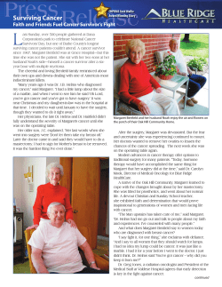


© Copyright 2026