
Provisional PDF - Journal of Medical Case Reports
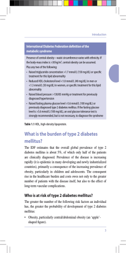
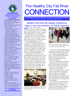
Journal of Medical Case
Reports
This Provisional PDF corresponds to the article as it appeared upon acceptance. Fully formatted
PDF and full text (HTML) versions will be made available soon.
Aminaphtone therapy in patients with type 1 diabetes and albuminuria: a case
report
Journal of Medical Case Reports 2014, 8:443
doi:10.1186/1752-1947-8-443
Concetta Romano ([email protected])
Consuelo Tamburella ([email protected])
Martino Costa ([email protected])
Marco Messina ([email protected])
Anna Lisa Fassari ([email protected])
Marco Bertini ([email protected])
ISSN
1752-1947
Article type
Case report
Submission date
2 July 2014
Acceptance date
30 October 2014
Publication date
19 December 2014
Article URL
http://www.jmedicalcasereports.com/content/8/1/443
This peer-reviewed article can be downloaded, printed and distributed freely for any purposes (see
copyright notice below).
Articles in Journal of Medical Case Reports are listed in PubMed and archived at PubMed Central.
For information about publishing your research in Journal of Medical Case Reports or any BioMed
Central journal, go to
http://www.jmedicalcasereports.com/authors/instructions/
For information about other BioMed Central publications go to
http://www.biomedcentral.com/
© 2014 Romano et al.
This is an Open Access article distributed under the terms of the Creative Commons Attribution License (http://creativecommons.org/licenses/by/4.0), which
permits unrestricted use, distribution, and reproduction in any medium, provided the original work is properly credited. The Creative Commons Public Domain
Dedication waiver (http://creativecommons.org/publicdomain/zero/1.0/) applies to the data made available in this article, unless otherwise stated.
Aminaphtone therapy in patients with type 1
diabetes and albuminuria: a case report
Concetta Romano1
Email: [email protected]
Consuelo Tamburella1
Email: [email protected]
Martino Costa1
Email: [email protected]
Marco Messina1
Email: [email protected]
Anna Lisa Fassari2
Email: [email protected]
Marco Bertini3*
*
Corresponding author
Email: [email protected]
1
Department of Cardiology, Acireale Hospital, Provincial Health Centre of
Catania, Catania, Italy
2
Department of Ophthalmology, Acireale Hospital, Provincial Health Centre of
Catania, Catania, Italy
3
Medical Department, Laboratori Baldacci SpA, Via San Michele degli Scalzi
73, 56124 Pisa, Italy
Abstract
Introduction
Microalbuminuria in type 1 diabetes is the earliest manifestation of diabetic microangiopathy
(nephropathy). To date, the pharmacological approach to microangiopathy has not been
shown to be useful. By using aminaphtone to control nephrologic complications of insulindependent diabetes mellitus we first obtained a significant improvement in microalbuminuria
confirming this new pharmacological approach for insulin-dependent diabetes mellitus
organospecific complications control.
Case presentation
After being treated with standard therapy for insulin-dependent diabetes mellitus (insulin) for
more than 20 years, a 49-year-old white man affected by insulin-dependent diabetes mellitus
adopted the standard therapy aminaphtone for a period of 2 months.
This therapy allowed a significant reduction of proteinuria from baseline evaluation that
immediately increased after he stopped aminaphtone therapy.
Conclusions
Aminaphtone therapy, used globally in the treatment and prevention of endothelial
dysfunctions, could be an interesting option for patients with insulin-dependent diabetes
mellitus with the express purpose of preventing diabetic nephropathy.
Keywords
Insulin-dependent diabetes mellitus, Microangiopathy
Introduction
Microangiopathic complications of diabetes include retinopathy, nephropathy and
neuropathy. These complications are specific to diabetes and arise as a result of
hyperglycaemia that lasts a long time. Undoubtedly, other metabolic factors, and
environmental and genetic factors are involved in the pathogenesis. Diabetic nephropathy
affects 20 to 40% of patients with insulin-dependent diabetes mellitus (IDDM), particularly
those with onset before puberty [1] and, probably, those with a hereditary predisposition to
hypertension [2]. Patients with diabetes with incipient nephropathy (persistent
microalbuminuria) have a 5 to 10 times greater risk of developing proliferative retinopathy
than those without albuminuria; furthermore, diabetic nephropathy is almost invariably
accompanied by retinopathy. The functional alterations that occur early in the natural history
of diabetic nephropathy include microalbuminuria and glomerular hyperfiltration.
Microalbuminuria is defined as a subclinical increase in the rate of urinary albumin excretion
in the range of 30 to 300mg/day. Microalbuminuria is due to the increased permeability of the
glomerular capillaries, probably secondary to increased glomerular capillary pressure [3] and
to the loss of negative charge at the level of the glomerular basement membrane.
Patients with IDDM with microalbuminuria have a 20 times greater risk of developing a
clinical nephropathy compared to those with a normal albumin excretion [4].
Microalbuminuria is considered a risk factor for diabetic nephropathy and progressive renal
failure in diabetes [5-8]. Longitudinal and cross-sectional studies conducted on type 2
diabetes have allowed the identification of risk factors associated with the development of
microalbuminuria and the progression of microalbuminuria to diabetic nephropathy. These
include: low body mass index, early onset of diabetes, hyperglycaemia, hypertension,
dyslipidaemia, cigarette smoking, and a family history of hypertension [9-14]. Although
microalbuminuria is considered by many authors to be the early stage of an irreversible
process, recent research has not confirmed this view, showing that microalbuminuria often
returns to its normal value [15,16]. The histopathological features characteristic of diabetic
kidney disease occur in the glomerulus. The main changes are: increase in the thickening of
the glomerular basement membrane, increase of volume of the mesangium, the presence of
hyaline deposits and global glomerular sclerosis [17]. The glomerular filtration rate (GFR) is
closely related to the surface of the basal membrane of the glomerular capillaries (filtration
surface) which is in turn determined by the number of clusters present at the time of
diagnosis, the entity of the expansion of the mesangium, the expansion capacity and the
number of sclerotic glomeruli. Urinary albumin excretion is related to the size of the pores of
filtration. The vascular endothelium plays a central role in the regulation of vascular tone.
Endothelin (ET) is a potent vasoconstrictor produced by endothelium that contributes to basal
vascular tone. Vasoconstriction in response to altered endogenous ET may lead to
hyperperfusion and subsequent microvascular damage. Hyperperfusion is one of the keys to
the onset and progression of microvascular complications in diabetes. Such a haemodynamic
condition can be determined by the excessive release of vasodilator substances, as for
example during ketonic decompensation, or for a reduced action of vasoconstrictive
substances. This interpretation may be the pathophysiological basis of what has been
observed in diabetes. In patients with diabetes there is a deficit of action of ET that, unlike in
the normal patient, does not induce vasoconstriction. In fact, individuals with diabetes have
levels of ET that would appear to be determined mainly by triglycerides and insulin.
Aminaphtone (2-hydroxy-3-methyl-1,4-napthohydroquinone-2-p-aminobenzoate) is a
synthetic molecule derived from four aminobenzoic acids which is currently employed for
“capillary disorders” and for chronic venous insufficiency [18]. This drug has recently
demonstrated the ability to downregulate ET-1 production in ECV304 cells by interfering
with transcription of preproET-1 (PPET-1) gene expression [19]. At the same time,
cytofluorometry has shown that aminaphtone significantly reduces the expression of Eselectin (endothelial-leukocyte adhesion molecule 1; ELAM-1) both in resting and in ET-B activated ECV304 cells in a dose-dependent manner [20]. In vivo, in patients affected by
systemic sclerosis, 12 weeks of aminaphtone treatment has demonstrated the ability to
downregulate sELAM-1 (soluble E-selectin adhesion molecules 1) and sVCAM-1 (soluble
vascular cell adhesion molecule 1) [21]. In a rat model of monocrotaline-induced pulmonary
hypertension, the administration of aminaphtone (30mg/kg/day or 150mg/kg/day)
significantly lowered rat mortality and significantly reduced plasma ET-1 concentration [22].
Aminaphtone has also demonstrated antiphlogistic activity on endothelial cells [23]. In order
to control vascular microangiopathy in patients affected by IDDM with proteinuria and
hypertension, we added aminaphtone to standard therapy for IDDM (insulin) and
hypertension (angiotensin-converting enzyme inhibitors).
Case presentation
A 49-year-old white man with type 1 diabetes has, from the age of 20, been treated with
NovoRapid® (insulin aspart; 3U breakfast time, 8U lunch time, and 10U dinner time) and
Lantus® (insulin glargine; 14U 10 p.m.) for glycaemic control for more than 20 years. He has
been hypertensive since 2006 and receives treatment with ramipril: one tablet 5mg at
breakfast time. He has also been treated with Torvast (atorvastatin; one tablet 10mg/day) for
hypercholesterolaemia. Microalbuminuria has been present in this patient since 2006
independently of any pharmacological treatments; no alterations have been reported in his
fundus oculi during the time. An examination of his nailfold periungual videocapillaroscopy
shows the presence of beanpole loops with dilated venous tract and presence of pearl-like
microbleeds with clear signs of microangiopathy (Figure 1). His microalbuminuria value was
487.1mg/L; GFR was under 58mL/minute; creatinine was 1.31mg/dL and arterial blood
pressure was 140/85mmHg. He began treatment with aminaphtone 75mg one tablet twice a
day for 2 months. After 2 months of therapy he was re-evaluated by videocapillaroscopic
examination showing a clear resolution of profuse bleeding (Figure 2). At the same time his
microalbuminuria value significantly decreased (98.3mg/L); his GFR (under 57mL/minute)
and creatinine (1.33mg/dL) were substantially unchanged but his arterial blood pressure
decreased to 130/80mmHg. Taking into account the results obtained with the above
treatment, he continues to take aminaphtone; however, intake was spontaneously interrupted
for 2 months. After 2 months without aminaphtone treatment he repeated proteinuria
evaluation: his microalbuminuria significantly increased to 573.0mg/L while his GFR (under
58mL/minute) and creatinine (1.31mg/dL) were unchanged and his arterial blood pressure
again increased to 140/85mmHg. No side effects related to aminaphtone were detected during
the trial.
Figure 1 Periungual videocapillaroscopic: microbleeds spread “pearl parades”.
Figure 2 Periungual videocapillaroscopic: resolution of profuse bleeding.
Discussion
This case report initially describes a significant reduction of proteinuria in a patient with
diabetes type 1, due to the use of aminaphtone. Our patient was first evaluated after 2 months
of treatment (aminaphtone capsules 75mg twice a day), showing a significant decrease in
microalbuminuria (from 487.1mg/dL to 98.3mg/dL = −388.8mg/dL) confirmed by an
improvement in the capillaroscopic examination, while GFR and creatinine remained
substantially unchanged. Surprisingly, arterial blood pressure decreased from 140/85mmHg
to 130/80mmHg after treatment. Subsequently, he continued treatment with aminaphtone for
2 months, but spontaneously interrupted the treatment. He was re-evaluated 2 months after
discontinuation of the drug and, again, showed an increase in proteinuria (from 98.3mg/dL to
573mg/dL = +474.7mg/dL), substantially the same GFR and creatinine values, and a new
increase in arterial blood pressure to 140/85mmHg: this kind of re-challenge clearly
demonstrates the relationship between proteinuria and aminaphtone therapy. The recent
preclinical findings about aminaphtone (anti-ET-1 and anti-E-selectin activities together with
a significant endothelial antiphlogistic effect) [19-23] seem to correlate with this result,
confirming that microangiopathy is the leading cause of microalbuminuria in patients with
IDDM.
Conclusions
Aminaphtone therapy seems to be useful for proteinuria control in patients with type 1
diabetes. The correlation between proteinuria and arterial blood pressure that significantly
decreased after aminaphtone treatment and increased once treatment was stopped was
surprising. To the best of our knowledge, descriptions of aminaphtone’s activity have not
included a report of a novel therapeutic option. For the purposes of therapeutic effectiveness,
the duration of treatment (more than 2 months) together with the compliance of daily
administration of the drug appears to be relevant. Further controlled studies, including
randomised double-blind controlled trials, are needed to definitively evaluate aminaphtone’s
efficacy over the long term in patients with IDDM. Nevertheless, the lesson to take from this
case report is that a pharmacological treatment of diabetic microangiopathy with a vascular
drug like aminaphtone could be useful for proteinuria control postponing IDDM nephrologic
complications.
Consent
Written informed consent was obtained from the patient for publication of this case report
and accompanying images. A copy of the written consent is available for review by the
Editor-in-Chief of this journal.
Abbreviations
ET, Endothelin; GFR, Glomerular filtration rate; IDDM, Insulin-dependent diabetes mellitus.
Competing interests
C Romano, C Tamburella, M Costa, M Messina and AL Fassari declare that they have no
competing interests.
Marco Bertini has competing interests because he is the Medical Department Director of
Laboratori Baldacci, the company that produces aminaphtone.
Authors’ contributions
CR, CT, MC and MM clinically followed the patient during his hospitalisation time; CR
performed videocapillaroscopy examination; ALF followed the time course of the retinal
complications of the patient; MB conceived of the study, collected the data and helped to
draft the manuscript. All authors read and approved the final manuscript.
Acknowledgments
We would like to thank Mr Alessandro Moscatelli for his technical support in preparing the
final manuscript.
References
1. Kofoed-Enevoldsen A, Borch-Johnsen K, Kreiner S, Nerup J, Deckert T: Declining
incidence of proteinuria in type 1 (insulin-dependent) diabetic patients in Denmark.
Diabetes 1987, 36:205–209.
2. Krolewski AS, Caressa N, Warram JH, Laeffe LMB, Christlieb AR, Knowler WC, Rand
LI: Predisposition to hypertension and susceptibility to renal disease in insulindependent diabetes. N Engl J Med 1988, 318:140–145.
3. Myers BD, Winets JA, Chui F, Michaels AS: Mechanism of proteinuria in diabetic
nephropathy: a study of glomerular barrier function. Kidney Int 1982, 21:633–641.
4. Viberti GC, Hill RD, Jarrett RJ, Argyropoulos A, Mahmud U, Keen H:
Microalbuminuria as a predictor of clinical nephropathy in insulin-dependent diabetes
mellitus. Lancet 1982, i:1430–1432.
5. Ritz E: Nephropaty in type 2 diabetes. J Int Med 1999, 245:111–126.
6. Alzaid AA: Microalbuminuria in patients with NIDDM: an overview (Review).
Diabetes Care 1996, 19:69–89.
7. American Diabetes Association: Diabetic nephropathy (Position Statement). Diabetes
Care 2000, 23(Suppl. 1):S29–S72.
8. Mogensen CE: Microalbuminuria, blood pressure and diabetic renal disease: origin
and development of ideas. Diabetologia 1999, 42:263–285.
9. Ritz E, Reinhold O: Nephropathy in patients with type 2 diabetes mellitus. N Engl J
Med 1999, 341:1127–1133.
10. Niskanen LK, Penttila I, Parviainen M, Vusitupa MIJ: Evolution, risk factors, and
prognostic implications of albuminuria in NIDDM. Diabetes Care 1996, 19:486–493.
11. The Microalbuminuria Collaborative Study Group: Predictors of the development of
microalbuminuria in patients with type 1 diabetes mellitus: a seven-year prospective
study. Diabet Med 1999, 16:918–925.
12. Klein R, Klein BE, Moss SE: Relation of glycemic control to diabetic microvascular
complications in diabetes mellitus. Ann Intern Med 1996, 124:90–96.
13. Villar G, Garcia Y, Goicolea I, Vasquez JA: Determinants of development of
microalbuminuria in normotensive patients with type 1 and type 2 diabetes. Diabet
Metab 1999, 25:246–254.
14. Warram JH, Scott LJ, Hanna LS, Wantam M, Cohen SE, Laffel LMB, Ryan L, Krolewski
AS: Progression of microalbuminuria to proteinuria in type 1 diabetes: nonlinear
relationship with hyperglycemia. Diabetes 2000, 49:94–100.
15. Ismail N, Beckner B, Strtzelczyk P, Ritz E: Renal disease and hypertension in noninsulin-dependent diabetes mellitus. Kidney Int 1999, 55:1–28.
16. Caramori ML, Fioretto P, Mauer M: The need for early predictors of diabetic
nephropathy risk. Diabetes 2000, 49(9):1399–1408.
17. Olgemoller B, Schleicher E: Alterations of glomerular proteins in the pathogenesis of
diabetic nephropathy. Clin Invest 1993, 71:S13–S19.
18. Atenas JG: The efficacy of aminaphtone in patients suffering from chronic venous
insufficiency. Ann Exp Clin Med 1995, 2:81–85.
19. Scorza R, Santaniello A, Salazar G, Lenna S, Colombo G, Turcatti F, Beretta L:
Aminaftone, a derivative of 4-aminobenzoic acid, downregulates endothelin-1
production in ECV304 Cells: an in vitro Study. Drugs RD 2008, 9(4):251–257.
20. Lenna S, Salazar G, Della Bella S, Antonioli R, Beretta L, Santaniello A, Scorza R: Novel
Mode of Action of The Aminaphtone: Down-Regulation of E-Selectin expression in ECV304
Cells. Lisbon (Portugal): Abstract Book of the XXII World Congress of the International
Union of Angiology; 2006.
21. Scorza R, Santaniello A, Salazar G, Lenna S, Della Bella S, Antonioli R, Toussoun K,
Beretta L: Effects of aminaftone 75mg TID on soluble adhesion molecules: a 12-week,
randomized, open label pilot study in patients with systemic sclerosis. Clin Ther 2008,
30(5):924–929.
22. Zambelli V, Santaniello A, Fumagalli F, Masson S, Scorza R, Beretta L, Latini R:
Efficacy of aminaftone in a rat model of monocrotaline-induced pulmonary
hypertension. Eur J Pharmacol 2011, 667:287–291.
23. Scorza R, Salazar G, Bellocchi C: Aminaftone: un possibile ruolo nella sclerosi
sistemica?. Gruppo Italiano per la Lotta alla Sclerodermia (GILS). Rubrica Medica – Giugno
2013.
Figure 1
Figure 2


© Copyright 2026