
origin - Stomatološki vjesnik
Stomatološki vjesnik Stomatological review Stomatološki vjesnik 2014; 3(2) ISSN 0350-5499 UDK 616.31 Izdavač / Publisher: Stomatološki fakultet Univerziteta u Sarajevu / Faculty of Dentistry, University of Sarajevo Za izdavača / For publisher: Sead Redžepagić ČLANOVI UREĐIVAČKOG ODBORA / EDITORIAL BOARD : Glavni urednik / Editor in chief: Sadeta Šečić Sekretar uređivačkog odbora / Secretary of editorial board: Selma Zukić Članovi /Members: Sead Redžepagić, Samir Prohić, Muhamed Ajanović, Amira Dedić, Sedin Kobašlija, Tarik Mašić, Amra Vuković, Enita Nakaš MEĐUNARODNI UREĐIVAČKI ODBOR / INTERNATIONAL EDITORIAL BOARD: Anwar Barakat Bataineh (Irbid, Jordan), Jasenka Živko-Babić (Zagreb, Hrvatska), Andrija Petar Bošnjak (Rijeka, Hrvatska), Hrvoje Brkić (Zagreb , Hrvatska), Dolores Biočina Lukenda (Split, Hrvatska), Davor Katanec (Zagreb, Hrvatska), Šahza Hatibović Koffman (London Ontario Kanada), Mladen Kuftinec (USA), Darko Macan (Zagreb, Hrvatska), Berislav Perić (Zagreb, Hrvatska), Tore Solheim (Oslo, Norveška), Dragoslav Stamenković (Beograd, Srbija), Marin Vodanović (Zagreb, Hrvatska) Lektor za engleski jezik / English language editor: Nermana Bičakčić Tehničko uređenje / Technical editor: Branislav Trogrančić Štampa / Printed by: Štamparija Fojnica Dizajn naslovnice / Cover page design: Lana Malić Tiraž/ Number of copies: 200 KONTAKT / CONTACT: Stomatološki vjesnik Stomatološki fakultet sa klinikama Bolnička 4a, 71000 Sarajevo Bosna i Hercegovina Telefon: + 387(33)214 294 e-mail: [email protected] Web: www.stomatoloskivjesnik.ba TRANSAKCIJSKI RACUN / TRANSFER ACCOUNT: 33386902296551066 UniCredit Bank dd Printed on acid free paper Svrha i i cilj : Stomatološki vjesnik je neprofitni naučno stručni časopis koji publicira originalne naučne radove, prikaze slučajeva, pisma uredniku, savremene perspektive, editorijale, preliminarne komunikacije u oblasti stomatologije i drugih biomedicinskih nauka. Radovi su na Bosanskom/Hrvatskom/Srpskom jeziku sa naslovom, sažetkom i ključnim riječima bilingvalnim B/H/S i engleskom jeziku. Radovi se mogu koristiti u edukacijske svrhe bez predhodnog odobrenja, a uz obavezno navođenje izvora. Korištenje cijelih ili dijelova članaka u komercijalne svrhe nije dozvoljeno bez predhodnog pismenog odobrenja izdavača Autorska prava posjeduje izdavač: Stomatološki fakultet sa klinikama Univerziteta u Sarajevu. Aim and Scope: Stomatološki vijesnik / Stomatological review is a non-profit scientific journal that publishes original articles, case reports, letters to the editors, current perspectives, editorials, fast-track articles in a field of dentistry and other bio-medical sciences. Papers are in Bosnian/ Croatian/Serbian language with at least title, abstract and key words bilingual in B/C/S and English language. All manuscripts undergo the peer review process before can be accepted for publishing in Stomatološki vjesnik/ Stomatolgical review. Papers can be used for educational purposes without prior consent only with adequate citation of the sources. Using whole or parts of articles for commercial purposes is not permitted without prior written permission of the publisher. Copyright owns the publisher: Faculty of Dentistry with Clinics, University of Sarajevo. Časopis Stomatološki vjesnik je oslobođen poreza na promet prema Mišljenju Federalnog ministarstva obrazovanja, nauke, kulture i sporta br: 04-15-661/2002. Journal Stomatological review is tax exempt according to the opinion of the Federal Ministry of Education Science Culture and Sports no: 04-15-661/2002. Indexed in: IIC (Index Copernicus International), DOAJ (Directory of Open Access Journal), EBSCO, HINARI, INFOBASE, EZB (Electronishe Zeitschriftenbibliothek), SJIF (Scientific Journal Impact Factor Value 2.502), GOOGLE SCHOLAR Stomatološki vjesnik CONTENTS / SADRŽAJ ORIGINAL SCIENTIFIC ARTICLES / ORIGINALNI NAUČNI RADOVI DENTAL ASPECTS OF CHILD ABUSE AND NEGLECT - KNOWLEADGE AND ATTITUDES OF DENTISTS IN BOSNIA AND HERZEGOVINA Marković N, Arslanagić Muratbegović A, Kobašlija S, Bajrić E, Selimović-Dragaš M, Huseinbegović A, Čuković-Bagić I 83 DEVELOPMENT STAGES OF THE THIRD MOLAR IN THE BOSNIAN AND HERZEGOVINIAN CHILDREN AND ADOLESCENTS Selmanagić A, Vuković A, Brkić H , Galić I, Zukić S, Prohić S 91 EVALUATION OF ORAL HEALTH IN 12-YEAR OLD CHILDREN IN THE VOGOŠĆA MUNICIPALITY, THE SARAJEVO CANTON 97 Porović S, Koradžić-Zuban S, Spahić-Dizdarević M, BrkaniĆ B, Branković B, Cilović-Lagarija Š COMPARISON OF INFLUENCE OF TWO DIFFERENT EPINEPHRINE CONCENTRATIONS IN LOCAL ANESTHESIA ON HEMO-DYNAMIC PARAMETERS IN PATIENTS WITH HYPERTENSION Trninić S, Šečić S, Trninić L 103 RELATIONSHIP BETWEEN PERIODONTAL STATUS AND OSTEOPOROSIS AMONG WOMEN IN MENOPAUSE Gojkov Vukelić M, Hadžić S, Dedić A, Pašić E 111 REVIEW ARTICLE / PREGLEDNI ČLANAK CURRENT PERSPECTIVES ON GLASS-IONOMER CEMENTS IN PAEDIATRIC DENTISTRY Negovetić Vranić D, Majstorović M, Ferk P, Jurić H 117 STRUČNI RAD /PROFESSIONAL ARTICLE LASER THERAPY IN BURNING MOUTH SYNDROME Pavlić V, Adamović T, Gojkov-Vukelić M, Markovic Djuric LJ 123 CASE REPORT / PRIKAZ SLUČAJA MAXILARY CANINE IMPACTION – EXTRACTION OR NOT? Kapusevska B, Dereban N, Benedetti A 129 DENTICLES ( PULP STONES) – A CHALLENGE IN ENDODONTIC THERAPY- MULTIPLE CASE REVIEW Konjhodžić- Prcić A, Korać S, Jakupović S, Vuković A 141 BOOK REVIEW / PRIKAZ KNJIGE ORTHODONTIC AND SURGICAL MANAGEMENT OF IMPACTED TEETH Kokich Vincent G , Mathews David P 147 Stomatološki vjesnik 2014; 3 (2) EDITOR'S ADDRESS Esteemed colleagues, It is our pleasure to present to you the new issue of "STOMATOLOŠKI VJESNIK". In this, sixth issue, we prepared nine articles representing 6 scientific, 1 expert work and 2 case studies. As it is already our practice, you may also find an overview of a book intended not only for oral surgeons and orthodontists, but also for dentists of other specialties. We sincerely hope to satisfy your expectations by this issue as well. With gratitude to the contributions you have given so far through your numerous articles and works with international characteristics, and extremely strict reviews, we succeeded in indexing "STOMATOLOSKI VJESNIK" into EBSCO, HINARI, INFOBASE and GOOGLE SCHOLAR data bases. Expecting your further support and active participation in the preparation of this journal, we wish you lot of personal and professional success in the forthcoming 2015. Sadeta Šečić Editor in chief 82 Stomatološki vjesnik 2014; 3 (2) ORIGINAL SCIENTIFIC ARTICLE / ORIGINALNI NAUČNI RAD DENTAL ASPECTS OF CHILD ABUSE AND NEGLECT - KNOWLEADGE AND ATTITUDES OF DENTISTS IN BOSNIA AND HERZEGOVINA 1 1 *Corresponding author 1 1 Marković N* , Arslanagić Muratbegović A , Kobašlija S , Bajrić E , Selimović-Dragaš M1, Huseinbegović A1, Čuković-Bagić I2 1 Department of Preventive and Pediatric Dentistry, Faculty of Dentistry, University of Sarajevo, Sarajevo, Bosnia and Herzegovina 2 Department of Paediatric and Preventive Dentistry, School of Dental Medicine, University of Zagreb, Zagreb, Croatia Nina Marković, Assistant Professor, PhD Department of Preventive and Pediatric Dentistry Faculty of Dentistry University of Sarajevo, Bolnicka 4a 71000 Sarajevo Bosnia and Herzegovina Phone: +387 33 214 249, ext. 107 e-mail: [email protected] ABSTRACT Dentists can play an important role in detecting and reporting cases of CAN, particularly in cases of medical and dental neglect of their young patients. The objective of this study was to assess the current status of knowledge and attitudes of dentists in Bosnia and Herzegovina regarding CAN, particularly in recognition of signs and symptoms and reporting procedure of suspected cases. Methods: Data were collected through a self-administrated structured questionnaire offered to 300 dentists. The questionnaire consisted of twenty- seven questions adopted and modified from previous studies. Final sample consisted of a total number of 210 questionnaires that were filled correctly and returned. Questions were analyzed by means of descriptive statistics. Non parametric statistical tests were used to analyze differences among respondents in respect of age, gender, professional experience, specialty, and workplace. The level of significance was set at (p<0.05). Results: Only 7.6% of respondents indicated that they had formal training about CAN, 66% of dentists had never suspected CAN. In the section of social indicators respondents usually indicated correct answer (more then 60% ). If they suspect, only 9% of them would report CAN. Prevailing reasons for not reporting suspected case of CAN was lack of knowledge of the reporting procedure (43%), never had a case and lack of knowledge about the procedure (31%) and lack of adequate history (15%). Conclusion: The study demonstrated a lack of overall understanding of the problem and also a lack of awareness of dentists' responsibilities regarding this issue. Key words: child abuse, child neglect, dental neglect, dentist, Bosnia and Herzegovina. Stomatološki vjesnik 2014; 3 (2) 83 DENTAL ASPECTS OF CHILD ABUSE AND NEGLECT - KNOWLEADGE AND ATTITUDES OF DENTISTS IN BOSNIA AND HERZEGOVINA Introduction Child abuse and neglect (CAN) are serious public problem present worldwide in all countries and socioeconomic groups. Despite global awareness, data on prevalence and incidence of CAN are lacking in many countries. Prevention and management strategies are very diverse [1]. Dentists can play an important role in detecting and reporting cases of CAN, particularly in cases of medical and dental neglect of their young patients. International studies emphasis the role of dentists in detecting CAN [1, 2, 3, 4, 5, 6, 7]. Dental health care providers are most likely the first who recognize the evidence of physical abuse. Oro-facial trauma is present in approximately 50 to 75 percent of all reported cases of physical child abuse [1, 6, 8]. The reason may be in the fact that these areas are being exposed and accessible or because it is considered to represent the whole personality [1]. It is well documented that parents and guardians avoid returning to the same physician, but they repeat visits to the same dentists [6]. Different nationwide studies reported low percentage of dentist's participation in reporting suspected cases of CAN [4,6,9,10]. Reasons could be incomplete knowledge of reporting procedures of signs and symptoms of CAN, or lack of awareness of their roll in all important aspects of CAN. Therefore, adequate education and training of dental practitioners is an important part for managing CAN in society. Properly trained dentists would be more confident in their ability to recognize child abuse, and more cases would be diagnosed and reported by dental profession. In Bosnia and Herzegovina (BH), the protection of children from abuse is mostly regulated by the Criminal Law, but some aspects of CAN, including dental neglect, are not legally regulated at all [11]. Health workers and other BH citizens, have a legal obligation to report suspected cases of CAN to the relevant authorities [11]. Dentists do not diagnose CAN, but they do have legal, moral, ethical and professional obligation to report suspected cases [11]. Few studies in Bosnia and Herzegovina analyzed the role of healthcare workers in detecting and reporting CAN [11]. Dentists' level of knowledge in important aspects of this issue has not been investigated so far. 84 Objective The objective of this study was to assess the current status of knowledge and attitudes of dentists in Bosnia and Herzegovina regarding CAN, particularly in recognition of signs and symptoms and reporting procedure of suspected cases. Methods The survey was carried out between March and June 2013 as a cross-sectional study. Data were collected through a self-administrated structured questionnaire offered to 300 dentists that were working in 7 different towns in BH. General dentists and dental practitioners from all specialties who worked with children on daily bases were included in the study. A total number of 210 (70%) dentists agreed to participate in the study and returned questionnaire filled correctly. The questionnaire was reviewed by two independent professionals with knowledge in this field. The questionnaire consisted of twenty-seven questions adopted and modified from previous studies [1,5,6,7]. The cover letter included information about nature and the purpose of the study, consent of the participation on voluntary bases, state on confidentiality of the information provided. The questionnaire was distributed to the dentists by hand from an administrator. The average time to complete the survey was approximately ten minutes. The questionnaire was returned to the administrator in closed envelope. The questionnaire included 27 questions in 5 sections regarding demographic characteristics of the respondents, knowledge of social indicators, level of education, knowledge and attitudes of physical abuse and reporting procedure. All questions about knowledge and attitudes were formulated as a statement using multiple choice or true/false formats. Data were coded and entered into statistical program SPSS (Statistical Package for the Social Sciences, v. 13) and analyzed by using descriptive analysis for responses to each question. Question by question were tabulated and analyzed using frequencies and percentages from responses to each Stomatološki vjesnik 2014; 3 (2) Marković N, Arslanagić Muratbegović A, Kobašlija S, Bajrić E, Selimović-Dragaš M, Huseinbegović A, Čuković-Bagić I questions to compile prevalence data. Non parametric statistical tests (The chi-square test and Kruzskal Wallis H test) were used to analyze differences among respondents in respect of age, gender, professional experience, specialty, and workplace. The level of significance was set at p<0.05. Results All subjects (n=210) indicated to treat pediatric patients on daily bases. Study sample was described by distribution of frequencies and percentages and it is presented in Table 1. Education about CAN Yes (%) No (%) Any kind of education 8 92 Provided with any kind of informations 16 84 Have had formal training 7.6 92.4 Table 2. Level of educational training about CAN The level of educational training and self education regarding CAN are presented in Table 2. Only 7.6% of respondents said that they had formal training on CAN. Knowledge of social indicators related to CAN and distribution of answers were presented in Table 3. Percentage Questions/social indicators 64 30 % 146 70% CAN is associated with poverty and low- income earners Number Gender Male Female 30-49 years >50 years 36 17% 140 67% 34 16% Years of working experience 101 48% 10-19 65 31% 20-30 25 12% >30 19 9% <10 145 69 No* 15 7 Don't know 50 24 Yes 36 17 No* 84 40 Don't know 90 46 Yes 41 19 No* 73 35 Don't know 96 46 Yes* If a child readily states that adult has caused No harm, the accusation should be addressed. Don't know 137 65 14 7 59 28 Yes* 96 46 No 23 11 Don't know 50 24 Yes* 145 69 No 15 7 Don't know 50 24 Younger children tell someone soon after the abuse. Elder children tell someone soon after the abuse. Dental specialties 130 62% 80 38% Public practice 147 70% Private practice 63 30% 210 100 % General dentists Specialties working with children Children are not removed from parents` homes in cases of abuse. Practice Total Table 1. Description of participants in the study Stomatološki vjesnik 2014; 3 (2) Number Percentage Yes Age <30 years Answer Parent delays seeking medical attention in child abuse cases * Correct answer is indicated with asterix. Table 3. Knowledge of social indicators of CAN among BH dentists 85 DENTAL ASPECTS OF CHILD ABUSE AND NEGLECT - KNOWLEADGE AND ATTITUDES OF DENTISTS IN BOSNIA AND HERZEGOVINA In this section respondents usually indicated correct answer. However, the fact that the answer "don't know" was frequently indicated revealed a lack of knowledge related to social indicators of CAN. Statistically significant difference was found for the question: "CAN are usually associated with poverty and low-income earners, and rarely occur among middleor high-income earners", between dentists from pri2 vate and public practice (c (2,n = 210) = 7.08 = p = 0.029, Cranmer`s V = 0,184), with correct answer mostly prevailing among dentist in public practice. More females selected "yes" in question: "Elder children who have been abused usually tell someone soon after the abuse." (c2 (2,n = 210) = 7.39 = p = 0.025, Cranmer`s V=0,082). Significantly more general dentists answered correctly that: "Child abuse may be indicated if a parent delays seeking medical attention"(c2 (2,n = 210) = 7.28 = p = 0.026, Cranmer`s V=0,082). Statistical significant differences between other independent variables in this section were not found Results of knowledge of physical indicators of CAN were given in Table 4. Respondents usually indicated correct answer except for question number 6 and 7 were "don't know" was the most prevalent answer. The last, tenth question was different. Respondents were asked to indicate what could be possible indicator of CAN. Only 15 % gave correct answer indicated "all of the above", 21% indicated psychosomatic complaints and the most prevalent answer was "psychosomatic complaints, unusual knowledge about sexual matters, untidy appearance"(46%). Statistical differences were found only between general dentists and specialists to the questions regarding observed bite-marks as possible indicator of CAN (Question 1) and bruises on cheeks and neck as possible indicators of CAN (Question 6). Whereas correct answer was more frequent among general dentist. Question 1: (c2 (2,n = 210) = 10.91 = p = 0.004, Cranmer`s V=0,228). Question 6:(c2 (2,n = 210) = 8.45 = p = 0.009, Cranmer`s V=0,201). Majority of BH dentists have never suspected CAN (66%), and only 9% of them if suspected, would report. Significant differences were observed between dentists in private and public practice (c2 (1,n = 210) = 8.83 = p = 0.03, fi = -0,216). Results of knowledge and attitudes regarding legal and ethical responsibilities should be omitted since they can be seen in Table 5. 86 Questions /possible indicators observed during normal course of a dental visit 1. Observed bite marks 2. Bruises on the cheek 3. Additional bruises in areas overlying bony prominences 4. Poor oral health as a part of physical neglect 5. Repeated injury to the dentition 6. Bruises on the neck are usually associated with accidental trauma 7. Burns associated with child abuse cases have shapes of hot objects 8. Parent describes a child injury as a self-inflicted injury 9. Parent describes a repeated child injury as a injury made by brothers or sisters Answer Number Percentage True* 128 61 False 26 12 Don't know 56 27 True* 163 78 False 22 10 Don't know 25 12 True* 102 49 False 59 28 Don't know 49 23 True* 148 70 False 43 21 Don't know 19 9 True* 41 19 False 73 35 Don't know 96 46 True 31 15 False* 136 65 Don't know 43 20 True* 101 48 False 25 12 Don't know 84 40 True* 162 77 False 12 6 Don't know 36 17 True* 129 61 False 22 11 Don't know Psychosomatic complaints 59 28 39 19 8 4 Untidy appearance, untidy clothes 14 7 Psychosomatic complaints, unusual knowledge about sexual matters, untidy appearance 96 46 All of the above* 32 15 Don't know 21 9 Seductive behavior, unusual knowledge about sexual matters, avoiding eye contact 10. Possible indicators of suspected CAN * Correct answer is indicated with asterix. Table 4. Knowledge of signs of physical abuse among BH dentists Stomatološki vjesnik 2014; 3 (2) Marković N, Arslanagić Muratbegović A, Kobašlija S, Bajrić E, Selimović-Dragaš M, Huseinbegović A, Čuković-Bagić I Questions Did you ever suspect CAN If suspect did you report case of CAN Were to report suspected CAN Reasons for not reporting suspicions of CAN Answer Number Percentage Yes 71 34 No 139 66 Yes 18 9 No 107 51 No answer 85 40 Social service* 160 76 Police 24 11 Social service and police 26 12 No* 121 58 No answer 21 11 Lack of adequate history 31 15 Lack of knowledge and the procedure of reporting 90 43 Lack of confidence that reports will be correctly investigated and fear that it may cause more harm than good 11 5.1 Never have a case, lack of knowledge about procedure of reporting 66 31 No answer 12 6 * Correct answer is indicated with asterix. Table 5. Knowledge and attitudes regarding legal and ethical responsibilities among BH dentists The most common reason for not reporting suspicions of CAN was lack of knowledge regarding the subject including the reporting procedure (43%), followed by lack of knowledge about procedure of reporting together with "did not have a case of CAN" (31%), lack of adequate history (15%), and lack of Stomatološki vjesnik 2014; 3 (2) confidence that report would be investigated correctly along with fear that it could cause more harm than good (5%). This question did not answer 6% of respondents. The survey was approved by the Federal Ministry of Education and Science of Federation of Bosnia and Herzegovina and Faculty of Dentistry of University in Sarajevo, Bosnia and Herzegovina. Discussion Child abuse and neglect (CAN) has been defined by the World Health Organization (WHO,1999) as "Every kind of physical, sexual, emotional abuse, neglect or negligent treatment, commercial or other exploitation resulting in actual or potential harm to the child's health, survival, development or dignity in the context of a relationship of responsibility, trust or power."[12]. Abuse can be physical, sexual, psychological and economic [11]. Neglect can be physical, psychological, medical (health) and educational [11]. Health neglect is defined as intentional failure to provide necessary medical treatment and health protection [11]. American Academy of Pediatric Dentistry (AAPD) defined dental neglect, as willful failure of a parent or guardian to seek and follow through with whatever treatment is necessary to ensure a level of oral health essential for adequate chewing function and freedom from pain and infection [13]. WHO estimated that almost 53,000 children died worldwide in 2002 from homicide [8]. In the United States each year almost 2,000 infants and young children die from being abused or neglected by caretakers [6]. A ten year study of 29 fatal cases of child abuse found a high incidence of facial injuries, with laceration of the mucosa of the upper lip that were occurring in almost half of the cases [2]. These types of injuries have been recognized in the literature as pathogmonic of a child abuse [2]. The problem is more extended in low economic countries associated with low incomes, poverty, high rate of unemployment, increase level of stress and unstable political situation [4, 8,12]. Bosnia and Herzegovina has all of the listed risk factors. Child abuse and neglect are significant problems worldwide. Numerous studies had showmen that oro -facial region, head and neck are common sites of 87 DENTAL ASPECTS OF CHILD ABUSE AND NEGLECT - KNOWLEADGE AND ATTITUDES OF DENTISTS IN BOSNIA AND HERZEGOVINA trauma of child abuse [2, 4,5,6,]. Statistics have reported that 50% to 70% of all cases of child abuse include trauma to the mouth, face and head [5]. The lower number of intra-oral injuries recorded in comparison to high number of head and face injuries suggests that they may be overlooked due to the unfamiliarity of the medical practitioners with interior examinations [1]. Dental professionals account for only small percentage of reports of overall cases of CAN. In the United States only 1% of all reported cases were made by dentists [5,6]. Results of available studies indicated that dentists failed to report suspected cases of CAN, mainly due to lack of knowledge and awareness of their ethical (and/or legal) responsibilities [1,34,5,6,9,10]. There is a lack of epidemiological reports of CAN for BH population. Few studies in BH have analyzed the role of healthcare workers in recognizing and reporting CAN [11]. Suspected evidence of child abuse was found in 19% of children who underwent regular medical checkups [11]. The findings of this cross-sectional study present baseline survey of knowledge and attitudes of dentists in BH towards CAN. The findings of the present survey indicated a national problem of lack of knowledge in many areas related to the CAN issue. The most of dentists (92%) had no education related to CAN, 84% stated that they were not provided so far with any kind of related information. Only 7.6% of respondents have been trained on issues related to CAN. Having in mind that education is very important in this issue, and that this issue is very important for community, the lack of education and training courses is serious problem for all authorities involved. Subject of CAN has not been included so far in curriculum for undergraduate and postgraduate students in dental schools in BH. Findings of numerous studies emphasize the need for mandated training related to recognition of CAN through long-life learning courses [1, 4,5,7,8,14]. Obviously, many correct answers in the study were drawn by logical conclusion rather than decision making based on knowledge. Knowledge of social issues related to CAN was evaluated in six questions were respondents indicated answers such as: yes, no or don't know. In this section respondents usually indicated correct answer, but "don't know" as an option was selected frequently as well indicating a lack of knowledge related to social indicators of CAN. 88 Many BH dentists recognized a bite –marks and bruises in oro-facial region as a sign of a physical abuse (61 % and 78%), but repeated injury to the dentition were recognized by only 19% of them. General knowledge of trauma may be the reason for frequent indicating correct answers in section of signs and symptoms of physical abuse. Several articles have documented evidence of cutaneous bitemarks and bruises in oro-facial region in patients suspected to child abuse [2,5,6]. Bite- marks injuries are rarely accidental and are good indicator of physical abuse [2]. Principal injuries reported in cases of suspected child abuse were fractures of teeth, oral bruises, and oral lacerations, fractures of maxilla and mandible and oral burns [2]. Dentists are usually the first health care providers who see patients with dental trauma; therefore they have to be aware of the importance of the issue and of their role in recognizing and reporting suspected cases. This is very important for prevention of further injuries and maltreatment and for providing better treatment for young domestic violence victims. Reporting suspicions of CAN on time can save life of young victim or victims if more than one individual is involved. Psychosomatic complaints, unusual knowledge about sexual matters, untidy appearance were most often indicated by respondents as indicators of CAN. Seventy percents of BH dentist associated poor oral health with physical neglect. Poor oral health could be considered as a serious dental neglect. By the definition of the American Academy of Pediatric dentistry dental neglect is considered a form of CAN when caregiver is aware of a child need for dental care; bat willfully denies the children from dental care [13]. It is considered that untreated caries, periodontal diseases, and other conditions can cause pain, infection and lost of function [4,11,15]. Dental neglect affects nutrition, sleeping, learning, communication and other activities necessary for normal growth and development of infants [4]. The most frequent indicator of dental neglect is rampant early childhood caries [11,16]. Epidemiological findings of oral health status of children in BH, where dmft was more then 6, suggests that dental neglect is widely spread in the population [17,18]. However, it is necessary to reconsider weather poor oral health is due to parent's ignorance. In countries with low socioeconomic status and many problems in health care system, poor oral health of citizens is most likely due to financial deprivation, inaccessibility to dental Stomatološki vjesnik 2014; 3 (2) Marković N, Arslanagić Muratbegović A, Kobašlija S, Bajrić E, Selimović-Dragaš M, Huseinbegović A, Čuković-Bagić I health care or lack of prevention and public oral health organization and promotion activities. Caries and periodontal diseases are multi-factor diseases, and many aspects may influence their development and progression. Therefore, it is necessary to assess patient's detailed history and to be well trained and experienced to address poor oral health as neglect exclusively. Results of legal and ethical issues suggest that a few respondents would act on the suspicion on CAN. Only 9 % answered that they would report suspected CAN. A total of 34% of them had suspicions on CAN in their practice. The most common reasons for not reporting was lack of knowledge of the reporting procedure (43%), never had a case along with lack of knowledge about the procedure (31%) and lack of adequate history (15%). These findings are similar to those in other studies [2, 4, 6, 19]. Dentists from other surveyed studies indicated: lack of history (over 70%), uncertainty about diagnosis (over 70%), possible consequences to a child (over 60%), and effects of work litigation (over 50%) [2, 3, 4, 5, 6, 9, 10, 19]. Lack of awareness of the responsibility could be significant reason for not paying attention and not reporting CAN. According to results of the present study, significant proportions of respondents are aware of their responsibility to pay attention to CAN. Social and psychological barriers can be one more reason for under-reporting CAN. It is necessary to emphasize that dental professionals are only responsible for reporting their suspicions to social service that is in charge for further investigations and appropriate course action if necessary. High proportion of BH dentists new where to report suspected CAN (over 70%), and only half of respondents consider that they have professional obligation to pay attention to CAN. Significant differences between general dentists and specialists could be due to frequency of patients on daily bases. Dental professionals must become more aware of their moral, professional, legal and ethical responsibilities in recognizing and reporting child abuse and neglect. All health professionals including dental teams must be aware of the seriousness of the problem. It has to be seriously understood that abuse and neglect can lead to death as a result of maltreatment and molestation. Stomatološki vjesnik 2014; 3 (2) Conclusion The study revealed a lack of overall understanding of the problem and a lack of awareness of the responsibilities of dentists regarding this issue. In a view of the high likelihood of oro-dental injuries occurring in association with child abuse, there is a clear need for dentists to receive an adequate formal training at the levels of undergraduate and postgraduate studies and during continuing professional dental education trainings on topics regarding the recognition and reporting of CAN. Acknowledgments The permission and financial support of the Federal Ministry of Education and Science are acknowledged. Technical and administrative supports of Faculty of Dentistry, University of Sarajevo, Bosnia and Herzegovina are highly appreciated and acknowledged. Declaration of interests All authors clearly state that there is no conflict of interest for any person or institution included in this research project. References 1. Suhad HS, MDS, DCD, Moammar H, BDS. Jordanian dental students' knowledge and attitudes in regard to child physical abuse. J Dent Educ. 2010;74(10):1159-65. 2. Child abuse and dentistry: a study of knowledge and attitudes among dentists in Victoria, Australia. Aust Dent J. 1999;44(4):259-67. 3. Al-Dabaan R, Newton J.T, Asinakopoulou K, Knowledge, attitudes and experience of dentists living in Saudi Arabia toward child abuse and neglect, The Saudi Dental Journal.2014;26:7987. 89 DENTAL ASPECTS OF CHILD ABUSE AND NEGLECT - KNOWLEADGE AND ATTITUDES OF DENTISTS IN BOSNIA AND HERZEGOVINA 4. 5. 6. A survey of attitudes, knowledge and practice of dentists in London towards child protection. Are children receiving dental treatment at the Eastman Dental Hospital likely to be on the child protection register? Br Dent J. 2009; 28; 206(4): E7; discussion 212-3. Child maltreatment: a survey of dentists in southern Brazil. Braz Oral Res. 2012;26(1):5-11. Ramos-Gomez F, Rothman D, Blain S. Knowledge and attitudes among California dental care providers regarding child abuse and neglect. J Am Dent Assoc. 1998;129(3):340-8. 7. Thomas E.J, Straffon B.A. Inglehart RM, Child Abuse and Neglect; Dental and Dental Hygiene Students˛Educational Experiences and Knowledge. Journal of Dental Education. 2006; 70(5):558-565. 8. Croatian dental students' educational experiences and knowledge in regard to child abuse and neglect. J Dent Educ. 2012;76(11):1512-9. 9. Reporting of child abuse: a follow-up survey of Texas dentists. –Pediatr Dent. 2003;25(6):541-5. 10. Child abuse and dentistry: a study of knowledge and attitudes among Nigerian dentists. Afr J Med Med Sci. 2008;37(2):125-34. 11. Arslanagic- Muratbegovic A. Recognizing Child Abuse and Neglect in The Dental Office. Criminal Justice Issues. 2011; 5-6:47-58. 12. World Health Organization and International Society for Prevention of Child Abuse and Neglect. Preventing child maltreatment: a guide 90 to taking action and generating evidence. 2006. avail. : http://whqlibdoc.who.int/publications /2006/9241594365_eng.pdf. 13. American Academy of Pediatrics Committee on Early Childhood, Adoption, and Dependent Care: Oral and dental aspects of child abuse and neglect. Pediatrics. 1986;78(3):537-9. 24. 14. Child abuse and dentistry: what you should know. J Mich Dent Assoc. 1994;76(5):20, 22-7. 15. Čuković –Bagić I. The role of Dentist The role of the dentist in Recognition of Child Abuse. Acta Stomatol. Croat. 44(4):285-292. 16. Valencia-Rojas, N., Lawrence, H.P., Goodman, D. Prevalence of early childhood caries in a population of children with history of maltreatment.Journal of Public Health Dentistry.2008; 68(2), 94-101. 17. Arslanagić Muratbegović A, Marković N, Zukanović A, Selimović Dragaš Mediha, Kobašlija S, Jurić H. Oral Health Related to Demographic Features in Bosnian Children Aged Six. Coll. Antropol. 2010; 34(3): 1027-1033. 18. Marković N, Arslanagić Muratbegović A, Kobašlija S, Bajrić E, Selimović-Dragaš M, Huseinbegović A. Caries prevalence of children and adolescents in Bosnia and Herzegovina. Acta Medica Academica 2013; 42(2):108-116. 19. Owais AI, Qudeimat MA, Qodceih S. Dentists' involvement in identification and reporting of child physical abuse: Jordan as a case study. Int J Paediatr Dent. 2009;19(4):291-6. Stomatološki vjesnik 2014; 3 (2) ORIGINAL SCIENTIFIC ARTICLE / ORIGINALNI NAUČNI RAD DEVELOPMENT STAGES OF THE THIRD MOLAR IN THE BOSNIAN AND HERZEGOVINIAN CHILDREN AND ADOLESCENTS Selmanagić A*1, Vuković A1, Brkić H2 , Galić I3, Zukić S1, Prohić S4 1 Department of Tooth Morphology with Dental Anthropology and Forensics, Faculty of Dentistry, University of Sarajevo, Sarajevo, Bosnia and Herzegovina 2 Department of Dental Anthropology, Faculty of Dentistry, University of Zagreb, Zagreb, Croatia 3 Study of Dental Medicine at the Faculty of Medicine, University of Split, Split, Croatia 4 Department of Oral Surgery, Faculty of Dentistry, University of Sarajevo, Sarajevo, Bosnia and Herzegovina *Corresponding author Aida Selmanagić, MSc Department of Tooth Morphology, Dental Anthropology and Forensics University of Sarajevo Faculty of Dentistry Bolnička 4a 71000 Sarajevo Bosnia and Herzegovina Phone: +387 61 803 342 e-mail: [email protected] ABSTRACT The precision of age determination using third molars is crucially important when there is a need to determine the juvenile or adult status of an individual when no valid document is available. Furthermore, this form of age estimation can be applied to assess the age of patient suffering from amnesia and also specimens of forensic or anthropological importance. Aim: The aim of this study was to assess correlation of eruptive stages of third molars between right and left side in upper and lower jaws. Material and methods: Sample of this study consisted of 529 individuals, whose OPGs have been analised- 264 (49,9%) male and 265 (50,1%) female, aged 8 to 25 years, divided into six groups, with known chronological age, by Olze method. Results: There is a high Spearman's correlation coefficient between eruptive stages of rigt and left lower and upper third molars. Overall, no significant differences were found in the third molar eruption between males and females. Conclusion: We recommend the use of third molars in lower jaws for assesing dental age by Olze method in the population of Bosnian and Herzegovian, according to our results. Key words: Third molar development, Age estimation by teeth, Bosnian and Herzegovinian children and adolescents. Stomatološki vjesnik 2014; 3 (2) 91 DEVELOPMENT STAGES OF THE THIRD MOLAR IN THE BOSNIAN AND HERZEGOVINIAN CHILDREN AND ADOLESCENTS Introduction Third molars, contrary to other permanent teeth, show their variability in position, size, time of formation and time of eruption [1]. The third molar or "wisdom tooth" is unique in its development and has the highest rate of agenesis [2]. The timing and sequence of its development are more variable than for all other teeth. The relation of third molar development and eruption to dental age, skeletal age, and level of maturity, as well as its effect on malocclusion and the possibility of impaction are of interest for planning therapy procedures [2]. The dental age can be assesed amongst children with greater accuracy. This is because many teeth are undergoing development and calcification simultaneously. However, this accuracy decreases with the completion of dental development [3-6]. Age estimation is especially difficult after the age of 14 years because all of the teeth, except unerupted ones, are in the process of completing their apical formation [7]. Third molars teeth remain the most reliable biologic indicator available for estimation of age during adolescent and early adult period , because only this group of teeth continue to develop [1, 8] and thereby is very important for dental age calculation [1]. The precision of age determination using third molars is crucially important when there is a need to determine the juvenile or adult status of an individual when no valid document is available [1]. Especcialy, procedures of dental age estimation of living persons are gaining frequency in Europe due to a high number of illegal immigrants [1]. The Homeland War that took place in Bosnia and Herzegovina from 1992 till 1995 urged the need for dental identification in Bosnia and Herzegovina and for dental age estimation as a part of it. Furthermore, this form of age estimation can be applied to assess the age of patient suffering from amnesia and also specimens of forensic or anthropological importance [3]. The aim of this study was to show correlation of eruptive developmental phases of third molars between the right and left side in the upper and lower jaws. Materials and Methods Sample of this study consisted of 529 individuals, whose OPGs have been analised- 264 (49,9%) male and 265 (50,1%) female, aged 8 to 25 years, divided into six groups, with known dates of birth. Panoramic radiographs were collected during two years (2011-2012) from the archive of the Faculty of Dentistry, University of Sarajevo. We used OPGs of Bosnian and Herzegovinian patients exclusively, being made according to strict specialist indications.This investigation used only OPGs with present all of four third molars, with at least two of them of normal shape. In total, 2116 third molars were analyzed; there were 1058 upper and 1058 lower molars. Numerous methods are currently used for dental age estimation. Most of them are based on developmen phases of the third molars as seen on panoramic radiographs (OPGs)[1]. Eruptive phases of third molars teeth (Figure 14) were classified into 4 stages, from A to D, and Figure 1. Mandibular third molar in Stage A. The crown is completly formed within the dental follicle. The root has not yet begun to develop. The occlusal plane is covered with alveolar bone. 92 Stomatološki vjesnik 2014; 3 (2) Selmanagić A, Vuković A, Brkić H , Galić I, Zukić S, Prohić S Figure 2. Mandibular third molar in Stage B. The roots have started to develop and there is complete resorption of alveolar bone over occlusal plane - Alveolar emergence. Figure 3. Mandibular third molar in Stage C. The roots have developed to two-thirds of their length and the tooth has started to erupt, although it has not yet reached the occlusal plane of the adjacent second molar - Gingival emergence. Figure 4. Mandibular third molar in stage D. The roots have completly developed, including closure of the apices, and the tooth has reached the level of the occlusal plane of the adjacent second molar - Complete emergence in occlusal plane. analized by Olze method. Stage A: Occlusal plane covered with alveolar bone. Stage B: Alveolar emergence; complete resorption of alveolar bone over occlusal plane. Stage C: Gingival emergence; penetration of gingival by at least one dental cusp. Stage D: Complete emergence in occlusal plane [1, 9-14]. Statistical analysis Spearman's correlation coefficient were computed to assess correlation between eruptive stages of Stomatološki vjesnik 2014; 3 (2) left and right third molars in both jaws. Statistical analysis was performed by SPSS program, 15 version. Results Distribution of chronological age of sample individuals is shown in Figure 5. On a repeated random sample of 53 OPGs identical findings were observed, so kappa value (confirming intra-examiner 93 DEVELOPMENT STAGES OF THE THIRD MOLAR IN THE BOSNIAN AND HERZEGOVINIAN CHILDREN AND ADOLESCENTS Discussion Third molars development is important for dental age estimation in adolescents and in early adult period. Several studies showed that chronological course of wisdom teeth mineralization varies slightly between different populations and races [1,3]. We have found no significant difference in eruptive phases of third molars between rigt and left sides in both jaws in our investigation. Majority of authors agree with this statement. Some of them assess eruptive stages, but the others assess development of the third molars teeth. Sarnat et al. were estimating dental age in Israeli children, aged 7-16 , using the method by Gat et al. They found very few significant differences in the development of the third molar between the right and left sides and the upper and lower jaws [2]. Golovencu et al. estimated dental age in Romanian population, aged 11-25, and they concluded that there weren't significant differences between development of wisdom teeth in both sides of jaws [3]. Prieto et al. found in their investigation regarding development of wisdom teeth on Spanish population, aged 14 to 21, very high correlation coefficient between rigt and left observed third molars in both males and females. They concluded that the wisdom teeth reached the same phases in Spanish population prior to Scandinavian, American, German, Japanese and South African population, too (15). Kurita et al. didn't find statisticly 94 25 Male Female 20 Frequency reliability) was 1. Statistical analysis revealed high Spearman's correlation coefficient between eruptive developmental stages of rigt and left lower and upper third molars (Table1). This coefficient showes the highest value (ro=0,989) (Figure 6) of third molars eruptive development phase between right side in upper and left side in lower jaw. Follows ro=0,987 between righ side in lower and left side in upper jaw (Figure 7); ro=0,966 between left sides in both jaws; ro=0,964 between right side in lower and left side in upper jaw. While Spearman's correlation coefficient has the smallest value(ro=0,961) comparing rigt sides in both jaws. And, it is same value (ro=0,961) for third molars eruptive phases on right side in upper and left side in lower jaw. The correlation between chronological age and developmental stages are shown in (Figure 8.). 15 10 5 0 5 10 15 20 25 30 5 10 15 20 25 30 Chronological age Figure 5. Distribution of chronologic age of participants according to their sex Upper (right) Lower (right) Lower (left) 0,989* 0,961* 0,961* 0,000 0,000 0,000 526 526 526 1,000 0,964* 0,966* 0,000 . 0,000 0,000 526 526 526 526 1,000 0,987* Spear- Upper Correl. Coefficient 1,000 man's (right) Sig. ( p ) . rho n 526 Upper Correl. Coefficient 0,989* (left) Sig. ( p ) n Upper (left) Lower Correl. Coefficient 0,961* 0,964* (right) Sig. ( p ) n 0,000 0,000 . 0,000 526 526 526 526 Lower Correl. Coefficient 0,961* 0,966* 0,987* (left) Sig. ( p ) n 1,000 0,000 0,000 0,000 . 526 526 526 526 * Statistically significant on p<0,01 Table 1. Spearman rank correlation- Third molars' eruptive phases on the right and left sides in the upper and lower jaw significant difference in eruption stages of third molars teeth between right and left side, i.e. the wisdom teeth were reached the same eruptive phases in the same time approximatly in Brazilian children. (16) Legović et al. didn't find significant difference between development of third molars on both sides in jaws in their investigation of eruptive phases of these teeth according to chronological age in Croatian (Istra) population of children and adolescent. (17) Li Guo et al. estimated dental age on Chinese children and adolescent population. They didn't find significant difference between development on right and left sides in jaws, too. (18) Orhan et al. concluded in Stomatološki vjesnik 2014; 3 (2) Selmanagić A, Vuković A, Brkić H , Galić I, Zukić S, Prohić S 200 ro = 0,989; p < 0,000 Count 150 100 50 0 A B C D Figure 6. Correlation proportion of third molars eruptive phases between rigt and left side in upper jaw their age estimation on Turkish population of children and adolescents similar to previous authors. (19) On the other side, Van Vlierberghe et al. found no correlation in eruptive phases between rigt and left sides of maxilla. In the upper jaws, the rigt side seemed to have a higher score than the left side but this difference was only significant for males. So, scoring the upper molars is sometimes difficult when the bootom of the maxillary sinus or the maxillary tuberositas eclipse the roots. Authors recommend using wisdom teeth in lower jaws for assesing dental age to overcome this problem. (20) Knell (21) and Mincer (22) have found right-left assimetry in developing teeth roots in both jaws. Conclusion 200 ro = 0,987; p < 0,000 Count 150 100 50 0 A B C D Figure 7. Correlation proportion of third molars eruptive phases between rigt and left side in lower jaw Based on this study, we can recommend wisdom teeth in lower jaws for assesing dental age by Olze method on the Bosnian and Herzegovian population. It is important to mention that forensic odontologist must be warned of the fact that assimetry in development and eruption are possible. Both sides should be independently evaluated. Relation of third molar development and eruption to dental age, skeletal age, level of maturity as well as its effect on malocclusion icluding the possibility of impaction, are of interest for planning therapy procedures in dental practice. eta squared = 0,820 p < 0,000 Mean Chronological age 22 References 20 18 Sex male female 16 1. Brkić H., Vodanović M., Dumančić J., Lovrić Ž., Čuković Bagić I., Petrovečki M. The Chronology of Third Molar Eruption in the Croatian Population. Coll. Antropol. 35, 2011; 2:353-357. Original scientific paper. 2. Sarnat H., Kaffe I., Porat J., Amir E. Developmental Stages of the Third Molar in Israeli Children. Pediatric Dent. 2003; 25:373-377. 3. Golovencu L., Scripcaru C., Zegan G. Third molar development in relation to chronological age in Romanian children and young adults. Romanian J Leg Med 2009; 17(4): 277-282. 14 12 10 A B C Lower jaw left side D Figure 8. Correlation degree between chronological and dental age in the left side of lower jaw. Stomatološki vjesnik 2014; 3 (2) 95 DEVELOPMENT STAGES OF THE THIRD MOLAR IN THE BOSNIAN AND HERZEGOVINIAN CHILDREN AND ADOLESCENTS 4. Galić I., Nakaš E., Prohić S., Selimović E., Obradović B., Petrovečki M. Dental age estimation among children aged 5-14 years using the Demirjian method in Bosnia-Herzegovina. Acta Stomatol Croat. 2010; 44(1):17-25. 13. Schmeling A, Olze A, Pynn BR, Kraul V, Schulz R, Heinecke A, Pfeiffer H. Dental age estimation based on third molar eruption in first nations people of Canada. J Forensic Odontostomatol, 2010; 28:1:32-38. 5. Galić I., Vodanović M., Cameriere R., Nakaš E., Galić E., Selimović E., et al. Accuracy of Cameriere, Haaviko and Willems radiographic methods on age estimation on Bosnian-Herzegovinian children age groups 6-13. Int J Legal Med. 2011; 125(2):315-21. 14. Olze A, Pynn B, Schmeling A. Dental age estimation in Living Individuals. Oral Health Journal, 2008; 1-9. 6. Galić I., Vodanović M., Janković S., Mihanović F., Nakaš E., Prohić S., et al. Dental age estimation on Bosnian-Herzegovinian children aged 6-14 years: evaluation of Chaillet's international maturity standards. J Forensic Leg Med. 2013; 20(1):40-5. 7. Bolanos M.V., Moussa H., Manrique M.C., Bolanos M.J. Radiographic evaluation of third molar development in Spanish children and young people. Forensic Science International 133, 2003; 212219. 8. Karadayi B., Kaya A., Kolusayin M.O., Afsin H., Ozaslan A. Radiological age estimation: based on third molar mineralization and eruption in Turkish children and young adults. Deutsche Zeitschrift fur die Gesamte Gerichtliche Medizin. 09/2012; 126(6):933-42. 9. Olze A., Niekerk van P., Ishikawa T., Zhu B.L., Schulz R., Maeda H., Schmeling A. Comparative study on the effect of ethnicity on wisdom tooth eruption. Int J Legal Med, 2007; 121: 445-448. 10. Olze A, Niekerk van P, Schulz R, Schmeling A. Studies of the Chronological Course of Wisdom Tooth Eruption in a Black African Population. J Forensic Sci, 2007;5 (52):1161-1163. 11. Olze A, Peschke C, Schulz R, Schmeling A. Studies of the chronological course of wisdom tooth eruption in a German population. Journal of Forensic and Legal Medicine, 2008; 7(15): 426429. 12. Olze A, Ishikawa T, Zhu BL, Schulz R, Heinecke A, Maeda H, Schmeling A. Studies of the chronological course of wisdom tooth eruption in a Japanese population. Forensic Science International, 2008; 174:203-206. 96 15. Prieto JL, Barberia E, Ortega R, Magana C. Evaluation of chronological age based on third molar development in the Spanish population. Int J Legal Med, 2005; 119:349-354. 16. Kurita LM. Dental maturity as an indicator of chronological age: radiographic assessment of dental age in a Brazilian population. Journal of Applied Oral Science, 2007; 15(2): 1-8. 17. Legović M, Sasso A, Legović I, Brumini G, Ćabov T, Šlaj M, Vančura I, Lapter M. The Reliability of Chronological Age Determination by Means of Mandibular Third Molar Development in Subject in Croatia. J Forensic Sci, 2010; 55(1):14-18. 18. Li G, Ren J, Zhao S, Liu Y, Li N, Wu W, Yuan S, Wang H. Dental age estimation from the developmental stage of the third molars in western Chinese population. Forensic Science International, 2012; Article in press. 19. Orhan K, Ozer L, Orhan AI, Dogan S, Paksoy CS. Radiographic evaluation of third molar development in relation to chronological age among Turkish children and youth. Forensic Science International 165, 2007; 46-51. 20. Van Vlierberghe M, Boltacz- Rzepkowska E, Van Langenhove L, Laszkiewicz J, Wyns B, Devlaminck D, Boullart L, Thevissen P, Willems G. A comparative study of two different regression methods for radiographs in Polish youngsters estimating chronological age on third molars. Forensic Science International, 2010; 201: 86-94. 21. Knell B, Ruhstaller P, Prieels F, Schmelling A. Dental age diagnostics by means of radiographical evaluation of the growth stages of lower wisdom teeth. Int J Legal Med. 2009; 123(6):465-9. 22. Mincer HH, Harris EF, Berryman HE. The A.B.F.O. study of third molar development and its use as an estimator of chronological age. J Forensic Sci. 1993; 38(2):379-90. Stomatološki vjesnik 2014; 3 (2) ORIGINAL SCIENTIFIC ARTICLE / ORIGINALNI NAUČNI RAD EVALUATION OF ORAL HEALTH IN 12-YEAR-OLD CHILDREN IN THE VOGOŠĆA MUNICIPALITY, THE SARAJEVO CANTON Porović S*¹, Koradžić-Zuban S1, Spahić-Dizdarević M1, Brkanic B1, Branković S2, Cilović-Lagarija Š3 1 Public Health Centre of the Sarajevo Canton, Sarajevo, Bosnia and Herzegovina Faculty of Health Studies, University of Sarajevo, Sarajevo, Bosnia and Herzegovina 3 Public Health Institute of the Federation of Bosnia and Herzegovina, Sarajevo, Bosnia and Herzegovina 2 *Corresponding author Porović Selma Health Centre of the Sarajevo Canton, Infirmary Podhrastovi Bardakčije 26 71000 Sarajevo Bosnia and Herzegovina Phone: + 38761282557 e-mail: [email protected] ABSTRACT Objective: The aim of this study was to present epidemiological parameters of caries prevalence and oral hygiene in 12 years old municipality Vogošća, Sarajevo Canton. Material and methods: The study was conducted during 2013 year for children aged 12. An oral helath survey was performed on a total number of 269 children in line with World Health Organisation methodology and criteria. Results for caries prevalnce and oral hygiene index were presented and discussed in this paper. Results: Mean DMFT (decay, missing, filled teeth for permanent dentition) for children aged twelve was 4,29 ± 2,62, and average value of oral hygiene index was 2,72±2,079. SiC index value was 7,2. Conclusion: The present study provides some evidence of relatively high caries prevalence and severity in comparison with Western European countries. It is necessary to devote more attention to the oral health of children and adolescents. Key words: 12 years old, oral health. Stomatološki vjesnik 2014; 3 (2) 97 EVALUATION OF ORAL HEALTH IN 12-YEAR-OLD CHILDREN IN THE VOGOŠĆA MUNICIPALITY, THE SARAJEVO CANTON Introduction Oral health is an important part of general health. According to the WHO definition, oral health is defined as a quite normal status of teeth function and supported tissue [1]. It is obvious that dental caries represents great health problem today. Complications of dental caries, as pain and other, have influence on psychical development, general health and quality of life, too. It is especially important that caries is a disease of children and adolescents thus making these problems more serious and greater [2]. The recent World Health Organization (WHO) report and other studies suggest dental caries to be a major public health problem in most of the developing countries, affecting 60-90% of the school children in spite of the declining trends in the most developed countries. Decline in dental caries among children in highly developed countries started to emerge around 1970 and the percentages of caries free children in different age categories have increased since then. This was mainly attributed to the increased use of fluorides from all sources, especially toothpastes [3]. This disparity between developed and developing countries has been attributed to preventive oral health care programs adopted by the former and changes in dietary habits coupled with inadequate exposure to fluorides in developing countries. Most European countries periodically conduct oral health surveys for specific age defined population groups. Recommended age groups in pediatric population to evaluate oral health status are 5 to 6 years, 12 years and 15 years of age [4]. Establishing baseline data on children dental caries and other oral health issues through regular national surveys is crucial for planning and developing intervention programs [5]. The aim of this study was to present oral health in 12-year-old school children from urban and rural parts of the Vogošća Municipality, Sarajevo Canton, capital city of Bosnia and Herzegovina. Material and Methods Municipality Vogošća is one of nine municipalities of the Sarajevo Canton, capital city of Bosnia and Her98 zegovina. It covers 71,7 square km and is about 6 km distant from the Sarajevo city centre. Municipality Vogošća has population of 25.450 inhabitants, distributed in 21 places, according to the 1991 data [6]. The survey was conducted during October 2013, as a cross-sectional study. The samples were 12-year-old school children of five primary schools in Vogošća municipality, attending 6th grade. Schools are deployed in urban and rural areas of the Municipality. The schools included in this research are: "Mirsad Prnjavorac" and "Zahid Baručija" from urban site of Municipality Vogošća, and "Porodice ef. Ramić" Semizovac, "Izet Šabić" Hotonj, and "Zajko Delić" Kobilja Glava, belonging to rural site of Municipality Vogošća. The study sample was 269, 12-year-old school children. Written consent for the participation of the children in the study was obtained from the principals of the concerned schools, and their parents. The examinations were performed in the school-room with children in a seated position on a school chair; the examiner sat in front of them. Two calibrated dentists performed the clinical examination under adequate natural light using a plane mirror and CPI ball-ended probe. Data were recorded into modified WHO form for this kind of research. Dental findings were recorded with DMFT index, recorded sealed fissures and dental extractions due to orthodontic reasons. Caries was measured using the DMFT index according to WHO criteria [4]. It was detected at the cavitation level only (detectable softened floor, undermined enamel or softened wall). Criteria of "catching" or "retention" of the explorer was not used to detect caries. SiC Indeks was also calculated and presented in results. 'Significant Caries Index' (SiC) was proposed in the year 2000, in order to put attention to the individuals having the highest caries scores in each population. The SiC Index is the mean DMFT of the one third of the study group with the highest caries score. The index is used as a complement to the mean DMFT value [7]. Oral hygiene was assessed using the Simplified Oral Hygiene Index (OHI-S) of Green and Vermillon (1964), including component Debris indeks. Score was recorded from six index teeth (all first molars), upper right and lower left central incisors per child. Labial surfaces were examined for all teeth with the exception of the lower molars, where the lingual surfaces were examined. Codes for the Debris index were as follows: 0= Absence of debris or extrinsic Stomatološki vjesnik 2014; 3 (2) Porović S, Koradžić-Zuban S, Spahić-Dizdarević M, Brkanic B, Branković S, Cilović-Lagarija Š Good hygiene < = 0,9 % (n) Fair hygiene 1,0-1,9 % (n) Poor hygiene ≥2 % (n) OHI-S values 56,1 (151) 11,9 (32) 32,0 (86) Males 55,3% (78) 12,1% (17) 32,6% (46) Females 57,0% (73) 11,7% (15) 31,3% (40) Urban 58,1% (68) 15,4% (18) 26,5% (31) Rural 54,6% (83) 9,2% (14) 36,2% (55) Table 1. OHI –S values by sex and by urban and rural areas (This table describes OHI-s values divided by sex and by urban and rural areas of living) stain, 1= debris covering not more than one third of the tooth surface, 2= debris covering more than 1/3 but not more than 2/3 of the tooth surface regardless of the presence of extrinsic stain, 3= Soft debris covering more than two thirds of the examined tooth surface. Oral hygiene of each child was recorded as good, fair and poor. Results of OHI-S values were: poor (≥ 2), fair (1, 0 – 1, 9) and good (≤ 0.9). Poor and fair categories were used for "poor" oral hygiene [8]. Statistical analysis The research results are presented in tables. Descriptive statistical analysis was used, calculated frequencies and percentages, arithmetical value and standard deviation. Significances between variables were calculated with nonparametric Pirsons hiquadrate test and Mann-Whitney test. Data analysis was made by using Program R. Results The total number of examined was 269 students, 141 males (52, 4%) and 128 females (47, 6%). About 56, 5% (152) of the total sample was from rurally located primary schools, and 43, 5% (117) from the urban located schools. Average DMFT value was 4, 29 (±2, 62). The most part of DMFT were filled teeth in total - FT= 2, 32 (±2,035), than caries - DT=1, 81 (± 2, 21), while Stomatološki vjesnik 2014; 3 (2) component MT was 0, 16 (± 0.455). Mann-Whitney test proved there was no significant differences of DMFT index between males and females - (Z=-1,059, p=0, 29). There was no significant differences of DMFT index between urban and rural schools - (Z=0,197, p=0,843). Only 5, 2% (14) students had no caries of the total number, while SiC index value was 7, 2. Out of the total number, 87, 4% (235) students had no sealed fissures, 5, 6% (15) had two sealed fissure, and only one had 4, 8% (13) students. Teeth extraction due to orthodontic reasons had 0, 4%, from 2-4 teeth. Average value of oral hygiene index was 2, 72 (±2,079). According to OHI-S values, 32, 0% (86) students had poor, 11,9% (32) fair and more than half students had good oral hygiene 56,1% (151) (Table 1). Hi2-test showed the next result – only 31,3% examined females and 32,6 % males had poor oral hygiene, while almost 55,3% male gender examined persons and 57,0% female gender examined persons had good oral hygiene, and there was no significant differences between them (x2=0,081, df=2, p=0,960) (Table 1). According to the type of living places, greater percent of examined persons living in urban parts had good oral hygiene 58,1%, while 54,6% had the same in rural parts. The differences were not 2 significant (x =4.205, df=2, p=0.122) (Table 1). 99 EVALUATION OF ORAL HEALTH IN 12-YEAR-OLD CHILDREN IN THE VOGOŠĆA MUNICIPALITY, THE SARAJEVO CANTON Discussion This research made the average analysis of oral health of 12-year-old children of the Vogošća municipality, part of the Sarajevo Canton, capital city of Bosnia and Herzegovina, during the year 2013. This research showed that DMFT index in 12-yearold children was 4, 29 ± 2, 62. This result does not significantly differ from the results of similar research realized during 2004 in Sarajevo, where value of DMFT index was 3,9 ± 2,9 [9], and are also comparable with another similar research provided in Sarajevo Canton during 2003, when DMFT index in 12 year olds was 4,81 ± 3,24 [10]. Comparing with research results from 1999 in the Sarajevo Canton, when DMFT index in 12 year olds was 7, 18 [11], significant decrease of this value was recorded 14 years after. Possible explanation for this result could be the fact that period 1995-2000 was the post-war period, when faced with hard economic challenges, it was likely that most people gave less priority to oral health issues. People were occupied with basic existential things, and there was not enough information about oral health. It could be the reason for such great DMFT index differences between 1999 and 2013. In spite of such index decrease, its value (4, 29) is very high, and it showed the lack of national preventive programme, education, and small number of paediatric dentists in Bosnia and Herzegovina. In 1979 WHO Council adopted important goal for children's oral health – average DMFT index should not be above 3, 0 and 50% of children at age of 6 should be without caries [12]. Main values of DMFT index in majority of countries of North and West Europe are under 3, 0, and in USA under 2, 0 [13] respectively. The value of SiC index of 7,2 and the fact that only 5,2% examined persons had no caries, showed that Bosnia and Herzegovina has high caries prevalence rate and bad education of population regarding the significance of oral health. By definition, SiC is a part of DMFT index calculated on one third of examined persons with highest values of calculated DMFT index [14]. Comparing SiC Index with neighbouring countries – Croatia, where SiC was 7, 4 [15], and Montenegro 6, 100 35 [16], our values are not significantly different, but in comparison with Germany in 12 year old persons, where SiC value was 2.29 [17], we realised alarming difference between the values. Surrounding countries have DMFT index in 12 year olds as it follows: Croatia 4,8 [15], Kosovo 5,8 [18], Montenegro 3,43 [16], and these values does not significantly differ from our results. Average value of oral hygiene index in this research was 2, 72 ± 2,079, belonging to poor oral hygiene. Finding of high DMFT index is expected due to poor oral hygiene. Bad socioeconomic environment, population knowledge regarding oral health and caries preventive measures are possible reasons for such bad oral health in our survey. Comparing with similar research provided in Montenegro with the same methodology presenting the value of Debris index in 12 year olds as 1,086 [16], and another research provided in Sudan with the OHI-S being 1, 3 ± 0, 9 [8], we can conclude that values obtained by our research are significantly different from mentioned results in these two countries in the sense of poor oral hygiene. Conclusion This study demonstrated the significance of epidemiological studies, which are rare in our country. High DMFT index and very bad index of oral hygiene estrange us decades away from the world trends in oral health. Relevant oral health promotion and treatment programs need to be established urgently. Poor oral health is not only a health problem but also a social one. References 1. Turčić A. Oralno zdravlje i prevencija karijesa. Hrvatski časopis za javno zdravstvo. 2010;6(22). Dostupna na www.hsjz.hr 2. Kobašlija S. Huseinbegović A. Selimović-Dragaš M. Berhamović E. Karijes zuba - primarna prevencija i kontrola. Sarajevo: Stomatološki fakultet Univerziteta u Sarajevu; 2010. Stomatološki vjesnik 2014; 3 (2) Porović S, Koradžić-Zuban S, Spahić-Dizdarević M, Brkanic B, Branković S, Cilović-Lagarija Š 3. Pradesh A. Sukhabogi IJR. Parthasarathi P. Anjum S. Shekar BRC. Padma CM. Rani AS. Dental Fluorosis and Dental Caries Prevalence among 12 and 15-Year-Old School Children in Nalgonda District. Ann Med Health Sci Res. 2014; 4(3): S245–S252. 4. WHO- Oral Health Surveys- Basic methods, 4th ed. Geneva 1997. 5. Mafuvadze BT. Mahachi L. Mafuvadze B. Dental caries and oral health practice among 12 year old school children from low socio-economic status background in Zimbabwe. Pan Afr Med J. 2013; 14: 164 6. Federalni zavod za statistiku, Kanton Sarajevo u brojkama. Sarajevo; 2013. 7. Nurelhuda NM. Trovik TA. Ali RW. Ahmed MF. Oral health status of 12-year-old school children in Khartoum state, the Sudan; a school-based survey. BMC Oral Health 2009, 9:15. 8. 9. Tagelsir et al. Oral health of visually impaired schoolchildren in Khartoum State, Sudan. BMC Oral Health. 2013; 13:33. Markovic N, Muratbegovic AA, Kobaslija S, Bajric E, Selimovic-Dragas M, Huseinbegovic A. Caries prevalence of children and adolescents in Bosnia and Herzegovina. Acta Medica Academica 2013;42(2):108-116. 10. Zukanović A, Bešlagić E, Dedić A, Ganibegović M. Evaluacija efikasnosti pojedinih riziko-faktora u Stomatološki vjesnik 2014; 3 (2) procjeni rizika za nastanak karijesa kod dvanaestogodišnjaka. Stomatološki vijesnik 2012;1(1). 11. Vuković A. Stanje oralnog zdravlja u korelaciji sa znanjem, stavom i praksom odrasle populacije u Kantonu Sarajevo; magistarski rad, Sarajevo: Stomatološki fakultet, 2003. 12. Han DH, Kim JB, Park DY. The decline in dental caries among children of different ages in Korea, 2000-2006. Int Dent J. 2010;60(5):329-35. 13. Marthaler TM. Changes in dental caries 19532003. Caries Res. 2004;38(3):173-81. 14. Bratthall D. Introducing Significant Caries Indeks with a proposal for a new global oral helath for 12-year-olds. Int Dent J. 2000;50(6):378-84. 15. Dukić W, Delija B, Lulić Dukić O. Caries prevalence among school children in Zagreb, Croatia. Croat Med J. 2011;52(6):665-71. 16. Djuricković M, Ivanović M. The state of oral health in children at the age of 12 in Montenegro. Vojnosanit Pregl. 2011 ;68(7):550-5. 17. Pieper K, Lange J, Jablonski-Momeni A, Schulte AG. Caries prevalence in 12-year-old children from Germany: results of the 2009 national survey. Community Dent Health. 2013;30(3):13842. 18. Begzati A, Meqa K, Siegenthaler D, Berisha M, Mautsch W. Dental health evaluation in children in Kosovo. Eur J Dent.2011;5(1):32-9. 101 ORIGINAL SCIENTIFIC ARTICLE / ORIGINALNI NAUČNI RAD COMPARISON OF INFLUENCE OF TWO DIFFERENT EPINEPHRINE CONCENTRATIONS IN LOCAL ANESTHESIA ON HEMO-DYNAMIC PARAMETERS IN PATIENTS WITH HYPERTENSION Trninić S*1, Šečić S1, Trninić L2 *Corresponding author Slobodan Trninić, MSc Department of Oral Surgery Faculty of Dentistry University of Sarajevo Bolnička 4a 71000 Sarajevo Bosnia and Herzegovina Phone: +387 33 214 249 e-mail: [email protected] 1 Clinic for Oral Surgery, Faculty of Dentistry with Clinics, University of Sarajevo, Sarajevo, Bosnia and Herzegovina 2 Private dental practice "dr. Vejsilovic", Sarajevo, Bosnia and Herzegovina ABSTRACT Introduction: "Divinum opus est sedare dolor" these are the words of Hippocrates and means "It's divine to alleviate pain". Chemical agents that accomplish this goal are called local anesthetics. Adrenaline (epinephrine) is a vaso-constrictor, being most commonly used in local anesthetics. The aim of the study: Compare the effect of two different concentrations of epinephrine (1:80.000 and 1: 160.000) of local anesthesia (2% lidocaine) on hemodynamic parameters (heart rate, systolic and diastolic pressure) in patients with hypertension. Patients and Methods: This is a prospective, controlled, clinical-experimental, single-blind study. The study includes a total of 30 patients of both sexes and all age groups. After the first dental examination, each patient extracted at least one tooth, and we performed two applications of local anesthesia in two different concentrations in two visits. Results: Results of the Kruskal-Wallis test showed no significant statistical difference between the value of systolic and diastolic blood pressure and pulse rate after anesthesia in hypertensive patients. Conclusion: Based on these analyzes, we recommend using of local anesthetic lidocaine 2% the concentration of adrenaline 1: 80.000 in oral surgery requiring more consistent hemostasis, but in the routine work we recommend the use of local anesthesia with adrenaline at concentration of 1: 160.000, because we can control pain and bleeding successfully. Key words: epinephrine, local anesthesia, hypertensia, haemodynamic parameters Stomatološki vjesnik 2014; 3 (2) 103 COMPARISON OF INFLUENCE OF TWO DIFFERENT EPINEPHRINE CONCENTRATIONS IN LOCAL ANESTHESIA ON HEMO-DYNAMIC PARAMETERS IN PATIENTS WITH HYPERTENSION Introduction "Divinum opus est sedare dolor" thes are the words of Hippocrates and means "It's divine to alleviate pain". Chemical agents that accomplish this goal are called local anesthetics, local anesthetic solutions or local analgesia. Perhaps the term local analgesia is didactically more correct term to use from the conventional local anesthesia [1], because with this procedure the patient does not loose consciousness as in general anesthesia, than is achieved by reversible absence of pain – analgesia of certain regions. Local anesthetics are leading to inability to cell depolarization and subsequent creation of a nerve impulse by reducing the permeability of sodium channel [2]. Local anesthetic agents themselves produce vasodilatation (except cocaine) during infiltration. Vasoconstrictors are added to local anesthetics due to the slowdown of absorption, prolonging the anesthetic action, reducing the increase in the concentration of anesthetic in plasma and blood flow reduction in the operative field. Adrenaline (epinephrine) is a vasoconstrictor, which is most commonly used in local anesthetics. It acts directly on alpha and beta adrenergic receptors, but the effect of beta is dominant. Among many systemic effects on heart, adrenaline acts via beta 1 receptor positive inotropic and chronotropic, increasing cardiac output and heart rate, and causes vasodilation of the coronary arteries thus increasing the blood flow. It affects the cardiovascular dynamics by direct stimulation, increasing systolic and diastolic pressure, heart rate, stroke volume, cardiac muscle contractions and oxygen consumption. Adrenaline is often used for local hemostasis due to activation of alpha receptors. This effect is transient, because decreasing amounts of adrenaline leads to a predominance of beta 2 receptors and consequent vasodilatation. Peterson recommended that the maximum dose of the local anesthetic in patients with compromised cardiovascular status may not exceed 4 ml of the anesthetic and adrenaline 0.04 for a period of 30 min. [3, 4]. Hypertension (Greek hyper - over, over and fluent - tension, weigh up, spread out), namely arterial hypertension (AH) is a disease of high blood pressure. According to the agreed standards of the World Health Organization (WHO), adults, ie. persons aged 104 20 years or more, are considered hypertensive if the values of blood pressure are above 140/90 mmHg on constant basis, when determined auscultatory by using a mercury manometer, given diastolic pressure is determined at the level of phase V Korotkovljev noise [5]. Malamed [6] divides hypertensive patients into groups of ASA (American Society of Anesthesiologists Physical Status Classification System) determining their indications and contraindications for the use of local anesthesia with vasoconstrictor. - ASA I patients - healthy patients without systemic disease, normotensive - ASA II patients - mild systemic disease, blood pressure S (systolic) 140-159 mmHg, D (DBP) 9094 mmHg - ASA III patients - severe systemic disease that limits activity, blood pressure 160-199 mmHg, S, D 95-114 mmHg - ASA IV patients - patients with severe systemic diseases that threaten life, blood pressure S> 200 mmHg, D> 115 mm Hg - ASA V patients - patients who are not expected to survive 24 hours In ASA III patients local anesthesia with vasoconstrictor is not contraindicated. In ASA IV patients local anesthesia and dental treatment are contraindicated until blood pressure is corrected. The aim of the study Compare the effect of two different concentrations of epinephrine (1:80.000 and 1: 160.000) of local anesthesia (2% lidocaine) on hemodynamic parameters (heart rate, systolic and diastolic pressure) in patients with hypertension Patients and Methods This is a prospective, controlled, clinical-experimental, single-blind study. The study includes a total of 30 patients of both sexes and all age groups. After the first dental examination, each patient extracted at least one tooth, ie. we performed two applications of local anesthesia. All patients have verified essential hypertension. The study does not include patients with clinically Stomatološki vjesnik 2014; 3 (2) Trninic S, Šečić S, Trninić L significant cardiac disease, pregnant women, patients with impaired renal function, patients with blood pressure S> 180 mmHg and / or D> 110 mm Hg, patients with symptomatic hypertensive patients undergoing therapy with non-selective beta. Tooth extractions and all measurements were done at the Department of Oral Surgery, Faculty of Dentistry in Sarajevo. All patients signed a voluntary consent. Patients are administered local anesthetic, a vasoconstrictor A or B with a dose of 2 ml, which does not exceed the dose for patients at risk per visit, 0.04 mg / 2 ml [6, 4]. A local anesthetic - 2% lidocaine with epinephrine concentration 1: 80,000, corresponding to 40 mg / 0,025 mg (lidocaine / epinephrine) in 2 ml. Local anesthetic B - 2% lidocaine with epinephrine concentration 1: 160,000, corresponding to 40 mg / mg 0.0125 (lidocaine / epinephrine) in 2 ml. The anesthetic is applied with plexus (terminal) anesthesia, after negative aspiration. Patients were divided into 2 groups, each of which comprising of 30 patients who are given anesthetic A or B. At the first visit, each patient was given 2% lidocaine with epinephrine at a concentration of 1: 160.000, and then at the second visit the same anesthetic with increased levels of adrenaline 1: 80,000. Over all patients, whether they are in group A or B group, we performed monitoring of blood pressure and pulse with the Norav Medical Company machine NBP-24. Each patient goes through four stages (F1, F2, F3, F4). F1 phase is preoperative (stabilization of vegetative nervous system (VNS), measured blood pressure and heart rate), F2 and F3 are operative (application of anesthesia and tooth extraction, measured blood pressure and pulse), and postoperative F4 (stabilization of the VNS, measured blood pressure and heart rate). The patient was scheduled for the next tooth extraction when he was given a local anesthetic with a different concentration of adrenaline. The minimum interval between tooth extractions at the same patient was two days. Female 50% Male 50% 15 patients 15 patients Figure 1. The percentage of male and female patients 1,2 1,0 0,8 0,6 0,4 0,2 0,0 elevation of systole pressure in mmHg % of elevation a - conc. 1:80.000 1,01 0,64 b - conc. 1:160.000 0,94 0,59 Figure 2. Comparative values of systolic pressure after aplication of anesthesia 2% lidocaine with concentration of epinephrine 1:80.000 and 1:160.000 (Phase 2) Statistical analysis was done in Epi Info program. Test results showed that systolic blood pressure slightly increases after application of 2% lignocaine with adrenaline concentration of 1: 80,000 in relation to systolic pressure after application of 2% lignocaine with adrenaline concentration of 1: 160,000 (F2 stage). 10 8 6 4 2 0 Results The study included a total of 30 patients who addressed to the Department of Oral Surgery for tooth extraction. All patients had verified essential hypertension. Stomatološki vjesnik 2014; 3 (2) decrease of systole pressure in mmHg % of decrease a - conc. 1:80.000 9,49 6,01 b - conc. 1:160.000 5,54 3,49 Figure 3. Comparative values of systolic pressure at the end of dental treatment at hypertensive patients who recived anesthetic 2% lidocaine with epinephrine concentration 1.80.000 and 1:160.000 (Phase 4) 105 COMPARISON OF INFLUENCE OF TWO DIFFERENT EPINEPHRINE CONCENTRATIONS IN LOCAL ANESTHESIA ON HEMO-DYNAMIC PARAMETERS IN PATIENTS WITH HYPERTENSION Test results showed that on average the systolic pressure is more reduced 20 minutes after tooth extraction (F4 stage) when administered 2% lidocaine at a concentration of adrenaline 1: 80,000, but in the same individual with a concentration of adrenaline 1: 160,000 compared to the F1 stage. Results of the Kruskal-Wallis test showed no significant statistical difference between the value of systolic pressure after anesthesia (F2), after tooth extraction (F3) and 20 minutes after tooth extraction (F4) in hypertensive patients who at firt visit were administered with solution of local anesthetic 2% lidocaine with epinephrine concentration of 1: 80,000, and at the second visit a concentration of adrenaline 1: 160 000, H = 3.429, p> 0.05 (p = 0.333). This study showed that diastolic blood pressure was slightly more reduced after administration of 2% lidocaine with epinephrine concentration of 1: 80,000 compared to diastolic blood pressure after anesthesia with lidocaine 2% concentration of 1: 160,000 (Phase 2). The study showed that 20 minutes after tooth extraction in hypertensive patients there was a greater reduction in diastolic blood pressure after 2% lidocaine with epinephrine concentration of 1: 160,000 compared to patients who were applied 2% lidocaine with epinephrine concentration of 1: 80,000 compared to F1 stage. Results Kruskal-Wallis test showed no significant difference between the values of diastolic blood pressure after anesthesia (F2), after tooth extraction (F3) and 20 minutes after tooth extraction (F4) in hypertensive patients who were administered in the first visit a solution of local anesthetic 2% lidocaine with epinephrine concentration of 1: 80,000, and in the second visit with the concentration of adrenaline 1: 160 000, H = 3.244, p> 0.05 (p = 0.476). These studies have shown that the pulse slightly increases after application of 2% lignocaine with adrenaline concentration of 1: 160,000 in relation to the pulse following the application of 2% lidocaine with epinephrine concentration of 1: 80,000 (F2 stage). Examination has shown that 20 minutes after tooth extraction, hypertensive patients had a greater decrease in heart rate after 2% lidocaine with epinephrine concentration of 1: 80,000 compared to patients who were applied 2% lidocaine with adrenaline at a concentration of 1: 160,000 compared to F1 stage. 106 0,7 0,6 0,5 0,4 0,3 0,2 0,1 0,0 decrease of dyastole pressure in mmHg % of decrease a - conc. 1:80.000 0,06 0,666 b - conc. 1:160.000 0,03 0,033 Figure 4. Comparative values of dyastolic pressure after aplication of anesthesia 2% lidocaine with concentration of epinephrine 1:80.000 and 1:160.000 (Phase 2) 5,0 4,5 4,0 3,5 3,0 2,5 2,0 1,5 1,0 0,5 0,0 decrease of dyastole pressure in mmHg % of decrease a - conc. 1:80.000 1,77 1,94 b - conc. 1:160.000 4,37 4,82 Figure 5. Comparative values of dyastolic pressure at the end of dental treatment at hypertensive patients who recived anesthetic 2% lidocaine with epinephrine concentration 1.80.000 and 1:160.000 (Phase 4) 1,2 1,0 0,8 0,6 0,4 0,2 0,0 elevation of pulse in beats/min. % of elevation a - conc. 1:80.000 0,87 0,98 b - conc. 1:160.000 1,10 1,20 Figure 6. Comparative values of pulse after aplication of anesthesia 2% lidocaine with concentration of epinephrine 1:80.000 and 1:160.000 (Phase 2) Stomatološki vjesnik 2014; 3 (2) Trninic S, Šečić S, Trninić L 8 7 6 5 4 3 2 1 0 decrease of pulse in beats/min. % of decrease a - conc. 1:80.000 5,89 7,88 b - conc. 1:160.000 2,28 2,56 Figure 7. Comparative values of pulse at the end of dental treatment at hypertensive patients who recived anesthetic 2% lidocaine with epinephrine concentration 1.80.000 and 1:160.000 (Phase 4) Results of the Kruskal-Wallis test showed no significant statistical difference between the value of pulse rate after anesthesia in hypertensive patients (F2), after tooth extraction (F3) and 20 minutes after tooth extraction (F4) in hypertensive patients who were administered in the first visit a solution of local anesthetic lidocaine 2% with epinephrine concentration of 1: 80,000, and in the second visit with the concentration of adrenaline 1: 160 000, H = 4.098, p> 0.05 (p = 0.343). Discussion Patients with hypertension are frequent patients in a dental office. Data NHANES 3 studies show that 20% of the population, one in five, have hypertension, and only 21% of these patients have successfully egulated blood pressure levels [7]. Modern dentistry is unimaginable without the use of local anesthesia or analgesia and is one of the criteria for quality dental services. Local anesthetics, except cocaine, cause vasodilatation during the infiltration, and don't have the necessary length and depth of anesthesia [8, 9], thus vasoconstrictors are added to the local anesthetic solutions, usually epinephrine, to minimize systemic absorption and consequently toxic effect of anesthetics, to prolong the effect of [10] as well as the depth of anesthesia [11], and to decrease bleeding in the operative field [12]. In the case of fear and pain during dental Stomatološki vjesnik 2014; 3 (2) interventions these adrenaline values can increase up to 40 times [13, 14]. The pain may occur during dental interventions due to inadequate anesthesia and it should be avoided. Therefore, the use of anesthetics with minimal amounts of exogenous entered adrenaline, extravasculary administered, has its justification rather than to leave the patient to suffer pain during dental treatment when endogenous catecholamines are released directly into the circulation. These catecholamines inevitably lead to changes in hemodynamic parameters that can not be controlled, because the amounts released are different in each patient, instead of the known quantities we apply in the local anesthetic solution. Therefore, pain control is imperative in dentistry for all patients, especially to those who are medically compromised. Most of the recommendations advise caution in the use of epinephrine in local anesthesia in hypertensive patients [15], while others are against its use [16]. At the first visit, our patients are always applied with 2% lidocaine with adrenaline at a concentration of 1: 160,000, and then, at the second visit, with the same anesthetic but with increased levels of adrenaline 1: 80,000. The study was conducted as a single-blind, so the patient did not know what kind of anesthetic is administered. This is done to exclude fear as a provoking factor that exists in the dental intervention, particularly in oral surgery. We tried to reduce the secretion of endogenous catecholamines, especially norepinephrine, due to its adverse effects to the cardiovascular dynamics. This explains why the pulse rate and systolic blood pressure in some patients were higher after application of anesthesia with a concentration of adrenaline 1: 160,000. Systolic blood pressure and pulse are slowly increasing due to beta-1 receptors, and diastolic blood pressure falls due to beta 2 receptors. Drop in blood pressure and heart rate in relation to the value after the application of anesthesia and return to approximately the same value or less in the patient before the intervention goes in favor of reducing the secretion of endogenous catecholamines and stabilization of vegetative nervous system. These results are consistent with results published by Petronijević, Sulejmanagić and colleagues [17], who stated that the increase in blood pressure and pulse rate after application of a local anesthetic as well as an easy tend to decrease after the inter107 COMPARISON OF INFLUENCE OF TWO DIFFERENT EPINEPHRINE CONCENTRATIONS IN LOCAL ANESTHESIA ON HEMO-DYNAMIC PARAMETERS IN PATIENTS WITH HYPERTENSION vention may be explained by neurovascular reactions, ie. psychogenic factors. Bader and his associates in their work in 2002 [18] tried to answer the question: 'What are the additional adverse cardiovascular risks caused by local anesthesia with adrenaline in controlled and uncontrolled hypertensive patients in need of dental treatment? He browsed the Medline and Embase database, then Cochran list of controlled studies and referenced relevant studies authors, and came to 373 scientific papers that dealt with local anesthesia, out of which only six were related to the issue raised by the author. In these 6 studies, 325 patients were included, of which 177 hypertensive. Of the total number of hypertensive patients 14% were taking antihypertensive medication. In all studies, the effects of 2% lidocaine with epinephrine concentration of 1: 80,000 and 1: 100,000 have been examined. The total quantity of anesthetic administered was 2 to 4.5 ml, and blood pressure and pulse were measured. Patients required tooth extraction in 5 out of these 6 studies. Blood pressure monitoring in three studies was made 5 minutes after anesthesia, while in other two studies half-minute and 2 minutes after injection respectively. On the basis of these studies, the authors come to the conclusion that the maximum change in blood pressure from baseline in hypertensive patients for systolic pressure was 15.3 mmHg, diastolic 2.3 mm Hg and heart rate 9.3 beats / min. These results are correlating to ours, except for diastolic blood pressure, being shown as the total value with a slight increase, possibly due to the amount of anesthetic administered and the time measurement of blood pressure and pulse. It is because the biotransformation of adrenaline is relatively quick, it enters into circulation, and takes place over kateholortometiltransferaze, so immediate effects may remain undetected. Gangormus [19] 2003 conducted research to evaluate changes in blood pressure and heart rate in hypertensive patients requiring dental extractions. Blood pressure and heart rate were measured by a digital pressure gauge immediately after anesthesia, before extraction and five minutes after extraction. The fluctuation of the average systolic blood pressure in hypertensive was -5.21 at the end of the test. The results are consistent with ours, with minor variations at the end of the test. The differences in the depth of anesthesia with 2% 108 lidocaine with adrenaline at a concentration of 1: 80,000 and 1: 160,000 don't exist, except for differences in hemostasis [20]. The maximum dose for patients with KVS problems is 0.04 mg per visit [10], or one ampoule 2 ml of the anesthetic lidocaine 2% with epinephrine at a concentration of 1: 80,000. In those patients, we should consider applying anesthesia with reduced levels of adrenaline 1: 160,000, which will enable us good anesthesia of pulpa, soft tissue anesthesis and solid hemostasis comparing to the 2% lidocaine at a concentration of adrenaline 1: 80,000. This anesthesia gives us the possibility of applying 2 ampoules 2 ml to medically compromised patients because it does not exceed the dose, and at the anesthesia failure it gives us the ability to repeat the anesthesia. Anesthesia 1: 160,000 is good considering its depth, but the effect of hemostasis is bad. It could be of use in tooth extractions and minor oral surgical interventions when we do not expect major hemorrhage. Conclusions 1. No significant statistical difference in blood pressure and pulse rate after application of the local anesthetic lidocaine 2% with adrenaline in concentrations of 1: 80,000 and 1: 160,000, systolic pressure H = 3.429, p> 0.05 (p = 0.333), diastolic pressure H = 3.244, p> 0.05 (p = 0.476) and pulse H = 4.098, p> 0.05 (p = 0.343). 2. Based on these analyzes, and the fact that even in these groups of patients with hypertension during the administration of the local anesthetic lidocaine 2% with adrenaline at a concentration of 1: 80,000 and 1: 160,000 we found significant statistical differences in clinical practice, we recommend the use of local anesthetic lidocaine 2% the concentration of adrenaline 1: 80,000 in oral surgery requiring more consistent hemostasis. However, in the routine work of a dental practitioner with tooth extraction in these patients, we recommend the use of local anesthesia of 2% lignocaine with adrenaline at a concentration of 1: 160,000, because we may control pain and bleeding successfully. Stomatološki vjesnik 2014; 3 (2) Trninic S, Šečić S, Trninić L References 1. Todorović Lj, et al. Anestezija u stomatologiji. Beograd: Univerzitet u Beogradu; 1997. 2. Hille B, et al. Rate and site of action of local anesthetics in myelinated nerve fibers. In Fink BR, editor:Molecular mechanisms of anesthesia. New York, Rven Press; 1975:13-20 3. 4. Peterson L., Ellis E., Hupp J., Tucker M. Contemporary Oral and Maxillofacial Surgery, 4th edition. St. Louis: Mosby; 2003. Kućanski B, Sulejmanagić H, Mustagrudić D, Gojkov T. Oralna hirurgija, I dio, II izdanje, urednik: Sulejmanagić H. Sarajevo: USBiH; 1998 5. Vrhovac B, et al. Interna medicina. Zagreb: Naklada Ljevak; 2003:566-83 6. Malamed FS. Sedation. 4th edition: Mosby; 2003 7. Working group report on primary prevention of hypertension: National high blood pressure education program. Bethesada MD, National institute of health, national heart lung and blood institute. 1993:93. 8. Kessling F, Hinds C. Optimal concentration of epinefrin in lidocain solution.J Am Dent Assoc. 1993;66 :337-340. 9. Cowan A. Minimum dosage techniqe in the clinical comparasion of representative modern local anaestetic techniques.J Dent Res. 1964;43:12281249. 10. Jage J. Circulatory effects of vasoconstrictors combined with local anesthetics. Anesth Pain Cont Dent. 1993;2:81-86. 12. Syngcuk K, et al. Haemostasis in endodontic surgery. Dent Clin North Amer. 1997;41:499-511. 13. Selaković S. Naučna stomatološka misao u Vojvodini: Povodom 25 godina rada Klinike za stomatologiju u Novom Sadu. Novi Sad: Univerzitet u Novom Sadu, Klinika za stomatologiju; 2003;326. 14. Glover J. Vasoconstrictors in dental anesthetics. Contraindications-Fact or Fallacy? Aust Dent J. 1968;13(1):65-69. 15. Glick M, New guidelines for prevention, detection, evaluation and treatment of high blood pressure. J Amer dent Assoc. 1998;129:1588-94. 16. Goulet JP, et al. Contraindications to vasocontrictors in dentistry. Part 3. Pharmacologic interactions. Oral Surg Oral Med Oral Pathol. 1992; 74:692-7. 17. Petronijević S, Sulejmanagić H, Beganović M, Radičević N. Kliničko ispitivanje djelovanja lidokaina, prokaina i butanilkaina. Stomatološki glasnik Srbije. 1981;28(3):159-64. 18. Bader JD, Bonito AJ, Shugars DA. A systematic review of cardiovascular effects of epinephrine on hypertensive dental patients. Oral Surg Oral Med Oral Pathol Oral Radiol Endod. 2002;93(6): 647-53. 19. Gungormus M., Buyukkurt M.C. The evaluation of the changes in blood pressure and puls rate of hypertensive patients during tooth extraction. Acta Med Austriaca. 2003;30(5):127-9. 20. Johman K. Lokalni anestetici u stomatološkoj praksi: Smjernice u izboru odgovarajućeg anestetika. Medix. 1999;25/26(5):99-101. 11. Falaiye VO, et al. Influence of adrenalin on the onset time of lignocaine anesthesia. J Dent. 1990;18:221-23. Stomatološki vjesnik 2014; 3 (2) 109 ORIGINAL SCIENTIFIC ARTICLE / ORIGINALNI NAUČNI RAD RELATIONSHIP BETWEEN PERIODONTAL STATUS AND OSTEOPOROSIS AMONG WOMEN IN MENOPAUSE Gojkov Vukelić M*¹, Hadžić S¹, Dedić A¹, Pašić E¹ 1 Department of Oral Medicine and Periodontology, Faculty of Dentistry, University of Sarajevo, Sarajevo, Bosnia and Herzegovina *Corresponding author Mirjana Gojkov Vukelić, Assitant Professor, PhD Department of Oral Medicine and Periodontology Faculty of Dentistry, University of Sarajevo Bolnička 4a 71000 Sarajevo Bosnia and Herzegovina Phone: +387 33 210 683 e-mail : [email protected] ABSTRACT Introduction: The periodontal disease and osteoporosis are resorptive bone diseases. Periodontitis is an inflammatory disease characterized by the resorption of the alveolar bone and loss of the soft connective tissue around teeth and constitutes the most frequent cause of loss of teeth in adults. Osteoporosis is a metabolical disease that is characterized by a reduction of bone tissue quantity per unit of volume of anatomic bone thus making the bone mechanically less valuable and increasing the possibility of break occurrence even with small trauma. The objective: The aim of this study was to evaluate relationship between periodontal status and osteoporosis. Material and methods: Our study included 76 women in menopause. Based on densitometry results, they were divided into three groups. All participants went through the anamnesis protocol, clinical examination and an orthopantomogram was made. Based on the orthopantomogram analysis, the type of mandibular bones resorption was determined. Results: All participants had an adequate oral hygiene with a medium value of the plaque index and papilla bleeding index up to 1.69 participants of the analyzed sample (90.8%) have a periodontal bone resorption. In 83% of patients, the horizontal type of periodontal resorption dominates, 8% of participants have the combined type of resorption, and in 9% of participants there was no resorption of the alveolar bone. The combined type of resorption was perceived at 5 participants with osteoporosis, only 1 participant with osteopenia and there were no such cases of healthy participants. Conclusion: This study suggested that there was a significant relationship between periodontal disease and bone mineral density. Further well-controlled studies are needed for better analysis of the correlation between the systematic and alveolar bone loss. Key words: periodontitis, alveolar bone loss, osteoporosis, menopause. Stomatološki vjesnik 2014; 3 (2) 111 RELATIONSHIP BETWEEN PERIODONTAL STATUS AND OSTEOPOROSIS AMONG WOMEN IN MENOPAUSE Introduction Periodontal disease and osteoporosis are resorptive bone diseases. Periodontitis is an inflammatory disease characterized by the alveolar bone resorption and loss of the soft connective tissue around teeth and it is the most frequent cause of teeth loss in adults. The loss of the alveolar bone is the most critical moment in the pathogenesis of the periodontal disease. The loss of bone actually signals the irreversibility of the pathological process. The alveolar bone resorption may be explained by means of different mechanisms. The common denominator of all of these is the activation of the osteoclasts that demineralize the bone and degrade the non-collagen matrix [1,2]. Since the alveolar bone loss is the most pronounced characteristic of the periodontal disease, more severe forms of osteoporosis/osteopenia might be viewed as an aggravating condition in the case of periodontal destruction [3]. Osteoporosis is a metabolical disease characterized by the reduction of bone tissue quantity per unit of anatomic bone volume, making bone mechanically less valuable and increasing the possibility for the break occurrence even with a small trauma [4]. The loss of bone mass in osteoporosis is produced by the activity of osteoclasts being either stronger or expanded, thus making the resorption great. Today, the prevailing opinion is that osteoporosis is not a single disease, but rather a heterogeneous disturbance with several different causes [5]. The disease primarily occurs in women, and it is particularly pronounced in menopause. Over the past several years, there has been an increase in the correlation between the systematic osteoporosis, oral loss of bone mass, loss of teeth and periodontal disease. The basic goal of this research was to evaluate the correlation of periodontal status and bone mineral density in postmenopausal women. index and measurement of periodontal pocket depth) and an orthopantomogram was made. The plaque evaluation was conducted by means of the plaque index (Silness and Loe, 1964), the existence of inflammation and activity of pockets was examined by applying the papilla bleeding index – PBI (Saxer and Mullemann, 1975). Depth of periodontal pockets was measured for every tooth from two sides (mesial and distal) following the enamel and cement line to the bottom of the pocket. Measurements were conducted by applying the Williams periodontal probe graduated by 2 mm. Based on the analysis of the orthopantogram, the type of mandibular bone resorption was determined: The horizontal type of alveolar bone resorption represented a line drawn by the resorptive bone surface that forms a right angle with the longitudinal tooth axe – the supra-bone pocket. The vertical type of alveolar bone resorption represented a line drawn on the resorptive bone surface that forms a sharp angle with the longitudinal tooth axe – the infra-bone pocket. The combined type of alveolar bone resorption on the orthopantogram was visible as a combination of the horizontal and vertical type of resorption (infra-bone and suprabone pockets). All relevant anamnesis data and analyzed parameters were entered in the file that was specifically prepared for this research. In the process of statistical data processing were used: descriptive statistics, chi-square (χ2) test, Pearson correlation, T-test for independent samples and ANOVA (analysis of variance). Results In the analyzed sample, 69 participants (90,8%) have periodontal bone resorption, and only 7 participants (9,2%) do not have periodontal bone resorption (Figure 1). In 83% of patients, the horizontal Material and methods Our study included 76 patients in menopause. Based on densitometry result, they were divided into three groups (24 healthy women, 26 women with osteopenia and 26 women with osteoporosis). All participants went through the anamnesis protocol, clinical examination (plaque index, papilla bleeding 112 9,2 90,8 bone resorption no bone resorption Figure 1. Percentual presentation of periodontal resorption for the whole sample. Stomatološki vjesnik 2014; 3 (2) Gojkov Vukelić M, Hadžić S, Dedić A, Pašić E Healthy patients Presence of the combined type of bone resorption Yes No Total Groups Patients with osteopenia Patients with osteoporosis Total Frequency 0 1 5 6 % among groups 0.0% 16.7% 83.3% 100.0% Frequency 24 25 21 70 % among groups 34.3% 35.7% 30.0% 100.0% Frequency 24 26 26 76 % among groups 31.6% 34.2% 34.2% 100.0% Table 1. Correlation between the presence of the combined type of periodontal resorption and groups. type of periodontal resorption dominates, in 8% of participatns' cases, there is a combined type of periodontal resorption, and in 9% of participants, there is no alveolar bone resorption. In all three groups there was a pronounced horizontal alveolar bone resorption. A combined type of resorption was identified in the case of 5 participants with osteoporosis, in only 1 participant with osteopenia and there were no healthy participants. By analyzing the correlation between the presence of the combined type of periodontal resorption and groups, we established that the presence of combined type of bone resorption was more frequent in case of patients suffering from osteoporosis (f = 5) as compared to healthy patients (f = 0) and patients suffering from osteopenia (f = 1) (Table 1). The significance of the chi-square ( (χ2) = 7.24, p = 0.03.) points to a correlation between the presence of the combined type of bone resorption and occurrence of osteoporosis (i.e. groups). All participants had an adequate oral hygiene with a medium value of plaque index and papilla bleeding index up to 1. Plaque index Clinically measured pocket depth Pocket depth on the radiograph Pearson's correlation 0.434 0.415 Significance 0.000 0.000 N 76 76 Table 2. Correlation between the plaque index and pocket depth (clinically measured pocket depths and pocket depth on the radiograph). Stomatološki vjesnik 2014; 3 (2) By analyzing the correlation between the plaque index and depth of the periodontal pocket, we identified that the average values of the plaque index were statistically significantly related (at a level with a 99% certainty) to the average values of the clinically measured pocket depth (r=0.43) and pocket depth measured by a radiograph (r = 0.42) (Table 2). By analyzing the correlation between the papilla bleeding index and periodontal pocked depth, we established that the average values of the papilla bleeding index are statistically significantly related (with a reliability of 99%) to the average values of the clinically measured periodontal pocket depth (r =0 .46) and pocket depth measured by means of a radiograph (r = 0.46) (Table 3). The T-test for independent samples has shown that the average value of papilla bleeding index regarding the presence of periodontal resorption (M = 0.94) is considerably higher (with a reliability of 96%) compared to cases with no bone resorption (M = 0.47) (Table 4). The average value of the papilla bleeding index increases with the alveolar bone resorption. Pearson's correlation Papilla bleeding Significance index N Clinically measured pocket depth Pocket depth on the radiograph 0.465 0.463 0.000 76 76 Table 3. Correlation between the papilla bleeding index and periodontal pocket depth (measured clinically and by means of a radiograph). 113 RELATIONSHIP BETWEEN PERIODONTAL STATUS AND OSTEOPOROSIS AMONG WOMEN IN MENOPAUSE By analyzing the correlation between periodontal pocket depth with combined type of alveolar bone resorption Levens's test of equality of variance ( A - F (1.74) = 0.42,p=0.52. B- F (1.74) = 0.53, p = 0.47.) showed no statistically significant indication that the variance of the average values of pocket depth (clinically measured pocket depths and pocket depth on the radiograph) is statistically different in relation to the presence of the combined type of bone resorption (Table 5). A. The T-test established that the average value of the pocket depth mesured cliniclly in the case of the combined resorption of the alveolar bone presence (M=5.32) is considerably higher ( at 99% significancy level) in comparison to cases in which the combined type of bone resorption was not recorded (M= 2.86) B. The T-test established that the average value of the pocket depth mesured on the radiograph in the case of the presence of combined resorption of the alveolar bone (M=4.22) is considerably higher ( at 99% significancy level) in comparison to cases in which the combined type of bone resorption was not recorded (M= 3.57) (Table 6). T - test T 2.136 df 74 Sig. 0.036 Mdiff 0.47 Se 0.22 95% Reliability Interval Lower Upper 0.03 0.91 Table 4. T-test for independent samples between papilla bleeding index and periodontal resorption. Combined type of alveolar bone resorption N M SD Se A. Clinically measured pocket depth Yes 6 4.22 0.73 0.29 No 70 2.86 0.56 0.07 B. Pocket depth on the radiograph Yes 6 5.32 0.83 0.34 No 70 3.57 0.66 0.08 Table 5. Relationship between periodontal pocket depth with combined type of alveolar bone resorption. T - test Discussion Only several studies reported on the statistically relevant correlation between a decrease in BMD in different locations and loss of teeth [6,7]. Recent studies have provided stronger evidence on the correlation between osteoporosis and loss of the epithelial tissue as a characteristic of periodontitis [8,9,10]. Authors Guiglia et al., in addition to the presence of common risk factors, also believe that there may be common factors in the pathogenesis of both of these diseases. In their research, they state that microorganisms can change the normal homeostasis of bone tissue by increasing the osteoclastic activity and decreasing both the local and systematic bone density through direct impact (release of toxins) and/or indirect mechanisms (release of infection mediators) [11]. In our clinical study, the results have shown that periodontitis is characterized by the presence of a combined type of alveolar bone resorption and in case of mandibular bones is more frequent in patients suffering from osteoporosis as compared to healthy patients and patients suffering from osteo114 T df Sig. Mdiff Se 95% Reliability Interval Lower Upper 5.542 74 0.000 1.36 0.24 0.87 1.85 6.120 74 0.000 1.75 0.29 1.18 2.32 Table 6. T–tests for independent samples between periodontal pocket depth and combined type of alveolar bone resorption. penia. The goal of the study conducted by Vishwanath et al. was to assess the correlation between the periodontal status and bone mineral density (BMD) in women in menopause, and the skeletal bone density of BMD was measured by means of the quantitative ultrasound technique. Based on the obtained results, it was concluded that BMD is related to the loss of the alveolar bone and it implies that the loss of BMD in menopause may be a risk indicator for periodontitis in women [12]. These results correspond to the results of our research, although we used the densitometry analysis for determining the skeletal bone density (two-energy absorption measurement of x-rays – DXA) due to the possibility of precise Stomatološki vjesnik 2014; 3 (2) Gojkov Vukelić M, Hadžić S, Dedić A, Pašić E measurement of skeletal density in several locations (spinal column, hip) and the least exposure of patients to x-rays, as well as due to the research conducted by Al Habashneh et al. They examined the relation between the skeletal bone density (BMD), radiographic finding of mandibular bones and clinical parameters of the periodontal status of women in menopause in Jordan. The analysis of the obtained results has shown that it is more probable that women with osteoporosis will face a serious loss of the alveolar crestal height (ACH) and periodontitis, and they believe that osteoporosis is considerably related to the loss of the alveolar crestal height and prevalence of periodontitis in women in menopause in Jordan [13]. A decreased BMD, together with an increase in the clinical parameters of the periodontal status, independently from the plaque index, suggests that there is a correlation between BMD and periodontal status, which is also in compliance with the findings of Passos et al., Estrugo-Deves et al., and Kim et al. [14,15,16] and findings of our research. Moeintaghavi et al. analyzed the potential correlation between osteoporosis and periodontal indices, such as the plaque index, pocket depth, loss of connective tissue, loss of alveolar bone and loss of teeth among women in menopause in Iran in order to explain some of the existing controversies. In conclusion, within the parameters of this research, changes in the analyzed periodontal indices were not related to the decrease in BMD, so they share the opinion that further studies with larger samples are needed in order to support these conclusions [17]. These findings do not correspond with our findings, although we can agree with the conclusion that better controlled studies with larger samples are needed in order to obtain more precise results. Aspalli et al. also believe that further studies are needed in order to better understand the relationship between these two diseases, and we agree with it. The authors believe that osteoporosis may be considered as risk factor for periodontitis. An analysis of clinical findings suggests that patients suffering from osteoporosis should be advised to pay more attention to their oral health in order to prevent any periodontal problems [18]. Due to the prevalence of osteoporosis, understanding of its etiology and actual treatment regime in case of patients suffering from a low bone density is crucial for dentists. Millions of patients today are taking osteoporosis medication (e.g. biosphosphonates), and periodontology Stomatološki vjesnik 2014; 3 (2) specialists should be aware of the numerous treatment possibilities of these drugs [19]. It is well known that biosphosphonates are not indicated as part of the periodontitis therapy, especially du to the possible risk of osteonecrosis. Dentists should closely cooperate with their physician colleagues who prescribe a biophosphonates therapy in order to ensure the best possible periodontal and teeth health before patients start with a biophosphonates therapy. Although our research did not include patients using an osteoporosis therapy, we believe that a good cooperation with physicians is crucial due to the correlation between the loss of skeletal bone density and alveolar bone resorption in case of periodontitis. Conclusion This study suggested that there was a significant relationship between periodontal disease and bone mineral density. Further well-controlled studies are needed for a better analysis of the correlation between the systematic and alveolar bone loss. References 1. Topić, B. Parodontologija: biologija, imunopatogeneza, praksa. Sarajevo: Stomatološki fakultet; Zagreb: Medicinska naklada; 2005. 2. Lyons K. Dental Applications for Bone Morfogenetic Proteins. intranet.biolchem.ucla.edu /bc205/lyons/04docs/pptpdfs2004/lec3bmp. pdf - Supplemental 3. Yoshihara A, Hanada N, Miyazaka HA. A longitudinal study of the relationship between periodontal disease and bone mineral density in community dwelling older adults. J Clin Periodontol. 2004;31:680-684 4. Rumenjak V. Oksidacijski stres kao rizični čimbenik u nastanku osteoporoze, Bochemia Medica, 2005;15:1-2. 5. Curtis WS, Whyte W, Lenrow D. Osteoporosis (Secondary). EMedicine.com 2005. 115 RELATIONSHIP BETWEEN PERIODONTAL STATUS AND OSTEOPOROSIS AMONG WOMEN IN MENOPAUSE 6. Megson E, Kapellas K, Bartold PM Relationship between periodontal disease and osteoporosis. Int J Evid Based Healthc. 2010 Sep;8(3):129-39. periodontal disease and osteoporosis in postmenopausal women in jordan. 2010;81(11): 1613-21. 7. Kribbs PJ. Comparison of mandibular bone in normal and osteoporotic women. J Prosthet Dent 1990;63:218-22. 8. Otomo-Corgel J. Osteoporosis and osteopenia: Implications for periodontal and implant therapy. Periodontol 2000 2012;59:111-39. 14. Passos JS, Vianna MI, Gomes-Filho IS, Cruz SS, Barreto ML, Adan L, et al. Osteoporosis/osteopenia as an independent factor associated with periodontitis in postmenopausal women: a casecontrol study. 2013;24(4):1275-83. 9. Martínez-Maestre MÁ, González-Cejudo C, Machuca G, Torrejón R, Castelo-Branco C. Periodontitis and osteoporosis: A systematic review. Climacteric 2010;13:523-9. 10. Shum I, Leung PC, Kwok A, Corbet EF, Orwoll ES, Phipps KR, et al. Periodontal conditions in elderly men with and without osteoporosis or osteopenia. J Periodontol 2010;81:1396-402. 11. Guiglia R, Di Fede O, Lo Russo L, Sprini D, Rini GB, Campisi G. Osteoporosis, jawbones and periodontal disease. Med Oral Patol Oral Cir Bucal 2013;18:e93-9. 12. Vishwanath SB, Kumar V, Kumar S, Shashikumar P, Shashikumar Y, Patel PV. Correlation of periodontal status and bone mineral density in postmenopausal women: a digital radiographic and quantitative ultrasound study. 2011;22(2): 270-6. 15. Estrugo-Devesa A, Gómez-Vaquero C, LópezLópez J Osteoporosis and oral diseases. 2013; 140(4):169-74. 16. Kim JW, Kong KA, Kim HY, Lee HS, Kim SJ, Lee SH, et al. The association between bone mineral density and periodontitis in Korean adults (KNHANES 2008-2010). 2013 Aug 26. 17. Moeintaghavi A, Pourjavad M, Dadgar S, Tabbakh NS. Evaluation of the association between periodontal parameters, osteoporosis and osteopenia in post menopausal women. J Dent (Tehran). 2013;10(5):443-8. 18. Aspalli SS, Shetty V S, Parab PG, Nagappa G, Devnoorkar A, Devarathnamma M V. Osteoporosis and periodontitis: Is there a possible link?. Indian J Dent Res 2014;25:316-20 19. Anil S, Preethanath RS, AlMoharib HS, Kamath KP, Anand PS. Impact of osteoporosis and its treatment on oral health. 2013;346(5):396-401. 13. Al Habashneh R, Alchalabi H, Khader YS, Hazza'a AM, Odat Z, Johnson GK. Association between 116 Stomatološki vjesnik 2014; 3 (2) REVIEW ARTICLE / PREGLEDNI ČLANAK CURRENT PERSPECTIVES ON GLASS-IONOMER CEMENTS IN PAEDIATRIC DENTISTRY Negovetic Vranić D*1, Majstorović M1, Ferk P, Jurić H1 1 Department of Paedodontics, Faculty of Dentistry, University of Zagreb, Zagreb, Croatia *Corresponding author Dubravka Negovetić Vranić, Professor, PhD Department of Paedodontics Faculty of Dentistry University of Zagreb Gundulićeva 5 10000 Zagreb Croatia. Phone: +38514802110 fax:+38514802159 e-mail: [email protected]. ABSTRACT New formulations of glass-ionomer cements with higher ability of fluoride release have high significance in secondary caries prevention. Aim of this study was to evaluate improved manipulating techniques as application of heat during cement setting that are claimed to increase preventive efficacy of these materials. Despite the relatively inferior mechanical properties compared to compomers and composites, the main advantage of glass-ionomer cements is their strong preventive effect on surrounding tissues produced by releasing fluoride ions. Introduction of new formulations of glass-ionomers with substantially higher level of fluoride release, makes their preventive effect even more important. Apart from chemical properties, new manipulating techniques, such as the application of heat during setting process, are claimed to have improved effects on mechanical and aesthetic material properties and their efficacy in clinical work in paediatric dentistry. Key words: Glass-ionomer cements, preventive effect, heat application Stomatološki vjesnik 2014; 3 (2) 117 CURRENT PERSPECTIVES ON GLASS-IONOMER CEMENTS IN PAEDIATRIC DENTISTRY Introduction Glass-ionomer cements (GIC) are very important materials for primary teeth restorations and treatment of patients with high caries risk in both dentitions. A restoration in the primary is different from a restoration in the permanent dentition due to the limited lifespan of primary teeth and the lower biting forces of small children. As early as 1977, it was suggested that glass-ionomer cements (GIC) could offer particular advantages as restorative materials in the primary dentition because of their ability to release fluoride and to adhere to dental hard tissues. They also require a short time to fill the cavity, glassionomer cements present an additional advantage when treating young children. GIC have a large number of applications in dentistry. The preventive effect of material is defined as prevention of marginal leakage and formation of secondary caries. It also includes prevention of bacterial penetration, pulp infection as well as local demineralization of hard dental tissues and systemic influence on surrounding teeth [1, 2, 3]. (Figure 1.) GIC is predominantly used as cements in dentistry; however, they have some disadvantages. Most important disadvantage is lack of adequate strength and toughness. In an attempt to improve the mechanical properties of the conventional GIC, resinmodified glass-ionomers have been added, with hydrophilic monomers, such as hydroxyethyl methacrylated (HEMA). As filling material, glassionomer cement mimics tooth colour but not as good as composites showing faster surface loss by wear, but since it is less technically sensitive, it may serve in many ways more successfully than resin-based composites. One of the most important advantages of GIC, compared to other restorative materials, is that they would be placed in cavities without any need for bonding agents. [3, 4]. Some studies showed that RMGIs generally have much higher flexural strength compared to conventional GIC (approximately, 71 MPa vs. 11 MPa) [5] – which show the historical improvement of the material solidity. Despite relatively inferior mechanical properties compared to compomers and composites, the main advantage of glass-ionomer cements is their strong preventive effect on surrounding tissues produced by releasing 2+ 3+ fluoride and other remineralisation ions like Sr , Al and F ). With the introduction of new formulations of glass-ionomers with substantially higher level of fluoride release, their preventive effect became even more important. Apart from chemical properties, new manipulating techniques, such as the application of heat during setting process, are claimed to have improved effects on material properties and their efficacy [4, 5]. For direct restorative dentistry, the slow setting of conventional glass-ionomers is considered as an inconvenience. Apart from the necessity of waiting for restoration completion, a drawback of the slow setting is laying in fact that the water content of the freshly placed cement can easily be altered either by dehydration or water uptake from the saliva. New technique was introduced, by which the cements hardening is substantially accelerated with heat treatment thus giving a solution for many problems related to the slow setting of glassionomers. GIC were heated with external heat source or polymerisation lamp. Mechanical properties are also significantly enhanced by heat treatment [4, 5]. (Figure 2a. - Figure 2d.) Discussion Figure 1. Restoration after two years made by Fuji IX® GP Extra, (GC Corp., Japan) 118 Since the introduction of GIC to the dental profession almost 40 years ago, numerous steps in their development have taken place. Early on, a major breakthrough the setting characteristics came with the addition of tartaric acid by Wilson and coworkers. Further improvements have been achieved Stomatološki vjesnik 2014; 3 (2) Negovetic Vranić D, Majstorović M, Ferk P, Jurić H by the inclusion of more reactive polyacids (e.g. copolymers of acrylic and maleic acid), through pretreatment of the glass surfaces and with modifed glass compositions. Addition of elements like lanthanum or strontium to the glass formulation led to the creation of radiopaque cements. Sintering the glass together with metals (e.g. silver) resulted in the so-called cermets, where the metal part can absorb mechanical stress to a certain extent and subsequently reduce the brittleness of the cement system [6,7,8]. Figure 2a. Cavity preparation in dentin Figure 2b. Removal of caries by minimaly invasive technique Figure 2c. Placement of the GIC on the bottom of the restoration Figure 2d. Restoration after 12 months Stomatološki vjesnik 2014; 3 (2) The evolution of the GICs over the last decade has resulted in changes in both the glass powder component and the polycarboxylic acid liquid. Very recently, the acid base setting reaction between these two components has been modified by introduction of water-soluble polymers and polymerizable monomers into the composition. The ultimate goal in prevention of development of secondary caries lesions is to prevent marginal leakage of fluids, bacteria and chemical agents between tooth tissue and the filling [6, 7]. Dionysopoulos et al. showed that formation of cavity wall lesions is the direct result of microleakage of acidic products from dental plaque or acidified solution along the tooth-restoration interface [8]. The preventive effect of glass-ionomer cements can be attributed not only to their capacity to release fluoride and other remineralisation ions but also to their chemical adhesion to hard dental tissues (enamel, dentine and cement). Their biocompatibility with dental structures and mechanical properties enable achievement regarding acceptable abrasion and fracture resistance [9, 10, 11, 12]. Papagiannoulis et al. showed in vitro preventive effect of GIC in the cases without the presence of marginal gap. Cases with marginal gasps presented the incidence of caries lesions comparable to composite fillings [13]. The same study showed in vivo that there were no lesions in gap free regions. To the contrary, regions with marginal gaps along GIC fillings showed a higher incidence of caries lesions. The lesion dimensions were also greater compared to composite fillings [11]. Contrary to resin bonding, the adhesion of glassionomer to tooth structure is not technically sensitive and its quality increases with time. Good GIC adhesion to dental tissues and marginal imperviousness 119 CURRENT PERSPECTIVES ON GLASS-IONOMER CEMENTS IN PAEDIATRIC DENTISTRY enable ion exchange and deposition of fluoride in surrounding areas. Reductions of secondary caries in contact with freshly placed fluoride releasing materials (GIC) were observed by Fontana et al. [14]. Similar results were found by Arai et al. They observed that two weeks of exposure to demineralising plaque, fluoride released from glass ionomer cement enhanced remineralization of neighbouring demineralised enamel in vivo [15]. Incorporation of fluoride ions in mineralised tissue of enamel and cement make them more resistant to demineralization process. The fluoride release depends on material chemical composition and manipulating technique. Mechanism of fluoride release is related to the setting reaction of GIC. It includes reaction of ion leachable aluminosilicate glass with aqueous solution of polyalkenoic acid. During reaction, acid degrades glass component and simultaneously release metal cations that are chelated by carboxylate groups. As a result cross linking of polyacid chains occur [16]. Such acid base reaction is exothermic in nature, accompanied with heat release [17, 18]. Available studies dealing with the effect of heat with light-curing on setting reaction show increased mechanical properties of material after such treatment during polymerisation reaction. As described by Al-Khateeb et al. the addition of fluoride can initially accelerate mineralization of enamel tissue [19]. Besides the improvement of mechanical properties, application of heat during setting of GIC should enable better preventive effect of material through fluoride release. The influence of accelerating the setting rate with heat has showed better clinical results. Clinical experience has resulted in improved formulations of the material and more controlled techniques [20, 21]. (Figure 3a., Figure 3b.) Conclusion Studies so far have reached the conclusion that application of heat while using polymerisation units has effect on accelerating setting time of GIC. Also, application of heat with light curing unit during setting of GIC enables better preventive effect of material through fluoride release. With these improvements of combined properties of enhanced strength and fluoride release, this material becomes 120 Figure 3a. Cavity adequate for preparation and GIC filling Figure 3b. Varnish for protection of GIC restorations. an attractive choice for most restorative procedures. For the paediatric dentist, this material has special value due to its preventive characteristics, ease of placement and aesthetics. References 1. Kilpatrick NM, Murray JJ, McCabe JF. The use of a reinforced glass-ionomer cermet for the restoration of primary molars: a clinical trial. Br Dent J 1995; 179: 175-9. Stomatološki vjesnik 2014; 3 (2) Negovetic Vranić D, Majstorović M, Ferk P, Jurić H 2. Hickel R, Voss A. A comparison of glass cermet cement and amalgam restorations in primary molars. ASDC J Dent Child 1990; 57:184-8. 3. Tyas MJ. Placement and replacement of restorations by selected practitioners. Aust Dental Journal 2005; 50 (2): 81-9. 4. Salama F, Riad MI, Abdel Megid FY. Microleakage and marginal gap formation of glass ionomer resin restorations. J Clin Pediatr Dent 1995; 20: 31-6. 12. Ngo H, Mount GJ, Peters MCRB. A study of glassionomer cement and its interface with enamel and dentin using low temperature, high-resolution scanning electron microscopic technique. Quintes Int 1997; 28: 63-9. 13. Papagiannoulis L, Kakabura A, Eliades G. In vivo vs. in vitro anticariogenic behaviour of glassionomer and resin composite restorative materials. Dent Mater 2002; 18: 561-69. 5. Nicholson JW, Czarnecka B. The biocompatibility of resin-modified glass-ionomer cements for dentistry, Dent Mater (2008) doi: 10.1016/ j.dental. 2008.04.005 14. Fontana M, Gonzalez-Cabezas, Haider A, Wilson ME, Sordelli A, Stookey GK. Inhibition of secondary caries around freshly placed, fluoride releasing dental materials. Caries Res 2000; 34: 340(Abs.93) 6. Kleverlaan CJ, van Duinen RMB, Feilzer AJ. Mechanical properties of glass ionomer cements affected by curing methods. Dent Mater 2004; 20: 45-50. 15. Arai K, Kato K, Nakagaki H, Robinson C. Plaque fluoride released from the glass ionomer cement and remineralization of neighbouring enamel in vivo. Caries Res 2000; 34: 340(Abs.92). 7. Yap AUJ, Pek YS, Kumar RA, Cheang P, Khor KA. Experimental studies on a new bioactive material: HAIonomer cements. Biomaterials 2002; 23: 955-62. 8. Dionysopoulos P, Kotsanos N, Papadogiannis Y, Konstantinidis A. Artificial secondary caries around two new F containing restoratives. Oper Dent1998; 23: 81-6. 16. Towler MR, Bushby AJ, Billington RW, Hill RG. A preliminary comparison of the mechanical properties of chemically cured and ultrasonically cured glass ionomer cements using nanoindentation techniques. Biomaterials 2001; 22:14016. 9. Hicks MJ, Flaitz CM, Silverstone LM. Secondary caries formation in vitro around glass ionomer restorations. Quintes Int 1986; 17: 527-32. 10. Kidd EAM, Silverstone LM. Remineralisation in vitro of artificial caries-like lesions produced in relation to amalgam restorations. Caries Research 1978; 12: 238-42 11. Mazzoui SA, Burrow MF, Tyas MJ. Fluoride release from glass ionomer cements and resin composites coated with dentin adhesive. Dent Mater2000; 16: 166-71. Stomatološki vjesnik 2014; 3 (2) 17. Fleming GJ. Advances in dental materials. Prim Dent J. 2014;3(2):54-61. 18. GC. Personal communication. 2005. 19. Al-Khateeb S, Exterkate R, Angmar-Mänsson B, ten Cate B. Effect of acid-etching on remineralization of enamel white spot lesions. Acta Odontol Scan 2000; 58: 31-6. 20. Prentice LH, Tyas MJ, Burrow MF. The effect of particle size distribution on experimental glassionomer cement. Dent Mater 2005; 21: 505-10. 21. Nicholson JW, Czarnecka B. The biocompatibility of resin-modified glass-ionomer cements for dentistry. Dent Mater 2008;24:1702–8. 121 STRUČNI RAD /PROFESSIONAL ARTICLE LASER THERAPY IN BURNING MOUTH SYNDROME *Corresponding author Pavlić V*1, Adamović T1, Gojkov-Vukelić M², Markovic Djuric LJ ³ Verica Pavlic, DDS, PhD Department of Periodontology and Oral Medicine Institute of Dentistry, Zdrave Korde 4 78000 Banja Luka Bosnia and Herzegovina Phone/fax: +387 66 769-844 e-mail: [email protected] 1 Department of Periodontology and Oral Medicine, Institute of Dentistry, Banja Luka, Bosnia and Herzegovina 2 Department of Oral Medicine and Periodontology, University of Sarajevo, Faculty of Dentistry, Sarajevo, Bosnia and Herzegovina 3 Public Health Centre, Kneževo, Bosnia and Herzegovina ABSTRACT Burning mouth syndrome (BMS) is a distinct clinical condition that causes burning and painful sensations of the mouth, in the absence of significant mucosal abnormalities. Incomplete understanding of the etiology and pathophysiology of BMS, lack of appropriate diagnostic criteria and effective treatments are making management of BMS very challenging. Recently, laser therapy (low-level laser therapy- LLLT; photo-bio-stimulation) has been described as an efficient therapy option for BMS, due to possibility to reduce the pain/burning sensations related to the BMS. Results of this study demonstrated significant reduction of pain/burning sensations following LLLT. Even though majority of the available literature demonstrated beneficial effects of LLLT in treatment of BMS, these results should be interpreted with caution, due to the possibility that beneficial outcome might be a result of placebo response. Further, different design and sets of laser irradiation parameters of a limited number of available studies prevent us from making definite conclusion on the efficiency of using LLLT in therapy of BMS. Key words: “burning mouth syndrome”, “stomatodynia”, “stomatopyrosis”, “laser therapy”, “low-level laser irradiation”. Stomatološki vjesnik 2014; 3 (2) 123 LASER THERAPY IN BURNING MOUTH SYNDROME Introduction Burning mouth syndrome (BMS) is a distinct clinical condition that causes burning and painful sensations of the mouth, in the absence of significant mucosal abnormalities or other clinical signs of organic disease [1]. Although BMS may appear in patients of any age and gender, it is 7 times more frequent in the postmenopausal middle aged women (mean age: 50-60 years) than men [1, 2]. Most commonly BMS affects the tongue, especially in the anterior region, followed by the lips, hard palate, buccal mucosa and oropharynx. When burning sensation is limited to the tongue, it is said that patients with BMS are having glossodynia and/or glossopyrosis, and if present in other oral regions, which can be multiple including tongue, it is called stomatopyrosis, stomatodynia and recently, oral dysesthesia [3]. BMS is typically characterized by a continuous, spontaneous and often intense burning sensation as if the mouth or tongue were scalded or on fire [4]. The patients also complain on xerostomia and loss or altered sense of taste [5]. BMS may present an idiopathic condition (primary BMS type) distinct from the symptom of oral burning that can potentially arise from various local or systemic abnormalities (secondary BMS type), including nutritional and vitamin deficiencies (iron, folate, vitamin B complex), hormonal changes associated with menopause, local oral infections (candidiasis), denture-related lesions, oral habits (tongue thrusting, bruxism), depression/anxiety, xerostomia, hypersensitivity reactions, medication side effects (topiramate, carbidopa/levodopa), systemic diseases (diabetes mellitus) and/or autoimmune type connective tissue diseases, such as systemic lupus erythematosus and Sjögren's syndrome [4]. Because of its chronic nature and unknown etiology, BMS is usually treated symptomatically with a combination of several drugs, including anxiolytics, anticonvulsants, antidepressants, atypical antipsychotics, histamine receptor antagonist and/or dopamine agonists. In addition, other therapies and treatments are being considered, such as natural supplements catuama, zinc, capsaicin, antioxidant alpha lipoic acid, sucralose and aloe vera [3, 6], tongue protectors, salivary mechanical stimulation therapy, acupuncture, cognitive therapy, electroconvulsive therapy and lingual 124 nerve block [1, 3, 6, 7]. Recently, laser therapy (lowlevel laser therapy- LLLT; photo-bio-stimulation) has been described as an efficient alternative or supplementary therapy option for many painful oral mucosal diseases, due to possibility to reduce the pain of various etiologies [8]. Therefore, the aim of this study was to demonstrate effects of laser therapy in treatment of BMS. Materials and Methods The articles published until December 31st 2013, were obtained from the Medline/PubMed online database. A systematic summary of published articles was carried out using following search terms and key words: "burning mouth syndrome" AND "laser therapy", "burning mouth syndrome" AND "low-level laser irradiation", "stomatodynia" AND "laser therapy", "stomatopyrosis" AND "laser therapy" and "burning mouth syndrome". Secondary sources included papers cited by articles retrieved from the above mentioned studies. Effects of laser therapy on BMS According to author's knowledge only 7 studies reported the effects of LLLT on BMS (Table 1). All studies had the same main outcome measure: pain/burning sensations, which were assessed by visual analogue scale (VAS) before and immediately after each laser session. Five out of seven studies reported beneficial effects of LLLT on BMS [1, 2, 5, 9, 10], and remaining 2 studies suggested that beneficial effects of LLLT could be considered due to the placebo [11, 12]. Yang et al. demonstrated effectiveness of diode laser (wavelength: 800 nm; power 3W, frequency 10 Hz and irradiation time of 70 s) in the complete resolution of BMS symptoms [1]. Authors [1] reported an average of 47.6% of pain reduction after LLLT (average pain score before laser treatment was 6.7 compared to 3.5 following LLLT). Dos Santos et al. [2] demonstrated significant pain reduction (up to 58.2% in the tenth laser session) following InGaAlP diode laser (wavelength: 660 nm; power 40 mW, 2 fluence 20 J/cm and irradiation time of 10 s). PezeljRibaric et al. [5] demonstrated effects of low-level GaAlAs laser irradiation (wavelength: 685 nm) on 40 Stomatološki vjesnik 2014; 3 (2) Pavlić V, Adamović T, Gojkov-Vukelić M, Markovic Djuric LJ patients (20 patients-laser group; 20 patientsplacebo group). 20 patients were irradiated with laser (power: 30 mW, fluence: 3 J/cm2, irradiation time: 10 min). Apart from significant reduction of the pain/burning sensations, authors [5] also reported statistically significant reduction in salivary levels of proinflammatory cytokines (tumor necrosis factor alpha; TNF-α and interleukin 6; IL-6). Similarly, Kato et al. [9] reported significant reduction of the pain/ burning sensations (80.4% with three laser sessions) after low-level diode laser (wavelength: 790 nm; 2 power 120 mW, fluence 6 J/cm and irradiation time of 10 s). Cekić-Arambašin et al. [10] also reported a significant decrease of the burning symptoms in patients with BMS using low-level Ga-As laser irradiation (wavelength: 904 nm; power: 20 W, irradiation time: 3 min). Placebo Effects On the other side, Vukoja et al. [11] employed pulsed diode laser (wavelength: 685 nm) on 40 patients (20 patients-laser; 20 patients-placebo) in order to compare an effectiveness of laser to placebo. Laser parameters used were: power 30 mW, frequen2 cy 4.56 Hz and irradiation dose 2 J/cm . Irradiation was applied five times a week for 2 weeks. As a result, Vukoja et al. demonstrated reduction of pain/burning sensation following LLLT, but they also reported symptom reduction in placebo group [11]. Therefore, they concluded that beneficial effects of LLLT could be considered as placebo effect. These results were similar to results obtained by Hansen et al. [12], who investigated the effects of diode laser therapy (wavelength: 904 nm) in 28 patients with BMS out of 40 patients with different painful oral lesions enrolled. Employed laser parameters were as follows: power 2 30 mW, fluency 4.7 J/cm and 60s of irradiation time, in direct contact with irradiated tissue, two times in four weeks. Authors [12] also demonstrated that effects of LLLT were based on the placebo effect, as a decrease in symptoms was also seen in patients who were treated with inactive placebo laser probe. Discussion BMS is a multifactorial complex chronic disorder of orofacial sensation that is distressing both for the Stomatološki vjesnik 2014; 3 (2) patient and clinician [1]. Incomplete understanding of the etiology, pathophysiology and lack of diagnostic criteria are always a big challenge in selection of effective therapy for BMS [6]. To date, selected therapy options are usually symptomatic, therefore somewhat disappointing. Also, selected pharmacological means (systemic and topic) are known to have many side effects, especially during long-term use. Therefore, new efficient therapy alternatives for BMS were constantly searched. Recently introduced LLLT was described as non-destructive, non-thermal and painless procedure with no thermal damage effects visible in the oral mucosa [13]. It has been widely used in oral medicine, especially in therapy of painful oral lesions, such as oral lichen planus, oral mucositis, recurrent aphthous stomatitis, etc. [7, 13]. Therefore, it was reasonable to expect that it would be efficient therapy approach for pain/burning sensations in BMS, too. The results from this study demonstrated that majority of the selected articles are confirming the efficacy of LLLT in therapy of BMS, due to analgesic effect [1, 2, 5, 9, 10]. LLLT was effective in reducing symptoms caused by BMS, decreasing burning sensations. Also, LLLT is not associated with any side effects contrary to pharmacological usually used for BMS. To date, several possible mechanisms responsible for reduction of the BMS symptoms have been proposed. However, the exact underlying mechanism is still unknown. There is wide range of reported positive effects of LLLT in analgesia, mainly its ability to improve peripheral and local blood circulation, such as reported decrease in burning sensations in BMS by increase in tongue blood flow following LLLT [14]. It is known that LLLT inhibits secretion of inflammatory mediators, such as prostaglandin E2 and interleukin 1-beta. Further, LLLT is claimed to reduce the level and inhibit cyclooxygenase and substance P activity and/or lymphocyte metabolism that could lead to reducing of edema, and to further reduction of inflammatory processes [15-17]. Also, release of endogenous pain relievers - endorphins and enkephalins [7], increase in production of serotonin and suppression of bradikinin activity [13] had been suggested. However, it is believed that not just one, but two or more co-existing mechanisms are responsible for the beneficial outcome of LLLT in analgesia [13]. 125 LASER THERAPY IN BURNING MOUTH SYNDROME Even though, results from our study supported the statement that LLLT is promising tool in therapy of BMS, these results should be interpreted with caution, mainly due to a limited number of, so far, published studies. Also, each and every study had its own protocol, including number of patients enrolled, female-male ratio and patients' main age (Table 2). Further, employed laser devices used completely different wavelengths and sets of irradiation parameters, such as power, fluency, irradiation time etc. (Table 1). Also, pain assessment, as the major outcome compared, was evaluated by VAS. Even though, VAS has been established as a valid and reliable method in a range of clinical applications, being strictly subjective and highly sensitive method. Consideration of the VAS in terms of its physical structure and the patient's behavior when confronted with the scale, casts doubt on its validity [18]. Wide range of positive effects of LLLT, or lasers generally, can be considered due to the placebo. Placebo effect is mainly determined by the doctor-patient relationship and further by beliefs and anticipation of the patients [12]. Some authors are claiming that, even though placebo-controlled studies are trying to assure patients' blindness by using laser active and inactive probe (sham laser irradiation), patients can predict when and where active laser probe is applied, due to mild warmth on targeted oral spot when using laser. As a result, some of the authors are constantly suggesting that patients are "favoring" the outcome of laser application in clinical settings. Even so, placebo response represents a natural, safe and effective method of treatment that can be successfully used in therapy of pain/burning sensations [12]. Conclusion Majority of available literature suggests that LLLT can be efficiently used in treatment of pain/burning sensations in patients with BMS. Since to date there is no efficient approach to guide the treatment of BMS with LLLT, it is difficult to support the effectiveness of LLLT as superior to other known therapy options. Author and the year of the publication (reference) Case study design (number of patients) Output measures Laser device (wavelenght, emission mode) Yang HW. et al. 2010. (1) Laser (17 patients) Pain assement Diode laser/K-laser (800 nm; pulsed waves) dos Santos L de F. et al. 2011. (2) Laser (10 patients) Pain assement InGaAIP diode laser (660 nm; continuous waves) Pezelj-Ribaric S. et al. 2010. (5) Laser vs. placebo (40 patients) Pain assement salivary flow rate, salivary levels of TNF-α and IL-6 GaA1As diode laser (685 nm; continuous waves) Kato IT. et al. 2010. (10) Laser (11 patients) Pain assement Diode laser (790 nm; continuous waves) Cekic-Arambasin A. et al. 1990. (11) Laser vs. medicaments (40 patients) Pain assement Ga-As diode laser 904 nm; pulsed waves) Vukoja D. et al. 2011. (12) Laser vs. placebo (40 patients) Pain assement GaA1As diode laser (685 nm; continuous waves) Hansen HJ. et al. 1990. (13) Laser vs. placebo (28 patients) Pain assement Ga-As diode laser 904 nm; pulsed waves) Table 1. Effects of laser therapy in treatment of BMS (BMS - Burning mouth syndrome); TNF-α (Tumor necrosis factor alpha); IL-6 (Interleukin 6) 126 Stomatološki vjesnik 2014; 3 (2) Pavlić V, Adamović T, Gojkov-Vukelić M, Markovic Djuric LJ Author and the year of the publication (reference) Number of patients Gender (Female/Male ratio) Patients main age Yang HW. et al. 2010. (1) 17 13 F / 4 M 50,6 dos Santos L de F. et al. 2011. (2) 10 9F/1M 65.8 Pezelj-Ribaric S. et al. 2010. (5) 20 15 F / 5 M 61.2 Kato IT. et al. 2010. (10) 11 10 F / 1 M 64 Cekic-Arambasin A. et al. 1990. (11) 40 mainly F (not specified) 46-77 Table 2. Study protocol/general information (F - female, M - male) Also, these results should be interpreted with caution, due to the possibility that beneficial outcome could be considered as a result of placebo response. Therefore, further long-term randomized controlled clinical studies are necessary to give any valid suggestion regarding the use of LLLT in therapy of BMS. Laser parameters Laser application Observation period/ Follow up Findings Power 3W Fluence 105 J/cm2 Irradiation time 70 s Once a week Before-Immediately after irradiation/ once every month to once every 3 months up to 12 months Reduction of the pain following LLLT (45.6%) Power 40 mW Fluence 20 J/cm2 Irradiation time 10 s Once a week for 10 weeks Before-Immediately after irradiation/ every day up to 10 weeks Reduction of the pain following LLLT (in the 10th session by up to 58%) Power 30 mW Fluence 3 J/cm2 Irradiation time 10 s 5 times a week for 4 weeks Before-Immediately after irradiation/ up to 4 weeks Reduction of the pain Statisticaly significant reduction of TNF-α and IL-6 following LLLT Power 120 mW Fluence 6 J/cm2 Irradiation time 10 s Weekly for 3 consequitive weeks Before-Immediately after irradiation/ up to 6 weeks Reduction of the pain following LLLT (80.4% with three LLLT sessions) Power 20 W Irradiation time 3 min 5 consequitive weeks Before-Immediately after irradiation/ up to 5 days Reduction of the pain following LLLT Power 30 mW Fluence 2 J/cm2 Frequency 4.56 Hz 5 times a week for 2 weeks Before-Immediately after irradiation/ up to 10 days Reduction of the pain is result of placebo Power 30 mW Fluence 4.7 J/cm2 Irradiation time 60 s 2 times a week for 4 weeks Before-Immediately after irradiation/ up to 4 weeks Reduction of the pain is result of placebo Stomatološki vjesnik 2014; 3 (2) 127 LASER THERAPY IN BURNING MOUTH SYNDROME References burning mouth syndrome patients:a pilot study. Photomed Laser Surg 2010; 28:835-9 1. Yang HW, Huang YF. Treatment of burning mouth syndrome with low-level energy diode laser. Photomed Laser Surg 2011; 29:123-5 11. Cekic-Arambasin A, Durdevic-Matic A, MravakStipetic M, Bilic A. Use of soft laser in the treatment of oral symptoms. Acta Stomatol Croat 1990; 24:281-8 2. dos Santos Lde F, Carvalho Ade A, Leão JC, Cruz Perez DE, Castro JF. Effect of low-level laser therapy in the treatment of burning mouth syndrome: a case series. Photomed Laser Surg 2011; 29:793-6 12. Vukoja D, Alajbeg I, Vučićević Boras V, Brailo V, Alajbeg IZ, Andabak Rogulj A. Is effect of lowlevel laser therapy in patients with burning mouth syndrome result of a placebo? Photomed Laser Surg 2011; 29:647-8; discussion 648, 651 3. Charleston L. 4th. Burning mouth syndrome: a review of recent literature. Curr Pain Headache Rep 2013; 17:336 4. Maltsman-Tseikhin A, Moricca P, Niv D. Burning mouth syndrome: will better understanding yield better management? Pain Pract 2007; 7:151-62 13. Hansen HJ, Thorøe U. Low power laser biostimulation of chronic oro-facial pain. A doubleblind placebo controlled cross-over study in 40 patients. Pain 1990; 43:169-79 5. Pezelj-Ribarić S, Kqiku L, Brumini G, Urek MM, Antonić R, Kuiš D, Glažar I, Städtler P. Proinflammatory cytokine levels in saliva in patients with burning mouth syndrome before and after treatment with low-level laser therapy. Lasers Med Sci 2013; 28:297-301 6. Patton LL, Siegel MA, Benoliel R, De Laat A. Management of burning mouth syndrome: systematic review and management recommendations. Oral Surg Oral Med Oral Pathol Oral Radiol Endod 2007; 103:S39.e1-13 7. Quintão NL, Ferreira J, Beirith A, Campos MM, Calixto JB. Evaluation of the effects of the herbal product Catuama in inflammatory and neuropathic models of nociception in rats. Phytomedicine 2008; 15:245-52 8. 9. de Souza FT, Amaral TM, dos Santos TP, Abdo EN, Aguiar MC, Teixeira AL, Kummer AM, Abreu MH, Silva TA. Burning mouth syndrome: a therapeutic approach involving mechanical salivary stimulation. Headache 2012; 52:1026-34 Fulop AM, Dhimmer S, Deluca JR, Johanson DD, Lenz RV, Douris PC, Enwemeka CS. A metaanalysis of the efficacy of laser phototherapy on pain relief. Clin J Pain 2010; 26:729-36 10. Kato IT, Pellegrini VD, Prates RA, Ribeiro MS, Wetter NU, Sugaya NN. Low-level laser therapy in 128 14. Pavlic V, Vujic-Aleksic V. Phototherapy approaches in treatment of oral lichen planus. Photodermatol Photoimmunol Photomed 2014; 30: 15-24 15. Nakase M, Okumura K, Tamura T, Kamei T, Kada K, Nakamura S, Inui M, Tagawa T. Effects of nearinfrared irradiation to stellate ganglion in glossodynia. Oral Dis 2004; 10:217-20 16. Mafra de Lima F, Costa MS, Albertini R, Silva JA Jr, Aimbire F. Low level laser therapy (LLLT): attenuation of cholinergic hyperreactivity, beta(2)adrenergic hyporesponsiveness and TNF-alpha mRNA expression in rat bronchi segments in E. coli lipopolysaccharide-induced airway inflammation by a NF-kappa B dependent mechanism. Lasers Surg Med 2009; 41:68-74 17. Sakurai Y, Yamaguchi M, Abiko Y. Inhibitory effect of low-level laser irradiation on LPS-stimulated prostaglandin E2 production and cyclooxygenase-2 in human gingival fibroblasts. Eur J Oral Sci 2000; 108:29-34 18. Jimbo K, Noda K, Suzuki K, Yoda K. Suppressive effects of low-power laser irradiation on bradykinin evoked action potentials in cultured murine dorsal root ganglion cells. Neurosci Lett 1998; 240:93-6 19. Langley GB, Sheppeard H. The visual analogue scale: its use in pain measurement. Rheumatol Int 1985; 5:145-8 Stomatološki vjesnik 2014; 3 (2) CASE REPORT / PRIKAZ SLUČAJA MAXILARY CANINE IMPACTION – EXTRACTION OR NOT? Kapusevska B*¹, Dereban N², Benedetti A³ 1 Department of Prosthodontics, Faculty of Dentistry, University "St. Cyril and Methodicus", Skopje, Republic of Macedonia. 2 P.H.I. Orto-gan, Struga, Republic of Macedonia. 3 Department of Maxillofacial Surgery, Faculty of Dentistry, University "St. Cyril and Methodicus", Skopje, Republic of Macedonia. *Corresponding author Biljana Kapusevska, MD, PhD Faculty of Dentistry University "St. Cyril and Methodicus" Majka Tereza PO BOX 17 Skopje 1000 Republic of Macedonia Phone: ++ 389 (02) 3115-647 e-mail: [email protected] ABSTRACT Introduction: The canine is a tooth rarely considered for extraction, because it determines the physiognomy of the facial profile. However, extraction is still imminent in secluded cases with precisely specified indications. Orthodontic reasons for extraction are attributed to severe canine malposition with high risk of orthodontic treatment failure or situations with complete loss of space for placing the canine and for ideal dental arch arrangement. Objective: The purpose of this paper is to simplify the eternal dilemma ''maxillary canine impaction - extraction or not?'' and thus help the clinicians to make a better distinction between cases with extraction of impacted maxillary canine and cases with orthodontic traction. Methods: From our clinical material composed of 25 clinical cases of patients with maxillary canine impaction, we selected and exhibited 3 carefully chosen cases with specific etiology and clinical expression: primary dental arch crowding, secondary dental arch crowding and supplementary teeth. The patients were diagnosed with maxillary canine impaction, then analysis was performed with use of palpation, inspection, periapical and orthopantomographic RTG scans. Results: Summing up the results from the different analysis in each of the 3 clinical cases, we determined the etiological cause, the discrepancy between chronological and dental age and then we made individual decisions to extract the maxillary canine or to perform an orthodontic traction. Conclusions: From this study we come to the conclusion that for the majority of clinical cases, the extraction of the canine is strictly contraindicated. The exceptions from this general rule should be cases with severely mal-positioned canine analyzed in tree special planes on the panoramic RTG. Key words: impaction, maxillary canine, RTG diagnosis, RTG prognosis, malposition, extraction, corticotomy, orthodontic traction. Stomatološki vjesnik 2014; 3 (2) 129 MAXILARY CANINE IMPACTION – EXTRACTION OR NOT? Introduction In the literature, the canines are also known as pillars of the occlusion and this is why their position in the dental arch is being used for classification of the inter-arch relationships. They also represent a boundary between the frontal and the lateral group of teeth, thus determining the shape of the dental arch. With their specific morphology they stand out from the rest of the dentition, because they are the only teeth without their own morphological substitute from the same group of teeth. Their massive root causes prominence on the facial side of the maxilla that touches and elevates the angles of the mouth and contributes to better personal esthetic appearance [1]. The long and massive root of this tooth makes it an ideal abutment for fixed partial dentures and together with its long eruptive path considerably contributes to the initiation and stimulation of the maxillary growth during the period of development of the facial bones. The canines are one of the most frequently impacted teeth in the dental arch. They are third in frequency after the impaction of the maxillary and mandibular third molars. The reasons for their impactions are multiple and vary from systematic to local and hereditary factors. One of the most common local factors is the long and painstaking eruptive path of the canine. The biggest part from the root of the canine is created before its eruption and therefore at the time of the eruption there is no growth impulse from the developing root. The eruption of the maxillary canine overlaps with the eruption of the first permanent premolar and therefore these two teeth often compete for space in the dental arch. The mesiodistal width of the deciduous canine is smaller in relation with the permanent canine, which is contrary to the morphology of the rest of the transcanine deciduous teeth. The calcification of the crown of the permanent upper canine begins between the 4th and the 5th month. By the age of one year the unerupted permanent maxillary canine is high in the front-nasal processus of the maxilla, beneath the floor of the orbit and above the bifurcation of the first deciduous molar. The canine crypt rapidly migrates forward in the maxilla and by the age of three years, the crown is 130 mesial and lingual positioned above the apex of the deciduous canine. Because of the bud of the canine being placed exactly behind the root of the deciduous predecessor, every peri-apical lesion caused by trauma or deep caries, affects the position and the angulation of the permanent canine [2]. As the root moves downward towards the occlusal plane, the crown lies in close proximity to the root of the permanent erupted lateral incisor. It travels down the distal aspect of the root from the lateral incisor until the age of 11-12 years when emerges in the oral cavity. The local factors that are considered as a cause for impaction could be the fibrous mucous of the alveolar ridge, prolonged retention and lack of resorption of the roots from the deciduous teeth [3], small, missing or roots with reduced height of the permanent lateral incisor [4], ankyloses of the permanent canine, crowding in the dental arch or lack of space for permanent canine placement, supplementary teeth, odontogenic cyst and odontoms [5]. The genetic factors are the most important systematic factors for canine impaction. These factors include patients with family predisposition, malposition of the canine bud [6], diminished length of the dental arch [7] and cleft palate. The systematic factors often include endocrine deficiency with reduced secretion of the growth hormone and infective diseases [8]. There are lot of dilemmas concerning the clinical solutions for the impaction of maxillary canine especially taking into account their undisputed value. The general principle implied in this treatment is inter-clinical cooperation between an orthodontist and an oral surgeon for placement of the canine in its appropriate position in the dental arch, even in the cases when we are forced to obtain the space necessary for its placement with extraction of another proximal tooth [9]. However, there are exceptions when the achievement of optimal arrangement of the teeth in the dental arch may lead us to a decision for extraction of the canine despite the clinical value of this tooth [10]. The reasoning that makes some of the impacted canines suitable for treatment and others indicated for extraction was the primal focus of this study, in which we closely considered the factors that affect the final decision making and prognostic evaluation for treatment. Stomatološki vjesnik 2014; 3 (2) Kapusevska B, Dereban N, Benedetti A Objective 2 3 a. 3 2 3 Material and Methods 2 Displaced source of radiation 3 2 b. The purpose of this paper is to simplify the eternal dilemma ''maxillary canine impaction - extraction or not?'' and thus help the clinicians to make better distinction between cases with extraction of impacted maxillary canine and cases with orthodontic traction. 2 3 2 3 Displaced source of radiation Figure 1.1. RTG prognosis on the favorability for orthodontic traction – de termininig the position of the impacted tooth with the use of parallax method a. mesially displaced source of radiation, b. distally displaced source of radiation As material for investigation in this study we used 25 clinical cases of patients with maxillary canine impaction. In an effort to set up relevant clinical guidelines for making a treatment decision between orthodontic traction and surgical extraction of the impacted canine, we reviewed 3 types of clinical cases with impaction of the upper maxillary canine. The patients from this study were divided into three groups: cases with teeth/arch discrepancy (primary dental crowding), missing space in the dental arch due to mesial migration of the permanent teeth (secondary dental arch crowding) and supplementary teeth. From the examined patients, we selected 3 clinical cases for presentation. The analysis of these clinical cases included: detailed medical and dental history, elaborate description of the teeth condition and arrangement found after the evaluation of the dental arch as well as estimated prognosis for success of outcome from the possible treatment with orthodontic traction. The prognosis for favorable or unfavorable outcome from the orthodontic traction was based on the position of the impacted canine in the patient's jaw. This position was accepted as one of the most relevant factors for the prognosis of the canine placement in the dental arch and was estimated using peri-apical and panoramic RTG scans. From the beginning, in each of the clinical cases, we determined the vestibule-oral placement of the canine with the use of the parallax method (Figure 1.1.). This method requires development of two subsequent retro-alveolar RTG scans in the area of the impacted tooth. The initial retro-alveolar RTG scan was taken according to the regulations for Stomatološki vjesnik 2014; 3 (2) 131 MAXILARY CANINE IMPACTION – EXTRACTION OR NOT? ortho-radial projection, meaning that the source of the radiation was under a perpendicular angle to the object that was being recorded and the film was placed on the opposite side of the dental arch. The second retro-alveolar RTG is always taken with a change of the radiation angle for at least 20 degrees. If the shadow of the impacted tooth from the first scan moves in the same direction as the source of the radiation in the second scan, this is a sign that the impacted tooth is positioned at the oral side of the dental arch. If the shadow of the impacted tooth in the second RTG scan moves opposite to the direction of the radiation source, it means that the impacted tooth is vestibular placed. More simplified method for determination of the vestibule-oral position of the impacted canine in the frontal plane is according to the RTG shadow of the root from the nearest lateral incisor (Figure 1.2). This method uses the localization of the tip from the crown of the impacted canine on the RTG in relation to the shadow of the root from the lateral incisor. The position of the shadow of the tip from the canine can be placed in front or behind the lateral incisor and this is also the vestibule-oral position of the entire canine. The oral position unlike the other locations for the impacted canine is less favorable for orthodontic traction than the vestibular placement. However, the vestibule-oral position of the canine is less significant than its angulation, upper-lower and mesio-distal position for the estimation of the prognosis of the orthodontic treatment. The prognostic value for orthodontic traction according to the position of the impacted canine in the frontal plane is determined by using the horizontal rule of thirds. In the frontal plane we evaluate the mesio-distal relationships of the impacted canine towards the lateral incisor. The analysis begins with the drawing of 3 standard straight lines on the panoramic RTG scan. The first line is drawn by connecting two dots that are located at the most distal contours of the crown and root of the lateral incisor. The second line is constructed by connecting the most mesial dots of the contours from the crown and root of the lateral incisor. The third line is constructed as a line of symmetry in the angle created by the other two lines and represents the axis of the lateral incisor. By drawing the 3 lines, we have created three areas, where the crown of the impacted 132 Poor Bad Good Figure 1.2. RTG prognosis on the favorability for orthodontic traction - based on the frontal position of the impacted canine Bad Poor Good Figure 1.3. RTG prognosis on the favorability for orthodontic traction – Vertical rule of thirds based on the vertical position of the impacted canine Stomatološki vjesnik 2014; 3 (2) Kapusevska B, Dereban N, Benedetti A Figure 1.4. RTG prognosis on the favorability for orthodontic traction – Determination of the angulations of the canine in relation to the bicondilar line canine can be placed in the frontal plane. In the first area, distal to the axis of the lateral incisor, the tip of the canine in the panoramic RTG scan is either barely touching or is not even overlapping with the lateral incisor and the prognosis for placement of the canine in the dental arch with orthodontic mechanisms is favorable. The second area is between the axis of the lateral incisor and the line of its most mesial contours. In the situations when the shadow of the tip from the canine overlaps the shadow of the lateral incisor, but doesn't go beyond the line of the most mesial contours of the lateral incisor, the prognosis for placement of the canine is uncertain. The third area is when the position of the tip from the crown of the impacted canine is mesial to the line of the most mesial contours of the lateral incisor. In this position, the tip from the crown of the canine completely overlaps and goes beyond the crown of the lateral incisor. When the canine is placed in this area, the prognosis for successful orthodontic traction in the correct position in the dental arch is poor. The prognosis on the favorability of the canine position in relation to the occlusal plane is determined by the vertical rule of thirds (Figure 1.3.). On a panoramic RTG, we draw three parallel straight lines in the area of the root from the lateral incisor. The first line goes through the most mesial and the most distal dot of the cement-enamel junction. The second line is parallel to the first and goes through the center of the root from the lateral incisor. The third line is also parallel to the previous two, but touches the apex of the lateral incisor. When the tip of the crown from the impacted canine is positioned in the gingival third of the lateral incisor in the RTG, the prognosis for orthoStomatološki vjesnik 2014; 3 (2) dontic treatment is favorable. In situations when the tip of the crown from the impacted canine is located in the middle third of the lateral incisor in the RTG, the outcome from the orthodontic traction of the canine is uncertain. In the cases when the impacted canine is positioned apically from the line touching the apex of the lateral incisor, the prognosis for orthodontic placement of the impacted tooth is poor. Some authors determine the angulation of the canine according to linea mediana while others use the occlusal plane or the bicondylar line (Figure 1.4). In this study we evaluated the angulation of the canine in the relation to the bicondylar line on the panoramic RTG by connecting the most superior dots from both condyles bilaterally. In each case we measured the internal angle that the bicondylar line makes with the axis of canine (straight line that goes through the tip of the crown and the apex of the canine). If this angle is 45 degrees or overpasses 45 degrees, the prognosis for orthodontic traction is favorable. When the angle of the canine with the bicondylar line is between 40-45 degrees, the prognosis for success of orthodontic treatment is uncertain. In the cases when this angle is lower than 40 degrees, the prognosis for treatment with orthodontic traction is poor. The final prognosis was made taking into account all of the coordinates for the position of the maxillary impacted canine in unison. This data were then considered collectively and a final decision was made towards the extraction or orthodontic traction of the impacted maxillary canine. The three clinical cases chosen for presentation in this study are as they follow: 133 MAXILARY CANINE IMPACTION – EXTRACTION OR NOT? a. b. c. Figure 2. Clinical case No. 1 Canine impaction due to teeth - arch discrepancies (primary crowding): a. retroalveolar RTG of the impacted canine, b. intraoral view of the patient, c. dental panoramic RTG Clinical case No. 1 Clinical case No. 2 The patient was referred to us at the age of 12 by the general practitioner because the presence of an impacted left maxillary canine was observed. The clinical examination confirmed the lack of the permanent maxilary canine as well as normoocclusion class I according to Angle without presence of other occlusal abnormalities (Figure 2). The patient was referred to us at age of 17 with purpose of esthetic reconstruction, when we determined an impaction of the right maxillary canine due to a delayed extraction of the deciduous teeth and mesial migration of the permanent canine (Figure 3). a. b. Figure 3. Clinical case No. 2. Canine impaction due to mesial migration of teeth buds (secondary crowding): a. intraoral view of the patient, b. panoramic RTG 134 Stomatološki vjesnik 2014; 3 (2) Kapusevska B, Dereban N, Benedetti A a. b. c. Figure 4. Clinical case No. 3. Canine impaction due to supplementary tooth (hyperdoncia): a. intraoral view of the patient, b. retroalveolar RTG, c. panoramic RTG Clinical case No.3 When the patient showed at the first appointment being 12 years of age, the panoramic RTG revealed a presence of supplementary tooth as a cause for maxillary canine impaction (Figure 4). Results As shown in Table 1, the selected clinical cases for presentation in this study were canine impaction from different causes: teeth/arch discrepancies (primary dental crowding), missing space in the dental arch due to mesial migration of the permanent teeth (secondary dental arch crowding) and supplementary teeth (Table 1). The collected data and results from our clinical investigation in clinical case No. 1 were placed in Table 1. The standard protocol in clinical cases when the canine is naturally replaced with the first premolar, without sufficient space in the dental arch for its correct placement and an ideal contact point between the lateral incisor and the first premolar, when the patient refuses orthodontic treatment is to Chronological age Dental age Cause for impaction Course of treatment Clinical case No.1 12 10-12 Primary dental arch crowding (teeth/arch discrepancy) Orthodontic traction Clinical case No.2 17 10-12 Secundary dental arch crowding (mesial migration of permanent teeth buds) Orthodontic traction Clinical case No.3 12 10-12 Supplementary tooth (hyperdoncia) Orthodontic traction Table 1. Etiology and treatment course of maxillary canine impaction Stomatološki vjesnik 2014; 3 (2) 135 MAXILARY CANINE IMPACTION – EXTRACTION OR NOT? perform an extraction. However after we performed a retro-alveolar RTG, we observed that the first premolar had a hypo-plastic root. This was why we reconsidered our standpoint and decided to do a premolar extraction. The position of the canine was analyzed in horizontal and vertical direction and was characterized as favorable for orthodontic extrusion. In the clinical case No. 2, as shown in Table 1, we preformed and elaborated an analysis on the position of the permanent canine. After we estimated the prognosis for orthodontic traction as favorable on the basis of the position of the permanent impacted maxillary canine, we preformed an extraction of the deciduous canine and placed a fixed orthodontic appliance. In the clinical case No.3 as seen in Table 1., it was determined that the patient is still in the mixed stage of dentition. We observed that all of the deciduous teeth in the maxillary dental arch were replaced with their permanent successors other than the presence of the deciduous left lateral incisor, left deciduous canine and first deciduous molar on the right side of the dental arch. The deciduous canines, first and second molars were still not replaced in the mandibular dental arch. A secondary retro-alveolar RTG was performed with the purpose of confirming the supplementary tooth in the maxilla. We made a decision to extract the supplementary tooth and to perform a serial extraction of the deciduous teeth. The deciduous lateral incisor was extracted from the upper right quadrant. In the upper left quadrant we preformed an extraction of the deciduous canine and first molar. Despite the interceptive actions, the canine was retained and remained impacted. An analysis was performed on the position of the canine on the panoramic RTG in relation to the contours of the lateral incisor and its position was estimated as favorable for orthodontic traction. Discussion It is important to stress out that in all of the situations that permit preservation and placement of the impacted canine in its rightful place in the dental arch, we should strive to accomplish this task. Only in a few secluded cases, in which this is not possible, we either close the empty space for the canine in the 136 dental arch, remodel the first premolar so as to mimic the canine, or place implants when the patient reaches the appropriate age. No matter to paradox, the extraction of the labial erupted canine in a situation of frontal crowding in the dental arch is contraindicated. This intervention may temporarily improve the esthetics, but it will complicate and even endanger the results from the subsequent orthodontic treatment [11]. Although the extraction of the impacted canine is rarely considered, it may be inevitable in situations in which the prognosis for the orthodontic traction is poor (the canine is placed too high, too mesial or horizontal with bad angulation) [12]. Other reasons that can guide us towards considering extraction are: 1. The impacted canine is ankylosed and it cannot be transplanted; 2. The impacted canine suffered an internal or external resorption of the root; 3. There is an evidence for resorption on the roots from the surrounding teeth produced by the pressure from eruption of the canine; 4. An acceptable occlusion is established with the first premolar in the position of the canine, where the contact between the first premolar and the lateral incisor is good, with exception of the impaction there is a functional occlusion with good arrangement of the teeth and the patient refuses orthodontic intervention [13]; 5. There are pathological changes such as a development of odontogenic cyst or infection surrounding the impacted canine; 6. Cases with delayed eruption of the permanent canine (patients with un-erupted canine over the age of 16). This cases are risky for orthodontic traction because of the unfavorable prognosis due to high rate of ankyloses of the canines, long duration of treatment and bad patient motivation and cooperation. The age of a patient at the start of treatment has been found to affect treatment time and, since this may be lengthy, older patients may find it unacceptable. The upper age limits suggested for successful alignment of an ectopic canine include 16 (McSherry, 1996) and 20 (Nordenram, 1987) years of age. [14]; Stomatološki vjesnik 2014; 3 (2) Kapusevska B, Dereban N, Benedetti A 7. Patients that refuse orthodontic treatment [15]; 8. Critical impaction with poor prognosis for placement of the canine in the appropriate place in the dental arch ( e.g. impacted canine is placed between the roots of the lateral and the central incisor and the orthodontic movement may compromise the integrity of these teeth) [16]. Many other parameters are also included in the determination of the clinical prognosis of the orthodontic treatment and the prognosis of the severity of the impaction of the canine such as: rotation, angulation in relation to the midline, the age of the patient, vestibular or palatal placement of the impacted canine, correspondence between the midlines of the dental arches, the arrangement of the teeth in the dental arch, the available space in the upper labial segment between the lateral incisor and the first premolar, vertical and horizontal position of the permanent canine, the condition of the deciduous canine and the presence of hypodontia of the lateral incisor [17]. Some of these factors have decisive significance for establishing the prognosis of the orthodontic treatment, while others only confirm the already established decision. The position of the impacted canine is the most important determinant for evaluating the prognosis of the orthodontic treatment of the impacted canine. It is determined in three directions, vertical, horizontal and labiapalatal. The labial position of the impacted canine is considered as more favorable for orthodontic treatment because the labial impacted canines may erupt even without involving a surgical procedure and be positioned ectopically in the dental arch. The palatal impacted canines on the other hand rarely erupt without surgical intervention. The hindered eruption of the palatal impacted canine is a result of the thickness in the palatal cortical bone as well as the resilient palatal mucous. The palatal impacted canines are often placed in a horizontal or inclined position unfavorable for orthodontic treatment while the labial impacted canines have more suitable vertical position [18]. The main reason for vestibular impaction is the frontal crowding in the dental arch and the crowding itself complicates the orthodontic treatment. The palatal impacted canines are less accompanied by frontal crowding in the dental arch and this is the only favorable feature for their Stomatološki vjesnik 2014; 3 (2) treatment. The favorability for orthodontic treatment in the frontal plane i.e. horizontal direction is determined by the degree of overlapping between the lateral and central incisors from the crown of the impacted canine. We determine the relationship between the crown of the impacted canine and the axis of the lateral incisor. The teeth placed distally from this line have 91% probability for favorable outcome from the orthodontic treatment. The impacted canines whose crowns are placed mesial from this line have poor prognosis and uncertain outcome from the orthodontic treatment with 60 % probability for their correct placement in the dental arch. The impacted canine with a crown placed mesial from the lateral incisor have generally poor prognosis and unfavorable outcome from the orthodontic treatment. In relation to the occlusal spatial plane, the position of the impacted canine is determined by partitioning the root of the nearest lateral incisor in three thirds or levels. If the tip from the crown of the impacted canine is placed in the first level between the enamel-cement junction and the middle of the root of the lateral incisor, the prognosis for orthodontic treatment is favorable. Opposite to this, if the position of the tip of the canine is in the middle partition of the root from the lateral incisor, the prognosis of the orthodontic treatment is with uncertain outcome. The position of the tip from the impacted canine in the apical third of the root from the lateral incisor is considered as unfavorable for orthodontic treatment. The next crucial parameter in determining the prognosis for orthodontic traction of the impacted canine is its angulation. Canines that are horizontally inclined are more difficult for orthodontic traction and have poor prognosis for their placement in the dental arch. The more inclination of the canine in relation to the midline increases, the greater is the probability for extraction instead of orthodontic traction. We can also estimate the angulation of the canine from the angle formed by its axis with the occlusal plane or the bi-condylar line. Lesser important factors that also affect the prognosis of the success from the orthodontic treatment are the cooperation with the patient and the oral hygienic habits. The chronological age of the patients prolongs the treatment time and the older patients may experience this as unacceptable. The upper age limit for successful treatment of maxillary ectopic canines according to some authors is 16 and according to others is 20. The crowding in the frontal 137 MAXILARY CANINE IMPACTION – EXTRACTION OR NOT? segment of the dental arch is unfavorable for orthodontic treatment because of difficulty in creating sufficient space for placement of the impacted canine. The presence of ideally arranged teeth in the frontal segment of the dental arch gives uncertain prognosis for the orthodontic treatment of impacted canines. On the other hand, the presence of diastema (medial teeth gap) and teeth spacing is considered as good prognostic sign. It is important to determine the distance between the lateral incisor and the first premolar. A distance of 4-7mm or more than 7 mm is a sign for good prognosis, 2-4 mm distance can prove difficult in obtaining space in the dental arch, and 0-2 mm distance has poor prognosis. The rotations in the frontal regions of the dental arch can be potentially bad for prognosis of the orthodontic treatment. A grater success from orthodontic treatment is expected with patients where the midlines from the maxillary and mandibular arch coincide or when the midline is displaced in direction of the impacted canine [19]. 6. 7. Acknowledgements The institutions that helped with the preparation of this scientific paper are as they follow: 1. Faculty of Dentistry, University “St. Cyril and Methodicus”, Skopje, Republic of Macedonia. 2. Department of Prosthodontics, Public Health Clinic ''St. Panteleimon, Skopje, Republic of Macedonia 3. Private Health Institution ”Orto-gan”, Struga, Republic of Macedonia. Conclusion From the treatment, evaluation and management of patients with impacted maxillary canines, we made the following conclusions: 1. The orthodontic reasons and indications for extraction of the impacted canine that refer to the unsuitable and unfavorable position of this tooth are rare; 2. The estimation of the position of the impacted canine in the jaw bone is the most relevant factor for the final prognosis of the success rate of the orthodontic treatment; 3. The position of the canine is determined in three directions: orally-vestibular, mesiodistal, corono-apical and by the angulation of the impacted tooth; 4. The vertical rule of thirds dictates that the prognosis of the orthodontic treatment is less favorable when the impacted canine is placed deeper in the patient's jaw; 5. The prognosis of the orthodontic treatment is less favorable in the cases when the angulation of the impacted canine approaches the horizontal plane; 138 The horizontal rule of thirds is determined in relation to the lateral incisor. The prognosis of the orthodontic treatment is unfavorable in the cases when the tip of the crown from the impacted canine overlaps or goes beyond the root of the lateral incisor; The most accurate method for determining the oral and vestibular position of the impacted canine is with parallactic method. The vestibular position is considered as more favorable outcome from the orthodontic treatment. Declaration of Interests None of the authors have any conflict of interest to disclose. References 1. Chaushu S, Bongart M, Aksoy A, Ben Bassat Y. Becker Buccal Ectopia of Maxillary Canines in the Absence of Crowding. Am J Orthod Dentofacial Orthop 2009; 136: 218–223. 2. da Costa. O. O. The unerupted maxillary canine – A review of literature, Nigerian Journal of Clinical Practice Dec. 2002, 5 (2); 91-98 3. Pinho T, Tavares P, Maciel P, Pollmann C. Developmental absence of maxillary lateral incisors in Stomatološki vjesnik 2014; 3 (2) Kapusevska B, Dereban N, Benedetti A the Portuguese population. Eur J Orthod 2005; 27: 443–449. 4. 5. 6. Ben Bassat Y, Brin I. Maxillary canines in patients with multiple congenitally missing teeth: a roentgenographic study. Semin Orthod 2010; 16: 193–198. Becker A, Chaushu S. Long-term follow-up of severely resorbed maxillary incisors following resolution of etiologically-associated canine impaction. Am J Orthod Dentofacial Orthop 2005; 127: 650–654. Rutledge MS, Hartsfield, JK Jr., Genetic Factors in the aetiology of Palatally Displaced Canines, Semin Orthod 2010; 16: 165–171. 7. Ely NJ, Sherriff M, Cobourne MT. Dental transposition as a disorder of genetic origin. Eur J Orthod 2006; 28: 145–151. 8. Yan B., Sun Z.,Fields H., Wang L., Luo L. Etiologic factors for buccal and palatal maxillary canine impaction: A perspective based on cone-beam computed tomography analyses. AJO-DO, 2013; 143( 4): 527-534. 9. Ferguson J W, Pitt S K J 2004 Management of unerupted maxillary canines where no orthodontic treatment is planned; a survey of UK consultant opinion. Journal of Orthodontics 32: 28–33. 10. Baccetti T, Mucedero M, Leonardi M, Cozza P. Interceptive treatment of palatal impaction of maxillary canines with rapid maxillary expansion: a randomized clinical trial. Am J Orthod Dentofac Orthop 2009; 136: 657–665. Stomatološki vjesnik 2014; 3 (2) 11. Fink D F, Smith R J 1992 The duration of orthodontic treatment. American Journal of Orthodontics and Dentofacial Orthopedics 102: 45–51 12. Baccetti T. Risk indicators and interceptive treatment alternatives for palatally displaced canines. Semin Orthod 2010; 16: 182–192. 13. Stivaros N, Mandall N A 2000 Radiographic factors affecting the management of impacted upper permanent canines. Journal of Orthodontics 27: 169–173 14. Pitt S., Hamdan A., Rock P., A treatment diffi culty index for unerupted maxillary canines. European Journal of Orthodontics 28, 2006; 141–144. 15. Vanarsdall RL.Jr. Efficient management of unerupted teeth: a timetested treatment modality. Semin Orthod 2010; 16: 212–221. 16. Becker A. Extreme tooth impaction and its resolution. Semin Orthod 2010; 16: 222–233. 17. Fleming PS, Scott P, Heidari N, di Biase AT. Influence of radiographic position of ectopic canines on the duration of orthodontic treatment. Angle Orthod 2009; 79: 442–446. 18. Daniels C, Richmond S 2000 The development of the Index of Complexity, Outcome and Need (ICON). Journal of Orthodontics 27: 149–162. 19. Kokich VG. Preorthodontic uncovering and autonomous eruption of palatally impacted maxillary canines. Semin Orthod 2010; 16: 205–211 139 CASE REPORT / PRIKAZ SLUČAJA DENTICLES (PULP STONES) - A CHALLENGE IN ENDODONTIC THERAPY - MULTIPLE CASE REVIEW Konjhodžić- Prcić A*1, Korać S1, Jakupović S1, Vuković A2 1 1Department of Restorative Dentistry and Endodontics, Faculty of Dentistry, University of Sarajevo, Sarajevo, Bosnia and Herzegovina 2 Department of Dental Morphology, Dental Antropology and Forensics, Faculty of Dentistry, University of Sarajevo, Sarajevo, Bosnia and Hercegovina *Corresponding author Konjhodžić Prcić Alma, Assistant Professor, PhD Department of Restorative Dentistry and Endodontics Faculty of Dentistry University of Sarajevo Bolnička 4a 71000 Sarajevo Bosnia and Hercegovina Phone: + 387 33 214 249 e-mail: [email protected] ABSTRACT Denticles are discrete calcified bodies in the pulp chamber. Denticle prevalence varies from 8% to 95%. By their structure they are classified as true and false, while by localization they can be embedded (interstitium), adherent (fixed) and free. Its etiology has not fully been cleared. Denticle represents a big problem in the endodontic therapy, because they, partially or fully, obliterate the pulp chamber or root canals, which aggravates or fully disables any endodontic treatment. This paper describes several cases of endodontic treatment with diagnosed denticles, as well as specific therapeutic procedures used in clinical work for each patient respectively. Key words: Pulp calcifications, denticles, dental pulp stone, pulp obliteration . Stomatološki vjesnik 2014; 3 (2) 141 DENTICLES (PULP STONES)- A CHALLENGE IN ENDODONTIC THERAPY- MULTIPLE CASE REVIEW Introduction Calcified bodies located in the pulp chamber or more rarely in the root canal are called denticles. The literature also called them pulpolites, pulp stones, corpora arenacea (grains of sand). They vary in size, are found separately or as multiple stones. In some cases they can be so large that they fill out the pulp chamber or extend to the root canal [1]. They can be diagnosed in all teeth, but most commonly in permanent molars [2, 3]. Etiology of pulp stones is not clear, although the literature mentions factors associated with the creation of pulp stones, such as: pulp degeneration, inductive interaction between the epithelium and pulp tissue, age , circulatory disorders in the pulp, tooth orthodontic movement, genetic predisposition, longterm irritation by caries, deep filling or chronic inflammation and idiopathic factors [4]. Presence of denticle in the pulp generally represents a local pulp response to certain irritating factors [3, 4]. Denticle prevalence varies from 8% to 95% [3]. Since it is radiologically possible to diagnose only bodies over 200 μm of size, the denticle prevalence determined radiologically is much lower than when confirmed histologically [5]. Certain studies indicate an identical denticle prevalence at men and women [4], while other studies showed much higher prevalence at women [3, 6]. Calcifications can be found in the pulp of people of all ages. Although there are statements indicating that the incidence of pulp calcifications increases with the age of the patient [7] some authors do not find a correlation between the age and occurrence of calcification [3]. Generalized pulp calcifications might be associated with systemic and genetic disorders of the dentin [2]. A generalized form of the denticle is found at people with various pathological conditions such as: tumoral calcinosis, dentine dysplasia Type II, Elfin facies syndrome, familial expansile osteolysis [7]. Increased incidence of pulp calcification is found in healthy pulps of patients with cardiovascular diseases [8], and significant correlation between coronary arterial stenosis and pulp calcifications has been established [9]. Structurally, they can be classified as true and false, while by localization, denticles can be embedded (interstitium), adherent (fixed) and free. True pulp 142 stones are built of dentin, while the false ones were made by calcification of degenerated pulp cells [7]. Since calcifications in the pulp usually do not cause pulp disease nor do they give subjective symptoms, it is still unclear whether they represent a pathology or a biological variation [3]. From a clinical point of view, denticles represent a problem because they partially or fully obliterate the pulp chamber thus closing entrances to the root canals, which aggravates or fully disables any endodontic treatment. This paper describes several cases of endodontic treatment with diagnosed denticles, as well as specific therapeutic procedures used in clinical work for each patient respectively. Case review No.1 – embedded (interstitium) denticle Patient I.B. (37) was reffered to the Department of Dental Pathology and Endodontics at Faculty of Dentistry of the University in Sarajevo because of strong pains of the tooth 27. Clinical examination and appropriate diagnostics found a symptomatic pulpitis of the tooth 27. Analyzing radiograph (Figure 1), an obliteration of the crown part of the pulp chamber was found caused by a large embedded denticle, which was also clinically diagnosed after opening the chamber. Since the denticle was embedded to the bottom of the pulp cavum (Figure 2), the morphological appearance of the cavum was altered, and entrances to the root canal blocked. The orientation of the therapist was aggravated, and there was a possibility of a cavum Figure 1. Radiograph before the endodontic treatment of tooth 27 (visible obliteration of the pulp chamber). Stomatološki vjesnik 2014; 3 (2) Konjhodžić- Prcić A, Korać S, Jakupović S, Vuković A Figure 5. Endodontic treatment of tooth 27 completed. Figure 2. Interstitial denticle at the pulp chamber floor of tooth 27 before identifying entrances to the roots canals. Figure 3. Treatment of the root canals entrances of tooth 27 using a CAV1 ultrasonic tips. Figure 4. CAV1 ultrasonic tip used for the opening of the root canals entrances. Stomatološki vjesnik 2014; 3 (2) perforation while looking for entrances in the root canals. Entrances to the roots canals were unblocked by CAV1 and a dentin removal tips (VDW Ultra), embedded on a SCALER ultrasonic device incorporated within an Adeco unit chair (Figures 3, 4). Advantages of this tips are shown through the posibility of a controlled, safe and efficient dentin removal by the means of discrete ultrasound vibrations, without rotating movements. Usage of these long and thin ultrasound extensions reduces working time and allow a better manual control, especially during an endodontic treatment of molars. After unblocking the entrances of the root canals, the endodontic treatment of the tooth is completed in accordance to standard protocols (Figure 5). Case No. 2 review – free denticle Patient M.K. (33) was reffered to the Department of Dental Pathology and Endodontics of the Faculty for Dentistry of the University in Sarajevo because of the pain occurred after the tooth preparation for dental crown placement. The right upper second molar was filed down some days ago. Since then patient was feeling pain occurring spontaneously, especially while lying on that side and disappearing by changing the position. Radiography showed a larger calcification in the crown part of the pulp chamber (Figure 6). After a plexus anesthesia and trepanation, the calcification of 4 mm in diameter was easily removed using an excavator and a probe 143 DENTICLES (PULP STONES)- A CHALLENGE IN ENDODONTIC THERAPY- MULTIPLE CASE REVIEW (Figure 7). Thus, root canal treatment was successfully performed (Figure 8). By analyzing the aforementioned symptomatology, clinical tests and radio-visiography of the second upper molar, we concluded that denticle was fixed to a layer of dentin, but not embedded into dentin. High-speed hand-peace vibrations, while preparing tooth for dental crown, led to the separation of denticle from dentin causing moving of the denticle. Odontalgia occurred specially in lying position whenever the denticle compressed nerve endings on that side of the pulp chamber. Figure 6. Visible calcification in the pulp chamber of tooth 17. Figure 7. Denticle found in pulp chamber of the tooh 17, dimeter 4 mm Figure 8. K - files in the root canals of tooth 17. 144 Case review No. 3 – adherent (fixed) denticle Patient F.B. (18) was reffered to the Department of Dental Pathology and Endodontics for endodontic treatment of tooth 16. X-rays revealed a denticle in tooth 16, as well as in all adjacent teeth (Figure 9). Denticle adherent to the bottom of the pulp chamber was found, which was separated from the remaining dentin by Gates-Gliden chisel and an excavator, and endodontic treatment was completed (Figure 10). A subsequent measuring found that a denticle 6 mm in diameter (Figure 11). Pato-histological findings colored by the HE technique demonstrated that the denticle was made of several smaller irregularryshaped dentin islands, with segments of connective and vascular pulp tissues. On a 250x magnafication, scleroticized dentin tubuli were observed. A morphological image of the sample corresponds to real denticle, with several false denticles alongside its edges (Figures 12, 13, 14, 15). Figure 9. Radiograph before the endodontic treatment of tooth 16. Visible pulp calcifications in pulp chambers of all teeth. Stomatološki vjesnik 2014; 3 (2) Konjhodžić- Prcić A, Korać S, Jakupović S, Vuković A Figure 10. Radiograph after endodontic treatment of tooth 16. Figure 14. Several smaller dentin islands Figure 11. Denticle extracted from the tooth 16, 6 mm in diameter. Figure 15. Several false denticles are spotted alongside the edges of the true denticle Discussion Figure 12. Denticle built of irregularry-shaped dentin, with segments of connective and vascular tissues Figure 13. Sclerotized dentin tubuli Stomatološki vjesnik 2014; 3 (2) This paper presents successful endodontic treatments of permanent molars with denticles. Denticles are created by calcification of the connective tissue, as a result of degenerative changes in the pulp and circulation reduction. Pulp calcifications can occur in several forms. Real denticle has canalicular structure similar to tertial dentin and on their periphery odontoblasts can be found, while the structure of the false denticle is amorphic. By localization, denticles can be embedded (interstitium), adherent (fixed) and free [7]. Free denticles are those found in the pulp tissue of the coronary or radix pulp. Adherent denticles are connected to a layer of dentin, but not embedded into it, while interstitium denticles are completely embedded into dentin. Free denticles are easily removed during extirpation whether they are in the radix or crown pulp, while the adherent and embedded denticles can significantly aggravate the endodontic treatment [7]. Denticles in molars are the most common, possibly because of the fact that they are the largest teeth in both jaws, with the best blood supply to pulp 145 DENTICLES (PULP STONES)- A CHALLENGE IN ENDODONTIC THERAPY- MULTIPLE CASE REVIEW tissues, which can result in calcium precipitation in the chamber [3]. Denticles rarely cause symptoms. Symptoms occur mostly when they are located by nerve fibers, and pressing them, they cause pulpitic pains. The character of the pain is neuralgiform, long-lasting and can occur on the intact teeth. In the case of odontalgia caused by denticle, endodontic therapy is indicated. If they are discovered without the presence of symptoms or pathological changes the endodontic therapy is indicative. Locating the entrances into the root canals, especially in the cases they are blocked by tertiary dentin and calcifications represents a challenge. Although denticles from the chamber can be removed by drill, it is far easier to use appropriate ultrasound instruments [7]. Ultrasound in modern endodontics is widely used. It is primarily used to improve irrigation, but can also be used for fine dentin treatment, which alleviates visualization and access to the root canal. It is important to mention that existence of individual or diffuse pulp calcification does not affect the threshold of electric pulp-testing [7]. Newer studies point out that dental radiographic determination of the presence or absence of pulp stones may have possibilities for use in cardiovascular disease screening [8]. Pulp stones may provide useful forensic information when examining dental records to identified deceased persons [10]. Conclusion Etiology of pulp calcification has not fully been cleared. Calcified structures in human dental pulps were usually found accidentally. Their presence in pulp chamber aggravates and compromises the endodontic treatment. Ultrasound proved to be useful addition to endodontic therapy. Using ultrasound, for this purpose, it is possible to reduce the time of operation and risks of root perforation during detection entrance of the root canals, as well as makes operating more comfortable to both, the therapist and the patient. As we demonstrated in this paper, presence of denticles in pulp chamber is not an insurmountable barrier in the endodontic therapy, and with adequate knowledge and tools, it is possible to complete the endodontic treatment lege artis. 146 References 1. Nanjannawar GS, Vagarali H, Nanjannawar LG, Prathasarathy B, Patil A, Bhandi S. Pulp stone-an endodontic challenge: successful retrieval of exceptionally long pulp stones measuring 14 and 9.5 mm from the palatal roots of maxillary molars. J Contemp Dent Pract. 2012;13(5):719722.2. 2. Ozkalayci N, Zengin AZ, Turk SE, Sumer AP, Bulucu B, Kirtiloglu T. Multiple Pulp Stones: A Case Report. Eur J Dent. 2011; 5(2):210–214.3. 3. Sener S, Cobankara FK, Akgünlü F. Calcifications of the pulp chamber: prevalence and implicated factors. Clin Oral Investig. 2009;13(2):209215.4. 4. Ranjitkar 1, Taylor JA, Townsend GC. A radiographic assessment of the prevalence of pulp stones in Australians. Aust Dent J. 2002;47(1):36-40.5. 5. Tomczyk J, Komarnitki J, Zalewska M, Wiśniewska E, Szopiński K, Olczyk-Kowalczyk D. The prevalence of pulp stones in historical populations from the middle euphrates valley (Syria). Am J Phys Anthropol. 2014;153(1):103-115.6. 6. Satheeshkumar PS, Mohan MP, Saji S, Sadanandan S, George G. Idiopathic dental pulp calcifications in a tertiary care setting in South India. J Conserv Dent. 2013;16(1):50-55.7. 7. Goga R, Chandler NP, Oginni AO. Pulp stones: a review. Int Endod J. 2008; 41(6):457-68.8. 8. Edds AC, Walden JE, Scheetz JP, Goldsmith LJ, Drisko CL, Eleazer PD. Pilot study of correlation of pulp stones with cardiovascular disease. J Endod. 2005;31(7):504-506.9. 9. Ezoddini-Ardakani F, Namayandeh SM, SadrBafghi SM, Fatehi F, Mohammadi Z, ShahrabiFarahani S, Hedayati AS, Rahmani-Baghemalek MJ. Association of pulp stones with coronary artery stenosis. Community Dent Health. 2011; 28(4):305-307. 10. Sisman Y, Aktan AM, Tarim-Ertas E, Ciftçi ME, Sekerci AE. The prevalence of pulp stones in a Turkish population. A radiographic survey.Med Oral Patol Oral Cir Bucal. 2012 Mar 1;17(2):212217. Stomatološki vjesnik 2014; 3 (2) BOOK REVIEW / PRIKAZ KNJIGE TITLE: Orthodontic and Surgical Management of Impacted Teeth AUTHORS: Kokich, Vincent G. and Mathews, David P. PUBLISHER: Quintessence Publishing Co, Inc; 4350 Chandler Drive, Hanover Park, IL 60133 Media Ogled d.o.o. Zagreb, Croatia LANGUAGE: Croatian FORMAT: 220 x 225 NUMBER OF PAGES: 188 NUMBER OF PHOTOGRAPHIES: 590 DATE OF ISSUE: 014 ISBN ISBN 978-953-7862-06-0 The advances in interdisciplinary clinical management of impacted teeth have been made in recent years. Impacted teeth are a commonplace encounter in dental practice, yet currently there is not a comprehensive book detailing the surgical treatments and orthodontic therapy to erupt or manage these teeth. You have in front of you the most complete, organized and contemporary book concerning this topic. Written by undoubtly the best orthodontist in the world and his periodontist colleague of almost 40 years, this text covers the various types of impactions that an orthodontist and surgeon would encounter. The most commonly impacted teeth have been identified including maxillary central incisors, maxillary canines (both labially and palatally impacted), mandibular canines, premolars, and mandibular molars. Second, the cause of impaction has been discussed. In most situations, these unerupted teeth have been diverted or are angulated aberrantly during development. Once the root apex has closed, they lose their potential to erupt. Each chapter focuses on a specific impacted tooth and how it can be surgically uncovered and orthodontically moved. Careful coordination between the surgeon and orthodontist is essential to ensure that the teeth will fit correctly after the surgery. The various surgical procedures to uncover these impacted teeth have been described. Three different techniques (excision, apically positioned flap, and closed eruption technique) may be used to uncover the impacted tooth. The specific criteria used to select the proper surgical technique were stated. Stomatološki vjesnik 2014; 3 (2) Last, the orthodontic mechanics and integration of tooth movement and surgical procedures were delineated and illustrated for each of the various types of impactions and uncovering techniques.The book also presents a chapter on failed cases, explaining what went wrong and how to prevent such negative outcomes when treating similar cases. All chapters were enriched with photographs, as many as 590 giving the specific value to this book. Ideal for any dental practitioner who treats impacted teeth, this comprehensive and up-to-date book is a fitting legacy for Dr Kokich, whose impact within the field of orthodontics will be felt for years to come.This is the perfect tool for orthodontists, pediatric dentists, periodontics and oral surgeons who treat these cases. Finally, we can say this book will serve not only to oral surgeons and orthodontists, but also to general dental practitioners and students in order to better understand the principles of impacted teeth treatment. Contents 1. Impacted Maxillary Central Incisors 2. Labially Impacted Maxillary Canines 3. Palatally Impacted Canines 4. Impacted Mandibular Canines 5. Impacted Premolars 6. Impacted Mandibular Molars 7. Complications and Adverse Sequelae Sadeta Šečić 147 INSTRUCTIONS FOR THE AUTHORS INSTRUCTIONS FOR THE AUTHORS made in accordance with the recommendations of the International Committee of Medical Journal based on "Uniform Requirements for Manuscripts Submitted to Biomedical Journals" (http://www.icmje.org/). Submissions of manuscripts are made through the submission form available at web page of the Journal (www.stomatoloskivjesnik.ba) or by sending the email to Editorial office at radovi@stomatoloski vjesnik.ba E-mail must be composed of: A) Covering letter, in which authors explain the importance of their study (Explanation why we should publish your manuscript ie. what is new and what is important about your manuscript, etc). B) Title of the manuscript C) Authors' names and email addresses (mark corresponding author with *) D) Abstract E) Attached file of the Copyright assignment form and F) Manuscript. Authors should NOT in addition post a hard copy of the manuscript and submission letter, unless they are supplying artwork, letters or files that cannot be submitted electronically, or have been instructed to do so by the editorial office. Please read Instructions carefully to improve yours paper's chances for acceptance for publishing. Thank you for your interest in submitting an article to Stomatološki vjesnik. Type of papers suitable for publishing in Stomatološki vijesnik (Journal in following text): Original Articles, Case Reports, Letters to the Editors, Current Perspectives, Editorials, and Fast-Track Articles are suitable for publishing in Stomatološki vjesnik. Papers must be fully written in English with at least title, abstract and key words bilingual in Bosnian/Croatian/Serbian language (B/C/S) and English language. Stomatološki vjesnik 2014; 3 (2) Editorial process: All submitted manuscripts are initially evaluated by at least two scientific and academic members of editorial board. An initial decision is usually reached within 3–7 days. Submitted manuscripts may be rejected without detailed comments after initial review by editorial board if the manuscripts are considered inappropriate or of insufficient scientific priority for publication in Stomatološki vjesnik. If sent for review, each manuscript is reviewed by scientists in the relevant field. Decisions on reviewed manuscripts are usually reached within one month. When submission of a revised manuscript is invited following review, the revision must be received in short time of the decision date. Criteria for acceptance: Submitted manuscripts may be rejected without detailed comments after initial review by editorial board if the manuscripts are considered inappropriate or of insufficient scientific priority for publication in the Journal. All other manuscripts undergo a complete review by reviewers or other selected experts. Criteria for acceptance include originality, validity of data, clarity of writing, strength of the conclusions, and potential importance of the work to the field of dentistry and similar bio-medical sciences. Submitted manuscripts will not be reviewed if they do not meet the Instructions for authors, which are based on "Uniform Requirements for Manuscripts Submitted to Biomedical Journals" (http://www.icmje.org/). Authorship: All authors have to sign the copyright assignment form. We ask all authors to confirm that: they have met the criteria for authorship as established by the International Committee of Medical Journal Editors; they believe that the paper represents honest work, and are able to verify the validity of the results reported. 149 INSTRUCTIONS FOR THE AUTHORS Redundant or duplicate publication: We ask the authors to confirm in the copyright assignment form that the paper has not been published in its current form or a substantially similar form (in print or electronically, including on a web site), that it has not been accepted for publication elsewhere, and that it is not under consideration by another publication. The International Committee of Medical Journal Editors has provided details of what is and what is not duplicate or redundant publication (http://www.icmje.org). In the submission letter to the editors, authors are asked to draw attention to any published work that concerns the same patients or subjects as the present paper. Author Contributions: Authors are required to include a statement to verify the contributions of each co-author in the copyright assignment form (available at www. stomatoloskivjes nik.ba ). Patient consent forms: The protection of a patient's right to privacy is essential. The authors may be asked by the editorial board to send copy of patient consent forms on which patients or other subjects of the experiments clearly grant permission for the publication of photographs or other material that might identify them. If the consent form for the research did not specifically include this, authors should obtain it or remove the identifying material. Ethics committee approval: The authors must state clearly in the submission letter and in the Methods section that the conducted studies on human participants are with the approval of an appropriate named ethics committee. Please also look at the latest version of the Declaration of Helsinki (http:// www.wma.net/e/policy/b3.htm). Similarly, the authors must confirm that experiments involving animals adhered to ethical standards and must state the care of animal and licensing guidelines under which the study was performed. The editorial board may ask author(s) for copy of ethical committee approval. Declaration of Interest: We ask the authors to state all potential financial support received for the work. This applies to all 150 papers including editorials and letters to the editor (see below). If you are sure that there is no conflict of interest, please state that. Copyright: Copyright assignment form contains authors' statement that all the copyrights are transferred to the publisher if and when the manuscript is accepted for publishing. Subscribers may reproduce tables of contents or prepare lists of articles including abstracts for internal circulation within their institutions. Permission of the Publisher is required for resale or distribution outside the institution and for all other derivative works, including compilations and translations. If excerpts from other copyrighted works are included, the author(s) must obtain written permission from the publisher and credit the source(s) in the article. Formatting requirements: Manuscript should be written in Times New Roman 12, Normal, double spacing. Include the following sections, each starting on a separate page: Title Page, Abstract and Keywords, Text, Acknowledgements, Declaration of interest, References, Individual tables and figures with captions. Margins should be not less than 3 cm. Pages should be numbered consecutively, beginning with the Title Page, and the page number should be placed in the bottom right hand corner of each page. Abbreviations should be defined on their first appearance in the text; those not accepted by international bodies should be avoided. Use only standard abbreviations. Avoid abbreviations in the title and abstract. The full term for which an abbreviation stands should precede its first use in the text unless it is a standard unit of measurement. The Title Page should carry the full title of the paper and a short title to be used as a 'running head' (and which should be so identified). The first name, middle initial and last name of each author should appear marked with superscript numbers or/and symbols corresponding to their affiliation or/and note. Affiliations of the authors should be written below authors name list. Full name, address, phone and fax number and e-mail of the author responsible for correspondence should appear on the Title Page. Please include the word count of the abstract and word count of text on the title page. Stomatološki vjesnik 2014; 3 (2) INSTRUCTIONS FOR THE AUTHORS The second page should carry a structured abstract of no more than 250 words. The abstract should state the Objective(s) of the study or investigation, basic Methods (selection of study subjects or laboratory animals; obser-vational and analytical methods), main Results (giving specific data and their statistical significance, if possible), and the principal Conclusions. It should emphasize new and important aspects of the study or observations. The abstract should be followed by a list of 3–10 keywords or short phrases which will assist the cross-indexing of the article and which may be published. When possible, the terms used should be from the Medical Subject Headings list of the National Library of Medicine (http://www.nlm.nih.gov/ mesh/meshhome.html). Body of the manuscript text of an experimental or observational nature may be divided into sections headed Introduction, Materials and Methods (including ethical and statistical information), Results, Discussion and Conclusion, although reviews may require a different format. Original Articles: original experimental and clinical studies should not exceed 4400 words (up to 15 pages) including tables and references. With Case Reports: presentation of a clinical case which may suggest novel working hypotheses, with a short discussion on the pertinent literature. The text should not exceed 2400 words (up to 8 pages). Letters to the Editors should not exceed 500 words, should not be signed by more than three authors and should not have more than 5 references. Preferably, letters should be in reference to a Journal article published within the last 3 months or to novel hypotheses so as to stimulate comments on issues of common interest. Authors of the letters accepted for publication will receive the galley proofs. The Editors will generally solicit replies. The Editors reserve the right to modify the text. Current Perspectives: invited articles by recognized authorities, to include position papers, reviews, and special topics of general interest. Independent submission will also be considered. Editorials: invited articles or brief editorial comments that represent opinions of recognized leaders in biomedical research. Stomatološki vjesnik 2014; 3 (2) Fast-Track Articles: short articles on laboratory or clinical findings, representing important new insights or major advances, produced with established methods or new applications of an established or new method. The text should not exceed 2400 words. Acknowledgements: One or more statements should specify (a) contributions that need acknowledging but do not justify authorship, such as general support by a department chairman; (b) acknowledgements of technical help; (c) acknowledgements of financial and material support, specifying the nature of the support. Persons who have contributed intellectually to the paper but whose contributions do not justify authorship may be named and their function or contribution described – for example, 'scientific adviser', 'critical review of study proposal', 'data collection', 'participation in clinical trial'. Such persons must have given their permission to be named. Authors are responsible for obtaining written permission from persons acknowledged by name because readers may infer their endorsement of the data and conclusions. Declaration of Interest A statement must be provided listing all financial support received for the work and, for all authors, any financial involvement (including employment, fees, share ownership) or affiliation with any organization whose financial interests may be affected by material in the manuscript, or which might potentially bias it. This applies to all papers including editorials and letters to the editor. If you are sure that there is no conflict of interest, please state this. References References should be numbered consecutively in the order in which they first appear in the text. They should be assigned Arabic numerals, which should be given in brackets, e.g. [17]. References should include the names of all authors when six or fewer; when seven or more, list only the first six names and add et al. References should also include full title and source information (Vancouver style). Journal names should be abbreviated as in MEDLINE (http://www.medscape.com/Home/ Search/ IndexMedicus/IndexMedicus.html). Examples of citation: Standard journal article: 151 INSTRUCTIONS FOR THE AUTHORS Tashiro H, Shimokawa H, Sadamatu K, Yamamoto K. Prognostic significance of plasma concentrations of transforming growth factor-ß. Coron Artery Dis 2002; 13(3):139-143. More than six authors: Yetkin E, Senen K, Ileri M, Atak R, Tandogan I, Yetkin Ö, et al. Comparison of low-dose dobutamine stress echocardiography and echocardiography during glucose-insulin-potassium infusion for detection of myocardial viability after anterior myocardial infarction. Coron Artery Dis 2002; 13(3):145-149. Books: Heger JW, Niemann JT, Criley JM. Cardiology, 5th ed. Philadelphia: Lippincott, Williams & Wilkins; 2003. Chapter in a book: Braunwald E, Perloff JK. Physical examination of the heart and circulation. In; Braunwald E, Zipes DP, Libby P (eds). Heart disease; a textbook of cardiovascular medicine, 6th edn. Philadelphia: WB Saunders; 2001, pp. 45-81. Personal communications and unpublished work should not feature in the reference list but should appear in parentheses in the text. Unpublished work accepted for publication but not yet released should be included in the reference list with the words 'in press' in parentheses beside the name of the journal concerned. References must be verified by the author(s) against the original documents. Tables • Provide each table on a separate page of the manuscript after the references. • Each table should be typed on a separate sheet in double spacing. • Number the table according to their sequence in the text. The text should include references to all tables. Each table should be assigned an Arabic numeral, e.g. (Table 3). 152 • Include a brief and self-explanatory title with explanations essential to the understanding of the table at the bottom of the table. • Identify statistical measures of variations, such as standard deviation and standard error of the mean or other where appropriate. Figures • Provide each figure on a separate page of the manuscript after the references. Number the figures according to their sequence in the text. The text should include references to all figures. • Graphs and figures should be in black/white or greyscale format. Colour illustrations are acceptable but not guaranteed. Minimal quality 300dpi, figures should be filed in suitable format (*.JPG, *.PNG, .*TIFF) • If figures are not original provide source and permition. • All figures should be 100% of a suitable final size and have the printing resolution of 300dpi and be cropped to include the figure only (no blank space). Units of measurement Measurements of length, height, weight, and volume should be reported in metric units (meter, kilogram, or liter) or their decimal multiples. All hematologic and clinical chemistry measurements should be reported in the metric system in terms of the International System of Units (SI). Post acceptance All correspondence concerning the copy, editing and production of accepted manuscripts should be addressed to Stomatološki vjesnik. Charges Authors will not be charged fees in a first year of publishing Stomatološki vjesnik. Optional charges for colour reproduction of figures may apply. Authors will be informed about this when the poof is supplied to them. The charges must be completed before the article is released. Stomatološki vjesnik 2014; 3 (2)
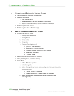

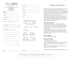

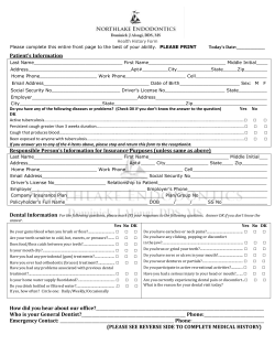
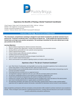


© Copyright 2026