
Estudo comparativo da anatomia de madeira e casca
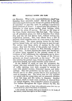
7º Congresso Florestal Nacional
“Florestas – Conhecimento e Inovação”
5 a 8 de Junho de 2013
Estudo comparativo da anatomia de madeira e casca de duas espécies de
eucalipto
1*
2
1
1,3
1
Marília Pirralho , Doahn Flores , Vicelina B. Sousa , Teresa Quilhó , Helena Pereira e
1
Sofia Knapic
1
Centro de Estudos Florestais, Instituto Superior, de Agronomia, Universidade Técnica de Lisboa, Tapada da
Ajuda, 1347-017 Lisboa, Portugal
2
Centro de Ciências Agrárias, Departamento de Ciências Florestais e da Madeira, Universidade Federal do
Espírito Santo, Avenida Governador Carlos Lindemberg, 316, Centro - 29550-000 - Jerônimo Monteiro, Espírito
Santo, Brasil
3
Centro das Florestas e Produtos Florestais, Instituto de Investigação Científica Tropical, Tapada da Ajuda,
1347-017 Lisboa, Portugal
e-mail: [email protected]
Resumo: Neste trabalho foram caracterizadas e comparadas, do ponto de vista da anatomia da
madeira e casca, duas espécies de eucalipto: Eucalyptus sideroxylon e E. viminalis. O trabalho
aqui apresentado faz parte do projecto “EucPlus-Novos processos e utilizações para madeira de
eucalipto” (PTDC/AGR-CFL/119752/2010), coordenado pelo Centro de Estudos Florestais.
Os eucaliptos, com 4 anos de idade, cresceram numa área experimental localizada no campus do
Instituto Superior de Agronomia da Universidade Técnica de Lisboa. A cada árvore foi retirada
uma rodela ao nível do DAP que serviu de base para o estudo da variação radial dos vasos,
fibras e raios.
A madeira foi estudada macroscópica e microscopicamente. Ao nível macroscópico foram
2
determinadas a área de vasos e o número de vasos por mm ao longo do raio (da medula para a
casca). Fizeram-se cortes histológicos (com utilização de micrómetro), segundo três planos tangencial (em três posições do raio 30%, 60% e 90%), transversal e radial - onde se
caracterizaram as duas espécies ao nível microscópico. A caracterização biométrica das fibras da
madeira foi realizada em material dissociado recolhido em três posições do raio (30%, 60% e
90%), determinando-se a largura, espessura e o comprimento das fibras. Os raios foram medidos
nas três posições no plano tangencial. Utilizaram-se técnicas de microscopia de luz transmitida e
análise de imagem. A casca foi caracterizada nos três planos: tangencial, transversal e radial. A
anatomia da casca destas duas espécies não tinha sido ainda investigada. A caracterização e
variabilidade anatómica (lenho e casca) destas duas espécies permitirão contribuir para a
avaliação do seu potencial comportamento do ponto de vista físico, mecânico e químico.
Palavras-chave: Eucaliptos, vasos, caracterização biométrica, fibras, casca.
Abstract: Two eucalypt species - Eucalyptus sideroxylon and E. viminalis - were characterized and
compared regarding the anatomy of wood and bark. This work is part of the project "New EucPlus
processes and uses for eucalyptus wood" (PTDC/AGR-CFL/119752/2010), coordinated by the Center
for Forest Studies.
The 4-year-old eucalypt trees grew up in an experimental area located on the campus of the School of
Agriculture, Technical University of Lisbon. From each tree a sample was taken at 1.30 m height
(DBH) and used for studying the radial variation of the vessels, fibres and rays.
The wood was studied at a macroscopic and microscopic level. Macroscopically the area and number
2
of vessels per mm along the ray (from pith to bark) was determined. Histological sections were
obtained (using a micrometer) in three directions - tangential (representing 30%, 60% and 90% of the
ray), transverse and radial - and the two species characterized at the microscopic level. The biometric
characterization of wood fibres was done in dissociated material at 30%, 60% and 90% of the ray, by
measuring width, thickness and length. The rays were measured at the three positions in the
tangential direction. Transmitted light microscopy and image analysis were used. The bark was
characterized in three directions: tangential, radial and transversal. The bark anatomy of these two
species has not yet been investigated. The characterization and anatomical variability (wood and bark)
of these two species will contribute to assess their potential behavior regarding physical, mechanical
and chemical properties.
Keywords: Eucalyptus, vessels, biometric characterization, bark.
1
Marília Pirralho, Doahn Flores, Vicelina B. Sousa, Teresa Quilhó, Helena Pereira e Sofia Knapic
1. INTRODUCTION
The knowledge of size and structure of anatomical elements which influence the wood and bark
properties in Eucalyptus are a useful tool to improve the quality of the final products (CARVALHO
1997, QUILHÓ et al. 2000, MIRANDA and PEREIRA 2002, RAMÍREZ et al. 2009, PEREIRA et al.
2011).
Although Eucalyptus had their origin in Australia, they are present today in other regions, as is the
case of Eucalyptus globulus in Portugal and Spain (PEREIRA et al. 2011). Only a few species are
from Indonesia and Papua New Guinea (GONZALEZ et al. 2011). Several species are exotics in
different regions i.e. E. nitens in Portugal and Spain as well as in Argentina and Chile; E. grandis in
the sub-tropical and tropical zones of Argentina, China, Brazil, India, South Africa, Uruguay and
Vietnam (FORRESTER et al. 2010). In spite of the numerous anatomical studies of wood
(CARVALHO 1997, MIRANDA and PEREIRA 2002, RAMÍREZ et al. 2009) and bark (CHATTAWAY
1955a, 1955b, 1955c; ALFONSO 1987, QUILHÓ et al. 1999, 2000) of the genus Eucalyptus, the
information on E. viminalis e E. sideroxylon is scarce.
The study of the anatomic characteristics is important because performance and potential value of
products depend on a wide range of interlinked fundamental wood characteristics (HUANG et al.
2003). The secondary xylem includes different cell types (i.e vessels, fibres, axial and radial
parenchyma in xylem or in phloem) originated by the vascular cambium; this cambium also
differentiates the secondary phloem with sieve tubes, fibres, axial and radial parenchyma and
sclereids (EVERT 2006). The secondary phloem, the periderm and rhytidome represent the bark
(RICHTER et al. 1996). For trees growing in plantations, these wood and bark cells are affected by
some factors such as site, ecological conditions, management, genetics and age (ZOBEL and VAN
BUIJTENEN 1989).
The main objective of this study was to characterize and compare the anatomic structure of wood and
bark of E. viminalis and E.sideroxylon.
2. MATERIAL & METHODS
This study was conducted in E. viminalis and E. sideroxylon trees with 4 years of age from an
experimental site located in the campus fields of the School of Agriculture, Technical University of
Lisbon, at Tapada da Ajuda, Lisboa, Portugal (38°42´N; 09°10´W). The region is under the influence of
a mesothermal humid climate, with a dry season in the summer extending from June to August, and
registering above 10°C in the coldest month and below or equal to 22°C in the hottest month. The soil
is a vertisol characterized by a fine, or medium to fine, texture, derived from tuffs or basalts, frequently
with limestone on the inferior horizons, or from calcareous rock (in much less extension).
In each tree samples with 10 cm thickness were collected at 1.30 of the tree height (DBH) and
wood and bark were separated.
The wood was studied macroscopically: vessel area was determined from pith to bark, using the
Leica software Qwin V 3.5.0, after acquisition of a sequence of images of each ray through a
digital camera Leica DFC 320 coupled to Leica Magnifier MZ6. Microscopically the wood was
characterized at three radial points: near pith, 30% of the radius; middle, 60%; and near the
cambium, 90%. Transversal, tangential and radial sections with 17 µm thickness were obtained,
2
Marília Pirralho, Doahn Flores, Vicelina B. Sousa, Teresa Quilhó, Helena Pereira e Sofia Knapic
using a micrometer, stained with safranin and mounted in Eukitt. Ray height and number of cells
were measured in 40 uniseriate rays, in tangential sections. The length, width and wall thickness
of 40 fibres were measured on dissociated material using image analysis LAS software V4.2
assisted by a digital camera EC3 coupled to a
transmitted light Dialux 22 EB microscope.
Descriptive terminology follows the IAWA List of Microscopic Feature for Hardwood Identification
(IAWA COMMITTEE 1989).
The bark samples were impregnated with DP 1500 polyethylene glycol. Transversal, tangential and
radial sections of approximately 17 µm thickness were prepared with a Leica SM 2400 microtome
using Tesafilm 106/4106 adhesive for sample retrieval (QUILHÓ et al. 1999). The sections were
stained with a triple staining of chrysodine/acridine red and astra blue and mounted on Eukitt. Light
microscopic observations were made using Leica DM LA and photomicrographs were taken with a
Nikon Microphot-FXA. The terminology follows RICHTER et al. (1996).
3. RESULTS & DISCUSSION
3.1. Anatomical characterization
Figure 1 shows the wood structure of E. viminalis (Fig. 1 A-C) and E. sideroxylon (Fig. 1D – E). The
two species are diffuse-porous with solitary vessels, which are circular to oval and arranged in a
diagonal pattern (Fig. 1A, D); the axial parenchyma is of different types i.e. apotracheal diffuse and
diffuse in aggregates or paratracheal vasicentric uni or circunvascular (Fig. 1A, D); the rays are mostly
uniseriate, occasionally biseriate (Fig. 1B, E) and homogenous with procumbent cells (Fig. 1C, F). The
same type of vessels, axial and radial parenchyma are found in other Eucalyptus spp. (OLIVEIRA and
FREITAS 1970, ALFONSO 1987, PEREIRA et al. 20011).
Figure1. Wood microscopic details: a Transverse section of E. viminalis; b Tangential section of E.
viminalis; c Radial section of E. viminalis; d Transverse section of E. sideroxylon; e Tangential section
of E. sideroxylon; f Radial section of E. sideroxylon. Scale bar = 300 µm.
Figures 2 and 3 show the bark structure of E. viminalis and E. sideroxylon. The bark of E.viminalis is
described as almost wholly smooth, or rough grey and persistent on the lower part of the trunk, shed
in long ribbons from the upper trunk and branches, leaving a smooth white or yellowish surface; in
3
Marília Pirralho, Doahn Flores, Vicelina B. Sousa, Teresa Quilhó, Helena Pereira e Sofia Knapic
contrast, E. sideroxylon has an ironbark, persistent to the small branches, hard and deeply furrowed,
dark brown to black (BOLAN et al. 1992). However the studied trees were very young and exhibited a
similar structure, in both species; this is in accordance with observations of CHATTAWAY (1955a,
1955b, 1955c) and ALFONSO (1987) from young eucalypt barks.
The barks present the following features: a periderm with two types of phellem cells (suberized and
lignified cells) and a poorly developed phelloderm (Fig. 2A and 3A); a thin layer of non-collapsed
phloem where tangential bands of fibres alternate with thin layers of axial parenchyma cells and sieve
tube elements (Fig. 2B and 3B); the collapsed phloem includes some dilatation tissue as a result of
tree growth i.e expanded axial parenchyma cells (Fig. 2C and 3C); the rays are mainly uniseriate (Fig.
2D and 3D) and homogenous (Fig. 2E and 3E); clusters of expanded parenchyma cells are present in
both species (Fig. 2E and 3E), more evident in E. viminalis. The bark structure of these species at this
age resembles the bark structure of E.globulus (QUILHÓ et al.1999).
Figure 2. Microscopic structure of E. viminalis bark. A – Periderm including the lignified (lig) and
suberized (sub) cells of the phellem and 2-3 cells of the phelloderm; deposits of suberin (arrow);
cortex (cx). (transverse section) B – phloem with non collapsed phloem and collapsed phloem
(transverse section); C- collapsed phloem (transverse section); D- phloem (tangential section); Ephloem (radial section). Scale bar = 50 µm.
Figure 3. Microscopic structure of E. sideroxylon bark. A – Periderm including the lignified (lig) and
suberized (sub) cells of the phellem and 1 cell of the phelloderm; deposits of suberin (arrow); cortex
(cx). (transverse section) B – phloem with non collapsed phloem and collapsed phloem (transverse
section); C- collapsed phloem (transverse section); D- phloem (tangential section); E- phloem (radial
section). Scale bar = 50 µm
4
Marília Pirralho, Doahn Flores, Vicelina B. Sousa, Teresa Quilhó, Helena Pereira e Sofia Knapic
3.2. Biometry
E. sideroxylon fibre length increased from an average 740.5 µm near the pith to 859.7 µm near the
cambium (Table 1). This trend was not observed for E. viminalis. The most frequent pattern of radial
variation of fibre length and wall thickness in the Eucalyptus genus is an increase from pith to near
cambium (PEREIRA et al. 2011). This pattern of radial variation was found in E. sideroxylon. E.
tereticornis showed a higher fibre length value of 850 at 4 years of age (RAO et al. 2002), when
compared to E. viminais and E. sideroxylon.
Fibre width increased radially in E. viminalis (19.8 µm near pith and 20.6 µm near bark), but decreased
in E. sideroxylon. Fibre wall thickness decreased from 3.8 µm to 3.0 µm and 2.7 µm, respectively, at
the pith, middle and near cambium positions in E. viminalis. In E. sideroxylon the fibre wall thickness
was not significantly different from pith to bark, with average values ranging from 3.8 µm of 4.2 µm and
to 3.8 µm in the three radial positions. These values of wall thickness are within the range of 1.7 µm to
6.6 µm reported for E.globulus at 9 and 12 years of age (PEREIRA et al. 2011).
Vessel area increased from pith to bark for E.viminalis and E. sideroxylon (Table 1). Vessels were
2
2
smaller near the pith (0.0078 mm for E. viminalis and 0.0034 mm for E. sideroxylon), increasing
2
2
afterwards (0.0079 mm for E.viminalis and 0.0035 mm for E. sideroxylon) and reaching its maximum
2
near the bark (0.0082 mm for E. viminalis and for E.sideroxylon). The same pattern of variation was
found in E. globulus (PEREIRA et al. 2011) i.e. vessels area increased from the pith to near cambium.
Table 1. Results of fibre variables (length, width and wall thickness) and vessel area measured at
tree radial position (30%, 60% and 90% of radius) of Eucalyptus viminalis and E. sideroxylon
Species
E. viminalis
E. sideroxylon
Position
(%)
Fibre length
(µm)
Fibre width
(µm)
19.8±3.9
Fibre wall
thickness
(µm)
3.8±0.8
30
616.1±83.2
60
Vessel area
2
(mm )
0.0078±0.003
908.8±84.7
17.2±2.8
3.0±0.38
0.0079±0.041
90
699.7±73.5
20.6±3.0
2.7±0.7
0.0082±0.004
30
740.5±68.7
13.3±1.5
3.8±0.45
0.0034±0.0009
60
816.8±97.8
20.4±3.0
4.2±0.76
0.0035±0.0009
90
859.7±74.8
13.3±1.3
3.8±0.59
0,0035±0.001
Table 2 shows the dimensions of uniseriate rays. The ray height increased radially from pith to bark in
E. sideroxylon (108.6 µm to 124.7 µm), and decreased in E. viminalis (166.5 µm to 153.3 µm). E.
sideroxylon showed 28% of bisseriate rays near pith and 34% near the cambium. In E. viminalis only
uniseriate rays were observed.
Regarding the variance analysis (table 3) results indicated that species and radial position and
interaction between species and position had a very significant effect in all the fibre biometric
variables (p<0.001). Interaction between species and position accounted for most of the variability
(35%, 52% and 24% of the total variation for fibre length, width and wall thickness, respectively).
Species had a significant effect in fibre wall thickness (28%). Between-fibre variation (error
component) was high at 50%, 64% to 60% of the total variation respectively.
5
Marília Pirralho, Doahn Flores, Vicelina B. Sousa, Teresa Quilhó, Helena Pereira e Sofia Knapic
Table 2.Variable of uniseriate rays measured at three radial position (30%,60%,90%) of E.viminalis
and E.sideroxylon
Specie
Position (%)
Height (µm)
30
166.5±45.7
E.viminalis
60
149.0±44.2
90
153.3±43.5
30
108.6±25.4
E.sideroxylon
60
107.9±28.1
90
124.7±38.9
Table 3. Summary of variance analysis for fibre length, width and wall thickness measured at tree
radial position (pith, middle and bark) for E.viminalis and E.sideroxylon
Species
Position
Species*Position
Error
Fibre length
Fibre width
Fibre wall thickness
Fibre length
Fibre width
Fibre wall thickness
Fibre length
Fibre width
Fibre wall thickness
Fibre length
Fibre width
Fibre wall thickness
df
1
1
1
2
2
2
2
2
2
F
36.6
95.1
83.1
101.1
15.1
13.5
55.1
85.8
23.9
234
234
234
P
<0.001
<0.001
<0.001
<0.001
<0.001
<0.001
<0.001
<0.001
<0.001
Exp.Var.( %)
8
19
28
4
5
7
35
52
24
26
25
41
4. CONCLUSIONS
The anatomic structure of wood and bark of E. viminalis and E. sideroxylon was compared. The wood
and bark structure of young E. viminalis and E. sideroxylon species are similar.
Both species presented the same radial pattern of the vessels area and similar to E .globulus i.e.
vessels area increased from pith to near cambium. Fibres of E. sideroxylon were higher and wider
than E. viminalis fibers, but their wall thickness was similar. E. viminalis reported a higher height of
unseriate rays. Interaction between species and the radial position was the most significant effect for
the variation of fibre biometric variables. E.sideroxylon showed a similar radial pattern of fibre length to
that found in the genus Eucalyptus.
6
Marília Pirralho, Doahn Flores, Vicelina B. Sousa, Teresa Quilhó, Helena Pereira e Sofia Knapic
ACKNOWLEDGMENTS
This study was funded by project “Eucplus - new processes and uses for eucalypt woods”
(PTDC/AGR-CFL/119752/2010) by FCT (Fundação para a Ciência e Tecnologia). The Centro de
Estudos Florestais is a research unit funded by FCT within the POCTI-FEDER programme. The
third author author acknowledges funding from FCT as a doctoral student, the last author
acknowledges funding from FCT as a post-doctoral researcher.
REFERENCES
ALFONSO, V. 1987. Caracterização anatómica do lenho e da casca das principais espécies de
Eucalyptus L' Herit, cultivados no Brasil. Tese de Doutoramento. Instituto de Biociências da
Universidade de Sao PauIo.
BOLAND D.J., BROOKER, M.I.H., CHIPPENDALE, G.M., HALL, N., HYLAND, B.P.M., JOHNSTON,
R.D., KLEINIG, D.A., Turner., J.D.,1992. Forest Trees of Australia. Over 200 of Australias most
important native trees described and illustrated, CSIRO Publ.
CARVALHO, A., 1997. Madeiras Portuguesas, estrutura anatómica, propriedades e utilizações. Vol II.
Direcção-Geral das Florestas, Lisboa, 340 pp.
CHATTAWAY, M.M., 1955a.Theanatomy ofbark.II.Oil gland in Eucalyptus species. Australian Journal
of Botany. 3: 23-27.
CHATTAWAY, M.M., 1955b. The anatomy of bark. III. Enlarged fibres in bloodwoods (Eucalyptus
spp.). Australian Journal of Botany 3: 28-38.
CHATTAWAY, M.M., 1955c. Theanatomy of bark.IV. Radially elongated cells in the phelloderm of
species of Eucalyptus. Australian Journal of Botany 3: 39-47.
EVERT, R.F., 2006. Esau's plant anatomy, meristems, cells, and tissues of the plant body, their
structure, function, and development. John Wiley & Sons Inc, New Jersey.
FORRESTER, D.I., MEDHURST, J.L., MATTHEW, W., BEADLE., CH.L., VALENCIA, C.J., 2010.
Growth and physiological responses to silviculture for producing solid-wood products from Eucalyptus
plantations: An Australian perspective. Forest Ecology and Management 259: 1819-1835.
GONZALEZ, R., TREASURE, T., WRIGHT, J.,SALONI, D., PHILLIPS, R., ABT, R., JAMEEL, H.,
2011. Exploring the potential of Eucalyptus for energy production in the Southern United States:
Financial analysis of delivered biomass. Part I. Biomass and Bioenergy 35 (2): 755-766.
HUANG, C.L., LINDSTRÖM, H., NAKATA, R., RALSTOM, J., 2003. Cell Wall struture and wood
poperties determined by acoustic-a selective review. Holz als Roh- und Werkstoff 61: 321-335.
7
Marília Pirralho, Doahn Flores, Vicelina B. Sousa, Teresa Quilhó, Helena Pereira e Sofia Knapic
IAWA Committee, 1989. List of microscopic features for hardwood identification. IAWA Bull, 112 pp.
MIRANDA, I., PEREIRA, H., 2002. Variation pulpwood quality with provenances and site in Eucalyptus
globulus. Annals of Forest Science 59:283-291.
OLIVEIRA, J.S., FREITAS, M.C., 1970. Eucaliptos da Namaacha. Universidade de Lourenço
Marques. Separata da Revista de Ciências Agronómicas 3(2) Série B, 1-230.
PEREIRA, H., MIRANDA, I.,GOMINHO, J., TAVARES, F., QUILHÓ, T., GRAÇA, J., RODRIGUES, J.,
SHATALOV, A., KNAPIC, S., Qualidade e utilização tecnológica do Eucalipto (E.globulus), edição
Centro de Estudos Florestais, Lisboa.
QUILHÓ, T., PEREIRA, H., RICHTER, H.G., 1999. Variability of bark structure in plantation-grown
Eucalyptus globulus. IAWA Journal 20: 171–180.
QUILHÓ, T., PEREIRA, H., RICHTER, H.G., 2000. Within-tree variationin phloem cell dimensions and
proportions in Eucalyptus globulus. IAWA Journal 22: 255-265.
RAMÍREZ, M., RODRÍGUEZ, J., PEREDO, M., VALENZUELA, S., MENDONÇA, R., 2009. Wood
anatomy and biometric parameters variation of Eucalyptus globulus clones. Wood Science and
Technology 43: 131-141.
RAO, R.V., SHASHIKALA, S., SREEVANI, P., KOTHIYAL, V., SARMA, C.R., LAL, P., 2002. Withintree variation in anatomical porperties of some clones of Eucalyptus terecticornis Sm. Wood Science
and Technology 36: 271-28.
RICHTER, H.G., VIVEIROS, S., ALVES, E., Luchi, A., Costa, C., 1996. Padronização de critérios
para a descrição anatómica da casca: lista de características e glossário de termos. IF Série
Registros, São Paulo 16: 1–25.
ZOBEL, B., VAN BUIJTENEN, B., 1989. Wood variation: its causes and control. Springer, New York,
USA, 363 pp.
8


© Copyright 2026