
Document 6782
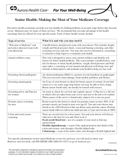
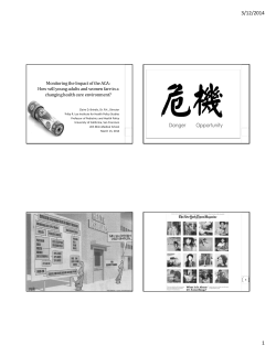
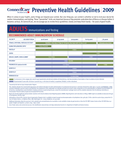
online © ML Comm
ORIGINAL ARTICLE
Korean J Audiol 2013;17:111-117
pISSN 2092-9862 / eISSN 2093-3797
http://dx.doi.org/10.7874/kja.2013.17.3.111
Clinical Application of 3D-FIESTA Image in Patients
with Unilateral Inner Ear Symptom
Jae Ho Oh, Jae Ho Chung, Hyun Jung Min,
Seok Hyun Cho, Chul Won Park and Seung Hwan Lee
Department of Otolaryngology-Head and Neck Surgery, College of Medicine, Hanyang University, Seoul, Korea
Received July 10, 2013
Revised September 20, 2013
Accepted October 13, 2013
Address for correspondence
Seung Hwan Lee, MD
Department of OtolaryngologyHead and Neck Surgery,
College of Medicine,
Hanyang University,
222 Wangsimni-ro, Seongdong-gu,
Seoul 133-792, Korea
Tel +82-31-560-2363
Fax +82-31-566-4884
E-mail [email protected]
Background and Objectives: Unilateral auditory dysfunction such as tinnitus and hearing
loss could be a warning sign of a retrocochlear lesion. Auditory brainstem response (ABR) and
internal auditory canal magnetic resonance image (MRI) are suggested as novel diagnostic
tools for retrocochlear lesions. However, the high cost of MRI and the low sensitivity of the ABR
test could be an obstacle when assessing patients with unilateral ear symptoms. The purpose
of this study was to introduce the clinical usefulness of three-dimensional fast imaging employing steady-state acquisition (3D-FIESTA) MRI in patients with unilateral ear symptoms. Subjects and Methods: Two hundred and fifty-three patients with unilateral tinnitus or unilateral
hearing loss who underwent 3D-FIESTA temporal bone MRI as a screening test were enrolled.
We reviewed the abnormal findings in the 3D-FIESTA images and ear symptoms using the
medical records. Results: In patients with unilateral ear symptoms, 51.0% of the patients had
tinnitus and 32.8% patients were assessed to have sudden sensory neural hearing loss. With
3D-FIESTA imaging, twelve patients were diagnosed with acoustic neuroma, four with enlarged
vestibular aqueduct syndrome, and two with posterior inferior cerebellar artery aneurysm.
Inner ear anomalies and vestibulocochlear nerve aplasia could be diagnosed with 3D-FIESTA
imaging. Conclusions: 3D-FIESTA imaging is a highly sensitive method for the diagnosis of
cochlear or retrocochlear lesions. 3D-FIESTA imaging is a useful screening tool for patients
with unilateral ear symptoms.
Korean J Audiol 2013;17:111-117
KEY WORDS: Retrocochlear disease · Hearing loss · Tinnitus · MRI.
Introduction
It is very important to identify the underlying cause of ear
symptoms such as tinnitus and hearing loss. If these symptoms
present unilaterally, a retocochlear lesion, vascular lesion or
inner ear anomaly should be considered in the diagnostic process. Many diagnostic tools have been suggested for use in the
evaluation of retrocochlear lesions. However, there is no single novel method to easily evaluate all lesions causing unilateral ear symptoms.
Since Selters first introduced auditory brainstem evoked
response (ABR) as a diagnostic method for retrocochlear leThis is an Open Access article distributed under the terms of the Creative
Commons Attribution Non-Commercial License (http://creativecommons.
org/licenses/by-nc/3.0/) which permits unrestricted non-commercial
use, distribution, and reproduction in any medium, provided the original
work is properly cited.
sions, the ABR test has been commonly used as a screening test.
However, the low sensitivity of the ABR test has lowered its
diagnostic value as a screening test, especially in small intracanalicular tumors.1,2)
After the introduction of magnetic resonance image (MRI)
technology, MRI testing was applied in the assessment of retrocochlear lesions with high sensitivity and high specificity.3)
Nevertheless, the high cost of conventional MRI testing, the use
of contrast media and the long image acquisition time might be
limitations for using it as a screening tool.
Recently, three-dimensional fast imaging employing steadystate acquisition (3D-FIESTA) images have been introduced.
3D-FIESTA imaging can give much higher spatial resolution
with outstanding image contrast between the cranial nerves
and cerebrospinal fluid. And it has a shorter image acquisition
time than conventional MRI scan and does not need contrast
Copyright © 2013 The Korean Audiological Society 111
3D-FIESTA Image in Patients with Unilateral Inner Ear Symptom
media.4,5)
In this study, we aimed to evaluate the usability of 3D-FIESTA imaging as a screening tool for inner ear lesions with applying 3D-FIESTA image in the diagnostic pathway of the patients with unilateral ear symptoms.
Subjects and Methods
From January 2011 to December 2011, 253 patients who presented with unilateral ear symptoms and underwent 3D-FIESTA imaging as a screening were enrolled in this study. Patients suspected to have Meniere’s disease and complained of
bilateral ear symptoms were excluded. A thorough medical review of the clinical presentation and audiometric results was
performed retrospectively.
All patients underwent 3D-FIESTA imaging as a screening
tool with a Philips Achieva (Best, the Netherlands) 3.0 tesla
MRI scanner. If an inner ear lesion or a brain lesion was suspected on the 3D-FIESTA image, an additional diagnostic
Epidermal cyst
Pons infarction
Vertebral a. calcification
Hypoplastic 8th n.
Inner ear anomaly
PICA aneurysm
EVAS
%
Acoustic neuroma
1.6
1.4
1.2
1.0
0.8
0.6
0.4
0.2
0.0
Fig. 1. Prevalence of each disease among patients with unilateral
ear symptoms. EVAS: enlarged vestibular aqueduct syndrome,
PICA: posterior inferior cerebellar artery.
A
112
B
Korean J Audiol 2013;17:111-117
brain MRI scan, a temporal MRI scan, or temporal bone CT
scans were performed to elucidate the final diagnosis.
The images from the screening and diagnostic imaging investigations were reviewed to compare their ability to find various cochlear, retrocochlear or vascular lesions.
Patients were divided into three groups according to their
chief complaint: tinnitus, unilateral hearing loss, and unilateral sudden idiopathic hearing loss, to compare their clinical
presentations and radiological results.
Clinical data and 3D-FIESTA results were analyzed to figure out the relationships between the clinical presentations of
acoustic neuroma and tumor size. The Kruskal-Wallis test was
applied for the statistical analysis of tumor size using commercially available software (Statistical Package for the Social
Sciences, Version 17.0; SPSS Inc., Chicago, IL, USA). A p value of <0.05 was considered as statistically significant.
Results
Among two hundred and fifty-three patients with a unilateral ear symptom, 105 (41.5%) patients were male and 148
(58.5%) were female. The mean age was 50.7±15.7 years.
Tinnitus was the most common symptoms of patients (129,
49.6%), sudden sensory neural hearing loss was the second
most common (83, 31.9) and unilateral hearing loss was the
next most common (41, 15.7%).
In the 3D-FIESTA imaging results, acoustic neuroma was
the most common retrocochlear lesion in patients with unilateral ear symptoms in this study (Fig. 1). Twelve patients were
diagnosed with acoustic neuroma (Figs. 2-4), and four patients were confirmed as having enlarged vestibular aqueduct
syndrome (Fig. 5). Other lesions such as a congenital anomaly, a vascular lesion, a mass and acute infarction could be detected with 3D-FIESTA imaging (Figs. 6-12).
Fig. 2. Temporal bone images of a
21 year old woman with left sudden
hearing loss. Acoustic neuroma extending to the cerebellopontine angle (arrows) was identified by screening using 3D fiesta imaging (A) and
was confirmed by Gd enhanced T1weighted imaging (B). 3D: three-dimensional.
Oh JH, et al.
We also tried to figure out the association between clinical
symptoms and the final diagnosis. Six patients who initially presented with tinnitus were diagnosed with acoustic neuroma
and two patients were diagnosed with posterior inferior cerebellar artery aneurysm. Abnormal findings in 3D-FIESTA
images in patients with hearing loss were acoustic neuroma,
Fig. 3. Temporal bone images of a
69 year old woman with tinnitus on
the right side. An intracanalicular
acoustic neuroma (arrows) was identified by screening using 3D fiesta
imaging (A) and it was confirmed by
Gd enhanced T1-weighted imaging
(B). 3D: three-dimensional.
Fig. 4. Temporal bone images of a
49 year-old man with right tinnitus. A
tiny fusiform acoustic neuroma (arrows) was identified by screening
with 3D fiesta imaging (A) and was
confirmed by Flair vista MR imaging
(B). 3D: three-dimensional.
Fig. 5. Temporal bone images of a
38 year-old woman with left sensorineural hearing loss. Enlarged vestibular aqueduct syndrome (arrows)
was identified by 3D fiesta screening (A) and confirmed by temporal
bone computed tomography (B).
3D: three-dimensional.
enlarged vestibular aqueduct syndrome, inner ear anomaly
and 8th nerve hypoplasia (Table 1).
The mean size of acoustic neuromas which were finally
diagnosed was 17×14 mm (4×3-62×64 mm), the mean
size of tumors in the tinnitus group, unilateral hearing loss
group and sudden idiopathic sensorineural hearing loss group
A
B
A
B
A
B
www.audiology.or.kr 113
3D-FIESTA Image in Patients with Unilateral Inner Ear Symptom
A
Fig. 6. Temporal bone image of a 69
year-old woman with bilateral continuous tinnitus. Posterior inferior
cerebellar artery aneurysm (arrows)
was identified by 3D fiesta screening
(A) and confirmed by computed tomography angiography (B). 3D: threedimensional.
B
A
B
was 13×10 mm, 52×23 mm, and 11×9 mm respectively. Table 2 shows the clinical characteristics of patients who were finally diagnosed with acoustic neuroma. The mean tumor size
of the unilateral hearing loss group tended to be smaller than
that of other groups. However, we could not find any significant
correlation between the groups (p=0.582).
Discussion
In patients with unilateral ear symptoms, it is important to
approach these patients with a high index of suspicion for cochlear or retrocochlear lesions such as an inner ear anomaly,
tumor, vascular or inflammatory lesion.
With conventional audiologic testing, retrocochlear lesions
can be suspected by a relatively low speech discrimination score
or low score in the short increment sensitivity index test. Retrochoelar lesions were diagnosed more easily after computed tomography (CT) and the ABR test were introduced.6,7)
But the temporal bone CT scan is inadequate for screening
tool due to the high false or negative positive rate for the diagnosis of retrocochlear lesions especially in the case of acous-
114
Korean J Audiol 2013;17:111-117
Fig. 7. Temporal bone images of a
16 year-old woman with right sensorineural hearing loss. Mondini dysplasia (arrows) was identified by 3D
fiesta screening (A) and confirmed
by temporal bone computed tomography (B). 3D: three-dimensional.
tic neuroma. ABR test has been used as a favored screening
tool for retrocochlear lesions; this test compares the amplitude and latency of waves produced by the auditory pathway.
However, Barrs, et al.8) reported that even though ABR demonstrated 97% of the tumors larger than 1.5 cm, 48% of neuromas are 1.5 cm or smaller. Ruckenstein, et al.9) reported
that the ABR screening test for retrocochlear pathology had
a sensitivity of 63% and a specificity of 64%. These findings
imply that ABR probably has a limited role in acoustic neuroma screening.
After the application of MRI for the diagnosis of retrocochlear lesions began in the 1980s, the specificity and sensitivity of
MRI was found to be up to 100% and it became to be considered the most accurate diagnostic tool.10,11) But MRI is a very
expensive test and it takes 40-60 minutes to get images with
contrast. Thus, MRI has limitations as a screening test due to
the low cost and time effectiveness.
With the development of the image acquisition technique
with MRI, new protocols for 3D-FIESTA sequences were introduced which provided much higher spatial resolution and
clearer depiction of small structures such as cranial nerves
Oh JH, et al.
especially within the cisternal spaces. The 3D-FIESTA uses
an ultrafast pulse sequence that produces 0.3 mm thin section
high resolution images with outstanding image contrast between the cerebrospinal fluid, vessels and cranial nerves. It
Fig. 8. Temporal bone images of a
1 month-old baby. Left internal auditory canal stenosis with labyrinthine
malformation (arrows) was identified
by 3D fiesta screening (A-C) and
confirmed by temporal bone computed tomography (D). 3D: three-dimensional.
usually takes 5 to 7 minutes to get a proper image and does not
need contrast media. The cost of 3D-FIESTA is just 1/5-1/6
that of conventional MRI. So it could be a very effective test
protocol for the screening of retrocochlear lesions in patients
A
B
C
D
Fig. 9. Temporal bone image of a 51 year-old woman with left sensorineural hearing loss. Hypoplastic vestibulocochlear nerve (arrow) was identified by 3D fiesta screening. 3D: three-dimensional.
Fig. 10. Temporal bone image of a 76 year-old man with left sudden sensorineural hearing loss. Vertebral artery calcification (arrow) was identified by 3D fiesta screening. 3D: three-dimensional.
www.audiology.or.kr 115
3D-FIESTA Image in Patients with Unilateral Inner Ear Symptom
with unilateral ear symptoms.
In this study, variable lesions such as small tumors less than
5 mm, inner ear anomalies and vascular malformations could
be identified using 3D-FIESTA imaging. And there is one report which showed that with 3D-FIESTA, there was decreased
vestibular signal intensity on the affected side in patients with
vestibular schwannoma, but that this was not present in those
with cerebellopontine angle meningiomas or in normal subjects.12)
Recently, MRI with high-dose contrast media has been introduced for detecting cochlear endolymphatic hydrops.13) 3DFIESTA images have a limitation in detecting inner ear lesions;
however, 3D-FIESTA images can detect inner ear anomalies
so it may be sufficient to use as a screening tool for retrocochlear lesions.
Acoustic neuroma, one of the most common retrocochlear
Table 2. Summary of acoustic neuroma patients
Case
No.
Fig. 11. Temporal bone image of a 73 year-old man with left tinnitus. Brain stem lesion, upper pons infarction (arrow) was identified
by 3D fiesta screening. 3D: three-dimensional.
A
Sex
Age
(years)
Chief
complaint*
Tumor
size (mm)
1
F
63
S
8×4
2
M
37
SS
10×50
3
M
35
S
12×50
4
F
67
S
21×17
5
F
56
SS
12×12
6
M
50
T
10×90
7
F
69
T
12×60
8
M
64
S
62×64
9
F
22
T
30×28
10
M
36
T
10×60
11
F
56
T
11×80
12
M
49
T
4×3
*chief complaint. S: sensorineural hearing loss, SS: sudden sen-
sorineural hearing loss, T: tinnitus
Fig. 12. Temporal bone images of
a 51 year-old man with right sensorineural hearing loss. Huge epidermal cyst (arrows) was identified by
3D fiesta screening (A) and confirmed by Gd enhanced T1-weighted
imaging (B). 3D: three-dimensional.
B
Table 1. Disease distribution and ratio according to the cause
Chief complaint
Tinnitus (129)
Acoustic neuroma
EVAS
6 (4.6%)
PICA aneurysm
Inner ear anomaly
8th n. hypoplasia
2 (1.5%)
SSNHL (83)
2 (2.4%)
1 (1.2%)
Unilat. HL (41)
4 (9.7%)
3 (7.3%)
1 (1.2%)
2 (4.8%)
1 (2.4%)
SSNHL: sudden sensorineural hearing loss, HL: hearing loss, EVAS: enlarged vestibular aqueduct syndrome, PICA: posterior inferior
cerebellar artery
116
Korean J Audiol 2013;17:111-117
Oh JH, et al.
lesions, is a benign tumor originating from the vestibular nerve
sheath in the internal auditory canal. In the early stage of acoustic neuromas, symptoms can be nonspecific but as the tumor
grows, progressive unilateral hearing loss and tinnitus are
typical symptoms. It is well known that about 10 to 20% of
acoustic neuroma patients can be present with sudden sensorineural hearing loss, and on the other hand only 1 to 3% of sudden seonsorineural hearing loss patients have acoustic neuroma.14-16)
In this study, 9.7% of unilateral hearing loss patients, 4.6%
of unilateral tinnitus and 2.4% of sudden sensorineural hearing loss patients were diagnosed with acoustic neuroma. The
mean size of acoustic neuroma varied from a tiny tumor smaller than 5 mm to a large tumor up to 64 mm. The mean size of
the tumor in the sudden hearing loss patients group was larger than the other two groups, but there was no statistically significant difference between the groups. In addition to acoustic
neuroma, many retrocochlear lesions, intracranial masses and
acute infarctions can be diagnosed with screening using 3DFIESTA imaging.
With these findings, we suggest that 3D-FIESTA imaging
could be a reliable screening test for patients who complain of
unilateral otologic symptoms. But considering that this study
was a simple retrospective observational study, further comparative study investigating the sensitivity and specificity of
3D-FIESTA imaging will be required.
Conclusion
3D-FIESTA imaging is a cost-effective and sensitive method for the diagnosis of cochlear or retrocochlear lesions. 3DFIESTA imaging is a useful screening tool for patients with
unilateral ear symptoms.
tests in the differential diagnosis of cochlear and retrocochlear hearing loss. Korean J Otolaryngol-Head Neck Surg 1999;42:955-60.
2) Cueva RA. Auditory brainstem response versus magnetic resonance
imaging for the evaluation of asymmetric sensorineural hearing loss.
Laryngoscope 2004;114:1686-92.
3) Wilson DF, Hodgson RS, Gustafson MF, Hogue S, Mills L. The sensitivity of auditory brainstem response testing in small acoustic neuromas. Laryngoscope 1992;102:961-4.
4) Erdogan N, Altay C, Akay E, Karakas L, Uluc E, Mete B, et al. MRI
assessment of internal acoustic canal variations using 3D-FIESTA
sequences. Eur Arch Otorhinolaryngol 2013;270:469-75.
5) Rigby PJ. Comparison of FIESTA and gadolinium-enhanced T1weighted sequences in magnetic resonance of acoustic schwannoma. Radiographer 2006;53:11-21.
6) Selters WA, Brackmann DE. Acoustic tumor detection with brain
stem electric response audiometry. Arch Otolaryngol 1977;103:181-7.
7) House JW, Waluch V, Jackler RK. Magnetic resonance imaging in
acoustic neuroma diagnosis. Ann Otol Rhinol Laryngol 1986;95(1
Pt 1):16-20.
8) Barrs DM, Brackmann DE, Olson JE, House WF. Changing concepts of acoustic neuroma diagnosis. Arch Otolaryngol 1985;111:1721.
9) Ruckenstein MJ, Cueva RA, Morrison DH, Press G. A prospective
study of ABR and MRI in the screening for vestibular schwannomas.
Am J Otol 1996;17:317-20.
10) Jackler RK, Shapiro MS, Dillon WP, Pitts L, Lanser MJ. Gadolinium-DTPA enhanced magnetic resonance imaging in acoustic neuroma diagnosis and management. Otolaryngol Head Neck Surg 1990;
102:670-7.
11) Glasscock ME 3rd, Hays JW, Minor LB, Haynes DS, Carrasco VN.
Preservation of hearing in surgery for acoustic neuromas. J Neurosurg 1993;78:864-70.
12) Ishikawa K, Haneda J, Okamoto K. Decreased vestibular signal intensity on 3D-FIESTA in vestibular schwannomas differentiating
from meningiomas. Neuroradiology 2013;55:261-70.
13) Seo YJ, Kim J, Choi JY, Lee WS. Visualization of endolymphatic hydrops and correlation with audio-vestibular functional testing in patients with definite Meniere’s disease. Auris Nasus Larynx 2013;
40:167-72.
14) Morrison GA, Sterkers JM. Unusual presentations of acoustic tumours. Clin Otolaryngol Allied Sci 1996;21:80-3.
15) Sataloff RT, Davies B, Myers DL. Acoustic neuromas presenting
as sudden deafness. Am J Otol 1985;6:349-52.
16) Lee YW, Lee KD, Yu TH, Kim HK. Neurotologic analysis of cerebellopontine angle tumor. Korean J Otolaryngol-Head Neck Surg
1998;41:454-60.
REFERENCES
1) Kim H, Lee YS, Chung JW, Yoon TH, Lee KS. Efficacy of audiologic
www.audiology.or.kr 117


© Copyright 2026