
Epidemiological study of canine parvovirus
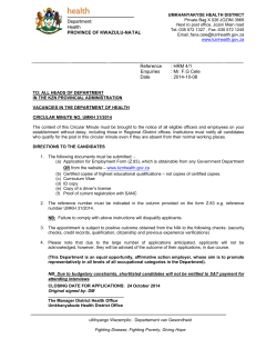
Veterinary World, EISSN: 2231-0916
Available at www.veterinaryworld.org/Vol.8/January-2015/7.pdf
RESEARCH ARTICLE
Open Access
Epidemiological study of canine parvovirus infection in and around
Bhubaneswar, Odisha, India
Monalisa Behera1, S. K. Panda1, P. K. Sahoo2, A. P. Acharya1, R. C. Patra3, Sweta Das2 and S. Pati1
1. Department of Veterinary Pathology, College of Veterinary Science and Animal Husbandry, Orissa University of
Agriculture and Technology, Bhubaneswar, Odisha, India; 2. Fish Health Management Division, Central Institute of
Freshwater Aquaculture, Kausalyaganga, Bhubaneswar, Odisha, India; 3. Department of Veterinary Medicine, College of
Veterinary Science and Animal Husbandry, Orissa University of Agriculture and Technology, Bhubaneswar, Odisha, India.
Corresponding author: Monalisa Behera, email: [email protected], SKP: [email protected],
PKS: [email protected], APA: [email protected], RCP: [email protected], SD: [email protected],
SP: [email protected]
Received: 22-10-2014, Revised: 29-11-2014, Accepted: 03-12-2014, Published: 09-01-2015
doi: 10.14202/vetworld.2015.33-37. How to cite this article: Behera M, Panda SK, Sahoo PK, Acharya AP, Patra RC,
Das S, Pati S (2015) Epidemiological study of canine parvovirus infection in and around Bhubaneswar, Odisha, India,
Veterinary World 8(1): 33-37.
Abstract
Aim: An epidemiological study of canine parvovirus infection in dogs in and around Bhubaneswar, Odisha was conducted
between December 2012 to March 2013 and prevalence rate was studied on the basis of age, breed, and sex.
Materials and Methods: A total of 71 fecal samples from suspected diarrheic dogs were collected in sterile phosphate
buffer saline (10% W/V) and examined by polymerase chain reaction (PCR) for detection of canine parvo virus infection,
followed by epidemiological study in relation to age, breed, and sex.
Results: Of 71 samples analyzed, 29 (40.85%) were found to be positive by PCR assay. The infection was higher in Deshi/
local breeds (34.48%), followed by German shepherd (17.24%), equal incidences in mixed and Labrador retriever (10.34%),
Rottweiler and German spitz showed 6.90% each and finally lower incidences in four breeds (3.45%) such as Dalmatians,
Nea politan mastiff, Pug and Great Dane. Age-wise prevalence study revealed the infection being more in the age group of
3-6 months (41.37%), followed by equal incidences of 27.59% in 1-3 months and 6-12 months age group, and a low incidence
in age groups above 12 months (3.45%). The incidence was predominantly higher in males (86.21%) than females (13.79%).
Conclusions: The epidemiological analysis revealed that the breed wise prevalence was found to be more in Deshi breeds
as compared to others, age groups below 6 months were found to be more prone to parvovirus infection and males were
mostly infected.
Keywords: canine parvovirus, epidemiology, polymerase chain reaction.
Introduction
Dog has become indispensable in man’s life as an
embodiment of love and affection. Dogs have diversified utility ranging from tracking, hunting, instruments
of war, bomb detecting squad and healer of both physical and emotional problems of humans, detecting criminals and guiding blind people [1]. In view of the need
of dogs in these above aspects it is necessary to study
the various diseases that are producing a high morbidity
and case fatality rate. Among the viral diseases the predominantly occurring diseases includes canine parvoviral infection, canine distemper, corona virus infection,
canine hepatitis, canine parainfluenza and rabies [2].
Canine parvo virus (CPV) infection is a highly
infectious viral disease of dogs of great concern to
pet owners, practicing veterinarians and scientists due
to its high morbidity and mortality rates. Parvo virus
infects dogs of all age groups, but puppies are most
severely affected than adults [2]. Parvo virus infection is most commonly manifested with signs like,
vomiting, bloody diarrhea and severe leukopenia.
Copyright: The authors. This article is an open access article licensed
under the terms of the Creative Commons Attributin License (http://
creative commons.org/licenses/by/2.0) which permits unrestricted
use, distribution and reproduction in any medium, provided the
work is properly cited.
Veterinary World, EISSN: 2231-0916
Outbreaks of CPV have been reported from many
countries including India. The prevalence study in
India was first reported by Balu and Thangaraj [3]
in Madras. The pattern of disease experienced in a
population is largely influenced by the susceptibility
of host, environmental conditions such as housing,
hygiene, population density, and pathogenicity of the
infectious agent [4].
The prevalence study may some way beneficial
to check the infection among the susceptible population. Furthermore, the epidemiology regarding CPV
in the study area is very scarce. Therefore, the present
study was conducted to determine the prevalence of
CPV infection in the Bhubaneswar city of Odisha, for
which effective control measures could be carried out.
Materials and Methods
Ethical approval
Ethical approval was not necessary. All the animals under this research were clinical cases and were
examined, diagnosed and treated as per standard treatment and examination procedure.
Study area
Dogs in and around Bhubaneswar suffering from
diarrhea and vomiting were selected for the study as
33
Available at www.veterinaryworld.org/Vol.8/January-2015/7.pdf
the suspected cases of parvovirus infection (Figure-1).
Suspected cases were mostly presented for treatment
to Teaching Veterinary Clinical Complex, College of
Veterinary Science and Animal Husbandry, OUAT,
Odisha from different parts of Bhubaneswar.
Sample collection
A total of 71 dogs with a history of vomiting
and diarrhea were taken in the study for collection
of samples during the period of December 2012 to
March 2013. They were of different age groups, different breeds and of both genders. In the above study,
dogs were categorized into different age groups such
as between 1 and 3 months, 3-6 months, 6-12 months
and above 12 months. All data regarding age, breed
and sex of the suspected dogs were recorded.
Fecal samples were collected from the suspected
dogs by introducing a swab into the anus of diarrheic
dogs and homogenized (10% w/v) in sterile phosphate
buffer saline (PBS). The samples were kept in 2 ml
collection tube with the swab. After thorough mixing of the sample in PBS, the swab was removed by
squeezing on the inner surface of the collection tube.
Then samples were stored at −20°C until further use.
analyzed on 1% agarose gel (Lonza, USA) with the
positive control and visualized under UV transilluminator (Alpha Innotech, USA).
Epidemiological analysis
Positive cases were analyzed as per different
breeds, age groups and sexes in order to conduct an
epidemiological analysis on the basis of PCR result.
The different breeds include Great Dane, German
shepherd, Labrador retriever, German spitz, Deshi,
Nea politan mastiff, Pug, Golden retriever, Dalmatian,
Doberman, Mixed, Chihuahua and Rottweiler. The
different breeds were classified under four groups
such as Group-1 (1-3 months), Group-2 (3-6 months),
Group-3 (6-12) and Group-4 (>12 months). All the
data regarding the breed, age and sex were given in
the Tables-1-3, respectively.
Results
The PCR screening of 71 suspected fecal samples
revealed 29 (40.85%) samples as positive for canine
parvovirus (Figure-2) showing band size of 583 bp
using the earlier published primers [6]. The incidence
study was calculated with respect to total positive (29)
Preparation of fecal samples
The fecal suspensions with PBS were processed
by centrifuging at 1500 xg for 10 min at 4°C. Then
supernatants were collected in 2 ml collection tube. The
supernatant (500 μl) was boiled for 10 min at 100°C to
inactivate the polymerase chain reaction (PCR) inhibitors and chilled on ice as described previously [5]. After
boiling the sample was centrifuged at 1500 xg for 5 min
and the collected supernatant was used as a source of
DNA template for PCR. The lysate was stored at −20°C
until further use. The commercial vaccine against CPV,
Canigen DHPPi (Virbac) was used as positive control
after a pretreatment by fast boiling method.
PCR
A reaction volume of 25 μl was prepared, which
consisted up 0.5 μl of each forward primer and reverse
primer (IDT, 10 pico mole concentration) (555 forward- 5’-CAGGAAGATATCCAGAAGGA-3’ and
555 reverse- 5’-GGTGCTAGTTGATATGTAATA
AACA-3’ primers used by Buonavoglia et al. [6]),
0.5 μl of 2 mM dNTPS (Fermentas), 2.5 μl of Taq
DNA buffer A 10X (tris with 15 mM of MgCl2) (Genei,
Bangalore, India), 0.25 μl of Taq DNA polymerase
(5 U/μl concentration, Genei, Bangalore) and finally
5 μl of lysate was added. Then the rest volume was
adjusted to 25 μl by addition of nuclease-free water
(Genei, Bangalore).
The reaction mixture was prepared in 200 μl
PCR tubes (Axygen). The amplification was performed in a thermocycler (Applied Biosystems) with
a reaction condition comprised of an initial denaturation at 94°C for 2 min, then 40 cycles of denaturation
at 94°C for 45 s, primer annealing at 61°C for 45 s and
extension at 72°C for 2 min and a final extension at
72°C for 10 min. The amplified PCR products were
Veterinary World, EISSN: 2231-0916
Figure-1: Dog showing bloody diarrhea.
Figure-2: Polymerase chain reaction amplicons of positive
samples showing band size of 583 bp. Lane M: Molecular
weight marker (100 bp Gene Ruler ladder). Lane 1-6:
Positive samples. P: Positive control.
34
Available at www.veterinaryworld.org/Vol.8/January-2015/7.pdf
Table-1: Breed-wise incidence of parvovirus infection in dogs.
Breed
PCR
positive
PCR
negative
Total
Incidence over
positive cases (%)
Incidence over
total cases (%)
3
2
10
1
5
0
1
0
0
3
2
1
1
29
3
4
15
2
4
3
0
1
1
3
5
0
1
42
6
6
25
3
9
3
1
1
1
6
7
1
2
71
10.34
6.90
34.48
3.45
17.24
0
3.45
0
0
10.34
6.90
3.45
3.45
100
4.22
2.82
14.09
1.41
7.04
0
1.41
0
0
4.22
2.82
1.41
1.41
40.85
Mixed
Rottweiler
Deshi
Dalmatians
German shepherd
Doberman
Nea politan mastiff
Chihuahua
Golden retriever
Labrador retriever
German spitz
Pug
Great Dane
Total
PCR=Polymerase chain reaction
Table-2: Age-wise incidence of parvovirus infection in dogs.
Age groups
in months
1-3
3-6
6-12
>12
Total
PCR
positive
PCR
negative
Total case
group wise
Incidence over
positive cases (%)
Incidence over
total cases (%)
8
12
8
1
29
12
10
16
4
42
20
22
24
5
71
27.59
41.37
27.59
3.45
100
11.27
16.90
11.27
1.41
40.85
PCR=Polymerase chain reaction
Table-3: Sex-wise incidence of parvovirus infection in dogs.
Sex
Male
Female
Total
PCR
PCR
Incidence
Incidence
positive negative over positive over total
cases (%)
cases (%)
25
4
29
25
17
42
86.21
13.79
100
35.21
5.63
40.85
PCR=Polymerase chain reaction
cases as well as total suspected cases (71). The breedwise incidence rate calculated over positive cases was
found to be higher in Deshi/local breeds (34.48%), followed by German shepherd (17.24%), equal incidence
in mixed and Labrador retriever (10.34%), Rottweiler
and German spitz showed 6.9% each and lower incidences in four breeds (3.45%) such as Dalmatians,
Nea politan mastiff, Pug and Great Dane (Table-1).
Similarly incidence rate over total screened cases was
found to be the highest in Deshi breeds (14.09%) and
lower incidence of 1.41% is shared equally in four
breeds such as Dalmatians, Nea politan mastiff, Pug
and Great Dane (Table-1). Age-wise prevalence study
over the positive cases revealed the infection being
more in the age group of 3-6 months (41.37%), followed by equal incidences in 1-3 months age group
and 6-12 months (27.59%), and a low incidence in
age groups above 12 months (3.45%) (Table-2). The
age-wise distribution pattern was represented in the
Figure-3. The incidence was predominantly higher in
males (86.21%) in comparison to females (13.79%)
with respect to total positive cases (Figure-4), whereas
the incidence rate over total suspected cases was
Veterinary World, EISSN: 2231-0916
18
16
14
12
10
8
6
4
2
0
Positive
Negative
1 to 3
3 to 6
6 to 12
Months
> 12
Figure-3: Age-wise distribution of parvovirus infection in
dogs.
30
25
20
15
Positive
10
Negative
5
0
Male
Female
Positive
25
4
Negative
25
17
Figure-4: Sex-wise distribution of parvovirus infection in
dogs.
found to be 35.21% in males and 5.63% in females
(Table-3).
35
Available at www.veterinaryworld.org/Vol.8/January-2015/7.pdf
Discussion
The study revealed an overall prevalence of
canine parvovirus infection in suspected dogs to be
40.85% on the basis of PCR assay. In the present
study, a low incidence rate was observed as other
cases screened positive for hookworm infection. The
above finding was in accordance with those of Phukan
et al. [7] to some extent, who reported 42.29% incidence of CPV infection among suspected dogs by
sandwich ELISA method in Assam, India. Srinivas
et al. [8] also reported 53.90% incidence by PCR
assay using H primer in five states of southern India.
However, higher incidences were reported by Phukan
et al. [9] such as 64% positive by sandwich ELISA
and 76% by indirect ELISA. Singh et al. [10] reported
a higher incidence of 63%. Such high incidence might
be due to prevalence of endemic infection in the
population under study. In contrast Joshi et al. [11]
reported lower incidences up to 23.02% by counter
immunoelectrophoresis, which might be due to the
less sensitivity of detection method. However, these
variations observed in the prevalence are difficult to
explain due to the different study area and difference
in the methods of sample analysis. Breed-wise distribution of CPV revealed Deshi/local breeds (34.48%)
were more prone to this infection than that of exotic
breeds. Similar findings were reported previously [12]
where 27.23% incidence was observed in the local
breed. Shukla et al. [13] also reported higher prevalence of 56.9% in nondescript dogs and a lower prevalence of 20% in Dalmatian breed. More incidences in
Deshi breeds might be due higher population density
of this breed making their close proximity to spread
the infection or poor vaccination schedule being followed by the owners of Deshi breed due to lack of
awareness among them. No specific comments can
be made on breed susceptibility as the population
density of breeds varies from one geographical area
to another [13]. Among the exotic breeds, German
shepherd and Labrador retriever were found to be
more susceptible with incidences of 17.24% and
10.34%, respectively. It was reported earlier that
medium and large breeds are more susceptible to CPV
infection [14-16]. Therefore, it might be a reason for
a higher incidence in German shepherd and Labrador
retriever. Singh et al. [10] reported breed wise prevalence of CPV was highest in Doberman (77.78%),
followed by Spitz (78.57%), German shepherd
(70.00%), Labrador (68.75%), Pomeranian (45.45%).
Mixed breeds were also susceptible to infection at the
same rate as that of Labrador retriever, followed by
lower incidences in other breeds such as Rottweiler,
Spitz, Dalmatian, Pug, Great Dane, Nea politan mastiff. These breeds are rarely kept by pet owners; hence,
this might be a reason for the lower incidence. Agewise prevalence was found to be more between the
age group of 3-6 months (below 6 months), followed
by age group of 1-3 months, 6-12 months and the least
Veterinary World, EISSN: 2231-0916
above 12 months. This higher prevalence rate below
6 months also reported earlier [8,17-19]. The higher
incidence of CPV below 6 months might be due to the
affinity of the virus for rapidly multiplying intestinal
crypt cells in weaning pups with higher mitotic index
due to changes in bacterial flora as well as in the diet
due to weaning [20,21]. The fall in maternal antibody
level after 3 months age might be one of the predisposing factors, which make the age group of 3-6 months
old more prone to CPV and as they advance in age
become prone to the infection in endemic areas due to
decline in protective titers [12,21]. Above 1 year age,
very less incidences were recorded, which might be
possible due to developed antibody in the adults either
due to vaccination schedule practiced or due to mild
exposure to virus leading to build up antibody in the
host or some other reasons that need to be explored.
In contrast Phukan et al. [7] reported highest incidence in the age group of 7-12 months followed by
1-6 month, 13 months and above. The prevalence in
the age of 7-12 month might be due to improper age or
timing of vaccination, non-boostering of the animals
and improper maintenance of cold chain for storage of
vaccines [22]. We found that males are mostly susceptible to parvovirus infection than that of females. Of
29 positive cases, 25 (86.21%) were males, and 4 were
females (13.79%). The present finding was corroborated with Gombac et al. [23]. Thomas et al. [24] also
reported a higher incidence of CPV (78.26%) in male
dogs. Reason behind this high incidence in males may
be because most of the admitted dogs (70.42%) were
male [24]. However again it can be explained that as
male dogs are mostly suffering from an infection, for
which more male dogs were being admitted for treatment. The higher incidence in males was also reported
by Phukan et al. [7], Srinivas et al. [8], Tajpara
et al. [12], Deka et al. [20] and Thomas et al. [24].
However, some workers reported that there was no
influence of sex on the incidence of CPV [25,26].
The high prevalence of CPV in male dogs might
be due to more chance of exposure due to certain
behavioral pattern and selective preference of keeping male dogs by pet owners [20,27].
Conclusion
The PCR screening of 71 suspected fecal samples revealed 29 samples positive for CPV with an
incidence of 40.85% in the area of study. The epidemiological analysis revealed that Deshi breeds are found
to be at a higher risk of getting this infection compared
to others, along with age susceptibility being more
below 6 months and the sex-wise incidence found to
be more in male dogs.
Authors’ Contributions
MB, SKP, PKS, APA and RCP designed the
experiments. MB, SD and SP carried out the experimental work. SKP, PKS and MB were involved in scientific discussion and analysis of the data. MB, SKP,
36
Available at www.veterinaryworld.org/Vol.8/January-2015/7.pdf
PKS and APA drafted and revised the manuscript. All
authors read and approved the final manuscript.
11.
Acknowledgments
The authors are thankful to the Director, Central
Institute of Freshwater Aquaculture, Kausalyaganga,
Bhubaneswar and Dean, College of Veterinary
Science and Animal Husbandry, Bhubaneswar for
providing necessary funds and facilities to conduct
the experiment.
The authors declare that they have no competing
interests.
References
2.
3.
4.
5.
6.
7.
8.
9.
10.
13.
14.
Competing Interests
1.
12.
Carmichael, L. (2003) Canine infectious diseases- A personal perspective. Proceedings in the international symposium on “Reunion Mundial de Lideres en la Education
Veterinaria” that commemorated the 150th anniversary
of veterinary education in the college of veterinary medicine, National autonomous university of Mexico: August
16th 2003, at D.F. Baker Institute for Animal Health, Cornell
University, Ithaca, New York, USA.
Bargujar, J., Ahuja, A., Bihani, D.K., Kataria, N. and
Dhuria, D. (2011) Studies on prevalence, clinical manifestations and therapeutic management in dogs suffering from
canine parvovirus infection. J. Canine Dev. Res., 7: 9-16.
Balu, P.A. and Thangaraj, T.M. (1981) Canine viral gastroenteritis a clinical report. Indian J. Vet. Med., 1: 73-77.
Nandi, S., Kumar, M., Chidri, S. and Chauhan, R.S. (2008)
Current status of canine parvo virus infection in dogs in
India and its pathogenesis. Indian J. Vet. Pathol., 32(2):
150-157.
Schunck, B., Kraft, W. and Truyen, U. (1995) A simple
touch-down polymerase chain reaction for the detection of
canine parvovirus and feline panleukopenia virus in faeces.
J. Virol. Methods, 55: 427-433.
Buonavoglia, C., Martella, V., Pratelli, A., Tempesta, M.,
Cavalli, A., Buonavoglia, D., Bozzo, G., Elia, G.,
Decaro, N. and Carmichael, L.E. (2001) Evidence for evolution of canine parvovirus type-2 in Italy. J. Gen. Virol.,
82: 1555-1560.
Phukan, A., Baishya, B., Deka, D. and Boro, P.K. (2010)
Prevalence of canine parvovirus infection in Assam. Indian
Vet. J., 87: 972-974.
Srinivas, V.M.V., Mukhopadhyay, H.K., Antony, P.X. and
Pillai, R.M (2013) Molecular epidemiology of canine parvovirus in Southern India. Vet. World, 6(10): 744-749.
Phukan, A., Sarma, D.K., Deka, D. and Boro, P. (2005)
Standardization of ELISA for detection of canine parvovirus infection. Indian Vet. J., 82: 355-356.
Singh, D., Verma, A. K., Kumar, A., Srivastava, M.,
Singh, S. K., Tripathi, A. K., Srivastava, A. and Ahmed, I.
(2013) Detection of canine parvo virus by polymerase chain
15.
16.
17.
18.
19.
20.
21.
22.
23.
24.
25.
26.
27.
reaction assay and its prevalence in dogs in and around
Mathura, Uttar Pradesh, India. Am J. Biochem. Mol. Biol.,
3(2): 264-270.
Joshi, D.V., Singh, S.P., Rao, V.D.P. and Patel, B.J. (2000)
Diagnosis of canine parvovirus infection by counter immunoelectrophoresis. Indian Vet. J., 77: 899-900.
Tajpara, M.M., Jhala, M.K., Rank, D.N. and Joshi, C.G.
(2009) Incidence of canine parvovirus in diarrhoeic dogs by
polymerase chain reaction. Indian Vet. J., 86: 238-241.
Archana, Shukla, P.C., Gupta, D.K. and Kumar, B. (2009)
Epidemiology on canine parvovirus infection. Indian J. Vet.
Res., 18(2): 42-44.
Sellon, R.K. (2005) Canine virus disease. In: Ettinger, S.J.
and Feldman, E.C., editors. Veterinary Internal Medicine.
6th ed. Elsevier Saunders, St. Louis, Missouri. p646-652.
McCaw, D.L. and Hoskins, J.D. (2006) Canine viral enteritis. In: Greene C.E., editor. Infectious Diseases of the Dog
and Cat. 3rd ed. Saunders Elsevier, St Louis, USA. p63-70.
Morais, M.P. and Costa, P.R. (2007) Parvoviridae. In:
Flores, E.F., editor. Virologia Veterinária. Ed da UFSM,
Santa Maria. p388-392.
Mohanraj, J., Mukhopadhyay, H.K., Thanislass, J.,
Antony, P.X. and Pillai, R.M. (2010) Isolation, molecular
characterization and phylogenetic analysis of canine parvovirus. Infect, Genet. Evol., 10: 1237-1241.
Xu, J., Guo, H.C., Wei, Y.Q., Shu, L., Wang, J., Li, J.S.,
Cao, S.Z. and Sun, S.Q. (2013) Phylogenetic analysis
of canine parvovirus isolates from Sichuan and Gansu
Provinces of China in 2011. Transbound Emerg. Dis.
Parthiban, S., Mukhopadhyay, H.K., Panneer, D.,
Antony, P.X. and Pillai, R.M. (2011) Isolation and typing of
canine parvovirus in CRFK cell line in Puducherry, South
India. Indian J. Microbiol., 51(4): 456-460.
Deka, D., Phukan, A. and Sarma, D.K. (2013) Epidemiology
of parvovirus and coronavirus infections in dogs in Assam.
Indian Vet. J., 90(9): 49-51.
Stepita, M.E., Bain, M.J. and Kass, P.H. (2013) Frequency
of CPV infection in vaccinated puppies that attended puppy
socialization classes. J. Am. Anim. Hosp. Assoc., 49: 95-100.
Carmichael, L.E., Joubert, J.C. and Pollock, R.V.H. (1983)
A modified live canine parvovirus vaccine II immune
response. Cornell Vet., 73: 13-29.
Gombac, M., Svara, T., Tadic, M. and Pogacnic, M. (2008)
Retrospective study of canine parvovirosis in Slovenia.
Case report. Slovenia Vet. Res., 45(2): 73-78.
Thomas, J, Singh, M., Goswami, T.K., Verma, S. and
Badasara, S.K. (2014) Polymerase chain reaction based epidemiological investigation of canine parvoviral disease in
dogs at Bareilly region. Vet. World, 7(11): 929-932.
Banja, B.K., Sahoo, N., Panda, H.K., Ray, S.K. and
Das, P.K. (2002) Epizootiological status of canine viral
haemorrhagic gastroenteritis in Bhubaneswar city. Indian
Vet. J., 79: 850-851.
Sanjukta, R., Kumar, M. and Mandakini, R. (2011)
Epidemiological study of canine parvovirus. Indian Vet. J.,
88(12): 77-79.
Anderson, N.V. (1980) Veterinary Gastroenterology. 1st ed.
Lea and Febiger, Philadelphia.
********
Veterinary World, EISSN: 2231-0916
37


© Copyright 2026