
Cognitive enhancement by omega-3 fatty acids from child-hood to old... Findings from animal and clinical studies
Neuropharmacology xxx (2012) 1e16
Contents lists available at SciVerse ScienceDirect
Neuropharmacology
journal homepage: www.elsevier.com/locate/neuropharm
Invited review
Cognitive enhancement by omega-3 fatty acids from child-hood to old age:
Findings from animal and clinical studies
Dirk W. Luchtman a, Cai Song b, c, *
a
Department of Pharmacology, University of Alberta, Edmonton, Canada
Chinese Academy Engineer Institute for the Development of Endangered Medicinal Resource in Southwest of China, Guangxi Botanic Garden of Medicinal Plants,
189 Changgang Road, Nanning 530023, China
c
Department of Psychology and Neuroscience, Dalhousie University, Life Science Building, Halifax, NS B3H 4R2, Canada
b
a r t i c l e i n f o
a b s t r a c t
Article history:
Received 1 April 2012
Received in revised form
6 July 2012
Accepted 8 July 2012
Omega-(n)-3 polyunsaturated fatty acids (PUFAs), including docosahexaenoic acid (DHA) and eicosapentaenoic acid (EPA) are major components of neuronal membranes and have a wide range of functions,
from modulating synaptic plasticity and neurochemistry, to neuroimmune-modulation and neuroprotection. Thus, it is not surprising that n-3 PUFA are widely acknowledged to have cognitive-enhancing
effects. Although clinical evidence is somewhat conflicting, probably in large part due to methodological
issues, animal studies have consistently demonstrated that n-3 PUFA are indispensable for proper brain
development, may enhance cognitive function in healthy, adult individuals and attenuate cognitive
impairment in aging and age-related disorders, such as dementia. This review discusses and integrates
up to date evidence from clinical and animal studies investigating the cognitive-enhancing effects of n-3
PUFA during development, child- and adult-hood, as well as old-age with associated neurodegenerative
diseases, such as Alzheimer’s disease. Furthermore, we cover the major underlying biochemical and
neurophysiological mechanisms by which n-3 PUFA mediate these effects on cognition.
This article is part of a Special Issue entitled ‘Cognitive Enhancers’.
Ó 2012 Elsevier Ltd. All rights reserved.
Keywords:
Polyunsaturated fatty acids
Docosahexaenoic acid
Eicosapentaenoic acid
Cognition
Development
Dementia
1. Introduction
The global health burden of human mental and neurological
disorders has surpassed that of both cardiovascular disease and
cancer (Collins et al., 2011). At the same time, the availability of
omega-(n)-3 polyunsaturated fatty acids (PUFAs) in the Western
diet has dramatically decreased during the last several decades
(Bazan et al., 2011). Increasing evidence has linked deficiency in
dietary intake of n-3 PUFAs to the burden of human mental and
neurological disorders (Zhang et al., 2011) but the role of these
Abbreviations: PUFAs, Polyunsaturated fatty acids; n-3, omega-3; n-6, omega-6;
LA, C18:2, n-6: linoleic-acid; AA, C20:4, n-6: arachidonic acid; DPA, C22:5, n-6:
Docosapentaenoic acid; ALA, C18:3, n-3: Alpha-linolenic acid; EPA, C20:5, n3:
Eicosapentaenoic acid; DPA, C22:5, n-3: Docosapentaenoic acid; DHA, C22:6, n-3:
Docosahexaenoic acid; AD, Alzheimer’s disease; RCT, randomized controlled clinical
trial.
* Corresponding author. Department of Psychology and Neuroscience, Dalhousie
University, Life Science Building, Halifax, NS B3H 4R2, Canada. Tel./fax: þ86 771 244
3036.
E-mail address: [email protected] (C. Song).
lipids in brain function is still incompletely understood. One aspect
of brain function that has been extensively studied in relation to n-3
PUFA is cognition. Although supported by somewhat conflicting
clinical evidence, it is thought that deficiency of n-3 PUFA has
detrimental effects on cognitive brain development, while
conversely, the dietary supplementation of n-3 PUFA may be
beneficial (Karr et al., 2011). Furthermore, evidence from clinical
and animal studies suggests that n-3 PUFA may have therapeutic
value for cognitive impairment associated with normal aging and
neurodegenerative disorders such as Alzheimer’s disease (AD)
(Zhang et al., 2011; Karr et al., 2011). With the increasing age of
Western population and rise of neurodegenerative disorders, there
is an urgent need for effective, mild therapy to prevent, delay or
cure these disorders. Thus, it is timely, to determine whether the
claimed benefits of otherwise safe and side-effect free n-3 PUFA can
be substantiated by recent high-quality evidence. In this review,
evidence from animal and clinical studies investigating the role of
n-3 PUFA in cognitive function and enhancement in the life stages
of development, child-and adulthood, and aging, will be discussed.
The mechanisms by which n-3 PUFA are thought to mediate their
effects are covered as well.
0028-3908/$ e see front matter Ó 2012 Elsevier Ltd. All rights reserved.
http://dx.doi.org/10.1016/j.neuropharm.2012.07.019
Please cite this article in press as: Luchtman, D.W., Song, C., Cognitive enhancement by omega-3 fatty acids from child-hood to old age: Findings
from animal and clinical studies, Neuropharmacology (2012), http://dx.doi.org/10.1016/j.neuropharm.2012.07.019
2
D.W. Luchtman, C. Song / Neuropharmacology xxx (2012) 1e16
2. Major PUFA and their transport to the brain
Omega-3 and omega-(n)-6) fatty acids are vital for the brain,
constituting about 30e35% of total brain fatty acids. In the
mammalian brain, lipids constitute 50e60% of the dry weight, and
the major brain lipid class is phospholipids (Youdim et al., 2000).
In phospholipids, the most abundant n-3 and n-6 PUFA are
respectively docosahexaenoic acid (DHA: C22:6, n-3) and arachidonic acid (AA: 20:4, n-6) (Youdim et al., 2000). See Fig. 1 for
a simple diagram of brain fatty acid composition. Importantly, n-3
similar to n-6 PUFAs are dietary essential, as the body cannot
synthesize them itself, and are critical for the development and
function of mammalian brain and, in case of DHA, also retina
(Youdim et al., 2000). DHA, the major n-3 PUFA in phospholipids
and eicosapentaenoic acid (EPA, C20:5, n-3), an another important
membrane n-3 PUFA, although far less abundant, are long chain n3 PUFA that can either be directly obtained from dietary sources,
in particular fish oils (fatty fish), or enzymatically derived from
alpha-linolenic acid (ALA, C18:3, n-3), which is an n-3 fatty acid
derived from vegetable sources, such as flax, soy, rapeseeds and
walnuts. Dietary intakes of a-linolenic acid among Western adults
are typically in the range of 0.5e2 g per day. In Western society,
intakes of fish or fish oils are also typically low. The major n-6
PUFA is arachidonic acid, which can be derived directly from meat
and dairy products, or enzymatically, from linoleic acid (LA, C18:2,
n-6) in vegetable sources. In Western society, the dietary intake of
LA is typically 5e20 times higher than that of ALA (Calder, 2012;
Youdim et al., 2000). See Fig. 2 for a simple overview of PUFA
metabolism.
The importance of PUFA in neuronal health and function is
suggested by the rigid maintenance of a unique membrane fatty
acid composition with high levels of palmitate and the polyunsaturated fatty acids (PUFA), including DHA and arachidonic acid
(AA), but very low levels of other PUFA, including EPA, a-linolenic
acid (ALA) and linolenic acid (LA) (Youdim et al., 2000). In order to
maintain relatively constant levels of PUFA, the brain relies on PUFA
uptake from plasma, which may be supplied from the diet and/or
the liver (Demar et al., 2005). Recent efforts have shed light on the
means of transport by which PUFA are transported to the brain,
which until recently was a matter of controversy and thought to
involve lipoproteins or plasma proteins such as albumen. Using the
in situ brain perfusion technique, Ouellet et al. (2009) demonstrated
that both EPA and DHA in free form are rapidly transported through
the blood brain barrier (BBB) as freely diffusible lipophilic drugs.
20
weight %
15
10
5
Since different PUFA appear to be equally well absorbed in the
brain, this cannot explain the unique profile of fatty acids in
neuronal membranes. For instance, brain phospholipids are much
more (about 300 times) enriched in DHA than EPA (Chen et al.,
2011). Much of this unbalanced distribution of membrane PUFA
has to with the unequal the partitioning of these fatty acids
between degradation and esterification processes. For instance,
while a-linolenic acid (ALA) can rapidly diffuse from plasma to
brain, it was demonstrated in adult rats fed a diet enriched in both
DHA and a-linolenic acid (ALA) that the majority of ALA in the adult
brain is recycled through b-oxidation; the remaining is incorporated unchanged into phospholipids (10%) and triglycerides (2%) or
as newly synthesized DHA (0.2%) (Demar et al., 2005). Similarly,
more than 86% of uptake plasma linolenic acid (LA) is b-oxidized in
the brain and only a small fraction (<1%) is used to synthesize
arachidonic acid (AA) (DeMar et al., 2006). A similar fate was
reported for EPA. For instance, Chen et al. (2011) reported that of
intracerebroventricularly (i.c.v) infused radiolabeled EPA, after 4
days, only 9% of intact EPA was recovered, the remaining was boxidized or lost by de-esterification from phospholipids. Yet AA
(Green et al., 2010) and DHA (DeMar et al., 2004) are much better
retained in the brain, making a case for direct intake of these longchain PUFA rather than their shorter-chained precursors, if one
were to ensure more efficient accumulation of these PUFA in the
brain. It should be noted that in rats fed inadequate amounts of
DHA, conversion of a-linolenic acid (ALA) to DHA in the liver is
upregulated, thereby establishing normal brain levels of DHA, as
long as ALA is provided (Demar et al., 2005). Thus, in cases of liver
damage, failure or age-related impairment of liver function, one
may need dietary intake of DHA to establish normal levels of DHA
in the brain. Furthermore, neuronal membrane n-3 PUFA content
may decrease with age and neurodegenerative diseases (Yehuda
et al., 2002).
While the lipid composition of neurons is rigid, it can be
modified to a certain degree by dietary alterations. For instance,
increased intake of EPA over the course of 6e8 weeks can significantly increase brain content of EPA and metabolite DPA (C22:5, n3) in mice, even though these are trace components of neuronal
membranes (Luchtman et al., 2012; Meng et al., 2010). Prolonged
intake of DHA will modestly increase brain DHA content, with
a reciprocal decrease in brain n-6 content, in particular DPA (C22:5,
n-6) (Bousquet et al., 2008). Conversely, complete long term dietary
n-3 PUFA deprivation will deplete the brain of DHA, but with
a reciprocal increase in brain n-6 content, in particular docosapentaenoic acid (DPA) (C22:5, n-6). In this way, the degree of
unsaturation is more or less maintained, but the function of these
fatty acids can be drastically different. For instance, high levels of
DPA (n-6) instead of DHA will lead to cognitive impairment, even
though these molecules differ only by a single double bond
(Fedorova and Salem, 2006). Because of the increased amount of n6 fatty acids in Western diet (increased use of cereals, vegetable oils
and meats), the ratio of n-6 to n-3 PUFA has increased substantially
during the last 150 years to 15:1e17:1 (reviewed in Simopoulos
(2002)), with potentially far reaching consequences for brain
function and mental/neurological health.
3. Function of N-3 PUFA
18
18
:2
,n
-6
20
:3
,n
-6
20
:4
,n
-6
22
:5
,n
-6
18
:3
,n
-3
20
:5
,n
-3
22
:5
,n
-3
22
:6
,n
-3
0
Fig. 1. Brain lipid profile (rat). Brain lipid profile, adapted from Palsdottir et al. (2012).
C18:0: stearic acid; C18:2, n-6: linoleic-acid (LA); C20:3, n-6: Dihomo-gamma-linolenic
acid (DGLA); C20:4, n-6: arachidonic acid (AA); C22:5, n-6: Docosapentaenoic acid
(DPA); C18:3, n-3: Alpha-linolenic acid (ALA); C20:5, n3: Eicosapentaenoic acid (EPA);
C22:5, n-3: Docosapentaenoic acid (DPA); C22:6, n-3: Docosahexaenoic acid (DHA).
Once in the brain, PUFAs (including arachidonic acid (AA) and
DHA) are converted by an acyl-CoA synthetase in acyl-CoAs and are
then esterified into glycerophospholipids at the sn-2 (stereospecific
numbered) position via the acyl-CoA transferase. The effects of
PUFA on cellular function are complex but, summarized, include
but are not limited to: 1) modulating the structure and function of
lipid rafts and neuronal membranes, including raft and membrane-
Please cite this article in press as: Luchtman, D.W., Song, C., Cognitive enhancement by omega-3 fatty acids from child-hood to old age: Findings
from animal and clinical studies, Neuropharmacology (2012), http://dx.doi.org/10.1016/j.neuropharm.2012.07.019
D.W. Luchtman, C. Song / Neuropharmacology xxx (2012) 1e16
Omega-6
3
Omega-3
Linoleic acid (LA) C18:2n-6
Alpha-linolenic acid (ALA) C18:3n-3
Delta-6 desaturatase
Stearidonic acid (SDA) C18:4n-3
Gamma-Linolenic acid (GLA)
C18:3n-6
Elongase
Eicosatetraenoic acid C20:4 n-3
Dihomo-gamma linolenic acid (DGLA) C20:3n-6
Delta-5 desaturatase
Eicosapentaenoic acid (EPA) C20:5 n-3
Arachidonic acid (AA) C20:4n-6
Elongase
Docosapentaenoic acid (DPA) C22:5 n-3
Docosatetraenoic acid C22:4n-6
Delta-4 desaturatase
Docosapentaenoic acid (DPA) C22:5n-6
Docosahexaenoic acid ((DHA)) C22:6 n-3
Fig. 2. Overview PUFA metabolism. Schematic representation of the chemical structure and conversion of omega-6 PUFA linoleic acid (C18:2, n-6) and omega-3 PUFA alphalinolenic acid (C18:3, n-3) to their longer-chain metabolites. The elongase and desaturase enzymes required for the metabolism as well as the intermediate fatty acids are
shown as well.
incorporated proteins, such as channels, receptors and signaling
proteins; 2) acting as direct ligands to transcription factors of genes
that play a role in a wide variety of processes, including fatty acid
metabolism, neurogenesis and synaptogenesis, differentiation,
inflammation and oxidative stress; 3) acting as precursors to
biosynthesis of lipid mediators which are involved in regulation of
many cell and tissue responses, particularly aspects of inflammation. Dietary intake of PUFA and the ratio of ingested n-6:n-3 PUFA
can in turn affect how PUFA affect all these cellular functions
(Calder, 2011).
3.1. Membrane fluidity
Long-chain esterified PUFA play an important role in modulating
the structure of neuronal membranes. The lipid bilayer tends to
exist at an optimum transition point between gel and liquid crystal,
and the maintenance of this state, often referred to as fluidity, is of
physiological importance and can be strongly influenced by fatty
acid composition. Transmembrane and peripheral proteins of
various shapes, molecular masses and charges (receptors, ion
channels, and membrane bound-enzymes) regulate important
cellular functions but their position, integration and functioning are
all affected by membrane fluidity and thus fatty acid composition.
Especially the double bonds in PUFA, which are exclusively in the cis
conformation, have strong influence and the replacement of even
a single double bond in these PUFAs is sufficient to exert a profound
effect on the physical properties of the membrane (Frisardi et al.,
2011; Youdim et al., 2000). Fatty acid composition of neuronal
membranes can be directly altered by dietary modifications.
Membrane fluidity also changes with age and neurodegenerative
diseases, for various reasons, including increased content of
cholesterol and ROS (Yehuda et al., 2002). ROS can lead to chemical
cross-linking of membrane proteins and lipids and a reduction in
membrane unsaturation. This not only decreases membrane
fluidity, but also results in inhibition of membrane-bound enzymes,
ion-channels, and receptors (Frisardi et al., 2011). Replenishment of
PUFA by means of dietary intake may compensate for these
neurodegenerative changes (Yehuda et al., 2002), although the risk
of peroxidability of PUFA is a matter of debate.
3.2. Gene expression related to synaptic function, neurochemistry
and inflammation
PUFAs are known to mediate widespread effects on gene
expression in the CNS (Calder, 2011; Kuperstein et al., 2008, 2005;
Kitajka et al., 2004; Rojas et al., 2002), either as free ligands, or
through their eicosanoid metabolites. Putative targets of n-3 PUFA
in regulating gene expression include ligand-activated transcription factors, including the retinoic acid receptors (RARs), retinoid X
receptors (RXRs), and peroxisome proliferator-activated receptors
(PPARs). Retinoid signaling pathways have been implicated in
regulating synaptic plasticity and learning and memory in rodents
(Dyall et al., 2010; Dyall and Michael-Titus, 2008; Kitajka et al.,
2004). Another important transcription factor involved with n-3
PUFA is cAMP response element binding protein (CREB), which is
important for synaptic integrity and synaptic transmission and
largely belonging to the CREB1 transcription pathway. CREB1dependent gene expression plays an important role in learning
and memory in mammals and is involved in hippocampal long
term potentiation (LTP), the neurophysiological process underlying
learning and memory (Sidhu et al., 2011; Cao et al., 2009). Brainderived neurotrophic factor (BDNF) is a major neurotrophic factor
transcribed by CREB1 and plays an important role in neuronal
survival (Rao et al., 2007). Through these effects on gene expression, PUFA play a role in neurogenesis and synaptogenesis
(reviewed in Su, 2010). PPARg, which PUFA can bind to, is able to
directly downregulate inflammatory gene expression, but it also
interferes with the activation of NFkB, a major inflammatory
Please cite this article in press as: Luchtman, D.W., Song, C., Cognitive enhancement by omega-3 fatty acids from child-hood to old age: Findings
from animal and clinical studies, Neuropharmacology (2012), http://dx.doi.org/10.1016/j.neuropharm.2012.07.019
4
D.W. Luchtman, C. Song / Neuropharmacology xxx (2012) 1e16
production. It is beyond the scope of this chapter to discuss the
eicosanoids derived from PUFA in detail, as there are excellent
reviews on this complex topic (e.g. Calder, 2011, 2010; Frisardi et al.,
2011; Russo, 2009; Serhan et al., 2008; Farooqui et al., 2007;
Youdim et al., 2000). This section summarizes some of the most
important aspects. Eicosanoids, which include prostanoids (prostaglandins, tromboxanes, prostacyclins) and leukotrienes are fast
acting bioactive molecules that are key mediators and regulators of
inflammation and immunity and are generated from 20-carbon
PUFAs, including arachidonic acid (AA) and EPA, by the metabolic
processes summarized in Fig. 3. Aside inflammation and immunity,
eicosanoids also play a role in sleep, synaptic plasticity and
neurotransmission. Through these widespread effects, eicosanoids
could potentially affect cognition, although the exact relationship
between these fast acting molecules and mental functions is
unclear (Tassoni et al., 2008).
An important aspect of eicosanoids is to distinguish those
derived from n-6 PUFA and those derived from n-3 PUFA, as they
tend to have differential affects on immunity. Essentially, n-6
derived eicosanoids have more potent and more pro-inflammatory
effects than n-3 derived eicosanoids. Dietary modification can
influence the production of eicosanoids. For instance, PGE2
production is increased by arachidonic acid (AA) feeding and
decreased by EPA or DHA feeding. COX-2 metabolism and other
aspects of the AA-metabolic cascade, which leads to PGE2
production, have been associated with cognitive impairment (Rao
et al., 2011). However, the role of AA-derived eicosanoids as
solely pro-inflammatory is an over-simplification as PGE2 was
shown to have both pro- and anti-inflammatory effects. Furthermore, another eicosanoid derived from AA, lipoxin A4, is antiinflammatory. Lipoxins, but also EPA and DHA-derived resolvins
play an important role in the resolution process of inflammation,
which is concerned with specific mechanisms to promote return to
homeostasis (Serhan et al., 2008). DHA can also give rise to neuroprotectins (docosanoids), biosynthesized via a lipoxygenasemediated pathway, one of which, neuroprotectin D1 (NPD1),
documented by Bazan (2009), is particular potent, can be induced
transcription factor responsible for the induction of proinflammatory cytokines and enzymes such as COX-2 and iNOS
(Calder, 2011; Bordet et al., 2006; Bernardo and Minghetti, 2006).
Anti-inflammatory effects may counter inflammation-induced
cognitive impairment in aging and neurodegenerative disorders
(further discussed in last section).
3.3. Synaptic function and neurochemistry
PUFA have widespread effects on synaptic function, integrity
and neurochemistry, as touched upon above. The exact mechanisms are unknown, but likely involve a complex interplay of
synergistic effects on neuronal membrane structure and function,
gene expression but also metabolism to eicosanoids/docosanoids.
Research over the past decade has demonstrated that quantitative
changes in n-3 fatty acids in the body are often paralleled by
quantitative changes of the monoamine concentration in the brain,
in particular the frontal cortex, nucleus accumbens and the striatum, but also the hippocampus (Chalon, 2006; Zimmer et al., 1998;
Chalon et al., 1998; Delion et al., 1997, 1994). Changes in PUFA
intake, for instance by dietary depletion during pre- and post-natal
life, but also throughout adulthood correlated with neurochemical
alterations, including down-regulation of the vesicular monoamine
transporter (VMAT-2) and a depletion of VMAT-associated vesicles
in the hippocampus (Chalon, 2006; Kuperstein et al., 2008). A
depletion of pre-synaptic vesicles can explain the neurotransmitter
depletion in conditions of n-3 PUFA deficiency. Some of these
neurochemical effects of n-3 PUFA depletion can be restored by
dietary repletion if done early in neonatal life (Chalon, 2006).
Furthermore, supplementation with n-3 PUFA, including EPA, has
substantial effects on monoamine and metabolite levels in the
brain of adult rodents (e.g. Luchtman et al., 2012; Meng et al., 2010).
3.4. Eicosanoid production and inflammation
A major way by which the ratio of n-3 and n-6 PUFA influence
brain function is by affecting eicosanoid and docosanoid
Arachidonic acid (AA)
PLA2
PLA2
free fatty acid
free fatty acid
free fatty acid
2-series
prostanoids
PGE2
LOX
4-series
leukotrienes
LOX
COX
LOX
More inflammatory
LTB4
3-series
prostanoids
lipoxins
e.g.
Docosahexaenoic acid (DHA)
PLA2
COX
e.g.
Eicosapentaenoic acid (EPA)
5-series
leukotrienes Less inflammatory
Anti- inflammatory
and resolving
E-series
resolvins
D-series
resolvins
LXA4
Anti-inflammatory
and neuroprotective
e.g.
D-series
protectins
NPD1
Fig. 3. Overview of eicosanoid/docosanoid metabolism. Simplified diagram of eicosanoid/docosanoid synthesis from AA, EPA and DHA. PGE2: prostaglandin E2; LTB4: leukotriene B4;
LXA4: lipoxin A4; NPD1: Neuroprotectin D1.
Please cite this article in press as: Luchtman, D.W., Song, C., Cognitive enhancement by omega-3 fatty acids from child-hood to old age: Findings
from animal and clinical studies, Neuropharmacology (2012), http://dx.doi.org/10.1016/j.neuropharm.2012.07.019
D.W. Luchtman, C. Song / Neuropharmacology xxx (2012) 1e16
by toxic conditions, such as oxidative stress, and plays a role in
resolving inflammation and promoting cell survival. NPD1 can
inhibit NFkB and COX-2-mediated inflammation as well as attenuate bcl-2 mediated apoptosis. This protection may be more relevant to aging and neurodegenerative diseases.
4. N-3 PUFA and cognitive development
While the preceding sections do not cover the full extent of
PUFA action in the brain, it should provide ample evidence to
appreciate that PUFA could improve cognition by multiple, possibly
synergistic mechanisms and that given the low side-effect profile,
they could be ideal candidates for cognition-enhancing interventions. The following sections evaluate up to date evidence on the
effects of n-3 PUFA depletion and/or supplementation on cognition
in young, adult or aged humans and laboratory animals.
4.1. Evaluation of infant studies
Since initial reports by Clandinin et al. (1980) of the rapid
accumulation of PUFA, in particular arachidonic acid (AA) and DHA,
in human during the last trimester of pregnancy, the potential roles
of DHA and AA in infant growth, development, and health have
been the subject of intense research for several decades (Hoffman
et al., 2009). Initial animal studies with rhesus monkeys fed
normal amounts of n-6 PUFA (linoleic acid, LA), for normal growth,
but inadequate amounts of n-3 PUFA (a-linolenic acid, ALA),
revealed retarded neurodevelopment in these animals (Carlson and
Neuringer, 1999). A large number of studies have confirmed these
findings in rodents, as discussed in detail in Section 4.2 (and
Table 1).
The increasing interest in the role of PUFAs in infant nutrition
was enhanced by observations that plasma and erythrocyte lipid
contents, as well as post-mortem brain content of these fatty acids,
primarily DHA, were lower in infants fed formula (prior to 2002)
than in breast-fed infants (Sanders and Naismith, 1979 and
reviewed in Hoffman et al., 2009; Heird and Lapillonne, 2005;
Auestad et al., 2003; Carlson and Neuringer, 1999). This is a particular concern in developing countries where access to animal
sources rich in long-chain PUFA such as arachidonic acid (AA) and
DHA are limited (Huffman et al., 2011). Aside the third trimester of
pregnancy, significant amounts of PUFA accumulate in the brain
(and retina) during the first postpartum year, making this period of
life (the brain “growth spurt”) potentially susceptible to the effects
of possible deficiencies. While breastmilk is considered the “gold
standard” model for infant formula, and recommended as the sole
source of nutrition for the vast majority of infants for approximately
the first 6 months of life, its PUFA and in particular DHA content,
was found to vary. While AA content is relatively consistent across
populations worldwide, a study by Brenna et al. (2007) showed
a greater than 20-fold range of means of DHA content across
populations worldwide, from 0.06% to 1.4%. Interestingly, the reliance of human and other mammalian brains on DHA appears to be
the result of evolution, as neurons first evolved in an aquatic
environment where high levels of DHA were readily available
(Lassek and Gaulin, 2011).
Yet the true benefit of maternal supplementation with n-3 PUFA
(during pregnancy as well as lactation) or formulas containing
supplemented n-3 PUFA is debated in literature reviewing the
clinical trials conducted to evaluate the effect of supplementation
on neurodevelopmental aspects such as psychomotor ability,
cognitive and visual function in both term and preterm infants
(Dziechciarz et al., 2010; Hoffman et al., 2009; Simmer et al., 2008a,
b; Cheatham et al., 2006; McCann and Ames, 2005; Heird and
Lapillonne, 2005; Carlson and Neuringer, 1999). While beneficial
5
effects of n-3 PUFA supplementation are reported, there is also
strong inconsistency in findings. Randomized controlled clinical
trials (RCTs) often used standardize tests such as the Bayley Scales
of Infant Development, which assess global abilities of infants from
birth to 42 months of age, and the Fagan Test of Infant Intelligence
(FTII), which focuses on novelty-preference (Heird and Lapillonne,
2005). Inconsistency in findings from studies utilizing these tests
is largely attributable to numerous study design heterogeneity and
variation in factors such as levels of PUFA added to the formula and
ratio of n-6 to n-3 content; duration of formula feeding and at what
stage of development supplementation occurred (e.g. during
pregnancy or lactation, or both); ages and numbers of infants
evaluated; and sources and quality of PUFA; and, importantly,
variation in PUFA requirements between infants, the latter of which
is a largely unexplored area (Dziechciarz et al., 2010; Heird and
Lapillonne, 2005). Furthermore, lack of significant effect of n-3
PUFA supplementation on cognitive performance may also be due
to adequate baseline maternal and infant nutrition, as most studies
are conducted in developed countries (Dziechciarz et al., 2010).
Background interacting genetic and environmental factors other
than diet are also known to affect infant cognitive development,
and thus may have contributed to the differences in outcomes
among the studies (Hoffman et al., 2009). Cheatham et al. (2006)
made an excellent point stating that global assessments of early
cognitive function (e.g., the Bayley Scales of Infant Development)
may not be sensitive enough to detect the potentially specific
effects of PUFA supplementation on certain aspects of cognitive
functions. Various cognitive functions develop independently, at
different stages, each of which could be affected differently by
nutritional interventions. For instance, Cheatham et al. (2006)
describe research that shows that specific cognitive outcomes
(e.g., problem solving, attention, and processing speed in infancy;
distractibility in toddlers; and attention in preschool and schoolaged children) are related to DHA intake or the amount of DHA in
circulating cells or plasma lipids. Despite researchers’ best efforts,
these results combined show that valid research on the true
benefits of n-3 PUFA supplementation during the brain growth
spurt of the developing infant is still in its “infancy”. Cheatham et al.
(2006) advise on the use of a more systemic developmental
cognitive neuroscientific approach targeted to more specific
aspects of cognitive functioning.
4.2. Evidence from animal studies
Due to the urgency of the topic, effects of n-3 PUFA deficiency and
supplementation on cognitive development have been extensively
researched in basic experimental studies with rodents (Fedorova
and Salem, 2006). A significant advantage of animal studies is that
more experimental control over factors such as maternal or pup
nutritional status can be achieved, without the complicating background aspects of demographic status. Furthermore, factors that
potentially confound a valid measure of cognitive performance,
such as anxiety, can be more easily controlled. Table 1 presents an
overview of rodent research on the developmental effects of n-3
PUFA deficiency/supplementation. Dietary depletion of n-3 PUFA, in
particular DHA, is most optimally achieved pre-natally in rodents, or
over successive generations, as n-3 (essentially, DHA) levels are
tenaciously retained by the adult brain (Chalon, 2006; Fedorova and
Salem, 2006, Table 1). However, significant loss of brain DHA can
also be achieved in a one-generation model in which both rats and
mice are artificially reared by methods using a bottleenipple system
to hand feed pups with artificial milk containing carefully dosed n-3
content, allowing total control over n-3 intake (Fedorova and Salem,
2006, Table 1). As long as n-6 PUFA are provided, dietary restriction
of only n-3 PUFA does not grossly affect development or growth, but
Please cite this article in press as: Luchtman, D.W., Song, C., Cognitive enhancement by omega-3 fatty acids from child-hood to old age: Findings
from animal and clinical studies, Neuropharmacology (2012), http://dx.doi.org/10.1016/j.neuropharm.2012.07.019
6
D.W. Luchtman, C. Song / Neuropharmacology xxx (2012) 1e16
Table 1
Effects of developmental n-3 supplementation/deficiency on cognition and hippocampal LTP.
Species
Test
Dosing/Exp/design essentials
Effect of n-3 deficiency/supplementation
References
Long Evans rat
Barnes circular
maze
Barnes circular
maze
SpragueeDawley
rat
Morris water
maze
Long Evans rat
Morris water
maze
Long Evans rat
Morris water
maze
Wistar rat
Passive
avoidance
Long Evans rat
Morris water
maze
Long-Evans rat
Morris water
maze
Two groups of dams were fed n-3 adequate
or deficient diet and male off-spring (F2 as
well as F3) weaned on these diets and
tested at 8e9 weeks.
Long-Evans rat
Olfactory-test
and Morris
water maze
Passive-avoidance
Two groups of dams were fed n-3 adequate
or deficient diet and F2 males weaned to
these diets and tested at 8e9 weeks.
Female rats received diet supplemented
with low or high dose of n-3 PUFA
throughout pregnancy and lactation.
Offspring was tested at post-natal
day 21 and 90.
Female mice were fed either a normal
(palm oil) or n-3 enriched diet during
pregnancy and lactation and offspring
were weaned and maintained on these
diets. Rats were tested at young (7e11
weeks), mature (9e10 months), and
old (17e19 months) age.
Spatial memory training (escape latency
and errors) and reversal was impaired,
but normal 2-week retention.
Spatial memory training (escape latency and
errors) impaired, but 10-day retention or
working memory not impaired.
Spatial and working memory (escape latency)
best in rats fed normal diet þ fish oil.
N-3 deficiency did impair performance but
this could be partially recovered by adding
fish oil
Spatial memory performance during
training (escape latency) as well as retention
test (probe trial) impaired in n-3
deficient animals.
On the Morris water maze learning
trials (escape latency) as well as retention
test (probe trial), DPA fed rats were as
impaired as n-3 deficient rats, while
DHA or DHA/DPA fed rats had similar
performance as controls.
Passive avoidance learning was impaired
in rats fed from n-3 deficient dams, but
repletion with DHA restored this
impairment.
N-3 deficiency significantly impaired
Morris water maze learning (escape
latency) and retention (probe trial), but
repletion with n-3 adequate diet at
birth and weaning restored this
impairment at both 9 and 13 weeks
of testing.
Repletion at adult age (7 weeks)
recovered performance only when
testing was done at 13 weeks.
Both the 2nd and especially 3rd
generation offspring of n-3 inadequate
fed dams were impaired on the spatial
memory training (escape latency). In the
retention test (probe trial), only 3rd
generation offspring was impaired.
Olfactory-based learning as well as Morris
water maze learning (escape latency) were
impaired in n-3 deficient animals.
While supplementation at both doses
benefited motor performance, there was
no effect on learning and memory
performance.
Fedorova et al. (2009)
Mice
Pregnant dams fed n-3 adequate/deficient diet;
then male newborns were lactated and weaned
on same (pelleted) diet for 8 weeks.
Mouse pups were artificially reared with n-3
adequate/deficient artificial milk, then
weaned on same (pelleted) diets for 7 weeks.
Pregnant dams fed normal, normal þ fish oil
supplemented, or n-3 deficient diet; then
male offspring was weaned on same diets
but half of deficient animals fed fish oil
from post-natal day 60.
Dams were fed n-3 adequate diet, then
male offspring either dam-fed, or artificially
reared (hand-fed) on n-3 deficient or adequate
diet, then weaned on these diets for 9 weeks.
Dams were fed n-3 adequate diet, then males
either dam-fed, or artificially reared (hand-fed)
on n-3 deficient diet or diets with DHA,
DPA (n-6) or both.
SpragueeDawley
Swiss OF1 mice
Morris water
maze/Active
avoidance
Long-Evans rats
Morris water
maze
C57Bl/6 mice
LTP
Newborn Wistar male rats were lactated by
dams fed normal diet or n-3 deficient diet,
but the latter were repleted with DHA and
AA during lactation in an additional group.
Female rats were fed n-3 adequate or deficient
diets through three generations and the male
F3 offspring provided with n-3 adequate diets
at birth and weaning, but also 7 weeks.
Behavioral testing was done at 9 and 13 weeks.
Rat pups were artificially reared from
day 5e18 on milk substitutes containing
adequate LA and ALA, but with low/high
AA or DHA added, then weaned and
maintained on these diets for 6 weeks.
Pregnant mice were fed with an n-3
fatty acid adequate (control) or deficient
diet throughout pregnancy and the
lactation period, then LTP induced in
brain slices of 18 day old pups.
Supplementation with n-3 PUFA did not
affect spatial leaning (escape latency) in
any group, but spatial memory in the
retention test (probe trial) was improved
in mature mice. In the avoidance test,
n-3 PUFA caused a decrement in
performance in mature and old mice,
while in juvenile mice, an improvement
was seen.
Performance on spatial learning and
memory (swimming distance) was not
different among the diet groups.
LTP was impaired in hippocampal slices
of DHA deficient pups.
Fedorova et al. (2007)
Chung et al. (2008)
Lim et al. (2005)
Lim et al. (2005)
García-Calatayud
et al. (2005)
Moriguchi and
Salem (2003)
Moriguchi et al. (2000)
Greiner et al. (1999)
Coluccia et al. (2009)
Carrié et al. (2000)
Wainwright et al.
(1999)
Cao et al. (2009)
Please cite this article in press as: Luchtman, D.W., Song, C., Cognitive enhancement by omega-3 fatty acids from child-hood to old age: Findings
from animal and clinical studies, Neuropharmacology (2012), http://dx.doi.org/10.1016/j.neuropharm.2012.07.019
D.W. Luchtman, C. Song / Neuropharmacology xxx (2012) 1e16
leads to a decrease of the brain levels of DHA, region specifically
(frontal cortex and hippocampus are susceptible), with a reciprocal
increases in the n-6 PUFA docosapentaenoic acid (DPA, 22:5, n-6) in
the nervous system (Chung et al., 2008; Levant et al., 2007; Chalon,
2006; Fedorova and Salem, 2006; Delion et al., 1997). DPA, however,
cannot replace DHA, for the adequate development of cognition,
despite the fact that these molecules only differ by a single double
bond at the n-3 position (Fedorova and Salem, 2006; Lim et al.,
2005).
Cognitive assessments are often done with maze-tests, such as
the Morris water maze and the Barne’s circular maze, but also
Radial Arm Maze, Olfactory Discrimination and Passive/Active
avoidance learning (Fedorova and Salem, 2006). The Morris water
maze is a very commonly used test, as it measures hippocampusdependent deficits in spatial memory (Sharma et al., 2010). A
disadvantage of the Morris water maze is that the stress, evoked by
the water, may interfere with memory performance. Thus,
compounds that reduce stress, such as n-3 PUFA could affect
memory performance indirectly, rather than having a direct
cognitive-enhancing effect. Conversely, deficiency of n-3 PUFA
would have a stronger effect in the water maze. For instance,
Fedorova et al. (2009, 2007) found a less strong memory deficit by
n-3 deficiency in the Barnes Maze than is generally reported in the
water maze (see Table 1). Thus, the advantage of animal studies
here once again is that one has the luxury of choosing the most
appropriate test, with least possibility of confounding effects of
anxiety. An obvious disadvantage of these studies, however, could
be the lack of translatability to the vastly more complex human
cognition and testing paradigms. Furthermore, animal studies are
often biased towards the male sex, but studies suggest that there
could be important sex difference in effects of n-3 intake on
cognition (Lassek and Gaulin, 2011).
As indicated in Table 1, an impressive amount of evidence
indicates that deficiency of n-3 PUFA, in particular DHA, during the
critical developmental stages described above significantly impairs
maze performance (Fedorova et al., 2009, 2007; Xiao et al., 2006;
Lim et al., 2005; García-Calatayud et al., 2005; Moriguchi et al.,
2000; Greiner et al., 1999), which can be restored by repletion of
the n-3 PUFA in the diet at birth or weaning (García-Calatayud et al.,
2005; Moriguchi and Salem, 2003) and young adult hood (Chung
et al., 2008; Moriguchi and Salem, 2003). DHA cannot be replaced
by its closest long-chain equivalent; n-6 PUFA docosapentaenoic
acid (DPA, C22:5, n-6), as pups fed a diet rich in LA and DPA but low
in DHA had impaired spatial memory performance compared to
rats fed a diet rich in DHA (Lim et al., 2005). While dietary depletion
of n-3 PUFA cause detrimental effects on brain development,
supplementation with n-3 PUFA of a normal non-depleted diet
during the early stages of development also has demonstrated
benefits on cognitive performance (Chung et al., 2008; Carrié et al.,
2000), although lack of effect on cognition has been found as well
(Coluccia et al., 2009; Wainwright et al., 1999; Carrié et al., 2000). In
general, the strongest effects of n-3 PUFA supplementation are
observed in conditions of deficiency. No evidence has been
demonstrated that deficiency or supplementation with EPA alone
has effects of cognitive development; DHA appears to be the critical
PUFA for cognitive enhancement, at least during development.
Indirect clues about the developmental effects of n-3 PUFA
deficiency/supplementation on cognition can also be obtained from
neurophysiological studies. In the fetal rat brain, the bulk of neurogenesis (when neuron precursors have ceased mitosis and
mature into cells with a definitive neuronal phenotype) is between
E14 and E17. During this period and especially shortly therefore
(second week post-natally), synaptogenesis is initiated. Green et al.
(1999) elegantly demonstrated that these periods of neurogenesis
and synaptogenesis coincide with large increases in brain fatty acid
7
accumulation, including DHA, in phospholipids, but DHA steeply
increased prior to the period of synaptogenesis while other fatty
acids reached plateau, implying its important role during this
critical period of development. Thus, depletion of DHA during these
critical periods of brain growth could catastrophically interfere
with neurogenesis and synaptogenesis (Coti Bertrand et al., 2006).
Indeed, a number of recent in-vitro and in-vivo studies have
directly related DHA deficiency or supplementation to neurogenesis (Kawakita et al., 2006) and major aspects of synapse
development and function, including neurite outgrowth (Robson
et al., 2010; Cao et al., 2009; Calderon and Kim, 2004); neuron
size (Ahmad et al., 2002); the expression of pre- and post-synaptic
proteins involved in vesicle trafficking and recycling processes, as
well as synaptic transmission, including synaptic puncta (synapsins
(a family of neuron-specific phosphoproteins associated with the
membranes of synaptic vesicles) associated with synaptic vesicles),
glutamate receptors and other proteins (Sidhu et al., 2011; Cao
et al., 2009; Cansev and Wurtman, 2007). The importance of DHA
was dramatically illustrated by Cao et al. (2009), who found that
gestational DHA-deprivation in fetal hippocampi inhibited neurite
outgrowth and synaptogenesis in cultured hippocampal neurons,
however in-vitro supplementation of 1 mM DHA (approximates
physiological concentration) but not arachidonic acid (AA), oleic
acid or docosapentaenoic acid (DPA, n-6) to these neurons could
restore the neurite outgrowth and synaptogenesis. The same group
also found that DHA metabolite DEA (N-docosahexaenoylethanolamide) contributes significantly to hippocampal
neuronal development, including hippocampal neurite growth and
synaptogenesis, at significantly lower concentrations than DHA
itself (Kim et al., 2011). Interestingly, in neonatal brains, EPA rather
than DHA was shown to play an important role in myelinogenesis
(Salvati et al., 2008).
N-3 PUFAs are well known to promote the expression and
activity of neurotrophins, which play a role in neuronal growth,
differentiation and survival. One of these neurotrophic factors,
brain-derived neurotrophic factor (BDNF) has been linked to DHA
status in the brain. For instance, 15 weeks of n-3 PUFA deprivation
in young rats reduced DHA in the frontal cortex, along with BDNF
mRNA and protein (Rao et al., 2007). We previously observed in our
lab that EPA administration to fully differentiated neuroblastoma
cells increases cell viability, through modulation of BDNF receptors
(Kou et al., 2008).
Increasing evidence has demonstrated that n-3 PUFA, including
DHA and EPA, or their combination, modulate LTP. In terms of
development, Cao et al. (2009) found that maternal dietary
depletion of DHA impairs LTP in offspring mice. Other findings
pertain more to adult or aged rodents, or rodents exposed to
a neurotoxic challenge, as will be discussed below. These studies
show that n-3 PUFA can influence one of the most important and
extensively described neurophysiological process underlying
learning and memory.
5. N-3 PUFA in young and adult individuals
The majority of research has focused on the developmental
effects of dietary intake of n-3 PUFA as well as its effects on agerelated cognitive decline and dementia, but few studies have
thoroughly examined the cognitive impact of n-3 PUFA during the
stage of life in between, during young or advanced, healthy
adulthood.
5.1. Evidence from studies on children and young adults
However, a few elegant studies have been done. For instance, in
a neuroimaging study, McNamara et al. (2010) showed in healthy
Please cite this article in press as: Luchtman, D.W., Song, C., Cognitive enhancement by omega-3 fatty acids from child-hood to old age: Findings
from animal and clinical studies, Neuropharmacology (2012), http://dx.doi.org/10.1016/j.neuropharm.2012.07.019
8
D.W. Luchtman, C. Song / Neuropharmacology xxx (2012) 1e16
boys (8e10 years old) that only 8 weeks of supplementation with
400 mg (low, normal dose) or 1200 mg (high dose) of DHA per day
dose dependently increased the erythrocyte membrane DHA
composition, which was positively correlated with functional
activity in the dorsolateral prefrontal cortex and inversely correlated with reaction time during performance on an attention task.
Fontani et al. (2005) who compared the effect of daily supplementation of fish oil (4 g) for 35 days in 33 healthy individuals
(aged 22e51) with placebo (16 participants) reported beneficial
effects of n-3 PUFA on reaction time in attention tasks, as well as an
improvement in mood assessments.
Not all positive effects have been found. In Kennedy et al. (2009),
90 healthy, cognitive intact 10e12 year old children were fed either
400 mg or 1000 mg of DHA per day for 8 weeks, but reported no
reliable beneficial effects of these diets on cognitive performance.
Karr et al. (2012) assessed the effects of a four week fish oil (480 mg
DHA/720 mg EPA) treatment in healthy college-aged individuals on
cognitive performance, but reported limited cognitive benefits,
although acknowledged that the dose of fish oil may have been subtherapeutic and sample size too small. Surprisingly, de Groot et al.
(2007) reported that higher plasma DHA levels were associated
with a slower learning curve in healthy well-nourished non-pregnant woman. This is in contrast with a study by Lassek and Gaulin
(2011), who showed that cognitive performance in young females
benefits from DHA intake, although the latter conclusion was based
on 24-h dietary recall surveys (further discussed below).
Mazereeuw et al. (2012) performed a meta-analyses of several
randomized double-blind placebo-controlled studies on the
cognitive benefits of n-3 PUFA supplementation in both cognitively
impaired and intact subjects and concluded that there were no
substantial beneficial effects in healthy subjects. Overall, few
studies have examined the cognitive effects of n-3 PUFA through
childhood, young adulthood, and middle age (Karr et al., 2011) and
the inconsistency in findings has likely to do with the methodological complexities involved in working with human samples (and
issues such as complex demographic backgrounds, variation in
DHA and/or EPA dosing and duration, sample size, etc).
5.1.1. Psychiatric conditions could interfere with cognition
One important aspect of adult cognitive performance that
should be mentioned is the possible presence of underlying
psychiatric disorders, such as attention-deficit disorders, dyslexia,
autism and depression, which may be present in children and
adults and could potentially affect cognitive performance. Studies
suggest that plasma n-3 and also n-6 fatty acids of children with
such deficits is lower than healthy individuals, possibly due to
prenatal PUFA deficits, either due to due to neural deficits in PUFA
accrual, or deficient maternal PUFA intake (Schuchardt et al., 2010;
Sinn et al., 2010; McNamara and Carlson, 2006). Conversely, various
clinical trials suggest that supplementation with PUFA may be
beneficial in managing these disorders, including cognitive symptoms, although inconsistency between findings is reported as well
(Sinn et al., 2012; Schuchardt et al., 2010; Sinn et al., 2010;
McNamara and Carlson, 2006).
While it has been suggested that EPA may be more effective than
DHA in managing these disorders, for reasons not completely
understood (Jazayeri et al., 2008; Song and Zhao, 2007; Peet and
Stokes, 2005), others reported similar effects on mood between
EPA and DHA or even an advantage of DHA compared to EPA (Sinn
et al., 2012). Milte et al. (2012) observed DHA to be more effective
than EPA in improving cognition in ADHD-afflicted subjects. The
EPA vs DHA debate on mental function is an important one and
largely underinvestigated. One unresolved question in comparing
EPA and DHA is to what extent EPAs contribution to improved
cognition and/or mood is due to conversion to DHA.
5.1.2. Sex difference in the relationship between dietary PUFA and
cognitive performance
Often ignored as well in scientific studies of the relationship
between n-3 PUFA and cognition is gender differences. Females
must provide DHA for the growth of the unusually large brains of
their offspring from maternal fat stored during childhood, so their
need for DHA is particularly great. Lassek and Gaulin (2011)
demonstrated in a sample of 4000 American children aged 6e16
from the Third National Health and Nutrition Examination Survey
that females also have a two-fold greater cognitive benefit from
DHA intake than males. Furthermore, they found that only in
females, n-6 PUFA contributed negatively to cognitive performance.
These results were particularly remarkable as 33 other fatty acids
and nutrients were not related to cognitive outcome measures. A
disadvantage of the study was that the authors estimated dietary
intake of nutrients by 24 h dietary recall, which may not be an
accurate measure of long-term nutrient and fatty acid intake (this
issue is further discussed in Section 6.1). Nonetheless, this study
does point out the importance of considering gender differences in
the relation between n-3 PUFA intake and cognition.
5.2. Findings from animal studies
While in (young) adults, brain development has been established, neuroplasticity is ongoing and a limited number of experiments with animal studies (Table 2) have shown that n-3 PUFA
supplementation in child- and adult-hood can modulate synaptic
plasticity (LTP) and neurochemistry, as well as cognitive
performance.
Pan et al. (2011) measured the effect of increasing doses of DHA
and found that appropriate doses (150 or 300 mg/kg/d) significantly improved learning and memory in the Morris water maze,
but that a higher intake (600 mg/kg/d) increased memory impairment. Unique about this study is that the researchers used a control
diet that was not deficient in n-3 PUFA, thus demonstrating a true
supplementation effect of DHA. This in contrast with Lim and
Suzuki (2000), who also demonstrated a memory-enhancing
effect of DHA, but only compared to controls that were fed a diet
deficient in n-3 PUFA, so the observed effect was not a true
supplementation effect, and merely an off-set of DHA-deficiency
effect. Similarly, Gamoh et al. (1999) and Moriguchi and Salem
(2003) found that DHA can improve memory performance in
young rats in the Radial Arm Maze and Morris water maze
respectively, but these rats were depleted with n-3 PUFA over three
generations. Nonetheless, it shows that post-developmental DHA
supplementation can restore effects of depletion. Similar to Pan
et al. (2011), it would be of interest to study more the effect of n3 supplementation in rats fed n-3 adequate diets, to determine the
true memory-enhancing potential of n-3 PUFA in healthy animals.
Thus, we have conducted a number of experiments demonstrating the effects of feeding a purified form of EPA (mostly 0.8%)
for 7e8 weeks on animal cognition and neurochemistry in adult
rats (Taepavarapruk and Song, 2010; Song et al., 2009; Song and
Horrobin, 2004) and mice (Luchtman et al., 2012) fed standard n3 adequate rodent chow (Table 2). Remarkably, in none of these
studies, EPA by itself modified learning and memory performance
in the water maze (Luchtman et al., 2012; Song et al., 2009; Song
and Horrobin, 2004), however, it did significantly modulate
neurochemistry,
including
hippocampal
dopamine
(DA),
noradrenaline (NA) and metabolites, and attenuated toxin-induced
impairments of memory (see below). Although brain fatty acid
concentration was not measured in most studies, in Luchtman et al.
(2012), it was found that the EPA diet in mice only increased brain
EPA and its direct metabolite DPA (C22:5, n-3) rather than DHA. We
also found in an unpublished study that EPA can enhance LTP
Please cite this article in press as: Luchtman, D.W., Song, C., Cognitive enhancement by omega-3 fatty acids from child-hood to old age: Findings
from animal and clinical studies, Neuropharmacology (2012), http://dx.doi.org/10.1016/j.neuropharm.2012.07.019
D.W. Luchtman, C. Song / Neuropharmacology xxx (2012) 1e16
9
Table 2
Effects of (young) adult n-3 supplementation on cognition and hippocampal LTP.
Species
Test
Dosing/exp/design essentials
Effect of n-3 supplementation
References
SpragueeDawley
Morris water maze
Two-month old rats were fed a diet
supplemented with appropriate
(150e300 mg/kg) or high doses
(600 mg/kg) of DHA for 1 month.
Pan et al. (2011)
Crj:CD-1 mice
Maze
Three-week old mice were fed a
diet supplemented with DHA ethyl
ester (DHA-EE) plus palm oil (rich
in palmitic and oleic acid, but no
n-3 PUFA) for 5 months.
Wistar rats
Radial Arm Maze
Long Evans rat
Morris water maze
Rats were depleted with n-3 PUFA
over three generations. Male offspring
was weaned on depleted diet and
at 5-weeks fed the diet supplemented
with DHA for 10 weeks.
Female rats were fed n-3 adequate
or deficient diets through three
generations and the male F3 offspring
provided with n-3 adequate diets at
birth and weaning, but also 7 weeks.
Behavioral testing was done at
9 and 13 weeks.
The appropriate doses of DHA
resulted in improved spatial
learning performance (escape
latency) as well as retention
(probe trial), but high dose
impaired performance.
Compared to mice fed standard
diet with added palm oil (control
diet), n-3 supplemented rats
performed better in the maze.
Note that the control diet was
deficient in n-3 PUFA.
DHA supplementation improved
reference but not working
memory errors in the maze.
Moriguchi and Salem
(2003)
C57Bl6 mice
Morris water maze
Six week old male mice were fed a
diet supplemented with EPA for
12 weeks, then tested.
SpragueeDawley
rat
Morris water maze
Two-month old rats were fed regular
rodent chow supplemented with
EPA for 7 weeks and tested on the
water maze.
Wistar rats
Morris water maze
Adult Wistar rats were fed regular
rodent chow supplemented with EPA
for 8 weeks and then tested on the
water maze.
Wistar rats
LTP
Wistar rats
Morris water maze
and LTP
Three-week old rats received chow
depleted of fish-oil, or this chow
(from 5 weeks) with ethyl-EPA
(E-EPA) added.
Young (3e4 months) rats were fed
standard rodent chow supplemented
with ethyl-EPA (E-EPA) or DPA (n-3)
for 8 weeks, then tested for memory
and LTP.
N-3 deficiency significantly
impaired Morris water maze
learning (escape latency) and
retention (probe trial), but
repletion with n-3 adequate diet
at birth and weaning restored this
impairment at both 9 and 13
weeks of testing.
Repletion at adult age (7 weeks)
recovered performance only when
testing was done at 13 weeks.
EPA did not improve spatial
learning (escape latency) or
retention (probe trial) compared
to control.
The EPA diet did not significantly
affect water maze performance
(but it attenuated an olfactorybulbectomy induced impairment
of memory).
The EPA diet did not significantly
affect water maze performance
(but it attenuated an interleukin-1
(IL-1) induced impairment of maze
performance).
EPA administration increased LTP
in the CA1 but not dentate gyrus
region of the hippocampus.
EPA but not DPA enhanced a
component of hippocampal LTP
(mean population EPSP slope),
but did not affect memory performance.
Kelly et al. (2011)
induction and amplitude. A similar effect was observed by Kelly
et al. (2011) after feeding 3e4 month old rats with EPA and its
metabolite DPA. Kawashima et al. (2010) also found an EPA-induced
enhancement of LTP in adult rats, but these rats were previously fed
a diet deficient in EPA and DHA, so this was not a true supplementation effect (Table 2). These results demonstrate that EPA and
not only DHA may have cognition enhancing effects.
Interestingly, Robson et al. (2010) compared the effects of
arachidonic acid (AA), EPA and DHA on neurite outgrowth of dorsal
root ganglion (DRG) cells from rats of different ages (post-natal day
3 (P3), and 9 (P9), adult stage (2e4 months) and 18e20-month-old)
and reported that all three fatty acids and especially DHA enhanced
neurite outgrowth, at all these life stages. The effects were
comparable to that of compounds such as nerve growth factor
(NGF) and all-trans retinoic acid (ATRA), which have welldocumented neurotrophic properties and roles in neuronal differentiation. While not direct evidence for cognitive enhance, it could
Lim and Suzuki (2000)
Gamoh et al. (1999)
Luchtman et al. (2012)
Song et al. (2009)
Song and Horrobin (2004)
Kawashima et al. (2010)
be an indirect support for the use of n-3 PUFA to improve neuronal
function.
6. Clinical findings in aged and demented individuals
Prevalence rates for all dementias, including AD, in the USA are
5e10% for population aged 65e85 and 25e45% for those older than
85. The rates will increase rapidly due to baby boomers now
reaching 65. The seriousness of the problem is significant and thus
the search for risk factors, therapeutic targets and potential
opportunities for therapy have been intensively researched (Cole
et al., 2009; Issa et al., 2006). Omega-3 PUFAs offer a desirable
therapeutic opportunity due to their accessibility, low cost and
generally safe and side-effect free long-term use (Cole et al., 2009).
Furthermore, Alzheimer’s disease is strongly correlated with
decreases in omega-3 PUFA levels in the brain and peripheral
tissues (Dyall and Michael-Titus, 2008).
Please cite this article in press as: Luchtman, D.W., Song, C., Cognitive enhancement by omega-3 fatty acids from child-hood to old age: Findings
from animal and clinical studies, Neuropharmacology (2012), http://dx.doi.org/10.1016/j.neuropharm.2012.07.019
10
D.W. Luchtman, C. Song / Neuropharmacology xxx (2012) 1e16
6.1. Findings from observational studies
The large majority of studies are observational of nature. While
informative, a common flaw in reported cross-sectional (e.g. Nurk
et al., 2007; Kalmijn et al., 2004) and prospective (e.g. Devore
et al., 2009; Van Gelder et al., 2007; Barberger-Gateau et al.,
2007; Huang et al., 2005; Morris et al., 2005; Engelhart et al.,
2002; Kalmijn et al., 1997) epidemiological studies is the limited
reliability of dietary recall surveys and crude food frequency
questionnaires to approximate blood levels of n-3 PUFA in relation
to cognitive decline (Tan et al., 2012; Cole et al., 2009 O’Brien et al.,
2009). While these studies (except Devore et al., 2009 and
Engelhart et al., 2002) generally report a positive association
between n-3 PUFA intake and lowered risk of cognitive decline,
their validity can thus be questioned.
Cross-sectional (Tan et al., 2012; Lopez et al., 2011; Wang et al.,
2008; Dullemeijer et al., 2007; Cherubini et al., 2007; Tully et al.,
2003 (serum); Conquer et al., 2000) or prospective (Samieri et al.,
2011; Whalley et al., 2008; Dullemeijer et al., 2007; Beydoun
et al., 2007; Schaefer et al., 2006; Laurin et al., 2003; Heude et al.,
2003) surveys that actually measured blood levels of n-3 PUFA
rather than historical dietary patterns may offer more scientifically
accurate insights and these studies generally support a protective
effect of n-3 PUFA against cognitive decline. In these studies, those
that measured fatty acid composition of phospholipids in red blood
cells (RBCs) (as in Tan et al., 2012; Whalley et al., 2008; Heude et al.,
2003) may be more valuable than those that measure fatty acids in
plasma (as in Samieri et al., 2011; Lopez et al., 2011; Wang et al.,
2008; Dullemeijer et al., 2007; Beydoun et al., 2007; Cherubini
et al., 2007; Schaefer et al., 2006; Tully et al., 2003 (serum);
Laurin et al., 2003; Conquer et al., 2000), as fatty acid composition
in RBCs more reliably represents long term dietary exposure to n-3
PUFA. Tan et al. (2012) used cognitive testing as well as brain
imagery and found that lower levels of RBC DHA and EPA in late
middle age were associated with markers of accelerated structural
and cognitive aging. Whalley et al. (2008) also found that RBC total
n-3 PUFA and DHA concentrations were associated with benefits for
cognition. Heude et al. (2003) reported that higher proportions of
both saturated and total n-6 PUFAs in RBCs were associated with
greater risk of cognitive decline, whereas a higher proportion of
total n-3 fatty acids was associated with a lower risk of cognitive
decline.
It should be noted that not all studies controlled for possession
of the ApoE4 allele, which is a known genetic risk factor for AD.
However, the relationship between n-3 PUFA and cognitive
impairment is still complex, even when ApoE4 carriage is
controlled for. For instance, those studies that did control for the
allele (e.g. Whalley et al., 2008; Barberger-Gateau et al., 2007;
Huang et al., 2005) reported less protection by n-3 PUFA in ApoE4
carriers. On the other hand, Samieri et al. (2011) and Van de Rest
et al. (2008) (randomize clinical study) found a beneficial effect of
n-3 PUFA (EPA and DHA) against cognitive impairment only in ApoE
phenotype carriers. Lopez et al. (2011) and Schaefer et al. (2006)
found that higher plasma levels and dietary intake of DHA was
associated with significantly reduced odds of AD, regardless of
ApoE phenotype. Yet Kröger et al. (2009), in a large-scare Canadian
prospective study found no association between blood measures of
total n-3 PUFAs, DHA or EPA with the incidence of dementia and AD
and their results were corrected for ApoE4, or blood mercury
content (which can be present in fish and neurotoxic).
6.2. Findings from clinical studies
A very limited number of clinical trials have been done to
determine the cognition-enhancing effects of n-3 PUFA in healthy
elderly (Danthiir et al., 2011 (ongoing study with reported methodology); Dangour et al., 2010; Van de Rest et al., 2008), elderly
wild mild (age-related) cognitive decline (Yurko-Mauro et al., 2010;
Chiu et al., 2008) and samples with dementia or AD (Quinn et al.,
2010; Chiu et al., 2008; Freund-Levi et al., 2006), or other groups
(Andreeva et al., 2011).
In studies with healthy elderly, mild beneficial effects of n-3
PUFA have been observed. For instance, Dangour et al. (2010)
observed a small benefit in the domain of attention in n-3 PUFA
treated participants that also carried the ApoE4 phenotype.
Yurko-Mauro et al. (2010) investigated the effects of daily DHA
supplementation (900 mg) for only 24 weeks in 485 adults over
55 years old with mild age-related cognitive decline. They
reported a doubling in plasma DHA values of treated participants,
along with a significant increase in performance on episodic
memory, but not executive function or working memory. In
studies with demented patients, those that benefit the most from
n-3 PUFA treatment are the milder cases. For instance, in a study
by Freund-Levi et al. (2006), 174 AD patients, with mean age of 74,
were subjected to a daily regimen of 1.7 g DHA and 0.6 g EPA for
a total of 12 months, and tested on a number of standard cognitive
assessments. While patients in the n-3 PUFA supplemented group
showed mean 2.4- and 3.6-fold increases in the ratios of DHA and
EPA respectively, compared to placebo, this did not result in
a significant overall improvement on cognitive performance.
However, in a subgroup with very mild cognitive dysfunction,
a significant reduction in the cognitive decline rate was observed
compared to placebo, suggesting that those with milder cognitive
impairment may benefit from n-3 PUFA treatment. Quinn et al.,
2010 suggested that intervention with n-3 PUFA prior to AD
onset would be more beneficial. Boston et al. (2004) conducted
a preliminary assay to investigate whether ethyl-EPA alone
(500 mg twice daily) can improve cognitive function in 20 AD
patients. They used a somewhat unusual design in which rate of
cognitive decline during 12 weeks was used as a baseline period to
compare rate of decline during the following 12 weeks during
which treatment with EPA took place. There was little difference
between treatment and baseline periods in the rate of cognitive
decline, suggesting the EPA treatment was not effective. Aside the
low sample size, it is possible that the treatment period was too
short, although RBC fatty acid measures indicated significant
increases in EPA and DPA (C22:5, n-3) content. As suggested
above, it is likely that the patients were in a too advanced stage of
disease for the treatment to have significant effect.
6.3. Findings from animal studies
Studies investigating the beneficial effects of n-3 PUFA on
cognitive performance in aging or age-related diseases generally
used aged rodents and standard models of neurodegenerative
disorders (Table 3). The following sections address these types of
studies individually.
6.3.1. Findings from studies with aged rodents
In terms of normal aging, literature on the effects of n-3 PUFA on
cognitive function in normal, aged rats is sparse. Most studies
challenged their animal subjects with inflammatory or neurotoxins
to model neurodegeneration. However, Gamoh et al. (2001)
showed that chronic administration of DHA in 100 weeks old rats
that were previously n-3 PUFA deficient could enhance Radial arm
maze performance, along with a reduction in hippocampal lipid
peroxidation, a common age-related neurotoxic process. While this
is not a true supplemental effect of n-3 PUFA at old age, it does show
that even at a very old age, n-3 PUFA can restore/enhance cognitive
performance. Effect of n-3 PUFA supplementation per se were
Please cite this article in press as: Luchtman, D.W., Song, C., Cognitive enhancement by omega-3 fatty acids from child-hood to old age: Findings
from animal and clinical studies, Neuropharmacology (2012), http://dx.doi.org/10.1016/j.neuropharm.2012.07.019
D.W. Luchtman, C. Song / Neuropharmacology xxx (2012) 1e16
11
Table 3
Effects of n-3 supplementation on LTP and cognition in aged rats or rats with neurodegeneration.
Species
Test
Dosing/exp/design essentials
Model
Effect of n-3 supplementation
References
Wistar rats
Morris water
maze and LTP
Aging
Radial Arm
Maze
Both EPA and DPA attenuated an
age-induced impairment of LTP
as well as spatial learning (escape
latency) with EPA having the
strongest effect.
Chronic administration of DHA
significantly decreased the number
of reference memory errors and
working memory errors.
Kelly et al. (2011)
Wistar rats
SAMP8 mice
T-Maze food
shock
avoidance
Aging
High dose of DHA improved
acquisition and retention in
the T-maze.
Petursdottir et al. (2008)
Wistar rats
LTP
Aging and i.c.v.b
Abd infusion
Ab inhibited LTP significantly
in aged rats and EPA
improved this.
Minogue et al. (2007) and
Lynch et al. (2007)
Wistar rats
LTP
I.P.c injection of LPS
LPS significantly inhibited
expression of LTP, but EPA
improved this effect.
Lonergan et al. (2004) and
Kavanagh et al. (2004)
Wistar rats
Radial Arm
Maze
I.c.v. Ab infusion
ActiveAvoidance
Wistar rats
Radial Arm
Maze
Wistar rats
Radial Arm
Maze
Long Evans rat
Radial Arm
Maze
Ab infusion impaired reference
and working memory errors in
fish-oil deficient rats, but
TAK-085 supplementation
improved performance.
Avoidance learning was
significantly impaired in
Ab infused rats, while DHA
treated rats infused with
Ab performed even better
than controls.
Ab infusion impaired reference
and working memory errors
in fish-oil deficient rats, but
DHA supplementation
improved performance.
Ab infusion impaired reference
and working memory errors
in fish-oil deficient rats, but
EPA supplementation
improved performance.
IL-1b d induced memory errors,
which were prevented by EPA.
Hashimoto et al. (2011)
Wistar rats
Aged (20e22 month rats) were
fed standard rodent chow
supplemented with ethyl-EPA
(E-EPA) or DPA (n-3) for
8 weeks.
Rats were depleted with n-3
PUFA over three generations.
Male offspring was weaned
on depleted diet and at
100-weeks fed the diet
supplemented with
ethyl-DHA for 10 weeks.
Ten month old mice
received a standard diet
supplemented with low
or high dose of DHA for
8 weeks.
Aged (20e22 month rats) were
fed standard rodent chow
supplemented with ethyl-EPA
(E-EPA) for 4 weeks, then
infused with Ab.
Adult rats were fed standard
rodent chow supplemented
with ethyl-EPA (E-EPA) for
4 weeks, then injected with LPS.
Rats were made fish-oil deficient
and 5-week old second generation
rats fed TAK-085 (purified fish oil
containing ethyl-EPA and ethyl-DHA)
for 12 weeks.a
Rats were made fish-oil deficient
though three generations. At 25
weeks, third generation rats were
then supplemented with ethyl-DHA
for 12 weeks, then Ab infused.a
Transgenic
APPswe/
PS1dE9
mice
Morris water
maze
Only a marginally significant
effect of the model was found
on spatial learning (escape
latency) and retention (probe
trial). The diets, irrespective of
n-3 content, did not reverse
this effect.
Oksman et al. (2006)
a
b
c
d
Rats were made fish-oil deficient
though three generations. At 20
weeks, third generation rats were
then supplemented with ethyl-DHA
for 12 weeks, then Ab infused.a
Rats were made fish-oil deficient
though three generations. At 5 weeks,
third generation rats were
supplemented with EPA for 12 weeks,
then Ab infused.a
Adult rats were fed a regular rodent
chow supplemented with ethyl-EPA
(E-EPA) for 7 weeks, then infused
with IL-1b.
Adult mice (6 months) were fed
several experimental diets
containing varying n-6/n-3-ratios,
saturated and polyunsaturated
fatty acid and cholesterol contents.
Aging
I.c.v. Ab infusion
I.c.v. Ab infusion
I.c.v. Ab infusion
I.c.v. IL-1b infusion
Accumulation of Ab
in transgenic mice
Gamoh et al. (2001)
Hashimoto et al.
(2006, 2002)
Hashimoto et al. (2005a,b)
Hashimoto et al. (2009)
Taepavarapruk and
Song (2010)
In these studies, the fish oil deficient diet did have n-3 PUFA, including ALA and EPA, but no DHA or AA.
I.c.v., intracerebroventricular.
I.p., intraperitoneal.
Ab, Amyloid-beta.
investigated in the following studies: Petursdottir et al. (2008) fed
senescence-accelerated prone 8 (SAMP8) mouse, which develop
learning and memory impairments at 8e12 months of age, for 8
weeks with DHA and found that it improved cognitive decline in
a maze test. Kelly et al. (2011) fed aged rats (20e22 months)
standard rodent chow supplemented with ethyl-EPA or its
metabolite DPA and found that both diets attenuated an ageinduced impairment of LTP as well as spatial learning in the
Morris water maze, with EPA having the strongest effect (Table 3).
Neurogenic and synaptogenic effects of DHA were found in
neonatal rats (Sidhu et al., 2011; Cao et al., 2009; Cansev and
Wurtman, 2007; Kawakita et al., 2006; Calderon and Kim,
2004) but Robson et al. (2010) also investigated whether these
effects could be found in adult and aged rodents. In that process,
the authors compared the effect of arachidonic acid (AA), EPA
and DHA at four different life stages of rat dorsal root ganglia
(DRG) cells and found that neurite-enhancing effects were still
seen at the adult and old stage, and while both AA and EPA had
Please cite this article in press as: Luchtman, D.W., Song, C., Cognitive enhancement by omega-3 fatty acids from child-hood to old age: Findings
from animal and clinical studies, Neuropharmacology (2012), http://dx.doi.org/10.1016/j.neuropharm.2012.07.019
12
D.W. Luchtman, C. Song / Neuropharmacology xxx (2012) 1e16
neurotrophic effects, DHA had the strongest effects. The same
research group also demonstrated that treatment of aged rats
with fish oil (EPA þ DHA) may reverse a decrease in neurogenesis that is observed with age in the dentate gyrus of the
hippocampus. This was paralleled by attenuation of a decrease
in retinoid receptor expression, which may play an important
role in neurogenesis (Dyall et al., 2010). In another study of the
group, the group demonstrated that treatment of aged rats with
fish oil reversed age-related decreases in the GluR2 and NR2B
subunits of respectively N-methyl-D-aspartate (NMDA) receptors
and the a-amino-3-hydroxy-5-methyl-4-isoxazole propionic
acid (AMPA) receptors. Signaling through these receptors plays
an important in synaptic plasticity underlying learning and
memory, such as LTP. Along these lines, Kelly et al. (2011)
demonstrated that n-3 PUFA EPA and its metabolite DPA
(C22:n-3), possibly through conversion to DHA, in aged rats can
improve memory impairment and LTP.
6.3.2. Findings from studies with models of neurodegeneration
Studies that investigate the effect of n-3 PUFA against age or ADrelated neurodegeneration (reviewed in Hooijmans et al., 2012;
Hashimoto and Hossain, 2011; Dyall and Michael-Titus, 2008).
generally use models of peripheral or central injection of proinflammatory agents, such as cytokines, or endotoxin, or use
brain infusion of amyloid-b peptides, as a more specific model of
AD, or mice transgenic for one or more mutations that induce AD.
With some exceptions (e.g. Oksman et al., 2006), these studies have
generally produced convincing evidence that DHA (Hashimoto
et al., 2006, 2005a,b; 2002;), EPA (Hashimoto et al., 2009;
Taepavarapruk and Song, 2010; Minogue et al., 2007; Lynch et al.,
2007; Lonergan et al., 2004; Kavanagh et al., 2004) or their
combination (Hashimoto et al., 2011) is effective at preventing
cognitive impairment, inhibition of LTP, and neurodegeneration.
Conversely, n-6 PUFA were demonstrated to aggravate it (Amtul
et al., 2012; Ma et al., 2009). While these studies do not provide
direct evidence of the mechanisms by which the n-3 PUFA protect,
it is likely through a synergy of protective mechanisms explained in
previous sections, including modulating membrane fluidity, gene
expression and eicosanoid/docosanoid metabolism, which can in
turn affect synaptic plasticity, neurochemistry, oxidative stress and
inflammation.
7. Summary and concluding remarks
While methodology in animal studies can still be improved
(Dyall, 2011), it is apparent when reviewing animal and clinical
studies, that with increasing control over experimental design,
remarkably potent beneficial effects of n-3 PUFA on cognitive
function are observed, across all age-cohorts, but in particular in
the developing (potentially n-3 deficient) and aged or cognitively
impaired cohort. Thus, where n-3 PUFA are not sufficiently
supplied, which may lead to cognitive impairment, or in elderly,
cognitively impaired individuals, n-3 PUFA supplementation may
help to restore cognitive function. On the other hand, in healthy
(young and adult) individuals with adequate n-3 PUFA intake,
evidence of cognition-enhancing effects by n-3 PUFA supplementation is less convincing, although less frequently studied as well.
Findings from animal studies are quite striking in the sense that
neuronal development and cognition appears dramatically
impaired, in-vivo as well as in-vitro in a DHA-void or inadequate
nutritional environment. These studies have in many cases also
shown that nutritional repletion with DHA can restore some of
these cognitive or neuronal deficits. These findings altogether
support that omega-3 PUFA should be adequately supplied during
human pregnancy and infancy.
Less consistency of n-3 PUFA effects on cognition is found in
studies conducted in the somewhat ambiguous period between
development and aging, possibly due to the large age-span and
thus inherent sampling variation that applies to these studies.
Some studies measure the effects of n-3 PUFA repletion in n-3
depleted (young) adult samples, whereas others report the effects
of true supplementation in n-3 adequate populations. The
conclusion from these studies is that repletion of the diet with n-3
PUFA in most cases restores n-3 deficiency induced cognitive
impairment. This may be a valuable outcome for Western society,
where intake of n-3 PUFA, including a-linolenic acid (ALA) and fish
oils, tends to be low. The cognitive enhancing effects of n-3 PUFA
supplementation per se (on a n-3 adequate nutritional background) is somewhat less convincing, as improvement in cognitive performance was observed, but also impairment after a high
dose of DHA. In studies conducted with EPA supplementation, no
effect on memory performance was observed, although the n-3
PUFA did modulate brain neurochemistry and LTP in these studies,
suggesting that EPA, despite its trace contents in the brain, can
potentially modulate neuronal function. A more direct comparison between EPA and DHA within studies would be valuable in
drawing more definite conclusions about the potential cognitive
enhancing effect of EPA compared to DHA. Important in such
studies would be to address to what extent EPA becomes converted to DHA, as the latter may contribute to cognitive
enhancement. Impaired performance due to supplementation of
large amounts of n-3 PUFA could possibly due to oxidation of the
fatty acids administered, as highly unsaturated fatty acids are
prone to oxidation and thus extra care should be taken when
ingesting large amounts of PUFA-rich oils.
Findings from studies in aged rodents suggest that supplementation with EPA or DHA improves cognitive impairment.
Similarly, studies with toxin-induced rodent models of neurodegeneration consistently report that EPA, DHA or their combination is beneficial in improving cognitive impairment, although in
many of these studies, animals were first made n-3 PUFA deficient,
which evoked the toxin-induced neurodegeneration. Thus, it is
unclear from these studies whether neurodegeneration and
cognitive impairment in n-3 adequate subjects could improve with
n-3 PUFA supplementation. Clinical trials do offer perspective on
such a question, although in a less experimentally controlled
environment.
Clinical trials on the developmental effects of n-3 supplementation are abundant, but lack of experimental control and methodological variation across studies impedes their validity in judging
the benefit of pre- and post-natal n-3 supplementation. Very little
controlled clinical research has been done to measure the potential
benefit of n-3 supplementation in healthy, cognitively intact young
and adult individuals, but based on findings from animal research,
there is promise for this large population to improve or restore
cognition by n-3 PUFA, especially if a deficiency was suffered early
in life. Individuals with cognitive impairment due to an underlying
affective or psychiatric condition may particularly benefit from n-3
PUFA intake. Studies on gender differences suggest that females
may benefit more from n-3 intake than males. More stringent,
however, is the need for improved, safe and side-effect free interventions for preventing, delaying or treating the cognitive decline
that is observed in the elderly, or worse, patients with dementia
and AD. While the majority of observational research has suggested
a relation between n-3 PUFA intake and reduced risk of cognitive
decline, these studies, even if they are prospective of nature, have
the least experimental control to draw valid conclusions about
causality between dietary intake of n-3 PUFA and prospective risk
of cognitive decline. The far more superior randomized clinical
studies (RTCs) have only demonstrated a benefit in case of mild
Please cite this article in press as: Luchtman, D.W., Song, C., Cognitive enhancement by omega-3 fatty acids from child-hood to old age: Findings
from animal and clinical studies, Neuropharmacology (2012), http://dx.doi.org/10.1016/j.neuropharm.2012.07.019
D.W. Luchtman, C. Song / Neuropharmacology xxx (2012) 1e16
cognitive impairment and early stages of AD, but relatively few
studies have been conducted to date.
While greater experimental control can be achieved in animal
studies and causality thus more validly and reliably inferred, these
studies also need to be treated with caution. The vast majority of
animal studies, for instance, use standard rodent memory tests, as
the Morris water maze, to evaluate cognitive performance. While
these studies have proven relevance to the integrity and function of
brain structures critically involved in human cognition, such as the
frontal cortex and hippocampus, outcomes from these tests are
difficult to interpret in terms of the much more complex human
cognitive performance. Furthermore, factors such as anxiety, which
strongly interfere with cognition, are easier to measure and control
for in animal studies than in human studies. Nonetheless, the
consistency by which animal studies link n-3 PUFA to cognition
warrant further clinical research.
A last remark that should be made is with regards to the type of
n-3 PUFA formulas used in testing. What is obviously lacking in
current research as is a systematic approach to elucidating whether
DHA, EPA or their combination at different proportions have the
most beneficial effects on cognition at all age cohorts. Research
with EPA or that compares EPA and DHA directly is lacking, especially in the setting of cognitive performance/impairment.
Furthermore, the role of n-6 PUFA in cognition is underinvestigated. The authors stress the importance of an overall
improved systematic approach towards the testing of PUFA in both
animal as well as human studies.
References
Ahmad, A., Murthy, M., Greiner, R.S., Moriguchi, T., Salem Jr., N., 2002. A decrease in
cell size accompanies a loss of docosahexaenoate in the rat hippocampus. Nutr.
Neurosci. 5, 103e113.
Amtul, Z., Uhrig, M., Wang, L., Rozmahel, R.F., Beyreuther, K., 2012. Detrimental
effects of arachidonic acid and its metabolites in cellular and mouse models of
Alzheimer’s disease: structural insight. Neurobiol. Aging 33. 831.e21e31.
Andreeva, V.A., Kesse-Guyot, E., Barberger-Gateau, P., Fezeu, L., Hercberg, S.,
Galan, P., 2011. Cognitive function after supplementation with B vitamins and
long-chain omega-3 fatty acids: ancillary findings from the SU.FOL.OM3
randomized trial. Am. J. Clin. Nutr. 94, 278e286.
Auestad, N., Scott, D.T., Janowsky, J.S., Jacobsen, C., Carroll, R.E., Montalto, M.B.,
Halter, R., Qiu, W., Jacobs, J.R., Connor, W.E., Connor, S.L., Taylor, J.A., 2003.
Neuringer M, Fitzgerald KM, Hall RT. Visual, cognitive, and language assessments at 39 months: a follow-up study of children fed formulas containing
long-chain polyunsaturated fatty acids to 1 year of age. Pediatrics 112,
177e183.
Barberger-Gateau, P., Raffaitin, C., Letenneur, L., Berr, C., Tzourio, C., Dartigues, J.F.,
Alpérovitch, A., 2007. Dietary patterns and risk of dementia: the Three-City
cohort study. Neurology 69, 1921e1930.
Bazan, N.G., 2009. Cellular and molecular events mediated by docosahexaenoic
acid-derived neuroprotectin D1 signaling in photoreceptor cell survival and
brain protection. Prostaglandins Leukot. Essent. Fatty Acids 81 (2e3), 205e211.
Bazan, N.G., Musto, A.E., Knott, E.J., 2011. Endogenous signaling by omega-3 docosahexaenoic acid-derived mediators sustains homeostatic synaptic and
circuitry integrity. Mol. Neurobiol. 44, 216e222.
Bernardo, A., Minghetti, L., 2006. PPAR-gamma agonists as regulators of microglial
activation and brain inflammation. Curr. Pharm. Des. 12, 93e109.
Beydoun, M.A., Kaufman, J.S., Satia, J.A., Rosamond, W., Folsom, A.R., 2007. Plasma n3 fatty acids and the risk of cognitive decline in older adults: the Atherosclerosis
Risk in Communities Study. Am. J. Clin. Nutr. 85, 1103e1111.
Bordet, R., Gelé, P., Duriez, P., Fruchart, J.C., 2006. PPARs: a new target for neuroprotection. J. Neurol. Neurosurg. Psychiatry 77, 285e287.
Boston, P.F., Bennett, A., Horrobin, D.F., Bennett, C.N., 2004. Ethyl-EPA in Alzheimer’s disease e a pilot study. Prostaglandins Leukot. Essent. Fatty Acids 71,
341e346.
Bousquet, M., Saint-Pierre, M., Julien, C., Salem Jr., N., Cicchetti, F., Calon, F., 2008.
Beneficial effects of dietary omega-3 polyunsaturated fatty acid on toxininduced neuronal degeneration in an animal model of Parkinson’s disease.
FASEB J. 22 (4), 1213e1225.
Brenna, J.T., Varamini, B., Jensen, R.G., Diersen-Schade, D.A., Boettcher, J.A.,
Arterburn, L.M., 2007. Docosahexaenoic and arachidonic acid concentrations in
human breast milk worldwide. Am. J. Clin. Nutr. 85 (6), 1457e1464.
Calder, P.C., 2010. Omega-3 fatty acids and inflammatory processes. Nutrients 2,
355e374.
Calder, P.C., 2011. Fatty acids and inflammation: the cutting edge between food and
pharma. Eur. J. Pharmacol. 668 (Suppl. 1), S50eS58.
13
Calder, P.C., 2012. Mechanisms of action of (n-3) fatty acids. J. Nutr. 142, 592Se599S.
Calderon, F., Kim, H.Y., 2004. Docosahexaenoic acid promotes neurite growth in
hippocampal neurons. J. Neurochem. 90, 979e988.
Cansev, M., Wurtman, R.J., 2007. Chronic administration of docosahexaenoic acid or
eicosapentaenoic acid, but not arachidonic acid, alone or in combination with
uridine, increases brain phosphatide and synaptic protein levels in gerbils.
Neuroscience 148, 421e431.
Cao, D., Kevala, K., Kim, J., Moon, H.S., Jun, S.B., Lovinger, D., Kim, H.Y., 2009.
Docosahexaenoic acid promotes hippocampal neuronal development and
synaptic function. J. Neurochem. 111, 510e521.
Carlson, S.E., Neuringer, M., 1999. Polyunsaturated fatty acid status and neurodevelopment: a summary and critical analysis of the literature. Lipids 34,
171e178.
Carrié, I., Guesnet, P., Bourre, J.M., Francès, H., 2000. Diets containing long-chain n-3
polyunsaturated fatty acids affect behaviour differently during development
than ageing in mice. Br. J. Nutr. 83, 439e447.
Chalon, S., 2006. Omega-3 fatty acids and monoamine neurotransmission. Prostaglandins Leukot. Essent. Fatty Acids 75, 259e269.
Chalon, S., Delion-Vancassel, S., Belzung, C., Guilloteau, D., Leguisquet, A.M.,
Besnard, J.C., Durand, G., 1998. Dietary fish oil affects monoaminergic neurotransmission and behavior in rats. J. Nutr. 128, 2512e2519.
Cheatham, C.L., Colombo, J., Carlson, S.E., 2006. N-3 fatty acids and cognitive and
visual acuity development: methodologic and conceptual considerations. Am. J.
Clin. Nutr. 83, 1458Se1466S.
Chen, C.T., Liu, Z., Bazinet, R.P., 2011. Rapid de-esterification and loss of eicosapentaenoic acid from rat brain phospholipids: an intracerebroventricular study.
J. Neurochem. 116, 363e373.
Cherubini, A., Andres-Lacueva, C., Martin, A., Lauretani, F., Iorio, A.D., Bartali, B.,
Corsi, A., Bandinelli, S., Mattson, M.P., Ferrucci, L., 2007. Low plasma N-3 fatty
acids and dementia in older persons: the InCHIANTI study. J. Gerontol. A. Biol.
Sci. Med. Sci. 62, 1120e1126.
Chiu, C.C., Su, K.P., Cheng, T.C., Liu, H.C., Chang, C.J., Dewey, M.E., Stewart, R.,
Huang, S.Y., 2008. The effects of omega-3 fatty acids monotherapy in Alzheimer’s disease and mild cognitive impairment: a preliminary randomized
double-blind placebo-controlled study. Prog. Neuropsychopharmacol. Biol.
Psychiatry 32, 1538e1544.
Chung, W.L., Chen, J.J., Su, H.M., 2008. Fish oil supplementation of control and (n-3)
fatty acid-deficient male rats enhances reference and working memory
performance and increases brain regional docosahexaenoic acid levels. J. Nutr.
138, 1165e1171.
Clandinin, M.T., Chappell, J.E., Leong, S., Heim, T., Swyer, P.R., Chance, G.W., 1980.
Extrauterine fatty acid accretion in infant brain: implications for fatty acid
requirements. Early. Hum. Dev. 4, 131e138.
Cole, G.M., Ma, Q.L., Frautschy, S.A., 2009. Omega-3 fatty acids and dementia.
Prostaglandins Leukot. Essent. Fatty Acids 81, 213e221.
Collins, P.Y., Patel, V., Joestl, S.S., March, D., Insel, T.R., Daar, A.S., Scientific Advisory
Board and the Executive Committee of the Grand Challenges on Global Mental
Health, Anderson, W., Dhansay, M.A., Phillips, A., Shurin, S., Walport, M.,
Ewart, W., Savill, S.J., Bordin, I.A., Costello, E.J., Durkin, M., Fairburn, C., Glass, R.I.,
Hall, W., Huang, Y., Hyman, S.E., Jamison, K., Kaaya, S., Kapur, S., Kleinman, A.,
Ogunniyi, A., Otero-Ojeda, A., Poo, M.M., Ravindranath, V., Sahakian, B.J.,
Saxena, S., Singer, P.A., Stein, D.J., 2011. Grand challenges in global mental
health. Nature 475, 27e30.
Coluccia, A., Borracci, P., Renna, G., Giustino, A., Latronico, T., Riccio, P., Carratù, M.R.,
2009. Developmental omega-3 supplementation improves motor skills in
juvenile-adult rats. Int. J. Dev. Neurosci. 27, 599e605.
Conquer, J.A., Tierney, M.C., Zecevic, J., Bettger, W.J., Fisher, R.H., 2000. Fatty acid
analysis of blood plasma of patients with Alzheimer’s disease, other types of
dementia, and cognitive impairment. Lipids 35, 1305e1312.
Coti Bertrand, P., O’Kusky, J.R., Innis, S.M., 2006. Maternal dietary (n-3) fatty acid
deficiency alters neurogenesis in the embryonic rat brain. J. Nutr. 136,
1570e1575.
Dangour, A.D., Allen, E., Elbourne, D., Fasey, N., Fletcher, A.E., Hardy, P., Holder, G.E.,
Knight, R., Letley, L., Richards, M., Uauy, R., 2010. Effect of 2-y n-3 long-chain
polyunsaturated fatty acid supplementation on cognitive function in older
people: a randomized, double-blind, controlled trial. Am. J. Clin. Nutr. 91,
1725e1732.
Danthiir, V., Burns, N.R., Nettelbeck, T., Wilson, C., Wittert, G., 2011. The older
people, omega-3, and cognitive health (EPOCH) trial design and methodology:
a randomised, double-blind, controlled trial investigating the effect of longchain omega-3 fatty acids on cognitive ageing and wellbeing in cognitively
healthy older adults. Nutr. J. 10, 117.
de Groot, R.H., Hornstra, G., Jolles, J., 2007. Exploratory study into the relation
between plasma phospholipid fatty acid status and cognitive performance.
Prostaglandins Leukot. Essent. Fatty Acids 76, 165e172.
Delion, S., Chalon, S., Hérault, J., Guilloteau, D., Besnard, J.C., Durand, G., 1994.
Chronic dietary alpha-linolenic acid deficiency alters dopaminergic and serotoninergic neurotransmission in rats. J. Nutr. 124, 2466e2476.
Delion, S., Chalon, S., Guilloteau, D., Lejeune, B., Besnard, J.C., Durand, G., 1997. Agerelated changes in phospholipid fatty acid composition and monoaminergic
neurotransmission in the hippocampus of rats fed a balanced or an n-3 polyunsaturated fatty acid-deficient diet. J. Lipid Res. 38, 680e689.
DeMar Jr., J.C., Ma, K., Bell, J.M., Rapoport, S.I., 2004. Half-lives of docosahexaenoic
acid in rat brain phospholipids are prolonged by 15 weeks of nutritional
deprivation of n-3 polyunsaturated fatty acids. J. Neurochem. 91, 1125e1137.
Please cite this article in press as: Luchtman, D.W., Song, C., Cognitive enhancement by omega-3 fatty acids from child-hood to old age: Findings
from animal and clinical studies, Neuropharmacology (2012), http://dx.doi.org/10.1016/j.neuropharm.2012.07.019
14
D.W. Luchtman, C. Song / Neuropharmacology xxx (2012) 1e16
Demar Jr., J.C., Ma, K., Chang, L., Bell, J.M., Rapoport, S.I., 2005. Alpha-Linolenic acid
does not contribute appreciably to docosahexaenoic acid within brain phospholipids of adult rats fed a diet enriched in docosahexaenoic acid. J. Neurochem. 94, 1063e1076.
DeMar Jr., J.C., Lee, H.J., Ma, K., Chang, L., Bell, J.M., Rapoport, S.I., Bazinet, R.P., 2006.
Brain elongation of linoleic acid is a negligible source of the arachidonate in
brain phospholipids of adult rats. Biochim. Biophys. Acta 1761, 1050e1059.
Devore, E.E., Stampfer, M.J., Breteler, M.M., Rosner, B., Hee Kang, J., Okereke, O.,
Hu, F.B., Grodstein, F., 2009. Dietary fat intake and cognitive decline in women
with type 2 diabetes. Diabetes Care 32, 635e640.
Dullemeijer, C., Durga, J., Brouwer, I.A., van de Rest, O., Kok, F.J., Brummer, R.J., van
Boxtel, M.P., Verhoef, P., 2007. N 3 fatty acid proportions in plasma and cognitive
performance in older adults. Am. J. Clin. Nutr. 86, 1479e1485.
Dyall, S.C., 2011. Methodological issues and inconsistencies in the field of omega-3
fatty acids research. Prostaglandins Leukot. Essent. Fatty Acids 85, 281e285.
Dyall, S.C., Michael-Titus, A.T., 2008. Neurological benefits of omega-3 fatty acids.
Neuromolecular Med. 10, 219e235.
Dyall, S.C., Michael, G.J., Michael-Titus, A.T., 2010. Omega-3 fatty acids reverse agerelated decreases in nuclear receptors and increase neurogenesis in old rats.
J. Neurosci. Res. 88, 2091e2102.
Dziechciarz, P., Horvath, A., Szajewska, H., 2010. Effects of n-3 long-chain polyunsaturated fatty acid supplementation during pregnancy and/or lactation on
neurodevelopment and visual function in children: a systematic review of
randomized controlled trials. J. Am. Coll. Nutr. 29, 443e454.
Engelhart, M.J., Geerlings, M.I., Ruitenberg, A., Van Swieten, J.C., Hofman, A.,
Witteman, J.C., Breteler, M.M., 2002. Diet and risk of dementia: does fat
matter?: the Rotterdam Study. Neurology 59, 1915e1921.
Farooqui, A.A., Horrocks, L.A., Farooqui, T., 2007. Modulation of inflammation in
brain: a matter of fat. J. Neurochem. 101 (3), 577e599.
Fedorova, I., Salem Jr., N., 2006. Omega-3 fatty acids and rodent behavior. Prostaglandins Leukot. Essent. Fatty Acids 75, 271e289.
Fedorova, I., Hussein, N., Di Martino, C., Moriguchi, T., Hoshiba, J., Majchrzak, S.,
Salem Jr., N., 2007. An n-3 fatty acid deficient diet affects mouse spatial learning
in the Barnes circular maze. Prostaglandins Leukot. Essent. Fatty Acids 77,
269e277.
Fedorova, I., Hussein, N., Baumann, M.H., Di Martino, C., Salem Jr., N., 2009. An n-3
fatty acid deficiency impairs rat spatial learning in the Barnes maze. Behav.
Neurosci. 123, 196e205.
Fontani, G., Corradeschi, F., Felici, A., Alfatti, F., Migliorini, S., Lodi, L., 2005. Cognitive
and physiological effects of Omega-3 polyunsaturated fatty acid supplementation in healthy subjects. Eur. J. Clin. Invest. 35, 691e699.
Freund-Levi, Y., Eriksdotter-Jönhagen, M., Cederholm, T., Basun, H., Faxén-Irving, G.,
Garlind, A., Vedin, I., Vessby, B., Wahlund, L.O., Palmblad, J., 2006. Omega-3 fatty
acid treatment in 174 patients with mild to moderate Alzheimer disease:
OmegAD study: a randomized double-blind trial. Arch. Neurol. 63, 1402e1408.
Frisardi, V., Panza, F., Seripa, D., Farooqui, T., Farooqui, A.A., 2011. Glycerophospholipids and glycerophospholipid-derived lipid mediators: a complex
meshwork in Alzheimer’s disease pathology. Prog. Lipid Res. 50, 313e330.
Gamoh, S., Hashimoto, M., Sugioka, K., Shahdat Hossain, M., Hata, N., Misawa, Y.,
Masumura, S., 1999. Chronic administration of docosahexaenoic acid improves
reference memory-related learning ability in young rats. Neuroscience 93,
237e241.
Gamoh, S., Hashimoto, M., Hossain, S., Masumura, S., 2001. Chronic administration
of docosahexaenoic acid improves the performance of radial arm maze task in
aged rats. Clin. Exp. Pharmacologic. Physiol. 28, 266e270.
García-Calatayud, S., Redondo, C., Martín, E., Ruiz, J.I., García-Fuentes, M.,
Sanjurjo, P., 2005. Brain docosahexaenoic acid status and learning in young rats
submitted to dietary long-chain polyunsaturated fatty acid deficiency and
supplementation limited to lactation. Pediatr. Res. 57, 719e723.
Green, P., Glozman, S., Kamensky, B., Yavin, E., 1999. Developmental changes in rat
brain membrane lipids and fatty acids. The preferential prenatal accumulation
of docosahexaenoic acid. J. Lipid Res. 40, 960e966.
Green, J.T., Liu, Z., Bazinet, R.P., 2010. Brain phospholipid arachidonic acid half-lives
are not altered following 15 weeks of N-3 polyunsaturated fatty acid adequate
or deprived diet. J. Lipid Res. 51, 535e543.
Greiner, R.S., Moriguchi, T., Hutton, A., Slotnick, B.M., Salem Jr., N., 1999. Rats with
low levels of brain docosahexaenoic acid show impaired performance in
olfactory-based and spatial learning tasks. Lipids 34, S239eS243.
Hashimoto, M., Hossain, S., 2011. Neuroprotective and ameliorative actions of
polyunsaturated fatty acids against neuronal diseases: beneficial effect of
docosahexaenoic acid on cognitive decline in Alzheimer’s disease. J. Pharmacol.
Sci. 116, 150e162.
Hashimoto, M., Hossain, S., Shimada, T., Sugioka, K., Yamasaki, H., Fujii, Y.,
Ishibashi, Y., Oka, J., Shido, O., 2002. Docosahexaenoic acid provides protection
from impairment of learning ability in Alzheimer’s disease model rats.
J. Neurochem. 81, 1084e1091.
Hashimoto, M., Tanabe, Y., Fujii, Y., Kikuta, T., Shibata, H., Shido, O., 2005a. Chronic
administration of docosahexaenoic acid ameliorates the impairment of spatial
cognition learning ability in amyloid beta-infused rats. J. Nutr. 135, 549e555.
Hashimoto, M., Hossain, S., Agdul, H., Shido, O., 2005b. Docosahexaenoic acidinduced amelioration on impairment of memory learning in amyloid betainfused rats relates to the decreases of amyloid beta and cholesterol levels in
detergent-insoluble membrane fractions. Biochim. Biophys. Acta 1738, 91e98.
Hashimoto, M., Hossain, S., Shimada, T., Shido, O., 2006. Docosahexaenoic acidinduced protective effect against impaired learning in amyloid beta-infused
rats is associated with increased synaptosomal membrane fluidity. Clin. Exp.
Pharmacol. Physiol. 33, 934e939.
Hashimoto, M., Hossain, S., Tanabe, Y., Kawashima, A., Harada, T., Yano, T.,
Mizuguchi, K., Shido, O., 2009. The protective effect of dietary eicosapentaenoic
acid against impairment of spatial cognition learning ability in rats infused with
amyloid beta(1-40). J. Nutr. Biochem. 20, 965e973.
Hashimoto, M., Tozawa, R., Katakura, M., Shahdat, H., Haque, A.M., Tanabe, Y.,
Gamoh, S., Shido, O., 2011. Protective effects of prescription n-3 fatty acids
against impairment of spatial cognitive learning ability in amyloid b-infused
rats. Food Funct. 2, 386e394.
Heird, W.C., Lapillonne, A., 2005. The role of essential fatty acids in development.
Annu. Rev. Nutr. 25, 549e571.
Heude, B., Ducimetière, P., Berr, C., 2003. EVA Study. Cognitive decline and fatty acid
composition of erythrocyte membranes-The EVA Study. Am. J. Clin. Nutr. 77,
803e808.
Hoffman, D.R., Boettcher, J.A., Diersen-Schade, D.A., 2009. Toward optimizing vision
and cognition in term infants by dietary docosahexaenoic and arachidonic acid
supplementation: a review of randomized controlled trials. Prostaglandins
Leukot. Essent. Fatty Acids 81, 151e158.
Hooijmans, C.R., Pasker-de Jong, P.C., de Vries, R.B., Ritskes-Hoitinga, M., 2012. The
effects of long-term omega-3 fatty acid supplementation on cognition and
Alzheimer’s pathology in animal models of Alzheimer’s disease: a systematic
review and meta-analysis. J. Alzheimers Dis. 28, 191e209.
Huang, T.L., Zandi, P.P., Tucker, K.L., Fitzpatrick, A.L., Kuller, L.H., Fried, L.P.,
Burke, G.L., Carlson, M.C., 2005. Benefits of fatty fish on dementia risk are
stronger for those without APOE epsilon4. Neurology 65, 1409e1414.
Huffman, S.L., Harika, R.K., Eilander, A., Osendarp, S.J., 2011. Essential fats: how do
they affect growth and development of infants and young children in developing countries? A literature review. Matern. Child. Nutr. 7, 44e65.
Issa, A.M., Mojica, W.A., Morton, S.C., Traina, S., Newberry, S.J., Hilton, L.G.,
Garland, R.H., Maclean, C.H., 2006. The efficacy of omega-3 fatty acids on
cognitive function in aging and dementia: a systematic review. Dement. Geriatr.
Cogn. Disord. 21, 88e96.
Jazayeri, S., Tehrani-Doost, M., Keshavarz, S.A., Hosseini, M., Djazayery, A., Amini, H.,
Jalali, M., Peet, M., 2008. Comparison of therapeutic effects of omega-3 fatty
acid eicosapentaenoic acid and fluoxetine, separately and in combination, in
major depressive disorder. Aust. N. Z. J. Psychiatry 42, 192e198.
Kalmijn, S., Feskens, E.J., Launer, L.J., Kromhout, D., 1997. Polyunsaturated fatty acids,
antioxidants, and cognitive function in very old men. Am. J. Epidemiol. 145,
33e41.
Kalmijn, S., van Boxtel, M.P., Ocké, M., Verschuren, W.M., Kromhout, D., Launer, L.J.,
2004. Dietary intake of fatty acids and fish in relation to cognitive performance
at middle age. Neurology 62, 275e280.
Karr, J.E., Alexander, J.E., Winningham, R.G., 2011. Omega-3 polyunsaturated fatty
acids and cognition throughout the lifespan: a review. Nutr. Neurosci. 14,
216e225.
Karr, J.E., Grindstaff, T.R., Alexander, J.E., Jan 16 2012. Omega-3 polyunsaturated
fatty acids and cognition in a college-aged population. Exp. Clin. Psychopharmacol. (Epub ahead of print).
Kavanagh, T., Lonergan, P.E., Lynch, M.A., 2004. Eicosapentaenoic acid and gammalinolenic acid increase hippocampal concentrations of IL-4 and IL-10 and
abrogate lipopolysaccharide-induced inhibition of long-term potentiation.
Prostaglandins Leukot. Essent. Fatty Acids 70, 391e397.
Kawakita, E., Hashimoto, M., Shido, O., 2006. Docosahexaenoic acid promotes
neurogenesis in vitro and in vivo. Neuroscience 139, 991e997.
Kawashima, A., Harada, T., Kami, H., Yano, T., Imada, K., Mizuguchi, K., 2010. Effects
of eicosapentaenoic acid on synaptic plasticity, fatty acid profile and phosphoinositide 3-kinase signaling in rat hippocampus and differentiated PC12
cells. J. Nutr. Biochem. 21, 268e277.
Kelly, L., Grehan, B., Chiesa, A.D., O’Mara, S.M., Downer, E., Sahyoun, G., Massey, K.A.,
Nicolaou, A., Lynch, M.A., 2011. The polyunsaturated fatty acids, EPA and DPA
exert a protective effect in the hippocampus of the aged rat. Neurobiol. Aging
32. 2318.e1e15.
Kennedy, D.O., Jackson, P.A., Elliott, J.M., Scholey, A.B., Robertson, B.C., Greer, J.,
Tiplady, B., Buchanan, T., Haskell, C.F., 2009. Cognitive and mood effects of 8
weeks’ supplementation with 400 mg or 1000 mg of the omega-3 essential
fatty acid docosahexaenoic acid (DHA) in healthy children aged 10e12 years.
Nutr. Neurosci. 12, 48e56.
Kim, H.Y., Moon, H.S., Cao, D., Lee, J., Kevala, K., Jun, S.B., Lovinger, D.M., Akbar, M.,
Huang, B.X., 2011. N-Docosahexaenoylethanolamide promotes development of
hippocampal neurons. Biochem. J. 435, 327e336.
Kitajka, K., Sinclair, A.J., Weisinger, R.S., Weisinger, H.S., Mathai, M., Jayasooriya, A.P.,
Halver, J.E., Puskás, L.G., 2004. Effects of dietary omega-3 polyunsaturated fatty
acids on brain gene expression. Proc. Natl. Acad. Sci. U. S. A. 101, 10931e10936.
Kou, W., Luchtman, D., Song, C., 2008. Eicosapentaenoic acid (EPA) increases cell
viability and expression of neurotrophin receptors in retinoic acid and brainderived neurotrophic factor differentiated SH-SY5Y cells. Eur. J. Nutr. 47,
104e113.
Kröger, E., Verreault, R., Carmichael, P.H., Lindsay, J., Julien, P., Dewailly, E., Ayotte, P.,
Laurin, D., 2009. Omega-3 fatty acids and risk of dementia: the Canadian study
of health and aging. Am. J. Clin. Nutr. 90, 184e192.
Kuperstein, F., Yakubov, E., Dinerman, P., Gil, S., Eylam, R., Salem Jr., N., Yavin, E.,
2005. Overexpression of dopamine receptor genes and their products in the
postnatal rat brain following maternal n-3 fatty acid dietary deficiency.
J. Neurochem. 95, 1550e1562.
Please cite this article in press as: Luchtman, D.W., Song, C., Cognitive enhancement by omega-3 fatty acids from child-hood to old age: Findings
from animal and clinical studies, Neuropharmacology (2012), http://dx.doi.org/10.1016/j.neuropharm.2012.07.019
D.W. Luchtman, C. Song / Neuropharmacology xxx (2012) 1e16
Kuperstein, F., Eilam, R., Yavin, E., 2008. Altered expression of key dopaminergic
regulatory proteins in the postnatal brain following perinatal n-3 fatty acid
dietary deficiency. J. Neurochem. 106, 662e671.
Lassek, W.D., Gaulin, S.J., 2011. Sex differences in the relationship of dietary Fatty
acids to cognitive measures in american children. Front. Evol. Neurosci. 3, 5.
Laurin, D., Verreault, R., Lindsay, J., Dewailly, E., Holub, B.J., 2003. Omega-3 fatty
acids and risk of cognitive impairment and dementia. J. Alzheimers Dis. 5,
315e322.
Levant, B., Ozias, M.K., Carlson, S.E., 2007. Specific brain regions of female rats are
differentially depleted of docosahexaenoic acid by reproductive activity and an
(n-3) fatty acid-deficient diet. J. Nutr. 137, 130e134.
Lim, S.Y., Suzuki, H., 2000. Intakes of dietary docosahexaenoic acid ethyl ester and
egg phosphatidylcholine improve maze-learning ability in young and old mice.
J. Nutr. 130, 1629e1632.
Lim, S.Y., Hoshiba, J., Salem Jr., N., 2005. An extraordinary degree of structural
specificity is required in neural phospholipids for optimal brain function: n-6
docosapentaenoic acid substitution for docosahexaenoic acid leads to a loss in
spatial task performance. J. Neurochem. 95, 848e857.
Lonergan, P.E., Martin, D.S., Horrobin, D.F., Lynch, M.A., 2004. Neuroprotective effect
of eicosapentaenoic acid in hippocampus of rats exposed to gamma-irradiation.
J. Biol. Chem. 277, 20804e20811.
Lonergan, P.E., Martin, D.S.D., Horrobin, D.F., Lynch, M.A., 2004. Neuroprotective
actions of eicosapentaenoic acid on lipopolysaccharide-induced dysfunction in
rat hippocampus. J. Neurochem. 91, 20e29.
Lopez, L.B., Kritz-Silverstein, D., Barrett Connor, E., 2011. High dietary and plasma
levels of the omega-3 fatty acid docosahexaenoic acid are associated with
decreased dementia risk: the Rancho Bernardo study. J. Nutr. Health Aging 15,
25e31.
Luchtman, D.W., Meng, Q., Song, C., 2012. Ethyl-eicosapentaenoate (E-EPA) attenuates motor impairments and inflammation in the MPTP-probenecid mouse
model of Parkinson’s disease. Behav. Brain Res. 226, 386e396.
Lynch, A.M., Loane, D.J., Minogue, A.M., Clarke, R.M., Kilroy, D., Nally, R.E., Roche, O.J.,
O’Connell, F., Lynch, M.A., 2007. Eicosapentaenoic acid confers neuroprotection
in the amyloid-beta challenged aged hippocampus. Neurobiol. Aging 28,
845e855.
Ma, Q.L., Yang, F., Rosario, E.R., Ubeda, O.J., Beech, W., Gant, D.J., Chen, P.P.,
Hudspeth, B., Chen, C., Zhao, Y., Vinters, H.V., Frautschy, S.A., Cole, G.M., 2009.
Beta-amyloid oligomers induce phosphorylation of tau and inactivation of
insulin receptor substrate via c-Jun N-terminal kinase signaling: suppression by
omega-3 fatty acids and curcumin. J. Neurosci. 29, 9078e9089.
Mazereeuw, G., Lanctôt, K.L., Chau, S.A., Swardfager, W., Herrmann, N., 2012. Effects
of omega-3 fatty acids on cognitive performance: a meta-analysis. Neurobiol.
Aging (Epub ahead of print).
McCann, J.C., Ames, B.N., 2005. Is docosahexaenoic acid, an n-3 long-chain polyunsaturated fatty acid, required for development of normal brain function? An
overview of evidence from cognitive and behavioral tests in humans and
animals. Am. J. Clin. Nutr. 82, 281e295.
McNamara, R.K., Carlson, S.E., 2006. Role of omega-3 fatty acids in brain development and function: potential implications for the pathogenesis and prevention
of psychopathology. Prostaglandins Leukot. Essent. Fatty Acids 75, 329e349.
McNamara, R.K., Able, J., Jandacek, R., Rider, T., Tso, P., Eliassen, J.C., Alfieri, D.,
Weber, W., Jarvis, K., DelBello, M.P., Strakowski, S.M., Adler, C.M., 2010. Docosahexaenoic acid supplementation increases prefrontal cortex activation during
sustained attention in healthy boys: a placebo-controlled, dose-ranging, functional magnetic resonance imaging study. Am. J. Clin. Nutr. 91, 1060e1067.
Meng, Q., Luchtman, D.W., El Bahh, B., Zidichouski, J.A., Yang, J., Song, C., 2010. Ethyleicosapentaenoate modulates changes in neurochemistry and brain lipids
induced by parkinsonian neurotoxin 1-methyl-4-phenylpyridinium in mouse
brain slices. Eur. J. Pharmacol. 649, 127e134.
Milte, C.M., Parletta, N., Buckley, J.D., Coates, A.M., Young, R.M., Howe, P.R., 2012.
Eicosapentaenoic and docosahexaenoic acids, cognition, and behavior in children with attention-deficit/hyperactivity disorder: a randomized controlled
trial. Nutrition 28, 670e677.
Minogue, A.M., Lynch, A.M., Loane, D.J., Herron, C.E., Lynch, M.A., 2007. Modulation
of amyloid-beta-induced and age-associated changes in rat hippocampus by
eicosapentaenoic acid. J. Neurochem. 103, 914e926.
Moriguchi, T., Salem Jr., N., 2003. Recovery of brain docosahexaenoate leads to
recovery of spatial task performance. J. Neurochem. 87, 297e309.
Moriguchi, T., Greiner, R.S., Salem Jr., N., 2000. Behavioral deficits associated with
dietary induction of decreased brain docosahexaenoic acid concentration.
J. Neurochem. 75, 2563e2573.
Morris, M.C., Evans, D.A., Tangney, C.C., Bienias, J.L., Wilson, R.S., 2005. Fish
consumption and cognitive decline with age in a large community study. Arch.
Neurol. 62, 1849e1853.
Nurk, E., Drevon, C.A., Refsum, H., Solvoll, K., Vollset, S.E., Nygård, O., Nygaard, H.A.,
Engedal, K., Tell, G.S., Smith, A.D., 2007. Cognitive performance among the
elderly and dietary fish intake: the Hordaland Health Study. Am. J. Clin. Nutr. 86,
1470e1478.
O’Brien, D.M., Kristal, A.R., Jeannet, M.A., Wilkinson, M.J., Bersamin, A., Luick, B.,
2009. Red blood cell delta15N: a novel biomarker of dietary eicosapentaenoic
acid and docosahexaenoic acid intake. Am. J. Clin. Nutr. 89, 913e919.
Oksman, M., Iivonen, H., Hogyes, E., Amtul, Z., Penke, B., Leenders, I., Broersen, L.,
Lütjohann, D., Hartmann, T., Tanila, H., 2006. Impact of different saturated fatty
acid, polyunsaturated fatty acid and cholesterol containing diets on betaamyloid accumulation in APP/PS1 transgenic mice. Neurobiol. Dis. 23, 563e572.
15
Ouellet, M., Emond, V., Chen, C.T., Julien, C., Bourasset, F., Oddo, S., LaFerla, F.,
Bazinet, R.P., Calon, F., 2009. Diffusion of docosahexaenoic and eicosapentaenoic
acids through the blood-brain barrier: an in situ cerebral perfusion study.
Neurochem. Int. 55, 476e482.
Palsdottir, V., Månsson, J.E., Blomqvist, M., Egecioglu, E., Olsson, B., 2012. Long-term
effects of perinatal essential fatty acid deficiency on anxiety-related behavior in
mice. Behav. Neurosci. 126 (2), 361e369.
Pan, J.P., Zhang, H.Q., Wei-Wang, Guo, Y.F., Na-Xiao, Cao, X.H., Liu, L.J., 2011. Some
subtypes of endocannabinoid/endovanilloid receptors mediate docosahexaenoic acid-induced enhanced spatial memory in rats. Brain Res. 1412, 18e27.
Peet, M., Stokes, C., 2005. Omega-3 fatty acids in the treatment of psychiatric
disorders. Drugs 65, 1051e1059.
Petursdottir, A.L., Farr, S.A., Morley, J.E., Banks, W.A., Skuladottir, G.V., 2008. Effect of
dietary n-3 polyunsaturated fatty acids on brain lipid fatty acid composition,
learning ability, and memory of senescence-accelerated mouse. J. Gerontol. A.
Biol. Sci. Med. Sci. 63, 1153e1160.
Quinn, J.F., Raman, R., Thomas, R.G., Yurko-Mauro, K., Nelson, E.B., Van Dyck, C.,
Galvin, J.E., Emond, J., Jack Jr., C.R., Weiner, M., Shinto, L., Aisen, P.S., 2010.
Docosahexaenoic acid supplementation and cognitive decline in Alzheimer
disease: a randomized trial. JAMA 304, 1903e1911.
Rao, J.S., Ertley, R.N., Lee, H.J., DeMar Jr., J.C., Arnold, J.T., Rapoport, S.I., Bazinet, R.P.,
2007. N-3 polyunsaturated fatty acid deprivation in rats decreases frontal cortex
BDNF via a p38 MAPK-dependent mechanism. Mol. Psychiatry 12, 36e46.
Rao, J.S., Kim, H.W., Kellom, M., Greenstein, D., Chen, M., Kraft, A.D., Harry, G.J.,
Rapoport, S.I., Basselin, M., 2011. Increased neuroinflammatory and arachidonic
acid cascade markers, and reduced synaptic proteins, in brain of HIV-1 transgenic rats. J. Neuroinflammation 8, 101.
Robson, L.G., Dyall, S., Sidloff, D., Michael-Titus, A.T., 2010. Omega-3 polyunsaturated fatty acids increase the neurite outgrowth of rat sensory neurones
throughout development and in aged animals. Neurobiol. Aging 31, 678e687.
Rojas, C.V., Greiner, R.S., Fuenzalida, L.C., Martinez, J.I., Salem Jr., N., Uauy, R., 2002.
Long-term n-3 FA deficiency modifies peroxisome proliferator-activated
receptor beta mRNA abundance in rat ocular tissues. Lipids 37, 367e374.
Russo, G.L., 2009. Dietary n-6 and n-3 polyunsaturated fatty acids: from biochemistry to clinical implications in cardiovascular prevention. Biochem. Pharmacol.
77, 937e946.
Salvati, S., Natali, F., Attorri, L., Di Benedetto, R., Leonardi, F., Di Biase, A., Ferri, F.,
Fortuna, S., Lorenzini, P., Sanchez, M., Ricceri, L., Vitelli, L., 2008. Eicosapentaenoic acid stimulates the expression of myelin proteins in rat brain.
J. Neurosci. Res. 86, 776e784.
Samieri, C., Féart, C., Proust-Lima, C., Peuchant, E., Dartigues, J.F., Amieva, H., Barberger-Gateau, P., 2011. Omega-3 fatty acids and cognitive decline: modulation
by ApoE34 allele and depression. Neurobiol. Aging 32. 2317.e13e22.
Sanders, T.A., Naismith, D.J., 1979. A comparison of the influence of breast-feeding
and bottle-feeding on the fatty acid composition of the erythrocytes. Br. J.
Nutr. 41, 619e623.
Schaefer, E.J., Bongard, V., Beiser, A.S., Lamon-Fava, S., Robins, S.J., Au, R., Tucker, K.L.,
Kyle, D.J., Wilson, P.W., Wolf, P.A., 2006. Plasma phosphatidylcholine docosahexaenoic acid content and risk of dementia and Alzheimer disease: the Framingham Heart Study. Arch. Neurol. 63, 1545e1550.
Schuchardt, J.P., Huss, M., Stauss-Grabo, M., Hahn, A., 2010. Significance of longchain polyunsaturated fatty acids (PUFAs) for the development and behaviour
of children. Eur. J. Pediatr. 169, 149e164.
Serhan, C.N., Chiang, N., Van Dyke, T.E., 2008. Resolving inflammation: dual antiinflammatory and pro-resolution lipid mediators. Nat. Rev. Immunol. 8, 349e361.
Sharma, S., Rakoczy, S., Brown-Borg, H., 2010. Assessment of spatial memory in
mice. Life Sci. 87, 521e536.
Sidhu, V.K., Huang, B.X., Kim, H.Y., 2011. Effects of docosahexaenoic acid on mouse
brain synaptic plasma membrane proteome analyzed by mass spectrometry
and (16)O/(18)O labeling. J. Proteome. Res. 10, 5472e5480.
Simmer, K., Schulzke, S.M., Patole, S., 2008a. Longchain polyunsaturated fatty acid
supplementation in preterm infants. Cochrane. Database Syst. Rev. 23,
CD000375.
Simmer, K., Patole, S.K., Rao, S.C., 2008b. Longchain polyunsaturated fatty acid
supplementation in infants born at term. Cochrane. Database Syst. Rev. 23,
CD000376.
Simopoulos, A.P., 2002. The importance of the ratio of omega-6/omega-3 essential
fatty acids. Biomed. Pharmacother. 56 (8), 365e379.
Sinn, N., Milte, C., Howe, P.R., 2010. Oiling the brain: a review of randomized
controlled trials of omega-3 fatty acids in psychopathology across the lifespan.
Nutrients 2, 128e170.
Sinn, N., Milte, C.M., Street, S.J., Buckley, J.D., Coates, A.M., Petkov, J., Howe, P.R.,
2012. Effects of n-3 fatty acids, EPA v. DHA, on depressive symptoms, quality of
life, memory and executive function in older adults with mild cognitive
impairment: a 6-month randomised controlled trial. Br. J. Nutr. 107, 1682e1693.
Song, C., Horrobin, D., 2004. Omega-3 fatty acid ethyl-eicosapentaenoate, but not
soybean oil, attenuates memory impairment induced by central IL-1beta
administration. J. Lipid Res. 45, 1112e1121.
Song, C., Zhao, S., 2007. Omega-3 fatty acid eicosapentaenoic acid. A new treatment
for psychiatric and neurodegenerative diseases: a review of clinical investigations. Expert Opin. Investig. Drugs 16, 1627e1638.
Song, C., Zhang, X.Y., Manku, M., 2009. Increased phospholipase A2 activity and
inflammatory response but decreased nerve growth factor expression in the
olfactory bulbectomized rat model of depression: effects of chronic ethyleicosapentaenoate treatment. J. Neurosci. 29, 14e22.
Please cite this article in press as: Luchtman, D.W., Song, C., Cognitive enhancement by omega-3 fatty acids from child-hood to old age: Findings
from animal and clinical studies, Neuropharmacology (2012), http://dx.doi.org/10.1016/j.neuropharm.2012.07.019
16
D.W. Luchtman, C. Song / Neuropharmacology xxx (2012) 1e16
Su, H.M., 2010. Mechanisms of n-3 fatty acid-mediated development and maintenance of learning memory performance. J. Nutr. Biochem. 21, 364e373.
Taepavarapruk, P., Song, C., 2010. Reductions of acetylcholine release and nerve
growth factor expression are correlated with memory impairment induced by
interleukin-1beta administrations: effects of omega-3 fatty acid EPA treatment.
J. Neurochem. 112, 1054e1064.
Tan, Z.S., Harris, W.S., Beiser, A.S., Au, R., Himali, J.J., Debette, S., Pikula, A., Decarli, C.,
Wolf, P.A., Vasan, R.S., Robins, S.J., Seshadri, S., 2012. Red blood cell omega-3
fatty acid levels and markers of accelerated brain aging. Neurology 78,
658e664.
Tassoni, D., Kaur, G., Weisinger, R.S., Sinclair, A.J., 2008. The role of eicosanoids in the
brain. Asia Pac. J. Clin. Nutr. 1, 220e228.
Tully, A.M., Roche, H.M., Doyle, R., Fallon, C., Bruce, I., Lawlor, B., Coakley, D.,
Gibney, M.J., 2003. Low serum cholesteryl ester-docosahexaenoic acid levels in
Alzheimer’s disease: a case-control study. Br. J. Nutr. 89, 483e489.
Van de Rest, O., Geleijnse, J.M., Kok, F.J., van Staveren, W.A., Dullemeijer, C.,
Olderikkert, M.G., Beekman, A.T., de Groot, C.P., 2008. Effect of fish oil on
cognitive performance in older subjects: a randomized, controlled trial.
Neurology 71, 430e438.
Van Gelder, B.M., Tijhuis, M., Kalmijn, S., Kromhout, D., 2007. Fish consumption, n-3
fatty acids, and subsequent 5-y cognitive decline in elderly men: the Zutphen
Elderly Study. Am. J. Clin. Nutr. 85, 1142e1147.
Wainwright, P.E., Xing, H.C., Ward, G.R., Huang, Y.S., Bobik, E., Auestad, N.,
Montalto, M., 1999. Water maze performance is unaffected in artificially reared
rats fed diets supplemented with arachidonic acid and docosahexaenoic acid.
J. Nutr. 129, 1079e1089.
Wang, W., Shinto, L., Connor, W.E., Quinn, J.F., 2008. Nutritional biomarkers in
Alzheimer’s disease: the association between carotenoids, n-3 fatty acids, and
dementia severity. J. Alzheimers Dis. 13, 31e38.
Whalley, L.J., Deary, I.J., Starr, J.M., Wahle, K.W., Rance, K.A., Bourne, V.J., Fox, H.C.,
2008. N-3 Fatty acid erythrocyte membrane content, APOE varepsilon4, and
cognitive variation: an observational follow-up study in late adulthood. Am. J.
Clin. Nutr. 87, 449e454.
Xiao, Y., Wang, L., Xu, R.J., Chen, Z.Y., 2006. DHA depletion in rat brain is associated
with impairment on spatial learning and memory. Biomed. Environ. Sci. 19,
474e480.
Yehuda, S., Rabinovitz, S., Carasso, R.L., Mostofsky, D.I., 2002. The role of polyunsaturated fatty acids in restoring the aging neuronal membrane. Neurobiol.
Aging 23, 843e853.
Youdim, K.A., Martin, A., Joseph, J.A., 2000. Essential fatty acids and the brain:
possible health implications. Int. J. Dev. Neurosci. 18, 383e399.
Yurko-Mauro, K., McCarthy, D., Rom, D., Nelson, E.B., Ryan, A.S., Blackwell, A.,
Salem Jr., N., Stedman, M., MIDAS Investigators, 2010. Beneficial effects of
docosahexaenoic acid on cognition in age-related cognitive decline. Alzheimers
Dement. 6, 456e464.
Zhang, W., Li, P., Hu, X., Zhang, F., Chen, J., Gao, Y., 2011. Omega-3 polyunsaturated
fatty acids in the brain: metabolism and neuroprotection. Front. Biosci. 17,
2653e2670.
Zimmer, L., Hembert, S., Durand, G., Breton, P., Guilloteau, D., Besnard, J.C.,
Chalon, S., 1998. Chronic n-3 polyunsaturated fatty acid diet-deficiency acts on
dopamine metabolism in the rat frontal cortex: a microdialysis study. Neurosci.
Lett. 240, 177e181.
Please cite this article in press as: Luchtman, D.W., Song, C., Cognitive enhancement by omega-3 fatty acids from child-hood to old age: Findings
from animal and clinical studies, Neuropharmacology (2012), http://dx.doi.org/10.1016/j.neuropharm.2012.07.019
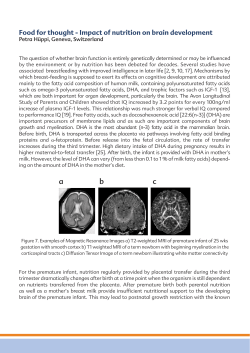


© Copyright 2026