
Department of Medicine, Division of Hematology Karolinska University Hospital Solna and
Department of Medicine, Division of Hematology
Karolinska University Hospital Solna and
Karolinska Institutet, Stockholm, Sweden
CHRONIC MYELOID LEUKEMIA
CLINICAL, EXPERIMENTAL
AND HEALTH ECONOMIC STUDIES,
WITH SPECIAL REFERENCE TO IMATINIB TREATMENT
Lotta Ohm
Stockholm 2013
All previously published papers were reproduced with permission from the publisher.
The cover image by Paulo Henrique Orlandi Mourao. Wikimedia Commons 2009.
Published by Karolinska Institutet. Printed by Repro print AB.
© Lotta Ohm, 2013
ISBN 978-91-7549-006-9
To my family and
in memory of my parents
ABSTRACT
CML is a malignant disease that originates in the bone marrow stem cell, carrying the Philadelphia chromosome
with the BCR-ABL fusion gene. This gene translates into an active tyrosine kinase, Bcr-Abl, affecting
hematopoiesis, particularly resulting in increased numbers of white blood cells in the peripheral blood. Left
untreated, CML progresses from a silent chronic phase (CP) to a life-threatening blastic phase (BP). After the
millennium shift imatinib was introduced for the treatment of CML. Specifically targeting the Bcr-Abl
oncoprotein, it was the first tyrosine kinase inhibitor (TKI) employed in cancer. It induced spectacular responses
among CML-CP patients, strikingly reducing the risk of disease progression, combined with excellent
tolerability. In this thesis we have studied various aspects of imatinib treatment in CML.
In a cohort of 45 newly diagnosed CML-CP patients initiated on imatinib, we consecutively assessed treatment
responses by FISH, PCR and chromosome banding analysis (CBA). In a landmark analysis, an early favourable
response, defined as less than 10% BCR-ABL-positive cells by FISH after 3 months of treatment, was identified
as a predictive marker of an improved long-term clinical outcome. Among evaluable patients 51% achieved this
response. A large majority, 95% of such responders, reached complete cytogenetic response within 12 months
and 100% an event-free survival at 48 months.
We assessed the effect of imatinib treatment on neutrophil leukotriene (LT) signaling to evaluate its possible
role as a clinical biomarker predictive of treatment response. Increased LT signaling has previously been
suggested as a driver of leukocytosis in CML. The activity and expression of LTC4S, catalyzing formation of
LTC4 from LTA4, were determined in neutrophils from 11 CML-CP patients during their initial phase of
imatinib treatment, and the results related to the parallel development of BCR-ABL-expression. CD16+
neutrophils were isolated, their LTC4S activity measured and LTC4S expression determined at the protein and
mRNA levels. In parallel, BCR-ABL expression was assessed by bone marrow CBA and by FISH on peripheral
blood cells, including a combined May Grünewald Giemsa staining and FISH technique (MGG-FISH) to score
neutrophilic cells. An aberrant expression of LTC4S in CML neutrophils was typically found, but it was rapidly
normalized after initiation of imatinib treatment, later paralleled by a decreasing expression of BCR-ABL. The
findings indicate that increased expression and activity of LTC4S in CML is a down-stream step of BCR-ABL
activity, i.e. the Bcr-Abl protein directly or indirectly causes an upregulation of LTC4S. It is possible that an
early evaluation of LTC4S expression during imatinib treatment could serve as a more rapid way of assessing
treatment response than the current methods identifying BCR-ABL expression through CBA, FISH or qRTPCR.
We also defined real life outcome of patients with CML in Sweden during four decades and related the relative
survival (RS) patterns to imatinib treatment and other management strategies. We assessed trends in survival and
short-and long-term excess mortality among all patients (n=3,173) regardless of clinical trial enrollment. Patients
were categorized into five age groups (<50, 50-59, 60-69, 70-79 and >79 years) and five calendar periods (19731979, 1980-1986, 1987-1993, 1994-2000 and 2001-2008). We found that throughout all calendar periods, age
was a strong predictor of survival, with superior survival for the youngest patients. In analyses including age and
period of diagnosis, RS improved with calendar period in all age groups, but most markedly in patients younger
than 79 years of age, particulary those 70-79 years of age. Survival among all age groups was greatest in the last
calendar period, mainly as a result of an increasing use of imatinib. However, elderly patients still do poorly. The
Swedish CML registry data show that patients diagnosed 2002-2008, at the age of 70-79 years received TKI in
66% and patients >80 years in only 18% of the cases.
Finally, we compared the costs during the last decades with earlier decades treatment regimens and related the
costs to the expected improved survival. Using Swedish real world national data from CML patients diagnosed
in the country from 1973 to 2008 (n=1,778), we evaluated the incremental cost-effectiveness ratio (ICER)
between three periods associated with broad implementation of imatinib (III), interferon-α and allogeneic stem
cell transplantation (II), and symptomatic treatment (I), respectively. We observed substantial health gains over
time, paralleled by increased treatment costs. The mean survival was 2.9, 9.2 and 18.5 years during periods I-III,
respectively. The resulting ICER was £45 700 per QALY gained comparing periods III and II using a societal
perspective. In a separate analysis by groups of age at diagnosis showed lower ICERs for individuals <50 years
at diagnosis: £38 500 for the societal perspective. Since the prevalence of CML patients is increasing and
assuming that 75% of each incident cohort was to receive imatinib at current prices, the imatinib budget would
need to double by 2050. A future potential discontinuation of imatinib for selected excellent responders would
reduce the ICER per QALY gained. Reduced drug cost of imatinib linked to the patent expiry of the drug will
probably have a greater impact on ICER per QALY. An estimated price reduction of 80% (global competition)
or 30% (expected change for biological drugs) would be associated with an ICER of £20 000 and £36 000,
respectively, per QALY gained.
LIST OF PUBLICATIONS
This thesis is based on the following papers, which are referred to in text by their Roman
numerals:
I
Ohm L, Arvidsson I, Barbany G, Hast R, Stenke L.
Early landmark analysis of imatinib treatment in CML chronic phase: less than
10% BCR-ABL by FISH at 3 months associated with improved long-term
clinical outcome. Am J Hematol. 2012 Aug;87Aug; 87(8):760-5.
II
Roos C, Stenke L, Ohm L, Widell S, Kumlin M, Lindgren JA,
Tornhamre S. Clinical imatinib mesylate treatment induces early normalisation
of aberrant neutrophil leukotriene C4 synthase expression and activity in
chronic myeloid leukaemia. Br J Haematol. 2008 Sep;142(6):992-5.
III
Björkholm M, Ohm L, Eloranta S, Derolf A, Hultcrantz M, Sjöberg J,
Andersson T, Höglund M, Richter J, Landgren O, Kristinsson SY, Dickman
PW. Success story of targeted therapy in chronic myeloid leukemia: a
population-based study of patients diagnosed in Sweden from 1973 to 2008. J
Clin Oncol. 2011 Jun 20;29(18):2514-20.
IV
Ohm L, Lundqvist A, Dickman P W, Höglund M, Persson U, Stenke L, Steen
Carlsson K, Björkholm M. Real world cost-effectiveness in chronic myeloid
leukemia, from non targeted treatment to imatinib-the current and future price
of success. Submitted for publication.
TABLE OF CONTENTS
LIST OF ABBREVIATIONS
5-LO
ABL
ALL
AML
AP
ASCT
ATP
BCR
BCR-ABL
Bcr-Abl
BP
CBA
CCA
CCyR, CCgR
CgR
CHR
CML-CP
CMR
CP
CysLT
D-FISH
DNA
EFS
ELN
ES-FISH
FISH
HLA
HR
HU
IFN
IR
LO
LR
LT
LTA4
LTB4
LTC4
LTC4S
LTD4
LTE4
Five-lipoxygenase
Abelson 1(gene)
Acute lymphoblastic leukemia
Acute myeloid leukemia
Accelerated phase
Autologous stem cell transplantation
Adenosine-5'-triphosphate
Breakpoint cluster region (gene)
Breakpoint cluster region-Abelson fusion (gene)
Breakpoint cluster region-Abelson fusion (protein)
Blastic phase (or blastic crisis)
Chromosome banding analysis
Clonal chromosomal abnormalities
Complete cytogenetic response
Cytogenetic response
Complete hematologic response
Chronic myeloid leukemia chronic phase
Complete molecular response
Chronic phase
Cysteinyl Leukotriene
Dual fusion-FISH
Deoxyribonucleic acid
Event-free survival
European Leukemia Net
Extra signal-FISH
Fluorescence in situ hybridization
Human leukocyte antigen
High risk
Hydroxyurea
Interferon-alpha
Intermediate risk
Lipoxygenase
Low risk
Leukotriene
Leukotriene A4; 5(S) -trans-5,6-oxido-11,14-ciseicosatetraenoic acid
Leukotriene B4; 5(S), 12(R)-dihydroxy-6,14-cis-8,10-transeicosatetraenoic acid
Leukotriene C4; 5(S)-hydroxy-6(R)-S-glutathionyl-7,9-trans11,14-cis- eicosatetraenoic acid
Leukotriene C4 synthase
Leukotriene D4; 5(S)-hydroxy-6(R)-S-cysteinylglycyl-7,9-trans11,14-cis- eicosatetraenoic acid
Leukotriene E4; 5(S)-hydroxy-6(R)-S-cysteinyl-7,9-trans-11,14cis-eicosatetraenoic acid
M-BCR
m-BCR
MMR
µ-BCR
MRD
m-RNA
OS
Ph
RNA
RT-PCR
q-RT-PCR
RS
RSR
SCT
TKI
WBC
WHO
Major breakpoint cluster region
Minor breakpoint cluster region
Major molecular response
Micro breakpoint cluster
Minimal residual disease
Messenger-RNA
Overall survival
Philadelphia
Ribonucleic acid
Reverse-transcription polymerase chain reaction
(quantitative) Real time reverse-transcription polymerase chain
reaction
Relative survival
Relative survival ratio
Stem cell transplantation
Tyrosine kinase inhibitor
White blood cell
World Health Organization
1 INTRODUCTION TO CHRONIC MYELOID LEUKEMIA
(CML)
1.1 BACKGROUND
The term leukemia was coined by Virchow in 1845 as he recognized several cases of splenomegaly, anemia and massive granulocytosis and understood the neoplastic nature in patients
with ”purulent” blood.1,2 The disease origin from the bone marrow was clarified by Neumann
some years later.3 Nowell´s and Hungerford´s discovery of the Philadelphia chromosome in
1960 was a breakthrough in cancer biology and CML. For the first time it was demonstrated
that a chromosome change was associated with a specific type of leukemia.4. In 1973 Rowley
found that the Philadelphia chromosome (Ph) was a result of a reciprocal chromosomal
translocation between the long arms of chromosomes 9 and 22.5 It took another 10 years before
it was revealed that the proto-oncogene ABL on chromosome 9 and the previously unidentified
BCR gene on chromosome 22 was involved and that the deregulated Abl tyrosine kinase was
the pathogenic factor.6 7 8 In 1990 Daley et al reported the first evidence of ability of BCR-ABL
to transform primary myeloid cells and induce a CML-like disease in mice, which finally
confirmed that the BCR-ABL and the constitutively active Bcr-Abl tyrosine kinase was the
underlying pathogenic factor in CML.9
Figure1. The normal Chromosomes 9 and 22, the translocation, and the two derivate chromosomes 9q+
and 22q- (Ph).
Prior to the 1950s, splenic irradiation was the mainstay of CML therapy. The treatment had no
or minimal effect on survival. In the mid 1950s busulphan became the prevailing palliative
9
treatment of CML, reducing the leukocytosis and splenomegaly. Busulphan was some decades
later replaced by hydroxyurea (HU) as the treatment of choice, due to less toxicity of the latter.
Neither drugs affected cytogenetic response or progression to BP resulting in a median survival
of 3.2 years in the 1970s.10 Until the 1980s, CML was regarded as incurable and inexorably
fatal.
In the 1980s it became clear that allogeneic stem cell transplantation (SCT), despite a relatively
high transplantation-related mortality especially in the early years, could induce long-term
Philadelphia chromosome negativity. It became the treatment of choice for eligible younger
patients with access to a donor since it offered a chance of cure. Efforts to extend SCT to all
patients with CML failed, due to lack of suitable donors and the increased incidence of lethal
graft-versus-host disease (GVHD).11 The standard therapy for the majority of patients in CML
chronic phase (CML-CP) in the mid 1980s was recombinant interferon-α (IFN). All studies that
reported IFN as first line treatment, tested the drug in combination with other agents mostly
hydroxyurea, low dose cytarabin or both. IFN prolonged life of patients in all ages, but lower
doses were used in elderly patients. IFN showed promising results in different studies with 10year overall survival (OS) of 25-53%12 13 and a median survival of 5-7.5years.14 However, IFN
therapy was associated with side effects, including fever, chills, muscle pain, asthenia, fatigue,
that lead to considerably reduced quality of life and problems to maintain the required high
doses of IFN.
In 1984 the Swedish CML Study Group was formed, and presented national recommendations
for the management of patients with CML the same year. Between 1984 and 1989 the Swedish
CML Study Group randomly allocated patients to treatment with either HU or busulphan.
Approximately 35% of all patients with newly diagnosed CML (n=179) were included.15 No
difference in overall or blast crisis-free survival was observed. Patients who underwent
allogeneic SCT fared significantly better with a median survival of 4.7 years in comparison
with 3.3 years in patients who did not. As a consequence of the study, younger patients were
offered SCT. In patients younger than 55 years of age without a donor, the Swedish CML Study
Group during the 1990s explored combined treatment with HU and IFN followed by one to
three courses of intensive chemotherapy. Patients who achieved significant Ph reduction and
negativity underwent high-dose chemotherapy and autologous SCT to further minimize the Phpositive clone.16 During the same period (1989 to 1997), the Swedish CML Study Group used
an intensive chemotherapy protocol in patients in accelerated phase (AP) and blastic phase (BP)
in an attempt to restore CP, including allogeneic or double autologous (i.e., cells harvested in
early chronic phase) SCT. The 1-year survival was 70% for allo-geneic SCT/AutologousSCT(ASCT) patients (median survival 21 months), 50% in responding patients overall, but only
7% in non-responders.17
10
1.2 CML – CLINICAL ASPECTS
1.2.1 Definition, diagnostic criteria, methods and predictors of prognosis.
CML is a myeloproliferative disease that originates in an abnormal pluripotent bone marrow
stem cell carrying the Philadelphia (Ph) chromosome and/or the BCR-ABL fusion gene. It is
found in all myeloid cell lineages, but also in some lymphoid cells,18 although the myelopoiesis
is dominating. At diagnosis parallel to the malignant clone there is a suppressed normal
hematopoesis.19,20
The diagnosis of CML should be suspected based on abnormal blood counts (leukocytosis,
thrombocytosis), the presence of an enlarged spleen and/or general symptoms such as fatigue,
weight loss, sweating and is formally diagnosed by
1) Typical morphological assessment of blood or bone marrow smears
AND
2) Detection of the BCR-ABL fusion gene in cells from blood or bone marrow.
Morphology
The peripheral blood shows increased white blood cell (WBC) counts, due mainly to
segmented neutrophils and neutrophils in different stages of maturation such as myelocytes,
metamyelocytes. Basophilia is invariably present and many patients may have eosinophilia as
well. A low number of myeloblasts are often seen. The platelet count is normal or increased and
may exceed 1000x109/l. Thrombocytopenia in chronic phase is rare. Most patients have a mild
anemia.
The bone marrow shows hypercellularity due to increased numbers of neutrophils and their
precursors. In chronic phase the blasts are usually fewer than 5%, while 10-19% indicates
transformation to an accelerated and ≥ 20% to a blastic phase.21 The megakaryocytes are
characteristically small and have hypolobated nuclei.22 Forty per cent of the patients display an
increase of reticulin fibers that generally correlates with increased numbers of megakaryocytes,
enlarged spleen and more severe degree of anemia.
Detection of BCR-ABL fusion gene
The presence of BCR-ABL can be detected by three different methods;
1) Chromosome metaphase analysis (chromosome level) shows an elongated chromosome 9
and a truncated chromosome 22, i.e. the Philadelphia chromosome. Karyotyping ("G-banding”
or "conventional cytogenetics") is a screening analysis where all chromosomes are evaluated in
metaphases. Normally 20-30 metaphases are analyzed. Since relatively few metaphases are
studied the sensitivity is rather low. An advantage of this technique is that additional aberrations
than Ph chromosome can be identified since the whole genome is analyzed. However, cryptic
fusions between the BCR and ABL genes can not be detected by karyotyping.
10
Karyotyping is used to measure the response to therapy. The cytogenetic response is associated
with prognostic significance, why this method is used until a complete cytogenetic response
(CCyR)= 0% Ph chromosomes, has been achieved.
2) FISH (Fluorescence In Situ Hybridization) (DNA level) is a targeted analysis, showing the
gene fusion BCR-ABL. In addition to the typical fusion gene, it is possible to detect cryptic
fusions. FISH allows the assessment of a larger number of non-dividing (interphase) cells, both
from peripheral blood23,24 25 26 and bone marrow and is thus more sensitive than chromosome
banding analysis (CBA). The method can also be performed on cultured metaphase cells, hypermetaphase FISH. 27 28
Probes targeting the ABL and BCR genes are used and nucleated cells are then examined in an
epiflourescence microscope. The fusion gene will appear yellow. In interphase FISH it is
recommended that at least 200 cells should be analyzed. The sensitivity for FISH is higher than
for karyotyping, less than 1 Ph-positive cell per 100 nucleated cells can be detected. The
method has so far not been used to give prognostic information similar to the degree of
cytogenetic responses using CBA (i.e. complete cytogenetic response, major cytogenetic
response etc.) 29
3) PCR (Polymerase Chain Reaction) (mRNA level) can be performed on bone marrow and
blood, usually on blood. Qualitative (RT-PCR) reveals the presence or absence of BCR-ABL
mRNA. Quantitative (qRT-PCR), reveals the amount of mRNA. The method is highly
sensitive and can detect 1 out of 105 transcripts of mRNA. Q-RT-PCR is the only method that
can measure minimal residual disease (MRD), after the patient reaches normal findings by
karyotyping (CCgR) or FISH. 30-32 The results of qRT-PCR are expressed as the ratio BCRABL/reference gene (ABL and/or GUS) as a percentage. A problem with the PCR technique is
that the methods and results vary between laboratories. An international scale (IS) has been
established to make results from different laboratories comparable, by giving each laboratory a
conversion factor related to a reference laboratory. All over the world laboratories undergo a
standardization work, in order to express their results according to the IS.31 33
Clinical course
The natural course of the CML disease is generally divided into three stages/phases. The
disease can present in any phase but most patients (90%) present in the chronic phase, during
which mature granulocytes are still produced but with an increased numbers of myeloid
progenitors in the peripheral blood. This early phase is often asymptomatic. In the absence of
effective treatment, the disease progresses eventually into the more aggressive phases, AP and
BP.11 In the pre-TKI era the average duration of the chronic phase was approximately 3-5
years.34,35 The following shorter AP is a poorly defined phase characterized by an increasing
number of myeloblasts and basophils in peripheral blood and bone marrow, persistent
thrombocytosis, and increasing spleen size unresponsive to therapy. In some cases there are
evidence of clonal evolution. The most common cytogenetic events in the clonal evolution are
the appearance of +8, +Ph, and i(17q) that denotes the activation of other oncogenes than BCRABL or a deletion of tumour suppressor genes.11 36 There are several definitions for the AP
including the definitions by European Leukemia Net (ELN),37 World Health Organization
11
(WHO)21 and the American National Comprehensive Cancer Network (NCCN).38 In Sweden,
the WHO classification from 2008 is the most widely used. The AP is a transition to the final
blastic phase. Here the percentage of blast cell in the bone marrow or peripheral blood is
increased to ≥20%, and the blasts can either be of lymphoid or myeloid origin.39-42 Myeloid BP
is the most common BP 50%, lymphoid 25% and undifferentiated BP in 25%.43 Occasional
patients have a combined myeloid/lymphoid CML-BP.44 In the CML-BP there is a clear
maturation stop in the myelopoiesis and the clinical features are more like an acute leukemia.
Isolated extramedullary proliferation of blasts that can be seen in the skin, lymph nodes, spleen,
bone or in the central nervous system constitutes a CML-BP, independent of the picture in the
peripheral blood and the bone marrow.
CML-AP may be made when one or more of the following are present:
Blasts 10-19% of WBC in peripheral blood and/or of nucleated bone marrow cells
Peripheral blood basophils ≥20%
9
Persistent thrombocytopenia (<100x10 /l ) unrelated to therapy or persistent
9
Thrombocytosis (>1000x10 /l) unresponsive to therapy
Increasing spleen size and increasing WBC count unresponsive to therapy
Cytogenetic evidence of clonal evolution
CML-BP may be made if one or more is present:
Blasts ≥ 20% of WBC in peripheral blood and/or of nucleated bone marrow cells
Extramedullary blast proliferation
Large foci or clusters of blasts in the bone marrow biopsy
Table 1 .WHO definition of accelerated phase.
Prediction of prognosis
At diagnosis the most important clinical prognostic factor for survival is to define whether the
patient is in the chronic, accelerated or blastic phase.21 In chronic phase the risk of progression
to a more advanced phase can be calculated by the Sokal and Hasford scores, respectively. The
two scores are calculated in a slightly different way, based on age of the patient, spleen size and
blood counts. Depending on the results the patients will be designated into one of three risk
groups; low, intermediate or high.
The Sokal score uses age at diagnosis, spleen size, platelet count and percentage of blasts in
blood. It predicts survival of newly diagnosed CML patients treated with hydroxyurea.45 Data
now indicate that Sokal score also predicts the chance of achieving CCgR and risk of
progression to AP/BP in patients treated with imatinib.32,46
The Hasford (Euro) score, is a further development of the Sokal score, predicts survival of
newly diagnosed CML patients with IFN treatment.47 It is based on data from 1303 patients
treated within twelve different studies. It is possible, but not yet clearly demonstrated, that
Hasford score can also be applied to patient groups treated with imatinib. Hasford score is
calculated based on age, spleen size, platelet count and percentage of blasts, eosinophils and
basophils in peripheral blood at diagnosis.
12
The EUTOS score is a novel, not yet widely spread score. It is calculated from the percentage of
basophils and spleen size and predicts outcome of imatinib therapy.48 The score may be used to
identify CML patients with significantly lower probabilities of responding to therapy and
survival.
The risk scoring systems may have lost some of its impact, since the most important individual
prognostic factor today is the degree and timing of the hematologic, cytogenetic and molecular
responses.37 49
1.2.2 Epidemiology
CML is a rare disease that has a reported world-wide incidence of 1.0-1.5 cases per 100 000
population per year.50 51 The incidence in Sweden during the last 8 year period was 0.8-1.0 per
100 000, 52 which means that about 90 patients are diagnosed with CML in Sweden annually.
CML is diagnosed in all ages, but the incidence increases with age. The median age at diagnosis
in Sweden is approximately 60 years. Males are slightly more affected than females (1.3:1).51,53
Due to more effective treatment in the last decade, prevalence increases in all Western
countries.54 No geographical or ethnical differences exist regarding the incidence, but the
prevalence differs, likely due to differences in management strategies between countries,
depending of availability of expensive drugs, modern diagnostic technologies and health-care
system.55
1.2.3 Etiology
Exposure to benzene and ionizing radiation constitute risk factors. Studies have shown that
BCR-ABL transcripts can be induced in hematopoietic stem cells by ionizing radiation in
vitro.56 A sharp increase of the incidence of CML was seen after the atomic bomb in Hiroshima
194557,58,59. No similar increase in CML incidence has been seen in connection with the
Chernobyl accident in the Soviet Union in 1986, where the radiation doses were significantly
lower compared to Hiroshima-Nagasaki.60
An explanation for the spontaneous appearance of the fusion gene BCR-ABL may be the short
physical distance between BCR and ABL genes in human lymphocytes61 and CD34 + cells that
could predispose to translocation between the genes.62,63 The mere presence of the BCR-ABL
translocation in a hematopoietic cell is not enough alone to cause CML. It is known that the
BCR-ABL fusion transcripts of M-BCR and m-BCR type are detected in low frequency among
30% of healthy individuals.64 65 It is unclear why CML occurs in only a minority of these
individuals. In healthy subjects the translocation possibly occurs in the terminal portion of
maturation and is eliminated by the normal immune system. Indirect evidence for the immune
system effects is that certain HLA types (HLA-B8 HLA-A3) appear to protect against CML.66
It is also likely that the chromosomal change must occur in a sufficiently primitive
hematopoietic progenitor cell, in order for the clone to expand. Another possibility is that BCRABL is not the only genetic lesion to induce CML-CP.67 However, in the vast majority of
patients the etiology of the disease is still unknown and there does not appear to be a hereditary
facto
13
1.2.4 Patophysiology
Blood cells develop from a multipotent hematopoietic stem cell located in the bone marrow.68
Normally the maturation from the stem cell into functional blood cells is very well regulated. In
CML a reciprocal translocation of genes between the long arms of chromosome 9 and 22 has
occurred in a stem cell.5,11 A part of the Abelson 1 gene (ABL1, hereafter called ABL) on
chromosome 9 is translocated and forms a fusion gene with a part of the breakpoint cluster
region gene (BCR), located on chromosome 22. The shorter derivate chromosome 22 carrying
the BCR-ABL fusion gene, is now called the Ph chromosome t(9;22)(q34;q11).11,69
The normal ABL proto-oncogene encodes a cytoplasmic and nuclear protein tyrosine kinase
that has been implicated in processes of cell differentiation, cell division, cell adhesion, and
stress response.67,70,71 The activity of the ABL gene is negatively regulated by its SH3
domain,67,72 and deletion of the SH3 domain turns ABL into an oncogene.73 After the t(9;22)
translocation the negative regulation of ABL is lost. The new fusion gene, BCR-ABL encodes
an unregulated, cytoplasm-targeted tyrosine kinase that allows the cells to proliferate without
being regulated by cytokines. Although the ABL gene product and BCR-ABL fusion protein
has been extensively studied, the function of the normal BCR gene product is not clear. Parts of
the ABL gene remains on the derived chromosome 9 and parts of the BCR gene translocates to
the 9th chromosome, resulting in a ABL-BCR gene. This fusion gene is encoding a protein
without any known function.
Fig 2. After the translocation between chromosomes 9 and 22, the BCR-ABL fusion gene undergoes
transcription. The resulting BCR-ABL messenger RNA (mRNA) is then translated into the Bcr-Abl
tyrosine kinase protein, which enhances cell proliferation, adhesion and survival.
9
The site of the breakpoint in the BCR gene differs, but in CML it is almost always in the major
breakpoint cluster region [M-BCR, BCR-ABL junctions e13a2 (b2a2) and e14a2 (b3a2)].
Rarely the breakpoint occurs in the minor breakpoint cluster region (m-BCR e1a2) as in Ph
positive acute lymphoblastic leukemia (ALL), or in the micro-region (µ-BCR, e19a2) as in
chronic granulocytic leukemia.11
The BCR–ABL fusion gene is transcribed into a messenger-RNA (mRNA) which will be
translated to a protein, a tyrosine kinase called Bcr-Abl, which activates mediators of the cell
cycle regulation system, leading to CML. The Bcr-Abl protein has different weight depending
of the different breakpoint in the BCR gene, 190kDa (m-region), 210 kDa (M-region) or
230kDa, (µ-region) but typically 210 kDa in CML.11
The Bcr-Abl tyrosine kinase protein is constitutively active, catalyzing the transfer of phosphate
from ATP to a tyrosine residue on a substrate protein downstream, resulting in the CML
phenotype, i.e. inhibition of apoptosis, increased proliferation and decreased adhesion to stroma
cells via effects on Bcr-Abl substrates like CRKL, CBL, RIN, GAP and paxillin and further
phosphorylation activates intracellular signal pathways like RAS, MYC and STAT.11,74
The mechanism behind disease progression and clonal evolution, when additional cytogenetic
aberrations occur, is still largely unknown. Genetic instability as a consequence of the the BCRABL translocation might be one explanation, alternatively, the mechanisms responsible for the
translocation might trigger the changes leading to progression of the disease.55
Five to 10% of CML patients have a variant translocation, which means that one or two
additional chromosomes are involved in the 9:22 chromosomal translocation. All chromosomes
have been described as participating in these variants, as an example t(3:9:22).75,76
1.2.5 Clinical signs and symptoms
About 20-40% of the CML patients are asymptomatic at diagnosis.51 The disease is quite often
detected at a routine medical examination. Others have symptoms like fatigue, sweats, weight
loss and abdominal fullness. Sometimes symptoms caused by the large number of circulating
leukocytes are seen eg. visual disturbances (due to vascular effects, including bleeding in the
fundus of the eye), pain in the left flank (due to splenic infarction or enlarged spleen) and
priapism.
In CML-CP, the peripheral blood shows an increasing amount of both mature and immature
leukocytes together with mild anemia and sometimes increased number of platelets. Atypically
the disease presents in CML-AP or CML-BP, without a previously detected CP. The more
advanced phases of CML are generally accompanied by worsened performance status and by
symptoms related to severe anemia, thrombocytopenia and/or marked splenic enlargement.
10
Blood count
Reference
values
B-Hb (g/l)
105 134-170
9
B-Platelets (10 /l)
694 145-348
9
B-Leukocytes (10 /l)
124 3.5-8.8
B-Neutrophil granulocytes
47
1.8-7.5
B-Eosinophil granulocytes
2.5
0.04-0.4
B-Basophil granulocytes
3.7
0-0.1
B-Lymphocytes
9.9
2.9-4.5
B-Monocytes
9.9
0.1-1.0
Immature myeloid cells
B-Band-formed granulocytes
17.4 0
B-Metamyelocytes
8.7
0
B-Myelocytes
14.9 0
B-Promyelocytes
5.0
0
B-Blasts
3.7
0
B-Erythroblasts
1.2
0
Table 2. Typical full blood count in a newly diagnosed CML-CP patient.
1.2.6 Treatment
Tyrosine Kinase Inhibitors
With improved understanding of the molecular mechanism involved in CML, the development
of targeted therapies, such as tyrosine kinase inhibitors (TKI) has gradually improved. TKI
inhibit the Bcr-Abl protein activity, thus stopping the leukemic phenotype. The first TKI for
CML, imatinib mesylate was a major breakthrough in CML treatment. The large phase 3
clinical trial International Randomized IFN versus STI571=imatinib mesylate (IRIS) for newly
diagnosed CML-CP patients started in 2000. The superiority of imatinib over IFN soon became
obvious. After 12 months of treatment 69% of the imatinib treated patients had achieved CCyR
compared to 7% in the IFN group.77 After 6 years of treatment the OS was 88% (95% when
only CML related deaths were considered) in the imatinib arm. Since a majority of the patients
(65%) randomized to the IFN arm, crossed over to the imatinib arm mainly due to intolerance 46
it is difficult to correctly compare data between the two arms. However, in the British CML III
study the 6-year OS was only 5% for patients treated with the IFN.78 At a median of 8 years of
follow up of the IRIS trial, the estimated OS of all patients randomized to receive imatinib was
85%. When only CML-related deaths was considered the figure was 93%.79
Imatinib and later generations of TKI have improved the prognosis for CML patients in all
disease phases, but most particularly for patients in CML-CP.80 TKI induce a much faster
reduction of BCR-ABL transcript levels than previously available drug therapies. Today TKI is
formally approved as first line treatment for CML-CP in all ages. Currently there are three TKI
approved by European Medicines Agency (EMA) and Food and Drug Administration (FDA) as
first-line treatment in CML; imatinib, nilotinib and dasatinib. Additional TKIs (bosutinib and
ponatinib) are currently being evaluated in clinical trials.
9
A recent French study has shown that approximately 40% of 100 imatinib treated patients with
a deep molecular response could discontinue treatment, without any signs of recurrent disease
within 2-3 years.81,82 Since 2nd generation TKIs as first line treatment seem to generate a larger
number of patients with deep molecular response, is possible that more CML-CP patients can
discontinue the treatment in the future.
Although imatinib has dramatically increased survival for CML patients in CP the outcome of
patients in more advanced phases has not been as successful. A treatment effect can be seen, but
is usually transient. TKIs may play a role as a bridge to allogeneic SCT for patients who
progress to BP. For patients progressing to AP during imatinib treatment a switch to a 2nd
generation TKI may revert the disease into the CP.
Imatinib (Gleevec® or Glivec®) is a small molecule, created by using the structure of the ATP
binding site of the ABL protein kinase as a template. It binds to the adenosine triphosphatebinding (ATP) site of Abl, thereby keeping the protein in an inactive form, thus inhibiting the
phosphorylation of substrate and subsequent blocking the activity of the Bcr-Abl tyrosine
kinase. The transmission of the oncogenic signal to the nucleus is by this interrupted and
thereby the malignant transformation, since unphosphorylated Bcr-Abl tyrosine kinase is
inactive.83 Imatinib is relative specific for Bcr-Abl tyrosine kinase but also active against
platelet-derived growth factor (PDGF) receptor84,85 and c-KIT receptor kinase.86 In Sweden
imatinib was approved for clinical use in 2001 and was the only TKI approved for newly
diagnosed CML patients until 2010. The most frequent side-effects are oedema, muscle cramps,
rash, nausea, diarrhoea, and hepatotoxicity.87 88 89
Imatinib is also used in the treatment of gastrointestinal stroma tumor (GIST)90 91 in hypereosinophilic syndrome (HES)92 and chronic eosinophilic leukemia (CEL)93 with FIP1L1-PDGFRα
rearrangement,92,94,95 (PDGFR) gene, in unresectable dermatofibrosarcoma protuberans
(DFSP)96 and in Ph positive acute leukemias, mainly in ALL. 97,98 99 100 However, except for
newly diagnosed CML-CP patients, there are no controlled trials demonstrating a clinical
benefit or increased survival for these diseases.
Dasatinib (Sprycel®) a second generation TKI, is a dual Abl/Src kinase inhibitor that binds to
Abl kinase domain irrespective of the configuration of the activation loop. Dasatinib inhibits a
lot more tyrosine kinases than imatinib and nilotinib. Dasatinib was approved in 2006 for CML
(CP, AP and BC) with resistance or intolerance to imatinib and to Ph positive ALL in resistance
or intolerance to prior therapy. It was approved for treatment of newly diagnosed CML patients
in 2010 but the Swedish The Dental and Pharmaceutical Benefits Agency (TLV) does not
reimburse the drug as first line treatment. Consequently it is not used beyond clinical trials as
first line drug.
The DASISION (Dasatinib vs Imatinib Study in Treatment–Naïve CML patients) study in
newly diagnosed patients, has shown a significantly superior response for the dasatinib vs.
imatinib with a major molecular response (MMR) rates at 3 years of 68% vs. 55% treated.101
The rate of transformation to AP or BP was numerically (but not statistically significantly)
lower for dasatinib: 4.7% vs. 6.7%.101 Treatment with dasatinib cause pleural effusion in 14-
10
26% of the patients 102,103 104 105 106 107 which seems more frequent in patients with previous
cardiovascular/ pulmonary disease, and autoimmune diseases.108
Nilotinib (Tasigna®) a second generation TKI derived from imatinib, is a selective Abl inhibitor
that binds to the inactive/closed conformation of the Abl kinase that also inhibits c-KIT, ARG,
PDGFα and PDGFβ. Nilotinib was approved in 2007 for treatment of CML patients with
resistance or intolerance to imatinib and was in 2010 approved for newly diagnosed patients
since the large randomized ENESTnd (Nilotinib Efficacy and Safety in Clinical trial-Newly
Diagnosed Patients) study showed that nilotinib compared to imatinib resulted in deeper and
faster short term treatment responses .109,110 The cumulative rates of MMR by 3 years was 70%
to 73% with nilotinib (two different doses) and 53% with imatinib, combined with a
significantly lower rate of transformation to AP or BP was observed, 2.1-3.2% vs 6.7%,
respectively.111 Nilotinib may cause pancreatitis (about 1%), liver (5-10%), lipase increase
(about 10%) and cause hyperglycaemia.112
Imatinb Nilotinb Dasatinib ABL, ARG, BCR-‐ABL, KIT, PDGFR, DDR1/2, NQO2 ABL, ARG, BCR-‐ABL, KIT, PDGFR, DDR1/2, NQO2 ABL, ARG, BCR-‐ABL, KIT, PDGFR, DDR1/2, SRC, YES, FYN, LYN HCK, LCK, FRG, BLK, FRK, CSK, BTK, TEC, BMX, TXK, ACK, ACTR2B, ACVR2, BRAF, EGFR/ERBB ,EPHA2, EPHA3, EPHA4 EPHA5, FAK, GAK, GCK, HH498/TNN13K ILK LIMK1, LIMK2 MYT1, NLK, PTK6/Brk, QIK, QSK, RAF1, RET, RIPK2, SLK, STK36/ULK, SYK, TAO3, TESK2, TYK2, ZAK Table 3. Tyrosine kinase “targets” of imatinib, nilotinib and dasatinib.113,114
Treatment objectives
Today the key objectives of front-line therapy in CML chronic phase are to
i) Prevent progression to the advanced phases (AP and BP)
ii) Maximize achievement of CCyR and MMR which gives the patient a platform to achieve
CMR and thereby a possibility of drug cessation.
There are three clinical milestones (interim targets) in the treatment response of CML-CP
(Table 4)
1) Complete hematologic response (CHR)
2) Complete cytogenetic response (CCyR)
3) Deep molecular response, i.e. achieving major molecular response (MMR) and “complete
molecular response” (CMR, signifying non-detectable transcripts, normally <MR4.0 – MR5.0).
The response to TKI treatment is generally fast, with normalization of leukocytosis/
thrombocytosis within weeks, followed by a gradual reduction of Ph positive cells in blood
and bone marrow until achieving CCyR and MMR/CMR. To guide the physicians European
Leukemia Net (ELN) and other organizations give valuable recommendations, which among
other things specify treatment goals at certain time points.37,49
11
In trials, second generation TKIs as first line treatment seem to give a more rapid treatment
response and with fewer patients progressing to AP and BP, compared to imatinib, but so far,
no overall survival benefits have yet been reported.
Definitions of treatment response
Hematologic response
Complete (CHR):
B-WBC <10 109/l
B-Basophils< 5% of WBC
B-Platelets <450 109/l
No leukocyte precursors in peripheral blood
No palpable spleen
Cytogenetic response
(% Ph positive metaphases)
0
Complete (CCyR)
Partial
1-35
Minor
36-65
Minimal
66-95
No
>95
Molecular response
BCR-ABL % International scale (IS)
Major (MMR, 3.0 log
reduction)
≤0.1
4.0
MR (4.0 log reduction) ≤0.01
MR4.5 (4.5 log reduction) ≤0.0032
MR5.0 (5.0 log reduction) ≤0.001
Table 4. Definitions of treatment response.
Time
At diagnosis
3 month
6 month
12 month
18 months
Any time
Optimal
CHR and at least minor
CR
At least PCgR
CCgR
MMR
Stable or improving
MMR
Suboptimal
Failure
No CgR
Less than PCgR
PCgR
Less than MMR
Less than CHR
No CgR
Less than PCgR
Less than CCgR
Loss of CHR or
CCgR
Mutations B
CCA/ Ph+
Loss of MMR
Mutations A
Warnings
High risk. CCA/Ph+
Less than MMR
CCA/Ph-
Table 5. Definitions according to ELN, of optimal and suboptimal response, failure and warnings for
previously untreated patients with early chronic phase CML, treated with imatinib at certain time points.
Mutation A= Still sensitive to imatinib. Mutation B= Poorly sensitive to imatinib. CCA= Clonal
chromosome abnormalities. 37,49
Treatment in AP and BP
Patients diagnosed in AP may initiate treatment with a 2nd generation TKI. They should be
carefully monitored, and if the patients do not reach the criteria for CP, allogeneic SCT should
10
be considered. For imatinib treated patients progressing to AP a switch to a 2nd generation TKI
or SCT is needed.
For patients diagnosed in or that have progressed to BP, the prognosis is very poor. If CML is
diagnosed in BP the patients should start TKI treatment in combination with ALL- or AMLlike chemotherapy (depending on lymphoid or myeloid phenotype) and be considered for
allogeneic SCT as soon as possible. If progressing to BP during imatinib treatment the patients
should be prepared for allogeneic SCT if possible, and a switch to high dose dasatinib is then
preferred (unless mutations resistant to dasatinib) to optimize the conditions for SCT. AML - or
ALL–like chemotherapy could be given before transplantation.
The response to TKI in BP is expected to be transient. Allogeneic SCT has a curative potential
for CML and the best results are achieved for patients transplanted in CP.115-118 SCT in overt BP
is not recommended.116
Treatment failure
If imatinib treatment of a CML-CP patient does not meet the ELN criteria for “optimal”
response at a certain time point (see table 1), the response may be regarded as “suboptimal” or
as a “failure”. There are several explanations for not achieving optimal response. Both BCRABL dependent and independent mechanisms have been suggested; poor compliance, reduced
bioavailability, mutations in the Bcr-Abl tyrosine protein pocket, clonal evolution or
resistance to the drug etc. When treatment failure is a fact, a change of treatment is needed.
Both dasatinib and nilotinib have been shown to have clinical activity in CML patients with
intolerance or resistance to imatinb.119-123 Another options is allogeneic SCT.
In the case of BCR-ABL independent factors, a dose escalation of imatinib may be successful.
There are currently about 100 known mutations, rendering imatinib therapy suboptimal or
failing. In most cases, 2nd generation TKI can overcome this.124,125 Nilotinib is sometimes a
better choice than dasatinib, or vice versa, related to specific mutations. Dasatinib thus seems to
be more effective than nilotinib in the presence of BCR-ABL mutations such as Y253F/H,
E255K/V, F359C/V while in the presence of Q252H, V299L and F317L, nilotinib is preferable
to dasatinib.126 Only one mutation, T315I (threonine 315 isoleucine) is completely resistant to
imatinib, dasatinib and nilotinib.127 However, ponatinib, a multi-targeted tyrosine kinase
inhibitor, has in an on-going clinical trial shown efficacy against this mutation.128 The AP may
after a switch of TKI be induced to revert into the CP. In BP the effect of TKIs is very limited.
If any effect, it is not durable, but can act as a bridge to allogeneic SCT, which is the only
treatment offering long-term survival for patients in BP.129
Surrogate markers for long-term outcome
CCyR, MMR and CMR are the therapeutic goal in treatment of CML-CP in order to minimize
the risk of transformation into advances phases. For many years, the main goal of treatment was
to achieve CCyR, since studies had shown that CCyR was associated with survival benefits.130132
With the introduction of imatinib treatment, a majority of the patients achived CCyR. Many
had an even better response, major molecular response (MMR), which gave further improved
OS 46,49,130,131,133,134 compared to patients with CCyR but without MMR.135 Furthermore, 2nd
generation TKI (dasatinib and nilotinib) as first line treatment seem to give an even deeper and
10
faster response, and the proportion of patients achieving MMR is higher at 12 months than with
imatinib. 109,136,137 Today the IRIS study has given the clinicians more than 10 years of
experience regarding imatinib treatment. At 5-year the cumulative incidence of CCyR in the
imatinib arm was 87%.130 At that time 93% of the patients had not progressed to the accelerated
or blastic phases. The estimated 5 year OS was 89%.130 However at 5 years 31% of the patients
in the IRIS study had discontinued imatinib treatment and were censored.131 Clinical trials using
“intention to treat” criteria may be difficult to interpret as for instance in the IRIS study. The
achieved OS is not only reflecting imatinib treatment, but eventually also second line TKI
and/or allogeneic SCT, thus the OS in the IRIS study is likely overestimated, reflecting only
imatinib treatment.
However, achieving CCyR and MMR seems to give the patients long-term event free and
overall survival, but there is also a time aspect when the goals are achieved. Reducing the
tumour load is important to reduce the risk of progression to more advanced phases. Having
achieved CCyR and MMR the annual risk of progressing is low. For example, in the IRIS study
no patient that had achieved MMR at 12 months subsequently progressed into AP or BP in the
5-year follow-up130. Detailed recommendations are available for monitoring and timing of
treatment goals, provided by groups and individual centers. In Sweden the national
recommendations138 are used which are similar to the guidelines of ELN. To further improve
the outcome, we probably need to act earlier in the disease process, at an earlier time than
guidelines advise us today.
1.3 SWEDISH POPULATION REGISTRIES
Since 1947 all Swedish citizens are given a unique identification code at birth. For each
individual, date of death is centrally registered in the nation wide “Cause of Death
Registry”139. Furthermore, every physician, pathologist and cytologist are obliged by law to
report occurrence of cancer to the population-based national wide “Swedish Cancer
Registry”140 established in 1958. Through these high quality registers it is possible to follow
patients from the date of cancer diagnosis to death. The Swedish CML Registry, established in
2002, collects information once a year from physicians treating CML patients throughout the
course of the disease. The registry is unique, with nearly all CML cases in Sweden included.
10
2 AIMS
Overall aims
To improve management of patients with CML by
i)
identifying early biological markers linked to long-term clinical outcome of
imatinib treatment
and
ii)
establishing population-based survival data including health economic aspects
with special focus on imatinib treatment
Specific aims
I.
To evaluate the impact of early reduction of disease burden on long-term outcome (i.e.
landmark analysis) in imatinib treated patients.
II.
To evaluate the effect of imatinib treatment on neutrophil leukotriene signaling and assess its
possible role as a clinical marker of CML treatment response.
III.
To define real life outcome of patients with CML in Sweden during four decades and to relate
the survival patterns to imatinib treatment and other management strategies.
IV.
To evaluate health economic aspects of real life treatment of CML in Sweden, comparing
imatinib with previous and alternative therapeutic strategies.
10
3 STUDY OF PROGNOSTIC FACTORS-LANDMARK
ANALYSIS (I)
3.1 METHODOLOGICAL ASPECTS
Imatinib has dramatically improved the outcome in CML-CP, but still some patients continue to
respond sub optimally or become resistant to imatinib. These patients need alternative treatment
with an early switch to 2nd generation TKI or allogeneic SCT. Standard dose imatinib (400mg
q.d.) has been reported to induce CHR in the majority of patients and CCyR in 69% within 12
months of treatment, but a minority of the patients still does poorly. In the IRIS study, 34% of
the patients had discontinued their initial imatinib therapy at 5 years because of different
reasons, mostly intolerance or resistance. Seven percent of the IRIS study patients had
progressed to AP or BP within 2 years.130 The risk of progression seems to peak during the first
year of treatment, before the tumour burden radically has been decreased. Apparently, we need
to act early of signs of non-optimal response, to improve outcome in imatinib treated patients.
CBA has for many years been the gold standard for evaluating treatment effects and to reach a
status of non-detectable Ph-chromosomes (CCyR) by 12 months, has been regarded as one of
the treatment goals. Since 30% of the patients do not reach that goal it is of interest to early
identify patients having an increased risk of progression of the disease.
In an attempt to identify markers of early non-responders, we followed 45 newly diagnosed
CML-CP patients initiated on imatinib treatment at standard dose at Karolinska University
Hospital. We evaluated the treatment responses by repeated FISH, PCR and cytogenetics
assessments initially at 3 months intervals.
3.1.1 Cytogenetic Banding Analysis (CBA)
Conventional cytogenetic analyses of bone marrow samples were performed at the Department
of Clinical Genetics, Karolinska University Hospital. All investigations were done using
standard G-banding technique and followed the rules of International System for Human
Cytogenetic Nomenclature, ISCN. Cytogenetic response evaluations were regularly based on
full analysis of at least 20 metaphases. Of a total of 257 samples in this study, 246 contained 20
or more metaphases, with a median of 29 metaphases analysed per sample.
3.1.2 Fluorescence In Situ Hybridization (FISH)
DNA-probes directed against the BCR and ABL genes are added to a smear of blood or bone
marrow cells and are analyzed in an epifluorescence microcope. The reliability of FISH is
largely dependent on the quality of the DNA probe used.141,142,143 Different probes show
different signal patterns.144 The Extra Signal Dual Color Translocation Probe-FISH (ES-FISH)
reduces the number of falsely BCR-ABL positive cells, compared to the earlier used Dual Color
Single Fusion Translocation Probe-FISH (S-FISH).143,145 Our control studies showed that the
ES probe, in our hands, had high sensitivity and specificity, similar to the Dual Color Dual
9
Fusion translocation Probe (D-FISH).146 One disadvantage with ES-FISH compared to D-FISH
is that Ph-clones with minor deletions of the long arm of chromosome 9 can be missed because
these aberrations will give the typical signal pattern 1G1O1F.
In our study we performed interphase FISH on unseparated nucleated cells on bone marrow
smears, using an extra signal dual-color DNA probe (LSI BCR-ABL ES Dual Color
Translocation probe, Vysis, IL). The probe is a mixture of the LSI ABL probe labeled with
spectrum orange and the LSI BCR probe labeled with spectrum green. The spanning ABL
probe is approximately 650kb extending from an area centromeric of ASS gene well telomeric
of the last ABL exon. The spectrum green BCR probe is approximately 300 kb beginning
between BCR exons 13 and 14 (M-BCR region) A cell lacking the BCR-ABL fusion gene will
exhibit a two orange and two green signal pattern (2O2G). In a cell carrying the fusion gene one
green (native BCR) one large orange (native ABL), one smaller orange (rest signal of ABL on
the derivate chromosome 9) and one fused green/orange (BCR-ABL) (2O1G1F).
On each patient smear a median of 400 nucleated cells were analysed.
Figure 3. A BCR-ABL positive cell with the typical ES probe signal pattern 2OIGI.
(Photo Ingrid Arvidsson)
Control studies for FISH
Bone marrow smears from nine Ph-negative controls were analyzed using the ES probe. In each
case 500 cells were scored. The mean percentage of “not true” BCR-ABL positive cells among
these controls was 0.031% (SD 0.075%) For the purpose of our study (I) we decided to define
the cut-off level for a “true” positive sample to ≥0.25% (mean +2.576xSD).
10
Further cytospin preparations from the CML cell line K562 were used as positive controls. All
(100%) of 2500 K562 cells examined with the ES-probe depicted the typical fusion pattern
(2O1G1F).
To compare DS-FISH with ES-FISH, we analyzed nine patient samples with both probes. We
found a strong correlation (r= 0.87;p= 0.0003).
3.1.3 Quantitative reverse-transcriptase Polymerase Chain Reaction (qRT-PCR)
The Department of Clinical Genetics, Karolinska University Hospital, Stockholm, performed
the PCR analysis. For samples prior to October 2008 peripheral blood mononuclear cells were
separated by the Ficoll-Paque TMPLUS (GE Healthcare, UK) and total RNA was isolated
using Trizol® reagent (Invitrogen, Carlsbad). From October 2008 and onwards, total blood
leukocytes were used for RNA isolation. RNA (1µg) was reverse-transcribed into cDNA as
described elsewhere using pd(N)6 random hexamer (GE Healthcare, UK) and M-MLV
enzyme (Invitrogen, Carlsbad).147 ABL and GUS were used as control genes to correct for
variations in RNA quality and quantity.148 The BCR-ABLp210 and BCR-ABLp190 mRNA
transcripts were assessed by quantitative real-time RT-PCR, as described previously.148,149
ABL and GUS were used as reference genes. In this studies, we consequently expressed the
amount of BCR-ABL transcript as a ratio of BCR-ABL copy number relative to 100 ABL
copies, since the IS not yet was established in our laboratory at that time.
3.1.4 Statistical methods
The probabilities of OS and event free survival (EFS), where events were defined as response
loss, progression or death, in line with ELN recommendations.49 OS and EFS were calculated
using Kaplan-Meier method150 according to the intention-to-treat principle. The association
between early determinations of BCR-ABL or Ph-expression, as assessed by the three different
techniques and the long-term clinical outcome was examined using contingency tables and
Fisher´s exact test.
3.2 RESULTS AND DISCUSSION
The median follow up was 58 (range 15-115) months. Within 12 months of treatment 80%
achieved CCyR and 42% MMR. Corresponding figures at 24 months were 97 and 84%. After 3
months of treatment the median BCR-ABL expression was 9.8 (range 0-95)% according to
FISH and 16.0 (range 0-100)% according to PCR. Corresponding figures for 6 months were
0.25 (range 0-65)% for FISH, 1.2 (range 0-85)% for PCR and 0 (range 0-100)% for CBA.
Four patients (8.8%) progressed to AP within 16 months. One patient later progressed into BP
after initially having achieved CCyR.
In a landmark analysis, an early favourable response, defined as < 10% BCR-ABL positivity by
FISH after 3 month of treatment, was identified as a predictive marker of an improved longterm clinical outcome. Thus of evaluable patients, 51% achieved this response. Ninety-five per
cent of such responders reached CCyR within 12 months and 100% had an event-free survival
10
at 48 months as compared to 67 and 65%, respectively, of patients with higher (≥ 10%) BCRABL positivity at 3 months.
BCR-ABL positive cells by FISH
BCR-ABL mRNA by PCR
<10%
≥10%
P-value <10%
≥10%
P-value
CCyR at 12 months
18/19 (95%)
12/18 (67%)
0.042
11/12 (92%)
11/15 (73%)
0.342 ns
EFS at 36 months
19/19 (100%)
12/18 (67%)
0.008
10/11 (91%)
11/16 (69%)
0.350 ns
EFS at 48 months
19/19 (100%)
11/17 (65%)
0.006
9/10 (90%)
11/16 (69%)
0.350 ns
CCyR at 12 months
21/24 (88%)
1/4 (25%)
0.022
23/27 (85%)
5/8 (63%)
0.312 ns
EFS at 36 months
21/24 (88%)
2/5 (40%)
0.046
24/27 (89%)
7/8 (87%)
1.0 ns
EFS at 48 months
21/24 (88%)
2/5 (40%)
0.046
21/24 (88%)
6/7 (86%)
1.0 ns
EFS at 36 months
17/18 (94%)
0/3 (0%)
0.003
31/34 (91%)
0/2 (0%)
0.016
EFS at 48 months
17/18 (94%)
0/3 (0%)
0.003
27/30 (90%)
0/2 (0%)
0.020
3-month landmark
6-months landmark
12-months landmark
Table 6. Number of patients with CCyR at 12 months, EFS at 36 months and EFS at 48 months, related
to responses at 3 and 6 months assessed by FISH and PCR.
We showed that significantly more patients with a reduction of the malignant clone to <10% at
3 months of imatinib treatment achieved CCyR within 12 months compared to those that failed
to reach this level of reduction. They had also a significantly better EFS both at 36 and 48
months compared to patients with BCR-ABL ≥10% by FISH at 3 months. This finding is
consistent with other research groups,151,152 using the qRT-PCR method, observing that patients
with PCR levels ≥10% at 3 month are likely to respond poorly on imatinib therapy. Marin et al
found that patients with transcript levels of more than 9.84% (n = 68) at 3 months had
significantly lower 8-year probabilities of OS (56.9% v 93.3%; p <0.001), progression-free
survival, cumulative incidence of CCyR, and CMR than those with lower transcript levels.151
Hanfstein et al found persistence of BCR-ABL transcript levels >10%, according to the IS at 3
months, to separate a high-risk group (28% of patients; 5-year OS: 87%) from a group with 110% BCR-ABL.152 In contrast, when we performed landmark analysis on our patient cohort
with PCR, we did not observe the same predictive value as with FISH. The fact that we did not
use IS in our study may explain why we did not find the same predictive value with PCR<10%
as other groups have. Although PCR has become the dominating method for monitoring TKI
treatment, particularly to detect minimal residual disease (MRD), the technique can be
inconsistent with considerable intralaboratory and interlaboratory variations. Thus, this will be a
minor problem when laboratories have harmonized the PCR standard to the IS. Despite this,
many physicians do not have access to laboratory service with the semi-quantitative PCR
method. The well-established FISH method could then be of great value.
10
In our study four patients (8.8%) progressed to AP within 16 months using the WHO
classification, which is slightly more than is reported in the IRIS trial where 7.9% progressed
within 18 months.77 However, in the IRIS study, a different definition of AP and BP was used.
In the IRIS study definition of AP was ≥15% blasts in peripheral blood or bone marrow and for
BP ≥30% blasts, compared to 10-19% and ≥20% respectively, in the WHO classification. If
WHO classification would have been used in the IRIS study, probably more patients would
have been considered to have progressed to AP or BP.
All of our four patients had clonal evolution in their Ph+ cell clones. Thus, undoubtedly, they
had progressed to AP. They were all scored as Sokal intermediate risk at diagnosis. Two of
these four patients regained chronic phase after treatment with second generation TKI, while the
remaining two died. All patients with Sokal high-risk score achieved CCyR within 12 months,
although high-risk patients in general have an increased risk of progression.130,153 154
Our data, even though based on a limited patient cohort, indicate that FISH can effectively be
used in the early assessment of remaining Ph-positive cells to identify patients at risk for a longterm non-optimal response to imatinib. In clinical practice; sufficient data presently indicate that
a change of treatment to 2nd generation TKI should be considered if BCR-ABL levels persist
above 10% after 3 months of treatment with imatinib, as measured with PCR or FISH or PCR.
11
4 STUDY OF PROGNOSTIC FACTORS - LEUKOTRIENE
SIGNALING (II)
4.1 BACKGROUND
Leukotrienes (LTs) are a family of lipid mediators derived from arachnoid acid with important
signaling molecules in inflammatory and allergic conditions.155-158,156-159 Leukocytes can form
various types of leukotrienes (hence their name) via an initial conversion of arachnoid acid to
LTA4, a reaction mediated by the 5-lipoxygenase (5-LO) enzyme. Members of our group have
previous reported data indicating a role for certain leukotrienes also in CML. Thus, we have
found that the bioactive cysteinyl-containing leukotriene C4 (LTC4) can stimulate normal
human myelopoiesis160 and that myeloid cells from CML patients have a markedly increased
capacity to synthesize LTC4.160,161 Moreover, neutrophils from CML patients were noted to
possess significant leukotriene C4 synthase (LTC4S) expression and activity, while this is not
the case for normal neutrophils.162,163 Recently, the 5-LO was suggested as a crucial growth
regulator for leukemia stem cells (LSC) in CML, since gene expression profiles using DNA
micro array technique identified Alox5, the gene encoding 5-LO, as highly and selectively
expressed in LSC. Blocking the Alox5 activity impaired LSC function and prolonged survival
in mice with CML.164 These data indicate a role for Alox5 and LTC4 in CML leukemogenesis.
In this study we examined and followed the response of imatinib treated CML-patients over
time, to analyse whether imatinib could regulate neutrophil LTC4S expression and activity in
vivo and if so, whether such changes were paralleled by changes in the expression of BCRABL. If so, a normalization of neutrophil LTC4S and/or activity might be used as a novel
indirect biomarker of early treatment efficacy. Furthermore, we also aimed at estimating the
LTC4 production in vivo, by assessing urinary excretion of the LTC4 metabolite LTE4 in
individual patients with CML and healthy controls.
9
Fig 4. Enzymatic biosynthesis of leukotrienes from arachidonic acid. 5-LO= 5-lipoxygenase.
LTC4S=Leukotriene C4 synthase.
4.2 PATIENTS AND METHODS
We sequentially studied the activity and expression of LTC4S in neutrophils from CML patients
subjected to imatinib treatment, and related these data to the parallel development of BCRABL-expression. A total of eleven CML-CP patients, three newly diagnosed and eight with
previous IFN therapy, were assessed. Blood samples were drawn before start of imatinib
therapy and then after 2 weeks, 1, 4, 6 and 9 months of treatment. Peripheral blood CD16+
neutrophils were isolated using magnetic cell sorting (MACS). Their LTC4S activity assessed
(i.e. Formation of LTC4 from LTA4) as measured by high performance liquid chromatography,
(HPLC) and LTC4S expression determined at the protein (immunoblot) and mRNA (RT-PCR)
levels. In parallel, Ph-chromosome expression was assessed by bone marrow cytogenetics and
by performing FISH on smears of peripheral blood identifying and specifically scoring
neutrophilic granulocytes by a combined May-Grünwald-Giemsa staining and regular FISH
technique cells, which makes it possible to allocate specific chromosomal aberrations (in this
study BCR-ABL) in morphologically defined cells.165,166
10
4.2.1 Statistical methods
The two-sided Student`s test for unpaired samples was employed to compare mean values of
LT production in the different cell fractions
4.3 RESULTS AND DISCUSSION
We demonstrated that in vivo-treatment of CML patients with imatinib led to a normalization of
the aberrant LTC4S expression and activity. In general, this normalization occurred in parallel to
a reduced DNA-expression of BCR-ABL, but the time courses of these changes were different.
Already after 14 days of imatinib treatment we found a significant decrease of LTC4S activity
in the neutrophils while the BCR-ABL expression remained unaltered in these cells at the same
time point. We propose that a block of p210/Bcr-Abl protein activity in neutrophils by imatinib
blocks down-stream 5-LO/Alox5 activity and thereby LTC4S and LTC4 formation. This can
occur in CML cells that still express the Ph chromosome. Later, when imatinib treatment has
resulted in replacement of Ph-positive CML cells with normal Ph-negative cells, LTC4S activity
remains low or non-detectable while FISH and cytogenetics show no or low BCR-ABL
expression.
Thus, it is possible that analyses of neutrophil LTC4S and/or LTC4 expression may serve as
early biomarkers of initial phase clinical imatinib response in CML-CP. Although TKIs are the
standard treatment of CML today, resistance to these drugs remains a problem for certain
patients. Targeting complementary, aberrant signaling pathways down-streams of or parallel to
Bcr-Abl tyrosine kinase may provide a constructive option.
Furthermore, urine samples from 23 CML-CP patients were analyzed and compared to urine
samples from 18 healthy controls to look for differences in LTE4 excretion, which is considered
to represent the combined in vivo formation of the LTC4, LTD4 and LTE4, the cysteinyl LTs
(cysLTs) in humans.167 Notably, a significantly higher level of LTE4 was observed in the CML
group. This indicates indeed a higher in vivo production of LTC4 in the leukemia patients and
further supports a pathophysiological role for the cysLTs in CML.
The recent results by Chen et al demonstrating that blocking the 5-LO with the asthma drug
zileuton inhibited the leukomogenesis in CML induced mice are consistent with our findings of
a relationship between 5-LO, LTC4 and BCR-ABL in CML. An ongoing clinical study in USA
is examining the effect of combining the 5-LO inhibitor zileuton with imatinib in newly
diagnosed CML-CP patients. This study is planed to be completed in 2014.
11
Figure 5. LTC4S activity in CD16 (+) neutrophils (left panel) and the parallel occurrence of BCR-ABL,
investigated by FISH (right panel) from CML patients before, after 7 and 14 days of treatment with
imatinib. The lines represent individual patients and the bars show mean+/- SEM (n=4) of experiments
perforemd in duplicate.
10
5 POPULATION-BASED STUDIES OF SURVIVAL IN CML (III)
5.1 PATIENTS AND METHODS
Our aim was to assess trends in survival, among all patients diagnosed with CML in Sweden,
regardless of clinical trial enrollment, during a 36-year period. In the 1970s, at the start of the
study period, busulphan was the dominating therapeutic agent. It was followed by a more
widespread use of hydroxyurea and the introduction of IFN and allogeneic SCT. Most
importantly, imatinib was available in study protocols from 2000 and it was approved by the
Swedish Medical Products Agency (MPA) in November 2001 for CML patients in advanced
phase CML and in “interferon failing” CP.
We included information on all newly diagnosed CML patients (n=3173; 1796 males and 1377
females; median age 62 years), reported to the Swedish Cancer Registry140 from 1973 to 2008
using the International Classification of Diseases Version 7 (ICD 7). The diagnosis was coded
as 2051. Patients in all phases of CML were included. The exact proportion of patients
diagnosed in the advanced phases is known only for period the 2002-2008: 4% in AP and 3% in
BP. The Swedish CML Registry, to which more detailed information about disease characteristics is reported, was established in 2002,53. Information from the Swedish Cancer
Registry140 included date of birth, gender, date of diagnosis, region, and hospital where the
diagnosis was made. All patients were observed from the date of diagnosis until death,
emigration, or end of follow-up (December 31, 2009). We excluded individuals who were
diagnosed incidentally at autopsy. The choice to include patients from 1973 was based on the
fact that by then, the Swedish Cancer Registry, established 1958, had reached a high rate of
coverage.168,169 Patients with a preceding cancer diagnosis, including those with a hematological
malignancy, were included. Date of death was obtained from Cause of Death Registry.139
Information of the use of upfront imatinib was collected from the Swedish CML registry.
Information on the number of SCTs performed in CML patients during this time period was
obtained from the registry of the European Group for Blood and Marrow Transplantation
(EBMT), established in 1974.
5.1.1 Statistical methods
Relative survival ratios (RSR) were computed as measures of CML survival.170,171 The
important advantage of using RS is that it does not rely on the accurate classification of cause of
death. Instead, in this project it provides a measure of total excess mortality associated with the
diagnosis of CML, irrespective of whether the excess mortality was a direct or indirect result of
the cancer (here CML). As such, the RSR allows capturing the excess mortality resulting from,
e.g. infection or second primary malignancies, which is not possible when using cause-specific
survival. The RSR is defined as observed survival in the patient group (where all deaths are
considered as events) divided by the expected survival of a comparable group from the general
population, which is assumed to be free from the cancer in question. One, 5 and 10 year RSRs
can be interpreted as the proportion of patients with CML who survived the malignancy at 1, 5
and 10 years, respectively. Expected survival was estimated using the Ederer II method172 from
11
Swedish population life tables stratified by age, sex, and calendar period. One, 5, and 10 year
RSRs were calculated for patients diagnosed during five calendar periods and five age
categories. Poisson regression was used to model excess mortality173 to estimate the effects of
the factor described, while controlling for potential confounding factors. Parameter estimates
from this model are interpreted as excess mortality rate ratios (EMRRs). An EMRR of e.g. 1.5
for men/women indicates that men experience a 50% higher excess mortality rate (difference
between observed and expected mortality) than women. All calculations were performed using
Stata 11 (StataCorp, College Station, TX).
5.2 RESULTS AND DISCUSSION
Using data from the population-based Swedish Cancer Registry and population life tables, we
estimated RS for all patients diagnosed with CML between 1973-2008. Patients were
categorized into five age groups and five calendar periods (<50, 50-59, 60-69, 70-79, >79 years;
and 1973-1979, 1980-1986, 1987-1993, 1994-2000, 2001-2008.)
Relative survival improved with each calendar period, with the greatest improvement between
1994-2000 and 2001-2008. Five-year cumulative relative survival ratios (95% Cls) were 0.21
(0.17-0.24) for patients diagnosed 1973-1979, 0.23 (0.20-0.27), 0.37 (0.33-0.41) 0.54 (0.500.58) 1994-2000, and 0.80 (0.75-0.83) for 1980-1986, 1987-1993, 1994-2000 and 2001-2008,
respectively. This improvement was confined to patients younger than 79 years of age. Fiveyear RSRs for patients diagnosed from 2001 to 2008 were 0.91 (95% CI, 0.85 to 0.94) and 0.25
(95% CI, 0.10-0.47) for patients younger than 50 and older than 79 years, respectively. Men had
inferior outcome. Upfront overall use of imatinib increased from 40% (2002) to 84% (2006).
Only 18% of patients older than 80 years of age received imatinib as first-line therapy.
Throughout all calendar periods age was a strong predictor of survival, with superior survival
in the youngest patients. In analyses including age and period of diagnosis, RS clearly
improved with calendar period in all age groups, but was most pronounced in patients
younger than 79 years of age, particularly those 70 to 79 years of age. The use of imatinib as
first line treatment was confirmed by data from the national CML registry. Imatinib was used
in 40% of the newly diagnosed CML patients in 2002, in 78% 2004, and in 90% in 2007. This
large population-based study reveals a major improvement in outcome of patients with CML up
to 79 years of age diagnosed from 2001 to 2008, mainly caused by an increasing use of
imatinib. The elderly patients still had a poor outcome, partly because of a limited use of
imatinib. However, according to a later report from the Swedish CML registry53, the primary
use of imatinib (or other TKI) in patients >80 years of age, diagnosed 2007 - 2010, has
increased to 72%, showing an attitude change in the approach to the treatment of CML in the
elderly, in favor of more active treatment.
10
Figure 6. Cumulative relative survival ratios by calendar period of diagnosis.
Figure 7. Cumulative relative survival by age (years) and period of diagnosis.
10
6 HEALTH ECONOMIC ASPECTS OF CML TREATMENT (IV)
6.1 PATIENTS AND METHODS
Medical advances have improved the outcome for patients with CML, especially during the
most recent decade. The tyrosine kinase inhibitor imatinib was introduced in the treatment of
CML 2000-2001 and has been a breakthrough in treatment of CML. Imatinib allows a faster,
deeper and more pronounced reduction of BCR-ABL transcript levels than previously available
drug therapy. This is associated with a lower rate of progression and an improvement of
survival130 leading to an increased prevalence. One disadvantage with imatinib and other TKIs
is that they are costly to the society. We wanted to compare the costs of different decades and to
relate the costs to the expected improved survival. Using the unique real world national data
from CML patients diagnosed in Sweden from 1973 to 2008, (n=1778) we evaluated the
incremental cost-effectiveness ratio (ICER) between periods associated with broad
implementation of imatinib, (Glivec®), IFN and allogeneic SCT, and symptomatic treatment,
respectively.
6.1.1 Statistical methods
A lifetime cohort cost-effectiveness model with three age cohorts (<50; 50-69; and 70+ years)
diagnosed during three calendar periods (I: 1973−1979; II: 1991−1997; III: 2002−2008) was
developed. The number of patients diagnosed in each calendar period was I: 609, II: 576, III:
593. The three periods were selected in order to capture broad implementation of three different
treatment therapies prevailing at the respective time period, namely; I. Busulphan II. Allogeneic
SCT or a combination of IFN and hydroxyurea and III. Imatinib. The stratification of data into
age cohorts reflected the fact that treatment decisions were made partly with respect to age,
where patients eligible to allogeneic SCT were typically younger than 50 years. The model
allowed separate distributions of therapies (type, resource use) and outcome (survival, quality of
life) for each age cohort and period (I-III)
We compared the quality adjusted life years (QALYs) and lifetime costs for an average person
if treated for CML with therapies as provided in each of the three periods. The value of
continued medical advances was compared by calculating the incremental cost-effectiveness
ratio (ICER) per QALY gained for each period compared to the previous period. For example,
the ICER of period III versus period II was:
!"#$ =
!"#$%"&$ !"#$# (!"#$%& !!!) − !"#$%"&$ !"#$# (!"#$%& !!)
!"#$% (!"#$%& !!!) − !"#$% (!"#$%& !!)
We applied a health-sector perspective and a societal perspective. All prices reflected Swedish
prices in year 2011 and were expressed in pound sterling (1GBP=10.4 SEK). Costs and health
gains were discounted by 3% following Swedish health economic guidelines.174 Survival data
from all patients included was obtained from the national wide Swedish Cancer Registry and
Cause of Death Registry. All patients were followed from date of diagnosis until death,
emigration or end of follow up, December 31st 2009. Health-care costs for each therapy were
9
estimated from Swedish clinical practice, following the Swedish guidelines. We included
resources as blood tests, bone marrow tests, chromosomal analyses, qRT-PCR, allogeneic SCT,
special visits, inpatient days, and costs for the drugs (busulphan, IFN, hydroxyurea and
imatinib) and finally care related to BP. According to Swedish health economic guidelines,
economic evaluations should also consider the effect of net consumption (total consumption
minus total production) from increased survival.174 The health-sector perspective only includes
health care costs while the societal perspective includes both health care costs and net
consumption.
A recently published study by Mahon et al82 has suggested that imatinib may be successfully
discontinued in a subpopulation of patients with excellent treatment response showing no PCRdetectable minimal residual disease. Furthermore, since the imatinib (Glivec®) patent in
Sweden will expire in 2016, opening the market for biosimilars, it is estimated that the overall
drug costs for CML may then be reduced by approximately 30-80%. To compute these future
perspective we also analysed two different, possible upcoming scenarios A) Discontinuation of
imatinib and B) Price reduction of imatinib.
6.2 RESULTS AND DISCUSSION
We observed substantial health gains over time, paralleled by increased treatment costs. The
mean survival was 2.9, 9.2 and 18.5 years during periods I-III, respectively. The resulting ICER
was £45 700 per QALY gained comparing periods III and II using a societal perspective and a
similar result adopting the limited health-sector perspective. In a separate analysis by age
groups showed lower ICERs for individuals <50 years at diagnosis: £38 500 for the societal
perspective and £47 700 per QALY gained for the health-sector perspective. Since the
prevalence of CML patients is increasing and assuming that 75% of each incident cohort was to
receive imatinib at current prices, the imatinib budget would need to double by 2050. A future
potential discontinuation of imatinib for selected excellent responders would reduce the ICER
per QALY gained. Reduced drug cost of imatinib linked to the patent expiry of Glivec will
probably have a greater impact of ICER per QALY. An estimated price reduction of 80%
(global competition) or 30% (expected change for biological drugs) would be associated with
an ICER of £20 000 and £36 000, respectively, per QALY gained.
Our study focused on imatinib treated patients. However, imatinib has for the last 5 years
gradually been replaced by 2nd generation TKIs for many patients, partly as second line
treatment because of intolerance or resistance to imatinib and partly as first line treatment, as
trials have shown that these drugs provide a faster and deeper molecular response. This change
in prescribing pattern is not taken into account in this analysis.
Nevertheless these health economic analyses reveal a dramatically prolonged survival for CML
patients during four decades of development, paralleled by incremental cost-effectiveness ratios
at levels generally accepted by some, but not all Western national health authorities.
10
15
Per 100000 inhabitants
Prevalence:
10
Observed
Predicted
5
0
400
300
200
100
0
1980
1990
2000
2010
2020
Year
2030
2040
Costs of imatinib
per 100000 inhabitants (in £1000)
Prevalence
per 100 000 inhabitants
20
Costs of imatinib in 1000 £
Price per 400 mg dose:
£74.72 (year 2011)
£52.31 (ï30%)
£14.94 (ï80%)
2050
Figure 8. Prevalence of CML per 100 000 inhabitants year 1980-2050 and the predicted drug costs for
imatinib years 2008-2050 using three levels after patent expire in year 2016.
10
7 SUMMARY AND CONCLUSIONS
I
Our landmark data indicate that ES-FISH may be used already after 3 months of imatinib
treatment, to identify a patient cohort with inferior long-term EFS. From our data FISH appears
to be more sensitive than PCR. The observation that PCR did not give the predictive values as
other groups have found, may be explained by the fact that we did not use the IS. Of patients
with FISH levels < 10% after 3 months of imatinib treatment 95% achieved CCyR within 12
months and 100% had an event free survival at 48 months. FISH can effectively be used in the
early assessment of remaining Ph-positive cells to identify patients at risk for a long-term nonoptimal response to imatinib.
II
Neutrophils from CML patients showed an increased expression and activity of LTC4S. In vivo
treatment with imatinib normalized this pattern. This normalization occurred faster than the
cytogenetic response. The observation indicates that expression and activity of LTC4S may be a
down-stream step in the signal transduction events initiated by the Bcr-Abl tyrosine kinase. It is
possible that an early evaluation of LTC4S expression during imatinib treatment could be a
more rapid way of assessing treatment response than by conventional methods identifying
BCR-ABL expression (through CBA, FISH or qRT-PCR).
III
Survival in CML has improved significantly since the 1970s. In all calendar periods, age was a
strong predictor of survival, with superior survival for the youngest patients.
In analyses including age and period of diagnosis, RS improved with calendar period in all age
groups, but most pronounced in patients younger than 79 years of age, particular those 70-79
years of age. We observed a moderate but statistically significant better survival for women,
when adjusted for age and calendar period.
The improvement of survival started 1987-1993. Major contributing factor to the improvement
observed were an increasing number of allogeneic SCT, the introduction of IFN, better
supportive care and more aggressive treatment in both chronic and accelerated phases.
Survival among all age groups was greatest in the last calendar period, but patients older than
79 still do poorly. The Swedish CML registry data shows that patients diagnosed 2002-2008,
70-79 years old received TKIs in 66% and patients >80 years in only 18%. This observation has
resulted in an increased use of the TKIs in elderly patients. A recent report from the CML
registry, reveals that the primary use of imatinib in patients >80 years diagnosed 2007-2010 has
increased to 72%. This shows an attitude change in the approach to treatment of CML in elderly
individuals.
IV
Substantial health gains were noted over the three calendar periods (I: 1973-1979, II: 19911997, and III: 2002-2008), paralleled by increased treatment costs. The mean survival was 2.9,
11
9.2 and 18.5 years during periods I-III. The health economic analyses reveal dramatically
prolonged survival for CML patients during the four decades, with incremental costeffectiveness ratios at levels generally accepted by Swedish and some but not all Western
national health authorities.
10
8 ACKNOWLEDGEMENTS
To my own surprise I finished this thesis, but it would not have been possible without help and
support of many important persons around me. In particular I wish to thank:
Leif Stenke, my main supervisor, for sharing your enormous knowledge of CML with me. For
never giving up on me when I was grumpy… you are such an encouraging person. For eveningand weekend-meetings, working on this project. For all the fun we have and together with
others, making music together.
Robert Hast, my co-supervisor and former main supervisor. You hooked me up on hematology
and introduced me into the world of research and learned me everything worth knowing about
being a hematologist. You have been such a great guide.
Magnus Björkholm my co-supervisor, for sharing your vast knowledge, for support,
encouragement and for guiding me in this thesis fight.
Ingrid Arvidsson for teaching me about FISH and for being a great co-author and friend.
Martin Höglund, Johan Richter, Ola Landgren, Malin Hultcrantz, Janne Sjöberg,
Susanne Widell, Maria Kumlin, Jan Åke Lindgren, Gisela Barbany, Susanne Tornhamre
and especially Cecilia Roos, Adam Lundqvist and Katarina Steen Carlsson, my co-authors
for great discussions and great collaboration.
Paul Dickman, Sandra Eloranta, Therese Andersson my co-authors, for invaluable statistical
discussions and work.
Per Ljungman and Viktoria Hjalmar for providing an environment where it is possible to
combine research and clinical work.
Ana Petrescu who conjured with the schedule, letting me do my research when I needed and
for all the laughs (especially when talking about everyday experiences).
Pelle Bernell my clinical tutor, for all encouragement and trusts you have been given me over
the years and for great friendship.
Åsa Derolf, my co-author, for your practical advice, how to finish this project.
Sigurdur Yngvi Kristinsson, my co-author for guiding me in to the world of RSRs.
Olga Stromberg, my roommate, for being such a good example of being a doctor and a great
friend. You are bringing joy!
All my colleagues and friends at Division of Hematology, for great collaboration, genuine
friendship, fruitful discussions and social activities. It is such a privilege working together with
dedicated people.
Harriet Ryblom, the perfect study nurse, for great collaboration and for reminding me of all
kind of stuff.
Ninni Petersen for all super-secretarial help, always being one step ahead!
Members of the Swedish CML Group and the Nordic CML Study Group, for all exciting
collaboration in clinical research.
All CML patients, for participating in our studies and thus contribute to increase the
knowledge of CML.
My friends, for just being there in good and bad times
and finally to
My warm-hearted beloved family, for everything. This thesis is dedicated to you.
11
9 REFERENCES
1.
Degos L: John Hughes Bennett, Rudolph Virchow... and Alfred Donne: the first
description of leukemia. The hematology journal : the official journal of the European
Haematology Association / EHA 2:1, 2001
2.
Seufert W, Seufert WD: The recognition of leukemia as a systemic disease. Journal of
the history of medicine and allied sciences 37:34-50, 1982
3.
E. N: Uber myelogene leukemia. Berliner Klinische Wochenschreibe 15:69-72, 1878
4.
Nowell PC, Hungerford DA: Chromosome studies on normal and leukemic human
leukocytes. Journal of the National Cancer Institute 25:85-109, 1960
5.
Rowley JD: Letter: A new consistent chromosomal abnormality in chronic
myelogenous leukaemia identified by quinacrine fluorescence and Giemsa staining.
Nature 243:290-3, 1973
6.
Bartram CR, de Klein A, Hagemeijer A, et al: Translocation of c-ab1 oncogene
correlates with the presence of a Philadelphia chromosome in chronic myelocytic
leukaemia. Nature 306:277-80, 1983
7.
Groffen J, Stephenson JR, Heisterkamp N, et al: Philadelphia chromosomal
breakpoints are clustered within a limited region, bcr, on chromosome 22. Cell 36:939, 1984
8.
Shtivelman E, Lifshitz B, Gale RP, et al: Fused transcript of abl and bcr genes in
chronic myelogenous leukaemia. Nature 315:550-4, 1985
9.
Daley GQ, Van Etten RA, Baltimore D: Induction of chronic myelogenous leukemia
in mice by the P210bcr/abl gene of the Philadelphia chromosome. Science 247:824-30,
1990
10.
Wedelin C, Bjorkholm M, Mellstedt H, et al: Clinical findings and prognostic factors
in chronic myeloid leukemias. Acta medica Scandinavica 220:255-60, 1986
11.
Goldman JM, Melo JV: Chronic myeloid leukemia--advances in biology and new
approaches to treatment. The New England journal of medicine 349:1451-64, 2003
12.
Hehlmann R, Berger U, Pfirrmann M, et al: Randomized comparison of interferon
alpha and hydroxyurea with hydroxyurea monotherapy in chronic myeloid leukemia
(CML-study II): prolongation of survival by the combination of interferon alpha and
hydroxyurea. Leukemia : official journal of the Leukemia Society of America,
Leukemia Research Fund, U.K 17:1529-37, 2003
13.
Baccarani M, Russo D, Rosti G, et al: Interferon-alfa for chronic myeloid leukemia.
Seminars in hematology 40:22-33, 2003
14.
Garcia-Manero G, Faderl S, O'Brien S, et al: Chronic myelogenous leukemia: a review
and update of therapeutic strategies. Cancer 98:437-57, 2003
15.
Olsson-Stromberg U, Simonsson B, Ahlgren T, et al: Comparison of busulphan,
hydroxyurea and allogeneic bone marrow transplantation (BMT) in chronic myeloid
leukaemia: BMT prolongs survival. The hematology journal : the official journal of the
European Haematology Association / EHA 5:462-6, 2004
16.
Simonsson B, Oberg G, Bjoreman M, et al: Intensive treatment in order to minimize
the Ph-positive clone in CML. Danish-Swedish CML Group. Bone marrow
transplantation 17 Suppl 3:S63-4, 1996
12
17.
Axdorph U, Stenke L, Grimfors G, et al: Intensive chemotherapy in patients with
chronic myelogenous leukaemia (CML) in accelerated or blastic phase--a report from
the Swedish CML Group. British journal of haematology 118:1048-54, 2002
18.
Takahashi N, Miura I, Saitoh K, et al: Lineage involvement of stem cells bearing the
philadelphia chromosome in chronic myeloid leukemia in the chronic phase as shown
by a combination of fluorescence-activated cell sorting and fluorescence in situ
hybridization. Blood 92:4758-63, 1998
19.
Coulombel L, Kalousek DK, Eaves CJ, et al: Long-term marrow culture reveals
chromosomally normal hematopoietic progenitor cells in patients with Philadelphia
chromosome-positive chronic myelogenous leukemia. The New England journal of
medicine 308:1493-8, 1983
20.
Petzer AL, Eaves CJ, Lansdorp PM, et al: Characterization of primitive subpopulations
of normal and leukemic cells present in the blood of patients with newly diagnosed as
well as established chronic myeloid leukemia. Blood 88:2162-71, 1996
21.
Vardiman JW, Thiele J, Arber DA, et al: The 2008 revision of the World Health
Organization (WHO) classification of myeloid neoplasms and acute leukemia:
rationale and important changes. Blood 114:937-51, 2009
22.
Jaffe ES, Harris NL, Diebold J, et al: World Health Organization classification of
neoplastic diseases of the hematopoietic and lymphoid tissues. A progress report.
American journal of clinical pathology 111:S8-12, 1999
23.
Akel S, Kolialexi A, Mavrou A, et al: Evaluation at single cell level of residual
Philadelphia negative hemopoietic stem cells in chronic phase CML patients. Cancer
genetics and cytogenetics 122:93-100, 2000
24.
Akel S, Kolialexi A, Mavrou A, et al: Efficiency of interphase fluorescence in situ
hybridization for BCR/ABL on peripheral blood smears for monitoring of CML
patients: a comparison with bone marrow findings. Clinical and laboratory
haematology 24:361-7, 2002
25.
Landstrom AP, Ketterling RP, Knudson RA, et al: Utility of peripheral blood dual
color, double fusion fluorescent in situ hybridization for BCR/ABL fusion to assess
cytogenetic remission status in chronic myeloid leukemia. Leukemia & lymphoma
47:2055-61, 2006
26.
Le Gouill S, Talmant P, Milpied N, et al: Fluorescence in situ hybridization on
peripheral-blood specimens is a reliable method to evaluate cytogenetic response in
chronic myeloid leukemia. Journal of clinical oncology : official journal of the
American Society of Clinical Oncology 18:1533-8, 2000
27.
Duba HC, Hilbe W, Mehringer A, et al: Hypermetaphase and interphase fluorescence
in situ hybridisation for monitoring of remission status in Philadelphia chromosome
positive chronic myeloid leukaemia. International journal of oncology 17:1245-9, 2000
28.
Schoch C, Schnittger S, Bursch S, et al: Comparison of chromosome banding analysis,
interphase- and hypermetaphase-FISH, qualitative and quantitative PCR for diagnosis
and for follow-up in chronic myeloid leukemia: a study on 350 cases. Leukemia :
official journal of the Leukemia Society of America, Leukemia Research Fund, U.K
16:53-9, 2002
29.
Testoni N, Marzocchi G, Luatti S, et al: Chronic myeloid leukemia: a prospective
comparison of interphase fluorescence in situ hybridization and chromosome banding
analysis for the definition of complete cytogenetic response: a study of the GIMEMA
CML WP. Blood 114:4939-43, 2009
13
30.
Cross NC: Minimal residual disease in chronic myeloid leukaemia. Hematology and
cell therapy 40:224-8, 1998
31.
Hughes T, Deininger M, Hochhaus A, et al: Monitoring CML patients responding to
treatment with tyrosine kinase inhibitors: review and recommendations for
harmonizing current methodology for detecting BCR-ABL transcripts and kinase
domain mutations and for expressing results. Blood 108:28-37, 2006
32.
Hughes TP, Kaeda J, Branford S, et al: Frequency of major molecular responses to
imatinib or interferon alfa plus cytarabine in newly diagnosed chronic myeloid
leukemia. The New England journal of medicine 349:1423-32, 2003
33.
Branford S, Fletcher L, Cross NC, et al: Desirable performance characteristics for
BCR-ABL measurement on an international reporting scale to allow consistent
interpretation of individual patient response and comparison of response rates between
clinical trials. Blood 112:3330-8, 2008
34.
Kantarjian H, Sawyers C, Hochhaus A, et al: Hematologic and cytogenetic responses
to imatinib mesylate in chronic myelogenous leukemia. The New England journal of
medicine 346:645-52, 2002
35.
Kantarjian HM, Talpaz M, Giles F, et al: New insights into the pathophysiology of
chronic myeloid leukemia and imatinib resistance. Annals of internal medicine
145:913-23, 2006
36.
Chase A, Huntly BJ, Cross NC: Cytogenetics of chronic myeloid leukaemia. Best
practice & research. Clinical haematology 14:553-71, 2001
37.
Baccarani M, Saglio G, Goldman J, et al: Evolving concepts in the management of
chronic myeloid leukemia: recommendations from an expert panel on behalf of the
European LeukemiaNet. Blood 108:1809-20, 2006
38.
Network NCC, http://www.ncc.org
39.
Saikia T, Advani S, Dasgupta A, et al: Characterisation of blast cells during blastic
phase of chronic myeloid leukaemia by immunophenotyping--experience in 60
patients. Leukemia research 12:499-506, 1988
40.
Khalidi HS, Brynes RK, Medeiros LJ, et al: The immunophenotype of blast
transformation of chronic myelogenous leukemia: a high frequency of mixed lineage
phenotype in "lymphoid" blasts and A comparison of morphologic,
immunophenotypic, and molecular findings. Modern pathology : an official journal of
the United States and Canadian Academy of Pathology, Inc 11:1211-21, 1998
41.
Nair C, Chopra H, Shinde S, et al: Immunophenotype and ultrastructural studies in
blast crisis of chronic myeloid leukemia. Leukemia & lymphoma 19:309-13, 1995
42.
Derderian PM, Kantarjian HM, Talpaz M, et al: Chronic myelogenous leukemia in the
lymphoid blastic phase: characteristics, treatment response, and prognosis. The
American journal of medicine 94:69-74, 1993
43.
O'Brien S, Abboud CN, Akhtari M, et al: Chronic myelogenous leukemia. Journal of
the National Comprehensive Cancer Network : JNCCN 10:64-110, 2012
44.
Ilaria RL, Jr.: Pathobiology of lymphoid and myeloid blast crisis and management
issues. Hematology / the Education Program of the American Society of Hematology.
American Society of Hematology. Education Program:188-94, 2005
45.
Sokal JE, Cox EB, Baccarani M, et al: Prognostic discrimination in "good-risk"
chronic granulocytic leukemia. Blood 63:789-99, 1984
14
46.
Hochhaus A, O'Brien SG, Guilhot F, et al: Six-year follow-up of patients receiving
imatinib for the first-line treatment of chronic myeloid leukemia. Leukemia : official
journal of the Leukemia Society of America, Leukemia Research Fund, U.K 23:105461, 2009
47.
Hasford J, Pfirrmann M, Hehlmann R, et al: A new prognostic score for survival of
patients with chronic myeloid leukemia treated with interferon alfa. Writing
Committee for the Collaborative CML Prognostic Factors Project Group. Journal of
the National Cancer Institute 90:850-8, 1998
48.
Hasford J, Baccarani M, Hoffmann V, et al: Predicting complete cytogenetic response
and subsequent progression-free survival in 2060 patients with CML on imatinib
treatment: the EUTOS score. Blood 118:686-92, 2011
49.
Baccarani M, Cortes J, Pane F, et al: Chronic myeloid leukemia: an update of concepts
and management recommendations of European LeukemiaNet. Journal of clinical
oncology : official journal of the American Society of Clinical Oncology 27:6041-51,
2009
50.
Young JL, Jr., Percy CL, Asire AJ, et al: Cancer incidence and mortality in the United
States, 1973-77. National Cancer Institute monograph:1-187, 1981
51.
Faderl S, Talpaz M, Estrov Z, et al: Chronic myelogenous leukemia: biology and
therapy. Annals of internal medicine 131:207-19, 1999
52.
Cancercentrum HMR: Redovisning av material 2002-2010, Blodcancerregistret,
Nationellt register för Kronisk Myeloisk Leukemi (KML).2011
53.
Höglund: Nationellt Register för kronisk Myeloisk Leukemi Rapport nr 5,
http//http://www.roc.se/hematologi/KML, 2011
54.
Rohrbacher M, Hasford J: Epidemiology of chronic myeloid leukaemia (CML). Best
practice & research. Clinical haematology 22:295-302, 2009
55.
Hehlmann R, Hochhaus A, Baccarani M: Chronic myeloid leukaemia. Lancet
370:342-50, 2007
56.
Deininger MW, Bose S, Gora-Tybor J, et al: Selective induction of leukemiaassociated fusion genes by high-dose ionizing radiation. Cancer research 58:421-5,
1998
57.
Moloney WC: Radiogenic leukemia revisited. Blood 70:905-8, 1987
58.
Tanaka K, Takechi M, Hong J, et al: 9;22 translocation and bcr rearrangements in
chronic myelocytic leukemia patients among atomic bomb survivors. Journal of
radiation research 30:352-8, 1989
59.
Corso A, Lazzarino M, Morra E, et al: Chronic myelogenous leukemia and exposure to
ionizing radiation--a retrospective study of 443 patients. Annals of hematology 70:7982, 1995
60.
UNSCEAR: Health effects due to radiation from Chernobyl accident., in radiation
UUNSCoEoA (ed), 2011
61.
Kozubek S, Lukasova E, Ryznar L, et al: Distribution of ABL and BCR genes in cell
nuclei of normal and irradiated lymphocytes. Blood 89:4537-45, 1997
62.
Neves H, Ramos C, da Silva MG, et al: The nuclear topography of ABL, BCR, PML,
and RARalpha genes: evidence for gene proximity in specific phases of the cell cycle
and stages of hematopoietic differentiation. Blood 93:1197-207, 1999
15
63.
Saglio G, Storlazzi CT, Giugliano E, et al: A 76-kb duplicon maps close to the BCR
gene on chromosome 22 and the ABL gene on chromosome 9: possible involvement in
the genesis of the Philadelphia chromosome translocation. Proceedings of the National
Academy of Sciences of the United States of America 99:9882-7, 2002
64.
Bose S, Deininger M, Gora-Tybor J, et al: The presence of typical and atypical BCRABL fusion genes in leukocytes of normal individuals: biologic significance and
implications for the assessment of minimal residual disease. Blood 92:3362-7, 1998
65.
Biernaux C, Sels A, Huez G, et al: Very low level of major BCR-ABL expression in
blood of some healthy individuals. Bone marrow transplantation 17 Suppl 3:S45-7,
1996
66.
Posthuma EF, Falkenburg JH, Apperley JF, et al: HLA-B8 and HLA-A3 coexpressed
with HLA-B8 are associated with a reduced risk of the development of chronic
myeloid leukemia. The Chronic Leukemia Working Party of the EBMT. Blood
93:3863-5, 1999
67.
Deininger MW, Goldman JM, Melo JV: The molecular biology of chronic myeloid
leukemia. Blood 96:3343-56, 2000
68.
Fialkow PJ, Jacobson RJ, Papayannopoulou T: Chronic myelocytic leukemia: clonal
origin in a stem cell common to the granulocyte, erythrocyte, platelet and
monocyte/macrophage. The American journal of medicine 63:125-30, 1977
69.
Heisterkamp N, Stam K, Groffen J, et al: Structural organization of the bcr gene and its
role in the Ph' translocation. Nature 315:758-61, 1985
70.
Sawyers CL, McLaughlin J, Goga A, et al: The nuclear tyrosine kinase c-Abl
negatively regulates cell growth. Cell 77:121-31, 1994
71.
Apperley JF: Part I: mechanisms of resistance to imatinib in chronic myeloid
leukaemia. The lancet oncology 8:1018-29, 2007
72.
Cohen GB, Ren R, Baltimore D: Modular binding domains in signal transduction
proteins. Cell 80:237-48, 1995
73.
ABL1 c-abl oncogene 1, non-receptor tyrosine kinase [homo sapiens], NCBI 2013
74.
Sawyers CL: Chronic myeloid leukemia. The New England journal of medicine
340:1330-40, 1999
75.
Pasternak G, Hochhaus A, Schultheis B, et al: Chronic myelogenous leukemia:
molecular and cellular aspects. Journal of cancer research and clinical oncology
124:643-60, 1998
76.
Johansson B, Fioretos T, Mitelman F: Cytogenetic and molecular genetic evolution of
chronic myeloid leukemia. Acta haematologica 107:76-94, 2002
77.
O'Brien SG, Guilhot F, Larson RA, et al: Imatinib compared with interferon and lowdose cytarabine for newly diagnosed chronic-phase chronic myeloid leukemia. The
New England journal of medicine 348:994-1004, 2003
78.
Allan NC, Richards SM, Shepherd PC: UK Medical Research Council randomised,
multicentre trial of interferon-alpha n1 for chronic myeloid leukaemia: improved
survival irrespective of cytogenetic response. The UK Medical Research Council's
Working Parties for Therapeutic Trials in Adult Leukaemia. Lancet 345:1392-7, 1995
79.
Deininger M OBS, Guilhot F, et al.: International randomized study of interferon vs
STI571 (IRIS) 8-year follow up: sustained survival and low risk for progression or
16
event in chronic phase (CML-CP) treated with imatinib [ Abstract]. Blood 114:1126,
2009
80.
Kantarjian H, O'Brien S, Jabbour E, et al: Improved survival in chronic myeloid
leukemia since the introduction of imatinib therapy: a single-institution historical
experience. Blood 119:1981-7, 2012
81.
Rousselot P, Huguet F, Rea D, et al: Imatinib mesylate discontinuation in patients with
chronic myelogenous leukemia in complete molecular remission for more than 2 years.
Blood 109:58-60, 2007
82.
Mahon FX, Rea D, Guilhot J, et al: Discontinuation of imatinib in patients with
chronic myeloid leukaemia who have maintained complete molecular remission for at
least 2 years: the prospective, multicentre Stop Imatinib (STIM) trial. The lancet
oncology 11:1029-35, 2010
83.
Schindler T, Bornmann W, Pellicena P, et al: Structural mechanism for STI-571
inhibition of abelson tyrosine kinase. Science 289:1938-42, 2000
84.
Buchdunger E, Zimmermann J, Mett H, et al: Inhibition of the Abl protein-tyrosine
kinase in vitro and in vivo by a 2-phenylaminopyrimidine derivative. Cancer research
56:100-4, 1996
85.
Buchdunger E, Cioffi CL, Law N, et al: Abl protein-tyrosine kinase inhibitor STI571
inhibits in vitro signal transduction mediated by c-kit and platelet-derived growth
factor receptors. The Journal of pharmacology and experimental therapeutics 295:13945, 2000
86.
Druker BJ, Lydon NB: Lessons learned from the development of an abl tyrosine kinase
inhibitor for chronic myelogenous leukemia. The Journal of clinical investigation
105:3-7, 2000
87.
Deininger MW, O'Brien SG, Ford JM, et al: Practical management of patients with
chronic myeloid leukemia receiving imatinib. Journal of clinical oncology : official
journal of the American Society of Clinical Oncology 21:1637-47, 2003
88.
Mauro MJ, Deininger MW: Management of drug toxicities in chronic myeloid
leukaemia. Best practice & research. Clinical haematology 22:409-29, 2009
89.
Marin D, Marktel S, Bua M, et al: The use of imatinib (STI571) in chronic myelod
leukemia: some practical considerations. Haematologica 87:979-88, 2002
90.
Bumming P, Andersson J, Meis-Kindblom JM, et al: Neoadjuvant, adjuvant and
palliative treatment of gastrointestinal stromal tumours (GIST) with imatinib: a centrebased study of 17 patients. British journal of cancer 89:460-4, 2003
91.
Dematteo RP, Ballman KV, Antonescu CR, et al: Adjuvant imatinib mesylate after
resection of localised, primary gastrointestinal stromal tumour: a randomised, doubleblind, placebo-controlled trial. Lancet 373:1097-104, 2009
92.
Jonsson S, Lewerin C, Jacobsson S, et al: [Hypereosinophilic syndrome--difficult-tocatch diagnosis. Targeted molecular diagnostics and treatment now possible thanks to
gene discovery]. Lakartidningen 103:2556-9, 2006
93.
Bain BJ: Eosinophilic leukemia and idiopathic hypereosinophilic syndrome are
mutually exclusive diagnoses. Blood 104:3836; author reply 3836-7, 2004
94.
Coutre S, Gotlib J: Targeted treatment of hypereosinophilic syndromes and chronic
eosinophilic leukemias with imatinib mesylate. Seminars in cancer biology 14:23-31,
2004
17
95.
Pardanani A, Tefferi A: Imatinib therapy for hypereosinophilic syndrome and
eosinophilia-associated myeloproliferative disorders. Leukemia research 28 Suppl
1:S47-52, 2004
96.
Abrams TA, Schuetze SM: Targeted therapy for dermatofibrosarcoma protuberans.
Current oncology reports 8:291-6, 2006
97.
Towatari M, Yanada M, Usui N, et al: Combination of intensive chemotherapy and
imatinib can rapidly induce high-quality complete remission for a majority of patients
with newly diagnosed BCR-ABL-positive acute lymphoblastic leukemia. Blood
104:3507-12, 2004
98.
Thomas DA, Faderl S, Cortes J, et al: Treatment of Philadelphia chromosome-positive
acute lymphocytic leukemia with hyper-CVAD and imatinib mesylate. Blood
103:4396-407, 2004
99.
Ottmann OG, Pfeifer H: Management of Philadelphia chromosome-positive acute
lymphoblastic leukemia (Ph+ ALL). Hematology / the Education Program of the
American Society of Hematology. American Society of Hematology. Education
Program:371-81, 2009
100. Soupir CP, Vergilio JA, Dal Cin P, et al: Philadelphia chromosome-positive acute
myeloid leukemia: a rare aggressive leukemia with clinicopathologic features distinct
from chronic myeloid leukemia in myeloid blast crisis. American journal of clinical
pathology 127:642-50, 2007
101. Hochhaus A: Molecular response and BCR-ABL reductions in patients with newly
diagnosed chronic myeloid leukemia in chronic phase receiving dasatinib versus
imatinib: Dasision 3-Year Follow-up. Atlanta USA, ASH meeting 2012, 2012
102. Porkka K, Khoury HJ, Paquette RL, et al: Dasatinib 100 mg once daily minimizes the
occurrence of pleural effusion in patients with chronic myeloid leukemia in chronic
phase and efficacy is unaffected in patients who develop pleural effusion. Cancer
116:377-86, 2010
103. Apperley JF, Cortes JE, Kim DW, et al: Dasatinib in the treatment of chronic myeloid
leukemia in accelerated phase after imatinib failure: the START a trial. Journal of
clinical oncology : official journal of the American Society of Clinical Oncology
27:3472-9, 2009
104. Shah NP, Kim DW, Kantarjian H, et al: Potent, transient inhibition of BCR-ABL with
dasatinib 100 mg daily achieves rapid and durable cytogenetic responses and high
transformation-free survival rates in chronic phase chronic myeloid leukemia patients
with resistance, suboptimal response or intolerance to imatinib. Haematologica 95:23240, 2010
105. Cortes J, Rousselot P, Kim DW, et al: Dasatinib induces complete hematologic and
cytogenetic responses in patients with imatinib-resistant or -intolerant chronic myeloid
leukemia in blast crisis. Blood 109:3207-13, 2007
106. Hochhaus A, Kantarjian HM, Baccarani M, et al: Dasatinib induces notable
hematologic and cytogenetic responses in chronic-phase chronic myeloid leukemia
after failure of imatinib therapy. Blood 109:2303-9, 2007
107. Mustjoki S, Auvinen K, Kreutzman A, et al: Rapid mobilization of cytotoxic
lymphocytes induced by dasatinib therapy. Leukemia : official journal of the Leukemia
Society of America, Leukemia Research Fund, U.K, 2012
18
108. de Lavallade H, Punnialingam S, Milojkovic D, et al: Pleural effusions in patients with
chronic myeloid leukaemia treated with dasatinib may have an immune-mediated
pathogenesis. British journal of haematology 141:745-7, 2008
109. Saglio G, Kim DW, Issaragrisil S, et al: Nilotinib versus imatinib for newly diagnosed
chronic myeloid leukemia. The New England journal of medicine 362:2251-9, 2010
110. Larson RA, Hochhaus A, Hughes TP, et al: Nilotinib vs imatinib in patients with
newly diagnosed Philadelphia chromosome-positive chronic myeloid leukemia in
chronic phase: ENESTnd 3-year follow-up. Leukemia : official journal of the
Leukemia Society of America, Leukemia Research Fund, U.K 26:2302, 2012
111. Kantarjian H: Enestnd 4-Year (y) Update: Continued Superiority of Nilotinib Vs
Imatinib in Patients (pts) with Newly Diagnosed Philadelphia Chromosome–Positive
(Ph+) Chronic Myeloid Leukemia in Chronic Phase (CML-CP). Atlanta, 2012
112. Deremer DL, Ustun C, Natarajan K: Nilotinib: a second-generation tyrosine kinase
inhibitor for the treatment of chronic myelogenous leukemia. Clinical therapeutics
30:1956-75, 2008
113. Hantschel O, Rix U, Superti-Furga G: Target spectrum of the BCR-ABL inhibitors
imatinib, nilotinib and dasatinib. Leukemia & lymphoma 49:615-9, 2008
114. Rix U, Hantschel O, Durnberger G, et al: Chemical proteomic profiles of the BCRABL inhibitors imatinib, nilotinib, and dasatinib reveal novel kinase and nonkinase
targets. Blood 110:4055-63, 2007
115. Palandri F, Castagnetti F, Testoni N, et al: Chronic myeloid leukemia in blast crisis
treated with imatinib 600 mg: outcome of the patients alive after a 6-year follow-up.
Haematologica 93:1792-6, 2008
116. Ljungman P, Bregni M, Brune M, et al: Allogeneic and autologous transplantation for
haematological diseases, solid tumours and immune disorders: current practice in
Europe 2009. Bone marrow transplantation 45:219-34, 2010
117. Copelan EA, Crilley PA, Szer J, et al: Late mortality and relapse following BuCy2 and
HLA-identical sibling marrow transplantation for chronic myelogenous leukemia.
Biology of blood and marrow transplantation : journal of the American Society for
Blood and Marrow Transplantation 15:851-5, 2009
118. Kiss TL, Abdolell M, Jamal N, et al: Long-term medical outcomes and quality-of-life
assessment of patients with chronic myeloid leukemia followed at least 10 years after
allogeneic bone marrow transplantation. Journal of clinical oncology : official journal
of the American Society of Clinical Oncology 20:2334-43, 2002
119. Guilhot F, Apperley J, Kim DW, et al: Dasatinib induces significant hematologic and
cytogenetic responses in patients with imatinib-resistant or -intolerant chronic myeloid
leukemia in accelerated phase. Blood 109:4143-50, 2007
120. Kantarjian HM, Giles F, Gattermann N, et al: Nilotinib (formerly AMN107), a highly
selective BCR-ABL tyrosine kinase inhibitor, is effective in patients with Philadelphia
chromosome-positive chronic myelogenous leukemia in chronic phase following
imatinib resistance and intolerance. Blood 110:3540-6, 2007
121. Cortes J, Kim DW, Raffoux E, et al: Efficacy and safety of dasatinib in imatinibresistant or -intolerant patients with chronic myeloid leukemia in blast phase.
Leukemia : official journal of the Leukemia Society of America, Leukemia Research
Fund, U.K 22:2176-83, 2008
19
122. le Coutre P, Ottmann OG, Giles F, et al: Nilotinib (formerly AMN107), a highly
selective BCR-ABL tyrosine kinase inhibitor, is active in patients with imatinibresistant or -intolerant accelerated-phase chronic myelogenous leukemia. Blood
111:1834-9, 2008
123. Hochhaus A, Baccarani M, Deininger M, et al: Dasatinib induces durable cytogenetic
responses in patients with chronic myelogenous leukemia in chronic phase with
resistance or intolerance to imatinib. Leukemia : official journal of the Leukemia
Society of America, Leukemia Research Fund, U.K 22:1200-6, 2008
124. Tokarski JS, Newitt JA, Chang CY, et al: The structure of Dasatinib (BMS-354825)
bound to activated ABL kinase domain elucidates its inhibitory activity against
imatinib-resistant ABL mutants. Cancer research 66:5790-7, 2006
125. O'Hare T, Walters DK, Stoffregen EP, et al: In vitro activity of Bcr-Abl inhibitors
AMN107 and BMS-354825 against clinically relevant imatinib-resistant Abl kinase
domain mutants. Cancer research 65:4500-5, 2005
126. Branford S, Melo JV, Hughes TP: Selecting optimal second-line tyrosine kinase
inhibitor therapy for chronic myeloid leukemia patients after imatinib failure: does the
BCR-ABL mutation status really matter? Blood 114:5426-35, 2009
127. Branford S, Rudzki Z, Walsh S, et al: Detection of BCR-ABL mutations in patients
with CML treated with imatinib is virtually always accompanied by clinical resistance,
and mutations in the ATP phosphate-binding loop (P-loop) are associated with a poor
prognosis. Blood 102:276-83, 2003
128. Santos FP, Kantarjian H, Quintas-Cardama A, et al: Evolution of therapies for chronic
myelogenous leukemia. Cancer journal 17:465-76, 2011
129. Goldman JM: Initial treatment for patients with CML. Hematology / the Education
Program of the American Society of Hematology. American Society of Hematology.
Education Program:453-60, 2009
130. Druker BJ, Guilhot F, O'Brien SG, et al: Five-year follow-up of patients receiving
imatinib for chronic myeloid leukemia. The New England journal of medicine
355:2408-17, 2006
131. de Lavallade H, Apperley JF, Khorashad JS, et al: Imatinib for newly diagnosed
patients with chronic myeloid leukemia: incidence of sustained responses in an
intention-to-treat analysis. Journal of clinical oncology : official journal of the
American Society of Clinical Oncology 26:3358-63, 2008
132. Bonifazi F, de Vivo A, Rosti G, et al: Chronic myeloid leukemia and interferon-alpha:
a study of complete cytogenetic responders. Blood 98:3074-81, 2001
133. Quintas-Cardama A, Kantarjian H, Jones D, et al: Delayed achievement of cytogenetic
and molecular response is associated with increased risk of progression among patients
with chronic myeloid leukemia in early chronic phase receiving high-dose or standarddose imatinib therapy. Blood 113:6315-21, 2009
134. Roy L, Guilhot J, Krahnke T, et al: Survival advantage from imatinib compared with
the combination interferon-alpha plus cytarabine in chronic-phase chronic
myelogenous leukemia: historical comparison between two phase 3 trials. Blood
108:1478-84, 2006
135. Marin D, Milojkovic D, Olavarria E, et al: European LeukemiaNet criteria for failure
or suboptimal response reliably identify patients with CML in early chronic phase
treated with imatinib whose eventual outcome is poor. Blood 112:4437-44, 2008
20
136. Rosti G, Palandri F, Castagnetti F, et al: Nilotinib for the frontline treatment of Ph(+)
chronic myeloid leukemia. Blood 114:4933-8, 2009
137. Kantarjian H, Shah NP, Hochhaus A, et al: Dasatinib versus imatinib in newly
diagnosed chronic-phase chronic myeloid leukemia. The New England journal of
medicine 362:2260-70, 2010
138. gruppen SK: Nationella Riktlinjer för diagnostik och behandling av Kronisk Myeloisk
Leukemi, 2012
139. Socialstyrelsen: Dödsorsaksstatistik, 2011
140. Socialstyrelsen: Cancerstatistik, 2011
141. Kim YR, Cho HI, Yoon SS, et al: Interpretation of submicroscopic deletions of the
BCR or ABL gene should not depend on extra signal-FISH: problems in interpretation
of submicroscopic deletion of the BCR or ABL gene with extra signal-FISH. Genes,
chromosomes & cancer 43:37-44, 2005
142. Buno I, Wyatt WA, Zinsmeister AR, et al: A special fluorescent in situ hybridization
technique to study peripheral blood and assess the effectiveness of interferon therapy
in chronic myeloid leukemia. Blood 92:2315-21, 1998
143. Sinclair PB, Green AR, Grace C, et al: Improved sensitivity of BCR-ABL detection: a
triple-probe three-color fluorescence in situ hybridization system. Blood 90:1395-402,
1997
144. Primo D, Tabernero MD, Rasillo A, et al: Patterns of BCR/ABL gene rearrangements
by interphase fluorescence in situ hybridization (FISH) in BCR/ABL+ leukemias:
incidence and underlying genetic abnormalities. Leukemia : official journal of the
Leukemia Society of America, Leukemia Research Fund, U.K 17:1124-9, 2003
145. Najfeld V: High performance of the BCR/ABL-extra sensitive (ES) probe for detection
of der(9q) deletion in t(9;22) (q34;q11.2). Cancer genetics and cytogenetics 132:171,
2002
146. Pelz AF, Kroning H, Franke A, et al: High reliability and sensitivity of the BCR/ABL1
D-FISH test for the detection of BCR/ABL rearrangements. Annals of hematology
81:147-53, 2002
147. Thorn I, Olsson-Stromberg U, Ohlsen C, et al: The impact of RNA stabilization on
minimal residual disease assessment in chronic myeloid leukemia. Haematologica
90:1471-6, 2005
148. Beillard E, Pallisgaard N, van der Velden VH, et al: Evaluation of candidate control
genes for diagnosis and residual disease detection in leukemic patients using 'real-time'
quantitative reverse-transcriptase polymerase chain reaction (RQ-PCR) - a Europe
against cancer program. Leukemia : official journal of the Leukemia Society of
America, Leukemia Research Fund, U.K 17:2474-86, 2003
149. Gabert J, Beillard E, van der Velden VH, et al: Standardization and quality control
studies of 'real-time' quantitative reverse transcriptase polymerase chain reaction of
fusion gene transcripts for residual disease detection in leukemia - a Europe Against
Cancer program. Leukemia : official journal of the Leukemia Society of America,
Leukemia Research Fund, U.K 17:2318-57, 2003
150. Kaplan AP, Meier HL, Mandle R, Jr.: The Hageman factor dependent pathways of
coagulation, fibrinolysis, and kinin-generation. Seminars in thrombosis and hemostasis
3:1-26, 1976
21
151. Marin D, Ibrahim AR, Lucas C, et al: Assessment of BCR-ABL1 transcript levels at 3
months is the only requirement for predicting outcome for patients with chronic
myeloid leukemia treated with tyrosine kinase inhibitors. Journal of clinical oncology :
official journal of the American Society of Clinical Oncology 30:232-8, 2012
152. Hanfstein B, Muller MC, Hehlmann R, et al: Early molecular and cytogenetic response
is predictive for long-term progression-free and overall survival in chronic myeloid
leukemia (CML). Leukemia : official journal of the Leukemia Society of America,
Leukemia Research Fund, U.K 26:2096-102, 2012
153. Simonsson B, Kloke O, Stahel RA: ESMO Minimum Clinical Recommendations for
the diagnosis, treatment and follow-up of chronic myelogenous leukemia (CML).
Annals of oncology : official journal of the European Society for Medical Oncology /
ESMO 16 Suppl 1:i52-3, 2005
154. Hehlmann R, Ansari H, Hasford J, et al: Comparative analysis of the impact of risk
profile and of drug therapy on survival in CML using Sokal's index and a new score.
German chronic myeloid leukaemia (CML)-Study Group. British journal of
haematology 97:76-85, 1997
155. Rinaldo-Matthis A, Haeggstrom JZ: Structures and mechanisms of enzymes in the
leukotriene cascade. Biochimie 92:676-81, 2010
156. Peck MJ, Piper PJ, Williams TJ: The effect of leukotrienes C4 and D4 on the
microvasculature of guinea-pig skin. Prostaglandins 21:315-21, 1981
157. Dahlen SE, Bjork J, Hedqvist P, et al: Leukotrienes promote plasma leakage and
leukocyte adhesion in postcapillary venules: in vivo effects with relevance to the acute
inflammatory response. Proceedings of the National Academy of Sciences of the
United States of America 78:3887-91, 1981
158. Marom Z, Shelhamer JH, Bach MK, et al: Slow-reacting substances, leukotrienes C4
and D4, increase the release of mucus from human airways in vitro. The American
review of respiratory disease 126:449-51, 1982
159. Funk CD: Prostaglandins and leukotrienes: advances in eicosanoid biology. Science
294:1871-5, 2001
160. Stenke L, Mansour M, Reizenstein P, et al: Stimulation of human myelopoiesis by
leukotrienes B4 and C4: interactions with granulocyte-macrophage colony-stimulating
factor. Blood 81:352-6, 1993
161. Stenke L, Samuelsson J, Palmblad J, et al: Elevated white blood cell synthesis of
leukotriene C4 in chronic myelogenous leukaemia but not in polycythaemia vera.
British journal of haematology 74:257-63, 1990
162. Tornhamre S, Stenke L, Granzelius A, et al: Inverse relationship between myeloid
maturation and leukotriene C4 synthase expression in normal and leukemic
myelopoiesis-consistent overexpression of the enzyme in myeloid cells from patients
with chronic myeloid leukemia. Experimental hematology 31:122-30, 2003
163. Sjolinder M, Stenke L, Nasman-Glaser B, et al: Aberrant expression of active
leukotriene C(4) synthase in CD16(+) neutrophils from patients with chronic myeloid
leukemia. Blood 95:1456-64, 2000
164. Chen L: The Alox5 gene is a novel therapeutic target incancer stem cells of chronic
myeloid leukemia. Cell Cykle 8:21:3488-3492, 2009
165. Haferlach T, Winkemann M, Nickenig C, et al: Which compartments are involved in
Philadelphia-chromosome positive chronic myeloid leukaemia? An answer at the
22
single cell level by combining May-Grunwald-Giemsa staining and fluorescence in
situ hybridization techniques. British journal of haematology 97:99-106, 1997
166. Jacobsson B, Bernell P, Arvidsson I, et al: Classical morphology, esterase
cytochemistry, and interphase cytogenetics of peripheral blood and bone marrow
smears. The journal of histochemistry and cytochemistry : official journal of the
Histochemistry Society 44:1303-9, 1996
167. Sala A, Voelkel N, Maclouf J, et al: Leukotriene E4 elimination and metabolism in
normal human subjects. The Journal of biological chemistry 265:21771-8, 1990
168. Mattsson B, Wallgren A: Completeness of the Swedish Cancer Register. Non-notified
cancer cases recorded on death certificates in 1978. Acta radiologica. Oncology
23:305-13, 1984
169. Barlow L, Westergren K, Holmberg L, et al: The completeness of the Swedish Cancer
Register: a sample survey for year 1998. Acta oncologica 48:27-33, 2009
170. Henson DE, Ries LA: The relative survival rate. Cancer 76:1687-8, 1995
171. Dickman PW, Adami HO: Interpreting trends in cancer patient survival. Journal of
internal medicine 260:103-17, 2006
172. Ederer F HH: Instructions to IBM 650 programmers in processing survival
computations: Methodological note No 10 Bethesda, MD National Cancer Institute.
1959
173. Dickman PW, Sloggett A, Hills M, et al: Regression models for relative survival.
Statistics in medicine 23:51-64, 2004
174. LFN: General Guidelines for Economic Evaluation from the Pharmaceutical Benefits
Board, 2003
23


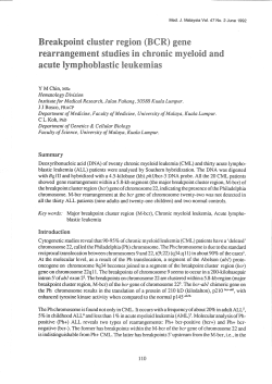


© Copyright 2026