
Human West Nile virus infection in Bosnia and
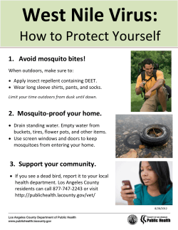
ORIGINAL ARTICLE
Human West Nile virus infection in Bosnia and Herzegovina
Sead Ahmetagić1, Jovan Petković1, Mirsada Hukić2, Arnela Smriko-Nuhanović1, Dilista Piljić1
1
Clinic for Infectious Diseases, University Clinical Centre Tuzla, Tuzla, 2 International Burch University, Sarajevo; Bosnia and Herzegovina
ABSTRACT
Aim To describe the first two cases of West Nile virus (WNV)
neuroinvasive infections in Bosnia and Herzegovina.
Methods At the Clinic for Infectious Diseases of the University
Clinical Centre Tuzla, Bosnia and Herzegovina (B&H), specific
screening for WNV infection was performed on patients with neuroinvasive diseases from 1 August to 31 October 2013. Serum
samples were tested for the presence of WNV IgM and IgG antibodies using enzyme-linked immunosorbent assay (ELISA); positive serum samples were further analyzed by detection of WNV
nucleic acid of two distinct lineages (lineage 1 and lineage 2) in
sera by RT-PCR.
Corresponding author:
Arnela Smriko-Nuhanović
Clinic for Infectious Diseases,
University Clinical Centre Tuzla
Trnovac bb, 75 000 Tuzla,
Bosnia and Herzegovina
Phone: +387 35 303 326;
Fax: +387 35 303 480;
E-mail: [email protected]
Original submission:
Results Three (out of nine) patients met clinical criteria, and two
of them had high serum titre of WNV specific IgM antibodies (3.5
and 5.2). Serum RT-PCR testing was negative. Conformation by
neutralization testing was not performed. Both cases represented
with encephalitis. None of these cases had recent travel history in
WNW endemic areas, or history of blood transfusion and organ
transplantation, so they represented autochthonous cases.
Conclusion Although there were no previous reports of flavivirus
infections in B&H, described cases had high titre of WNV specific
antibodies in serum, and negative flavivirus-vaccination history,
they were defined as probable cases because recommended testing
for case confirmation was not performed. The West Nile virus
should be considered a possible causative pathogen in this area,
probably in patients with mild influenza-like disease of unknown
origin and those with neuroinvasive disease during late summer
and early autumn.
Key words: neuroinvasive infections, encephalitis, flaviviruses
14 August 2014;
Revised submission:
24 October 2014;
Accepted:
18 December 2014.
Med Glas (Zenica) 2015; 12(1):47-51
47
Medicinski Glasnik, Volume 12, Number 1, February 2015
INTRODUCTION
The genus Flavivirus comprises 53 viruses, and
many of them are human pathogens of concern. In
Europe, many flaviviruses are endemic (West Nile,
Usutu, tick-borne encephalitis viruses) or occasionally imported (dengue, yellow fever viruses) (1).
West Nile virus (WNV) is transmitted in an avian
cycle by ornithophilic mosquitoes, chiefly of the
genus Culex. Mammals can also be infected, but
are considered dead end hosts because viraemia is
generally too low to infect mosquitoes (2). West
Nile virus was first identified in the West Nile district of Uganda in 1937 in a woman who presented with a mild febrile illness (3). During the next
decades, the virus spread through Africa and Asia.
It was first described in Europe in the 1960s when
seropositive animals or virus isolates were reported in France, Portugal and Cyprus (4). West Nile
virus has historically been considered less pathogenic in humans than dengue virus or yellow fever virus; however, more virulent genotypes have
emerged since 1998. Isolates from Israel (1998),
Hungary (2003), or North America (1999) belonging to the Israeli-American cluster of WNV lineage 1a are highly pathogenic in birds and mammals,
and lineage 2 viruses have caused an increasing
number of WNV outbreaks in Europe since 2008
(2). In Europe, WNV has mainly been reported in
central and south-eastern Europe, regions in which
WNV infections and virulence have recently increased, and the implicated viruses have spread to
new areas, including Bulgaria and Greece in 2010,
Albania and Macedonia in 2011, and Croatia, Serbia, and Kosovo in 2012 (1,2,5). The first laboratory-confirmed cases of the West Nile virus neuroinvasive infection in Croatia were diagnosed in
September 2012 in three eastern Croatian counties,
although specific antibodies to West Nile virus in
humans, horses, and European brown bears have
been previously detected (6-9). Also, in Serbia first
outbreak of West Nile virus infection was reported
in humans in August to October 2012, and evidence of detected virus activity in horses, wild birds
and mosquitos (10-12). There are no reports of
WNV or other flaviviruses activities in humans or
animals in Bosnia and Herzegovina.
PATIENTS AND METODS
At the Clinic for Infectious Diseases University
Clinical Centre Tuzla, Bosnia and Herzegovina
48
(B&H), specific screening for WNV infection was
performed on patients with neuroinvasive diseases
with negative CSF bacterial cultures and negative
serological tests for other common causes of bacterial, viral or protozoan meningitis and/or encephalitis (rubella, measles, mumps, varicella-zoster
viruses, adenovirus type 3, Epstein-Barr, herpes
simplex virus-1, herpes simplex virus-2, cytomegalovirus, coxackie B, coxsackie A7, echovirus
type 7 viruses, Treponemaa pallidum, Borrelia
burgdorfer sensu stricto, Borrelia garinii, Borrelia afzelii, Haemophilus influenza, Listeria monocytogenes 1/2a and 4b, Toxoplasma gondii) from
1 August to 31 October 2013. There was no data
about specific WNV screening performed before
in medical centres in B&H. The study had been
approved by the Research Ethics Committee of the
University Clinical Centre Tuzla.
Case definition
The case definition and case classification were
established according to European Union case
definitions for West Nile fever (13). Persons with
fever (≥37.5 °C) and meningitis and/or encephalitis were included. Meningitis was defined as
presence of fever, clinical signs of meningeal inflammation (including headache, nuchal rigidity,
Kernig’s sign or Brudzinski signs, photophobia
or phonophobia), and the presence of cerebrospinal (CSF) pleocytosis (>5 leucocytes/mm3),
elevated protein levels (>0.45 g/L; normal range
0.15-0.45 g/L) and normal (2.6-3.1 mmol/L, 5060% serum glucose levels) or mildly decreased
(2.4-2.6 mmol/L) CSF glucose level. Encephalitis was defined as the presence of fever, encephalopathy (decreased or altered level of consciousness, lethargy or personality change) and/or
focal neurological signs (weakness, cranial nerve palsy), seizures or movement disorders (tremor, parkinsonisam, ataxia), and the presence of
CSF pleocytosis (>5 leucocytes/mm3), elevated
protein levels (>0.45 g/L), and normal (2.6-3.1
mmol/L, 50-60% serum glucose levels) or mildly
decreased (2.4-2.6 mmol/L) CSF glucose level.
Laboratory criteria for a probable case were the
presence of WNV-specific antibody response in
serum (IgM or IgM and IgG). Laboratory criteria
for a case conformation were isolation of WNV
from blood or CSF, detection of WNV nucleic
acid in blood or CSF, WNV specific antibody
Ahmetagić et al. Human WNV infection in B&H
response (IgM) in CSF, WNV IgM high titre and
detection of WNV IgG, and conformation by neutralisation.
Serology and molecular tests
Serum samples were tested at the Institute of
Clinical Microbiology, University Clinical
Center of Sarajevo, for the presence of WNV
IgM and IgG antibodies using the West Nile Virus IgM Capture DxSelectTM ELISA and West
Nile Virus IgG DxSelectTM ELISA kits (Focus
Diagnostics, Cypress, California, USA). Serum
samples, which tested positive in the ELISA,
were further analyzed by detection of WNV
nucleic acid of two distinct lineages (lineage 1
and lineage 2) in sera by RT-PCR. Results were
confirmed at the Institute of Microbiology and
Immunology, Medical Faculty University of
Ljubljana, Slovenia. Diagnostic tests for WNV
isolation from blood, specific CSF testing
(WNV isolation, specific antibody response, or
detection of nucleic acid), and conformation by
neutralization were not performed.
Patient data
Patient’s demographic characteristics (age, sex,
region of residence), travel history, blood transfusions, transplants, and vaccination status,
comorbidity, presenting symptoms and signs,
clinical findings, hematology and blood biochemistry analysis, CSF cell count and biochemical
analysis were recorded, along with serology and
molecular diagnostic tests results, as well as the
outcome at hospital discharge.
RESULTS
During the period 01 August to 31 October 2013
only three patients (out of nine) met clinical criteria, and two of them were identified as probable cases according to the case definition (Table
1); conformation by neutralization testing was
not performed.
Table 1. Results of serology and molecular testing of two
patients with neuroinvasive West Nile virus infection
Day of samSample
IgM ELISA IgG ELISA
Case
pling after
RT-PCR
type
(titre)
(titre)
illness onset
1
Serum
20
Positive (3.5) Positive (2.2) Negative
2
Serum
12
Positive (5.2) Negative (0.27) Negative
First case had onset of symptoms at the beginning
of August, and second case at the beginning of September 2013. None of these cases had recent travel
history in areas where WNV was endemic, nor the
history of blood transfusion and organ transplantation, and had negative flavivirus-vaccination
history (demographic characteristics and comorbidities are shown in Table 2). They were both represented with encephalitis (self-reported symptoms
and clinical signs are shown in Table 3).
Table 2. Demographic characteristics and comorbidities of
two patients with neuroinvasive West Nile virus infection
Age
Case
Sex
(years)
1
84
2
68
Male
Immunosuppression Hyper- Other chronic
including tension
illness
diabetes
Vertigo
Tuzla
No
Yes
Chronic bronchitis
City
area
Female Kladanj
No
Yes
Hypertensive
heart disease
Table 3. Self-reported symptoms and clinical signs of two
patients with neuroinvasive West Nile virus infection
Signs and symptoms
Fever ≥ 37.5 °C
Neurological manifestation
Fatigue
Consciousness impairment
Rash
Case 1
+
+
+
+
+
Case 2
+
+
+
+
-
+, present; -, absent;
Neurological manifestation, including neurological
deficit and consciousness impairment were recorded on day 10 after symptom onset in both cases.
Neurological manifestations in case 1 included
horizontal nystagmus, positive Romberg’s sign,
positive Kernig’s sign, neck and extremities
muscle stiffness with lively deep tendon reflexes,
and urine retention. In this case qualitative (confusion, disorientation) and quantitative (somnolence) consciousness impairment were present,
and mood changes in form of anxiety.
Neurological manifestations in case 2 included
ataxia, horizontal nystagmus, positive Romberg’s
sign, nuchal rigidity, and diminished deep tendon
reflexes, with qualitative (confusion, disorientation) and quantitative (somnolence) consciousness
impairment, and incoherent speech.
Cerebrospinal fluid analysis results of these two
cases are shown in Table 4.
None of these patients developed respiratory failure requiring mechanical ventilation. Both patients recovered by the time they were discharged,
case 2 had complete recovery, and case 1 still had
49
Medicinski Glasnik, Volume 12, Number 1, February 2015
mild horizontal nystagmus, positive Romber’s
sign and urine retention.
Table 4. Cerebrospinal fluid analyses results of 2 patients
with neuroinvasive West Nile virus infection
Parameter
WBC (per mm3)
WBC differential
Protein (g/L)
Glucose (mmol/L)
Normal value/range
<5
No predominance
<0.45
2.6-3.1 mmol/L
Case 1
136
60% L
1.55
3.3
Case 2
960
62% N
1.45
2.7
WBC, white blood cells; L, lymphocytes; N, neutrophiles;
DISCUSSION
Taking in consideration that large WNV outbreaks happened in Eastern Croatia (6) and Serbia
(10) in 2012, we analyzed appearance of the human neuroinvasive disease in B&H, performing
a screening for patients hospitalized throughout
summer and autumn 2013 for WNV infection
with the aim to investigate presence of the virus
in this location. Although only patients with neuroinvasive disease were tested, of whom two
were positive, we assumed that there were more
patients with WNV infection who had influenzalike febrile illness who were not tested, or who
did not seek medical help due to the symptoms
being too mild or were treated in primary care.
This is a single-center study at regional level, and
a large study at state level should be planned so
that the incidence rates can be calculated.
Both presented patients had clinical signs of encephalitis that could not be distinguished from
the patients with encephalitis with a different etiology (14), but large sample case-control studies
might reveal specific characteristic of encephalitis due to WNV infection (14).
Serological diagnosis of flavivirus infections is
complicated by the antigenic similarities among
the Flavivirus genus because most flavivirus antibodies are directed against the highly immunogenic envelope protein, which contains both flavivirus cross-reactive and virus-specific epitope (1).
Serological assay results should thus be interpreted with care and confirmed by comparative neutralization test using a panel of viruses known to
REFERENCES
1.
50
Beck C, Jimenez-Clavero MA, Leblond A, Durand
B, Nowotny N, Leparc-Goffart I, Zientara S, Jourdain E, Lecollinet S. Flavivirus in Europe: Complex
circulation patterns and their consequences for the
diagnosis and control of West Nile Disease. Int J Environ Res Public Health 2013;10:6049-83.
circulate in Europe (1,15). Although there have
been no previous reports of other flavivirus infections in B&H, the cases presented in this report had high titre of WNV specific antibodies
in serum, and had negative flavivirus-vaccination
history, and accordingly, they were defined as
probable because we were not able to perform
recommended testing for case conformation.
Knowledge of flavivirus diversity at the local level is essential before rigorous serological surveys can be undertaken (1). Veterinary and entomologic investigations related to WNV and other
flaviviruses have not been performed in B&H, so
we can only assume from the published regional
data (6-12) the circulating pattern of the virus in
this area. Systematic research is needed to understand epidemiology and ecology of WNV in
B&H. It is very important to develop a project to
detect WNV in mosquitoes in urban regions and
near rivers in various parts of the state, as well as
surveillance of human cases.
Considering the vectors and recent floods, a number of public health measures can be undertaken
(intensifying activities to reduce the number of
mosquitos in environments using insecticide, destroying mosquito habitat, systematic extermination of larvae and adult form of larvae and adult
form of mosquitoes, education of the population on
how to avoid or decrease the risk of being bitten by
potentially infected mosquitoes through posters, leaflets, television, and newspapers) (16). These actions should help avoid outbreaks of human WNV
infection and raise awareness of healthcare providers about emergence of the virus in B&H. In patients with mild influenza-like disease of unknown
origin and those with neuroinvasive disease during
late summer and early autumn, WNV should be
considered a possible causative pathogen.
FUNDING
No specific funding was received for this study.
TRANSPARENCY DECLARATION
Competing interests: None to declare.
2.
3.
Reiter P. West Nile virus in Europe: understanding the present to gauge the future. Eurosurvaill
2010. http:www.eurosurveillance.org/ViewArticle.
aspx?Articleid=19508. (3 Jun 2014)
Sithburn KC, Hughes TP, Burke AW Paul JH. A neutropic virus isolated from the blood of a native of
Uganda. Am J Tro Med 1940; 20:471-92.
Ahmetagić et al. Human WNV infection in B&H
4.
Murgue B, Murri S, Triki H, Deubel V, Zeller H.
West Nile in Mediterranean basin:1950-2000. Ann
N Y Acad Sci 2001; 951:117-26.
5. Nowotny N, Bakonyi T, Weissenbock H, Seidel B,
Kolodziejek J, Sekulin K, Lussy H. West Nile virus
infection in Europe – general features. Medica Sciences 2013; 39:123-4.
6. Merdić E, Perić L, Pandak N, Kurolt IC, Turić N, Vignjević G, Stolfa I, Milas J, Bogojević MS, Markotić
A. West Nile virus outbreak in humans in Croatia,
2012. Coll Antropol 2013; 37:943-7.
7. Madić J, Huber D, Lugović B. Serologic survey for
selected viral and rickettsial agents of brown bears
(Ursus arctos) in Croatia. J Wildl Dis 1993; 29:572-6.
8. Madić J, Savini G, Di Gennaro A, Monaco F, Jukić
B, Kovac S, Rudan N, Listes E. Serological evidence
for West Nile virus infection in horses in Croatia. Vet
Rec 2007; 160:772-3.
9. Barbić L, Listeš E, Katić S, Stevanović V, Madić J,
Starešina V, Labrović A, Di Gennaro A, Savini G.
Spreading of West Nile virus infection in Croatia.
Vet Microbiol 2012; 159:504-8.
10. Popović N, Milošević B, Urošević A, Poluga J, Lavadinović L, Nedelijković J, Jevtović D, Dulović
O. Outbreak of West Nile virus infection among
humans in Serbia, August to October 2012. Euro
Surveill 2013. http://www.eurosurveillance.org/ViewArticle.aspx?ArticleId=20613 (11 July 2014)
11. Petrović T, Blazquez AB, Lupulović D, Lazić G, Escribano-Romero E, Fabijan D, Kapetanov M, Lazić
S, Saiz J. Monitoring West Nile virus (WNV) infection in wild birds in Serbia during 2012: first isolati-
12.
13.
14.
15.
16.
on and characterisation of WNV strains from Serbia.
Euro Surveill 2013; 31:18. pii: 20622.
Lupulović D, Martín-Acebes MA, Lazić S, AlonsoPadilla J, Blázquez AB, Escribano-Romero E, Petrović T, Saiz JC. First serological evidence of West
Nile virus activity in horses in Serbia. Vector Borne
Zoonotic Dis 2011; 11:1303-5.
European Center for Disease Prevention and Control (ECDC). EU case definition. Stockholm: ECDC.
http://ecdc.europa.eu/en/healthtopics/west_nile_fever/EU-case-definition/Pages/EU-case-definition.
aspx (10 October 2013)
Sejvar JJ. Clinical manifestations and outcomes
of West Nile virus infection. Viruses 2014; 6:606-23. Sambri V, Capobianchi MR, Cavrini F, Charrel R,
Donoso-Mantke O, Escadafal C, Franco L, Gaibani
P, Gould EA, Niedrig M, Papa A, Pierro A, Rossini G, Sanchini A, Tenorio A, Varani S, Vázquez A,
Vocale C, Zeller H. Diagnosis of West Nile virus human infections: overview and proposal of diagnostic
protocols considering the results of external quality
assessment studies. Viruses 2013; 5:2329-48.
Radivojević S, Maris S, Ljubić B, Obrenović J. First
detection of West Nile fever in human population in
the territory of Belgrade. Proceedings Third International Epizootiology Days and XV Serbian Epizootiology Days, Niš/Serbia, May 8-11 2013. Faculty of
veterinary medicine Belgrade, University of Belgrade,
Department for infectious disease of animals and bee
diseases, Belgrade. FVM; 2013. p. 61-2. http://www.
vet.bg.ac.rs/uploads/file/seminari_simpozijumi/Epizootioloski.dani.2013.ZBORNIK.pdf (20 April 2014)
Humana infekcija virusom Zapadnog Nila u Bosni i Hercegovini
Sead Ahmetagić1, Jovan Petković1, Mirsada Hukić2, Arnela Smriko-Nuhanović1, Dilista Piljić1
1
Univerzitetski klinički centar Tuzla, Tuzla, 2International Burch University, Sarajevo; Bosna i Hercegovina
SAŽETAK
Cilj Prikazati prva dva slučaja neuroinvazivne infekcije uzrokovane virusom Zapadnog Nila (WNV) u
Bosni i Hercegovini (BiH).
Metode U Klinici za infektivne bolesti Univerzitetskog kliničkog centra Tuzla (BiH), u periodu od 1.
avgusta do 31. oktobra 2013. godine, kod pacijenata kojima je postavljena dijagnoza neuroinvazivne
infekcije provedena su specifična testiranja na WNV infekciju. Uzorci seruma testirani su na prisustvo
specifičnih WNV IgM i IgG antitijela pomoću enzimsko-imunološkog testa (ELISA), a pozitivni uzorci
seruma su dalje testirani ispitivanjem prisustva WNV nukleinske kiseline za dvije različite linije (linija
1 i linija 2) pomoću RT-PCR-a.
Rezultati Samo tri pacijenta (od ukupno devet) zadovoljavala su postavljene kliničke kriterije, od kojih
su dva imala visoke titrove WNV specifičnih IgM antitijela (3,5 i 5,2) u serumu. RT-PCR testovi iz istog
seruma bili su negativni. Potvrdni test neutralizacije nije urađen. Oba slučaja imala su kliničku sliku
encefalitisa. Pacijenti nisu boravili u područjima gdje je virus Zapadnog Nila endemičan, niti su primali
transfuzije krvi ili transplantaciju organa, tako da predstavljaju autohtone slučajeve.
Zaključak Iako nije bilo ranije prijavljenih flavivirusnih infekcija u BiH, u oba slučaja su u serumu
nađena specifična WNV antitijela u visokom titru i u oba slučaja nije bilo prethodne vakcinacije protiv
flavivirusa; oba slučaja su ostala nepotvrđena jer nije provedeno preporučeno testiranje za potvrdu
slučaja. Virus Zapadnog Nila je prisutan u ovoj regiji te ga uvijek treba razmotriti kao potencijalnog
patogena u pacijenata sa simptomima blage influence nepoznatog porijekla ili neuroinvazivne bolesti
koji se javljaju u kasno ljeto i ranu jesen.
Ključne riječi: neuroinvazivne infekcije, encefalitis, flavivirusi
51


© Copyright 2026