
Safety, Pharmacokinetics, and Activity of ABX-EGF, a
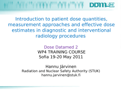
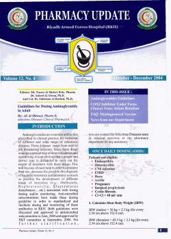
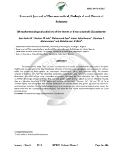
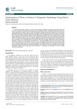
VOLUME 22 䡠 NUMBER 15 䡠 AUGUST 1 2004 JOURNAL OF CLINICAL ONCOLOGY O R I G I N A L R E P O R T Safety, Pharmacokinetics, and Activity of ABX-EGF, a Fully Human Anti–Epidermal Growth Factor Receptor Monoclonal Antibody in Patients With Metastatic Renal Cell Cancer Eric K. Rowinsky, Garry H. Schwartz, Jared A. Gollob, John A. Thompson, Nicholas J. Vogelzang, Robert Figlin, Ronald Bukowski, Naomi Haas, Pamela Lockbaum, Yu-Ping Li, Rosalin Arends, Kenneth A. Foon, Gisela Schwab, and Janice Dutcher From the Institute for Drug Development, Cancer Therapy and Research Center and Brook Army Medical Center, San Antonio, TX; Beth Israel Deaconess Cancer Center, Boston, MA; University of Washington, Seattle, WA; University of Chicago Cancer Research Center, Chicago, IL; Cleveland Clinic, Cleveland, OH; Fox Chase Cancer Center, Philadelphia, PA; UCLA School of Medicine, Los Angeles; and Abgenix Inc, Fremont, CA; and Our Lady of Mercy Cancer Center, New York Medical College, Bronx, NY. Submitted November 11, 2003; accepted March 31, 2004. Presented in part at the 38th annual meeting of the American Society of Clinical Oncology, Orlando, FL, May 18-21, 2002. Authors’ disclosures of potential conflicts of interest are found at the end of this article. Address reprint requests to Eric K. Rowinsky, MD, Institute for Drug Development, Cancer Therapy and Research Center, 7979 Wurzbach Rd, 4th Floor, Zeller Building, San Antonio, TX 78229; e-mail: [email protected]. A B S T R A C T Purpose To determine the antitumor activity of ABX-EGF, a fully human monoclonal antibody to the epidermal growth factor receptor (EGFr), in previously treated patients with metastatic renal cell carcinoma, and to characterize its toxicity, immunogenicity, pharmacokinetics, and pharmacodynamics. Patients and Methods The antitumor activity, as well as the toxicity, pharmacokinetics, pharmacodynamics, and immunogenicity of ABX-EGF, were assessed. Results Eighty-eight patients were treated with ABX-EGF doses of 1.0, 1.5, 2.0, or 2.5 mg/kg weekly with no loading dose. EGFr immunostaining was performed on 76 tumor biopsy specimens (86%), and 69 (91%) scored positive. Major responses occurred in three patients, and two patients had minor responses. Forty-four patients (50%) also had stable disease at their first 8-week assessment, and the median progression-free survival (PFS) was 100 days (95% CI, 58 to 140 days). Low hemoglobin and high alkaline phosphatase predicted for short PFS. The principal toxicity, an acneiform rash, occurred in 68%, 95%, 87%, and 100% of patients who received at least three doses of ABX-EGF at 1.0, 1.5, 2.0, and 2.5 mg/kg/wk, respectively. A trend indicated that the severity of the rash may relate to PFS. No human antihuman antibodies were detected. ABX-EGF pharmacokinetics fit a model that incorporated both linear and saturable EGFr-mediated clearance mechanisms, and interindividual variability was low. At 2.5 mg/kg/wk, ABX-EGF concentrations throughout treatment exceeded those estimated to saturate nonlinear clearance and inhibit xenograft growth by 90%. Conclusion ABX-EGF was generally well tolerated. The objective response rate was low in previously treated patients with metastatic renal cell carcinoma. Although skin rash may be a pharmacodynamic marker of drug action, its potential as a surrogate marker of clinical benefit requires further evaluation. J Clin Oncol 22:3003-3015. 0732-183X/04/2215-3003/$20.00 DOI: 10.1200/JCO.2004.11.061 INTRODUCTION The binding of epidermal growth factor (EGF) or transforming growth factor-alpha (TGF␣) to the epidermal growth factor receptor (EGFr) seems to trigger autophosphorylation and internalization of the receptor, which culminate in mitogenic signal transduction.1,2 The notion of targeting EGFr as a therapeutic strategy against cancer is supported by experimental evidence that aberrations of EGFr-mediated signal transduction play critical roles in tumorogenesis and cancer cell proliferation.1,2 The relevance of this strategy is further substantiated by EGFr expression, overexpression, or aberrant function in renal, lung, prostate, breast, head and neck, colorectal, and other epithelial malignancies, as well as evidence that the level of EGFr expression may be a 3003 Downloaded from jco.ascopubs.org on June 9, 2014. For personal use only. No other uses without permission. Copyright © 2004 American Society of Clinical Oncology. All rights reserved. Rowinsky et al determinant of tumor proliferation, invasiveness, and angiogenesis, and relates adversely to prognosis.3-14 Concomitant with EGFr expression, EGFr ligands, particularly TGF␣, are commonly upregulated, and therefore, blockade of autocrine loops is a rational strategy for the treatment of many epithelial malignancies.15-18 ABX-EGF (Abgenix Inc, Fremont, CA; Immunex Corp, a subsidiary of Amgen Inc, Thousand Oaks, CA), a high-affinity (Kd, 5 ⫻ 10⫺11M) fully human immunoglobulin G2 (IgG2) monoclonal antibody against human EGFr, was generated from human antibody-producing XenoMouse strains (Abgenix Inc), which were engineered to be deficient in mouse antibody production and contain integrated megabase-sized fragments from the human heavy and light chain loci with the majority of the human antibody repertoire.19 ABX-EGF completely blocks binding of EGF and TGF␣ to the EGFr and induces profound and rapid internalization of the receptor in EGFr-expressing human cancers.19,20 These actions abolish EGFr-dependent cellular responses, including EGFr tyrosine phosphorylation, extracellular acidification, angiogenesis, and cell proliferation.19,20 The treatment of athymic mice with ABX-EGF 0.2 mg intraperitoneally twice weekly for 3 weeks completely prevents the formation of EGFr-overexpressing human epidermoid carcinoma A431 xenografts.19,20 Furthermore, administration of ABX-EGF at doses as low as 0.3 mg intraperitoneally for 3 weeks without concomitant chemotherapy or radiation completely eradicates tumors as large as 1.2 cm3, and a total dose of 0.6 mg completely eradicates tumors in 65% of mice.19,20 Although similar results have been reported with other antibodies and therapeutics against EGFr, the results with ABX-EGF are unique in that no tumor recurrences have been noted for more than 8 months after the last antibody injection in a sizable proportion of animals.16,17,21-24 Similar therapeutic effects have been observed following treatment of a wide range of human tumor xenografts with variable EGFr expression levels ranging from 17,000 to 1,000,000 receptors/cell using renal, breast, pancreatic, ovarian, prostate, and colorectal cancer cell lines.19 Furthermore, ABX-EGF has not only been demonstrated to inhibit growth of other cancers that express high levels of EGFr, such as A431 and MDA-MB468, but also to inhibit growth of other cancers with much lower levels of EGFr expression.19,20 However, the magnitude of tumor growth inhibition achieved following ABX-EGF treatment seems to relate to the EGFr number; significant growth inhibition of xenografts expressing at least 17,000 receptors per cell has been noted, whereas xenografts expressing 11,000 or fewer receptors per cell seem not to be affected by ABX-EGF treatment.20 Toxicology, efficacy, and pharmacologic studies of ABX-EGF in mice and nonhuman primates have been used to simulate the pharmacokinetic profiles in humans and predict effective dosing regimens.25,26 Based on the pharmacokinetics of ABX-EGF in monkeys treated with ABXEGF doses ranging from 0.3 to 30 mg/kg, human clearance 3004 (CL) was predicted to be nonlinear, possibly due to progressive saturation of an EGFr sink.25 Allometric scaling was used to determine a dose-schedule for a phase I study, in which patients with malignancies known to express EGFr were treated with four weekly doses of ABX-EGF.26 The most common toxicity was a dose-dependent skin rash, occurring in 100% of patients treated with ABX-EGF at 2.0 mg/kg/wk or 2.5 mg/kg/wk. No allergic reactions, human antihuman antibody (HAHA) formation, nor serious adverse events were reported at doses up to 2.5 mg/kg/wk, which encompassed the dosing range evaluated in the present study. Furthermore, the interpatient variability in ABX-EGF exposure was low, and trough concentrations at doses of at least 2 mg/kg exceeded IC90 values derived from xenograft studies.19,20,25,26 This phase I study had not yet been completed when the present study began. The results of the aforementioned studies, as well as a high (70% to 90%) incidence of EGFr expression in patients with renal cell carcinoma, served as the rationale for this study. The principal objectives were to evaluate the antitumor activity of multiple weekly doses of ABX-EGF in patients with metastatic renal carcinoma who either failed to respond to treatment with interleukin-2 (IL-2) and/or interferon-alfa (IFN-␣) or in subjects in whom this therapy was felt to be contraindicated from a medical standpoint, as well as to characterize the safety, immunogenicity, and pharmacokinetics of ABX-EGF on a multidose schedule. PATIENTS AND METHODS Study Objectives The study was designed as a multicenter, dose-rising trial to principally evaluate the antitumor activity of multiple doses of ABX-EGF in patients with metastatic renal cell carcinoma. The toxicity, pharmacokinetics, pharmacodynamics, and immunogenicity of ABX-EGF were also assessed. Enrollment was planned for successive cohorts of 20 patients treated at 1.0, 1.5, 2.0, and 2.5 mg/kg weekly with no testing or loading dose. After each series of eight weekly treatments, antitumor activity was evaluated, and stable or responding patients were able to receive as many as five series of eight weekly treatments. Eligibility Subjects in whom IL-2 or IFN-␣ were felt to be contraindicated from a medical standpoint, or patients with histologically confirmed, metastatic renal cell carcinoma who had previously received and failed treatment with IL-2 or IFN-␣ in the metastatic setting, were eligible for this study. Patients who received chemotherapy and radiation in the adjuvant and/or metastatic settings were also eligible, but subjects who had been previously treated with EGFr-targeted therapeutics were ineligible. Although the eligibility requirements did not mandate documentation of EGFr expression before enrollment, the study required the availability of tumor tissue to characterize EGFr immunostaining. Eligibility criteria also included: age ⱖ 18 years; an Eastern Cooperative Oncology Group performance status less than 2; no prior anticancer treatment of any type or investigational agents within 30 days; JOURNAL OF CLINICAL ONCOLOGY Downloaded from jco.ascopubs.org on June 9, 2014. For personal use only. No other uses without permission. Copyright © 2004 American Society of Clinical Oncology. All rights reserved. ABX-EGF in Renal Cancer no hypercalcemia; adequate hematopoietic (absolute neutrophil count ⱖ 1,500/L, platelet count ⱖ 100,000/L), hepatic (total bilirubin ⱕ 1.5 mg/dL, transaminases and alkaline phosphatase ⱕ 3 times the institutional upper normal limit), renal (creatinine ⱕ 2.2 mg/dL), and cardiac (left ventricular ejection fraction ⱖ 45% as measured by multiple gated acquisition [MUGA] scan) functions; no prior treatment with anthracyclines or related therapeutics; no myocardial infarction within 1 year; measurable disease; and no coexisting medical problem of sufficient severity to limit compliance with the study. Patients with a history of brain metastases were eligible as long as there was no evidence of progressive disease or peritumoral edema on a magnetic resonance or computed tomography scan of the brain performed before treatment, symptomatic manifestations of brain metastases, nor requirement for corticosteroids. Patients with a history of other malignancies, except basal cell carcinoma of the skin and cervical carcinoma-in-situ, within 5 years before study, were ineligible. All patients of childbearing potential were required to use appropriate contraception during treatment. Patients gave written informed consent according to Federal and institutional guidelines before enrollment. Immunohistochemistry Paraffin-embedded sections and fresh tumor tissue from tumor biopsies were assessed in a central laboratory by IMPATH (Los Angeles, CA) to evaluate EGFr expression. The immunostaining procedure used an EGFr immunohistochemistry kit developed by Dako Cytomation Inc (Carpinteria, CA) in collaboration with Abgenix Inc for investigational purposes, and a two-step immunostaining procedure was routinely used to process paraffin-embedded tissues. Following incubation with the primary rabbit antibody to human EGFr protein, a ready-to-use visualization reagent based on dextran technology was used. This reagent consisted of both secondary goat antirabbit antibody and horseradish peroxidase molecules linked to a common dextran polymer backbone, thereby eliminating the need for sequential application of link antibody and peroxidase-conjugated antibody. The enzymatic conversion of the subsequently added chromogen resulted in the formation of a visible reaction product at the antigen site. The specimen was then counterstained, and a coverslip was applied. Control slides containing formalin-fixed, paraffin-embedded human cell lines with various levels of EGFr expression were provided to validate staining runs. EGFr expression was scored using light microscopy as follows: 0 (no staining), 1⫹ (weak staining of tumor cells), 2⫹ (moderate staining of tumor cells), and 3⫹ (strong staining of tumor cells). Staining was scored as positive if the stain was visualized within the cell membrane and/or cytoplasmic rim. In addition, an EGFr H score, which reflected both the intensity of staining and the percentage of tumor cells with the various immunostaining intensity scores, was calculated for each biopsy sample by adding the products of percentages of cells with weak, moderate, and strong immunostaining and weighting factors of 1, 2, and 3, respectively (ie, H scores ⫽ [% tumor cells with weak intensity ⫻ 1] ⫹ [% tumor cells with moderate intensity ⫻ 2] ⫹ [% tumor cells with strong intensity ⫻ 3]), so that the minimum and maximum H scores were 0 and 300, respectively. Drug Administration ABX-EGF was supplied by Abgenix Inc in 5-mL glass vials containing 50 mg of antibody at a concentration of 10 mg/mL in pyrogen-free 0.9% sodium chloride solution US Pharmacopeia (USP). The vials were stored at 2 to 8°C. The appropriate volume of the contents of the ABX-EGF vial, which was calculated according to dose level assignment, was diluted with 0.9% saline solution USP in a polyvinyl chloride infusion bag to yield an infusion volume ranging from 100 to 150 mL. The solution was infused intravenously over 60 minutes using a syringe or infusion pump with a 0.22 m filter in the intravenous line. Vital signs were recorded from 30 minutes before treatment, to 60 minutes posttreatment. Pretreatment and Follow-Up Evaluations Histories that included recording of performance status and concurrent medications, physical examination, MUGA scan, HAHA studies, an ECG, archived tumor tissues, and routine laboratory studies, were obtained before treatment. An interval history with recording of adverse events and a physical examination were repeated weekly. The physical examination included a dermatologic assessment, which consisted of descriptions of the type and location of lesions, related symptoms, and specific treatment rendered, as well as a quantitative assessment of proportion of skin affected, all of which was recorded on a designated form. Cutaneous manifestations were also photographed. Routine laboratory studies were performed every 4 weeks, and MUGA scans and HAHA studies were performed after each 8-week course. Routine laboratory studies included a CBC and differential WBC count, chemistries, coagulation studies, and cardiac enzymes. Toxicity was graded according to the National Cancer Institute Common Toxicity Criteria (NCI CTC; version 2.0). Although a prior phase I study had confirmed the safety of ABX-EGF in the dosing range evaluated in the present study, definitions for dose-limiting toxicity (DLT) and maximum-tolerated dose were established a priori. Dose-escalation was to cease if a maximum-tolerated dose, which was defined as the dose at which DLT occurred in at least 30% of patients (with at least two events), was observed in the first 8-week series of treatment. DLT was defined as any NCI CTC grade 3 or 4 adverse experience (except skin rash), any severe or lifethreatening complication not encompassed in the NCI CTC guidelines; severe skin toxicity defined as desquamation, generalized urticaria, or symptomatic skin-related toxicity requiring narcotics or systemic corticosteroids; or event felt to be intolerable by the patient. Dose reduction to 1.5 mg/kg/wk was permitted for patients who developed severe skin-related toxicity following resolution of the toxic event within 3 weeks after dose interruption. Relevant radiological studies for evaluation of all measurable or assessable sites of malignancy were performed pretreatment, after each 8-week course, and as needed to confirm response. Patients were able to continue treatment if they did not develop progressive disease. The response to treatment was assessed using the Response Evaluation Criteria in Solid Tumors criteria. All objective tumor responses were confirmed 4 weeks after response diagnosis and centrally verified by an independent radiologist. Statistical Analysis Descriptive statistics of relevant demographic and clinical features were compiled. Summary statistics were provided for tumor response assessment at the end of the initial 8-week cycle of treatment. The relationship between time to disease progression and each of the pertinent factors was analyzed using univariate survival analysis (the log-rank test for categoric variables and a score test based on the Cox proportional hazards model for continuous variables). Kaplan-Meier estimates, median values, 95% CIs, and both 25% and 75% quartile values were calculated. The 3005 www.jco.org Downloaded from jco.ascopubs.org on June 9, 2014. For personal use only. No other uses without permission. Copyright © 2004 American Society of Clinical Oncology. All rights reserved. Rowinsky et al rates of adverse events as a function of dose were determined. All analyses and summaries were based on the modified intent-totreat population, defined as patients who received at least one dose of ABX-EGF. The relationships between ABX-EGF dose and the incidence of cutaneous toxicity were described using the sigmoidal maximum effect (Emax) model of drug action (ie, incidence of cutaneous toxicity ⫽ Emax ⫻ Dose␥/(ED50␥ ⫹ Dose␥), where the Emax was fixed at 100%, and ED50 was the dose at which the effect is 50% of the maximal effect. The exponent ␥ is a constant that describes the sigmoidicity or steepness of the curve.27 From the ED50, the ABX-EGF dose predicted to yield a 90% incidence of skin rash (ED90) was derived as a secondary parameter. Pharmacokinetics and Immunogenicity To measure the relevant parameters indicative of exposure to ABX-EGF, blood samples were collected within 30 minutes before and 30 minutes following each of the first eight weekly infusions of ABX-EGF, and every 4 weeks thereafter. The samples were allowed to clot for 30 to 120 minutes followed by centrifugation at 3,000 rpm for 15 minutes. The serum was transferred to a sample tube, which was frozen at ⫺80°C until assayed. Serum ABX-EGF concentrations were measured using a validated electrochemiluminescence assay based on Origen technology (IGEN International Inc, Gaithersburg, MD) with a lower limit of quantitation (LLOQ) of 0.079 g/mL. Samples with ABX-EGF concentrations below the LLOQ of the electrochemiluminescence assay were considered zero in calculating mean concentration values. An enzyme-linked immunosorbent assay was used to detect the formation of HAHA in serum sampled immediately before each 8-week course, and 4 weeks after the eighth dose of the last course. Noncompartmental analysis of the ABX-EGF serum concentration data was accomplished using WINNonlin Professional, Version 4.0 (Pharsight Corp, Mountain View, CA) to estimate the steady-state area under the serum concentration-time curve for the weekly dosing interval (AUC0-7 days) after the seventh dose. If the data for concentration (C; Cmax and Ctrough) were incomplete for the seventh dose, the steady-state AUC0-7 days of an earlier dose, as close as possible to the seventh dose, was calculated. To ensure steady-state condition, this alternate dose interval had to be after the third dose at the 1.0-mg/kg dose level, after the fourth dose at the 1.5-mg/kg dose level, and after the fifth dose at the 2.0- and 2.5-mg/kg dose levels. AUC0-7 days values were calculated from time 0 to using the linear trapezoidal rule, where is the time (7 days) at the end of the dosing interval. The systemic CL was calculated by dividing the weekly dose by AUC0-7 days. The steady-state ABX-EGF Cmax ⫾ SEM values were calculated based on the average values for doses three to eight in the 1.0-mg/kg dose group, doses four to eight in the 1.5-mg/kg dose group, and doses five to eight for the 2.0-mg/kg and 2.5-mg/kg dose groups. The steady-state ABX-EGF Ctrough ⫾ SEM values were calculated based on the average values for doses three to seven in the 1.0-mg/kg dose group, doses four to seven in the 1.5-mg/kg dose group, and doses five to seven for the 2.0-mg/kg and 2.5-mg/kg dose groups. The serum ABX-EGF concentration profile was described by a two-compartment model. Drug was introduced into the central compartment from which elimination occurred in parallel by first-order elimination (linear kinetics) and capacity-limited elimination (nonlinear kinetics). Distribution of ABX-EGF was described by interchange of drug between the central compartment 3006 and the second or peripheral compartment according to the intercompartmental rates k12 (input from central to peripheral) and k21 (input from peripheral back into central). The nonlinear pathway was set according to Michaelis-Menten kinetics characterized by the parameters Vmax and Km, where Vmax represents the maximum elimination rate for the nonlinear pathway and Km is the serum ABX-EGF concentration at which ABX-EGF is eliminated at 50% of Vmax. From the Km, the IC90 was derived as a secondary parameter representing the serum ABX-EGF concentration at which ABX-EGF is eliminated at 10% of Vmax by the nonlinear elimination pathway. Nonlinear regression analysis of skin rash data and serum ABX-EGF concentration data was accomplished with SAAM II version 1.2 (SAAM Institute Inc, Seattle, WA). The goodness of model fit for the pharmacokinetic or the pharmacodynamic model was guided by inspection of the SEs of the mean parameter estimates, the minimization of the objective function (SAAM II User Guide 1997-1998; SAAM Institute Inc) and the minimization of the Akaike information criterion.28 Reduction of the model was deemed appropriate if the SEs of the relevant parameters were either improved or were not adversely affected, the visual fit of the data improved or remained acceptable, and the objective function and/or the Akaike information criterion did not increase. RESULTS General Ninety-five total patients, whose characteristics are presented in Table 1, were enrolled; 88 patients received at least one dose of ABX-EGF and are considered a modified intent-to-treat population for assessment of antitumor response and toxicity. Seven patients provided informed consent but were withdrawn from study before receiving drug. Four patients withdrew consent, two patients were found to be ineligible, and one patient experienced an adverse event (femoral fracture). Seventy-nine patients (90%) underwent nephrectomy before enrollment. Only eight patients (9%) had not previously received systemic biologic and/or chemotherapy, whereas 75 (85%), 19 (22%), and 27 patients (31%) received prior treatment with IL-2, chemotherapy, and/or radiation, respectively. The median number of prior biologic and/or chemotherapy regimens per patient was two, and 32 patients (36%) received at least three prior regimens. EGFr Immunostaining EGFr immunostaining was performed on the tumor biopsy specimens of 76 patients (86%), and 69 patients (91%) scored positive (2⫹ to 3⫹ immunostaining in ⱖ 10% of cells). Immunostaining was positive in 10 (77%) of 13 patients with non– clear-cell cancer whose malignant tissues were assessable, in 56 (93%) of 60 patients with clear-cell cancer, and in all three (100%) patients in whom no histologic subtype information was available. H scores were at least 200 in 42 patients (55%), and below 200 in 34 patients (45%). JOURNAL OF CLINICAL ONCOLOGY Downloaded from jco.ascopubs.org on June 9, 2014. For personal use only. No other uses without permission. Copyright © 2004 American Society of Clinical Oncology. All rights reserved. ABX-EGF in Renal Cancer Table 1. Patient Demographics According to ABX-EGF Dose Level Dose (mg/kg) 1.0 (n ⫽ 22) Characteristic No. of Patients Mean age, years Sex Female Male Performance status (ECOG) 0 1 or 2 Prior nephrectomy Yes No No. of prior biological and/or chemotherapy regimens 0 1-2 ⱖ3 Prior additional cancer surgery Yes No Prior biological therapy Yes No Prior radiation therapy Yes No Prior chemotherapy Yes No Prior hormonal therapy Yes No 1.5 (n ⫽ 22) % No. of Patients 56 2.0 (n ⫽ 23) % No. of Patients 57 2.5 (n ⫽ 21) % No. of Patients 58 Total (N ⫽ 88) % No. of Patients 60 % 58 9 13 41 59 8 14 36 64 4 19 17 83 3 18 14 86 24 64 27 73 16 6 73 27 9 13 41 59 15 8 65 35 11 10 52 48 51 37 58 42 20 2 91 9 19 3 86 14 21 2 91 19 19 2 90 10 79 9 90 10 2 13 7 9 59 32 1 11 10 5 50 45 3 14 6 13 61 26 2 10 9 10 48 43 8 48 32 9 55 36 9 13 41 59 14 8 64 36 11 12 48 52 9 12 43 57 43 45 49 51 16 6 73 27 20 2 91 9 20 3 87 13 19 2 90 10 75 13 85 15 5 17 23 77 8 14 36 64 6 17 26 74 8 13 38 62 27 61 31 69 6 16 27 73 7 15 32 68 3 20 13 87 3 18 14 86 19 69 22 78 0 22 100 1 21 95 0 23 100 0 21 100 0 88 100 Abbreviation: ECOG, Eastern Cooperative Oncology Group. Safety Overview ABX-EGF was generally well tolerated with no hypersensitivity reactions. The most common toxicity was skin rash, which was qualitatively similar in most affected patients and typically involved the face in a periorificial distribution, as well as the upper trunk. It usually consisted of clusters of monomorphous pustular lesions that resembled an acneiform-type drug eruption; however, a maculopapular eruption predominated in some individuals. The rash was usually evident by the second or third week, and the intensity was maximal by weeks 3 to 5. In most patients, the intensity of the eruption gradually decreased despite uninterrupted treatment. The cutaneous manifestations were usually associated with minimal or no symptoms. The incidence of rash as a function of ABX-EGF dose in patients receiving a minimum of three doses is shown in Figure 1A, and the distribution of the worst grades of rash as a function of dose level is displayed in Figure 1B. The incidences of any grade of cutaneous toxicity in patients treated with a mini- mum of three doses of ABX-EGF were 68%, 95%, 87%, and 100% at the 1.0-, 1.5-, 2.0-, and 2.5-mg/kg/wk dose levels, respectively. At the 2.5-mg/kg dose level, 100% of patients developed skin toxicity, and 75% of patients developed either grade 2 or 3 toxicity compared with 32% to 52% of patients treated with ABX-EGF at the three lower doses. The relationship between the incidence of skin rash and ABXEGF dose fits a sigmoidal model, as shown in Figure 1A. Based on this model, the ABX-EGF dose predicted to yield a 50% incidence in skin rash (ED50 ⫾ SEM) was 0.78 ⫾ 0.13 mg/kg (95% CI, 0.51 to 1.0 mg/kg), whereas ␥ was estimated as 3.3 ⫾ 1.4 mg/kg (95%, CI 0.42 to 6.2 mg/kg). This latter parameter ␥ (ie, the factor that accommodates the shape of the curve, being estimated at a value greater than 1) reflects the narrow dose range within which the incidence of skin rash increases from 0% to 100%. The ABX-EGF dose predicted to result in a 90% incidence of skin rash (ED90) was estimated to be 1.5 ⫾ 0.26 mg/kg (95% CI, 1.0 to 2.0 mg/kg). 3007 www.jco.org Downloaded from jco.ascopubs.org on June 9, 2014. For personal use only. No other uses without permission. Copyright © 2004 American Society of Clinical Oncology. All rights reserved. Rowinsky et al Fig 1. (A) Scatter graph of the incidence of rash at different ABX-EGF doses (F), with the line representing the percentage of rash predicted using a sigmoidal Emax model. This model predicted that 90% of patients would develop rash at 1.5 ⫾ 0.26 mg/kg/wk (95%CI, 1.0 to 2.0 mg/kg/wk). (B) The percentages of subjects with a particular grade of rash at different doses. Patients treated with ABX-EGF also complained of asthenia, diarrhea, stomatitis, and nausea, but these effects were generally mild to moderate in severity and not doserelated in the dosing range evaluated. The incidences of adverse effects that were graded as at least grade 2 in severity in at least 5% of patients are listed in Table 2. Five serious adverse events that were possibly related to ABX-EGF included dys- pnea and diarrhea at 1.0 mg/kg, deep venous thrombosis at 1.5 mg/kg, and vomiting and rigors at 1.5 mg/kg. Antitumor Activity Of the 88 patients who received at least one dose of ABXEGF, three whose pertinent disease- and treatment-related details are presented in Table 3 had partial or complete Table 2. Incidence of Moderate and Severe Adverse Events As a Function of ABX-EGF Dose Dose (mg/kg) 1 (n ⫽ 22) 1.5 (n ⫽ 22) 2.0 (n ⫽ 23) 2.5 (n ⫽ 21) Total (N ⫽ 88) Adverse Event No. of Patients % No. of Patients % No. of Patients % No. of Patients % No. of Patients % Asthenia Pain Abdominal pain Back pain Anorexia Constipation Nausea Peripheral edema Hyperglycemia Cough Dyspnea Diarrhea Bone pain Thrombophlebitis 2 2 1 2 0 1 1 2 1 0 1 2 0 2 9 9 5 9 6 5 2 6 2 2 2 0 1 2 2 1 1 2 27 23 9 27 9 9 9 4 4 2 1 1 2 1 1 1 1 2 0 1 0 17 17 9 4 4 9 4 4 4 4 9 1 0 0 2 1 0 0 1 1 5 4 2 2 0 5 13 11 5 11 4 5 4 4 4 8 9 5 4 4 15 13 6 13 5 6 5 5 5 9 10 6 5 5 5 5 9 5 5 9 9 5 9 9 5 5 9 4 10 5 5 5 24 31 10 10 NOTE. Includes moderate and severe adverse events with ⱖ 5% incidence level. 3008 JOURNAL OF CLINICAL ONCOLOGY Downloaded from jco.ascopubs.org on June 9, 2014. For personal use only. No other uses without permission. Copyright © 2004 American Society of Clinical Oncology. All rights reserved. ABX-EGF in Renal Cancer Table 3. Pertinent Characteristics of Patients Experiencing Objective Disease Regression Patient No. ABX-EGF Dose (mg/kg) Disease Response Time to Disease Progression (days)ⴱ EGFr Expression (H score) No. of Prior Treatment Regimens Hemoglobin Nephrectomy Skin Rash 1 2 3 4 5 1.0 1.0 1.5 2.5 2.5 PR MR CR MR PR 128 631 ⫹ 540 ⫹ 78 221 ⱖ 200 ⬍ 200 ⬍ 200 ⬍ 200 NA 4 0 3 2 1 ⬍ LLN Normal Normal Normal Normal No Yes Yes Yes Yes Moderate Moderate Mild Moderate Moderate Abbreviations: EGFr, epidermal growth factor receptor; PR, partial response; LLN, lower limit of normal; MR, minor response; CR, complete response; NA, not available. ⴱ Tabulated as of August 18, 2003. responses, and two other individuals had objective evidence of tumor regression that did not meet the criteria for a partial response. In these patients, tumor regression was noted at the planned assessment following the first 8-week course and was confirmed 4 weeks later. The complete response was documented in a 60-year-old woman who developed multiple large biopsy-confirmed pulmonary metastases 3 months following nephrectomy, that did not respond to treatment with high-dose IL-2, ending 2 months before ABX-EGF treatment. As shown in the computerized tomographic scans in Figure 2, a partial response was documented following eight weekly treatments with ABX-EGF at the 1.5-mg/kg dose level, and further disease regression occurred during additional therapy. The patient elected to discontinue ABX-EGF after 10 months of treatment and experienced progressive regression of the multiple pulmonary metastases over the next year until complete disappearance of all disease was documented on computed tomography scan 23 months following the start of ABXEGF treatment. Forty-four other patients (50%) had stable disease as their best response at their first 8-week assessment, and received additional treatment. The median progression-free survival (PFS) was 100 days (95% CI, 58 to 140 days). At this juncture, follow-up is too short to estimate parameters pertaining to overall survival. As presented in Table 4, neither PFS nor response was related to ABX-EGF dose. Furthermore, neither the intensity of EGFr immunostaining by H score, Eastern Cooperative Oncology Group performance status, or number of prior systemic therapies correlated with PFS. However, low hemoglobin and high alkaline phosphatase values were related adversely to PFS. PFS was significantly longer for the 60 patients who entered the study with normal hemoglobin levels than for the 27 patients with low hemoglobin levels (median, 140 days; [95% CI, 88 to 165 days] v 52 days [95% CI, 46 to 57 days]). In addition, the PFS of 74 patients with normal serum alkaline phosphatase was significantly longer than that of the 14 patients with elevated serum alkaline phosphatase (median, 108 days [95% CI, 80 to 161 days] v 51 days [95% CI, 46 to 56 days]; log-rank P ⫽ .0023). Although the number of patients with non– clear-cell histology was relatively low, patients with this histology seemed to have a better overall outcome than those with clear-cell histology. Among the 14 patients with non– clear-cell carcinoma, there were two partial responders, one minor responder, and six patients with stable disease as their best response. Patients with non– clear-cell carcinoma had a longer, though not statistically significant PFS than those with clear-cell carcinoma (median, 92 days [95% CI, 57 to 140 days] v 56 days [95% CI, 46 to 272 days]). As shown in Figure 3, an exploratory analysis of PFS as a function of skin toxicity demonstrated that the PFS of 43 patients who had moderate or severe skin toxicity was significantly longer than that of 34 patients with mild skin toxicity, and the PFS of each of the aforementioned groups was significantly longer than that of the 11 patients who did not experience skin toxicity (median, 128 days [95% CI, 80 to 168 days] v 76 days [95% CI, 53 to 161 days] v 56 days [95% CI, 41 to 115 days], respectively; log-rank P ⫽ .0139); however, these results should be interpreted with caution because of the small numbers of patients and broadly overlapping CIs, particularly in the latter group. Pharmacokinetics The mean ABX-EGF Cmax and Ctrough concentrations during the first course of treatment are shown in Figure 4. The steady-state ABX-EGF Cmax ⫾ SEM values, which were calculated based on at least four average values, were 22.0 ⫾ 0.350, 42.2 ⫾ 0.555, 70.1 ⫾ 3.10, and 130 ⫾ 10.2 g/mL for patients treated with ABX-EGF doses of 1.0, 1.5, 2.0, and 2.5 mg/kg/wk, respectively. The steady-state ABX-EGF Ctrough ⫾ SEM values were calculated based on three or more average values (see Patients and Methods) and were 0.473 ⫾ 0.0300, 9.69 ⫾ 0.611, 27.4 ⫾ 0.195, and 48.4 ⫾ 1.37 g/mL for patients treated with ABX-EGF doses of 1.0, 1.5, 2.0, and 2.5 mg/kg/wk, respectively. ABX-EGF concentrations increased nonlinearly with dose, which was most likely due to progressive saturation of a fixed EGFr sink. EGFr-mediated CL requires occupancy of the EGFr by ABX-EGF before internalization. A pharmacokinetic 3009 www.jco.org Downloaded from jco.ascopubs.org on June 9, 2014. For personal use only. No other uses without permission. Copyright © 2004 American Society of Clinical Oncology. All rights reserved. Rowinsky et al Fig 2. Computed tomography scans in a patient who experienced a CR. (A and D) Two pulmonary metastases before treatment with ABX-EGF (1.5 mg/kg), and (B and E) following the first 8-week course. All lesions decreased until the end of 10 months of therapy, at which time the patient discontinued treatment. (C and F) Lesions decreased on observation only until a complete remission was documented 1 year after discontinuing ABX-EGF. into the systemic circulation at 0.908 ⫾ 0.208 day⫺1 (95% CI, 0.490 to 1.33 day⫺1). The mean ⫾ SEM estimates for linear CL and volume of distribution were 2.59 ⫾ 0.196 mL/d/kg (95% CI, 2.19 to 2.98 mL/d/kg), and 41.8 ⫾ 0.675 mL/kg (95% CI, 40.4 to 43.1 mL/d/kg), respectively. The parameters characterizing the nonlinear CL, Vmax, and Km, model, which incorporates both linear CL and nonlinear Michaelis-Menten saturable CL, was fit to the ABX-EGF peak and trough data, as shown in Figure 4. The distribution of ABX-EGF was reflected by the rate constant out of the systemic circulation, k12 at 0.366 ⫾ 0.0798 day⫺1 (95% CI, 0.206 to 0.527 day⫺1), and k21, the rate constant back Table 4. Best Objective Antitumor Response and Progression-Free Survival in the Modified Intent-to-Treat Population As a Function of ABX-EGF Dose Level Dose (mg/kg) 1.0 (n ⫽ 22) CR, PR/MR Stable disease PD Not available Kaplan-Meier estimate of time to disease progression, days Median 95% CI 1.5 (n ⫽ 22) 2.0 (n ⫽ 23) 2.5 (n ⫽ 21) Total (N ⫽ 88) No. of Patients % No. of Patients % No. of Patients % No. of Patients % No. of Patients % 1/1 11 8 1 9 50 36 5 1/0 12 8 1 5 55 36 5 0/0 9 11 3 0 39 48 13 1/1 12 6 1 10 57 29 5 5 44 33 6 6 50 38 7 108 56 to 40 165 54 to 246 53 46 to 100 103 60 to 162 100 8 to 140 Abbreviations: CR, complete response; PR/MR, partial response/minor response; PD, progressive disease. 3010 JOURNAL OF CLINICAL ONCOLOGY Downloaded from jco.ascopubs.org on June 9, 2014. For personal use only. No other uses without permission. Copyright © 2004 American Society of Clinical Oncology. All rights reserved. ABX-EGF in Renal Cancer Fig 3. Kaplan and Meier curves showing progression-free survival as a function of skin rash intensity classified as none, mild, or moderate to severe. were estimated at 165 ⫾ 7.16 g/d/kg (95% CI, 151 to 179 g/d/kg) and 1.24 ⫾ 0.186 g/mL (95% CI, 0.777 to 1.70 g/d/kg), respectively. The half-life, which was derived as a secondary parameter expected when the nonlinear CL pathway is fully saturated, averaged 15.9 ⫾ 1.57 days (95% CI, 12.7 to 19.1 days). Also derived as a secondary parameter, was the ABX-EGF concentration at which 90% saturation of EGFr-mediated CL occurs and was estimated at 11.2 ⫾ 1.86 g/mL (95% CI, 6.70 to 15.3 g/mL). Mean CL ⫾ SEM values, derived by noncompartmental methods as a function of dose are plotted in Figure 5. Mean ⫾ SEM ABX-EGF CL values decreased from 14 ⫾ 1.3, 11 ⫾ 1.5, 8.5 ⫾ 1.5, to 4.8 ⫾ 0.49 mL/d/kg as the dose of ABX-EGF increased from 1.0 (n ⫽ 20), to 1.5 (n ⫽ 20), to Fig 4. Mean (⫾SEM) ABX-EGF concentration-time data fit to the model described in the Patients and Methods section. Graph shows the observed ABX-EGF concentrations represented as symbols with (F), (E), (), and (ƒ) for the 1.0-, 1.5-, 2.0-, and 2.5-mg/kg doses, respectively. The different lines represent the model fits for each dose. The horizontal line represents the IC90 for saturation of the nonlinear clearance. Fig 5. Plot graph of the mean (⫾ SEM) clearance values of ABX-EGF as a function of dose. 2.0 (n ⫽ 19), to 2.5 (n ⫽ 14) mg/kg. At the 2.5-mg/kg dose, the CL of ABX-EGF (4.8 mL/d/kg) was close to the typical CL range (1 to 4 mL/d/kg) of human IgG antibodies that are not subject to an antigen sink (Adalimumab prescribing information; North Chicago, IL; Abbott Laboratories 2002; ABX-IL8, Abgenix data on file), but are cleared via the reticuloendothelial system.29,30 This observation supports that the majority of the EGFr sink was saturated at the ABXEGF exposure associated with 2.5 mg/kg administered weekly. DISCUSSION Nonspecific cytotoxic therapeutics have had negligible impact, if any, on the treatment of advanced renal cell cancer.31-33 Although some biologic therapies, particularly IL-2, can induce durable responses, the response rate is low and the toxicity of IL-2 limits the number of patients who can receive this treatment.31-33 The transduction of proliferative signals mediated by extracellular growth factor receptors such as the EGFr, is an attractive target for therapeutic development against renal cell cancer and a wide range of other malignancies.3 The notion that EGFr may be a strategic target is supported by studies that have demonstrated that EGFr is expressed, overexpressed, or mutated in most epithelial malignancies, and receptor expression is an adverse prognostic determinant in some settings.1-10 Since antibodies against the extracellular domain of EGFr and small molecule inhibitors of the EGFr tyrosine kinase have been demonstrated to inhibit EGFr-dependent tumor proliferation, angiogenesis, invasion, and metastases in preclinical studies, these therapeutic strategies are being 3011 www.jco.org Downloaded from jco.ascopubs.org on June 9, 2014. For personal use only. No other uses without permission. Copyright © 2004 American Society of Clinical Oncology. All rights reserved. Rowinsky et al evaluated in the clinic, and the results of these efforts in many disease settings have been encouraging.15-18,34-39 In the present study, the antitumor activity of four dose levels of ABX-EGF, a fully human monoclonal antibody against EGFr, was evaluated in previously treated patients with metastatic renal carcinoma. Although EGFr receptor number in experimental models of renal cancer has been related to sensitivity to ABX-EGF, receptor number is not readily quantifiable on archival biopsy specimens. In addition, since most renal cell carcinomas are known to express EGFr as determined immunohistochemically, and EGFr immunostaining has not been related to antitumor activity in clinical trials of EGFr inhibitors to date, EGFr immunostaining was not used as a criterion for study eligibility.10,34 In fact, EGFr immunostaining was performed on 76 tumor biopsy specimens (86%) in this study, and most (69 specimens [91%]) demonstrated 2⫹ to 3⫹ EGFr immunostaining. Nevertheless, because of the lack of reliable patient enrichment strategies to facilitate the establishment of proof of principle in disease-directed studies of EGFrtargeting therapeutics, the low overall response rates noted in studies of EGFr-targeted therapeutics to date, and the evaluation of a wide dosing range in the present study, the trial was designed so that approximately 80 patients (20 patients at each dose level—is larger than traditional phase II studies of nonspecific cytotoxic therapies) could be treated to provide adequate power to detect a low, albeit potentially relevant, level of overall antitumor activity and to assess dose-dependent effects. ABX-EGF produced a low rate of objective antitumor responses, with one complete response, two partial responses, and two minor responses observed in the patients in this study, the overwhelming majority of whom had been treated with at least one prior biotherapy regimen (85%). Furthermore, 22% and 31% of the patients had received prior chemotherapy and/or radiotherapy, respectively. In addition, 50% of the patients had stable disease as their best response, and the median PFS was 100 days (95% CI, 8 to 140 days). No relationships between the dose and parameters indicative of clinical benefit were evident between doses ranging from 1.0 to 2.5 mg/kg/wk. In addition, EGFr expression did not relate to objective activity or PFS, but these results must be interpreted with caution since EGFr immunostaining was negative in only a few patients, thereby limiting the statistical power of this analysis. Interestingly, both low serum hemoglobin and elevated alkaline phosphatase strongly correlated with short PFS. Similar predictive variables have been reported by Motzer et al in their derivation of a multivariate model that was predictive of survival in 670 patients with metastatic renal cancer.32 In this model, low serum hemoglobin, low performance status, high serum lactate dehydrogenase, high serum calcium, and short time from diagnosis to treatment, predicted for short survival; alkaline phosphatase, however, was not included 3012 in the analysis. Median survival times were 20, 10, and 4 months for patients with 0, 1 to 2, and ⱖ 3 of these risk factors, respectively, and patients in these respective groups were classified as low-, intermediate-, and high-risk. To roughly gauge the categorical distribution of the patients in the present study in terms of risk, these predictive factors were modified with alkaline phosphatase, and time from the last treatment until ABX-EGF, substituting for lactate dehydrogenase and time from diagnosis to initial treatment, respectively. Using this modified risk scoring scheme, the distribution of scores in the present study veered toward the poorer prognostic groups, with 2%, 72%, and 26% of patients fitting into low-, intermediate-, and high-risk groups, respectively. Furthermore, PFS was significantly related to this modified risk group distribution (median PFS, 274 days [95% CI, NA] v 110 days [95% CI, 82 to 162 days] v 50 days [95% CI, 42 to 56]; log-rank P ⬍ .0002). Relevant differences between EGFr-targeting therapeutics with respect to their antitumor spectra have been emerging. Both cetuximab, a chimeric monoclonal antibody that has demonstrated notable activity in advanced colorectal cancer, and gefitinib, a small-molecule inhibitor of EGFr tyrosine kinase that has received regulatory approval for treatment of patients with recurrent non–smallcell lung cancer, were devoid of objective activity in phase II studies in renal cell cancer.40,41 No antitumor activity was reported in 55 previously untreated patients with metastatic renal carcinoma who received cetuximab.40 The median PFS was 57 days, which was similar to that reported with IFN-␣.31-33 Additionally, no objective activity was noted with gefinitib in a phase II study of patients with advanced renal cell carcinoma.41 Although the potential for a drug to induce tumor response and a relevant degree of clinical benefit seems to be discordant in patients with renal cell carcinoma, tumor response may serve as a rough screen for therapeutics of ultimate impact. The principal toxicity of ABX-EGF exposure at the dosing rate of 1.0 to 2.5 mg/kg/wk, skin rash, was generally tolerable and predictable. Skin toxicity was generally well tolerated, even by patients in whom the rash was graded as severe by NCI CTC, and typically improved, reaching a steadychronic state, during treatment. Although rash was a doserelated effect, it was not dose limiting. Of note, diarrhea has been reported rarely in patients treated with ABX-EGF, and has been uncommon in patients treated with both chimeric and humanized antibodies against EGFr.14-18,26,34-46 In contrast, diarrhea has been the dose-limiting toxicity for most small-molecule inhibitors of EGFr tyrosine kinase.15-18,26,34-46 Furthermore, neither hypersensitivity reactions, nor anaphylactic reactions, qualitatively similar to those that have been reported with other EGFr-targeting small molecules and antibodies have been noted following treatment with ABX-EGF.15-18,26,34-46 These differences compared with other EGFr-targeting antibodies may be due to the fact that JOURNAL OF CLINICAL ONCOLOGY Downloaded from jco.ascopubs.org on June 9, 2014. For personal use only. No other uses without permission. Copyright © 2004 American Society of Clinical Oncology. All rights reserved. ABX-EGF in Renal Cancer ABX-EGF is fully human, whereas cetuximab and other antibodies such as EMD 72,000 are comprised of various degrees of murine-derived amino acid sequences. These antibodies seem to require premedication with antihistamines and corticosteroids, which is apparently obviated by using a fully human antibody.16-18,26, 34-36 The most notable aspect of the dermatologic toxicity was its remarkably low interindividual variability in both severity and temporal onset as exemplified by the development of skin-related toxicity in 90% of patients treated with at least three doses of ABX-EGF at the 1.5- or 2.0-mg/kg/wk dosing rates and 100% of patients treated with 2.5 mg/kg/ wk. The potential importance of this finding is illustrated by several retrospective analyses of phase II studies of both EGFr-targeting small molecules and antibodies, in which the propensity to develop skin-related toxicity, as well as the severity of skin toxicity, seemed to correlate with the magnitude of therapeutic benefit.42,43 In a retrospective review of various phase I and II studies with the chimeric EGFr antibody cetuximab, the duration of survival was related to the development and severity of skin rash.42 Similarly, a strong relationship between the occurrence and severity of drugrelated skin rash and overall survival has been reported in patients with advanced non–small-cell lung cancer treated with the EGFr tyrosine kinase inhibitor erlotinib.43 An exploratory analysis of PFS as a function of skin toxicity demonstrated that the PFS of 43 patients who had moderate or severe skin toxicity was significantly longer than that of 34 patients with mild skin toxicity, and the PFS of each of the aforementioned groups was significantly longer than that of the 11 patients who did not experience skin toxicity (median, 128 days [95% CI, 80 to 168 days] v 76 days [95% CI, 53 to 161 days] v 56 days [95% CI, 41 to 115]; log-rank P ⫽ .0139); however, these results should be interpreted with caution due to the small numbers of patients and broadly overlapping CIs, particularly in the latter group. Therefore, the therapeutic significance of skin toxicity following treatment with EGFr-targeting agents will require large prospective studies. At this juncture, these exploratory analyses do not necessarily indicate true biologic concordance between the drug-related effects in skin and malignant tissues, or that skin toxicity is a true surrogate for therapeutic benefit. Although additional clinical studies, particularly larger evaluations in which specific grades of toxicity are prospectively targeted, would be required to precisely define the relationship between skin toxicity and therapeutic benefit, the incidence of skin rash likely represents a pharmacodynamic index relevant to antitumor activity. If this is the case, with a consistent pharmacokinetic and toxicity profile, ABX-EGF may confer a higher therapeutic index than other EGFr-targeting therapeutics, especially small molecules. In general, small-molecule inhibitors of EGFr tyrosine kinase exhibit highly variable exposure across individuals, which is likely due in part to variability in gastrointestinal absorption, first-pass hepatic processing, and/or hepatic metabolism.44-46 These intersubject differences may be further compounded by the variability in tumor penetration and intracellular drug transport, all of which may cumulatively produce profound inter-individual variability in drug behavior, not to mention all of the, as of yet undefined, determinants of biologic responsiveness in both toxicity and antitumor benefit. ABX-EGF was administered at dosing rates projected to yield antibody concentrations that exceed relevant concentrations in xenograft models. Indeed, at all dosing rates evaluated, at some time during treatment, serum ABX-EGF concentrations exceeded such levels. At steady-state, trough concentrations at the 2.0- and 2.5-mg/kg/wk dose rates exceeded the xenograft IC90. The estimated IC90 of the nonlinear CL pathway (11.2 g/mL) based on the clinical pharmacokinetic data was exceeded at steady-state at trough by both the 2.0- and 2.5-mg/kg/wk ABX-EGF doses, and approached by the 1.5 mg/kg/wk dose (Fig 4). Interestingly, the ABX-EGF dosing rate is at least 50% less than the dose of the chimerized anti-EGFr antibody cetuximab selected for clinical trials.17,18,34,35 The disparity may reflect the four-fold higher affinity of ABX-EGF (5 ⫻ 10⫺11M) compared with cetuximab (2 ⫻ 10⫺10M), as well as differences in their propensity to generate human antihuman immune responses. The potential implications of these estimates are based to the reasoning that treatment of patients with ABX-EGF in the high end of its feasible dosing range will maximize the occupancy of EGFr, thereby maximizing therapeutic efficacy. Although relationships between dose and either objective response or PFS were not apparent, the pharmacokinetic and pharmacodynamic results support the selection of a dosing rate for subsequent evaluations that achieves serum trough ABX-EGF concentrations at least as high as what has been observed in this study at the 2.5-mg/ kg/wk dose rate. At this dose rate, repetitive treatment was also well tolerated, and HAHA was not detected despite chronic long-term treatment. The lack of HAHA may be due to the inherent absence of immunogenicity of this fully human antibody. It also suggests an absence of alterations in pharmacokinetics over time, which was indeed observed in this study. ABX-EGF demonstrated a low rate of objective antitumor activity in previously treated patients with metastatic renal cell carcinoma, most of whom were of intermediateor high-risk and heavily-pretreated. The highly consistent toxicologic, pharmacokinetic, and pharmacodynamic profiles of ABX-EGF are encouraging. At 2.5 mg/kg/wk, 100% of patients had skin rash and ABX-EGF concentrations were maintained above the IC90 values estimated from xenograft studies and from the clinical nonlinear CL pathway. Although skin rash may be a pharmacodynamic marker of drug action, its potential as a surrogate marker of clinical benefit requires further evaluation. ■ ■ ■ 3013 www.jco.org Downloaded from jco.ascopubs.org on June 9, 2014. For personal use only. No other uses without permission. Copyright © 2004 American Society of Clinical Oncology. All rights reserved. Rowinsky et al Authors’ Disclosures of Potential Conflicts of Interest The following authors or their immediate family members have indicated a financial interest. No conflict exists for drugs or devices used in a study if they are not being evaluated as part of the investigation. Owns stock (not including shares held through a public mutual fund): Gisela Schwab, Abgenix. Acted as a consultant within the last 2 years: Eric K. Rowinsky, Abgenix; Robert Figlin, Abgenix; Ronald Bukowski, Abgenix. Performed contract work within the last REFERENCES 1. Yarden Y, Sliwkowski MX: Untangling the ErbB signalling network. Nat Rev Mol Cell Biol 2:127-137, 2001 2. Walker RA: The erbB/HER type 1 tyrosine kinase receptor family. J Pathol 185:234-235, 1998 3. Sargent ER, Gomella EG, Belldegrun A, et al: Epidermal growth factor gene expression in normal human kidney and renal cell carcinoma. J Urol 142:1364-1368, 1989 4. Yoshida K, Tosaka A, Takeuchi S, et al: Epidermal growth factor receptor content in human renal cell carcinomas. Cancer 73:19131918, 1994 5. Weidner U, Peter S, Strohmeyer T, et al: Inverse relationship of epidermal growth factor receptor and Her2/neu gene expression in human renal cell carcinoma. Cancer Res 50:45044509, 1990 6. Weber KL, Doucet M, Price JE, et al: Blockade of epidermal growth factor receptor signaling leads to inhibition of renal cell carcinoma growth in the bone of nude mice. Cancer Res 63:2940-2947, 2003 7. Yao M, Shuin T, Misaki H, et al: Enhanced expression of c-myc and epidermal growth factor receptor (C-erbB-1) genes in primary human renal cancer. Cancer Res 48:6753-6757, 1988 8. Stumm G, Eberwein S, Rostock-Wolf S, et al: Concomitant overexpression of the EGFR and erbB-2 genes in renal cell carcinoma (RCC) is correlated with dedifferentiation and metastasis. Int J Cancer 69:17-22, 1996 9. Scher HI, Sarkis A, Reuter V, et al: Changing pattern of the epidermal growth factor receptor and transforming growth factor alpha in the progression of prostatic neoplasms. Clin Cancer Res 1:545-550, 1995 10. Shirai H, Ueno E, Osaki M, et al: Expression of growth factors and their receptors in human early colorectal carcinomas: Immunohistochemical study. Anticancer Res 15:2889-2894, 1995 11. Porebska I, Harlozinska A, Bojarowski T: Expression of the tyrosine kinase activity growth factor receptors (EGFR, ERB B2, ERB B3) in colorectal adenocarcinoma and adenomas. Tumour Biol 21:105-115, 2000 12. Radinsky R, Risin, Fan, et al: Level and function of epidermal growth factor receptor predict the metastatic potential of human colon carcinoma cells. Clin Cancer Res 1:19-31, 1995 13. Itakura Y, Sasano H, Shiga C, et al: Epidermal growth factor receptor overexpression in 3014 2 years: Eric K. Rowinsky, Abgenix; Robert Figlin, Abgenix; Ronald Bukowski, Abgenix; Jared A. Gollob, Abgenix; John A. Thompson, Abgenix; Nicholas J. Vogelzang, Abgenix; Naomi Haas, Abgenix; Pamela Lockbaum, Abgenix; Yu-Ping Li, Abgenix; Rosalin Arends, Abgenix; Kenneth A. Foon, Abgenix; Gisela Schwab, Abgenix. Served as an officer or member of the Board of a company: Gisela Schwab, Abgenix. Received more than $2,000 a year from a company for either of the last 2 years: Eric K. Rowinsky, Abgenix; Robert Figlin, Abgenix; Ronald Bukowski, Abgenix. esophageal carcinoma: An immunohistochemical study correlated with clinicopathologic findings and DNA amplification. Cancer 74:795-804, 1994 14. Fan Z, Mendelsohn J: Therapeutic application of anti-growth factor receptor antibodies. Curr Opin Oncol 10:67-73, 1998 15. Strawn LM, Shawver LK: Tyrosine kinases in disease: Overview of kinase inhibitors as therapeutic agents and current drugs in clinical trials. Exp Opin Invest Drugs 7:553-573, 1998 16. Baselga J, Mendelsohn J: Type I receptor tyrosine kinases as targets for therapy in breast cancer. J Mammary Gland Biol Neoplasia 2:165174, 1997 17. Mendelsohn J: Epidermal growth factor receptor inhibition by a monoclonal antibody as anticancer therapy. Clin Cancer Res 3:27032707, 1997 18. Arteaga CL, Baselga J: Clinical trial design and endpoints for epidermal growth factor receptor-targeted therapies: Implications for drug development and practice. Clin Cancer Res 9:1579-1589, 2003 19. Yang XD, Jia XC, Corvalan JRF, et al: Development of ABX-EGF, a fully human antiEGF receptor monoclonal antibody for cancer therapy. Crit Rev Oncol Hematol 38:17-23, 2001 20. Yang X-D, Jia X-C, Corvalan JRF, et al: Eradication of established tumors by a fully human monoclonal antibody to the epidermal growth factor receptor without concomitant chemotherapy. Cancer Res 59:1236-1243, 1999 21. Ciardiello F, Caputo R, Bianco R, et al: Antitumor effect and potentiation of cytotoxic drugs activity in human cancer cells by ZD-1839 (Iressa), an epidermal growth factor receptorselective tyrosine kinase inhibitor. Clin Cancer Res 6:2053-2063, 2000 22. Fan Z, Baselga J, Masui H, et al: Antitumor effect of anti-epidermal growth factor receptor monoclonal antibodies plus cisdiamminedichloroplatinum on well established A431 cell xenografts. Cancer Res 53:46374642, 1993 23. Moyer JD, Barbacci EG, Iwata KK, et al: Induction of apoptosis and cell cycle arrest by CP-358,774, an inhibitor of epidermal growth factor receptor tyrosine kinase. Cancer Res 57: 4838-4848, 1997 24. Fry DW: Inhibition of the epidermal growth factor receptor family of tyrosine kinases as an approach to cancer chemotherapy: Progression from reversible to irreversible inhibitors. Pharmacol Ther 82:207-218, 1999 25. Roskos L, Lohner M, Schwab G, et al: A biomathematical model of neoplastic cell growth and prediction on silico of effective doses of ABX-EGF in cancer patients. Proc Am Assoc Cancer Res 42:833, 2001 (abstr 4471) 26. Figlin RA, Belldegrun A, Crawford J, et al: ABX-EGF, a fully human anti-epidermal growth factor receptor monoclonal antibody in patients with advanced cancer: Phase I clinical results. Proc Am Soc Clin Oncol 21:10a, 2002 (abstr 35) 27. Lalonde RL: Pharmacodynamics, in Evans WE, Schentag JJ, Jusko WJ (eds): Applied Pharmacokinetics: Principles of Therapeutic Drug Monitoring. Vancouver, WA, Applied Therapeutics, 1992, pp 4-1– 4-33 28. Ludden TM, Beal SL, Sheiner LB: Comparison of the Akaike Information Criterion, the Schwarz criterion and the F test as guides to model selection. J Pharmacokinet Biopharm 22: 431-445, 1994 29. Ober RJ, Radu CG, Ghetie V, et al: Differences in promiscuity for antibody-FcRn interactions across species: Implications for therapeutic antibodies. Int Immunol 13:1551-1559, 2001 30. Ghetie V, Ward ES: Transcytosis and catabolism of antibody. Immunol Res 25:97-113, 2002 31. Yang JC, Sherry RM, Steinberg SM, et al: Randomized study of high-dose and low-dose interleukin-2 in patients with metastatic renal cancer. J Clin Oncol 21:3127-3132, 2003 32. Motzer RJ, Mazumdar M, Bacik J, et al: Survival and prognostic stratification of 670 patients with advanced renal cell carcinoma. J Clin Oncol 17:2530-2540, 1999 33. Motzer RJ: Renal cell carcinoma: A priority malignancy for development and study of novel therapies. J Clin Oncol 21:1193-1194, 2003 34. Mendelsohn J, Baselga J: Status of epidermal growth factor antagonists in the biology and treatment of cancer. J Clin Oncol 21:27872799, 2003 35. Cunningham D, Humblet Y, Siena S, et al: Cetuximab (C225) alone or in combination with irinotecan (CPT-11) in patients with epidermal growth factor receptor-positve, irinotecanrefractory metastatic colorectal cancer. Proc Am Soc Clin Oncol 22:252, 2003 (abstr 1012) 36. Meropol NJ, Berlin J, Hecht JR, et al: Multicenter study of ABX-EGF monotherapy in patients with metastatic colorectal cancer. Proc Am Soc Clin Oncol 22:256, 2003 (abstr 1026) 37. Fukuoka M, Yano S, Giaccone G, et al: Multi-institutional randomized phase II trial of gefitinib for previously treated patients with advanced non-small-cell lung cancer. J Clin Oncol 21:2237-2246, 2003 38. Perez-Soler R, Chachoua A, Huberman M, et al: A phase II trial of the epidermal growth JOURNAL OF CLINICAL ONCOLOGY Downloaded from jco.ascopubs.org on June 9, 2014. For personal use only. No other uses without permission. Copyright © 2004 American Society of Clinical Oncology. All rights reserved. ABX-EGF in Renal Cancer factor receptor tyrosine kinase inhibitor OSI-774, following platinum-based chemotherapy, in patients with advanced, EGFR-expressing, nonsmall cell lung cancer. Proc Am Soc Clin Oncol 20:310, 2001 (abstr 1235) 39. Tabernero J, Rejo F, Jimenez E, et al: A phase I, PK , and serial tumor and skin pharmacodynamic study of weekly, every 2-week, or every 3-week 1-hour infusion EMD72000, a humanized monoclonal anti-epidermal growth factor receptor antibody in patients with advanced tumors. Proc Am Soc Clin Oncol 22:192, 2003 (abstr 770) 40. Motzer RJ, Amato R, Todd M, et al: Phase II trial of antiepidermal growth factor receptor antibody C225 in patients with ad- vanced renal carcinoma. Invest New Drugs 21:99-101, 2003 41. Drucker BJ, Schwartz L, Marion S, et al: Phase II trial of ZD1839 (Iressa), an EGF receptor inhibitor, in patients with advanced renal cell carcinoma. Proc Am Soc Clin Oncol 21:181, 2002 (abstract 720) 42. Saltz L, Kies M, Abbruzzese JL, et al: The presence and intensity of the cetuximab-induced acne-like rash predicts for increased survival in studies across multiple malignancies. Proc Am Soc Clin Oncol 22:204, 2003 (abstr 817) 43. Clark GM, Perez-Soler R, Siu L, et al: Rash is predictive of increased survival with erlotinib. Proc Am Soc Clin Oncol 22:196, 2003 (abstr 786) 44. Ranson M, Hammond LA, Ferry D, et al: ZD1839, a selective oral epidermal growth factor receptor-tyrosine kinase inhibitor, is well tolerated and active in patients with solid malignant tumors: Results of a phase I trial. J Clin Oncol 20:2240-2250, 2002 45. Nakagawa K, Yamamoto N, Kudoh S, et al: A phase I intermittent dose-escalation trial of ZD1839 (Iressa) in Japanese patients with solid malignant tumors. Proc Am Soc Clin Oncol 19: 183, 2000 (abstr 711) 46. Hidalgo M, Siu L, Nemunaitis J, et al: Phase I and pharmacologic study of OSI-774, an epidermal growth factor receptor tyrosine kinase inhibitor in patients with advanced solid malignancies. J Clin Oncol 19:3267-3279, 2001 3015 www.jco.org Downloaded from jco.ascopubs.org on June 9, 2014. For personal use only. No other uses without permission. Copyright © 2004 American Society of Clinical Oncology. All rights reserved.


© Copyright 2026