
| case report | research
issn 1868-3207 Vol. 16 • Issue 1/2015 implants international magazine of oral implantology 1 2015 | case report Non-compromised aesthetics with multiple single implants in the anterior maxillae Gingival recessions using a 3-D collagen matrix | research Aadva implant in private practice The IMPLAnt Clinical experience since 1963 Premium quality at a fair price Part of the digital workflow For more information, please visit www.schuetz-dental.com 4DIàU[%FOUBM(NC)t%JFTFMTUSt3PTCBDI(FSNBOZ 5FM t'BY XXXTDIVFU[EFOUBMDPNtFYQPSU!TDIVFU[EFOUBMEF editorial The role of implantology in the upcoming years I Dr Rolf Vollmer Dear colleagues, “Has everything been seen before? Concepts in dental implantology”—this was the topic of the 44th annual meeting of the German Association of Dental Implantology in Düsseldorf last year. I have been active in the field of implantology since the early seventies, and I have seen many trends and designs come and go. The entire time, I was convinced that dental implantology would develop into a scientifically recognised dental discipline based on a large number of trial and error attempts. This vision was realised in 1982 when implantology was formally recognised as an advanced field in dentistry by the Deutsche Gesellschaft für Zahn-, Mund- und Kieferheilkunde (German Association of Oral and Maxillofacial Dentistry). Nowadays, implantology is formally recognised as a dental discipline. Considering the current trends, particularly in geriatric dentistry, it becomes obvious that implantology will play an important role in the rehabilitation of older patients in the next 20 years. Furthermore, in view of the increasing number of edentulous patients, there is significant potential for treating many people who can benefit from implantology. On the one hand, a trend towards simpler and cheaper implants and treatment concepts is evident in the industry; on the other hand, even the market leaders offer special types of implants with difficult implantation procedures at excessively high prices. As a logical consequence, treatment can be very expensive owing to high material costs. This is in contrast to treatment concepts like “All-on-4®”, on which multicentre studies have been conducted, that are promoted to make implants and their benefits affordable for more patients on a social-based level. In my opinion, the current development has both positive and negative aspects. I believe that it poses a particular problem for the newcomer in terms of deciding on standard, large or small, short, or mini implants, or implants of different materials, such as ceramic, as well computerguided navigation systems. It is suggested that the smaller the implants the more easily they can be inserted into the jaw without problems, perhaps with navigation, maybe without requiring a flap procedure. Daily practice often shows us other results and veterans in our discipline will smile because they know about the problems and failures. Consider that the surgeon is not a robot and behind the implant there is a human being. Comparison here to the speed of vehicles seems fitting: exceeding the speed limit on the highway may result in a fine; exceeding the speed limit in implantology may lead to implant failure and court for the implantologist. Let’s see what the upcoming International Dental Show in Cologne will present to us. With best regards, Dr Rolf Vollmer First Vice-President and Treasurer of the German Association of Dental Implantology implants 1 _ 2015 I 03 I content page 6 page 22 I editorial I interview 03 38 The role of implantology in the upcoming years | Dr Rolf Vollmer “DGZI and its training programme is internationally highly regarded” | Markus Brakel I case report 06 page 32 Non-compromised aesthetics with multiple single implants in the anterior maxillae I meetings 44 IDS 2015 — Implantology with innovation potential | Dr Nikolaos Papagiannoulis et al. 12 Gingival recessions using a 3-D collagen matrix I obituary 46 | Prof. Dr Gregor-Georg Zafiropoulos | Frederic Love I research 16 The man who made people smile Aadva implant in private practice | Dr Jean Pierre Brun et al. I news 34 Manufacturer News I industry report 48 News 22 I about the publisher Use of a full-arch bridge in the maxilla—A case report | Dr Dr Richard Marcelat 30 How a modern implant system is developed 50 | imprint | DENTSPLY Implants 32 Straumann provides exclusive insights into production site | Claudia Duschek I practice management 36 Becoming Fred—How to deliver extraordinary results | Fred Heppner page 38 04 I implants 1_ 2015 page 44 Cover image courtesy of Schütz Dental GmbH www.schuetz-dental.de Original Background: ©style_TTT Artwork by Sarah Fuhrmann, OEMUS MEDIA AG. page 46 Welcome to GCTech.Europe . Gmbh Aadva Implants & CAD/CAM Production Centre Implants & CAD/CAM MUTUAL RESPECT Visit us! Hall 11.2 Stand P040 GC Tech.Europe GmbH Member of GC Group . Harkortstrasse 2, . D-58339 Breckerfeld . T: +49.2338.801980 . F: +49.2338.8019 . www.gctech.eu Austrian Office . Tallak 124 . A-8103 Rein bei Graz . T: +43.3124.54020 . F: +43.3124.54020.40 France Office . 9 bis , avenue des Boutons d’Or BP 166 . F-94384 Bonneuil sur Marne . Tel :01.49.80.01.32 . Mail : [email protected] I case report Non-compromised aesthetics with multiple single implants in the anterior maxillae Authors_Dr Nikolaos Papagiannoulis & Dr Marius Steigmann, Germany Fig. 1a_Initial situation before extraction. Fig. 1b_Extraction sockets immediately after extraction. Fig. 2_Soft-tissue quality and anatomy after extraction. _Tooth mobility is a clinical finding that indicates several difficulties regarding the treatment possibilities of the patients affected. Regardless of the mobility’s cause, periodontal disease, occlusal trauma or a combination, the prosthetic rehabilitation of such patients is challenging. As this case report shows, conventional single-unit prostheses, such as full-ceramic crowns, may solve the aesthetic problems. The aesthetic outcome may be satisfactory at the beginning, but in the medium term the soft tissue will continue to retract. At the same time, the main problem will not have been resolved. Mobility, especially in cases of untreated periodontal disease, will proceed despite the prostheses, which will eventually lose functionality, and a new treatment plan will be needed. Periodontal treatments have priority over every other treatment. Depending on the attachment loss, tooth mobility can persist, requiring a long-term stability solution. In this case report, the clinical examination found a tooth mobility of Grade II for teeth #12–23 as a result of an attachment loss that persisted even after successful conservative periodontal treatment. As mentioned, fixed prostheses are not an alternative, and fixing the teeth with a bridge would only accelerate further attachment loss, although it would reduce the occlusal load. A removable denture was not an option for the patient. An implant solution was thus deemed the only acceptable treatment. A re- Fig. 1a Fig. 1b Fig. 2 06 I implants 1_ 2015 movable temporary denture was not an option for us and therefore we decided to replace each extracted tooth with an implant with immediate loading. In such cases, surgeons have to deal with tooth loss, epithelial proliferation, bone resorption and loss of the periodontal ligament. In this case, we could clearly see in the pretreatment analysis that major bone resorption had occurred both horizontally and vertically. The bony defects affected more than one wall, but the bone resorption around the root was not infiltrated with soft tissue. _Clinical and radiographic findings The clinical examination found severe periodontal defects with a screening index of Grade IV, pocket depths up to 4mm and tooth mobility. The functionality was very limited and the aesthetic situation unsatisfactory. The radiographic findings confirmed that all four maxillary incisors and the left canine needed to be extracted (Figs. 1 & 2). The patient had a low scalloped gingiva with a middle thick gingival biotype, rectangular teeth and a bright smile. _Treatment plan A removable denture was not acceptable, nor was a temporary or definitive denture. Although the ma- Implant planning made easy Planmeca PlanScan® 33D Planmeca ProMax 3D ® Planmeca Romexis® software RHUVDFRPSOHWHO\LQWHJUDWHGDQGGLJLWDOZRUNȵRZ IRUPRGHUQLPSODQWRORJ\)URPLQWUDRUDOVFDQQLQJWRHDV\SURVWKHWLFGHVLJQDQGUHDOLVWLF LPSODQWOLEUDULHVWKHPRVWVRSKLVWLFDWHGLPSODQWSODQQLQJWRROVDUHMXVWDIHZFOLFNVDZD\ · $OOWKHVFDQQHGDQGGHVLJQGDWDIRUSURVWKHWLFZRUNVLVLPPHGLDWHO\DYDLODEOH DQGFDQEHPDSSHGZLWKWKHSDWLHQWȇV&%&7GDWD · 8VHFURZQOLEUDU\RUSDWLHQWVSHFLȴFFURZQIURP&$'V\VWHP · 9HULI\WKHLPSODQWSODQZLWKYHULȴFDWLRQWRRO · 2UGHUVXUJLFDOJXLGHGLUHFWO\IURPWKHVRIWZDUH · 6KDUHGDWDHDVLO\ZLWKSDUWQHUVWKURXJKPlanmeca Romexis Cloud ® LPDJHWUDQVIHUVHUYLFH )LQGPRUHLQIRDQG\RXUORFDOGHDOHU www.planmeca.com Planmeca Oy Asentajankatu 6, 00880 Helsinki, Finland Tel. +358 20 7795 500, fax +358 20 7795 555, [email protected] 6HH\RXDW IDS 2015LQ&RORJQH +DOO6WDQGV*++ I case report Fig. 4 Fig. 3 Fig. 5 Fig. 6 Fig. 3_Flap raising and implant insertion, showing the bone morphology after extraction. Fig. 4_Implant positioning, frontal view. Fig. 5_Guided bone regeneration: filling the gap to the buccal plate and the interproximal space. Fig. 6_Flap closure, coronal view. jor focus of treatment was on functional rehabilitation, aesthetics should not be underestimated in such cases. Once functionality has been obtained, the patient’s attention turns to his or her appearance. The patient was to receive implants for teeth #12–23 in an immediate implantation with simultaneous guided bone regeneration. The implants were to be loaded immediately with a high-filler resin temporary bridge. giva to prevent scar formation through vertical cuts at the mucosa. The low vestibule made a split-thickness or periosteal pocket flap the less logical choice. Mobilising soft tissue from the lips too, through other flap designs, would have caused functional limitations, suture tension and a second gingival surgery to reposition the coronally transpositioned soft tissue. The wound margins were cut back to remove excess epithelium and the bone defects freed from soft-tissue ingrowth (Figs. 7–10). _Surgery With a wax-up on the situation model, an optimal form was created to support and manipulate soft tissue during the healing phase. At the same time, the temporary bridge functions as wound coverage if primary closure is not possible (Figs. 3–6).1–4 Fig. 7_Flap closure, frontal view. Fig. 8_Provisorium and temporary bridgework. In the next step, teeth #12–23 were extracted. The flap outline preserved the papillae of the adjacent teeth by an incision at the papilla base. Owing to the interproximal bone defects, papilla raising in this region would have led to severe recession. The vertical bone defects were obvious after raising a full-thickness flap. A releasing incision was made only mesiodistally at tooth #12 and only in attached gin- Fig. 8 Fig. 7 08 I implants 1_ 2015 The horizontal bone loss was moderate. The implants were placed slightly sub-crestally. Although the gap between the implants and buccal plate was due to the resorption of approximately 1–1.5 mm and the buccal plate thickness of less than 1 mm, we decided on 3.8 mm implants, leaving a 1.5 mm gap from the buccal plate.5–10 The inter-implant space and the buccal plate were augmented with a combination of allograft and xenograft materials. Autologous bone obtained with a bone scraper was placed directly on the implant surface and covered with a mixture of allograft and xenograft materials. A pericardium membrane was used as barrier (Fig. 11). © MIS Corporation. All rights reserved. MCENTER Open w ir surgical e-frame 3D prin te alllloows ted irrigationtemplate + a n e s allll angle thesia fr o tools elims. Surgical drills m and in a te th guidanc e keys, e need for fre hands & saving ti eing-up me. AM AD/C tomizedy C n s o cu rar isi Prec ation of s tempo c u fabri ents pl ges in a m t d i u r ab ns & b rials. crowe of mate rang te cura lant c a p y l m Highi-level i lanning t p l mu ement r more plac are, forgical softwrate su & less accuedures procr-time. chai ON THE CUTTING EDGE OF DIGITAL IMPLANT DENTISTRY MAKE IT SIMPLE ® All MCENTER products and services, from the initial plan to temporary restoration, are available in one location. MSOFT, MGUIDE and MLAB systems provide doctors with optimum support for quicker, more accurate surgical procedures resulting in better esthetics, predictable outcomes and reduced chair-time. Learn more at: www.mis-implants.com I case report Fig. 9 Fig. 10 Fig. 11 Fig. 12 Fig. 9_Aesthetics with temporary bridgework. Fig. 10_Soft-tissue healing three months post-op. Fig. 11_Soft-tissue quantity and quality before loading. Fig. 12_Soft-tissue healing, coronal view. The anatomy of the maxillae and the low vestibule did not allow primary closure. To protect the augmentation and the membrane from proteolytic resorption, we placed two layers of collagen tissue fleece above the membrane. Through the collagen fleece and the protection of the provisional bridge, free granulation of the extraction socket was expected after two weeks (Figs. 11 & 12). The patient was recalled weekly for prophylaxis and hygiene instructions. Three weeks post-operatively, the sutures were removed. The tissue was not inflamed and the wound healing and closure ideal (Fig. 13). necessary emergence profile. With the help of convex or concave prostheses, soft tissue can be manipulated in the direction desired for aesthetic reasons (Figs. 15 & 16).13–16 The final crowns showed great results. The papillae and pseudo-papillae filled the interproximal space. The interproximal contact had to be deeper and wider than normal in order to compensate for the previous vertical bone loss, especially in regions #11 and 12. Nevertheless, no black triangles could be seen, the patient was satisfied and it was expected that with the proper hygiene the aesthetic outcome would be optimised in the next several months. Therefore, there was no need to use gingival ceramics. _Re-entry and prostheses Fig. 13_Zirconia abutments before loading. Fig. 14_Fixed single-unit prosthesis. Fig. 13 Three months post-operatively, an impression was taken without removing the abutments using special impression screws. The abutments were not removed (except for photographs) until the zirconia abutments had been fabricated. The healed situation showed optimal soft-tissue quality and an adequate quantity of attached gingiva. Above the implant necks, we measured a soft-tissue height of 2–2.5 mm, enough for the Fig. 14 10 I implants 1_ 2015 _Discussion In a periodontally compromised situation, it is important to decide whether a curative periodontal treatment offers satisfactory long-term results. As was the case on this occasion, an extraction at the crucial time helps us to preserve what we have, use it to the maximum for implant surgery and risk no further case report bone loss or recession. Any other procedure would have led to a two-stage surgical approach and probably to a removable prosthesis. The patient’s thick biotype, particularly the low lip line, was very favourable. The quantity of soft tissue was evident. Tension on the flap closure was prevented through the surgical protocol and free granulation of the wound. The bone quantity ensured primary stability of the implant. The immediate implantation provided stability for the augmentation and reduced the amount of material required. The positioning of the implant allowed us to create an optimal emergence profile, making complicated softtissue procedures unnecessary.17–19 Through the positioning of the implants and the free granulation of the extraction wound, we enhanced the soft tissue, a major advantage for the reentry and prosthesis.20–22 The implants placed have microgrooves of 1 mm in height on the implant neck. This laser-manufactured design imitates biology and promises improved cell adhesion to this surface. Such modern designs, combined with the advantages of platform switching, result in high-tech products. Modern crestal bone maintenance works by means of the protection of the crestal bone. When implants are placed sub-crestally or crestally, a soft-tissue ring is built up on the platform to protect the bone below. When implants are placed supra-crestally, the implant neck designs secure the crestal bone below through soft-tissue fibre attachment to their necks, implants can be placed closer to each other, cases like this can be treated successfully with single implants, and fibre attachment to the surface and between the implants secures the crestal bone, building a natural barrier.23, 24 In cases in which primary closure is not possible or mobilisation of adjacent soft tissue through other flap designs is not desired, temporary prostheses are essential. The soft-tissue manipulation begins from the very first moment and is crucial for the aesthetic outcome.25–27 Owing to the implants used and the immediate loading, the soft tissue did not have to be manipulated. The implant system allowed us to take the impressions without having to remove the abutments. The continuous removal and insertion of implant components may introduce bacteria under the soft tissue. Every aesthetic try-in could also be performed on the initial abutments. In this protocol, we only removed the temporary abutments once the fixed single-unit crowns had been fabricated. The clinical situation at the point of implant loading with the crowns showed optimal soft-tissue quality and quantity. No individual abutments were I Fig. 15_Radiographic control immediately after loading. Fig. 16_Radiographic control one year after loading. Fig. 15 Fig. 16 needed. The aesthetic achieved was more than satisfactory, especially regarding the soft-tissue outcome.13–15 The combination of these biomaterials forms part of our standard augmentation protocol and is well documented. The results of guided bone regeneration are predictable and can be planned, even in case of major defects. The structure of the combined biomaterials is very important. Rocky and edgy particles help to establish internal stabilisation at the augmentation area. Often, external stabilisation with pins or screws is unnecessary. The porosity of the particles is defined by their biology. This is the reason that we do not prefer alloplastic biomaterials and take advantage of the benefits of allografts and xenografts through their combination. These are the requirements of modern biomaterials, including of course osteoinductivity and osteoconductivity.28–30 _Conclusion Periodontal disease is frequently a limiting factor in oral implantology, but there are situations in which periodontal disease presents no contra-indication for implantology. Prerequisites for similar procedures are an understanding and knowledge of biology, surgery and prosthetics. There are no algorithms for such procedures, rather the treatment outcome depends on proper diagnosis, analysis and planning for every individual patient and the selection of the right implant system and biomaterials. As the presented case has shown, modern implantology provides all of the tools for successful implant treatment._ contact Dr Nikolaos Papagiannoulis Dental Esthetics www.fsde.com.gr Dr Marius Steigmann Steigmann Implant Institute www.steigmann-institute.com implants 1 _ 2015 I 11 I case report Gingival recessions using a 3-D collagen matrix Author_Prof. Dr Gregor-Georg Zafiropoulos, Germany _Nowadays, increasing patient demand for covering of gingival recessions has resulted in growing interest in improved treatment options. In recent decades, recession coverage by the combination of a connective tissue graft (CTG) with various flap techniques (envelope technique, laterally repositioned flap, coronally repositioned flap, tunnel technique) has become the standard treatment of recessions, providing very good results with regard to aesthetics and function. Compared with a free gingival graft, the CTG offers the advantage of good adaptation to the gingival colour, a minimal risk of keloid formation and the possibility of closing the palatal harvesting site, thereby minimising the risk of post-operative complications and bleeding. The shortage of donor tissue is a significant limitation of the CTG. Furthermore, it is a delicate technique that requires profound surgical experience, since it poses the risk of injury to the palatine artery. Even though the end-results are often satisfying, the second surgical intervention at the palatal harvesting site has low patient acceptance. Fig. 1_Regio 13, initial clinical situation. Fig. 2_Regio 13, horizontal incision. Fig. 3_Regio 13, de-epithelialisation and preparation of triangles. Fig. 4_Regio 13, vertical incisions. 12 I implants 1_ 2015 Fig. 1 Fig. 2 Fig. 3 Fig. 4 case report I Fig. 5_Regio 13, mobilisation of the mucosal flap. Fig. 6_Regio 13, root surface planning. Fig. 5 Fig. 6 Biomaterials have been frequently applied in periodontal and implant surgery for regeneration and/or augmentation of bone defects and extraction sockets for many years. Recently, new biomaterials, such a 3-D collagen matrix, were introduced as an alternative to CTG application for soft-tissue augmentation. The structure of the matrix allows ingrowth of soft-tissue cells and blood vessels, and subsequent integration into the surrounding tissue. This report presents a representative case for the treatment of gingival recessions using a 3-D collagen matrix. _Initial clinical situation and treatment summary The patient (male, 51 years old, non-smoking) presented with gingival recession with cold sensitivity in regions 13 and 23. Once informed consent had been obtained, the clinical study was commenced. The recession in region 13 (Fig. 1) was treated with a 3-D collagen matrix (Type I/III collagen, originating from porcine dermis; mucoderm, botiss dental), while the recession in region 23 was treated with a CTG. The two surgical sites were AD 8 years 4 months 14 years 7 years 6.0 x 5.7 mm 6.0 x 5.0 mm 14 years 4.0 x 5.0 mm 5.0 x 5.0 mm 5.0 x 6.0 mm 6.0 x 5.0 mm 6.0 x 5.7 mm 5.0 x 6.0 mm 5.0 x 6.0 mm 6.0 x 6.0 mm 4.0 x 6.0 mm 4.5 x 6.0 mm 3.0 x 6.0 mm 4.0 x 5.0 mm 8 years 6.0 x 5.7 mm Visit us at IDS: Hall 4.2 Booth: G070 J079 SHORTEST IMPLANTS LONGEST HISTORY Bicon Europe Ltd. Q Hauptstrasse 1 Q 55491 Buechenbeuren Q Fon: +49 (0)65 43 / 81 82 00 Q Fax: +49 (0)65 43 / 81 82 01 [email protected] Q www.bicon.com implants 1 _ 2015 I 13 I case report Fig. 7 Fig. 8 Fig. 9 Fig. 10 Fig. 11 Fig. 12 Fig. 7_Regio 13, 3-D collagen matrix in situ. Fig. 8_Regio 13, coronal repositioning of the flap. Fig. 9_Regio 13, clinical situation two weeks post-op. Fig. 10_Regio 13, clinical situation six months post-op (3-D collagen matrix). Fig. 11_Regio 23, initial clinical situation. Fig. 12_Regio 23, clinical situation six months post-op (treated using CTG). 14 I implants 1_ 2015 treated on the same day, and both transplants were combined with a coronally advanced flap. _Surgical procedure First, a horizontal incision at the height of the cemento-enamel junction in the region of the interdental papillae was performed (Fig. 2). The coronal side of the papillae was de-epithelialised; the resulting connective tissue triangles served for the later fixation of the mucosal flap (Fig. 3). The full width of the interdental papillae was maintained. Two vertical incisions were made and a mucosal flap was raised without compromising the connective tissue triangles in the area of the interdental papillae (Figs. 4 & 5). Subsequently, the roots of the affected teeth were cleaned and planed (Fig. 6). The 3-D collagen matrix was first rehydrated in sterile saline, then cut to shape and fixed to the periosteum with resorbable sutures (Fig. 7). Next, a periosteal incision at the apical end of the mucosal flap was performed to enable repositioning of the flap and tension-free fixation to the connective tissue triangles of the interdental papillae with non-resorbable sutures (Fig. 8). Plaque-inhibiting agents were prescribed for the first two weeks after surgery. The sutures were removed two weeks post-operatively (Fig. 9). Figure 10 demonstrates the clinical result six months post-operatively. The recession in region 23 was treated with a CTG and a coronally advanced flap. Figures 11 and 12 show the situation preoperatively and six months after recession coverage. There was no visible difference between the two differently treated regions. _Conclusion The CTG has successfully been applied in periodontal surgery for a long time. Because of better colour matching with the gingiva, the CTG shows better aesthetic results compared with free gingival grafts. The new 3-D collagen matrix offers a valid alternative to the application of a CTG. The surgical technique (coronally advanced flap, tunnel technique, etc.) does not require adaptation and can be selected according to the individual case. The advantage of using the 3-D collagen matrix is circumvention of a second surgical site for harvesting of the transplant, while achieving the desired aesthetic and functional results. In my practice, more than 50 gingival recessions have been treated with a combination of a 3-D collagen matrix with a coronally advanced flap over the last several years, and no complications have been observed. Furthermore, the results of an ongoing clinical study by our team indicate that there is no difference between the results of recession coverage after treatment with a CTG or with a 3-D collagen matrix._ contact Prof. Dr Gregor-Georg Zafiropoulos Specialist for Periodontology (DGP – German Soc. Periodontology) Expert for Oral Implantology (DGOI_German Soc. Oral Implantology) Diplomate of ICOI Sternstr. 61, 40479 Düsseldorf [email protected] Straumann® Bone Level Tapered Implant More than primary stability. The new tapered standard. The perfect symbiosis of design, material and surface – the Straumann® Bone Level Tapered Implant: • Roxolid® material – Reducing invasiveness with smaller implants • SLActive® surface – Designed to maximize your treatment success and predictability • Apically tapered – Excellent primary stability even in compromised bone situations • CrossFit® connection – Simplified handling, legacy of Bone Level System In combination with: I research Aadva implant in private practice Author_Dr Jean Pierre Brun, Dr Philippe Leclercq, Prof. Joe Merheb, Dr Willem Frederik Simons, Prof. Bart Van Meerbeek, Prof. Marc Quirynen, Belgium Fig. 1_Morphology of the Aadva implant with the various locations where roughness was measured and where SEM images were taken. SEM image of micro-threads at the shoulder; SEM image of the middle of the implant; and SEM image of the implant apex. _Several long-term studies have confirmed that oral implants can offer a predictable solution for the replacement of one or more teeth.1, 2 The number of failures during the first years is limited. However, there are currently numerous disturbing reports about late infections around implants. Some authors have reported incidences of peri-implantitis above 50 per cent after 10 years of loading3, while others have published more favourable data.4, 5 Of course, unlike the original, very strict protocol (with a healing period of 6 months after extraction, an osseointegration period of 3 to 6 months, splinting of the implants, minimum ridge width >7 mm, minimum implant length of 10 mm, etc.)6, 7, the more recent procedures are much more flexible and perhaps even too flexible (immediate placement, immediate loading, narrow ridge, limited bone height, guided bone regeneration, etc.). Figs. 2a & b_Cumulative percentage of bone loss around the implants. Changes between the loading time and 1 (upper) or 2 (lower graph) years later respectively. The implants themselves have also undergone a tremendous evolution. Their design has been adjusted (body shape, threads, connection type, platform switch) and a lot of changes have been made to the implant surface. This has come in response to fundamental research8 which showed that a roughened im- Fig. 2b Fig. 2a 16 I implants 1_ 2015 plant surface would increase the chances of osseointegration and in particular accelerate osseointegration (ideal for fast loading). Today, implants are categorised as minimally rough implants with Sa <1 µm, moderately rough implants with Sa 1–2 µm, and rough implants with Sa >2 µm.9 Very rough implants (for example, implants with Sa >3 µm) appear to be more susceptible to peri-implantitis, probably because of accelerated biofilm formation.2 Moderately rough implants show a clearly higher chance of integration at the expense of only a slightly increased risk of peri-implantitis.10-13 Some major risk factors for peri-implantitis have now been identified. For example, it was found that a history of chronic adult periodontitis and especially of aggressive periodontitis significantly increases the risk of peri-implantits.14–17 This can probably be explained by the absence of an effective immune system. In such patients it is extremely important to offer a thorough follow-up programme.18, 19 Early bone loss can also be induced by the surgeon, for example through excessive bone compression,20 failure to respect the biological dimensions,21 or repeated removal of an abutment.22 VIS SIT US! Hall 11 Booth A0 .3, 10 – B01 9 I research Fig. 3a_Teeth loss following trauma rendered hard and soft tissue reconstruction necessary. Fig. 3b_The vertical positioning of the implant combined with a conical connection will guide the prosthetic emergence profile. Figs. 3c & d_Stable soft tissues and nice biological width. Fig. 3a Fig. 3b Fig. 3c Fig. 3d However, there is still a very strong desire to further improve oral implants and/or surgical procedures, and companies are inclined to keep on marketing new implant variants, unfortunately sometimes even without clinical validation. The aim of this study was to clinically evaluate a new implant with a moderately rough surface before it became commercially available. First, the implant's surface roughness was examined. Two private practices were also asked to treat a series of patients with different indications, medical backgrounds and jawbone dimensions using this new implant. _Materials and methods Tab. 1_Intra-oral distribution of installed implants according to the position in the jaw. Central incisor The implant's surface roughness was examined at three levels (Fig. 1): at the implant's shoulder, in the middle of the implant body and at the apex. This analysis was done with a Wyko Optical Profiler (Veeco, New York, USA) and a magnification of 50x. Electronic scans of these areas were also made with a SEM, JSM-6610LV (JEOL, Tokyo, Japan). This retrospective clinical study was performed at two private practices in France (Jean Pierre Brun and Ph. Leclercq). A number of “consecutive” patients, who received one or more implants to replace one or several teeth in the upper or lower jaw, were included. The implants were placed in extraction holes and in healed sites, sometimes in combination with guided bone regeneration. The protocol was usually performed in two stages. The average age of patients receiving implant placement was 59.6 years. 137 patients were included: 56 men and 81 women. No special inclusion or exclusion criteria were used. Patients were not admitted to the study if they presented one of the following exceptional situations: (1) excessive alcohol or medication use; (2) a health condition not allowing surgical procedures; (3) unfavourable circumstances such as Lateral incisor Canine First premolar Second premolar First molar Second molar Upper jaw (n = 248, 63.1 %) Number 30 30 25 48 42 46 27 % 7.6 7.6 6.4 12.2 10.7 11.7 6.9 Lower jaw (n = 145, 36.9 %) Number 11 5 15 12 18 47 37 % 2.8 1.3 3.8 3.1 4.6 12.0 9.4 18 I implants 1_ 2015 research Fig. 3e Fig. 3f Fig. 3g Fig. 3h tumours, chronic bone diseases or prior radiation of the area of the planned implants; (4) severe bruxism; (5) a psychiatric condition or related problems; (6) inability to give consent for the treatment. The patients were recruited between 16/11/2009 and 18/12/2012. The clinical procedure was performed according to the manufacturer's guidelines. Depending on the bone density, a wider final drill was used to prevent overcompression of the bone. Both clinicians saw the patients again after 3 months, 6 months and every year after that, unless check-ups were performed by the colleague who referred the patient to them. For the calculation of the implants' cumulative survival rate, the patients who did not have any check-ups were contacted by phone to verify the proper functioning of the implants. Panoramic images or preferably intraoral X-rays (using the long-cone, parallel technique) were made at the time of placement, at the time of loading and every year after that. Two independent Interval in months I Fig. 3e_The tissue contour is improved by the crestal design of the Aadva implant. Fig. 3f_The radiographic control at the impression stage shows the importance of implant positioning. Fig. 3g_Missing central Incisor (21) treated with an Aadva implant; 1 year follow-up. Fig. 3h_Same clinical case; 4 years follow up. clinical researchers (J. Merheb and W.-F. Simons) evaluated the X-rays. An extra analysis was performed in cases where there was a difference ≥ 1 mm. _Results The GC Aadva implant is made from grade V titanium and is cylindrical in shape, slightly tapered towards the apex to improve its self-tapping characteristics. The neck of the implant (1.8 to 2.5 mm wide) has micro-threads. More apically the threads are larger towards the apex, with a spacing of 1 mm. At the apex, there are several cut-aways to make room for any bone released when the implant is screwed in. The implant is available in diameters of 3.3, 4.0 and 5.0 mm and in lengths of 8, 10, 12 and 14 mm. The surface of the implant has been sand-blasted, except the shoulder, which is very smoothly polished. This section tapers inward to provide a platform switch in order to promote Tab. 2_Implant cumulative survival rate. Implants interval Failed implants Interval survival Cumulative survival percentage 0–6 300 3 99.0 99.0 7 – 12 297 2 99.3 98.3 13 – 18 259 0 100 98.3 19 – 24 158 0 100 98.3 25 – 30 86 0 100 98.3 31 – 36 24 0 100 98.3 37 – 42 6 0 100 98.3 implants 1 _ 2015 I 19 I research Fig. 3i_Contained tooth gap treated with 2 Aadva implants; 2 years follow-up. Fig. 3j_Posterior tooth gap treated with 3 Aadva implants; 3 years follow-up. Fig. 3k_Posterior tooth gap treated with 3 Aadva implants; 4 years follow-up. Fig. 3l_Posterior tooth gap treated with 3 Aadva implants; 5 years follow-up. Fig. 3i Fig. 3j Fig. 3k Fig. 3l a strong soft tissue collar. The internal connection consists of a machine taper (11°) and a hexagonal index. The implant shows a fairly homogenous roughness over the entire surface with a Sa-value ranging from 2.0 to 2.3 µm. The corresponding Ra-values vary from 1.3 to 2.5 µm. This means that this implant falls just within the category of moderately rough implants. A total of 393 GC Aadva implants were placed. Their intra-oral distribution is summarised in table 1. The implants were primarily placed in the upper jaw (248 implants, 63.1 %) and often in the premolar area (120 implants, 30.5 %) or the molar area (157 implants, 39.9 %). The diameter of most implants was 4 mm (n = 284), but narrow (n = 69) and wide implants (n = 40) were used as well. Several implant lengths were used: 8 mm (57), 10 mm (144), 12 mm (160) and 14 mm (32). Most implants were placed in bone quality type 2 (79.9 %), while 10.4 % were placed in type 1 bone and 9.7 % were placed in type 3 bone.23 Several patients presented risk factors: 10 % of the patients were smokers; bone dehiscence occurred in 12.9 % and pre-operative guided bone regeneration was necessary at 6 % of the sites. A sinus floor elevation was required in 11 % of the cases, and 11.5 % of the implants had only limited primary stability at the time of placement. A total of 5 implants were lost. These losses were probably due to an excess of clinical indications in order to push the capabilities of the implant Aadva. A Kaplan-Meier analysis (Tab. 2) showed a 98.5 % cumulative success rate for the implants after 42 months. For 334 implants (118 patients) the marginal bone loss could be followed longitudinally (Tab. 3). The cross-sectional data (not al- 20 I implants 1_ 2015 ways with the same implants at any given time) revealed a 0.2 mm bone loss between placement and loading, 0.2 and 0.4 mm during the first and second years, and no further loss afterwards. The longitudinal analyses (with the same implant observed at several points in time) showed a 0.3 mm relative bone loss during the first and second year of loading, with an unchanged situation afterwards (Fig. 2). The number of implants with more than 1 mm bone loss was 5.5 % during the first year and 8.8 % during the first two years. _Discussion Initial bone remodelling after implant placement and loading is presently a focus of industrial competition. Some companies advertise their implant as having minimal bone loss during this period of remodelling. With some implant designs, connections and topographies, bone level was sometimes reported to be as low as the first or second macro-thread in the first months after loading. The data of this study showed a 0.4 mm average bone loss during the healing period, which is similar to the best performing implants currently on the market. These observations contrast with studies on other implant designs that report much higher bone losses during this period.24,25 Bone level appears to subsequently remain relatively stable with an average loss of 0.3 mm during the first and second year. Afterwards it was found that this bone resorption could be further reduced. It should nevertheless be pointed out that this paper reports on a field study, far away from the academic environment but research Marginal bone level at a specific observation time (cross sectional data) Time Number of Implants Average bone level S.D. Placement 225 0.46 0.46 Loading 170 0.67 0.64 After 1 year 167 0.88 0.66 After 2 years 156 1.26 0.80 After 3 years 115 1.26 0.73 Number of Implants Average bone level S.D. Placement – 1 y in use 129 0.47 0.61 Placement – 2 y in use 103 0.75 0.84 Placement – 3 y in use 81 0.80 0.75 Placement – loading 113 0.40 0.72 Loading – 1 y in use 75 0.27 0.52 Loading – 2 y in use 62 0.65 0.69 Loading – 3 y in use 51 0.57 0.47 I Tab. 3_Marginal bone level (cross-sectional observations) and longitudinal bone loss around GC Aadva implants. Marginal bone loss (longitudinal observations) Interval probably closer to clinical reality. Clinical studies in an academic setting are often very strictly managed, with stringent inclusion and exclusion criteria and strict patient follow-up. All these factors, which can only improve the results, were not present in this study. Clinical observations showed almost no soft tissue recession, as illustrated in a case (Fig. 3). It is assumed that this is due to the favourable crestal bone height and the internal connection (platform switching). The new implant performed well in various situations, from a single tooth implant to full-fixed dental restorations in all tooth positions and in different bone types. No significant changes were observed in the survival rate between treatment options (immediate placement, GBR, etc.). The survival rate (98.5 % after 3.5 years) is within or better than the survival rates reported in clinical studies until now.26, 27 In the current study, only 5 out of 399 implants were lost, probably due to insufficient primary stability. The recently introduced implant design showed stable bone and soft tissue levels. This is a promising result, but a long-term study is required to confirm these initial very favourable results._ These findings can further be supported with data from an in-vitro study in pigs by Joke Duyck's group, comparing the osseointegration process between the GC Aadva and Osseospeed Astra Tech implants. After 1 and 3 months, only very limited differences were observed in many parameters such as bone-toimplant contact, marginal bone level, etc.28 contact _Conclusion Editorial note: A list of references is available from the publisher. Marc Quirynen Department of Periodontology Faculty of Medicine, Katholieke Universiteit Leuven Kapucijnenvoer 33, B-3000 Leuven, Belgium [email protected] implants 1 _ 2015 I 21 [PICTURE: ©LIGHTHUNTER] I industry report Use of a full-arch bridge in the maxilla A case report Author_Dr Dr Richard Marcelat, France _In dental implantology, optimal and truly passive fit of the framework is mandatory due to the physiology of bone tissue around implants. Fig. 1a_Initial status. Fig. 1b_Preoperative panoramic X-ray. Actually, it is key to the long-term success of a restoration. As a matter of fact, for a multiple-unit implant-supported restoration, a traditional pouring technique is rather complex and challenging. The difficulty to achieve a passive fit is directly correlated with the number of components used and the volume of the framework. In contrast, CAD/CAM technology provides such a high level of accuracy that it has revolutionised the field of restorative dentistry. Today, many implant manufacturers partner with industrial companies to develop state-of-the-art machining solutions for their implant-supported frameworks. In that regard, the concept developed by Simeda® is innovative and yet supported by many years of proven success in the fabrication of CAD/CAM dental restorations. The major advantage of CAD/CAM technology is that it guarantees a highly accurate and predictable fit (<10 microns). This clinical case is very representative of the high potential of this novel digital solution. _Patient Presentation This male former smoker patient was 51 years old when the treatment was initiated. He presented with high blood pressure and took Tahor® on a daily basis. In addition, he had been on Kardegic® therapy since his heart attack in 2005. For functional and aesthetic reasons, he wanted a fixed prosthesis in his maxillary arch (Figs. 1a & b). Fig. 1a _Debridement and pre-implant surgery Due to the periodontal condition of his remaining maxillary teeth, all of them were atraumatically removed. Then, an alveolar curettage was performed Fig. 1b 22 I implants 1_ 2015 industry report I through mechanical debridement and copious irrigation with Betadine®. A maxillary complete overdenture was fabricated and placed on the same day of the extractions. After a healing period of 4 months, Dentascans were obtained to evaluate the bone heights. The scans showed significant bone resorption in the posterior sectors of the maxilla (Figs. 2a–c): SA-4 according to the Misch classification, since classification was a residual ridge height less than 5 mm. Sinus grafting was necessary and implant placement had to be delayed by 5-6 months, until complete healing and good initial stability were achieved. Bilateral sinus lift was performed under local anaesthesia from a lateral approach using the technique described by Tatum. The Schneider membrane was gently lifted. As there were no perforations, PRF was used for coverage of the sinus floor. Maxgraft® allografts were placed to elevate the maxillary sinus floor, and then coated with a Bio-Gide® collagen membrane and PRF. After a healing period of 5 months, the patient underwent CT scan, wearing the scan prosthesis that consisted in acrylic resin and commercially available teeth for visibility of the desired tooth location in CT images (Fig. 3). CT examination showed an adequate bone volume in the grafted posterior regions, and an even sinus floor with homogeneous allografted areas. The dome-like shape of the vestibulo-lingual crosssections was indicative of the absence of material leakage into the maxillary sinuses (Fig. 5a). _Osteogenic activation I performed an osteogenic activation of the processed Maxgraft® bone used for sinus lift elevation using the technique described by Scortecci. A transparietal approach was used for insertion of the matrix osteotensors following a minimally-invasive flapless protocol (Fig. 4). Endosteal stimulation results in osteogenic activation and allows the evaluation of the mechanical strength of the grafted areas to probing. Thanks to this simple and minimally-invasive technique, the initial quality of the future recipient bone site is easily assessed. These techniques have been successfully used in orthopaedics for ten years. In view of the excellent response to osteogenic activation, it was decided that implants would be placed 45 days later. _Treatment planning The case was planned in the SIMPLANT® treatment planning software. The scan prosthesis is critical for determination of the correct position and axial alignment of the implants, visualisation of the emergence profile, and determination of the size, position and axial alignment of the abutments. Furthermore, it allows making the most use of the available bone height. At this stage, special attention should be paid to 3-D positioning of Fig. 2a Figs. 2a–2c_Scan images. Fig. 2b Fig. 2c the implants and more particularly to the emergence profile in order to facilitate the fabrication process of the final restoration. Straight or angled conical abutments are now clearly visible on the vestibulo-lingual cross-sections. Ten Anthogyr AXIOM® PX implants were planned for a maxillary screwretained bridge restoration (Figs. 5a–c). _Implant placement Implant placement was performed under local anaesthesia using the case-specific surgical guide. For this patient, I used a specific implant design (Axiom® PX) with symmetrical double-lead threads (self-drilling and self-tapping) and a reverse conical neck (Fig. 6). Its unique design combined with a special drilling protocol promotes bone condensation even in soft bone, which ensures excellent initial fixation. The BCP (biphasic calcium phosphate) sandblasting technique provides an implant surface with superior osteoconductive properties which positively influence the development of osteoblastic cells in the early stage of osseointegration. A flapless technique was used for implant placement. The flapless technique has clear-cut advantages: preservation of the subperiosteal blood vessels. _Temporary bridge and immediate loading It was agreed with the patient that the implants would be immediately loaded provided that good initial stability was obtained. This way, the temporary removable prosthesis would be worn for a limited time. Fortunately, adequate stability was achieved, allowing for immediate loading. Each implant (except number 27) was torqued to ≥ 35 Ncm or more. The same day, an impression was made using the pick-up technique, with a previously prepared impression tray. First, the final straight conical abutments were hand-tightened into the im- implants 1 _ 2015 I 23 I industry report Fig. 3_Scan prosthesis. Fig. 4_Osteotensor. Figs. 5a & b_Implant placement planning in SIMPLANT® software. Figs. 5c-d_CT cross-sections. Fig. 6_Anthogyr AXIOM® PX implant. Fig. 8_Healing status at 6 months postoperative. Fig. 7a_Panoramic X-ray showing the temporary bridge placed 48 hours earlier. Figs. 7b & c_High-rigidity temporary bridge made of CoCr and Resin. Fig. 3 Fig. 4 Fig. 5a 24 I implants 1_ 2015 Fig. 5b Fig. 5c Fig. 6 Fig. 7a Fig. 7b Fig. 7c Fig. 5d industry report I Fig. 9a Fig. 8 Fig. 9d Fig. 9b Fig. 9c Fig. 10 plants using a torque of 15 Ncm. They were intended to accommodate the screw-retained provisional, then the final screw-retained prosthesis. The AXIOM® PX implant system offers two major advantages: platform switching and indexing trilobe Morse taper connection. The latter greatly facilitates abutment placement. A tight stable connection guarantees integrity of the soft tissue (Fig. 8). In the laboratory, the master model with the embedded analogs was used to fabricate a master plaster cast. A high-rigidity CoCr/resin temporary bridge was fabricated, tried in, and transferred to the patient’s mouth 48 hours after the implants had been placed. This provisional device would serve as an external fixator during osseointegration of the implants. A control X-ray was taken to confirm the passive fit of the framework. The temporary bridge was hand-tightened to a torque of 10 Ncm. Occlusion was accurately adjusted (Figs. 7a-c). The patient wore the temporary bridge for 6 months. During that period, a number of parameters were evaluated, including: occlusion, osseointe- gration status, oral hygiene, mastication, phonetics, aesthetics, lip support etc. The temporary bridge should be rigid (framework) while easily removable (screw fixation). Site 27 healed uneventfully, protected as it was from mechanical stress. _Final bridge At the end of the 6-month healing period, preparation for the final restoration could start. Wearing the temporary bridge had allowed adjustment of the above mentioned parameters (i.e. aesthetics, phonetics, lip support) and validation of the vertical dimension and intermaxillary relationship. The temporary bridge was removed, an implant stability percussion test was performed, and control X-rays were taken. The straight conical abutments that had been placed concomitantly with the implants were tightened to 25 Ncm (as recommended by the manufacturer), except abutment 23, Fig. 8_Healing status at 6 months postoperative. Fig. 9a_Impression. Fig. 9b_Pick-up transfer copings interconnected. Fig. 9c_Wax bite block. Fig. 9d_Master model. Fig. 10_Wax-up of the framework. Fig. 11a_SimedaScan. implants 1 _ 2015 I 25 I industry report Fig. 11b Fig. 11c Fig. 11d Fig. 12a Fig. 11b_Master Model Scanning. Figs. 11c & d_CAD model. Figs. 12a–c_Machined from a titanium block. Figs. 13a & b_Machined titanium framework. Figs. 13c–f_Final bridge. Fig. 13a which was angled (Fig. 8). Impression of the final bridge was taken with the same impression tray as for the temporary bridge. Pick-up transfer copings were interconnected using Luxabite® resin, and the impression was made using Impregum®. The master model including the conical abutment analogs and silicone soft tissue (representing the patient’s gingiva) was fabricated, then validated in the dentist’s office via a wax bite block (into which extra hard plaster material was poured). Then, the wax bite was tried in (Figs. 9a–d). Using silicone indexes (vestibular, occlusal, palatal) from the temporary bridge, a wax-up was fabricated in the laboratory (Fig. 10). The wax-up must meet the aesthetic demand of the patient and should be the exact replica of the temporary bridge (both anatomically and aesthetically). The validated master model and wax-up were forwarded to the Simeda® machining centre where the master model was scanned. Then, a CAD model was designed (Figs. 11a–d). A PDF 3D file is used to validate the design, after which the manufacturing process can be initiated. All Fig. 12b Fig. 12c pieces are machined from titanium blocks using highprecision 5-axis milling machines (Figs. 12a–c). Titanium is a lightweight material, and more importantly, it is highly biocompatible and has superior mechanical properties. It is four times lighter than commonly used semi-precious alloys. Actually, it is the lightest metal used in dentistry. Furthermore, titanium is a self-passivating metal: it readily reacts with oxygen in air to form a tough layer of oxide which protects from corrosion. Titanium is known to resist extremely well to corrosion and chemical attacks. It also has an additional key advantage for a dental implant: it is bactericidal. Material density is a crucial factor in implantology. We believe that the weight of a maxillary implant-supported prosthesis is the most important factor in influencing the outcome of the restoration. A few days later, we received the framework for a tryin. It had a perfect passive fit and was returned to the laboratory for veneering. First lab steps are metal preparation: sandblasting, titanium etching and application of opaquer porcelain to conceal the metal core. Then, the bisque bake was tried in to allow the patient to validate the aesthetics of the restoration. This step is necessary to assess static and dynamic occlusion and perform minor adjustments (Figs. 13a & g). The bisque bake was then returned to the laboratory for fine-tuning and glazing. Fig. 13b _CAD/CAM benefits Fig. 13c Fig. 13d 26 I implants 1_ 2015 Although conventional casting techniques have evolved, they are still fraught with inaccuracies due to the nature itself of the materials and to their handling. This includes: risk of errors during investment processing, risk of metal deformation, poor metal homogeneity etc. The CAD (computer-aided design) and CAM (computer-aided manufacturing) technologies used A Dentaurum Group company 2015 Years nto Impla l o g y. YES I WANT THE COMPLETE CAD/CAM SOLUTION. Innovation has a name: tioLogic® digital. Your step towards digital implantology. Say yes! Visit us! 10. – 14. March 2015 Hall 10.1 Stand E10/F11 Turnstraße 31 I 75228 Ispringen I Germany I Phone + 49 72 31 / 803 - 0 I Fax + 49 72 31 / 803 - 295 www.dentaurum-implants.de I [email protected] I industry report Fig. 13f Fig. 13e Fig. 13h Fig. 13g_Patient’s new smile. Fig. 13h_Postoperative panoramic X-ray with bridge in place. for metal frameworks are key to the quality of the final restoration (Fig. 13i). The CT scan data is converted into a format that allows the 3-D images to be utilised by the selected treatment planning software. The case is then planned in the software. CAD softwares have databases that allow creating virtual models of the desired restoration using different materials: zirconia, titanium, CoCr, E-max, PMMA etc. If the dental laboratory has its own scanner, an STL file is sent directly to the production centre by email. Otherwise, both the model and the wax-up are forwarded to the production centre via UPS. If computer settings are correct, you are ensured of a perfect reproducibility of the manufacturing process and consistency of the result (i.e. a truly passive framework fit). Optimal setting of the coping thickness parameter or the pontic connection parameter may prevent torsion or deformation of the framework during firing (baking) of the ceramic. Substractive manufacturing combined with digital modelling eliminates the risk of alteration of the material structure. The resulting metal framework will have optimal homogeneity and density. As regards fabrication of implant suprastructures, machining is definitely the technique of choice to achieve high precision and near passive fit. Practitioners can expect consistent and reproducible results, excellent framework fit, and regular, accurate prosthetic seals. _Conclusion Fig. 13i_Overview of Simeda® process stages. Fig. 13i 28 I implants 1_ 2015 Today, dental laboratories are using high-tech scanning equipment, which allows digitisation of the master model (to determine the implant index) and the waxup. CAD/CAM offers a level of quality and accuracy yet unsurpassed by any of the traditional techniques. Pas- Fig. 13g sive fit which is critical to the outcome of an implantsupported prosthesis is a determinant of the long-term success of a restoration. Passive fit of the framework for a long-span restoration is much easier to achieve and reproduce with CAD/CAM than with the traditional pouring techniques. The use of CAD/CAM machining for implant-supported restorations guarantees a highly accurate and predictable framework fit (<10 microns). In addition, machining centres can produce fully biocompatible materials such as titanium and zirconia. To take advantage of the accuracy of CAD/CAM, it’s required to use safe and reliable implant systems with superior biological and biomechanical characteristics. CAD/CAM will soon be a must-have. Current CAD/CAM solutions are easily accessible to any dentist while not changing fundamentally their work habits._ Thanks to G. Nauzes and J. Bellany, Laboratory Technicians (Socalab® laboratory). contact Dr Dr Richard Marcelat Post-graduate Certificate in Implantology, CURAIO, Lyon Academic Degree in Oral Implantology, Corte Academic Degree in Basal Implantology, Nice 11 cours des Platanes 26130 Saint Paul trois châteaux, France Tel: +33 475960011 [email protected] .1 11 49 L D AL 0 -H C4 S ID OTH BO simeda A ® customized & solutions D E F IMPLANTEO®a w IMPLANTS: Axiom®, a comprehensive solution for all clinical indications. w DIGITAL: Simeda® CAD-CAM solution, guided surgery. w EQUIPMENT: Motors, contra-angles, surgery and implantology handpieces, dynamometric wrench, bone collector, automatic crown and bridges remover. w SERVICES: Dental implantology seminars, patient education program. www.anthogyr.com 2 237, Avenue André Lasquin - 74700 Sallanches - FRANCE Phone: +33 (0)4 50 58 02 37 - Fax: +33 (0)4 50 93 78 60 I industry report How a modern implant system is developed Source_DENTSPLY Implants _A dental implant system consists of hundreds of components. It includes everything from the highly advanced implant to simple tweezers. For an optimal function of a system, all parts must interplay. They must fit together like cogwheels and create a smooth and well-functioning totality. If just one cog is misaligned, the entire system will suffer. And this may cause unnecessary problems for the dental team and ultimately the patient. the users. The set target was that the new ASTRA TECH Implant System EV should be the userfriendliest system on the market. Early on, the team realised that no matter how much they thought and tested on their own, there would always be a gap between what worked well on paper and in the laboratory compared to what worked in the everyday clinical reality. In the real world, one had to add unpredictable situations, users with different knowledge levels and the various needs of patients. _Mission started _A smart solution Per Aringskog and Agneta Broberg Jansson. Per Aringskog, R&D Director at DENTSPLY Implants, and his team were well aware of this. To start their development work, they had one of the most thoroughly documented dental implant systems in the business. Decades of research in areas as diverse as mechanical loading and osseous integration had created a product that functioned perfectly, with minimal bone loss and healthy soft tissue. With this as a foundation, the mission now was to create an implant system that was in every detail intuitive for The solution was obvious—let the users take part in the development work. That way you get a product that already at launch is tested and adapted to tackle the unpredictable. A product that has its origin where it will be used—the clinics. The solution is not unique, but it is smart and it works. The method of letting users take part in the development work exists in other businesses. In the software world they have worked with open source code for a long time. Some software developers even publish their software on the Internet. Users and other interested parties can then suggest improvements and further developments. In earlier development projects at the company, there have been smaller focus groups involved. This time however, the team took the idea to a whole new level—a group of 47 clinicians that work with dental implants on an everyday basis was formed. They became known as ambassadors. “The response to our initial contacts was very positive. Everyone we asked was enthusiastic about taking part,” says Agneta Broberg Jansson, responsible at Global Product Management for the ASTRA TECH Implant System at DENTSPLY Implants. 30 I implants 1_ 2015 A smaller group whose members had long professional experience with dental implants, was contacted first. The R&D and Product Management team had by then developed a system. Now, it was time for their efforts in the laboratory to face reality. The group was asked to evaluate the core system and contribute to the further development and refinement of the system. “The input given at this stage contributed to changes in parts of the system. Some designs were improved in ways we could never have imagined if we had not been open about our work,” says Per Aringskog. Even if openness and participation turned out to be the key to success, the contents of the project had to be kept secret. The company operates in a highly competitive market where many smaller players are very interested in using smart solutions, preferably without having to invest in the development work. Secrecy was of the utmost importance for this and similar future projects if they were to bear the expenses. Investing in research and development and constantly challenging and improving is part of the company philosophy. industry report I implants 1 I 31 nents. But, the saying “Confide in one, never in two; confide in three and the whole world knows” was refuted once and for all. ”It is amazing that we managed to keep the contents of the project secret. But, the participants were so dedicated that they saw this as their own project. We became one big project team with a great internal loyalty,” says Per Aringskog. By now, the work intensified. Six employees visited the ambassadors in their everyday business and held concept handling sessions. The ambassadors also gathered a few times to exchange experiences and thoughts in the early project phase, and the feedback kept coming in. As the project progressed, Per Aringskog and his colleagues adjusted the system and new tests took place. After five years of work, only fine-tuning of details remained and eventually everything was ready to be launched. “Each individual point of view might seem tiny, but put together everyone has contributed to the final result,” says Agneta Broberg Jansson, one of those who worked closest to the ambassadors._ _One big project Following the initial phase, the more basic parts started to fall into place. Now it was time to expand the group of ambassadors and to gather broader and more detailed feedback. But, allowing the group to grow was risky seen from a secrecy perspective. From the initial single-digit group of clinicians, the group now grew to almost 50 ambassadors on three conti- contact DENTSPLY Implants Steinzeugstraße 50 68229 Mannheim, Germany www.dentsplyimplants.com _ 2015 I industry report Straumann provides exclusive insights into production site Author_Claudia Duschek, Germany sation, take place in Villeret. Raw materials, of which the company holds a strategic supply for 18 months, are stored and tested for their mechanical and chemical properties on-site. A considerable number of high-tech machines manufacture the implants from titanium and ceramic bars under the constant supervision of operators. After every production step, the workpieces are checked for quality assurance. According to Andrew Lowe, head of the Straumann Villeret facility, who led the two-hour tour through the production plant, only 16 in one million implants are returned by customers to the facility owing to manufacture-related issues. Straumann collaborates closely with other technical companies in the region. For instance, PRECITRAME MACHINES, a company specialising in the development of state-of-the-art automated production equipment, designed a CNC machine with 12 workstations for Straumann that works ten times faster than any other CNC machine currently available on the market. Fig. 1_The Straumann production site is located in Villeret, a French-speaking municipality in the canton of Bern in Switzerland. Fig. 2_Packaging in Villeret. _Straumann’s manufacturing plant in Villeret in Switzerland recently opened its doors to representatives of the press to learn more about the manufacture of its dental implant products. The plant is Straumann’s most important production site and one of the largest state-of-the-art facilities in the dental implant segment. It currently produces more than five million components per year. The production plant in Villeret was established in 2000, and expanded in 2004 and 2009. Today, it covers about 15,000 m² and employs around 360 people. About 1,500 different types of finished products, including implants, prostheses and cutting tools, are manufactured in the facility. In total, 4,500 different components are produced at the site, which are then distributed through Straumann’s headquarters in the Swiss capital, Basel. Almost all manufacturing processes for Straumann implants, except for sterili- 32 I implants 1_ 2015 After the tour, Dr Gerhard Bauer, Head of Research, Development and Operations at Straumann, informed the journalists that, at a production capacity of 90 per cent in Villeret, Straumann has been able to reduce costs by 5 to 8 per cent in recent years. This has mainly been accomplished through increasing automation and efficiency in the production system and a comprehensive insourcing programme, Bauer said. Moreover, Straumann is planning to make the production process paperless within the next two to three years. The implementation of new software will render most of the paperwork required for such complex production unnecessary, according to Bauer._ contact Straumann AG Peter Merian-Weg 12 4002 Basel, Switzerland www.straumann.com I manufacturer news Manufacturer News Dentaurum Implants Anthogyr Complete solution for CAD/CAM processes With tioLogic® digital, Dentaurum Implants offers the complete solution for CAD/CAM processes on tioLogic® implants. The product range comprises all data and materials necessary for the fabrication of customised one-piece abutments, hybrid abutments as well as bar and bridge restorations using CAD/CAM technology. Two types of scan bodies were specially designed to allow a precise digital capture of the geometry of all indications: scan abutments directly from the interface for customised one-piece abutments and hybrid abutments as well as scan caps for bar-borne restorations and bridgeworks, which are fixed onto the respective abutment. Manufacturing centres certified by the company can use the original tioLogic® CAD/CAM titanium blocs for the fabrication of customised one-piece abutments. Titanium bases are used to fabricate customised hybrid abutments. The zirconia ceramic mesostructures fabricated using CAD/CAM technology are bonded to these bases. booth: 10.1, E0 10 The geometry of the titanium bases was designed to ensure reliable, aesthetic bonding with the ceramic mesostructure. The respective scan caps for bridge and bar restorations guarantee a precise and user-friendly scan data transfer for the digital creation directly on tioLogic® abutment series for bridges, bars and AngleFix. Performing osteotomy safely A solution for performing osteotomies through impacted crestal access, booth: Osteo Safe® is a pre-cali11.1, C0 40/ D049 brated automatic impaction instrument that is connected to a micromotor. It can be used for all indications related to implant site preparation and bone remodeling in the context of vertical and lateral bone augmentation. Indications for the instrument are: Sinus lift procedures through crestal access as well as bone compaction in order to obtain sufficient primary stability for the placement of Anthogyr Axiom® REG or PX implants (low bone density in the upper jaw bone type III or IV according to LEKHOLM & ZARB Classification, 1985). The Dentaurum Implants GmbH Turnstr. 31 75228 Ispringen, Germany www.dentaurum-implants.de Schütz Dental The IMPLA family has a new member booth: 1 0.1 G010-H , 019 Schütz Dental’s IMPLA implant system has been used successfully for clinical application since 1963. IMPLA represents tested safety and quality-controlled products made in Germany and offered at fair prices. Eight different implant lines and two different connection types (Cone Connection and Hex Connection) offer implantologists the right implant for nearly every indication. The cylindrical “allround implant” IMPLA Cylindrical Cone Connection is the new member of our IMPLA family. The self-tapping threads give implantologists high flexibility when inserting the implants and can, in some cases, decrease the complexity of the surgery. The good primary stability supports a quick and safe osseointegration. Each of these implants features two synchronous threads, complementing the cylindrical shape up to the implant shoulder. By means of this feature, the insertion depth can be adjusted quickly in many cases. Another benefit of the implant line is the internal cone connection combined with an internal hex. This connection type minimises the microgap and, at the same time, provides a safe protection against rotation between implant and abutment, thus assisting the prevention of peri-implantitis and bone loss. instrument offers greater patient comfort and safety. Due to its ergonomic and practicability, gripping with a single hand enables improved visibility during surgery. Reproducible and accurate, Osteo Safe® ensures controlled and regulated movement during impaction. The instrument is simple and quick in use for the placement of Axiom® REG/PX implants for all clinicians. Schütz Dental GmbH Dieselstr. 5–6 61191 Rosbach, Germany www.schuetz-dental.com ANTHOGYR SAS 2237, avenue André Lasquin 74700 Sallanches, France www.anthogyr.com 34 I implants 1_ 2015 manufacturer news MIS Bicon New Digital Dentistry Hub in Berlin As a dentist, what are your driving goals? Most likely it’s to provide the best possible treatment for your patients, while building your professional reputation into a successful practice. It’s a perfectly balanced win-win situation whereby patient satisfaction directly impacts profit. This process can be greatly accelerated using Digital Dentistry technology. “Success has never been more attainable and the MIS MCENTER truly makes it simple,” says Mr. Christian Hebbecker, the new MCEN- TER Europe Manager. “We provide doctors with optimum support for quicker, more accurate surgical procedures, reduced chair-time, less patient visits, plus beautiful and predictable outcomes.” Christian explains that the new MCENTER Europe offers expert Digital Dentistry capabilities in support of the fast growing MIS customer base in the region by concentrating all MIS digital dentistry products and services (from the booth: 04.1, B0 30 initial plan to temporary restoration), in one convenient, well-equipped location. The centre provides a comprehensive range of services divided into three main categories. The MSOFT: 3-D & 2-D virtual implant planning software and prosthetic driven planning, the MGUIDE: Exclusively designed 3-D printed template and dedicated Surgical Kit, and the MLAB (CAD/CAM): For the fabrication of customized abutments and temporary crowns. “MCENTER products represent some very exciting and innovative advances in Digital Dentistry technology, exclusive to MIS Implants,” continues Hebbecker. “The MGUIDE surgical template or guide is a lightweight, open wireframe design that allows delivery of irrigation and anesthesia through the template. Special slots built-in to the drill permit irrigation to penetrate even while the drill is fully inserted in the sleeve.Also no drill guidance keys are needed, freeing up dentist’s hands for a quicker and more accurate procedure. The system includes the MIS Surgical Kit (patent pending), where all drills can be used as final drills and actually help collect bone during the drilling process.” MIS Implants Technologies GmbH Simeonscarré 2 32423 Minden, Germany www.mis-implants.com Nobel Biocare Stability and flexibility in parallel The NobelParallel Conical Connection implant system by Nobel Biocare is straightforward in design and application. It is designed for universal use in all bone qualities and for a wide range of indications. With implant sizes ranging from a 3.75 mm Narrow Platform variant to a 5.5 mm Wide Platform option, NobelParallel Conical Connection can be used in both the anterior and the posterior. The advanced internal conical connection opens the door to a wide range of innovative restorative options. These include the NobelProcera ASC (angulated screw channel) Abutment for easier access and increased aesthetic possibilities and the NobelProcera FCZ (full-contour zirconia) Implant Crown, which possesses the I booth: 04.1, A0 91 A090/B / 099 strength required to deal with high occlusal forces in the posterior. Among others, these cement-free solutions mean NobelParallel Conical Connection can achieve optimised results without any of the risks associated with excess cement. 30th anniversary Since 1985, the Bicon Dental Implant Sysbooth: 4 tem has offered den.2. G 70, J 7 9 tists a proven solution for missing dentition. The Bicon implant design comprises plateaus, sloping shoulders and a bacteriallysealed, 1.5° locking taper implant to abutment connection. With the plateau design, cortical like bone forms around and between each plateau. This Haversian bone allows for the routine use of 5.0 mm short implants. The sloping shoulder provides the necessary room for bone to support interdental papillae that are gingivally aesthetic. Bicon’s 360° of universal abutment positioning provides for the revolutionary cementless and screwless Integrated Abutment Crown™, which consistently provides for a non-metallic aesthetic gingival margin. Bicon Dental Implants Arborway 501 Boston, MA 02130, USA www.bicon.com AD Whether used at the back or the front of the mouth, the straightforward surgical protocol will be appreciated by both experienced clinicians and those early in their implant careers. It offers flexibility and shortens treatment time. Nobel Biocare Services AG P.O. Box 8058 Zurich-Airport, Switzerland www.nobelbiocare.com Hand in your article. Please contact Georg Isbaner [email protected] implants 1 _ 2015 I 35 I practice management Becoming Fred — How to deliver extraordinary results Author_Fred Heppner, USA Hi, Fred. I’ve noticed quite a few books on business, management, and leadership. Which one would you recommend for me to read and apply to my dental practice? _Often times, a client will ask about leadership and management skills to help reduce the stress and frustration that may come from managing people in a dental practice. There are many great books on this subject, and recently, I’ve come across a great read, “Fred 2.0: New Ideas on How to Keep Delivering Extraordinary Results”, written by Mark Sanborn. As an aside, it’s great to know there are dentists out there who also enjoy something other than the dental journals which provide the knowledge and understanding of the healing arts of dentistry! Every now and then, there is a book that is written where it doesn’t have anything to do with dentistry, but it has everything to do with operating a business that can be incorporated into running a dental practice; this is one of them. _Who is Fred? [PICT URE: ©RE YDES IGN] Fred is a real-life mailman, Fred Shea, who has adopted a code of success and who is a person who is passionate about his ordinary job, treating those on his mail route as his friends. In the process, he has turned an ordinary job into something truly extraordinary! 36 I implants 1_ 2015 The author, Mark Sanborn, reminds us that our lives are what we make of them. And if we are bold enough to embrace the code of success, we will also reach even higher and do more. He wrote the book, “The Fred Factor” in 2004, and the ideas and principles have been adopted by many companies across the country and other parts of the world. Not surprising, as you’ll see from what follows. But before I do, make sure you get the book—at a book store, downloaded onto an iPad or Kindle, or however you exercise your mind today. _Fred Principles and Checklist 1) 2) 3) 4) In simple, the four basic Fred Principles are: Everybody makes a difference. It is all built on relationship. You can add value to everything you do. You can reinvent yourself continually. And, the Fred Checklist (maybe we should all use this as a test on our ability to become extraordinary) where Fred: Goes beyond what is expected. Isn’t content with being “normal”. Does ordinary things in an extraordinary way. Loves his/her job. Cares about the people he/she works with and for. practice management [PIC TUR E: © BER KUT ] I Staying the same isn’t enough because yesterday’s success can easily become tomorrow’s mediocrity. Pessimism is tied to this underlying fear: not only is the glass half-empty, but it has no hope of getting a refill. Leaders don’t just tell a better story; they make the story better. Integrity is the distance between our lips and our lives. The only thing more powerful than a committed individual is a team of committed individuals. Going the extra mile isn’t just the right thing to do; it also provides a competitive advantage. _Become the Fred of your dental practice _Choose to be extraordinary! I truly enjoyed reading this book; and the highlights for me are the little pearls that appear in the margins of the text. Here is what you can look forward to when reading the book for yourself: Don’t settle for normal. Choose to be extraordinary! If you want more out of life, go for more. Raise your expectations. Settle up, rather than down. Fred does what he does because he knows it’s the right thing to do. Take life one day at a time, and make each day better than the last. Word of mouth can hurt your business, but word of mouse can really sink your boat. Being a Fred isn’t about the job you hold but how you do the job. All Fred’s do ordinary work. Why? Because that’s the only kind of work there is. A Commitment without a goal is like a trip without a map: odds are you won’t get to where you want to be. Passion should come before profit. Passion without commitment and hard work is like a cart without a horse—it’s not going anywhere. The important question is not, how creative am I? But how can I be creative? Creativity rarely emerges under pressure. Indifference is the opposite of making a difference. We are responsible for living in a way that shows others who we truly are and what we believe. Why is it that we’re so quick to give ourselves the benefit of the doubt but so slow to do so for others? An elevated experience happens when you are expecting something standard or run of the mill and you end up with something more. A Professional is someone who is more worried about the solutions to your problems and needs than you are. What would it look like if we took the Fred spirit— choosing to be exceptional rather than ordinary—into our communities? When you know what is important to you in your life and work, you should apportion your talents and efforts so you can give the best you have to those things. Having too many priorities isn’t much better than no priorities at all. Pick just one thing you can do better today. I really hope you get the book; it’s so well done, and goes beyond scratching the surface of how to be better at what we do. Enjoy! Become the Fred of your dental practice. And drop me a line; let me know what you think after you’ve read it yourself!_ about the author Fred Heppner has been serving the dental industry since 1983. He has enhanced dental offices across the country as a Management Consultant, and with his experience and knowledge in Practice Transitions, has helped dentists sell and buy practices with “Win-Win” results. His business management firm, PROACTIVE PRACTICE MANAGEMENT, specialises in professional, objective practice guidance for dental professionals nationwide, and ARIZONA TRANSITIONS assists dentists in valuing, analysing, buying and selling practices. contact Fred Heppner Proactive Practice Management Arizona Transitions Tel.: +1 602 320-8073 Fax: +1 480 513-0472 [email protected] [email protected] implants 1 _ 2015 I 37 I interview “DGZI and its training programme is internationally highly regarded” Author_Markus Brakel, Germany The German Association of Dental Implantology (DGZI) is the oldest dental professional organisation for implantology in Europe and thus one of the leaders in its field. The association, which has around 4,000 members, is an important part of dental implantology history and firmly rooted with diverse international contacts. DGZI has established co-operation with dental technicians and advanced implant training for dentists. The further development of the association is mainly the task of its executive board. In this issue, we present the First Vice-President, Dr Rolf Vollmer (Wissen, Germany). Prof. Dr Rolf Vollmer 38 I implants 1_ 2015 Dr Vollmer’s main duty for the DGZI (since 1996, in collaboration with DGZI secretary Katrin Mielke) is located in his dental office in Wissen, Germany. As First Vice-President and Treasurer of the DGZI, Prof.* Dr Rolf Vollmer (*Faculty of Oral and Dental Medicine Cairo University) is responsible for establishing international contacts and expanding DGZI activities beyond the national borders. In this interview, he offers insights into his involvement in oral implantology and the DGZI. _When did you become interested in dental implantology, and what was the status of this innovative therapy then? Dr Vollmer: I sat the dental examination in 1977. During my studies, which I completed in Bonn, the maxillofacial surgeons, Profs. E. Krueger and S. Lehnert, were not particularly skilled in the field of dental implantology. As students, we were taught that oral implantology is a little-used therapy applied in special cases only, for example when a singer or actor wanted to continue the last years of his or her career. When I took over the practice of my father, who died in 1976, I found that we had a significant number of patients with complete dentures. We could only provide dentures in the edentulous mandible with often unsatisfactory results using also the conventional manner without any stabilisation. I personally disliked the grinding of healthy teeth and I was already involved in dental implantology (in 1978) at an early stage. At that time, further training in the field of oral implantology, which was then not yet recognised by universities as an advanced or specialised field of dentistry, consisted mainly of courses offered by individual companies. These were, for example, the Linkow seminars with Prof. Hans L. Grafelmann (Bre- T 2./3. Oktob er 2015 2./3. Oktober 2015 Wiesbaden I Dorint Hotel Pallas Wissenschaftliche Leitung: Prof. (CAI) Dr. Roland Hille/DE IMPRESSIONEN 44. INTERNATIONALER JAHRESKONGRESS DER DGZI Zahntechnik und Implantologie – Schnittstelle zum Erfolg?! Goldsponsor Silbersponsor FAXANTWORT // 0341 48474-290 E KT LDU GSPU N SAVE THE DATE Internationaler Jahreskongress der DGZI N FOR 16 BI Bronzesponsor Praxisstempel Praxisstempel Bitte senden Sie mir das Programm zum 45. Internationalen Jahreskongress Der DGZI am 2./3. Oktober 2015 in Wiesbaden zu. implants 1/15 Fig. 1_DGZI meeting Dubai 2015. Fig. 2_GBOI exam – DGZI international annual congress 2007. men, Germany), as well as classes with Prof. Egon Brinkmann (Oldenburg, Germany) or in the US with Prof. Leonard I. Linkow (New York) in person. The DGZI was established as a practitioner association in 1970 by Prof. Grafelmann, initially without the support of the university teachers. It was only later that universities established the relevant associations. I have always believed that dental implantology would develop into a scientifically recognised dental discipline based on a large number of trial and error attempts. This vision became reality in 1982 when implantology was formally recognised as an advanced field in dentistry by the Deutsche Gesellschaft für Zahn-, Mund- und Kieferheilkunde (German Association of Oral and Maxillofacial Dentistry) and today it is recognised formally as a dental discipline. _When did you become involved in the DGZI and what motivated you to join? I joined the DGZI in July 1992 through contact with an Aachen colleague Dr Stephan Hausknecht, who had brought the idea of study groups from the US. He established a small study group with DGZI members in the Cologne area. However, he thought the concept 40 I implants 1_ 2015 should be applied differently in Germany, not be commercially oriented as was the case in the US. The idea was to establish small teaching and learning groups that would meet every three months to discuss individual cases and present mini-lectures—still using slides at that time. Within the DGZI, a department for study groups was then established. I later directed the groups together with Dr Hausknecht for several years. I was then, in 1996, elected to become the First VicePresident and Treasurer of the DGZI after my predecessor, Bernhard Hölscher, stepped down. As the financial situation of the DGZI under my predecessor was stable, it was in my interest to continue my predecessor’s progress. _Internationally, implantology is evolving rapidly. To what extent are international contacts relevant in this regard? International contacts are of great importance for us to broaden our knowledge. It is fascinating that in Europe different concepts have preference in different places. For example, in France, implants with a tricortical design, shape and support are frequently used; however, they are rather unpopular in Germany and other European countries. US and Japanese dentists too largely approach therapy in different ways. Even the German conical crowns are unknown overseas. Therefore, it is beneficial for dentists in Germany to learn about new developments and ideas overseas, and for the DGZI to recruit new speakers. While there may be new techniques, sometimes people try to sell something as new although it has been in use already. _How do you maintain foreign relations? Could you please name a few that are particularly valuable for you personally? IM 1/15 Membership Application Form I hereby to apply for membership of the DGZI – German Association of Dental Implantology (Deutschen Gesellschaft für Zahnärztliche Implantologie e.V.). Please send this form via FAX to +49 211 16970-66. Do you have experience in implantology? (mandatory) Yes No I hereby agree to have my personal data processed for all purposes of the DGZI. Full membership (outside Germany) Assistant doctors (outside Germany) Students/auxiliaries (outside Germany) 125 Euro p.a. 60 Euro p.a. 60 Euro p.a. I have transferred the annual fee to the DGZI bank account c/o Dr Rolf Vollmer: IBAN: DE33 5735 1030 0050 0304 36 | KSK Altenkirchen | SWIFT/BIC: MALADE51AKI Personal Data Name First name Title Citizenship Street City, ZIP code Phone, Country and Area code Fax E-Mail Homepage Special qualification Spoken languages Date of birth Country Payment (by credit card) Please use your: (Please mark as appropriate) Visa MasterCard Card holder’s name Card number Expiry date Signature Place, Date FOR FURTHER INFORMATION PLEASE CONTACT Deutsche Gesellschaft für Zahnärztliche Implantologie e.V. Central Office: Paulusstraße 1, 40237 Düsseldorf, Germany Phone: +49 211 16970-77 | FAX: +49 211 16970-66 | [email protected] | www.dgzi.de I interview Fig. 3_ Anatomy course: Dr Vollmer supports students at the University Dresden in 2013. Fig. 4_Visit and further education at Prof. Leonard Linkow’s, New York. Fig. 5_Dr Rolf Vollmer (left) welcomes a congress participant at the DGZI congress in Dubai 2005. My board colleagues and I maintain foreign relations by regularly attending international conferences in the US and Japan (which has 600 DGZI members currently), among others. During these congresses, we have meetings with the other boards, during which we discuss future joint events or projects, such as meetings and continuing education through curricula that can be developed jointly. We also invite our overseas colleagues to congresses in Germany in order to strengthen our contacts. For example, this year we have established contacts with a university in Mexico City. _How important is the DGZI training programme for international contacts? The training programme is highly regarded in the Arab world, which has unfortunately suffered political unrest in recent times. Background knowledge is necessary to understand the situation concerning the placement of dental implants. For example, in countries like Saudi Arabia or the United Arab Emirates, we face the following problem: placing a dental implant without a licence is illegal and punishable by law. Once colleagues in this region have completed a course in implantology similar to the German one, which we offer as a kind of licence issue through our representatives, they undergo an interview, which includes a written and oral examination, by the Ministry of Higher Education to be awarded the licence to legally place implants. In this regard, some countries have already made substantial progress compared with Germany, where dentists can place implants without any specific qualification. The regulatory bodies in these countries ensure that, most importantly, patients are treated satisfactorily and professionally and that their doctors have the requisite qualifications, as should be the case for any treatment. The government of Qatar, for instance, has taken the next step forward by issuing a law that implants placed by licensed implant dentists will be wholly subsidised by government health care insurance. This must be a paradise for implant dentists with postgraduate education. _In addition to running your own practice, you are internationally active and a professor in the Faculty of Oral and Dental Medicine at Cairo University. Could you tell us more about your activities abroad? My duties at the DGZI, as well as my practice, include promoting the DGZI’s reputation. This is usually achieved through lectures on postgraduate education in different countries, which I regularly attend. The intention is to promote the DGZI through sharing expertise. Anything made in Germany is automatically regarded as well-made and reliable. Concerning my professor role at Cairo University, I am there on a regular basis along with other colleagues from the DGZI board. In addition, invited speakers, not necessarily members of the DGZI, give presentations or hold lectures. We have also worked together 42 I implants 1_ 2015 interview with the dental faculty in developing a master’s programme, which is being launched this year, specific to the situation in Egypt, which will require a substantial amount of paper work. _So much work and travel certainly needs some balance. What do you do in your leisure time? In addition to having to balance my activities between the DGZI and my practice, I have a family with three children aged between 13 and 18. Every summer, I go to Spain with my family, where we relax doing all kinds of activities, such as water sports. Furthermore, I try to avoid the health problems that a dentist develops during the course of his or her professional life through poor posture by swimming regularly. I try to keep active during my free time with various activities in the garden and restoration of halftimbered houses. _Where do you see the future of DGZI in relation to other implantology associations? I foresee the DGZI in the future being among the top of the largest professional associations. To achieve this place, the DGZI board would have to continue pursuing personal contact with our members, which is not something other scientific associations (which might be more university oriented) have succeeded in doing well. from the simple practitioner association it was in the beginning. We have a great mixture of science-based connections to universities and contacts with practitioners. For example, our president, Prof. Dr Herbert Deppe (Clinic and Policlinic for Oral and Maxillofacial Surgery at the Technische Universitaet Munich hospital), is a university professor himself and the first assessor of the DGZI board. Prof. K.-O. Henkel is a senior physician and medical director of oral and maxillofacial surgery and plastic surgery in the government military hospital in Hamburg. Several scientific projects have been initiated in recent years, including studies on socket and alveolar ridge preservation, heat generation while drilling, a finite element study on current topics, such as the “All-on-4®” concept, by Dr Paulo Maló, and a three-year study by Prof. Werner Goetz from the Department of Orthodontics at the University of Bonn about the integration of bone replacement materials._ Dr Vollmer, thank your for taking your time for these interesting information. I Fig. 6_Dr Vollmer and his wife Dr Martina Vollmer at the 44th DGZI annual congress. Fig. 7_Japanese participants at the GBOI exam in 2007. Fig. 8_Congress in Orlando: (from left) John Minichetti (President AAID), Dr. Mazen Tamimi (DGZI representative Jordan and Middle East) and Dr Rolf Vollmer with his children Nikolaus, Bernadette and Leonard. In the future, we will focus on the concerns and interests of colleagues of the younger generation, for example in the young study groups. Positive developments in the Hamburg and Cologne groups have already been seen. A leader of one of the study groups has already been accepted to the extended DGZI board. We have also been addressing our young colleagues’ needs by means of a more modern approach, for instance our new e-learning curriculum. During the past thirty years, the DGZI has evolved immensely implants 1 _ 2015 I 43 I meetings IDS 2015 — Implantology with innovation potential _The figures provide confirmation: implantology is a growth area in dentistry. In Germany alone, over 800,000 implants are inserted each year. More than 1,300 different dental implants are currently available; around the world, implantological procedures will achieve an estimated sales volume of five billion US dollars this year—with a strong upward trend. This will also be taken into consideration at the International Dental Show (IDS) in Cologne: every two years, in particular the implantology specialists among the dentists and dental technicians use the world's largest trade fair in the dental sector to inform themselves about product innovations and current trends. It is vital to follow the diverse developments in this extremely innovative specialist field. However, it is not always easy to maintain an overview as the material is complex and sometimes requires interdisciplinary approaches. In this context, 44 I implants 1_ 2015 the indications for dental implants have become more extensive: even patients with reduced alveolar ridge width or with reduced mesiodistal gaps between individual teeth can now be provided with implants with reduced diameter. The usually twopart mini-implants comprise the same biocompatible materials as standard implants, can optionally be inserted using a flapless approach and—depending on the individual situation—are suitable for temporary right up to immediate implantation. In addition to new implant materials, for example heavy-duty zirconium and titanium alloys, modifications to implant surfaces are increasingly moving into the focus amongst industry experts. Optimisation of implant surfaces can be achieved both mechanically as well as biochemically. The two strategies complement each other: for example, osteoconduction can be accelerated by appropriate meetings adhesion of growth factors. Special processes have also been developed for modifying the roughness of titanium surfaces in the nanometre range, from classical sand blasting via plasma spray technology, anodic oxidation or acid etching, right up to nanotubes. The desired topographic configuration of the implant surfaces increases the BIC value and the adhesion of osteoblasts, from which advantages are also derived for osteointegration, such as in the case of immediate implantations. I [PICTURES: ©KOELNMESSE] Also of great importance with respect to bone and soft tissue regeneration are modern bone replacement materials, which are available to implantologists today in many forms. Here, the latest developments are bespoke CAD/CAM produced bone blocks based on 3-D X-ray data, which are precisely inserted and can increase the prospects of success e.g. in the case of augmentations or osteotransplantations. In Cologne, the results of these developments are comprehensively presented by experts from the dental industry—undoubtedly a domain of the IDS. Independent of the respective implantological indication, economic planning systems and methods for improving workflows are gaining in importance everywhere. Here, an important trend relates to 3-D implant navigation systems—current methods give the clinician the option to produce suitable templates themselves using CT or DVT images or to outsource these complex processes to specialist companies within the dental industry, because modern software systems now permit 3-D planning without having DVT equipment on-site—an interesting alternative, especially for smaller practices. The upcoming IDS also offers the implantologically-orientated trade visitor the perfect opportunity to comprehensively inform themselves about all innovations in their dynamic specialist area—an advantage that only the International Dental Show can offer, thanks to its unique size and concentrated competence. Whatever their personal focus, all visitors to the IDS from 10 to 14 March 2015 will find the solutions that suit them best: to this end, numerous experts will be on site to provide advice. Those that are planning their participation at the IDS in advance have the perfect opportunity to gain in- valuable stimuli and information for their own activities. “Implantologists have the unique opportunity to experience manufacturers and their products live at the IDS in Cologne. In this way, dentists and dental technicians can benefit directly from the professionalism of the dental industry, seek dialogues with competent experts and take away knowledge that is really practically relevant”, says Dr Markus Heibach, Executive Director of VDDI. The IDS (International Dental Show) takes place in Cologne every two years and is organised by the GFDI, Gesellschaft zur Förderung der Dental-Industrie mbH, the commercial enterprise of the Association of German Dental Manufacturers (VDDI). It is staged by the Koelnmesse GmbH, Cologne. www.ids-cologne.de implants 1 _ 2015 I 45 I obituary The man who made people smile Author_Frederic Love _On December 20, 2014, Per-Ingvar Brånemark died after a period of extended illness in his hometown of Gothenburg, Sweden. Without the work of Per-Ingvar Brånemark, the world might still be awaiting the advent of titanium implants. His observation, in the midtwentieth century, that the human body would not only tolerate titanium, but even integrate it into living bone tissue (under carefully controlled conditions) revolutionised the fields of dental, maxillofacial and orthopaedic rehabilitation. Based on his original scientific insight— subsequently substantiated and rigorously documented—innovative bone-anchored restorative solutions have improved the quality of millions of people’s lives around the world since then. 46 I implants 1_ 2015 _Choosing the right path Students of science say that luck combined with unique circumstances often dictate the direction in which any research project ultimately turns. No one was more aware of this than Per-Ingvar Brånemark. As a young researcher in his native Sweden in the 1950s, he was interested in neither titanium nor implants. He was working instead to advance the world’s knowledge of the anatomy of blood flow, and found himself using an optical device that happened to be enclosed in machined titanium. Attached to a rabbit’s leg, this device made it possible for him to study microcirculation in the bone tissue of rabbits through specially modified light microscopes. When it came time to re- obituary move the device from the bone, Brånemark was surprised to find that the bone and the titanium had become inseparable. but it wasn’t until the mid-1970s that the Swedish National Board of Health and Welfare were finally prepared to approve of the Brånemark method. In a subsequent study of microcirculation, approximately 20 students who volunteered to have titanium instruments inserted into their arms for several months showed no signs of rejecting the titanium-enclosed optics. At that point, Brånemark changed the direction of his work to investigate the body’s ability to tolerate titanium. _For the benefit of the patient _Breaking down borders Seeing that the body could peacefully coexist with titanium, perhaps indefinitely, Brånemark wanted to find out the reasons why. He realised that he would need to approach this new area of research from several different perspectives simultaneously. To gain a proper understanding of osseointegration—the term Brånemark coined for the integration of titanium into living bone tissue—he realised that one would need access to expertise in physics, chemistry and biology, at the very least. Under Brånemark’s leadership, physicians, dentists and biologists would all investigate the interplay between bone and titanium. Together they developed careful, methodical techniques for the insertion of implants. At the same time, engineers, physicists and metallurgists studied the metal’s surface and how the design of the implant might have an effect on bone healing and growth. _Meeting resistance Brånemark found himself working in a headwind. His findings that the body would accept titanium over the long term, and even allow it to integrate in bone, flew in the face of conventional wisdom. In the mid-1960s, physicians and dentists were still being taught that foreign, non-biological materials could not be integrated into living tissue. Initial inflammation and ultimate rejection were considered to be inevitable. Previous trials with implants had failed, after all, and caused patients considerable suffering. The academic world questioned Brånemark’s research, partly because of the failures of others in the past and partly because he was working in so many different academic disciplines at the same time. Funding from Swedish research organisations dried up. He was repeatedly turned down when he applied for renewed grants to study tissue anchored implants, yet he persevered. Eventually the US National Institute of Health stepped in and funded his research, which made it possible for him to repeatedly demonstrate the accuracy of his claims and the viability of osseointegration, I In 1965 a Swedish man, Gösta Larsson, became PerIngvar Brånemark’s first dental implant patient. Using a very cautious method that his research group had devised to show the greatest possible degree of respect to the living bone tissue, Brånemark inserted a set of titanium implants that Larsson would have for the rest of his life. This remarkable patient had been born with a deformed jaw, and the four titanium implants that he received that day meant that a set of new teeth could be attached to his jaw. For the first time in his life, he could eat and talk normally. When he died in 2006, his implants had worked without problems as the foundation for a series of oral prostheses for 40 years. Since then, well over ten million people worldwide have benefited from Per-Ingvar Brånemark’s discovery. Both in Sweden and abroad, Per-Ingvar Brånemark’s achievements in the field of osseointegration have opened up entire new areas of promising research. Some Brånemark-inspired research teams now focus on trying to better understand how the processes of healing and immune defense interact. Others focus on the surface structure and chemistry of titanium implants, in attempts to tweak the surface properties just enough to give the body an even better chance for rapid and safe healing. As the number of successfully treated patients explodes around the globe, yet other centres scientifically evaluate both new and well-established component designs to ensure that the highest possible standards of safety and efficacy continue to be maintained in the future. Per-Ingvar Brånemark’s greatest legacy may be the fact that medical and dental schools now teach the use of osseointegrated implants as a routine part of their normal curricula. The pursuit of learning for the sake of constant improvement was paramount in his professional life and reflected in this often repeated maxim: “We must never forget that from the patient’s point of view, the criteria which differentiate between success and failure are always the key issues we face as a team.”_ implants 1 _ 2015 I 47 NEWS Nobel Biocare introduces Launch of New chief financial officer Research Award 2014/2015 Nobel Biocare has announced the appointment of a new chief financial officer. As of 1 February, Tullio Di Dio will succeed Oliver Walker, who, according to Nobel Biocare, is leaving to pursue his career interests outside the company. Di Dio joins Nobel Biocare with 23 years of finance experience, including assignments at United Technologies, at Roche and during the past 12 years at Danaher. Most recently, he served as Vice-President of Finance at Beckman Coulter Europe, Middle East and Africa. He played an important role in integrating Beckman Coulter into Danaher. For the fourth time, the CAMLOG Foundation announces its renowned CAMLOG Foundation Research Award. The award is presented every two years at the company’s International Congress and is open to all young, talented scientists/researchers and dedicated professionals from universities, hospitals and practices up to 40 years of age. [PICTURE: ©JASON SALMON] “I wish to welcome Tullio Di Dio, who will provide valuable experience as Nobel Biocare integrates into Danaher,” commented Richard Laube, CEO of Nobel Biocare. “At the same time, I wish Oliver Walker much success in his new endeavors and would like to thank him for his efforts and contributions in helping to turn Nobel Biocare around and make it a more predictable and performing business during his two and a half years of service. Our finance team has become a significantly more capable and effective organisation with his leadership.” Dentist is Best job of 2015 Every year, US News & World Report ranks the top 100 jobs. This year, the publisher announced that dentist and dental hygienist are again among the best jobs in the country, with dentist at No. 1. This high ranking is mainly attributable to a considerable predicted employment growth rate, a low unemployment rate and the agreeable work-life balance in the dental profession. According to US News & World Report, seven of the top ten jobs are in the health care sector, with dentist claiming the No. 1 spot, followed by nurse practitioner at No. 2, physician at No. 3 and dental hygienist at No. 5. The jobs were ranked based on projected openings, rate of growth, job prospects, unemployment rate and job satisfaction. The US Department of Labor’s Bureau of Labor Statistics predicts an employment growth rate of nearly 16 per cent between 2012 and 2022 for the dentist profession, with more than 23,000 new openings. The estimated unemploy- 48 I implants 1_ 2015 [PICTURE: ©WAVEBREAKMEDIA] ment rate is 0.9 per cent. Dentist is also among the 2015 top best-paying jobs, US News & World Report stated, only preceded by physicians, who top the list with an average of US$ 188,440 earned in 2013. Dentists earned a median salary of US$ 146,340 in 2013. The best-paid earned more than US$ 187,999, while the lowest-paid earned less than US$ 72,240. Overall, dentists earned more than most other dental professionals. In 2013, dental assistants received an average salary of US$ 35,640 and dental hygienists earned about US$ 71,530. The expected scientific papers must be published in English in a recognised scientific journal. They should treat one of these topics in implant dentistry or related disciplines: – Diagnostics and planning in implant dentistry – Hard- and soft-tissue management in implant dentistry – Sustainability of implant-supported prosthetics – Physiological and pathophysiological aspects in implant dentistry – Advances in digital procedures in implant dentistry. The winner of the Research Prize 2014/2015 will be given the opportunity of presenting his/her work to a wider audience on the occasion of the International CAMLOG Congress 2016. Furthermore, the authors of the three best contributions will receive attractive cash prizes (EUR 10,000, EUR 6,000 and EUR 4,000 respectively). The entry conditions and the mandatory registration form can be downloaded from www.camlogfoundation.org/awards. Registration deadline is November 30, 2015. Dental practice Straumann announces cost-saving Costs in Germany keep increasing Measures to mitigate currency impact [PICTURE: ©RISTESKI GOCE] an existing practice instead of establishing their own. The costs involved in take-over amounted to approximately €300,000. “For medical care to continue at the current high level and to be comprehensive and offered close to the patient’s residence, we need enough dentists who take pleasure in their profession and practise it with commitment and are willing to take the risk of self-employment,” asserted Dr Wolfgang Eßer, head of the National Association of Statutory Health Insurance Dentists. According to a report published by the Institute of German Dentists, the costs for dentists establishing their own practice in Germany have increased significantly – approximately €427,000 in 2013, which are 5 per cent more than in the previous year. Sixty-eight per cent of dentists chose to take over For Eßer, politics contribute to the uncertain future of young professionals in the country. According to him, there is no planning security owing to frequent government intervention. In addition, excessive administrative burdens take up time necessary for treatment. Furthermore, practices are placed under significant pressure caused by increasing competition and the economisation of health care. In February, Straumann announced that it has initiated a number of measures to mitigate the consequences of the recent sudden appreciation of the Swiss franc against the major currencies in which the Group does business – especially the Euro.The measures focus on cost reductions, including compensation adjustments, with the goals of avoiding job losses in Switzerland and maintaining profitability at an acceptable level. [PICTURE: ©PSDESIGN1] Coated tissue scaffolds help the body Grow new bone to repair injuries or congenital defects MIT chemical engineers have devised a new implantable tissue scaffold coated with bone growth factors that are released slowly over a few weeks. When applied to bone injuries or defects, this coated scaffold induces the body to rapidly form new bone that looks and behaves just like the original tissue. This type of coated scaffold could offer a dramatic improvement over the current standard for treating [PICTURE: ©MIT] bone injuries, which involves transplanting bone from another part of the patient’s body — a painful process that does not always supply enough bone. Patients with severe bone injuries, such as soldiers wounded in battle; people who suffer from congenital bone defects, such as craniomaxillofacial disorders; and patients in need of bone augmentation prior to insertion of dental implants could benefit from the new tissue scaffold, the researchers say. “It’s been a truly challenging medical problem, and we have tried to provide one way to address that problem,” says Nisarg Shah, a recent PhD recipient and lead author of the paper, which appeared in the Proceedings of the National Academy of Sciences in February. Paula Hammond, the David H. Koch Professor in Engineering and a member of MIT’s Koch Institute for Integrative Cancer Research and Department of Chemical Engineering, is the paper’s senior author. Source: MIT Since 15 January 2015, the value of the Euro against the Swiss franc has tumbled from around CHF 1.20 to almost parity. Based on a general consensus, Straumann does not foresee a significant improvement for some time. As 95% of the Group’s business is outside Switzerland (approx. 40% of its revenues are in Euros) and 45% of its costs (production and operating) are in Switzerland, Straumann was among the worst affected companies, with its share price sliding 28 % in two weeks. “Almost overnight, we were thrown back to where we were in 2012 in terms of revenue and profits. If our key strategic initiatives, restructuring and cost reductions over the past 18 months had not been effective, the new situation would have meant severe job losses. To maintain our current level of employment and to protect our competitiveness going forward, we are announcing cost reductions, including compensation adjustments in Switzerland”, commented Marco Gadola, CEO. All these measures are precautionary and will be reviewed if the currency situation improves substantially. implants 1 _ 2015 I 49 I about the publisher implants international magazine of oral implantology Publisher Torsten R. Oemus [email protected] CEO Ingolf Döbbecke [email protected] Members of the Board Jürgen Isbaner [email protected] Lutz V. Hiller [email protected] Chief Editorial Manager Dr Torsten Hartmann (V. i. S. d. P.) [email protected] Editorial Council Dr Ralf Vollmer [email protected] Prof. Dr Dr Kurt Vinzenz [email protected] Dr Torsten Hartmann [email protected] Dr Suheil Boutros [email protected] Editorial Office Georg Isbaner [email protected] Katrin Maiterth [email protected] Claudia Jahn [email protected] Executive Producer Gernot Meyer [email protected] Designer Sarah Fuhrmann [email protected] Customer Service Marius Mezger [email protected] Published by OEMUS MEDIA AG Holbeinstraße 29 04229 Leipzig, Germany Tel.: +49 341 48474-0 Fax: +49 341 48474-290 [email protected] Printed by Silber Druck oHG Am Waldstrauch 1 34266 Niestetal, Germany implants international magazine of oral implantology is published in cooperation with the German Association of Dental Implantology (DGZI). DGZI Prof. Dr Herbert Deppe DGZI Central Office Paulusstraße 1, 40237 Düsseldorf, Germany Tel.: +49 211 16970-77 Fax: +49 211 16970-66 [email protected] www.dgzi.de www.oemus.com www.implants.de Copyright Regulations _implants international magazine of oral implantology is published by OEMUS MEDIA AG and will appear in 2015 with one issue every quarter. The magazine and all articles and illustrations therein are protected by copyright. Any utilization without the prior consent of editor and publisher is inadmissible and liable to prosecution. This applies in particular to duplicate copies, translations, microfilms, and storage and processing in electronic systems. Reproductions, including extracts, may only be made with the permission of the publisher. Given no statement to the contrary, any submissions to the editorial department are understood to be in agreement with a full or partial publishing of said submission. The editorial department reserves the right to check all submitted articles for formal errors and factual authority, and to make amendments if necessary. No responsibility shall be taken for unsolicited books and manuscripts. Articles bearing symbols other than that of the editorial department, or which are distinguished by the name of the author, represent the opinion of the afore-mentioned, and do not have to comply with the views of OEMUS MEDIA AG. Responsibility for such articles shall be borne by the author. Responsibility for advertisements and other specially labeled items shall not be borne by the editorial department. Likewise, no responsibility shall be assumed for information published about associations, companies and commercial markets. All cases of consequential liability arising from inaccurate or faulty representation are excluded. General terms and conditions apply, legal venue is Leipzig, Germany. 50 I implants 1_ 2015 implants international magazine of " oral implantology Subscribe now! I would like to subscribe to implants international magazine of oral implantology (4 issues per year) for € 44 including shipping and VAT for German customers, € 46 including shipping and VAT for customers outside Germany, unless a written cancellation is sent within 14 days of the receipt of the trial subscription. The subscription will be renewed automatically every year until a written cancellation is sent to OEMUS MEDIA AG, Holbeinstr. 29, 04229 Leipzig, Germany, six weeks prior to the renewal date. You can also subscribe via www.oemus.com/abo Last Name, First Name Company Street ZIP/City/County E-mail Signature Reply via Fax +49 341 48474-290 to implants 1/15 OEMUS MEDIA AG or per E-mail to [email protected] Notice of revocation: I am able to revoke the subscription within 14 days after my order by sending a written cancellation to OEMUS MEDIA AG, Holbeinstr. 29, 04229 Leipzig, Germany. Signature OEMUS MEDIA AG Holbeinstraße 29, 04229 Leipzig, Germany, Tel.: +49 341 48474-0, Fax: +49 341 48474-290, E-Mail: [email protected] A complete posterior solution Bringing innovation back To keep pushing forward, we’re bringing innovation to the back. Connect with your entire treatment team and achieve shorter time to teeth with Nobel Biocare’s complete posterior solution. It offers new ways to overcome the challenges of working in the posterior region while reducing complexity and risks. nobelbiocare.com/bringinginnovationback © Nobel Biocare Services AG, 2015. All rights reserved. Nobel Biocare, the Nobel Biocare logotype and all other trademarks are, if nothing else is stated or is evident from the context in a certain case, trademarks of Nobel Biocare. Please refer to nobelbiocare.com/trademarks for more information. Product images are not necessarily to scale. &KUENCKOGT5QOGRTQFWEVUOC[PQVDGTGIWNCVQT[ENGCTGFTGNGCUGFHQTUCNGKPCNNOCTMGVU2NGCUGEQPVCEVVJGNQECN0QDGN$KQECTGUCNGUQHƂEGHQTEWTTGPVRTQFWEVCUUQTVOGPVCPFCXCKNCDKNKV[


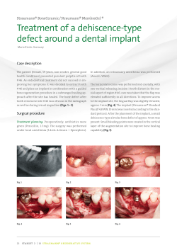


© Copyright 2026