
Introduction: Specialized cells
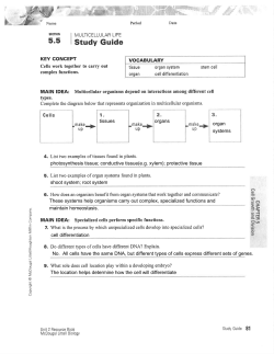
Introduction: Specialized cells The 3D printer is an amazing invention. Instead of spraying out ink to make words or images, a 3D printer uses plastic or metal to create objects. It works by building up layer after layer to a design created in a computer. We can already make parts for machinery and tools for astronauts to use in space. But imagine if we could make human body parts this way – an ear, a hand, maybe even a heart. Instead of using plastic or metal, we would use the body’s own living cells as the “ink”. Printed tissues and organs could help thousands of patients who are desperately waiting for an organ transplant – and donor organs are always in short supply. The good news is that scientists and engineers are working intensely at making life-saving bioprinting a reality. In fact they've already made some body parts that look nearly like the real thing. Almost every type of cell has been used to print tissues in this way, including muscle and brain cells. But researchers still face major challenges, such as figuring out how to provide printed tissues with a blood supply – essential for keeping cells fed with nutrients. Unless this problem is solved, the bold idea of printing fully-functioning organs on demand will remain a distant hope. Read the full Cosmos magazine article here. Scientists and engineers are now developing ways to bioprint human organs such as kidneys, ears and fingers. Poll 1 Reflect: Some people argue that bioprinting organs to save lives will inevitably lead to the printing of bigger muscles for athletes and more beautiful noses and ears – the start of a slippery slope that ends with Frankenstein's monster. How far do you think bioprinting of human tissues and organs should be allowed to go? Note: Once your teacher has made the results of the poll visible, they might ask you to enter into a class discussion of the issue. This poll is currently closed, so you can't vote It should not be allowed at all. It should be allowed only to save lives. It should be allowed only to treat medical conditions, whether they're life-threatening or not. It should be allowed for medical treatments and the creation of cosmetic or enhanced organs. It should be allowed even to print whole organisms, if this becomes possible. Gather: Specialized cells Most cells can only be seen through a light microscope (left) or a more powerful electron microscope. Two examples of what you might see: sperm cells (centre) and muscle tissue (right). Your cells and their organelles The basic building blocks of all organisms are cells and you are made up of tens of trillions of them! Most of the cells in your body are too small to see with the naked eye – you can get a better sense of their size here. Modern microscopes can magnify cells to thousands of times their actual size, allowing us to see their internal structure in incredible detail. What we discover is a variety of even smaller structures known as organelles – "little organs" – each of which has a specific role to play in the cell's complex machinery. 4:53 Question 1 Summarize: Complete the following table to summarize the structure and functions of some of the organelles commonly found in animal cells. Name of organelle cell membrane Structure very thin consists of three layers nucleus (plural: nuclei) cytoplasm "control centre" that sends messages to other organelles holds genetic information (DNA) needed for reproduction composed of a fluid (water, minerals, salts, etc.) and organelles other than the nucleus endoplasmic reticulum (plural: endoplasmic reticula) mitochondrion (plural: mitochondria) holds organelles in place and supports their activities "assembly line" where proteins are put together transportation network that connects organelles oval shape contains folded membranes lysosome golgi body Function/s contains enzymes that digest food particles "garbage disposal unit" that breaks down worn-out cell parts and collects waste products a group of flattened ovals Question 2 Identify: Label the organelles visible in the following electron microscope image of a plasma cell – a type of blood cell that produces antibodies to help fight infections. cell membrane | nucleus | endoplasmic reticulum | mitochondrion | golgi body Cell specialization Unicellular – single-celled – organisms such as bacteria need to perform all of the functions necessary for life on their own. In contrast, multicellular organisms can divide up these functions among different types of cells. This is known as cell specialization. Your body contains over 200 different types of specialized cells. Each type is adapted to do a particular job well and has developed special features to do it. Just as you can’t use a pair of scissors as a hammer, you can’t replace a bone cell with a muscle cell and expect to get the same result. Specialized cells vary widely with respect to: shape size number of organelles types of organelles For example, muscle cells are specially adapted to perform their key function of contraction. They: tend to form long fibres, range in size from about 1–40 mm long depending on the needs of the muscle they belong to, have larger numbers of mitochondria to meet their high energy demands and can have up to 100 nuclei to control activity along their length, and contain special cylindrical organelles called myofibrils that are involved in contraction. Question 3 Decide: Which of the following statements are true: Question 4 Analyze: Which features of muscle cells allow them to perform their special function of contraction? Select only one answer. All cells have the same types of organelles. Cell specialization is when different types of cells Large numbers of mitochondria perform different functions. Long, fibrous shape All cells have the same numbers of mitochondria. Myofibrils Some cells form long fibres whereas others are Multiple nuclei round or branching. All of the above Question 5 Match: Draw lines to match these specialized cells with their descriptions of what they do. Tissues Cells that work together to perform a particular function are organized into tissues. Some tissue types are familiar, such as muscle, bone and blood. Others have been discovered through dissection of the body and close examination of tissue samples under the microscope. Tissues present a whole new level of complexity compared to the cells that make them up. The reason bioprinting tissues is so difficult is that many different types of specialized cells must be organised in just the right way so that they can work together. 1:32 Question 6 Classify: In the mind map below, provide two examples of each of the four basic tissue types as described in the video. Choose from the following list: brain | glands | biceps | spinal cord | bone | skin | blood | heart epithelial muscle Tissue Types connective nervous Organs and organ systems Tissues that work together to perform a larger function form organs such as eyes, lungs and kidneys. Similarly, organs that work together to perform whole-body functions form organ systems. For example, together the heart and blood vessels form the circulatory system which is dedicated to delivering oxygen and nutrients to all parts of the body. Question 7 Order: When you first look at an animal, you see the whole animal. What you don't immediately recognize is that the animal is made up of a large number of parts organized at many different levels. Order the following parts of a multicellular organism from the smallest scale to the largest: cell | organ system | organelle | tissue | organism | molecule | organ Process: Specialized cells Left: A 3D printer ejects a stream of living cells to create a kidney. Right: 3D printed prosthetic nose and ear. Bioprinting Now that you've seen the intricate way in which our organs are built out of tissues – and tissues out of cells – you can understand how difficult it is to create a working organ with a 3D printer. The task is made easier by using a "bioink" containing cells that are already alive. But bioprinters must be able to place the right types of specialized cells in the right locations. And then there's the problem of providing an interconnected blood supply to keep them alive for more than a few hours or days. We'll now take a closer look at this process. 3:49 Question 1 Sequence: Order the steps involved in bioprinting a human blood vessel. The first step has been done for you. 1 The Cosmos magazine article "Make your own spare parts" describes the challenge of supplying printed tissues with a network of blood vessels. These are needed to supply the cells with oxygen and nutrients to keep them alive. The light microscope image on the right gives a cross section through a small human artery. It shows four types of cells: red blood cells filling the centre of the artery, endothelial cells in a single layer lining the inner wall (light pink), smooth muscle cells forming the remainder of the artery wall (dark pink), and connective tissue (light brown). Note that the cell nuclei in this image show up as darker circles and ovals. Question 2 Draw: It’s important for scientists to be able to draw accurate diagrams of what they observe, showing only what's needed – no more and no less. Draw a scientific diagram of the above micrograph image of a human artery and upload a photo of your drawing in the project space below. Be sure to follow these standard conventions: Use a very sharp pencil (preferably 2B) and plain white paper. Print a heading at the top of the page in capital letters and underline it using a ruler. State the magnification, in this case x250. Draw a large and clear view of the image as you observe it and include only the essential details. Use dots or hatching to show darker areas rather than colouring in or shading. Draw only what you see and not what you think you should see! Identify features using clear labels. These should be written horizontally in capital letters and arranged neatly around the drawing so that each label is close to the relevant feature. Draw a straight line without arrowheads between each label and its feature using a ruler. Question 3 Analyze: Which cells in the image you have drawn are distinguished by their lack of nuclei? Question 4 Infer: You may have wondered why artery walls contain muscle cells at all. This is so that they can change blood flow and blood pressure by either contracting or relaxing. Red blood cells Endothelial cells Given that fainting is caused by a sudden drop in blood pressure, what are the smooth muscle cells in your arteries most likely doing when you faint? Smooth muscle cells Contracting Connective tissue cells Relaxing Question 5 Calculate: The diagram shows blood vessel cells in orange being printed into a support structure of gel rods. Assuming that each blood vessel cell is spherical, has a diameter of 0.2 mm and is in contact with the cells around it, calculate the number of cells needed to print a blood vessel 12 cm long. The ethics of bioprinting You’ve been invited to join the studio audience for an episode of a current affairs program in which audience members put questions to a panel. This week's panellists: Panellist A is on a five year waiting list for a kidney donation and hoping to receive a bioprinted kidney instead. Panellist B is a biomedical engineer who is developing a technique to bioprint functioning organs. Panellist C is a federal politician who is involved in framing a law to place limits on bioprinting. Panellist D has set up a company to bioprint meat for human consumption. Panellist E is a human rights lawyer who supports medical bioprinting but opposes the bioprinting of enhanced organs and synthetic meat. Question 6 Debate: Think about some of the complex issues that bioprinting raises. Your teacher may divide the class into groups to discuss them. Then, either on your own or in groups: 1. Formulate a different question for each of the panellists to find out their opinions on the proper limits of bioprinting. Hint: The live poll in the Introduction and any class discussion may give you some ideas. 2. Share your questions with another classmate or group and propose answers to each other’s questions by imagining yourself as each of the five panellists. Apply: Specialized cells Put your cells to work Question 1 Compose: Imagine you're a specialized cell in the human body. Your task is to research your cell type and then prepare a resumé so that you can apply for a job that suits your skills. Organize your resumé as follows: Name: state which type of specialized cell you are. Address: identify where in the body you can be found. Place of birth: identify where in the body you were formed. Appearance: provide two images – a drawing and a microscope image – and label your main features, such as your shape, organelles and any special extensions. Skills: list your main roles in the body and explain how your various features allow you to perform them. Clubs and organizations: describe your role in forming tissues and organs and your associations with any other specialized cell types. Closing statement: write a paragraph to describe the type of job you're looking for and highlight how your skills would suit this job. Some examples of cells you might choose to be: nerve cell (neuron), red blood cell (erythrocyte), sperm cell, egg cell, photoreceptor cell, taste bud cell, heart muscle cell, fat cell (adipocyte), macrophage. Career: Specialized cells 1:51 Question 1 Imagine: If you were a biomedical engineer involved in bioprinting, which types of tissue would you like to create? What would you find most rewarding about this work? Cosmos Lessons team Lesson author: Hayley Bridgwood Introduction author: Bill Condie Editors: Jim Rountree and Campbell Edgar Art director: Wendy Johns Education director: Daniel Pikler Image credits: iStock, PathologyOutlines.com, CK-12 Foundation, Bloomberg, Science Photo Library, Modern Meadow, Dan Piraro/Cartoonist Group. Video credits: Films for the Humanities & Sciences/mrspereiraclass, qprmarsh, ABC/Chelsea Singletary and Pixel Planet on YouTube


© Copyright 2026