
ARTICLE Functional screening identifies miRNAs inducing cardiac regeneration
ARTICLE
doi:10.1038/nature11739
Functional screening identifies miRNAs
inducing cardiac regeneration
Ana Eulalio1{, Miguel Mano1, Matteo Dal Ferro1,2, Lorena Zentilin1, Gianfranco Sinagra2, Serena Zacchigna1 & Mauro Giacca1
In mammals, enlargement of the heart during embryonic development is primarily dependent on the increase in
cardiomyocyte numbers. Shortly after birth, however, cardiomyocytes stop proliferating and further growth of the
myocardium occurs through hypertrophic enlargement of the existing myocytes. As a consequence of the minimal
renewal of cardiomyocytes during adult life, repair of cardiac damage through myocardial regeneration is very
limited. Here we show that the exogenous administration of selected microRNAs (miRNAs) markedly stimulates
cardiomyocyte proliferation and promotes cardiac repair. We performed a high-content microscopy, high-throughput
functional screening for human miRNAs that promoted neonatal cardiomyocyte proliferation using a whole-genome
miRNA library. Forty miRNAs strongly increased both DNA synthesis and cytokinesis in neonatal mouse and rat
cardiomyocytes. Two of these miRNAs (hsa-miR-590 and hsa-miR-199a) were further selected for testing and were
shown to promote cell cycle re-entry of adult cardiomyocytes ex vivo and to promote cardiomyocyte proliferation in both
neonatal and adult animals. After myocardial infarction in mice, these miRNAs stimulated marked cardiac regeneration
and almost complete recovery of cardiac functional parameters. The miRNAs identified hold great promise for the
treatment of cardiac pathologies consequent to cardiomyocyte loss.
In mammals, cardiomyocyte (CM) proliferation rapidly ceases after
birth1–3. The transition of CMs from a proliferative state, characteristic
of embryonic stages, to the differentiated, hypertrophic phenotype typical of adult cells is a highly regulated process1–3. Although there is also
evidence of cardiac cell renewal in humans4,5, the proliferative capacity
of adult CMs remains limited. Consequently, the ability of the adult
heart to repair itself following injury, such as myocardial infarction or
heart failure, is very restricted. A number of reports have indicated that,
after cardiac injury, a few replicating CMs can be detected in the
damaged area, suggestive of a partial attempt at myocardial regeneration6–10; however, repair typically occurs through a scarring mechanism.
Notably, CMs isolated from neonatal hearts retain some proliferative capacity in culture, which becomes extinguished after a few days3.
This offers a valuable opportunity to identify the mechanisms that
maintain this process, with the goal to foster CM proliferation and
induce cardiac regeneration after injury.
MicroRNAs (miRNAs) regulate gene expression post-transcriptionally, by base-pairing to partially complementary sequences in target
messenger RNAs11,12. Impairment of the miRNA pathway in cardiac
muscle leads to heart failure and cardiomyopathy13,14. Furthermore,
altered miRNA expression patterns have been associated with various
cardiac pathologies15–18. Although some miRNAs have been shown to
inhibit CM proliferation, including miR-119,20, miR-13321 and members
of the miR-15 family22, to date and to our knowledge no miRNA has been
reported to increase CM proliferation.
Here, we set out to systematically identify miRNAs triggering CM
proliferation using a synthetic miRNA library of human origin, with
the ultimate purpose to identify potential human therapeutics.
Screening for miRNAs regulating CM proliferation
We performed a high-content, fluorescence-microscopy-based, highthroughput screening in neonatal rat CMs using a library of 875
miRNA mimics (988 mature miRNAs, 875 unique sequences,
miRBase release 13.0 (2009), http://mirbase.org; Fig. 1a). Cultures
of neonatal rat ventricular CMs, containing .90% CMs, were transfected with the library of miRNA mimics (.95% transfection efficiency; Supplementary Fig. 1a, b). After 72 h, the cells were stained
for sarcomeric a-actinin to distinguish CMs, for the proliferation antigen Ki-67 and for 5-ethynyl-29-deoxyuridine (EdU), a uridine analogue that is incorporated into newly synthesized DNA23
(Fig. 1b). There was a high correlation between the two proliferation
markers (Spearman r . 0.80; Fig. 1b and Supplementary Fig. 1c).
Image segmentation and analysis was performed to selectively quantify the number of proliferating CMs (a-actinin1, Ki-671, EdU1;
Fig. 1b). The screening was performed in duplicate; the replicates
showed very good reproducibility (Spearman r 5 0.94; Fig. 1c). On
average, approximately 2,500 cells were analysed per experimental
condition and replicate.
The screening identified 204 miRNAs that significantly increased
neonatal CM proliferation more than twofold when compared to
CMs in basal conditions or treated with the control miRNA celmiR-67 (from 12.5% up to more than 45% proliferating cells; red in
Fig. 1c and Supplementary Table 1); 331 miRNAs decreased EdU
incorporation and Ki-67 positivity (from 12.5% to 0%; blue in
Fig. 1c, Supplementary Fig. 2 and Supplementary Table 1), the majority without significantly affecting cell viability. Examples of miRNAs
strongly increasing or decreasing CM proliferation are shown in
Fig. 1d. The majority of the miRNAs that enhanced EdU and Ki-67
positivity also increased the number of CMs (Spearman r 5 0.63;
Fig. 1e). Of note, miRNAs belonging to the same family induced a
matched proliferation phenotype (Fig. 1f). No significant synergistic effect was observed upon pairwise administration of the top
four miRNAs (Supplementary Fig. 3). Comparison of miRNA expression profiles in neonatal and adult CMs showed that, out of the
1
Molecular Medicine Laboratory, International Centre for Genetic Engineering and Biotechnology (ICGEB), 34149 Trieste, Italy. 2Department of Medical, Surgical and Health Sciences, University of Trieste,
Trieste, Italy and Center for Translational Cardiology, Azienda Ospedaliero-Universitaria ‘‘Ospedali Riuniti di Trieste’’, 34129 Trieste, Italy. {Present address: Institute for Molecular Infection Biology (IMIB),
University of Würzburg, D-97080 Würzburg, Germany.
3 7 6 | N AT U R E | VO L 4 9 2 | 2 0 / 2 7 D E C E M B E R 2 0 1 2
©2012 Macmillan Publishers Limited. All rights reserved
ARTICLE RESEARCH
a
b
Reverse transfection
Rat cardiomyocytes
0h
α-actinin
EdU
Ki-67
cel-miR-67
hsa-miR-590-3p
hsa-miR-1825
hsa-let-7i
Hoechst α-actinin EdU
Image
segmentation
+ EdU
52 h
Hoechst
Microscopy
image
875 miRNA mimics
on 96-well plates
72 h
Fixation and
fluorescence staining
(Hoechst, α-actinin,
Ki-67 and EdU)
d
cel-miR-67
10
0
0
10
20
30
40
50
60
12.5%
% Ki-67+ EdU+ CMs (2)
f
2,000
1,000
0
10 20 30 40 50
% Ki-67+ EdU+ CMs
60
miR-15
20
10
Nucleus of
proliferating cell
Nucleus of
non-proliferating cell
Proliferating
cardiomyocyte
Non-proliferating
cardiomyocyte
g
40
30
0
0
let-7
0.9%
% Ki-67+ EdU+ CMs
(mouse)
3,000
miR-302
45.6%
40
cel-miR-67
% Ki-67+ Edu+ CMs
4,000
CM number
miR-518
50
5,000
hsa-miR-518a-3p
hsa-miR-518b
hsa-miR-518c
hsa-miR-518d-3p
hsa-miR-518e
hsa-miR-518f
e
48.5%
2× control
20
hsa-miR-15a
hsa-miR-15b
hsa-miR-16
hsa-miR-195
hsa-miR-497
30
Hoechst
α-actinin
EdU
hsa-let-7a
hsa-let-7b
hsa-let-7c
hsa-let-7d
hsa-let-7e
hsa-let-7f
hsa-let-7g
hsa-let-7i
hsa-miR-98
40
hsa-miR-302a
hsa-miR-302b
hsa-miR-302c
hsa-miR-302d
hsa-miR-302e
hsa-miR-302f
hsa-miR-372
hsa-miR-373
50
Microscopy
image
cel-miR-67
% Ki-67+ EdU+ CMs (1)
60
Image
segmentation
c
30
miR-199a-3p
miR-33b*
miR-590-3p
20
miR-1825
2× control
10
0
0
10
20 30 40 50
% Ki-67+ EdU+ CMs
(rat)
60
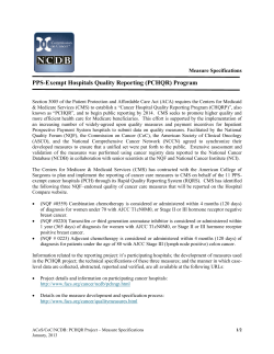
Figure 1 | High-content screening identifies miRNAs regulating CM
proliferation. a, Screening workflow. b, Microscopy images and image
reconstruction of rat CMs treated with cel-miR-67 (control). c, Percentage of
proliferating rat CMs following treatment with the 875 miRNAs. Red, .twofold
proliferation; blue, ,twofold proliferation. Approximately 2,500 cells were
analysed per miRNA/replicate. d, Rat CMs treated with selected miRNAs.
e, Correlation between percentage of rat proliferating CMs and CM number. Red,
.twofold proliferation; blue, ,twofold proliferation. f, Percentage of proliferating
rat CMs transfected with miRNAs of different families. g, Correlation between
percentage of proliferating rat and mouse CMs after treatment with the 204
miRNAs increasing rat CM proliferation more than twofold. Red, .twofold
proliferation for both mouse and rat CMs. Scale bars, 100 mm.
269 miRNAs expressed by rat neonatal CMs, the majority of the proproliferative miRNAs were less expressed in adult cells (Supplementary Fig. 4).
Because the screening was based on high-content imaging, we also
determined the effect of the 875 miRNAs on CM size. The top
miRNAs promoting CM proliferation were not among those inducing
hypertrophy (for example, hsa-miR-29b; Supplementary Fig. 5a, b), as
also confirmed by measuring the atrial natriuretic peptide levels
(Supplementary Fig. 5c–f).
To understand whether the identified miRNAs also increased proliferation of CMs from another species, thus increasing the likelihood
of the conservation of their functional effect in human cells, we retested the selected 204 miRNAs in neonatal mouse CMs. In the mouse
heart, CMs stop dividing sooner than those in the rat heart (shortly
after birth in the mouse and 3–4 days after birth in the rat24,25). Consequently, CMs isolated from newborn mice have a lower proliferative
capacity (approximately 5%). From the 204 miRNAs tested, 40
miRNAs also enhanced EdU incorporation and Ki-67 positivity in
mouse CMs at least twofold (Fig. 1g, Supplementary Table 1 and
Supplementary Fig. 6).
To demonstrate that the increase in CM DNA replication eventually resulted in an increase in karyokinesis and cytokinesis, we
treated rat CMs with the top 10 miRNAs from the rat screening
and mouse CMs with the top 10 from the mouse re-screening. After
72 h, the cells were stained for histone H3 phosphorylated on serine
10 (H3S10ph), a marker of late G2/mitosis, and for Aurora B
kinase localization in midbodies, transient structures formed during
cytokinesis (Fig. 2a, b). Treatment with the selected miRNAs significantly increased both the percentage of cells positive for H3S10ph,
and the number of cells presenting midbodies. Table 1 reports the
results of these quantifications. Notably, all the 10 plus 10 selected
miRNAs were effective in promoting karyokinesis and cell division in
both rat and mouse cells. Figure 2c, d shows the results for the best two
performers of each list. Consistent with an effective cell division, at
6 days after transfection we observed a significant increase in CM
number (Fig. 2e and Supplementary Fig. 7). None of the top performing miRNAs increased rat cardiac fibroblast proliferation
(Supplementary Fig. 8a, b), indicating that their effect for CMs was
rather selective.
miRNAs increase post-natal CM proliferation
Next we tested whether the identified miRNAs stimulated post-natal
CM proliferation. As a first approach, we used CMs isolated from 7days-old rats. At this age, CM proliferation was almost undetectable;
however, the percentage of EdU1 CMs significantly increased up to
13–20% upon treatment with hsa-miR-590-3p, hsa-miR-199a-3p,
hsa-miR-1825 and hsa-miR-33b* (Supplementary Fig. 9).
To test whether these miRNAs also induce re-entry into the cell
cycle of fully differentiated cells, CMs were isolated from adult
(2-months old) rats and transfected with cel-miR-67 or with hsamiR-590-3p and hsa-miR-199a-3p. Remarkably, treatment with the
two miRNAs determined a time-dependent re-entry of the cells into
the cell cycle (Fig. 2f–h), eventually leading to a significant increase of
the number of CMs (Fig. 2h).
2 0 / 2 7 D E C E M B E R 2 0 1 2 | VO L 4 9 2 | N AT U R E | 3 7 7
©2012 Macmillan Publishers Limited. All rights reserved
RESEARCH ARTICLE
f
Not treated
**
**
hsa-miR-590-3p
**
4
2
0
cel-miR-67
hsa-miR-590-3p
hsa-miR-199a-3p
hsa-miR-1825
hsa-miR-33b*
0
2
hsa-miR-199a-3p
2.0
**
***
***
***
***
8
6
3
**
2
4
**
0
**
1
2
g
Adult (day 7)
cel-miR-67
**
6
4
e
Mouse
4
0
h
0.5
2.0
8
**
6
*
4
*
CM number
(fold over cel-miR-67)
***
10
% EdU+ CMs
Hoechst α-actinin H3S10ph
1.0
0.0
12
Hoechst α-actinin EdU
*** ** *
1.5
cel-miR-67
hsa-miR-590-3p
hsa-miR-199a-3p
hsa-miR-590-3p
hsa-miR-199a-3p
***
cel-miR-67
hsa-miR-590-3p
hsa-miR-199a-3p
hsa-miR-1825
hsa-miR-33b*
6
*
8
Rat
10
CM number
(fold over cel-miR-67)
***
d
***
cel-miR-67
hsa-miR-590-3p
hsa-miR-199a-3p
hsa-miR-1825
hsa-miR-33b*
10
cel-miR-67
hsa-miR-590-3p
hsa-miR-199a-3p
hsa-miR-1825
hsa-miR-33b*
Mouse
**
cel-miR-67
hsa-miR-590-3p
hsa-miR-199a-3p
hsa-miR-1825
hsa-miR-33b*
Hoechst Aurora-B
α-actinin
Adult (day 1)
% H3S10ph+ CMs
hsa-miR-590-3p
cel-miR-67
Midbodies
b
Rat
8
% midbodies+ CMs
c
hsa-miR-590-3p
Hoechst H3S10ph
α-actinin
cel-miR-67
H3S10ph
a
*****
1.5
*
*
1.0
0.5
2
0
4
7
Days in culture
0.0
4
7
Days in culture
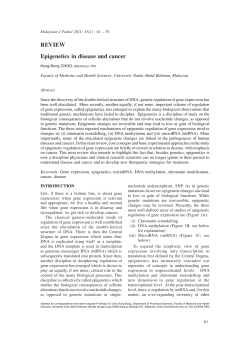
Figure 2 | miRNAs increase cytokinesis and proliferation of post-natal
CMs. a, b, H3S10ph (a) and Aurora B kinase (b) staining of miRNA-treated rat
CMs. Arrows, positive CMs. c, d, Percentage of CMs positive for H3S10ph
(c) and midbodies (d). n 5 3–5 per group. e, CM number, 6 days after miRNA
treatment. n 5 3 per group. f, EdU staining of adult rat CMs. Top,
epifluorescence; bottom, confocal. Arrows, proliferating CMs. g, H3S10ph
staining of miRNA-treated adult rat CMs, at day 7. h, Percentage of
proliferating adult rat CMs and CM number. n 5 3–6 per group. All panels,
mean 6 s.e.m.; *P , 0.05, **P , 0.01, ***P , 0.001 relative to cel-miR-67;
scale bars, 100 mm.
To obtain further molecular insights into the capacity of the
miRNAs to induce cell cycle re-entry of adult CMs, we compared
neonatal, adult CMs at the time of isolation and adult CMs transfected
with cel-miR-67, hsa-miR-590-3p and hsa-miR-199a-3p at day 7 for
the expression of genes related with cardiac differentiation26 and proliferation. CM culture itself, as described previously27–29, determined a
partial de-differentiation of adult CMs, as concluded from increased
expression of b-myosin heavy chain (MYH7), decrease in a-myosin
heavy chain (MYH6), and increase in DAB2, destrin and RUNX1
(Supplementary Fig. 10 and Fig. 2f for microscopy images showing
more rounded cells, with less organized contractile apparatus); these
changes in gene expression never reached those found in of neonatal
CMs, except for NKX2-5. This partial de-dedifferentiation, however,
did not result in cell cycle re-entry per se, unless the cells were stimulated with hsa-miR-590-3p and hsa-miR-199a-3p. Upon miRNA treatment, the marked increase in the levels of cyclin genes correlated with
their increased proliferation (Supplementary Fig. 10 and Fig. 2f-h).
Collectively, these experiments demonstrate that hsa-miR-5903p and hsa-miR-199a-3p are able to induce proliferation of postnatal CMs.
Table 1 | Effect of selected miRNAs on rat and mouse CMs.
miRNA
Rat CMs
Mouse CMs
cel-miR-67
hsa-miR-590-3p
hsa-miR-1825
hsa-miR-518a-3p
hsa-miR-1273
hsa-miR-302b*
hsa-miR-495
hsa-miR-99a
hsa-miR-518f
hsa-miR-885-5p
hsa-miR-302d*
cel-miR-67
hsa-miR-199a-3p
hsa-miR-33b*
hsa-miR-590-3p
hsa-miR-1244
hsa-miR-23b
hsa-miR-152
hsa-miR-19b-2*
hsa-miR-372
hsa-miR-130a*
hsa-miR-885-5p
Proliferating CMs (%){
H3S10ph1 CMs (%){
Midbodies1 CMs (%){
11.1 6 1.2
49.1 6 3.5
48.2 6 4.2
46.9 6 1.0
41.8 6 2.2
43.0 6 1.3
45.6 6 4.6
43.3 6 2.9
42.2 6 1.7
39.1 6 1.7
40.8 6 3.9
6.6 6 1.1
38.1 6 1.4
29.9 6 3.2
25.2 6 1.2
20.7 6 1.4
21.1 6 1.3
24.1 6 1.7
28.5 6 0.9
21.0 6 1.0
22.4 6 1.5
17.3 6 0.6
1.28 6 0.23
6.44 6 1.55
5.83 6 0.51
6.48 6 0.57
5.47 6 0.20
6.53 6 0.70
5.47 6 0.53
5.70 6 1.10
4.56 6 0.25
5.98 6 0.55
4.86 6 0.52
1.18 6 0.15
8.46 6 0.79
5.12 6 0.81
5.14 6 1.11
5.76 6 0.21
5.33 6 0.52
5.83 6 0.87
4.48 6 0.48
4.80 6 0.12
4.63 6 0.20
4.75 6 0.32
0.89 6 0.20
5.23 6 0.30
6.56 6 0.32
4.57 6 0.04
3.41 6 0.15
3.02 6 0.14
5.10 6 0.92
3.67 6 0.16
4.05 6 0.37
4.12 6 0.12
5.33 6 0.22
0.22 6 0.02
2.99 6 0.48
1.40 6 0.18
2.10 6 0.24
1.35 6 0.04
2.56 6 0.23
1.29 6 0.33
2.41 6 0.57
1.53 6 0.20
1.55 6 0.25
1.52 6 0.27
Percentage of proliferating (Ki-671 and EdU1), H3S10ph1 and midbodies1 rat and mouse CMs (a-actinin1) transfected with the 10 miRNAs that increase proliferation of rat and mouse CMs more efficiently.
Results for the control cel-miR-67 are shown for comparison. Data are shown as mean values 6 s.e.m.
{ Data are reported to the total number of CMs.
3 7 8 | N AT U R E | VO L 4 9 2 | 2 0 / 2 7 D E C E M B E R 2 0 1 2
©2012 Macmillan Publishers Limited. All rights reserved
ARTICLE RESEARCH
b
1
Neonatal rats
4
c
8
6
cel-miR-67
hsa-miR-590-3p
hsa-miR-199a-3p
4
2
e
f
g
12
h
4
2
0
**
**
CM cross-sectional area
(fold over control)
Heart/body weight
(mg/g)
10
6
AAV9-miR-199a
i
1.2
0.6
1.0
0.5
0.8
0.6
0.4
0.4
0.3
0.2
0.2
0.1
0.0
0.0
27.9%
siRNA Hopx
30
hsa-miR-590-3p
20
10
0
c
40
30
16.1%
12.9%
hsa-miR-199a-3p
20
10
0
Non-targeting
Crim1
C030006K11Rik
Cfl2
Kdm3a
Nfat5
Tcp11l2
LOC100047997
Zfp317
Dnm1l
Spcs3
Clip4
Pln
Nufip2
Homer1
Slc30a1
Calu
Ttc33
Sucla2
Lrrc10
Ankrd52
Lace1
Rictor
Epm2aip1
Otud6b
Hagh
Hopx
Gtf3c3
Ipo13
Ptpmt1
Apc
Itgav
Zfx
Ttn
Ifi47
Erlin2
Ppip5k2
Gm2058
Pja2
Clic5
Hook3
Me3
Lgmn
Slc8a1
% Ki-67+ EdU+ CMs
b
40
% Ki-67+ EdU+ CMs
Knockdown of 45 genes
increase proliferation >twofold
Hoechst
α-actinin EdU
5.6%
siRNA Homer1
Figure 3 | Genes downregulated by miR-590-3p and miR-199a-3p increase
CM proliferation. a, Workflow for miRNA target identification.
b, c, Percentage of proliferating mouse CMs after siRNA treatment. Targets
increasing proliferation .twofold are shown. Red, genes downregulated by
both miRNAs. d, EdU staining of mouse CMs treated with selected siRNAs.
Scale bar, 100 mm.
hsa-miR-199a-3p, whereas Clic5 is a direct target of the former miRNA
only (Supplementary Fig. 12).
Treatment with none of these siRNAs induced an increase in CM
proliferation as high as that observed with hsa-miR-590-3p or hsamiR-199a-3p. This demonstrates that the notable effect of the two
d
hsa-miR-590-3p
j
Figure 4 | miR-590 and miR-199a induce CM
proliferation in vivo. a, Schematic of in vivo
experiments using synthetic miRNAs. n 5 8 per
group. b, EdU and a-actinin staining of rat hearts
injected with miRNAs. Scale bars, 1 mm (top),
100 mm (bottom). c, Percentage of proliferating
cells. d, Confocal microscopy of rat hearts injected
with hsa-miR-590-3p and hsa-miR-199a-3p. Scale
bar, 10 mm. e, Schematic of in vivo experiments
using AAV vectors. n 5 9 per group. f, Mouse
hearts transduced with AAV vectors. Scale bar,
1 mm. g, Ratio between heart and body weight.
h, CM cross-sectional area. i, Percentage of cells
positive for H3S10ph. j, Confocal microscopy of
H3S10ph-stained mouse hearts transduced with
AAV. Scale bar, 10 mm. All panels, mean 6 s.e.m.;
**P , 0.01, ***P , 0.001 relative to control.
AAV9-miR-590
TOTO-3
H3S10ph α-actinin
Neonatal mice
8
AAV9-miR-590
Collection
% H3S10ph+ cells
Day: 0
AAV9-control
siRNA Crim1
siRNA screening
(601 genes)
hsa-miR-199a-3p
0
AAV9-miRNA
intraperitoneal
>1.5-fold change
697 mRNAs
1,056 mRNAs
downregulated (641 genes)
upregulated
TOTO-3 α-actinin EdU
% EdU+ cells
***
10
EdU
***
12
Hoechst
α-actinin EdU
14
Non-targeting siRNA
Transcriptomic analysis
(deep sequencing)
TOTO-3 α-actinin EdU
hsa-miR-199a-3p
Hoechst
α-actinin EdU
Day: 0
hsa-miR-590-3p
cel-miR-67
Collection
d
***
AAV9-miR-199a
***
AAV9-control
AAV9-miR-590
AAV9-miR-199a
TOTO-3
H3S10ph α-actinin
a
miRNA
intracardiac EdU
Transfection of
mouse cardiomyocytes
(hsa-miR-590-3p, hsa-miR-199a-3p)
Hoechst
α-actinin EdU
a
To identify the relevant targets of the selected miRNAs, we assessed
global transcriptome changes by deep-sequencing total neonatal
mouse CM RNA after transfection with hsa-miR-590-3p or hsamiR-199a-3p, which are the two miRNAs that were the most effective
in rat and mouse CMs, respectively.
This analysis identified 1,056 upregulated transcripts (283 for hsamiR-590-3p and 773 for hsa-miR-199a-3p; reads per kilobase of exon
model per million mapped reads (RPKM) . 5 and 1.5-fold upregulation) and 697 downregulated transcripts (95 for hsa-miR-590-3p and
602 for hsa-miR-199a-3p; RPKM . 5 and 1.5-fold downregulation,
including 35 mRNAs downregulated by both miRNAs; these corresponded to 641 different genes; Supplementary Table 2 and Fig. 3a).
Functional analysis of the upregulated transcripts revealed enrichment for genes belonging to the ‘cell cycle’, ‘cellular growth and proliferation’ and ‘DNA replication, recombination and repair’ categories
(Supplementary Fig. 11a). Analysis of the 641 downregulated genes
showed enrichment for genes belonging to the ‘skeletal and muscular
system development and function’ and ‘cellular assembly and organization’ categories (Supplementary Fig. 11b).
To identify, among the downregulated genes, those relevant in
controlling CM proliferation, we individually knocked down 601 of
these genes for which short interfering RNAs were available (Fig. 3a),
under conditions similar to those of the initial screening. Forty-five of
these siRNAs increased proliferation at least twofold (Supplementary
Table 3). Of these, 5 siRNAs targeted transcripts downregulated by
hsa-miR-590-3p (Fig. 3b) and 43 by hsa-miR-199a-3p (Fig. 3c); 3
siRNAs targeted transcripts downregulated by both miRNAs (red
in Fig. 3b, c); Fig. 3d shows representative images from these experiments. The transcripts downregulated by both miRNAs were Homer1,
involved in the regulation of calcium signalling in cardiac cells30, Hopx,
a regulator suppressing embryonic CM proliferation31, and Clic5.
Luciferase 39-untranslated region (UTR) reporter assays showed
that Homer1 and Hopx are directly regulated by hsa-miR-590-3p and
Non-targeting
Homer1
Hopx
5730455O13Rik
Clic5
1810030N24Rik
miRNA effect depends on multiple targets
2 0 / 2 7 D E C E M B E R 2 0 1 2 | VO L 4 9 2 | N AT U R E | 3 7 9
©2012 Macmillan Publishers Limited. All rights reserved
RESEARCH ARTICLE
miRNAs in increasing CM proliferation probably ensues as a cumulative effect on multiple, cellular mRNA targets.
a
Adult mice
Infarction
AAV9-miRNA EdU
Echo
Echo
Collection Echo Collection
AAV9-control
AAV9-miR-590
AAV9-miR-199a
miRNAs increase CM proliferation in vivo
miRNAs preserve cardiac function after infarction
Next we assessed whether hsa-miR-590 and hsa-miR-199a might
boost the normally ineffective myocardial repair that takes place after
myocardial infarction. Adult female CD1 mice (8–12-weeks old)
underwent permanent left anterior descending coronary artery ligation and were injected, in the peri-infarcted area, with AAV9 vectors
expressing the two miRNAs or a control vector (Fig. 5a). Our previous
experience indicates that this treatment results in efficient myocardial
transduction and month-long transgene expression36. As evaluated by
echocardiography, the left ventricular ejection fraction (LVEF,
Fig. 5b), fractional shortening (LVFS, Fig. 5c), end-systolic anterior
wall thickness (LVAW thickness-systolic, Fig. 5d), as well as other
parameters of cardiac function (Supplementary Fig. 18a–c), were significantly preserved over time in the infarcted mice injected with
AAV9-miR-590 or AAV9-miR-199a.
Analysis of trichrome-stained heart cross-sections clearly showed
that the infarct size was significantly reduced in mice treated
with both miRNAs (Fig. 5e, f). In the animals treated with the
two miRNAs, at 60 days after vector delivery and EdU treatment,
a significant number of EdU-positive nuclei was still detected in
the infarct border zone (Fig. 5g). Confocal microscopy revealed
that most of these nuclei belonged to mature CMs, well integrated
within the myocardial structure (Fig. 5h), indicative of active proliferation soon after infarction and treatment. Of note, administration of hsa-miR-590 and hsa-miR-199a did not protect CMs from
60
** **
12
**
*
d
30
LVFS (%)
50
LVEF (%)
60
35
*
40
30
c
**
**
*
**
**
*
*
25
20
15
10
30
5
20
e
0
12
30
60
Time after MI (days)
AAV9-control
AAV9-miR-590
LVAW thickness-systolic (mm)
Day: 0
b
12
30
60
Time after MI (days)
AAV9-miR-199a
1.25
******
1.00
******
**
*
0.75
0.50
0.25
0.00
12
30
60
Time after MI (days)
f
35
60 days
Infarct size (%LV)
12 days
30
25
20
** **
15
*** ***
10
5
0
h
g
12
60
Time after MI (days)
AAV9-miR-590
AAV9-miR-199a
***
0.8
0.6
***
AAV9-control
AAV9-miR-590
AAV9-miR-199a
0.4
0.2
TOTO-3 α-actinin EdU
1.0
TOTO-3 α-actinin EdU
1.2
% EdU+ cells
We went on to assess the in vivo effect of hsa-miR-590-3p and hsamiR-199a-3p. The synthetic miRNAs, complexed with a lipid transfection reagent, were injected directly into the heart of neonatal rats.
One day later, the animals were administered EdU intraperitoneally
(Fig. 4a). After three additional days, the left ventricle walls of the
hearts injected with the two miRNAs appeared markedly thicker than
those that received cel-miR-67 (Fig. 4b). Analysis of EdU incorporation revealed a marked increase in the number of EdU1 cells in these
hearts (Fig. 4b, c and Supplementary Fig. 13a). Confocal microscopy
analysis indicated that the majority of the proliferating cells were
indeed CMs (Fig. 4d). Consistent with the lack of induction of proliferation on cardiac fibroblasts in vitro, no increased fibrosis was
observed in the injected hearts (Supplementary Fig. 13b).
To explore the long-term effects of the miRNAs in the neonatal
heart, we exploited the high cardiac tropism and prolonged expression of viral vectors based on adeno-associated virus (AAV) serotype
9 upon systemic delivery32–34. AAV9 vectors expressing hsa-miR590 or hsa-miR-199a precursor miRNAs, or a control vector, were
injected intraperitoneally in neonatal mice (Fig. 4e). At 12 days after
injection, the hearts of these animals were morphologically normal,
but significantly enlarged (Fig. 4f, g). No difference in collagen content was observed (data not shown). Similarly, there were no signs of
CM hypertrophy (Fig. 4h and Supplementary Fig. 14).
The number of mitotic CMs, positive for H3S10ph, was significantly increased in the hearts of the animals injected with AAV9miR-590 or AAV9-miR-199a (Fig. 4i and Supplementary Fig. 15).
Confocal microscopy analysis of the proliferating cells revealed that
most of these cells were differentiated CMs; however, the cells showed
partial sarcomere disassembly, as would be expected in cells undergoing division35 (Fig. 4j). Intracardiac delivery of these miRNAs in
adult mice also resulted in increased cardiac cell proliferation
(Supplementary Fig. 16a–c). AAV vector transduction determined a
significant overexpression of the miRNAs both in neonatal and adult
animals (Supplementary Fig. 17a–d).
Collectively, these results show that hsa-miR-590 and hsa-miR199a significantly increase CM proliferation in vivo.
0.0
Figure 5 | miR-590-3p and miR-199a-3p induce marked cardiac
regeneration after myocardial infarction. a, Schematic of the myocardial
infarction (MI) experiments in adult mice. Echo, echocardiography. b–d, LVEF
(b), LVFS (c) and LVAW thickness-s (d). Dashed line, non-infarcted animals
injected with AAV9-control. n 5 10–16 per group. e, Masson trichrome
staining of heart cross sections. Scale bar, 1 mm. f, Infarct size. n 5 6–10 per
group. g, h, Percentage of cells positive for EdU (g) and confocal microscopy
(h) at the border zone of the infarct, 60 days post-MI. Scale bar, 10 mm. All
panels, mean 6 s.e.m.; *P , 0.05, **P , 0.01, ***P , 0.001 relative to control.
cardiotoxic stimuli in vitro (Supplementary Fig. 19a), nor did it have
a cardioprotective effect after myocardial infarction (Supplementary
Fig. 19b).
Together, these data indicate that the expression of hsa-miR-590
and hsa-miR-199a after infarction exerts a marked beneficial effect in
reducing infarct size and improving cardiac function, consistent with
the effect of these miRNAs in actively stimulating CM proliferation.
Discussion
To the best of our knowledge, this is the first demonstration that CM
proliferation can be stimulated by the exogenous administration of
miRNAs and, most pertinently, that such treatment can restore cardiac
mass and promote functional recovery after myocardial infarction in
adult animals. The marked proliferative effect exerted by these miRNAs
likely ensues as the sum of their effects on multiple, individual genes.
An important question prompted by our study concerns the identity of the cells upon which hsa-miR-590-3p and hsa-miR-199a-3p act
once delivered into the heart in vivo. In the neonate, depending on the
species, approximately 3–15% of the CMs are still in the cell cycle (see
refs. 24, 25, 37 and this manuscript), and thus it might be envisaged
that the miRNAs enhance the proliferative capacity of these cells.
3 8 0 | N AT U R E | VO L 4 9 2 | 2 0 / 2 7 D E C E M B E R 2 0 1 2
©2012 Macmillan Publishers Limited. All rights reserved
ARTICLE RESEARCH
Similarly, hsa-miR-590-3p and hsa-miR-199a-3p could act by increasing the normally limited CM proliferation that occurs in the adult heart
in the infarct border zone. Intriguingly, however, both these miRNAs
also induced re-entry into the cell cycle of adult CMs ex vivo and
promoted DNA synthesis and the appearance of mitotic figures in
CMs in vivo. These observations support the conclusion that these
miRNAs promote proliferation of already differentiated CMs, a condition known to associate with cardiac regeneration in zebrafish38–40
and in the neonatal mouse41.
Regeneration of the cardiac mass seems important in a variety of
pathological conditions associated with CM loss, leading from myocardial infarction to include heart failure and dilated cardiomyopathy42,43. Thus, the findings that exogenously administered miRNAs
have the potential to restore cardiac function to almost normal levels
after myocardial infarction have evident translational value. These
miRNAs, in addition, might be useful in expanding the proliferative
potential of CMs derived from human cardiac stem cells, which have
shown promise in recent clinical trials44,45.
METHODS SUMMARY
miRNA library screening. Ventricular CMs from neonatal rats were transfected
with a library of miRNA mimics (Dharmacon) using a standard reverse transfection protocol. EdU was added 52 h later; 72 h after plating cells were fixed and
stained. Image acquisition was performed using an ImageXpress Micro highcontent screening microscope (Molecular Devices). CMs were scored as proliferating only if positive for the two proliferation markers (Ki-67 and EdU).
Animal studies. Animals were injected with synthetic miRNAs complexed with
a transfection reagent or with AAV9 vectors encoding the microRNAs. Proliferation of cardiac cells was analysed 4 and 12 days after injection. Myocardial infarction was induced in adult CD1 mice by permanent left anterior descending
coronary artery ligation; cardiac function was evaluated by histological, morphological and echocardiographic analysis.
Full Methods and any associated references are available in the online version of
the paper.
Received 29 March; accepted 31 October 2012.
Published online 5 December 2012.
1.
2.
3.
4.
5.
6.
7.
8.
9.
10.
11.
12.
13.
14.
15.
16.
Ahuja, P., Sdek, P. & MacLellan, W. R. Cardiac myocyte cell cycle control in
development, disease, and regeneration. Physiol. Rev. 87, 521–544 (2007).
van Amerongen, M. J. & Engel, F. B. Features of CM proliferation and its potential for
cardiac regeneration. J. Cell. Mol. Med. 12, 2233–2244 (2008).
Bicknell, K. A., Coxon, C. H. & Brooks, G. Can the CM cell cycle be reprogrammed?
J. Mol. Cell. Cardiol. 42, 706–721 (2007).
Bergmann, O. et al. Evidence for CM renewal in humans. Science 324, 98–102 (2009).
Kajstura, J. et al. Cardiomyogenesis in the adult human heart. Circ. Res. 107,
305–315 (2010).
Beltrami, A. P. et al. Evidence that human cardiac myocytes divide after myocardial
infarction. N. Engl. J. Med. 344, 1750–1757 (2001).
Robledo, M. Myocardial regeneration in young rats. Am. J.Pathol.32, 1215–1239(1956).
Nag, A. C., Carey, T. R. & Cheng, M. DNA synthesis in rat heart cells after injury and
the regeneration of myocardia. Tissue Cell 15, 597–613 (1983).
Kajstura, J. et al. Myocyte cellular hyperplasia and myocyte cellular hypertrophy
contribute to chronic ventricular remodeling in coronary artery narrowinginduced cardiomyopathy in rats. Circ. Res. 74, 383–400 (1994).
Reiss, K., Kajstura, J., Capasso, J. M., Marino, T. A. & Anversa, P. Impairment of
myocyte contractility following coronary artery narrowing is associated with
activation of the myocyte IGF1 autocrine system, enhanced expression of late
growth related genes, DNA synthesis, and myocyte nuclear mitotic division in rats.
Exp. Cell Res. 207, 348–360 (1993).
Bartel, D. P. MiRNAs: target recognition and regulatory functions. Cell 136,
215–233 (2009).
Pasquinelli, A. E. MiRNAs and their targets: recognition, regulation and an
emerging reciprocal relationship. Nature Rev. Genet. 13, 271–282 (2012).
Chen, J. F. et al. Targeted deletion of Dicer in the heart leads to dilated
cardiomyopathy and heart failure. Proc. Natl Acad. Sci. USA 105, 2111–2116 (2008).
Rao, P. K. et al. Loss of cardiac miRNA-mediated regulation leads to dilated
cardiomyopathy and heart failure. Circ. Res. 105, 585–594 (2009).
Ikeda, S. et al. Altered miRNA expression in human heart disease. Physiol.
Genomics 31, 367–373 (2007).
Matkovich, S. J. et al. Reciprocal regulation of myocardial miRNAs and messenger
RNA in human cardiomyopathy and reversal of the miRNA signature by
biomechanical support. Circulation 119, 1263–1271 (2009).
17. Thum, T. et al. MiRNAs in the human heart: a clue to fetal gene reprogramming in
heart failure. Circulation 116, 258–267 (2007).
18. van Rooij, E. et al. A signature pattern of stress-responsive miRNAs that can evoke
cardiac hypertrophy and heart failure. Proc. Natl Acad. Sci. USA 103,
18255–18260 (2006).
19. Zhao, Y., Samal, E. & Srivastava, D. Serum response factor regulates a muscle-specific
miRNA that targets Hand2 during cardiogenesis. Nature 436, 214–220 (2005).
20. Zhao, Y. et al. Dysregulation of cardiogenesis, cardiac conduction, and cell cycle in
mice lacking miRNA-1–2. Cell 129, 303–317 (2007).
21. Liu, N. et al. miRNA-133a regulates CM proliferation and suppresses smooth
muscle gene expression in the heart. Genes Dev. 22, 3242–3254 (2008).
22. Porrello, E. R. et al. MiR-15 family regulates postnatal mitotic arrest of CMs. Circ.
Res. 109, 670–679 (2011).
23. Salic, A. & Mitchison, T. J. A chemical method for fast and sensitive detection of
DNA synthesis in vivo. Proc. Natl Acad. Sci. USA 105, 2415–2420 (2008).
24. Soonpaa, M. H., Kim, K. K., Pajak, L., Franklin, M. & Field, L. J. CM DNA synthesis and
binucleation during murine development. Am. J. Physiol. 271, H2183–H2189 (1996).
25. Li, F., Wang, X., Capasso, J. M. & Gerdes, A. M. Rapid transition of cardiac myocytes
from hyperplasia to hypertrophy during postnatal development. J. Mol. Cell.
Cardiol. 28, 1737–1746 (1996).
26. Kubin, T. et al. Oncostatin M is a major mediator of CM dedifferentiation and
remodeling. Cell Stem Cell 9, 420–432 (2011).
27. Fredj, S., Bescond, J., Louault, C. & Potreau, D. Interactions between cardiac cells
enhance CM hypertrophy and increase fibroblast proliferation. J. Cell. Physiol. 202,
891–899 (2005).
28. Zhang, Y. et al. Dedifferentiation and proliferation of mammalian CMs. PLoS ONE 5,
e12559 (2010).
29. Bird, S. D. et al. The human adult CM phenotype. Cardiovasc. Res. 58, 423–434 (2003).
30. Pouliquin, P. & Dulhunty, A. F. Homer and the ryanodine receptor. Eur. Biophys.
J. 39, 91–102 (2009).
31. Trivedi, C. M. et al. Hopx and Hdac2 interact to modulate Gata4 acetylation and
embryonic cardiac myocyte proliferation. Dev. Cell 19, 450–459 (2010).
32. Inagaki, K. et al. Robust systemic transduction with AAV9 vectors in mice: efficient
global cardiac gene transfer superior to that of AAV8. Mol. Ther. 14, 45–53 (2006).
33. Katare, R. et al. Intravenous gene therapy with PIM-1 via a cardiotropic viral vector
halts the progression of diabetic cardiomyopathy through promotion of
prosurvival signaling. Circ. Res. 108, 1238–1251 (2011).
34. Secchiero, P. et al. Systemic tumor necrosis factor-related apoptosis-inducing
ligand delivery shows antiatherosclerotic activity in apolipoprotein E-null diabetic
mice. Circulation 114, 1522–1530 (2006).
35. Ahuja, P., Perriard, E., Perriard, J. C. & Ehler, E. Sequential myofibrillar breakdown
accompanies mitotic division of mammalian CMs. J. Cell Sci. 117, 3295–3306 (2004).
36. Zentilin, L. et al. CM VEGFR-1 activation by VEGF-B induces compensatory
hypertrophy and preserves cardiac function after myocardial infarction. FASEB
J. 24, 1467–1478 (2010).
37. Collesi, C., Zentilin, L., Sinagra, G. & Giacca, M. Notch1 signaling stimulates
proliferation of immature CMs. J. Cell Biol. 183, 117–128 (2008).
38. Jopling, C. et al. Zebrafish heart regeneration occurs by CM dedifferentiation and
proliferation. Nature 464, 606–609 (2010).
39. Kikuchi, K. et al. Primary contribution to zebrafish heart regeneration by gata41
CMs. Nature 464, 601–605 (2010).
40. Poss, K. D., Wilson, L. G. & Keating, M. T. Heart regeneration in zebrafish. Science
298, 2188–2190 (2002).
41. Porrello, E. R. et al. Transient regenerative potential of the neonatal mouse heart.
Science 331, 1078–1080 (2011).
42. Dorn, G. W. II. Apoptotic and non-apoptotic programmed CM death in ventricular
remodelling. Cardiovasc. Res. 81, 465–473 (2009).
43. Nishida, K. & Otsu, K. Cell death in heart failure. Circ. J. 72 (Suppl. A) A17–A21 (2008).
44. Bolli, R. et al. Cardiac stem cells in patients with ischaemic cardiomyopathy (SCIPIO):
initial results of a randomised phase 1 trial. Lancet 378, 1847–1857 (2011).
45. Makkar, R. R. et al. Intracoronary cardiosphere-derived cells for heart regeneration
after myocardial infarction (CADUCEUS): a prospective, randomised phase 1 trial.
Lancet 379, 895–904 (2012).
Supplementary Information is available in the online version of the paper.
Acknowledgements The authors are grateful to M. Dapas and M. Zotti for technical
support in AAV production, to M. Sturnega for help in animal experimentation and to
S. Kerbavcic for editorial assistance. A.E. is recipient of a FEBS Long Term Fellowship.
This work was supported by Advanced Grant 250124 from the European Research
Council (ERC) to M.G. and from Project CTC from the Fondazione CRTrieste, Trieste,
Italy.
Author Contributions A.E., M.M. and M.G. designed the study. A.E., M.M., S.Z. and M.D.F.
performed the experiments and analysed the data. G.S. provided clinical consultancy
for the animal study. L.Z. was responsible for AAV production. A.E., M.M. and M.G. wrote
the manuscript.
Author Information The cardiomyocyte miRNA expression microarray and
transcriptomic data are deposited at GEO, under accession numbers GSE41537 and
GSE41538, respectively. Reprints and permissions information is available at
www.nature.com/reprints. The authors declare competing financial interests: details
are available in the online version of the paper. Readers are welcome to comment on
the online version of the paper. Correspondence and requests for materials should be
addressed to M.G. ([email protected]).
2 0 / 2 7 D E C E M B E R 2 0 1 2 | VO L 4 9 2 | N AT U R E | 3 8 1
©2012 Macmillan Publishers Limited. All rights reserved
RESEARCH ARTICLE
METHODS
Isolation of ventricular CMs from neonatal and 7-days-old animals. Wistar
rats and CD1 mice were purchased from Charles River Laboratories Italia Srl.
Animal care and treatments were conducted in conformity with institutional
guidelines in compliance with national and international laws and policies
(EEC Council Directive 86/609, OJL 358, 12 December 1987).
Ventricular CMs from neonatal rats or mice were isolated as described previously37, with minor modifications. In brief, ventricles from neonatal rats or mice
(post-natal day 0) were separated from the atria, cut into pieces and then dissociated in calcium and bicarbonate-free Hanks with HEPES (CBFHH) buffer
containing 1.75 mg ml21 trypsin (BD Difco) and 10 mg ml21 DNase II (Sigma),
under constant stirring. Digestion was performed at room temperature in eight to
ten 10-min steps, collecting the supernatant to fetal bovine serum (FBS, Life
Technologies) after each step. The collected supernatant was centrifuged to
separate the cells, which were then resuspended in Dulbecco’s modified
Eagle medium 4.5 g l21 glucose (DMEM, Life Technologies) supplemented with
5% FBS, 20 mg ml21 vitamin B12 (Sigma) and with 100 U ml21 of penicillin and
100 mg ml21 of streptomycin (Sigma). The collected cells were passed through a
cell strainer (40 mm, BD Falcon) and then seeded onto uncoated 100-mm plastic
dishes for 2 h at 37 uC in 5% CO2 and humidified atmosphere. The supernatant,
composed mostly of CMs, was then collected and pelleted. Cells were resuspended in antibiotic-free media, counted and plated at the appropriate density;
cultures of neonatal rat or mouse ventricular CMs prepared using this procedure
yielded consistently a purity of .90%.
Ventricular CMs from 7-days-old Wistar rats were isolated essentially as
described for neonatal rats, except that digestions were performed at 37 uC in
CBFHH buffer containing 0.25 mg ml21 of pancreatin (Sigma), 0.125 mg ml21
collagenase type II (Worthington Biochemical) and 10 mg ml21 DNase II (Sigma),
under constant stirring. Cells were washed thoroughly 24 h after seeding, and
transfected with the miRNAs, as described below.
Isolation of adult rat ventricular CMs. Ventricular CMs were isolated from
Langendorff-perfused hearts of adult female Wistar rats (2-months-old) as previously described46, with minor modifications. Briefly, hearts were extracted and
perfused retrogradely with calcium-free Krebs–Henseleit bicarbonate (KHB)
buffer. Hearts were then perfused with KHB buffer containing 1 mg ml21
Liberase (Roche) for 10 min. Following removal of the atria and great vessels,
the hearts were minced in KHB buffer and the cell mixture was filtered through a
cell strainer (100 mm, BD Falcon). The cells were then pelleted by centrifugation at
530g for 3 min at room temperature. The cell pellet was resuspended in a mixture
DMEM 1.0 g l21 glucose (Life Technologies) and perfusion buffer (1:1) and the
separation of CMs from other cell types was achieved by sedimentation on a 6%
bovine serum albumin (BSA, Sigma) cushion for 15 min. The CM pellet was resuspended and plated in DMEM 1.0 g l21 glucose supplemented with 2 g l21 BSA,
2 mM L-carnitine (Sigma), 5 mM creatine (Sigma), 5 mM taurine (Sigma), 1 mM
2,3-butanedione monoxime (BDM; Sigma) and with 100 U ml21 of penicillin and
100 mg ml21 of streptomycin. Cells were plated on 24-well plates coated with
laminin (Sigma), and kept at 37 uC in 5% CO2 and humidified atmosphere. The
medium was exchanged 24 h later to DMEM 4.5 g l21 glucose supplemented with
5% FBS, 20 mg ml21 vitamin B12 and the cells were transfected as described below.
miRNA transfection. The miRNA mimics library (miRIDIAN miRNA mimics)
corresponding to all the human mature miRNAs (988 miRNAs, 875 unique
sequences, miRBase 13.0), was obtained from Dharmacon, Thermo Scientific.
Sequences of all miRNAs and miRBase accession numbers are included in
Supplementary Table 1. For the miRNA screening in rat CMs, miRNAs were
transferred robotically from stock library plates to Primaria 96-well plates (BD
Falcon) leaving columns 1 and 12 empty for addition of controls (buffer, cel-miR67 and rno-miR-1). MiRNAs were transfected into neonatal rat CMs using
a standard reverse transfection protocol, at a final miRNA concentration of
25 nM. Briefly, the transfection reagent (Lipofectamine RNAiMAX, Life Technologies) was diluted in OPTI-MEM (Life Technologies) and added to the
miRNAs arrayed on 96-well plates; 30 min later, 1.0 3 104 cells were seeded per
well. Screening was performed in duplicate. Twenty-four hours after transfection,
culture medium was replaced by fresh medium; 28 h later, that is, 52 h after
plating, the culture medium was replaced with medium containing 5 mM 5ethynyl-29-deoxyuridine (EdU, Life Technologies) for 20 h. Cells were fixed at
72 h after plating and processed for immunofluorescence.
For the transfection experiments in mouse CMs, the selected miRNAs were
‘cherry-picked’ from miRNA library stock plates or transferred from individual
tubes and re-arrayed onto collagen-coated black clear-bottom 96-well plates
(PerkinElmer). Transfection was performed as described above, except that
1.5 3 104 cells were seeded per well and a final miRNA concentration of 50 nM
was used. For RNA isolation, 9 3 105 cells were seeded in Primaria 60-mm dishes
(BD Falcon) and transfected with miRNA mimics at a final concentration of
50 nM.
The screenings were performed at the ICGEB High-Throughput Screening
Facility (http://www.icgeb.org/high-throughput-screening.html).
Transfection of rat CMs isolated from 7-days-old and adult animals was performed essentially as described above for neonatal CMs, except that transfections
were performed 24 h after cell seeding using a forward transfection protocol at a
final miRNA concentration of 25 nM and 50 nM, respectively. For the 7-days-old
CMs the medium was replaced with medium containing 5 mM EdU 52 h after
transfection, and cells were fixed 20 h later. For the adult CMs, the medium was
replaced by medium containing 5 mM EdU 48 h after transfection and every 24 h
thereafter until the cells were fixed, 6 days after transfection.
siRNA transfection. For the siRNA transfection, selected siRNAs were ‘cherrypicked’ from the mouse genome-wide siRNA library stock plates (siGENOME
SMARTPools, Dharmacon, Thermo Scientific; gene IDs and gene symbols are
included in Supplementary Table 3) and re-arrayed onto collagen-coated black
clear-bottom 384-well plates (PerkinElmer). siRNA transfection was performed
at a final concentration of 50 nM, as described previously for the miRNA screening, except that 7.5 3 103 cells were seeded per well. Cells were fixed at 72 h after
plating and processed for immunofluorescence.
Hypertrophy studies. For a detailed analysis of the effect of miRNAs on CM size,
1.5 3 104 cells were seeded per well on Primaria 96-well plates (for microscopy)
or 6 3 105 cells were seeded in Primaria 60-mm dishes (for quantitative PCR);
24 h after cell seeding, the medium was exchanged to low-serum culture medium,
specifically DMEM 4.5 g l21 glucose supplemented with 0.1% FBS, and transfected as described above. Cells were fixed 72 h after transfection and processed
for immunofluorescence or qPCR, as described below.
Luciferase 39-UTR reporter assays. Constructs bearing the murine 39-UTR of
Homer1, Hopx and Clic5 were obtained by gene synthesis from BlueHeron
Biotech and subcloned into psiCHECK2 vector (Promega). HeLa cells were
transfected with cel-miR-67, hsa-miR-199a-3p or hsa-miR-590-3p at a final concentration of 50 nM in 96-well plates, through a standard reverse transfection
protocol similar to that described above for CMs. Twenty-four hours after
miRNA transfection, cells were transfected with 100 ng per well of the reporter
constructs or psiCHECK2 vector (control) using FuGENE HD transfection
reagent (Promega). Firefly and Renilla luciferase activities were measured 48 h
after plasmid transfection using the Dual Luciferase Reporter Assay System
(Promega), according to the manufacturer’s instructions.
Intracardiac injection of miRNA complexes in neonatal rats. Neonatal Wistar
rats (post-natal day 0) were anesthetized by cooling on an ice bed for 5 min.
Lateral thoracotomy at the fourth intercostal space was performed by dissection
of the intercostal muscles following skin incision. Following intracardiac injection of the miRNA complexes using an insulin syringe with incorporated 30gauge needle (Roche), the animals were removed from the ice bed and thoracic
wall and skin incisions were sutured. Rats were then placed under a heat lamp
and warmed for several minutes until recovery. The miRNA complexes were
prepared by mixing the miRNA (approximately 2.8 mg of cel-miR-67, hsa-miR590-3p or hsa-miR-199a-3p, Dharmacon, Thermo Scientific) with Lipofectamine
RNAiMAX transfection reagent for 30 min at room temperature. The total
volume of the mix injected per heart was 30 ml. EdU was administered intraperitoneally (350 mg per animal) 2 days after miRNA injection. The hearts of the
injected rats (n 5 8 per group) were collected 4 days after miRNA injection.
Production and purification of recombinant AAV vectors. The precursors of
hsa-miR-199a and hsa-miR-590 plus upstream and downstream flanking
sequences (total approximating 300 base pairs) were amplified from human
genomic DNA isolated from HeLa cells, using the QIAamp DNA mini kit
(Qiagen), according to the manufacturer’s instructions. The primers used to
amplify the precursor sequences were: hsa-miR-199a, forward 59-CGGAATTC
GCTCCGCCTCCCCCACTCTTTAG-39, reverse 59-GCTCTAGAATGGGGTG
GGGATGGCAGACTGA; hsa-miR-590, forward 59-CGGAATTCCCCCCAT
CAAAACAAAACCGGTGT-39, reverse 59-GCTCTAGAACTCCATCGAGTG
AGCCCACA-39. The amplified sequences were cloned into the pZac2.1 vector
(Gene Therapy Program, Penn Vector core, University of Pennsylvania, USA),
which was used to produce recombinant AAV vectors.
Recombinant AAV vectors were prepared in the AAV Vector Unit at ICGEB
Trieste, as described previously47. Briefly, AAV vectors of serotype 9 were generated in HEK293T cells, using a triple-plasmid co-transfection for packaging.
Viral stocks were obtained by CsCl2 gradient centrifugation. Titration of AAV
viral particles was performed by real-time PCR quantification of the number of
viral genomes, as described previously; the viral preparations had titres between
1 3 1013 and 3 3 1013 viral genome particles per ml.
Injection of AAV vectors in neonatal and adult mice. In the experiments using
AAV vectors, neonatal CD1 mice (post-natal day 1) were intraperitoneally
©2012 Macmillan Publishers Limited. All rights reserved
ARTICLE RESEARCH
injected with AAV9-LacZ (AAV9-control), AAV9-miR-590 or AAV9-miR-199a
at a dose of 1 3 1011 viral genome particles per animal, using an insulin syringe
with incorporated 30-gauge needle. The hearts of the injected mice (n 5 9 per
group) were collected 12 days after AAV injection.
Adult mice intracardiac injection of AAV vectors (AAV9-control, AAV9-miR590 or AAV9-miR-199a), at a dose of 1 3 1011 viral genome particles per animal,
was performed as described below for animals that underwent myocardial infarction. The hearts of the injected animals (n 5 3 per group) were collected 12 days
after AAV injection.
Myocardial infarction. Myocardial infarction was produced in adult female CD1
mice (8–12 weeks old), by permanent left anterior descending (LAD) coronary
artery ligation. Briefly, mice were anesthetized with an intraperitoneally injection
of ketamine and xylazine, endotracheally intubated and placed on a rodent ventilator. Body temperature was maintained at 37 uC on a heating pad. The beating
heart was accessed via a left thoracotomy. After removing the pericardium, a
descending branch of the LAD coronary artery was visualized with a stereomicroscope (Leica) and occluded with a nylon suture. Ligation was confirmed by
the whitening of a region of the left ventricle, immediately post-ligation.
Recombinant AAV vectors, at a dose of 1 3 1011 viral genome particles per
animal, were injected immediately after LAD ligation into the myocardium
bordering the infarct zone (single injection), using an insulin syringe with incorporated 30-gauge needle. Three groups of animals were studied, receiving AAV9control, AAV9-miR-590 or AAV9-miR-199a. The chest was closed, and the
animals moved to a prone position until the occurrence of spontaneous breathing.
EdU was administered intraperitoneally (500 mg per animal) every 2 days, for a
period of ten days. Echocardiography analysis was performed at days 12, 30 and
60 after infarction, as described below, and hearts were collected at 12 (n 5 6
animals per group) and 60 (n 5 10 animals per group) days after infarction.
Echocardiography analysis. To evaluate left ventricular function and dimensions, transthoracic two-dimensional echocardiography was performed on mice
sedated with 5% isoflurane at 12, 30 and 60 days after myocardial infarction, using
a Visual Sonics Vevo 770 Ultrasound (Visual Sonics) equipped with a 30-MHz
linear array transducer. M-mode tracings in parasternal short axis view were used
to measure left ventricular anterior and posterior wall thickness and left ventricular internal diameter at end-systole and end-diastole, which were used to
calculate left ventricular fractional shortening and ejection fraction.
Heart collection and histological analysis. At the end of the studies, animals
were anaesthetized with 5% isoflurane and then killed by injection of 10% KCl, to
stop the heart at diastole. The heart was excised, briefly washed in PBS, weighted,
fixed in 10% formalin at room temperature, embedded in paraffin and further processed for histology or immunofluorescence. Haematoxylin–eosin and
Masson’s trichrome staining were performed according to standard procedures,
and analysed for regular morphology and extent of fibrosis. Infarct size was
measured as the percentage of the total left ventricular area showing fibrosis.
Immunofluorescence. Cells were fixed with 4% paraformaldehyde for 15 min,
permeabilized with 0.5% Triton X-100 in phosphate buffered saline (PBS) solution for 10 min, followed by 30 min blocking in 1% BSA (Roche). Cells were then
stained overnight at 4 uC with the following primary antibodies diluted in blocking solution: mouse monoclonal antibody against sarcomeric a-actinin (Abcam),
rabbit antibodies against Ki-67 (Monosan), histone H3 phosphorylated at serine
10 (Millipore), Aurora B kinase (Sigma) or atrial natriuretic peptide (ANP,
Millipore). Cells were washed with PBS and incubated for 2 h with the respective
secondary antibodies conjugated to Alexa Fluor-488, -555 or -647 (Life Technologies). When indicated, cells were further processed using the Click-IT EdU
555 Imaging kit to reveal EdU incorporation, according to the manufacturer’s
instructions, and stained with Hoechst 33342 (Life Technologies).
Sections of rat or mouse hearts were deparaffinized, rehydrated and then
underwent antigen retrieval by boiling in sodium citrate solution for 20 min.
Slides were processed for immunofluorescence as described for cultured CMs.
Nuclei were identified by counter-staining sections with TOTO-3 (Life Technologies) and slides were then mounted in Vectashield with DAPI (Vector Labs).
For wheat germ agglutinin (WGA) staining, heart sections were deparaffinized,
rehydrated and then incubated for 1 h at room temperature with WGA conjugated to Alexa Fluor-594 (50 mg ml21, Life Technologies) in PBS. Slides were then
rinsed in PBS and mounted in Vectashield.
Image acquisition and analysis. For the screening experiments, image acquisition was performed using an ImageXpress Micro automated high-content screening fluorescence microscope at 310 magnification; a total of 16 images were
acquired per wavelength, well and replicate, corresponding to approximately
2.500 cells analysed per condition and replicate. Image analysis was performed
using the ‘Multi-Wavelenght Cell Scoring’ application module implemented in
MetaXpress software (Molecular Devices). Cells were scored as proliferating only
if positive for the two proliferation markers (Ki-67 and EdU). Quantification of
cells positive for histone H3 phosphorylated at serine 10, ANP and terminal
deoxynucleotidyl transferase dUTP nick end labelling (TUNEL) was performed
using the same methodology. Quantification of cells presenting Aurora B in
midbodies was performed by manual inspection and counting of 25 images
acquired per well, at a 320 magnification. In all quantifications, CMs were distinguished from other cells present in the primary cultures (for example, fibroblasts and endothelial cells) by their positivity for sarcomeric a-actinin.
Sections of rat or mouse hearts were imaged using an ImageXpress Micro
automated high-content screening fluorescence microscope or a laser scanning
confocal 510 META microscope (Carl Zeiss MicroImaging). Fluorescent images
of whole hearts were obtained by stitching of individual images, using MetaXpress software.
RNA isolation and quantitative real-time PCR. Total RNA, including small
RNA fraction, from isolated CMs or dissected ventricular heart tissue samples
was extracted using miRNeasy Mini Kit (Qiagen), according to the manufacturer’s instructions.
For quantification of gene expression in isolated CMs, total RNA was reverse
transcribed using hexameric random primers followed by qRT–PCR using predesigned TaqMan assays (Applied Biosystems) for genes related to cardiac differentiation (Myh6, Myh7, Nkx2.5, Dab2, destrin and Runx1), cell cycle (Ccne1,
Ccna2 and Ccnb1) and hypertrophy (ANP, also known as Nppa), according to
the manufacturer’s instructions. The housekeeping gene Gapdh was used for
normalization.
For miRNA quantification in samples of neonatal and adult mice at 12 days
after intraperitoneal or intracardiac injection, respectively, RNA was transcribed
using miRCURY LNA Universal cDNA synthesis kit (Exiqon) followed by qRT–
PCR using predesigned miRCURY LNA PCR primer sets (Exiqon) and
miRCURY LNA SYBR Green master mix, according to the manufacturer’s
instructions. Expression of selected miRNAs was normalized to expression levels
of 5S rRNA.
Fold changes were determined using the 2{DDCt method48.
miRNA microarray. The miRNA profiling was performed by Exiqon Services,
using a sixth generation miRCURY LNA miRNA Array (Exiqon), which contains
capture probes targeting all rat miRNAs annotated in miRBase 16.0. Briefly, total
RNA from neonatal or adult CMs, isolated as described above, was used for the
hybridizations; the quality of the total RNA was verified by an Agilent 2100
Bioanalyzer profile. 750 ng total RNA from each sample was labelled with Hy3
fluorescent label, using the miRCURY LNA miRNA Hi-Power Labelling Kit,
Hy3/Hy5 (Exiqon). The Hy3-labelled samples and a Hy5-labelled reference
RNA sample (a mix of all the samples used for the study) were mixed pair-wise
and hybridized to the array. The hybridization was performed using a Tecan HS
4800 hybridization station (Tecan). The miRCURY LNA array slides were
scanned using the Agilent G2565BA Microarray Scanner System (Agilent
Technologies) and the image analysis was carried out using the ImaGene 9.0
software (BioDiscovery). The quantified signals were background corrected
(Normexp with offset value 10) and normalized using the global Lowess (locally
weighted scatterplot smoothing) regression algorithm. Two-way hierarchical
clustering was performed using Genesis software49.
Transcriptomic analysis. For the transcriptomic analysis, mouse neonatal CMs
were mock-transfected or transfected with cel-miR-67, hsa-miR-590-3p or hsamiR-199a-3p as described above, except that 9.0 3 105 cells were seeded onto
Primaria 60-mm dishes. Total RNA was extracted 72 h after transfection, using
TRIzol (Life Technologies).
Deep-sequencing was performed by IGA Technology Services. Samples were
processed using TruSeq RNA-seq sample prep kit from Illumina. Briefly, the
poly-A containing mRNA molecules were purified using poly-T oligo-attached
magnetic beads, fragmented into small pieces using divalent cations under elevated temperature, cDNA was synthesized by reverse transcription and standard
blunt-ending plus added ‘A’ was performed. Then, Illumina TruSeq adapters with
indexes were ligated to the ends of the cDNA fragments. After ligation and
separation of non-ligated adapters, samples were amplified by PCR to selectively
enrich those cDNA fragments in the library having adaptor molecules at both
ends. A pool of 4 samples was loaded on one lane of an Illumina flowcell and
clusters created by Illumina cBot. The clusters were sequenced at ultra-highthroughput on the Illumina HiSeq2000. One lane in 4-plex was run obtaining
35–40 millions of single-reads per sample, 50-bp long.
CLC-Bio Genomics Workbench software (CLC Bio) was used to calculate gene
expression levels, as described previously50. Transcript expression levels are
expressed in terms of RPKM (reads per kilobase of exon model per million
mapped reads). Transcripts with a RPKM . 5 in the mock-transfected cells were
considered for further analysis. Fold-enrichment scores were obtained for each
transcript by expressing the RPKM of a given transcript in CMs transfected with
the selected miRNAs relative to mock-transfected cells. Only transcripts with a
©2012 Macmillan Publishers Limited. All rights reserved
RESEARCH ARTICLE
variation of less than 20% between mock-transfected and cel-miR-67 were considered for the enrichment analysis. A fold enrichment cut-off of 1.5 was set.
Functional gene analysis. Functional analysis was conducted using Ingenuity
Pathway Analysis Software (IPA, Ingenuity Systems) for the genes up or downregulated in CMs transfected with hsa-miR-590-3p or hsa-miR-199a-3p by more
than 1.5-fold compared to controls (1,056 and 697 transcripts, respectively). Data
sets were uploaded into IPA and analysed for functional enrichment in terms of
‘Molecular and Cellular Functions’ and ‘Physiological System Development and
Function’, based on the information in the Ingenuity Knowledge Base. Enrichment P values were calculated by IPA using the Fisher’s exact test.
TUNEL assay. For the TUNEL analysis in vitro, 48 h after CM transfection with
cel-miR-67, hsa-miR-590-3p or hsa-miR-199a-3p as described above, cells were
mock treated or treated with 1 mM doxorubicin or 1 mM epirubicin for 20 h. For
analysis of TUNEL in vivo, heart sections of animals collected 12 days after
myocardial infarction and intracardiac AAV injection. TUNEL assay was
performed using the Click-IT TUNEL Alexa Fluor-594 Imaging Assay (Life
Technologies), according to the manufacturer’s instructions.
Statistical analysis. All data are presented as mean 6 standard error of the mean
(s.e.m.). Statistical analysis was carried out using Prism Software (GraphPad). For
statistical comparison of two groups, unpaired, two-tailed Student’s t-test was
used; for the comparison of three or more groups, one-way ANOVA followed by
Tukey’s post-hoc test was used. A value of P , 0.05 was considered significant.
46.
47.
48.
49.
50.
Xiao, L. et al. MEK1/2-ERK1/2 mediates a1-adrenergic receptor-stimulated
hypertrophy in adult rat ventricular myocytes. J. Mol. Cell. Cardiol. 33, 779–787
(2001).
Arsic, N. et al. Induction of functional neovascularization by combined VEGF and
angiopoietin-1 gene transfer using AAV vectors. Mol. Ther. 7, 450–459 (2003).
Livak, K. J. & Schmittgen, T. D. Analysis of relative gene expression data using
real-time quantitative PCR and the 22DDCT Method. Methods 25, 402–408
(2001).
Sturn, A., Quackenbush, J. & Trajanoski, Z. Genesis: cluster analysis of microarray
data. Bioinformatics 18, 207–208 (2002).
Mortazavi, A., Williams, B. A., McCue, K., Schaeffer, L. & Wold, B. Mapping and
quantifying mammalian transcriptomes by RNA-Seq. Nature Methods 5,
621–628 (2008).
©2012 Macmillan Publishers Limited. All rights reserved


© Copyright 2026