
PDF - ACG Case Reports Journal
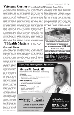
ACG CASE REPORTS JOURNAL
CASE REPORT | PEDIATRICS
Primary Pancreatic Lymphoma Simulating Acute Cholestatic
Hepatitis in a 7-Year-Old Child
Vikrant Sood, MD1, Nitesh Agrawal, MD, PDCC2, Seema Alam, MD1, Dinesh Rawat, MD,
MRCPCH, CCT1, Rajeev Khanna, MD, PDCC1, Kalpana Bansal, MD2, and Chhagan Bihari, MD3
Department of Pediatric Hepatology, Institute of Liver and Biliary Sciences, Vasant Kunj, New Delhi, India
Department of Radiology and Interventional Radiology, Institute of Liver and Biliary Sciences, Vasant Kunj,
New Delhi, India 3
Department of Pathology, Institute of Liver and Biliary Sciences, Vasant Kunj, New Delhi, India
1
2
Abstract
Primary pancreatic lymphoma in children has been described infrequently in literature, and its acute presentation as cholestatic hepatitis is similarly rare. We report a case of a 7-year-old child with primary pancreatic lymphoma presenting as acute infective hepatitis, leading to delay in correct diagnosis and management.
Introduction
Though seen commonly in adults, pancreatic malignancies are an exceedingly rare entity in the pediatric population.1-3 Lymphomatous involvement of pancreas can occur primarily, or, more commonly, as a secondary phenomenon of metastasis.1 Primary pancreatic lymphoma (PPL) in children has been described infrequently in literature,
constituting less than 2% of all extranodal non-Hodgkin’s lymphoma, and its acute presentation as cholestatic
hepatitis is similarly rare.4-9 In a recent 10-year retrospective study of malignant pancreatic tumors in children
involving 2 centers, no cases of PPL were found.2
Case Report
A 7-year-old child was admitted with complaints of yellow discoloration of eyes with dark-colored urine for 20
days. Jaundice was painless and gradually progressive, with development of pale stools and pruritus. It was
preceded by low-grade fever for 1 day with occasional episodes of nausea and non-bilious vomiting. There was
no associated pain in the abdomen, night sweats, weight loss, loss of appetite, bone tenderness, fatigue, skin
or mucosal bleeds, drug intake, abdominal distension, edema, delirium, symptoms of fat-soluble vitamin deficiency, or steatorrhea.
On examination, the child was well-developed with stable hemodynamics. Physical examination revealed pallor, icterus, and bilateral mild corneal xerosis. Abdominal examination was remarkable for a palpable liver 2 cm
below right costal margin in midclavicular line, with a total span of 7.5 cm, soft in consistency, non-tender with
smooth surface, and regular margins. We suspected acute infective hepatitis, which was initially managed conservatively with nutritional supplements.
Laboratory parameters showed evidence of conjugated hyperbilirubinemia and high cholestatic enzymes,
suggestive of obstructive etiology. Abdominal ultrasound showed a well-defined, hypoechoic mass (6.4 x 4.5
cm) with internal vascularity arising from head of pancreas reaching to porta, encasing the common bile duct
ACG Case Rep J 2015;2(3):190-192. doi:10.14309/crj.2015.51. Published online: April 10, 2015.
Correspondence: Vikrant Sood, Department of Pediatric Hepatology, Institute of Liver and Biliary Sciences, D-1, Vasant Kunj, New Delhi – 110070
([email protected]).
Copyright: © 2015 Sood et al. This work is licensed under a Creative Commons Attribution-NonCommercial-NoDerivatives 4.0 International
License. To view a copy of this license, visit http://creativecommons.org/licenses/by-nc-nd/4.0.
190
acgcasereports.gi.org
ACG Case Reports Journal | Volume 2 | Issue 3 | April 2015
PPL Simulating Acute Cholestatic Hepatitis
Sood et al
B
A
C
Figure 1. (A) Abdominal ultrasound showing hypoechoiec ill-defined mass in pancreatic head (short arrows) with encasement of common bile duct (red
arrow) and main pancreatic duct (not shown in figure). Note the echogenic wall (white arrow) of the common bile duct coursing through the lesion, which is
well appreciated in grayscale ultrasound. (B) Axial contrast-enhanced abdominal CT revealing poorly marginated hypodense homogenous hypoenhancing
mass in pancreatic head (short arrows) encasing spleno-portal confluence, entire main portal vein (white arrow), and celiac axis (red arrow). (C) Coronal
contrast-enhanced abdominal CT with minimum intensity projection reformation revealing gross dilatation of the main pancreatic duct (white arrow) and
common bile duct (red arrow) with bilobar intrahepatic duct dilatation.
(CBD) with upstream biliary dilation (Figure 1). Contrastenhanced abdominal computed tomography (CT) revealed
a large homogenously hypoenhancing lobulated mass (4.8
x 5.4 x 5.4 cm isodense to pancreas in plain scan) in the
pancreatic head encasing the common hepatic artery and
main portal vein (Figure 1). The mass was obstructing the
distal CBD and main pancreatic duct (MPD) with associated upstream ductal dilation (CBD 13 mm; MPD 4 mm)
and bilobar gross intrahepatic biliary dilatation, consistent
with double duct sign. Posteriorly, the mass encased the left
renal vein (Figure 1). There was no evidence of calcification/necrosis noted within the mass. Tumor markers showed
elevated alpha-fetoprotein (AFP) of 4.9 ng/mL, CA-125 of
57.5 U/mL, and CA19-9 of 783 U/mL.
Fine-needle aspiration cytology/cell block from the mass
showed atypical large lymphoid cells with enlarged, hyperchromatic nuclei with high nucleus: cytoplasm ratio,
conspicuous nucleoli, and scanty rim of deep basophilic
cytoplasm was suggestive of diffuse large B-cell lymphoma (CD20/CD45 positive and CD3/CD30 negative on immunohistochemistry; Figure 2). Contrast-enhanced chest
A
B
CT, bone marrow aspiration/biopsy, and cerebrospinal fluid
examination done for staging showed abnormal malignant
lymphoid cells in bone marrow, but none in cerebrospinal
fluid, and a prominent thymus in chest. Ann Arbor staging
was IVEA.10 The child was referred to a pediatric oncology
center for management, including chemotherapy.
Discussion
PPL is extremely rare, both in adults and children, with only
limited case reports or small case series available in world
literature; it accounts for <1% of extranodal lymphomas and
0.7% of all pancreatic malignancies.4-9,11 PPL is an extranodal lymphoma arising in the pancreas, with bulk of the tumor
localized to the pancreas.4 Standard diagnostic criteria include: 1) neither superficial lymphadenopathy nor enlargement of mediastinal lymph nodes on chest radiography, 2)
a normal leukocyte count in peripheral blood, 3) main mass
in the pancreas with lymph nodal involvement confined to
peripancreatic region, and 4) no hepatic or splenic involvement.12 Though no pediatric-specific definition is available,
our case fulfilled the standard definition.
C
Figure 2. Cytology smear from pancreatic mass showing (A) Geimsa stain of large malignant lymphoid cells (200x), (B) H&E stain of scattered malignant
lymphoid cells (200x), and (C) CD20 positivity on immunohistochemistry.
191
acgcasereports.gi.org
ACG Case Reports Journal | Volume 2 | Issue 3 | April 2015
PPL Simulating Acute Cholestatic Hepatitis
Sood et al
There are no specific clinical indications, but PPL usually
presents with abdominal pain, abdominal mass, weight loss,
jaundice, or acute pancreatitis.5,9 Presentation as obstructive
jaundice is rare, seen in only about one-third of adult cases,
and is much less common than in adenocarcinoma.5,7 Our
patient presented with features of obstructive jaundice leading to a false first impression of acute cholestatic hepatitis.
Diagnosis by ultrasound, CT, or endoscopic ultrasound is
suggested, but requires mandatory histological confirmation with tissue biopsy. Histologically, most of PPL are B-cell
type, with diffuse large-cell lymphoma as the predominant
histotype.9
Classical imaging findings include well-defined, sometimes bulky and infiltrating, homogeneous low-attenuation
masses relative to the enhancing pancreatic parenchyma,
with only mild enhancement on contrast-enhanced CT and
the presence of large lymph nodal masses.1,3,13 Lesion may
appear as a solitary hypoenhancing lesion, multiple noncontinguous lesions, or as a diffusely enlarged gland.1,3,13
The head of the pancreas is the most common location of
PPL.9 Vascular encasement or invasion, seen as irregularity
and caliber changes, is rarely seen, though vessels may be
stretched because of a mass effect. In our case, pancreatic head mass encased the common hepatic artery (and
its branches), main portal vein, and left renal vein with mild
attenuation, but no evidence of invasion. PPL can be distinguished from more common entities like pancreatoblastoma
and solid-pseudopapillary tumor by the multiplicity of large
nodal masses as the absence of internal heterogeneity, calcifications, necrosis, or vessel invasion.1,3 Double duct sign
is highly suggestive, but not diagnostic, of pancreatic head
malignancy or carcinoma of ampulla of Vater.14 Though ideal
management algorithms are still to be developed, chemoradiotherapy is the modality of choice worldwide, while primary surgery is reserved for treatment failure cases or with
diagnostic uncertainity.9
Disclosures
Author contributions: V. Sood is the article guarantor. V. Sood,
N. Agrawal, D. Rawat, and R. Khanna contributed equally to
assessing the clinical and laboratory information and to drafting the manuscript. V. Sood and N. Agrawal wrote the final
manuscript. S. Alam and K. Bansal edited and revised the
manuscript. C. Bihari assessed the pathology.
Financial disclosure: None to report.
Informed consent was obtained for this case report.
Received: December 28, 2014; Accepted: February 10, 2015
References
1.
2.
3.
4.
5.
6.
7.
8.
9.
10.
11.
12.
13.
14.
Shet NS, Cole BL, Iyer RS. Imaging of pediatric pancreatic neoplasms with radiologic-histopathologic correlation. Am J Roentgenol.
2014;202(6):1337–134.
Rojas Y, Warneke CL, Dhamne CA, et al. Primary malignant pancreatic
neoplasms in children and adolescents: A 20-year experience. J Pediatr
Surg. 2012;47(12):2199–2204.
Chung EM, Travis MD, Conran RM. Pancreatic tumors in children: Radiologic-pathologic correlation. RadioGraphics. 2006;26(4):1211–1238.
Hamilton SR, Lauri LA, eds. Pathology and genetics of tumours of the
digestive system. In: World Health Organization Classification of Tumours. Lyon, France: IARC Press; 2000.
Saif MW. Primary pancreatic lymphomas. JOP. 2006;7(3):262–73.
Arenas García BR. Primary pancreatic lymphoma in pediatric patients
[Article in Spanish]. Radiologia. 2007;49(2):125–127.
Pietsch JB, Shankar S, Ford C, Johnson JE. Obstructive jaundice secondary to lymphoma in childhood. J Pediatr Surg. 2001;36(12):1792–1795.
Eisenhuber E, Schoefl R, Wiesbauer P, Bankier AA. Primary pancreatic
lymphoma presenting as acute pancreatitis in a child. Med Pediatr Oncol. 2001;37(1):53–54.
Yoon WJ, Yoon YB, Kim YJ, et al. Primary pancreatic lymphoma in Korea:
A single center experience. J Korean Med Sci. 2010;25(4):536–540.
Rosenberg SA. Validity of the Ann Arbor staging classification for the
non-Hodgkin’s lymphomas. Cancer Treat Rep. 1977;61:1023-1027
Luo G, Jin C, Fu D, et al. Primary pancreatic lymphoma. Tumori.
2009;95(2):156–159.
Dawson IM, Cornes JS, Morson BC. Primary malignant lymphoid tumours of the intestinal tract. Report of 37 cases with a study of factors
influencing prognosis. Br J Surg. 1961;49:80–9.
Fujinaga Y, Lall C, Patel A, et al. MR features of primary and secondary
malignant lymphoma of the pancreas: A pictorial review. Insights Img.
2013;4(3):321–329.
Ahualli J. The double duct sign. Radiology. 2007;244(1):314–315.
Publish your work in ACG Case Reports Journal
ACG Case Reports Journal is a peer-reviewed, open-access publication that provides GI fellows, private practice clinicians, and other members of the
health care team an opportunity to share interesting case reports with their peers and with leaders in the field. Visit http://acgcasereports.gi.org for
submission guidelines. Submit your manuscript online at http://mc.manuscriptcentral.com/acgcr.
192
acgcasereports.gi.org
ACG Case Reports Journal | Volume 2 | Issue 3 | April 2015


© Copyright 2026