
Guide to the Care of the Hospitalized Patient with Ischemic Stroke
Guide to the Care of the Hospitalized Patient with Ischemic Stroke 2nd Edition, Revised AANN Clinical Practice Guideline Series This publication was supported by an educational grant from Genentech. 4700 W. Lake Avenue Glenview, IL 60025-1485 888/557-2266 International phone 847/375-4733 Fax 847/375-6430 [email protected] • www.AANN.org Content Authors Sue Pugh, MSN RN CNRN CRRN, Subcommittee Chair Claranne Mathiesen, MSN RN CNRN Melissa Meighan, MS RN CNRN Deborah Summers, MSN RN APCNS-BC CNRN CCRN FAHA Patricia Zrelak, PhD CNAA-BC CNRN Content Reviewers Carol Barch, MN CRNP Julia Curran, RN Janice L. Hinkle, PhD RN CNRN Norma McNair, MSN CCRN CNRN AANN Clinical Practice Guideline Series Editor Hilaire J. Thompson, PhD RN CNRN AANN Clinical Practice Guideline Series Editorial Board Patricia Blissitt, PhD RN APRN-BC CCRN CNRN CCNS CCM Matthew Hendell, MSN CNRN CPNP Tess Slazinski, MSN RN APRN CCRN CNRN Patricia Zrelak, PhD RN CNAA-BC CNRN AANN National Office Stacy Sochacki, MS Executive Director Kari L. Lee Managing Editor Sonya L. Jones Senior Graphic Designer Publisher’s Note The authors, editors, and publisher of this document neither represent nor guarantee that the practices described herein will, if followed, ensure safe and effective patient care. The authors, editors, and publisher further assume no liability or responsibility in connection with any information or recommendations contained in this document. These recommendations reflect the American Association of Neuroscience Nurses’ judgment regarding the state of general knowledge and practice in our field as of the date of publication and are subject to change based on the availability of new scientific information. Copyright © 2008, revised December 2009, by the American Association of Neuroscience Nurses. No part of this publication may be reproduced, photocopied, or republished in any form, print or electronic, in whole or in part, without written permission of the American Association of Neuroscience Nurses. Acknowledgment This publication was supported by an educational grant from Genentech. Contents Preface . . . . . . . . . . . . . . . . . . . . . . . . . . . . . . . . . . . . . . . . . . . . . . . . . . . . . . . . . . . . . . . . . . . . . . . . . . . . . . . . . . . . . . . . . . 4 Introduction . . . . . . . . . . . . . . . . . . . . . . . . . . . . . . . . . . . . . . . . . . . . . . . . . . . . . . . . . . . . . . . . . . . . . . . . . . . . . . . . . . . . . 5 Statement of the Problem and Guideline Goal . . . . . . . . . . . . . . . . . . . . . . . . . . . . . . . . . . . . . . . . . . . . . . . . . . . 5 Assessment of Scientific Evidence . . . . . . . . . . . . . . . . . . . . . . . . . . . . . . . . . . . . . . . . . . . . . . . . . . . . . . . . . . . . . 5 Background . . . . . . . . . . . . . . . . . . . . . . . . . . . . . . . . . . . . . . . . . . . . . . . . . . . . . . . . . . . . . . . . . . . . . . . . . . . . . . . . . . . . . . 5 Impact of Stroke . . . . . . . . . . . . . . . . . . . . . . . . . . . . . . . . . . . . . . . . . . . . . . . . . . . . . . . . . . . . . . . . . . . . . . . . . . 5 Stroke Centers of Excellence . . . . . . . . . . . . . . . . . . . . . . . . . . . . . . . . . . . . . . . . . . . . . . . . . . . . . . . . . . . . . . . . . 6 Etiology . . . . . . . . . . . . . . . . . . . . . . . . . . . . . . . . . . . . . . . . . . . . . . . . . . . . . . . . . . . . . . . . . . . . . . . . . . . . . . . . . 6 Supporting Data . . . . . . . . . . . . . . . . . . . . . . . . . . . . . . . . . . . . . . . . . . . . . . . . . . . . . . . . . . . . . . . . . . . . . . . . . . . 6 Methods, Procedures, Interventions, Education . . . . . . . . . . . . . . . . . . . . . . . . . . . . . . . . . . . . . . . . . . . . . . . . . . . . . . . 8 Assessment and Monitoring of a Patient with a Stroke . . . . . . . . . . . . . . . . . . . . . . . . . . . . . . . . . . . . . . . . . . . . 8 Interventions, Troubleshooting, and Patient Problems . . . . . . . . . . . . . . . . . . . . . . . . . . . . . . . . . . . . . . . . . . . . 11 Patient Problems . . . . . . . . . . . . . . . . . . . . . . . . . . . . . . . . . . . . . . . . . . . . . . . . . . . . . . . . . . . . . . . . . . . . . . . . . . . . . . . . 18 Initial Treatment Concerns . . . . . . . . . . . . . . . . . . . . . . . . . . . . . . . . . . . . . . . . . . . . . . . . . . . . . . . . . . . . . . . . . 18 General Supportive Care of a Patient with Stroke . . . . . . . . . . . . . . . . . . . . . . . . . . . . . . . . . . . . . . . . . . . . . . . 21 Patient and Family Education . . . . . . . . . . . . . . . . . . . . . . . . . . . . . . . . . . . . . . . . . . . . . . . . . . . . . . . . . . . . . . . . . . . . . 26 Expected Outcomes . . . . . . . . . . . . . . . . . . . . . . . . . . . . . . . . . . . . . . . . . . . . . . . . . . . . . . . . . . . . . . . . . . . . . . . . . . . . . . 27 Innovative Practices on the Horizon . . . . . . . . . . . . . . . . . . . . . . . . . . . . . . . . . . . . . . . . . . . . . . . . . . . . . . . . . . . . . . . . 27 Intra-Arterial Thrombolysis . . . . . . . . . . . . . . . . . . . . . . . . . . . . . . . . . . . . . . . . . . . . . . . . . . . . . . . . . . . . . . . . 27 Hypothermia . . . . . . . . . . . . . . . . . . . . . . . . . . . . . . . . . . . . . . . . . . . . . . . . . . . . . . . . . . . . . . . . . . . . . . . . . . . . 27 Neuroprotective Agents . . . . . . . . . . . . . . . . . . . . . . . . . . . . . . . . . . . . . . . . . . . . . . . . . . . . . . . . . . . . . . . . . . . . 27 Mechanical Stroke Interventions . . . . . . . . . . . . . . . . . . . . . . . . . . . . . . . . . . . . . . . . . . . . . . . . . . . . . . . . . . . . . 27 Documentation . . . . . . . . . . . . . . . . . . . . . . . . . . . . . . . . . . . . . . . . . . . . . . . . . . . . . . . . . . . . . . . . . . . . . . . . . . . . . . . . . . 28 References . . . . . . . . . . . . . . . . . . . . . . . . . . . . . . . . . . . . . . . . . . . . . . . . . . . . . . . . . . . . . . . . . . . . . . . . . . . . . . . . . . . . . . 29 Bibliography . . . . . . . . . . . . . . . . . . . . . . . . . . . . . . . . . . . . . . . . . . . . . . . . . . . . . . . . . . . . . . . . . . . . . . . . . . . . . . . . . . . . 34 Online Resources . . . . . . . . . . . . . . . . . . . . . . . . . . . . . . . . . . . . . . . . . . . . . . . . . . . . . . . . . . . . . . . . . . . . . . . . . . . . . . . . 35 Appendices . . . . . . . . . . . . . . . . . . . . . . . . . . . . . . . . . . . . . . . . . . . . . . . . . . . . . . . . . . . . . . . . . . . . . . . . . . . . . . . . . . . . . 36 The Barthel Index . . . . . . . . . . . . . . . . . . . . . . . . . . . . . . . . . . . . . . . . . . . . . . . . . . . . . . . . . . . . . . . . . . . . . . . . . 36 The Modified Rankin Scale . . . . . . . . . . . . . . . . . . . . . . . . . . . . . . . . . . . . . . . . . . . . . . . . . . . . . . . . . . . . . . . . . 38 Preface In 1997 the American Association of Neuroscience Nurses (AANN) created a series of guides to patient care called the AANN Reference Series for Clinical Practice to meet its members’ needs for educational tools. AANN changed the name of the series to AANN Clinical Practice Guideline (CPG) Series to better reflect the nature of the guides and the organization’s commitment to develop each guide based on current literature and evidence-based practice. A stroke, or “brain attack,” can be a devastating insult to the brain. Stroke is the leading cause of disability in the United States. Because nurses frequently are the professionals who witness the full impact of stroke and have specific skills that can alter the outcome of a patient’s recovery, it is important that they have a reliable resource to help them through this process. In 1997, as part of the original series of guides, AANN published Recommendations for the Nursing Management of the Hyperacute Ischemic Stroke Patient, and with the advent and availability of thrombolytics this guide was updated in 2004 as Care of the Patient with Ischemic Stroke. Despite the availability of thrombolytics, the care of the ischemic stroke patient remains challenging. Neuroscience nurses are faced with the complex challenge of educating the public about stroke prevention and encouraging individuals to recognize signs and symptoms and seek treatment for stroke when it occurs. Neuroscience nurses are Guide to the Care of the Hospitalized Patient with Ischemic Stroke also challenged to remain current with both basic and clinical research to provide optimal care for stroke patients. This guide is intended to translate the latest research into an easy-to-use reference. Its purpose is to provide nurses with a tool to help them deliver optimum-quality, patient-focused care for hospitalized individuals who have experienced an ischemic, cerebral infarct. Because of the high profile of stroke, new medical, nursing, and rehabilitation treatments are frequently emerging. Resources and recommendations for practice should inform best practices, which in turn enable the neuroscience nurse to provide optimal care of patients hospitalized with ischemic stroke. Accordingly, adherence to this guideline is voluntary. The ultimate determination about its application should be made by the practitioner who takes into consideration the unique circumstances presented by the individual patient. This reference is an essential resource for nurses who provide care to patients hospitalized with ischemic stroke. It is not intended to replace formal instruction but rather to augment the knowledge base of clinicians and provide a readily available reference tool. Nursing and AANN are indebted to the volunteers who have devoted their time and expertise to this valuable resource, which has been created for those who are committed to excellence in stroke-patient care. 4 I. Introduction A.Statement of the Problem and Guideline Goal Clinical management and treatment of cerebrovascular disease is an advancing science. New treatments and strategies for a systematic approach to stroke care have been summarized in several publications that detail formal consensus-based recommendations for both primary and comprehensive stroke-center care delivery (Alberts et al., 2005; Schramm et al., 2005). Recognition of stroke patients whose time to presentation is less than 3–6 hours has prompted an increase in emergency medical system (EMS) referrals to hospitals for hyperacute treatment. An ongoing challenge is to remove the barriers that prevent access to emergency stroke treatment. Important aspects of this process are to improve symptom recognition in both the healthcare and lay communities and to ensure timely transport to a hospital (Adams, 2003; Adams et al., 2007; Graf, Jahnke, & Zadrozny, 2003; Sacco et al., 2006). Optimal care for stroke patients depends on rapid diagnosis and aggressive implementation of evidence-based treatments. The purpose of this document is to help registered nurses, patient-care units, and institutions provide safe and effective care to patients hospitalized with ischemic stroke. The guideline offers a framework for providing excellent stroke care that is based on best evidence. The guideline, therefore, includes a review of available literature and covers issues such as assessing and monitoring patients who have had an ischemic stroke, neurological and physical examinations, laboratory evaluations, radiographic testing, and providing nursing care for patients receiving thrombolytic therapy. Initial treatment concerns for patients, such as managing blood pressure (BP) and assessing swallowing, as well as general strokecare issues such as risk management for deep vein thrombosis (DVT), are also discussed. B.Assessment of Scientific Evidence A review of the literature from January 1997 to February 2008 was conducted using the PubMed/ Medline and CINAHL Information Systems Web sites with the search terms stroke, ischemic stroke, and cerebrovascular disease. Monographs, textbooks, and review articles were also consulted. Studies not directly pertaining to the topic or not written in English were excluded from the review. Data quality is classified as follows for the AANN CPG Series: • Class I: Randomized controlled trial without significant limitations or meta-analysis • Class II: Randomized controlled trial with important limitations (e.g., methodological flaws, inconsistent results); observational study (e.g., cohort, case control) • Class III: Qualitative study, case study, or series Guide to the Care of the Hospitalized Patient with Ischemic Stroke • Class IV: Evidence from reports of expert committees and/or expert opinion of the guideline panel, standards of care, and clinical protocols that have been identified This CPG and recommendations for practice were based upon evaluation of the available evidence (American Association of Neuroscience Nurses [AANN], 2006 [adapted from Guyatt & Rennie, 2002; Melnyk, 2004]): • Level 1: Recommendations are supported by class I evidence. • Level 2: Recommendations are supported by class II evidence. • Level 3: Recommendations are supported by class III and class IV evidence. II. Background A.Impact of Stroke Approximately 780,000 cases of stroke occur in the United States each year (American Heart Association [AHA], 2008), making it the third leading cause of mortality in the United States. About 600,000 of these strokes are first events and 180,000 are recurrences. Stroke is also called a “brain attack” to emphasize the fact that it is an emergent condition. In the United States a stroke occurs, on average, approximately every 40 seconds, and someone dies of a stroke every 3–4 minutes (AHA, 2008). Stroke is the leading cause of serious, long-term disability in the United States and worldwide (AHA, 2008). The estimated cost of indirect and direct care for patients with stroke in the United States is $65.5 billion (AHA, 2008). According to the American Stroke Association (ASA) and the National Stroke Association (NSA), stroke accounts for about half of all hospitalizations for acute neurological disease. To reduce the number of brain attacks, a coordinated public-education effort, an integrated emergency response system, and a multidisciplinary-treatment-team approach are needed. Because “time is brain,” nursing professionals must be knowledgeable about new stroke-care standards to be able to manage care for these patients quickly and appropriately. In addition to those admitted for a chief complaint of stroke, 6.5%–15% of all strokes occur in hospitalized patients (Blacker, 2003). Most are related to perioperative or related high-risk cardiac procedures, and reasons for in-hospital stroke include iatrogenic surgery, line placement, embolism, hypoperfusion states (e.g., hypotension, dehydration, medication adjustments), and hematological etiologies such as procoagulant states (Blacker). Greater awareness of the need for assessment and timely intervention can reduce delays in stroke treatment for a hospitalized patient. Many facilities have a stroke team or rely on the medical emergency team to promptly evaluate stroke-like symptoms 5 and make referrals to neurological specialists. It is important to consider developing a response system for in-house events to help nursing staff ensure prompt consideration for thrombolytic and endovascular interventions. Nurses play a crucial role in communicating information about health risks and disease. Targeting individuals in the community who are the least aware of their risk and providing them with the opportunity to engage in risk-modifying activities and disease recognition also serve as ways to improve stroke care. In addition, nurses play a critical part in reducing the disability that may result from a stroke through their prompt recognition of stroke signs and symptoms, initiation of appropriate response efforts, and provision of interventions and education for preventing subsequent stroke. B.Stroke Centers of Excellence In 2000, the Brain Attack Coalition (BAC)—a group of professional, voluntary, and government entities dedicated to reducing the occurrence, disabilities, and deaths associated with stroke— along with the American Heart Association and American Stroke Association (AHA/ASA) pub lished its recommendations for primary stroke cen ter (PSC) designation (Alberts et al., 2000). Based on these recommendations, a formal PSC certification program was launched in 2003 as part of The Joint Commission Advanced Disease Specific Certification process. In addition to demonstrating use of a standardized method of delivering care based on the BAC Standards, certified PSCs must demonstrate that they (1) support a patient’s self-management activities, (2) tailor treatment and intervention to individual needs, (3) analyze and use standardized performance measure data to continually improve treatment plans, and (4) demonstrate their application of and compliance with the clinical practice guidelines published by the AHA/ASA or equivalent evidence-based guidelines. Effective January 1, 2010, certified PSCs must collect and report on eight National Inpatient Hospital Quality Measures for stroke. The measures include venous thromboembolism (VTE) prophylaxis, discharged on antithrombotic therapy, anticoagulation therapy for atrial fibrillation/flutter, thrombolytic therapy, antithrombotic therapy by end of hospital day two, discharged on statin medication stroke education, and assessed for rehabilitation. These measures have been endorsed by the National Quality Forum and approved as a core measure set for use in the Joint Commission’s ORYX program after October 1, 2009. As of 2010, PSCs are no longer required to report on dysphagia screening and antismoking cessation counseling. More information about PSC designationcan be found on The Joint Guide to the Care of the Hospitalized Patient with Ischemic Stroke Commission’s Web site at www.jointcommission. org/CertificationPrograms/PrimaryStrokeCenters. Some states have or are developing certification and regulations for Stroke Centers. Beyond PSC certification, there are efforts to credential facilities as comprehensive stroke centers (CSC), based on the BAC guidelines published in 2005 (Alberts et al., 2005), are under way. Credentialed CSC facilities or systems will have necessary personnel, infrastructure, expertise, and programs to diagnose and treat stroke patients who require a high degree of medical and surgical care, specialized tests, or interventional therapies. Both at the state and local level, government entities and hospital systems of care are looking at how they can better provide care to patients with stroke that includes an increasing reliance and emphasis on tele-stroke medicine (Lee, et al, 2009). C.Etiology Stroke is an acute vascular event that affects the brain. It involves neurological changes caused by an acute interruption of blood supply to a part of the brain. There are two main types of stroke. The first type is ischemic stroke, which results from decreased blood flow to a portion of the brain with consequent cell death. The second type is hemorrhagic stroke, which results from bleeding within the brain. 1. Ischemic stroke Ischemic stroke accounts for 80%–85% of all strokes and occurs when the blood supply to the brain is reduced or blocked; ischemic stroke is primarily a result of occlusive disease (Adams et al., 2003). Ischemic stroke is further classified into large-vessel thrombotic strokes, small-vessel thrombotic strokes, atheroembolic strokes (which originate from large arteries and move to distal branches), and cardioembolic strokes. The types of ischemic stroke and their frequency of occurrence are as follows (Adams et al., 1993): • 20% atherosclerotic cerebrovascular disease from hypoperfusion or arteriogenic emboli • 25% penetrating artery or proliferative disease (e.g., lacunes, subcortical stroke) • 20% cardiogenic embolism from atrial fibrillation, valve disease, ventricular thrombi, and other cardiac disorders • 30% cryptogenic stroke (i.e., stroke with unknown etiology) • 5% other, unusual causes such as prothrombic states, dissections, arteritis, migraines/ vasospasm, drug abuse, and others 2.Hemorrhagic stroke Hemorrhagic vascular disease accounts for 15%– 20% of strokes and occurs when a blood vessel ruptures (Adams et al., 2003). Hemorrhagic 6 stroke is caused mainly by hypertension, leading to bleeding in the deep structures of the brain such as the basal ganglia. Less commonly seen is subarachnoid hemorrhage (SAH) or arteriovenous malformations (AVMs). Subarachnoid hemorrhage occurs primarily as a result of the rupture of saccular aneurysms that form at branching points of the intracranial arteries at the circle of Willis (Alexander, Gallek, Presciutti, & Zrelak, 2007; Brisman, Song, & Newell, 2006). Refer to the AANN CPG Care of the Patient with Aneurysmal Subarachnoid Hemorrhage. D.Supporting Data 1.Stroke signs and symptoms Stroke symptoms usually occur acutely but may evolve progressively over hours or days and most often unilaterally. Strokes that present with maximal acute symptoms that then start to resolve are likely a result of embolus whereas strokes that progress over time are more likely to be the result of an artery thrombosis. Symptoms that resolve within 24 hours (usually within 60 minutes) and cause no permanent damage are known as transient ischemic attacks (TIAs). Stroke symptoms vary among patients and are related to the region of the brain and vessel involved (see section C.3). The inability of patients and bystanders to recognize stroke symptoms and quickly contact the EMS is the largest barrier to effective acute-stroke therapy (Barsan et al., 1994; Evenson, Rosamond, & Morris, 2001; Feldmann et al., 1993; Kothari et al., 1999; Morris, Rosamond, Madden, Schultz, & Hamilton, 2000; Rosamond, Gorton, Hinn, Hohenhaus, & Morris, 1998; Schroeder, Rosamond, Morris, Evenson, & Hinn, 2000; Williams, Bruno, Rouch, & Marriot, 1997). The clinical manifestations of an ischemic stroke, as established by several national organizations (i.e., ASA, 2002; National Institute of Neurological Disorders and Stroke, 2002; NSA, 2002), are as follows: • sudden numbness or weakness in the face, arm, or leg, especially on one side of the body • sudden confusion or trouble speaking or understanding • sudden trouble seeing in one or both eyes • sudden trouble walking, dizziness, or loss of balance or coordination • sudden severe headache with no known cause. This list of symptoms is often referred to as “the suddens.” Strokes are often overlooked by emergency and other healthcare providers. Some facilities and EMS providers have developed stroke triage or screening examinations based on FAST (i.e., Face, Arm, Speech, Time), such as the Guide to the Care of the Hospitalized Patient with Ischemic Stroke Los Angeles Motor Scale (LAMS)/Los Angeles Prehospital Stroke Screen (LAPSS) or the modified National Institutes of Health Stroke Scale (NIHSS; Level 2). FAST is based on the Cincinnati Prehospital Stroke Scale. It has not been determined whether the FAST message is easier for the public to recall than the “suddens” message (Kleindorfer et al., 2007). FAST captures 88.9% of patient with stroke or TIA (Kleindorfer et al., 2007). 2.Pathophysiology of stroke Ischemia occurs when the blood supply to a part of the brain is interrupted or completely occluded (Jones et al., 1981). Cerebral vessel occlusion produces ischemia in the brain tissue and edema in the surrounding tissue. Ischemia alters cerebral metabolism, and brain-cell survival depends on how long the brain is deprived of oxygen and metabolites. Cells in the center, or core, of the infarcted tissue die almost immediately after stroke onset; this area is often referred to as the site of primary neuronal injury. A zone of hypoperfused tissue exists around the core. This zone is referred to as the ischemic penumbra; it often can be salvaged during acute intervention (Jones et al.). A cascade of biochemical processes develops within minutes of the cerebral ischemic event. Release of neurotoxins such as oxygen free radicals, nitric oxide, and excitatory amino acids often occurs, prompting the development of local acidosis and membrane depolarization with an influx of sodium and calcium (Shi & Liu, 2007). This influx results in early cytotoxic edema and cell death followed by vasogenic edema (Rosand & Schwamm, 2001). Cerebral vessels that augment blood flow to the major circulatory vessels of the brain are termed collaterals. Differences in the number and size of these vessels explain the variations in severity of manifestations observed in patients. The extent of infarction depends on the size and location of the occluded artery and on the adequacy of collateral circulation to the area. 3.Clinical manifestations of stroke The symptoms that occur with stroke vary according to the location and extent of the occlusion. The more anterior the ischemic lesion, the more likely it is to produce symptoms associated with motor or speech functions; in more posterior lesions, the impairment is likely to be sensory and involve the visual field. The middle cerebral artery syndrome is the most common of all cerebral occlusions. Symptoms of the specific occlusion syndromes are described below (Hinkle, Guanci, Bowman, Hermann, & McGinty, 2004; TestaniDufour & Marano-Morrison, 1997): 7 a. Anterior circulation (1) Internal carotid artery symptoms include the following: • paralysis of the contralateral face, arm, and leg • sensory deficits of the contralateral face, arm, and leg • aphasia, if the dominant hemisphere is involved • hemianopia, ipsilateral episodes of visual blurring, or amaurosis fugax (i.e., temporary blindness in one eye) • carotid bruit. (2) Anterior cerebral artery symptoms include the following: • contralateral hemiparesis or hemiplegia of the foot and leg (Foot drop is a common finding.) • sensory loss in the toes, foot, and leg • mental status impairment including confusion, amnesia, perseveration, and personality changes such as apathy or flat effect • abulia (i.e., inability to make decisions or perform voluntary acts). (3) Middle cerebral artery symptoms include the following: • contralateral hemiparesis or hemiplegia of the face and arm (The leg is spared or has fewer deficits than the arm.) • contralateral hemisensory in the same area • contralateral vision loss (hemianopia; left hemisphere has right-visual field cuts and right hemisphere has leftvisual field cuts) • in left hemisphere, aphasia and difficulty reading, writing, and calculating are more likely • in right hemisphere, neglect of leftvisual spaces, extinction of left-sided stimuli, and spatial disorientation are more likely. b. Posterior circulation (1)Vertebral-basilar system symptoms include the following: • hemiplegia/hemiparesis or quadriplegia/quadriparesis • ipsilateral numbness and weakness of face • dysarthria and dysphagia • vertigo, nausea, and dizziness • ataxic gait and clumsiness • diplopia, homonymous hemianopia, nystagmus, conjugate gaze paralysis, and ophthalmoplegia • akinetic mutism (i.e., locked-in Guide to the Care of the Hospitalized Patient with Ischemic Stroke syndrome when the basilar artery is occluded). (2)Posterior cerebral artery symptoms include the following: • homonymous hemianopia, cortical blindness, lack of depth perception, peripheral visual-field loss, visual hallucinations • memory deficits • perseveration and dyslexia • thalamic or subthalamic involvement resulting in diffuse sensory loss, mild hemiparesis, and intentional tremor • brainstem involvement resulting in pupillary dysfunction, nystagmus, and loss of conjugate gaze. (3)Posterior-inferior cerebellar artery (referred to as Wallenberg syndrome) symptoms include the following: • dysarthria, dysphagia, and dysphonia • nystagmus • cerebellar signs, including unsteady gait, vertigo, and ataxia • ipsilateral Horner syndrome • nausea and vomiting • contralateral loss of pain and temperature sensation, loss of balance on affected side, and loss of pain and temperature sensation on the ipsilateral face. c. Brain stem/cerebellum Symptoms include the following: • motor or sensory loss in all four limbs; crossed signs • limb or gait ataxia • dysarthria • dysconjugate gaze, nystagmus, bilateral visual-field defects. d. Lacunar syndromes Symptoms include the following: • pure motor—involves only musclestrength deficits on one side • pure sensory—involves touch, pain, vibration, or heat or cold deficits on one side • clumsy hand dysarthria—involves garbled speech and clumsiness on one side • ataxic hemiparesis—involves motor incoordination on one side. Physicians and advanced practice nurses must rule out other disorders that can mimic a stroke, such as brain tumors, complicated migraine headache with visual transient neurological deficits, and metabolic abnormalities, especially hyperglycemia or hypoglycemia; subdural hematoma or other craniocerebral trauma; infections such as brain abscess, encephalitis, or other infection (even a urinary tract infection 8 [UTI] can mimic stroke in elderly patients); seizure with postictal paralysis; or brain tumor. Isolated vertigo or dizziness seldom indicate a TIA or stroke symptom but may be a result of Ménière disease. It is important to evaluate for associated vascular disorders and perform further diagnostic testing after completing the emergent evaluation and treating the stroke. III.Methods, Procedures, Interventions, Education A.Assessment and Monitoring of a Patient with a Stroke 1.Overview Neurological assessment of a patient with a stroke is critical for preventing reinjury of brain tissue or preserving viable tissue. Between 5% and 10% of stroke patients develop enough cerebral edema to cause obtundation or brain herniation. Early edema is cytotoxic in nature and is followed by vasogenic edema, which peaks on the second or third day but can cause mass effect for approximately 10 days (Rosand & Schwamm, 2001). Of all of the neurophysiologic monitoring modalities, the neurological examination of a conscious patient allows for the most comprehensive assessment of central nervous system function (Layton, Gabrielli, & Friedman, 2004). There are several adjuncts to the neurological assessment that range from highly invasive techniques, such as intraparanchymal oxygen monitoring and microdialysis monitoring of cerebral metabolism, to less invasive techniques, such as continuous electroencephalographic (EEG) monitoring. These technologies, however, are not used for the majority of ischemic strokes. Slowing of EEG activity reflects decreased oxygen activity. Interventions based on recognition of this activity may be performed in the same fashion as interventions made after recognition of electrocardiographic changes and may provide early warning of ischemia in thrombotic stroke (Layton et al., 2004). For patients with subarachnoid hemorrhage, monitoring brain tissue oxygenation has been found to provide an additional independent parameter for detecting hypoxic events (Väth, Kunze, Roosen, & Meixensberger, 2002). Microdialysis monitoring of cerebral metabolism may predict the occurrence of a neurological deficit (SkjothRasmussen, Schulz, Kristensen, & Bjerre, 2004), and early EEG monitoring also may add value to the clinical examination (Jordan, 2004). However, all of these technologies are adjuncts and are not replacements for a valid examination by an experienced clinician (Jordan), and the diagnosis remains related to early treatment of any deficit Guide to the Care of the Hospitalized Patient with Ischemic Stroke (Skjoth-Rasmussen et al.). The goal of monitoring is the same as that for treatment: the reduction of death and disability (Indredavik, 2004). Early mobilization is critical for the stroke patient, so any invasive adjunctive monitoring should be balanced with those goals. High-tech monitoring devices can direct much-needed attention away from early rehabilitation efforts (Indredavik). Therefore, the clinical bedside examination, for a variety of reasons, remains the gold standard of neurological assessment (Level 3; Jordan; Layton et al.). 2. History and physical examination Key areas for assessment during the patient’s history include onset of symptoms, recent events (e.g., stroke, myocardial infarction [MI], trauma, surgery, bleeding), comorbid illnesses (e.g., hypertension, diabetes, atrial fibrillation, seizures), use of medications (e.g., anticoagulants, insulin, antihypertensives), and substance-abuse history (Level 3; Adams et al., 2007). a. Neurological assessment The neurological assessment begins the moment the nurse enters the patient’s room. A great deal of neurological assessment can be done unobtrusively by a nurse who is engaged with the patient. (1) It is important to remember that a neurological emergency can occur rapidly, so every contact with a patient with a diagnosis of ischemic stroke, whether as an official examination or not, should consider the patient’s neurological status. The patient’s level of alertness may vary during the day; however, lethargy may be one of the first signs that the patient’s neurological status is declining (Hobdell et al., 2004). (2)Another important overriding feature of neurological assessment of patients with ischemic stroke is the evaluation of the cranial nerves and examination of bilateral motor ability. The patient should be assessed for visual, tactile and sensory, and motor extinction. (3) Depending on the setting, the neurological examination may be performed once every hour in the intensive care setting, once every 2 hours in a transitional care unit, and once every 4 hours in a neuroscience unit (Level 3; Hickey, 2003) during the acute phase. Individual hospital units may have differing policies. Patients with hyperacute stroke that are potential candidates for thrombolytic therapy should be assessed at minimum every 15 minutes prior to thrombolytic therapy and then for 2 hours after initiation of therapy, followed by 30 minutes 9 for 6 hours, and then every hour for the first 24 hours (Level 3; Adams et al, 2007). b. Neurological examination The neurological examination begins with assessment of the patient’s level of consciousness (LOC). Is the patient alert? Does the patient track the speaker with his or her eyes? Does the patient know his or her name, where he or she is, the date, and, if possible, why he or she is in the hospital? (1) First, the nurse assesses the patient for the least amount of stimulus that is required to arouse him or her. (2) As part of general good practice, the patient should be spoken to before he or she is touched. (3) The nurse should then assess the patient’s level of consciousness, basic motor function, and cranial nerves. (4) The patient should not be asked yes or no questions. The patient may be given a choice of answers, preferably with the wrong answer first. In scoring the NIHSS or the modified NIHSS, the first response the patient gives is scored. (5) Note both the speech and language. Is the speech clear or slurred? Does it make sense? There are three elements of language: fluency (use naming and conversation), comprehension (commands), and repetition. (6) The patient may be assessed for alertness and orientation even if he or she is intubated. (7) To assess cranial nerves II through XII (a) Check the pupils for reactivity. (b) Assess eye movements by having the patient follow an “H” configuration to determine if he or she can move the eyes from side to side and up and down (Hobdell et al., 2004). (c) Perform a gross examination of the visual fields by facing the patient directly and having him or her look at the examiner’s nose. Using the examiner’s own peripheral vision, the examiner holds up one or two fingers in the lower and upper quadrants of the patient’s left and right sides. The patient must look at the examiner’s nose and not directly at the examiner’s fingers. Test each eye separately and in all four quadrants. The patient must look at the examiner’s nose and not directly at the examiner’s fingers. Besides evaluating for field deficits, Guide to the Care of the Hospitalized Patient with Ischemic Stroke note if the there are signs of neglect (inability to detect double stimulation). (d) Assess the patient’s nasal fold for facial drooping at rest and while asking the patient to show his or her teeth or to smile. (e) Ask the patient to stick out the tongue and move it from side to side. Then have the patient press his or her head gently against the nurse’s hand to assess for sternomastoid muscle strength. The patient also may shrug the shoulders against resistance. (8) During motor assessment, the patient may be asked to squeeze and release the examiner’s hand with the nonparetic limb. The patient’s motor function should then be assessed by determining whether drift is present in the upper limbs when they are extended for 10 seconds (Adams et al., 2003). If the patient is in fairly good neurological condition, the examiner should ask him or her to close the eyes and hold out the hands, as if holding up a pizza, so that upper-extremity strength can be assessed. If the patient’s arm slowly drifts down, it is a sign of motor weakness and may be the first subtle sign that the patient is deteriorating neurologically. The patient may then be asked to press the arm against the examiner’s hand to be rated for strength on a 0–5 scale, with 5 being movement against gravity and resistance and 0 being no movement at all. The lower extremities may be assessed by having the patient hold each leg up off the bed for 5 seconds and assess for drift. Then have the patient press on the examiner’s foot (“press on the gas ) and then plantar flex against resistance. The best assessment of motor function is walking (Blumenfeld, 2002). However, this may not always be possible. (9) If the patient is comatose, movement may be assessed first by his or her response to central pain. The assessment is done by squeezing the shoulder muscles, not by rubbing the sternum because the patient may withdraw from central pain. The next question is whether the patient is able to localize pain, or move away from a painful stimulus in a particular area. This can be assessed by gently squeezing the earlobe. (10) If the patient is experiencing ataxia, cerebellum function can be tested by having the patient touch the nose. The examiner should then hold out a finger approximately arm’s length from the patient at varying 10 places. The patient’s ability to move the arm from one place to another in a controlled manner should be observed. (11) A patient’s sensory functioning may be assessed by using a clean testing pin while asking the patient to distinguish between the sharp and dull sides of the pin and asking whether those are “the same or different” on the arms and legs. An alert patient may also be asked if he or she is experiencing any numbness or tingling. A short but thorough neurological assessment of the stroke patient by an experienced practitioner may take approximately 5 minutes. 3.Assessment tools Several reliable and well-validated scoring systems for initial and serial assessment have been developed for the stroke patient and include the following: a. A widely used tool is the NIHSS (Level 2; Adams et al., 2003; Goldstein, Bertels, & Davis, 1989; Goldstein & Samsa, 1997). An online training and certification program is available for healthcare professionals to learn or review how to administer the NIHSS for acute stroke assessment at www.strokeassociation.org/presenter.jhtml?identifier=3023009. Continuous EEG monitoring has been found to correlate with the NIHSS (Jordan, 2004). The NIHSS is a quick and standardized measure of neurological function and stroke severity that ranges from 0 (no deficits) to 42 (severe deficits) (Level 2; Goldstein & Samsa, 1997). The NIHSS is a scored assessment tool that identifies neurological deficits including LOC, LOC questions, LOC commands, horizontal-gaze abnormality, visual-field cuts, facial weakness, motor weakness in arms and legs, limb ataxia, sensory loss, aphasia, dysarthria, and extinction or inattention. This tool provides a systematic assessment of the neurological deficits related to stroke and can be used as a measure of patient outcomes. Studies have shown that an increase or decrease of 4 points in the stroke score indicates important changes (Goldstein, 1994). A copy of the NIHSS can be retrieved at www.ninds.nih.gov/doctors/ NIH_Stroke_Scale.pdf. b. The Barthel Index measures functional capacity, takes 5–10 minutes to administer, and is highly reliable and valid. Nurses may administer this test to determine disability following a stroke (www.neuro.mcg.edu/ mcgstrok/Indices/Barthel_Ind.htm). (See Appendix A.) c. The modified Rankin scale (see Appendix B) Guide to the Care of the Hospitalized Patient with Ischemic Stroke is the most commonly used endpoint for clinical trials involving stroke. B.Interventions, Troubleshooting, and Patient Problems 1.Emergent evaluation Time is the most crucial factor for the optimal treatment of an individual who presents with clinical manifestations of a brain attack. The AHA and NSA offer consensus statements that help guide the initial care of a patient with an acute stroke (Adams et al., 2007). The recommendations of care are based on recent research indicating that newly discovered therapeutics must be instituted within the first 3–6 hours to have a positive effect on patient outcomes. Optimal response times for the management of stroke are highlighted in Figure 1. Figure 1. Optimal Response Times for Management of a Patient with Acute Stroke Time Goal <10 minutes Initial patient evaluation performed <15 minutes Stroke team notified <25 minutes CT scan initiated <45 minutes CT scan evaluated <60 minutes Door-to-drug (needle) ensured <3 hours Patient transferred to inpatient setting Note. CT = computed tomography. All times are based on the time of arrival of the patient to the emergency department. Information from National Institute of Neurological Disorders and Stroke (Ed.), December 12–13, 1996, Executive summary: Proceedings of a national symposium on rapid identification and treatment of acute stroke. Retrieved January 15, 2008, from www.ninds.nih.gov/news_and_events/proceedings/ stroke_proceedings/execsum.htm. 2.Prehospital care Individuals within the community must be educated about the clinical signs of brain attack and contacting the EMS (i.e., calling 911 or other applicable telephone number). Two of the five components of the education performance measures for stroke-center certification are recognizing the signs of stroke and calling 911. EMS personnel must be educated to rapidly recognize stroke signs and emergently evaluate the ABCs (i.e., Airway, Breathing, and Circulation). It is also important to establish the time of onset (Level 2; Adams, et. al. 2009). Onset should be determined based on the last time the patient was known to be well or at “baseline.” “Upon awakening” reflects that the time of onset was when the patient went to bed or if he or she had been seen up and well during the night. Time of awakening is not time of onset if symptoms are present upon awakening. If the patient is unresponsive or cannot speak because of aphasia, family members or 11 other observers must be questioned. The treatment generally consists of obtaining vital signs, monitoring cardiac rhythm, establishing intravenous (IV) access, administering supplemental oxygen based upon saturation levels, and notifying the nearest emergency facility of the expected arrival of a possible brain-attack patient (Level 2; Adams et al., 2007). Based on the EMS report, the computed tomography (CT) scanner should be cleared and appropriate staff notified so that they are ready when the patient arrives. If fluids are initiated, normal saline (or other nondextrose normotonic fluid) is recommended (Level 3) is the fluid of choice. 3.Hospital care emergent evaluation After the patient arrives in the emergency department (ED), personnel must systematically perform necessary evaluations and diagnostic testing. Patients should be seen by a physician within 10 minutes of arrival, and a stroke team member should be available, at least by telephone, within 15 minutes (Level 2; Alberts et al., 2005). ED personnel initially should evaluate and stabilize the ABCs while taking a brief, comprehensive history. In the initial acute management of the stroke, the goals are to control vital signs, confirm that the event is a stroke, determine the etiology, prevent decompensation and medical complications, and begin appropriate treatment (AHA, 2005). After the brief history and physical examination are completed and as soon as the ABCs are stabilized, an emergent CT scan of the brain must be taken. The BAC recommends that the CT be performed within 25 minutes of arrival to the ED. Results should be available within 20 minutes of completion or within 45 minutes of arrival. The patient’s symptoms, neurological examination, and medical examination should help determine the mechanism of stroke and compromised vascular territory. a. Acute management of acute ischemic stroke Acute management of an acute ischemic stroke includes the following steps (Adams et al., 2007): (1) Monitor airway and ensure that airway equipment is available. (2) The majority of patients with acute ischemic stroke do not require intubation; however, the risk for respiratory compromise is increased with large infarctions or infarctions that involve the brainstem. Monitor for signs of respiratory compromise and anticipate that the patient’s respiratory needs may require intubation. Emergent intubation may be necessary before the stroke outcome is known. Guide to the Care of the Hospitalized Patient with Ischemic Stroke Advise the family and help them make decisions about duration of intubation after the outcome is apparent. (3) Titrate oxygen to maintain oxygen saturation greater than 92% by using pulse oximetry. Respiratory failure can occur when there is brainstem involvement or increased intracranial pressure (ICP). Assessment of the patient’s respiratory status includes monitoring respiratory rate, lung auscultation, and continuous oxygen (O2) saturation. Supplemental oxygen of 2–4 L should be used if the patient is unable to maintain an O2 saturation greater than 92%. Arterial blood gases (ABGs) are indicated if the patient is unable to maintain an O2 saturation of 92%. (4) Establish IV access. Two sites should be maintained for patients who are candidates for Alteplase (recombinant tissue plasminogen activator or rt-PA) infusion (Level 3; Summer D, Leonard A, Wentworth D, Saver J.L, et al, 2009). (5) Perform STAT blood work. A complete blood count and platelet count, prothrombin time (PT)/international normalized ratio (INR) (especially if the patient is on Coumadin and/or to screen for a underlying hematologic [i.e., coagulation] disorder), blood glucose levels, and serum chemistries should be done. A cervical spine X ray is indicated if the patient is unresponsive and the possibility of trauma exists. An electrocardiogram (ECG) should be obtained and, if hypoxia or acute lung disease is suspected, a chest X ray should be taken. (6) Monitor the patient’s vital signs, neurological deficits, oxygen saturation, and cardiac rhythm frequently. The patient’s cardiac rhythm must be evaluated and treated if abnormalities are found. Cardiac arrhythmias can lead to decreased cardiac output and decreased cerebral perfusion pressure (CPP). A cardioembolic stroke should be suspected, and further investigation is needed if the patient has atrial fibrillation and is not therapeutically anticoagulated or not receiving anticoagulation therapy. An ECG should be performed in the ED to rule out myocardial ischemia. Myocardial infarction (MI) is the third leading cause of death for patients with acute stroke, and it is the leading cause of death 30 days after the occurrence of a TIA. ECG changes that mimic myocardial ischemia, such as peaked T waves, are not uncommon in SAH 12 and reflect an anterior circulation hemorrhage. Patients with acute stroke should be monitored with telemetry during the first 24 hours of care to detect potentially life-threatening arrhythmias. (7) Position the patient with the head midline. Keeping the bed flat may help improve cerebral perfusion while elevating the HOB 25°–30° can decrease intracranial pressure and prevent aspiration (Level 3; Summers, Leonard, Wentworth, Saver, et al, 2009). If the patient must lie flat, turn the patient on his or her side to minimize aspiration of secretions (Hickey, 2003). Stroke patients can have brainstem lesions that may lead to difficulty swallowing and controlling secretions, including respiratory failure. In such a situation, intubation should be considered to prevent the risk of aspiration, which can lead to further complications such as pneumonia and atelectasis. (8) Perform an emergent CT scan to determine whether the patient is a candidate for thrombolytics or other acute interventions. A CT scan without contrast of the brain rapidly excludes hemorrhagic strokes and other causes of neurological dysfunction. If subarachnoid hemorrhage is suspected but no blood or mass effect is seen on CT, cerebrospinal fluid examination should be considered. See the AANN Clinical Practice Guideline, Care of the Patient with Aneurysmal Subarachnoid Hemorrhage. CT angiograms are being used more frequently in acute stroke, especially if acute interventional treatment is under consideration (Latchaw, R.E., Alberts, M.J., Lev, M.H., Connors, J.J., & Harbaugh, R.E., et. al., 2009). (9) Monitor BP closely. It is common to see elevated blood pressure during acute stroke. Intervention may not be needed until BP is > 220/110 (See pp. 16–17 for BP management prior to rt-PA administration and for postrt-PA treatment management for treatment parameters). (10) Treat glucose levels higher than 150 mg/ dl because elevated glucose levels worsen outcomes (target glucose is 70–120 mg/dl according to National Institute of Neurological Diseases and Stroke [NINDS] guidelines). (11) Treat temperatures higher than 99.6°F because increased temperature worsens outcomes (Level 3). (12) Although very rare (i.e., incidence of 2%), anaphylactic reactions to rt-PA have been reported, so nurses must be prepared to intervene appropriately if symptoms occur. Guide to the Care of the Hospitalized Patient with Ischemic Stroke (13) Brain attack is a potentially life-threatening event that can have a major effect on the patient and family; it can challenge their beliefs and alter the patient’s definition of self. Education and support should start in the ED; a chaplain or minister may be provided if requested, patient and family privacy should be managed, and the patient’s modesty should be respected and maintained. In addition, the patient should be given adequate pain relief. Maintenance of the family unit is a desired outcome. (14) Patients who present with TIAs are at high risk for experiencing a stroke, especially during the week immediately following the TIA. These patients should have an acute ischemic workup and their risk factors should be addressed to minimize their risk of stroke. Symptoms that wax and wane or are relatively nondescript most likely are not secondary to a TIA (Johnston S.C., Gress D.R., Browner W.S., & Sidney S, 2000; Rothwell P.M. & Warlow C.P., 2005). (15) Although seizures are relatively uncommon (accounting for approximately 5% of strokes), they can intensify the brain injury caused by stroke and must be detected and treated in a timely manner. However, for most patients, pharmacological prophylaxis is not indicated (Level 2). b. Neurological and physical examination Using the NIHSS, the examiner can perform a neurological examination that can be referenced by all departments of the hospital (see section III.A.3.a). If the patient has a change in LOC, the examination should include assessment of pupil size and reactivity. Muscle-tone and reflex testing, along with additional cognition testing, can be done after the CT is completed. If the patient has a decreased LOC, the Glasgow Coma Scale (GCS) can be used to evaluate him or her. Although the GCS score is widely used, it was originally developed to reflect traumatic injury and does not necessarily effectively represent the localized nature of stroke (Weir, Bradford, & Lees, 2003). A copy of the GCS can be retrieved at www.ssgfx.com/ CP2020/medtech/glossary/glasgow.htm. A complete physical examination should include auscultation of the heart, lungs, and carotid arteries to evaluate for murmurs, crackles, and bruits. Carotid and peripheral pulses should be palpated to evaluate circulation. The head and neck should be inspected for signs of trauma or nuchal rigidity. 13 Nuchal rigidity, ocular hemorrhage, coma, and papilledema may be indicative of SAH, infections, tumors, or metabolic abnormalities (Hobdell et al., 2004). c. Laboratory evaluation STAT blood work should be completed within 30 minutes of arrival to the ED and include complete blood cell count with differential, platelet count, PT, partial thromboplastin time (PTT), INR, electrolytes, creatinine, blood urea nitrogen, and blood glucose (Level 3; Goldstein, 2007). A pregnancy test may be indicated with the STAT labs for females of childbearing age. The patient’s lipid profile (fasting) should be assessed within the first 24 hours but is not necessary during the emergent workup. Routine full chemistry, urinalysis, and cardiac enzymes should be done only if indicated. If the cause of the stroke cannot be established based on initial laboratory and radiographic evaluation, optional laboratory tests can be performed to screen for unusual causes of stroke. d. Radiographic evaluation (1) A CT without contrast to rule out hemorrhage should be performed immediately after the patient has been stabilized (Adams et al., 2007). All subsequent therapy and medical management depend on the results of the CT scan. An ischemic stroke or edema generally does not show up on the CT scan for 12–24 hours unless the patient has had a large infarction. Intracerebral hemorrhage greater than 1 cm can immediately be identified in the parenchyma. CT scans diagnose more than 95% of SAHs (Morgenstern et al., 1998), suggesting an aneurysm when blood is visualized in the subarachnoid space. Repeat CT scans are rarely needed during hospitalization unless there is clinical deterioration in the patient. (2) The use of CT angiography (CTA) and CT perfusion (CTP) is growing in popularity and usefulness for acute stroke management. Many EDs now include CTA as part of the initial stroke work-up, especially in young patients and those that may benefit from emergent interventional procedures (Lat, 2009). CTA/CTP imaging at admission assists in evaluating the cervical vessels and determining infarct localization and site of vascular occlusion. (3) A chest radiograph should be obtained in the ED or after the patient is admitted to the stroke center or neuroscience unit. A Guide to the Care of the Hospitalized Patient with Ischemic Stroke baseline chest film is necessary to evaluate the size of the heart (heart failure) and other comordibidities such as pneumonia (the second most common cause of stroke death; Bravata, DaShih-Yieh, Meehan, Brass, & Concato, 2007). (4) Magnetic resonance imaging (MRI) is not recommended routinely for emergency diagnosis of a stroke due to availability of personnel and equipment, particularly outside academic centers (Adams et al., 2003). In addition, patient conditions such as agitation or decreased LOC may preclude an MRI (Singer, Sitzer, du Mesnil de Rochemont, & Neumann-Haefelin, 2004). However, MRI’s diagnostic accuracy has been shown to be superior to that of CT (Chalela et al., 2007; Schellinger & Fiebach, 2005). Modern multisequence stroke-MRI protocols are an emerging routine for the assessment of stroke and may eventually replace CT (Adams, Adams, del Zoppo, & Goldstein, 2005; Fiebach et al., 2004). However, among currently available imaging technology, CT remains superior in its ability to detect the presence of blood. New techniques such as diffusion- and perfusion-weighted MRIs can delineate infarcted brain tissue and areas of hypoperfusion. Patients who are at excessive risk for bleeding may be excluded from thrombolytic therapy based on MRI. MRI may be used to guide rt-PA therapy beyond the 3-hour window (Schellinger, Fiebach, & Hacke, 2003). Many institutions obtain an MRI 24 hours after the initial stroke to “localize” the stroke, and the use of CTA is increasing. Because of increased bony artifact in the posterior fossa, MRI/magnetic resonance angiography (MRA) are also the preferred tests for brainstem or cerebellum stroke (Solenski, 2004). (5) For a definitive diagnosis of an aneurysm and its anatomical location, arteriography (or angiography) is indicated, especially if blood is seen in the subarachnoid space on the CT scan. A decision can be made at that time about whether the patient is a candidate for treatment of the aneurysm with ballooning, coil placement, or surgical clipping. Invasive-testing arteriography may be performed emergently if 3–6 hours have elapsed since the onset of stroke symptoms. Arteriography is used to diagnose stenosis or acute vascular thrombotic occlusions of large and small blood vessels in the head and neck. If a 14 radiologist specializing in neurointerventional procedures is available to perform acute revascularization with thrombolytics, the blood supply can be restored, the size of the infarct minimized, and the neurological deficit immediately improved. Angiography also allows for clot retrieval in centers that can perform this procedure. If an underlying stenosis is producing the ischemic event, a cerebral balloon angioplasty can be performed. If the patient is past the 3–6 hour window of opportunity, the arteriography may be scheduled at a more optimal time. At that point, arteriograms are performed to evaluate whether the patient has a stenosis of the carotid artery that requires surgery. (6) Noninvasive tests for the workup for causes of ischemic stroke are discussed as follows: (a) Carotid duplex scanning is the standard test used to initially screen for anterior stroke or patients with suspected cervical internal carotid stenosis. Differentiation between 95% and 100% occlusion is not possible, but demonstration of stenosis exceeding 60% is highly accurate. However, CTA of the cervical vessels or MRA may be used in place of carotid duplex scanning in some institutions. If a high-grade stenosis is demonstrated, a cerebral angiography should be done before a carotid endarterctomy is performed. Before an angiography is performed, it should be determined whether the patient is a surgical candidate. The angiography is the best tool to accurately evaluate the surface characteristics of the artery. Individuals are considered at high risk for stroke if ulcerated plaques are identified. (b) In addition to a basic echocardiogram, a transesophageal echo (TEE) should be included in the workup if the source of stroke is suspected to be cardioembolic or if the ECG is inconclusive. Patients with a history of cardiac disease, recent MI, and atrial fibrillations are at high risk of having an atrial thrombi. A TEE is sensitive for detecting apical thrombi as well as atrial septal defects or patent foramen ovales that may be the cause of the stroke. Before the TEE is performed, it must be determined whether the patient is a candidate for long-term anticoagulation or a surgical candidate for treatment of any cardiac lesions. Guide to the Care of the Hospitalized Patient with Ischemic Stroke The transthoracic echocardiography is sensitive only to ventricular thrombi and therefore generally is used for stroke patients only to evaluate their ventricular function. e.Acute pharmacological management of ischemic stroke An acute brain attack must be viewed as an evolving, dynamic process. The stroke event occurs suddenly, but the ischemic tissue that results from an infarct evolves over time. Recent research has shown that a window of opportunity exists for salvaging brain tissue. This information has led researchers to develop new treatments that may halt the progression of ischemic tissue to infarcted tissue. Three distinct strategies have emerged in an effort to acutely treat stroke: (a) urgent revascularization to restore perfusion to ischemic tissue, (b) protection of neurons from the ischemia, and (c) blocking the cascade of reperfusion injury. Before the recent research, pharmacological therapy focused only on the use of anticoagulants for prevention of further thrombotic events or antiplatelet therapy to prevent the formation of thrombus. f. Antithrombolytic therapy Thrombolytic therapy for a thrombus or embolus with rt-PA attempts to reestablish blood flow by dissolving the clot. Thrombosis is part of the normal hemostatic response that limits hemorrhage from vascular injury. Under normal conditions, a thrombus is usually confined to the immediate area of injury and does not obstruct flow to critical areas. However, if a vessel lumen is already diminished, as in atherosclerosis or other pathological conditions, a thrombus can propagate into an otherwise normal vessel and occlude blood flow. If this happens in the brain and blood flow is not restored before tissue is irreversibly damaged, an ischemic stroke may occur. When a vascular insult occurs, an immediate local cellular response takes place. Platelets migrate to the area of injury where they secrete several cellular factors and mediators. These mediators promote thrombus formation (i.e., clotting). In addition to the adherence and aggregation of platelets at the site of the clot, platelets activate, circulating prothrombin and converting fibrinogen to fibrin. Thrombolytic drugs work on recently formed thrombi by converting fibrin-bound plasminogen to plasmin. Plasmin is a natural fibrinolytic agent that lyses clots by breaking down fibrinogen and fibrin. Thrombolytic drugs typically are not effective on older thrombi because of the extensive fibrinpolymerization. 15 Tissue-type plasminogen activators are found principally in vascular endothelial cells. Because their activity is enhanced in the presence of fibrin, they have been described as clot specific despite the fact that they have some general systemic effects. Based on the updated results of the National Institute of Neurological Disorders and Stroke rtPA Stroke Study Group findings (NINDS I and II) and a post hoc analysis of the European Cooperative Acute Stroke Study (ECASS) trial, the Food and Drug Administration (FDA) approved the use of rt-PA (e.g., alteplase) in 1996 for the treatment of patients with acute ischemic stroke during the 0–3-hour window after symptom onset (Level 1; Hacke et al., 1995; Hacke et al., 1998; Kwiatkowski et al., 1999; National Institute of Neurological Disorders and Stroke [NINDS] rtPA Stroke Study Group, 1995, 1997). Since that time, results, including community hospital experience, have been reproduced (Albers et al., 2000; Grond et al., 1998; Krieger, 1999). Although the community experience almost uniformly confirms the results of the NINDS and ECASS studies, it also demonstrates that lower response rates and higher hemorrhage rates can be observed if the NINDS protocol is not followed (Katzan et al., 2004). See Figure 2 for inclusion and exclusion criteria for rt-PA (Activase). More recent trials have shown that extending the window for rt-PA administration to 3-4.5 hours is safe and beneficial, although the earlier the intervention leads to greater improvement (Wahlgren, et. al, 2008; Hacke, et al, 2008). These new studies have led to changes in national guidelines, although the FDA has not updated or changed their approval from 0–3 hours (del Zoppo, et al., 2009). Patients who are eligible for treatment with rtPA within 3 hours of onset of stroke should be treated as recommended in the 2007 guidelines (del Zoppo, et al., 2009). Additionally, the ASA guidelines call for treatment of eligible patients presenting within the 3–4.5 hour window treated within 3–4.5 hours (Level 1 Recommendation). The eligibility criteria for treatment in the extended time period are similar to those for persons treated within the 3-hour window, except with the following four exclusions: (1) older than 80 years, (2) taking oral anticoagulants regardless of their international normalized ratio (INR), (3) a baseline NIHSS >25, or (4) those with both a history of stroke and diabetes. rt-PA is currently available as alteplase or Activase and remains the only FDA-approved pharmacological treatment Guide to the Care of the Hospitalized Patient with Ischemic Stroke Figure 2. Inclusion and Exclusion Criteria for rt-PA (Activase) Inclusion Suspected hyperacute ischemic stroke Contraindications SBP >185 or DBP >110 mmHg despite treatment Seizure at onset Recent surgery/trauma (<15 days) Recent intracranial or spinal surgery, head trauma, or stroke (<3 months) History of ICH, brain aneurysm, or vascular malformation or brain tumor Active internal bleeding (<22 days) Platelets <100,000, PTT > 40 sec after heparin use, or PT >15, or INR>1.7, or known bleeding diathesis Suspicion of subarachnoid hemorrhage CT findings (ICH, SAH, or major infarct signs) Additional Contraindications for Patients Treated Between 3 and 4.5 Hours Patient > 80 years old Any anticoagulant use prior to admission (even if INR <1.7) NIHSS >25 Prior history of BOTH Stroke and Diabetes CT findings of > 1/3 Middle Cerebral Artery (MCA) territory Relative Contraindications Advanced Age Care team unable to determine eligibility Glucose < 50 or > 400 Increased risk of bleeding due to comorbid conditions Left heart thrombus Life expectancy < 1 year or severe co-morbid illness or CMO on admission Pregnancy Pt/Family Refused Rapid improvement or stroke severity too mild Stroke severity – Too severe (e.g. NISHH >22) Signs on CT scan (e.g., substantial edema, mass effect, or midline shift) Note. aPTT = activated partial thromboplastin time; CT = computed tomography; INR = international normalized ratio; NIHSS = National Institutes of Health Stroke Scale; PT = prothrombin time. Full prescribing and dosing information is available at www.activase. com/utilities/pi.jsp. for acute ischemic stroke. For every three patients treated with rt-PA, one patient will have less disability. The benefit of rt-PA can be seen in all subtypes of ischemic stroke. Patients receiving rt-PA were at least 30% more likely to have no disability or minimal disability at 3 months (NINDS rt-PA Stroke Study Group, 1995). A major concern with rt-PA is bleeding, especially symptomatic intracerebral hemorrhage. In the NINDS study, intracerebral hemorrhage occurred in 6.4% of the patients within 36 hours of the acute stroke. Newer data that include treatment up to 4.5 hours show the risk of ICH after rtPA at 24 hours to be 1.7% (95% confidence interval [CI] 1.4%– 2.0%) compared to 7.3% (95% CI 6.7%–7.9%) in the original NINDS trial. Mortality in both the earlier and later studies were insignificantly different between the treatment and 16 placebo groups, although in both the placebo group was slightly and insignificantly higher (NINDS rt-PA Stroke Study Group, 1995; del Zopp, 2009). (1) Current guidelines for the administration of intravenous rt-PA in acute stroke are based on the original NINDS study protocol and medication insert. Based on the efficacy of the NINDS phase 2B safety trial, a definitive phase 3 trial was not pursued. Therefore the protocol contains some idiosyncrasies that were not intended for inclusion in the FDA package insert with the exception of the additional four exclusion criteria and times for the expanded window. Because these criteria were not intended for clinical use, a physician’s judgment is required for individual cases (Mohr, Choi, Grotta, Weir, & Wolf, 2004). (2) Patients should present within the 3-hour window of stroke onset. Thrombolytic therapy needs to be initiated within this time window (Level 1; NINDS rt-PA Stroke Study Group, 1995). Patients presenting within the 3–4.5 hour window may still be treated based on individual institutional guidelines. Outcomes are better when the medication is administered within 90 minutes of symptom onset. Hospitals should aim at infusing rt-PA within 60 minutes of the patient’s presentation (Level 1; Adams et al., 2005). Patients presenting within the 3–4.5 hour window may still be treated based on individual institutional guidelines. (3) Thrombolysis should be implemented only when a physician with expertise in stroke establishes a diagnosis of ischemic stroke and a physician with appropriate expertise in reading this type of study assesses the brain CT (Level 1; Adams et al., 2007). The clinical diagnosis of ischemic stroke should include a measurable neurological deficit and should be based on an acceptable stroke-severity scale such as the NIHSS. (4) Glucose and platelet counts should be reviewed for all patients before drugs are administered. If there is no clinical history suggesting coagulopathy, rtPA administration should not have to wait until the INR and PTT results are available. Pregnancy, blood alcohol, and toxicology screens should be reviewed for certain patients. (5) BP at the time of administration should be equal to or below 185/110 mm Hg Guide to the Care of the Hospitalized Patient with Ischemic Stroke without aggressive antihypertensive therapy. Treatment options for BP include the following: • Labetolol bolus of 10–20 mg over 1–2 minutes; if there is inadequate response within 10–20 minutes, an additional dose may be administered • Nitropaste 1–2 inches. (6) While eligibility for rt-PA is being determined, rt-PA should be prepared for likely candidates to avoid wasting time. Genentech, the manufacturer of rt-PA, reimburses facilities for unused portions of the drug. (7) A total dose of 0.9 mg/kg of rt-PA is given, not to exceed a maximum dose of 90 mg. The first 10% of the dose is given as an IV bolus over 1 minute, and the remaining dose (90%) is given as an IV drip over the following hour (Minematsu, 2006). rt-PA should be diluted 1:1 in sterile water or normal saline, and the mixture should be gently swirled. If the patient’s weight is not known and cannot be quickly ascertained from self-report or by other means such as prior records, two healthcare workers should independently estimate the patient’s weight, and the resulting average estimate should be used as the approximate weight for drug administration. (8) Do not administer aspirin, heparin, or warfarin until 24 hours after rt-PA infusion and then only after the follow-up head CT or other, similar imaging has been performed. (9) The patient should be evaluated for any inclusions and exclusions prior to administration of rt-PA, which are provided in Figure 2. (10) Two IV lines should be established before drug initiation. Use isotonic saline and avoid glucose solutions. Use of central lines, arterial punctures, and nasogastric tubes should be restricted during the first 24 hours. If a central line or arterial puncture becomes necessary, a compressible site must be used. (11) If there is a question about whether the patient will be able to void, a urinary catheter should be inserted before infusion or held until at least 30 minutes after infusion. (12) The patient and family should be educated about the benefits and possible complications of rt-PA infusion, particularly the risk of bleeding. Consents are not 17 required for standard IV rt-PA therapy unless dictated by specific hospital policy. (13) Follow-up CT or MRI should be ordered for 24 hours (plus or minus 6 hours) after rt-PA administration. (14) After rt-PA administration, the patient should be admitted to an intensive care unit for 24 hours or to a dedicated stroke unit where he or she can be monitored appropriately. (15) Post-rt-PA blood pressure should be maintained at or below180 mg Hg systolic and/or 105 mg Hg diastolic for 24 hours. Options for hypertensive management include the following: • Labetalol, 10 mg intravenous push (IVP) over 1–2 minutes. The dosage may be repeated and/or doubled every 10 minutes, up to 100 mg in one period of treatment or 300 mg per day. An alternative is to give 10 mg IV followed by an infusion at 2–8 mg per minute, up to 300 mg per day. Caution must be used for patients with asthma, chronic obstructive pulmonary disease, left ventricular failure, second- or thirddegree heart block, and in patients with a heart rate <50 beats per minute (Adams et al., 2007). • Nicardipine, 5 mg per hour IV infusion as initial dose, titrated to desired effect by increasing 2.5 mg per hour every 5 minutes to a maximum dose of 15 mg per hour (Adams et al., 2007). Caution must be used for patients with left ventricular failure, aortic stenosis, and cardiac ischemia. Nicardipine is usually the next drug of choice after labetalol (Adams et al., 2007). • Sodium nitroprusside, IV infusion, 0.25–10 mcg/kg–1·per minute–1 IV may be considered if other agents are ineffective or if hypertension is extreme (Adams et al., 2007). Caution must be used for patients with elevated ICP, coronary artery disease, and renal insufficiency (Rose and Mayer, 2004). • Other options include hydralazine 10–20 mg IV or enalaprilat 0.625 mg IV every 6 hours. (16) In addition, hypotension should be avoided to maintain adequate perfusion pressure. Clinical hypotension should be treated to achieve a target mean arterial pressure of Guide to the Care of the Hospitalized Patient with Ischemic Stroke 120–130 mm Hg. The underlying cause of hypotension is treated with the use of fluid boluses and titration of a neosynephrine drip (i.e., 0.5–3 mcg/kg per minute). Caution must be used with patients with congestive heart failure, coronary artery disease, or renal insufficiency (Adams et al., 2007). (17) Patients must be monitored closely for complications. Neurological assessment and blood pressure should be checked every 15 minutes for the first 2 hours, every 30 minutes for the next 6 hours, and then every hour for the next 16 hours (Adams et al., 2007). g. Potential complications of rt-PA therapy There are several potential complications of rt-PA therapy. They include the following: (1) Intracranial hemorrhage Intracranial hemorrhage risk is highest during rt-PA infusion because rt-PA has a short half-life (i.e., 5–7 minutes). However, the risk of intracranial hemorrhage remains for 24–36 hours after infusion. If the patient develops severe headache, acute hypertension, nausea, vomiting, drowsiness, or neurological deterioration on examination, an intracranial hemorrhage should be suspected. Notify the physician, discontinue the infusion (but not unless another cause of the neurological deterioration is apparent), and obtain a STAT CT scan (or other test sensitive for the presence of blood). Draw blood for PT, PTT, platelet count, fibrinogen, and type and cross. Prepare for administration of 6–8 units of cryoprecipitate-containing factor VIII or 6–8 units of platelets. Remind the physician to alert the neurosurgeon or hematologist. (2) Angioedema Monitor the patient for angioedema. Angioedema occurs in 1%–2% of all patients with acute stroke treated with rtPA. It occurs more commonly in patients taking angiotensin-converting enzyme inhibitors and usually starts toward the end of the rt-PA infusion. Nursing staff should begin to examine the tongue 20 minutes before IV administration and repeat the examination several times until 20 minutes postinfusion. If angioedema is suspected, immediately consider discontinuation of the rt-PA, give 50 mg of diphenhydramine (Benadryl) IV and ranitidine 50 mg or famotidine 20 mg IV. If the tongue continues to enlarge after 18 the Benadryl and ranitidine/famotidine, give 80–100 mg IV of methylprednisolone (Solu-Medrol). If there is further increase in angioedema, epinephrine 0.1% 0.3 ml subcutaneously or 0.5 mg by nebulizer may be indicated. The patient may require intubation. If the tongue is too large for safe orotracheal intubation, prepare for a fiberoptic nasotracheal intubation or tracheostomy. (3) Bleeding (nonbrain) In addition to neurological assessment, the nurse must assess for signs of internal bleeding, such as tachycardia, a decrease in BP, pallor, or restlessness. Minor bleeding complications include oozing from catheter-insertion, venipuncture, or intramuscular sites; gingival bleeding; hemoptysis; superficial hematoma; ecchymosis; and purpura. All body secretions should be tested for occult blood (Level 1; NINDS rt-PA Stroke Study Group, 1995). Because stroke is an unexpected, acute event, the patient and family may be anxious during this time, so constant reassurance and explanation of interventions and assessments may help reduce stress and anxiety. h. Intra-arterial thrombolysis Many stroke centers are able to administer rt-PA intra-arterially (IA-tPA). Although this therapy is not FDA approved and is considered experimental, off-label use for acute ischemic stroke is common and may be considered for patients who arrive after the 3-hour window (Class I, Level 2; Adams et al., 2007) and who can be treated within a 6-hour window after stroke onset (Class II, Level 3; Adams et al., 2007). IAtPA is generally considered for patients with a NIHSS ≥ 10. IV rtPA should still be considered as well. When when using combination therapy, a low dose of IV rt-PA (0.6 mg/kg) is administered over 30 minutes (with 15% given as a bolus) followed by IA therapy (up to 22 mg). i. Mechanical thrombolysis Merci® Retriever was FDA approved in 2004 and the Penumbra System was approved in 2008 for treatment of acute ischemic stroke patients who are not rt-PA candidates or who have failed IV rt-PA therapy up to 8 hours after stroke onset. The devices are also an option for anticoagulated or postoperative patients who are not candidates for IV rt-PA and for patients with large clots that are resistent to IV rt-PA therapy (Level 2; Adams et al., 2007; Guide to the Care of the Hospitalized Patient with Ischemic Stroke Bose et al., 2008). The window for posterior circulation strokes in select cases may extend beyond the 8-hour window. j. Oral antiplatelet therapy Aspirin is the only oral antiplatelet agent that has been evaluated to date for the treatment of acute ischemic stroke. In two published trials, treatment with aspirin within 48 hours of stroke onset was associated with reduced mortality and disability, although this reduction was not significant (Chinese Acute Stroke Trial Collaborative Group, 1997; International Stroke Trial Collaborative Group, 1997). Aspirin therapy at an initial dose of 325 mg orally is now recommended for most patients within 24–48 hours of stroke onset (Level 1; Adams et al., 2007; Bose et al., 2008). IV. Patient Problems A. Initial Treatment Concerns The initial plan for the management of a patient with acute stroke is to control vital signs, prevent deterioration of the patient, and prevent medical complications of the stroke that worsen the patient’s outcome. Medical complications include respiratory failure, hypertension, hyperglycemia, cerebral edema, and fever. The nurse caring for the patient must coordinate the activities of an interdisciplinary team to provide high-quality care to the stroke patient. The critical path, clinical guidelines, and physician order sets provide a guide to the interdisciplinary team for managing the care of patients with complex presentations. Issues addressed on the critical-care path include patient assessment, diagnostic tests, medications, treatments, mobility and nutrition needs, bowel and bladder care, DVT prevention, interdisciplinary consultations necessary for optimal stroke care, and the psychosocial care and teaching needed for the patient and family prior to discharge (California Acute Stroke Pilot Registry Investigators, 2005; Kavanagh, Connolly, & Cohen, 2006; Kwan & Sandercock, 2004). 1.Vital Signs and Neurological Assessment After the patient is admitted to the hospital, vital signs and neurological assessment should be scheduled every 1–2 hours for the first 8 hours. 2.Arrhythmia The patient should be monitored on cardiac telemetry for the first 24 hours after a stroke (Level 2; Adams et al., 2007). Telemetry can be discontinued after 24 hours if no arrhythmias have been detected. If telemetry is unavailable, a Holter monitor can be used to evaluate for arrhythmias. Atrial fibrillation is commonly first detected at the time of the stroke. The nurse must monitor for arrhythmias and evaluate the patient’s hemodynamic status. Many 19 stroke patients have underlying cardiac problems and are at risk for an acute MI during the acute stages of a stroke. The patient may need to have an evaluation by a cardiologist during the acute stages of the stroke. 3.Oxygenation An oxygen saturation monitor should be used to evaluate the patient’s oxygenation. The patient should be placed on oxygen 2–4 L per minute and titrate to maintain an oxygen saturation of >92%. An ABG test should be performed and a chest film taken if a saturation of >92% cannot be obtained (Level 3; Adams et al., 2007). 4.Aspiration/Swallowing At the time of the stroke or during the acute stages of a stroke, patients may not be able to clear secretions and could be at high risk for aspiration. Aspiration can result in respiratory compromises due to infection or pulmonary edema. Nurses must frequently auscultate lungs, evaluate for signs of respiratory compromise, and evaluate for signs of dysphagia to prevent the occurrence of aspiration pneumonia. Initial interventions may include elevating the head of the bed (HOB) or turning the patient on his or her side, monitoring the patient during oral intake, and obtaining a formal swallowing evaluation if symptoms of choking are noted. Nurses must do or obtain a bedside swallowing assessment prior to the institution of any oral intake, including medications (Level 1). (Please also see section 7 for HOB effects on cerebral blood flow.) 5.Blood Pressure It is important to monitor BP frequently during the acute stages of a stroke. BP >160 is present in 60% of patients with an acute stroke (Adams et al., 2007). The brain raises the CPP to enhance blood flow to the damaged tissue. Aggressive use of antihypertensives can decrease the blood flow to the viable tissue surrounding the infarction and worsen the neurological deficits. The elevated BP generally declines by 28% within the first 24 hours, even without medication. BP should be decreased by 15%–25% during the first 24 hours. Overaggressive use of antihypertensive agents or calcium antagonists can decrease CPP dangerously. IV or oral labetalol, intravenous enalapril, or nicardipine are the recommended drugs for BP management (Level 3; Adams et al., 2007). Nipride should be avoided or used cautiously, especially in the presence of cerebral edema, because it causes cerebral vasodilation and may therefore lead to further increases in ICP (Rose & Mayer, 2004). Nurses must monitor BP frequently during the first 24 hours because patients are at highest risk for hypertension during that period (Braimah et al., 1997). Because increased BP is likely, Guide to the Care of the Hospitalized Patient with Ischemic Stroke the nurse must evaluate whether the increase is acceptable or due to causative factors such as hypoxia, increased ICP due to hemorrhagic transformation or herniation, full bladder, or pain. It may be necessary to notify the physician concerning antihypertensive medication if the BP remains elevated. Receiving BP parameters regarding the timing of pharmaceutical intervention is helpful. The nurse should check BP when the patient is in both lying and sitting positions before asking him or her to stand for the first time. If the patient is not a candidate for rt-PA, permissive hypertension up to 220/120 mmHg is allowed (Level 2; Adams et al., 2007) as long as there are no contraindications such as acute coronary disease. 6.Serum Glucose Monitoring serum blood glucose levels is important during the acute stages of ischemic stroke. Hypoglycemia may lead to neurological deficits that mimic a stroke and should be promptly treated if present. According to the AHA guidelines, a glucose <50 is an exclusion indicator to rt-PA. Hypoglycemia, although rare in patients with acute stroke, should be treated with dextrose 50% as needed (Level 2; Adams et al., 2007). Hyperglycemia is seen in two-thirds of all acute strokes (Lindsberg & Roine, 2004). The mechanism is poorly understood, but evidence has shown that hyperglycemia worsens the clinical outcome because of the increased anaerobic metabolism, lactic acidosis, and free-radical production. Because infarct expansion, hemorrhagic transformation and reduced recanalization with thrombolytics, poor clinical outcome, and increased length of stay have been associated with elevated glucose, hyperglycemia should be avoided (Level 2; Baird et al., 2003; Bruno et al., 2002; Els et al., 2002; Williams et al., 2002). Recent evidence-based clinical data have shown that management of serum glucose during the first 24 hours is a priority. Blood glucose >200 mg/dl was found to be an independent indicator of volume expansion in ischemic strokes and poorer neurological outcomes. According to the 2007 AHA guidelines, target blood glucose should be <140 mg/dl (Level 3; Adams et al., 2007). Alvarez-Sabín and colleagues (2004) found that the only predictor of outcome at 3 months was a blood glucose >140 mg/dl. Strict glucose control is possible with continuous insulin infusion or sliding-scale insulin. Also, IV solutions high in glucose or hypotonic solutions should not be used. IV administration of glucose-containing solutions should be avoided. A patient’s blood glucose should be maintained in the <140 mg/dl range. A serum glucose range of 140–185 mg/dl 20 should be treated with IV insulin (Level 3; Adams et al., 2007). 7.Intracranial Pressure/Edema a. Patients with MCA occlusion are at highest risk of increased ICP, which peaks approximately 4 days after the acute stroke. Cerebral edema rarely occurs during the first 24 hours after an ischemic stroke unless the stroke is a large multilobar or cerebellar infarct. Also, younger patients who generally have no cerebral atrophy are at higher risk of developing cerebral edema. b. Signs and symptoms of increasing intracranial pressure are as follows: • Early signs are decreased LOC (e.g., restlessness, confusion, change in orientation), headache, and visual disturbances. • Late signs are pupillary abnormalities, changes in BP (e.g., widening pulse pressure), heart rate (e.g., bradycardia), or changes in respiratory pattern with changes in ABGs. Respiratory patterns vary according to the area of the brain that is involved. The lower pons and medulla have regulatory centers, or automatic respiration. Strokes in these centers cause changes in the breathing patterns. These patterns include CheyneStokes (in the cerebral hemisphere), central neurogenic hyperventilation (in the midbrain), apneusis (in the pons), cluster (in the pons or medulla), and ataxia (Lee, Klassen, Heaney, & Resch, 1976). c. The goals of brain-edema management are to reduce ICP while maintaining CPP and to prevent the occurrence of brain herniation. The patient should be monitored and treated for hypoxemia, hypercarbia, and hyperthermia (Level 1; Adams et al., 2007). Immediate treatment includes hyperventilation; osmotic, cerebral spinal-fluid drainage; and decompressive surgery. The goal of hyperventilation management is to decrease the carbon dioxide concentration by 5–10 mm Hg, which lowers the ICP by 25%–30%. Hyperventilation should be done only on a short-term basis because brain perfusion may be compromised as vasoconstriction occurs (Level 2; Adams et al., 2007). The nurse may frequently assess the patient neurologically to monitor for changes in brain perfusion. Osmotic diuretics such as furosemide or mannitol are recommended for treatment of cerebral edema. Mannitol can be given every 6 hours. The nurse must closely monitor serum and urine osmolality (Level 2; Adams et al., 2007). d. If hydrocephalus is present, drainage of fluid through an intraventricular catheter can Guide to the Care of the Hospitalized Patient with Ischemic Stroke rapidly reduce the ICP. The two remaining treatment options for increased ICP are surgical decompression and evacuation of a large infarcted area (i.e., hemicraniectomy). Large cerebellar infarctions that compress the brainstem are best treated with surgical decompression. An evacuation may be done in patients with large hemispheric infarcts, but patients who survive these events, especially older patients and those with dominant infarctions, have severe residual neurological deficits (Kilincer et al., 2005). The timing for performing a hemicrainectomy is unclear despite recent refocused attention on this mode of treatment. Early treatment, within 24 hours of the malignant edema, appears to be associated with better outcomes (Schwab et al., 1998; Mayer, 2007). Large cerebellar infarcts can lead to brainstem compression and hydrocephalus. Suboccipital craniotomy can be performed to prevent herniation and relieve pressure from the hydrocephalus (Chen, Lee, & Wei, 1992; Mathew, Teasdale, Bannan, & OluochOlunya, 1995; Rieke et al., 1993). Randomized clinical trials continue, and recent studies have shown less favorable outcomes than prior studies (Fandino et al., 2004). The morbidity is unknown concerning surgical treatment for malignant edema of the cerebral hemisphere. Treatment may be life saving, but the patient’s outcome may include survival with severe disability (Level 2; Chen et al.; Fandino et al.; Mathew et al.; Rieke et al.; Schwab et al.). e. The AHA guidelines do not recommend corticosteroids for treating cerebral edema in patients with a stroke (Level 1; Adams et al., 2007; Bauer & Tellez, 1973; Mulley, Wilcox, & Mitchell, 1978; Norris & Hachinski, 1986). f. If signs and symptoms of increasing intracranial pressure are noted, the nurse should conduct a thorough neurologic assessment; the physician should be notified; an emergent CT scan should be performed; and airway, breathing, and circulation should be maintained. g. General measures for preventing elevation of ICP include the following: • HOB elevation should be 25–30 degrees (Level 2; Summer et. al, 2009) or according to practitioner specification; however, recent studies have found that in patients with MCA stroke, a head-flat position increased blood flow to the brain via transcranial Doppler technology (Wojner-Alexander, Garami, Chernyshev, & Alexandrov, 2005) and ICP/CPP monitoring (Schwarz, Georgiadis, Aschoff, & Schwab, 2002). For patients with MCA stroke, the neurological 21 benefits of horizontal (i.e., flat) positioning should be weighed against the potential risk of aspiration (Level 2; Schwarz et al.; WojnerAlexander et al.), and further study in other populations regarding the effect of HOB positioning on cerebral blood flow that examine benefits versus potential risk of aspiration is warranted (Level 3; Panel Consensus). • Good head/body alignment prevents increased intrathoracic pressure and allows for venous drainage. • Good pain control should be provided on a consistent basis. • Activities should be diffused to prevent overtiring or overstimulating the patient. • Normothermia should be maintained. 8.Temperature should be monitored. Temperature elevation has been associated with increased mortality and morbidity in an acute stroke. The fever increases metabolic demands of the brain, which can worsen the ischemia and lead to further tissue damage. Fever following an acute stroke may be due to infection or may be neurogenic. The patient should be treated with antipyretic agents and other cooling measures, evaluated for pneumonia and urinary tract infection, and treated accordingly. Research is studying the use of hypothermia for acute stroke and head injury, but data supporting its use are insufficient. The 2007 AHA guidelines recommend keeping the patient normothermic (Level 2; Adams et al., 2007). Antipyretics should be used to lower body temperature in febrile patients until further studies are completed (Level 3; Adams et al., 2007). 9.Seizures Seizures are a potential complication of stroke and, if not controlled, can be potentially life threatening. Seizures can occur at the time of the acute stroke or during the first few days or several months after the event. No study has specifically tested the usefulness of anticonvulsant medications for preventing or controlling seizures following stroke. Drugs that have been proven valuable for preventing seizures due to other causes, however, are recommended for patients who have had one or more seizures after a stroke. The routine prophylactic administration of anticonvulsants to stroke survivors who have not had seizures is not recommended (Level 3; Adams et al., 2007). If anticonvulsant medication is required, the nurse must assess the patient’s cognitive ability and readiness to learn. The nurse must educate the patient and family about the seizure condition, pharmacological management, and regular medication regimen for treatment of seizures. Guide to the Care of the Hospitalized Patient with Ischemic Stroke The patient must be advised never to adjust or take additional medications without consulting the physician. Some patients may never experience a seizure or may have a seizure after being discharged from the hospital. With this in mind, patients and families should be educated about the risk of seizure because it could occur several months after the stroke. B.General Supportive Care of a Patient with Stroke Medical and nursing management must focus on prevention of subacute complications of a stroke. Such complications can include malnutrition, aspiration pneumonia, UTI, bowel or bladder dysfunction, DVT, pulmonary embolism, contractures, joint abnormalities, pressure ulcers, and depression. 1. Fluid management and nutrition a. Fluid management Fluid management is crucial for the patient with acute stroke; both volume overload and depletion should be avoided. As with fluid overload, dehydration is associated with a less favorable outcome and is a common problem (Mohr et al., 2004). Dehydration may be preexisting and related to the cause of the acute prothrombotic event, associated with treatment delay, or may be due to difficulties with swallowing, resulting in unbalanced hydration needs. Patients who have difficulty with communication, cognitive problems, decreased mobility, or an infection or who are receiving diuretic therapy or are hyperthermic are also at risk. Critically ill stroke patients may also suffer from more complex electrolyte disturbances, such as the syndrome of inappropriate antidiuretic hormone or cerebral salt-wasting syndrome (for a review of signs and symptoms, see Dooling & Winkelman, 2004), and, in rare cases, diabetes insipidus. Intravenous therapy with isotonic fluids, such as Ringer’s solution or normal saline, should be instituted and continued for at least the first 24 hours after the acute prothrombotic event. Fluid balance during the first 24 hours should be more or less positive, depending on the level of dehydration on admission (Mohr et al., 2004). Hydration should be assessed by clinical observation; fluid intake and output, serum hematocrit, osmolarity, and sodium, as well as urine osmolarity, should be evaluated. In critically ill patients, fluid disturbances can be further assessed with central venous pressure measurements or pulmonary capillary wedge pressure via a pulmonary catheter. b. Nutrition Nutritional compromise interferes with stroke recovery. 22 A swallow assessment should be performed as soon as possible after admission to the hospital, no later than 48 hours after admission. Patients suspected of having swallowing problems should be given nothing by mouth until after a structured bedside swallowing assessment is performed that includes a water challenge (Level 2; Adams et al., 2007). Nutrition should be initiated within 48–72 hours after the swallowing assessment. It is suggested that enteral, rather than parenteral, nutrition be pursued in critically ill patients. The enteral route has several advantages, including simpler application, lower risk of infection, utilization of the normal physiological functions of digestion and absorption, maintenance of the intestinal mucosa, and lower cost (Mohr et al., 2004). Intestinal function and motility (as manifested by bowel sounds and aspiration of gastric residual) must be regularly monitored and, if necessary, supported by a stimulant such as metoclopramide. If motility is not restored or if dysphagia is expected to continue for more than 6–8 weeks, postpyloric feeding via a percutaneous endoscopic gastrostomy (PEG) should be considered. Parenteral nutrition is indicated in cases of imminent intubation or operation, gastrointestinal leakage, ileus, pancreatitis, and other conditions in which a patient’s gastrointestinal tract is unable to tolerate oral or enteral feedings for at least 5 to 7 days (Mohr et al.). c. Dysphagia (1) Nurses must monitor patients for clinically observable signs of dysphagia that include coughing or choking on saliva or food, pocketing of food in the mouth, garbled speech, facial muscle weakness, delayed or absent swallow reflex, drooling, watery eyes after any intake, or gurgling voice. Clinically observable signs of aspiration are not always evident because stroke patients can be “silent aspirators.” Patients at highest risk include those with infarctions in the brainstem, large hemispheric lesions, multiple strokes, or decreased LOC. Clinical interventions after the initial nursing swallow screen include consulting the speech and language pathologist (SLP) for formal evaluation and further recommendations on diet or techniques for decreasing the risk of aspiration. Also, nurses should perform aggressive oral care. Minimizing the bacterial count in the mouth can decrease the risk of developing aspiration pneumonia if the patient aspirates (Level 2; Abe, Ishihara, Adachi, & Okuda, 2006; Ferozali, Johnson, & Cavagnaro, 2007). Guide to the Care of the Hospitalized Patient with Ischemic Stroke (2) When the patient is determined to be at risk for aspiration, nurses must alert the physician or nurse practitioner to request a formal dietary consultation from a registered dietitian so that the patient’s metabolic and nutritional needs can be evaluated. Malnutrition has been proven to delay recovery and to increase the duration of hospital stay. However, nutritional supplements are not routinely recommended (Level 2; Adams et al., 2007). (3) Tests to evaluate for dysphagia include bedside videofluoroscopic and endoscopic studies. Videofluoroscopic procedures include barium esophagram and modified barium swallow. The patient is asked to swallow different textures of food coated with barium, and then he or she is watched for any aspiration. These tests must be conducted with fluoroscopy in the radiology department. Endoscopic studies include flexible endoscopic evaluation of swallowing (FEES) and flexible endoscopic evaluation of swallowing with sensory testing. These tests allow direct visualization of the laryngopharyngeal structure while the patient is given a variety of dyed food textures and consistencies. Both tests are used to evaluate for pooling, spillage, endotracheal penetration, and aspiration. The FEES test is popular because of its portability for patients in the intensive care unit (Tabaee et al., 2006). After these tests, the radiologist and SLP can make recommendations for safe food and liquid consistency. Patients who are aspirating or are at risk for aspiration with all types of food and liquids should receive nutrition through a soft feeding tube until swallowing is feasible. Alternatively, nutrition can be provided via a PEG if long-term feeding is anticipated. The dietitian can help determine the exact caloric needs and the correct commercially prepared formula. The dietitian follows the patient during the rehabilitation and adjusts his or her caloric needs as necessary. Most patients can tolerate an oral diet but may need to be taught special techniques, such as specific ways of positioning the head and neck and specified swallowing maneuvers. In addition, changes in consistency of food may be necessary during the acute phase of the stroke (Level 2; Huang, Zhang, Yao, Xia, & Fan, 2006; Ramsey, Smithard, & Kalra, 2003). Weight should be monitored at least once weekly to assess 23 for adequacy of nutrition (Level 3; CPG Panel Consensus). (4) An SLP will develop a feeding plan to decrease the risk of aspiration. Basic principles for preventing aspiration include the following: • The patient should be placed in a high Fowler’s position, preferably seated in a chair, for the meal and should remain seated for at least 30 minutes after the meal. • Mouth care should be performed prior to feeding because it can facilitate sensation and the production of saliva, which in turn can facilitate swallowing. Mouth care also should be performed after eating to observe whether the patient is pocketing food. Food fragments retained in the patient’s mouth can lead to aspiration. • The patient or care provider should be instructed to place the foods into the unaffected side of the patient’s mouth. • Pulmonary status should be assessed after eating. Suctioning apparatus should be kept near the patient at all times for possible use, and the patient should be monitored closely during his or her first meal. • Families must be educated about the feeding plan and the required special techniques for decreasing the risk of aspiration. In addition, patients should be fed small portions and allowed ample time for chewing and swallowing. Use of the chin-tuck method can help minimize aspiration during swallowing. Avoid allowing a patient to drink thin fluid from a straw while lying flat in bed; this is a dangerous feeding practice that can increase the risk of aspiration. Straws should be removed from the room and the family instructed not to give the patient a drink with a straw under the aforementioned circumstance. Nurses must be aware of whether the patient has a visual-field cut, because he or she may eat only the items on one side of the plate. Patients must be instructed to visually scan their meal tray and plate (Level 3; Barker, 2007). 2.Risk of infection Pneumonia and UTI are the most common infections that can occur during the acute-stroke phase. Death from pneumonia occurs in approximately 35% of patients with acute stroke (Hinchey et al., 2005). The highest incidence of pneumonia occurs in patients who require mechanical ventilation, in those with multiple stroke lesions or posterior circulation strokes, and in those with dysphagia. Immobility initially leads to development of atelectasia, which is followed by pneumonia. Pneumonia is associated with duration of hospital stay, higher hospital costs, and increased mortality. Guide to the Care of the Hospitalized Patient with Ischemic Stroke Airway and oxygenation should be monitored closely, and a structured swallowing assessment has been shown to be the best way to decrease the incidence of pneumonia in cases of acute stroke (Level 2; Hinchey et al.). Patients with suspected pneumonia or UTI should be treated with antibiotics (Level 1; Adams et al., 2007). Two additional, nurse-initiated interventions that have been shown to decrease the incidence of pneumonia are (a) initiation of early mobility and (b) good pulmonary toileting (Level 2; Hilker et al., 2003). UTIs occur in 15%–60% of stroke patients and have been shown independently to be a predictor of poor outcome (Langhorne et al., 2000). Stroke patients are at risk for a higher incidence of UTI because of changes in sphincter control and frequent use of an indwelling catheter. If at all possible, placement of indwelling cathethers should be avoided because of this risk (Level 3; Adams et al., 2007). A change in a patient’s LOC should lead to suspicion of a UTI if there are no other reasons for neurological deterioration. Urinalysis and cultures should be obtained if a UTI is suspected (Adams et al., 2007; Roth et al., 2001). 3.Bowel and bladder care Bowel and bladder dysfunction can lead to skin breakdown, UTIs, decreased self-esteem, depression, and interference with the progress of rehabilitation. The nurse must be responsible for evaluating the patient’s bowel and bladder routine and coordinating a retraining program that meets the needs of individual patients. Constipation is the most common bowel problem after a stroke, and to date virtually no interventional studies have been conducted in this important area. The nurse must assess for bowel sounds and abdominal distension and evaluate the patient’s fluid intake and hydration status. Nurses also should assess the patient’s premorbid bowel-elimination pattern. If, prior to the stroke, the patient usually had bowel movements in the morning, it would be ideal to attempt to duplicate this pattern; the use of medications may be necessary in this situation. The patient should be evaluated for impaction every 2 days. A bowel program for preventing constipation can integrate the use of stool softeners, laxatives, suppositories, digital stimulation, and enemas. Stool softeners should be given daily beginning with the acute phase. A laxative is necessary if the patient has not had a bowel movement for 2 days. At the end of the second day, it is ideal to give a laxative that requires 6–8 hours to work, after which bowel care should be attempted again in the morning. An enema should be used as a last resort if the laxative, suppository, or digital stimulation is ineffective after the third day. The nurse must assume the responsibility for 24 requesting the medications or developing a bowel program protocol or set of orders (Level 1; Harari, Norton, Lockwood, & Swift, 2004). The most common urinary complication after ischemic stroke is incontinence, which occurs in 30%–60% of patients during the acute phase (Gelber, Good, Laven, & Verhulst, 1993; Ween, Alexander, D’Esposito, & Roberts, 1996). AHA guidelines recommend avoiding indwelling catheters or, if they are medically necessary, they should be removed as soon as possible because of the increased risk of infection (see section IV.B.2). Indwelling catheters should not be placed for the convenience of nursing care. After the indwelling catheter is removed, intermittent catheterization may be necessary to retrain the bladder. A bladder scanner can be used to evaluate postvoid residuals (PVRs) and determine whether catheterization is necessary. The goal is to simulate normal physiological filling and emptying. If the PVR is >100, intermittent catheterization is recommended. This should also help to decrease the incidence of UTIs (Level 3; Chan, 1997). Daily intake and output should be monitored. The patient should be offered a commode, bedpan, or urinal every 2 hours during waking hours and every 4 hours at night. The patient should be taken to the bathroom regularly during the night or be encouraged to use a bedside commode at night to decrease the risk of falling. Also, if there are fluid restrictions, the nurse may encourage greater fluid intake during the day and decreased fluid intake during the evening before bedtime (Level 3; North American Nursing Diagnosis Association, 2007). 4.Risk of pulmonary embolism and deep vein thrombosis Studies have shown that 50% of mortalities after an acute stroke are due to a pulmonary embolism. Pulmonary embolism has been shown to occur between 3 and 120 days poststroke, with a median of 20 days poststroke. Pulmonary embolism usually originates as a DVT (Wijdicks, 1997). Following a stroke, patients are at risk for development of thrombophlebitis or DVT in the weak or paralyzed lower extremity. The DVT risk is related to both the paralysis of the leg and the immobility caused by the stroke. If the patient is unable to ambulate, passive range of motion or active range of motion can be started during the first 24 hours poststroke. Patients should be positioned to minimize the occurrence of dependent edema. Joint guidelines published by the ASA and the American Academy of Neurology recommend that subcutaneous unfractionated heparin, low-molecular-weight heparin, and heparinoids may be considered for DVT prophylaxis for at-risk patients with acute ischemic Guide to the Care of the Hospitalized Patient with Ischemic Stroke stroke, but the guidelines also acknowledge the lack of demonstrable benefit in the treatment of pulmonary embolism (Level 1; Adams et al., 2007; Coull et al., 2002). The guidelines’ authors caution that the relative benefits of these drugs must be weighed against the risk of hemorrhage. A recent metaanalysis reviewed the use of compression and pneumatic devices for DVT prevention for intensive-care patients. The conclusion of the review was that no significant difference existed among results obtained from three patient treatments: (a) no treatment, (b) treatment with low-molecularweight heparin, or (c) treatment with compression or mechanical devices (Level 1; Limpus, Chaboyer, McDonald, & Thalib, 2006). Compression devices should be used if anticoagulants are contraindicated (Level 2; Adams et al., 2007). If hemorrhage is a concern in the acute stroke, prophylactic prevention should include the use of bilateral-sequential compression devices. In addition, the most effective prevention of DVT is early mobilization of the patient. Early mobilization not only decreases the risk of DVT but also lessens the likelihood of major complications such as pneumonia and decubitus ulcers (Adams et al., 2007). The 2007 ischemic stroke guidelines (Adams et al., 2007) noted that studies examining enoxaparin as an agent for reducing the incidence of DVT are ongoing. Results from the PREVAIL trial (PREvention of VTE after Acute Ischemic Stroke with LMWH or Enoxaparin; Sherman et al., 2007) were released concurrently with the ischemic stroke guidelines. The PREVAIL study compared one daily injection of enoxaparin with two daily injections of heparin in ischemic stroke patients. The primary end study points were the presence of blood clots in the legs and lungs. The patients were additionally evaluated for intracerebral hemorrhage. The patients who had received enoxaparin had a 43% lower incidence of clots than the patients who had received unfractionated heparin. 5. Mobility and musculoskeletal system Immobility can lead to contractures, orthopedic complications, atrophy, and nerve-pressure palsies. Nursing interventions, including range-of-motion and positioning techniques, can prevent joint contractures and atrophy (Level 3). Nurses must assess for special deformities that may be found on the affected side, including shoulder adduction; flexion contractures of the hand, wrist, and elbow; external rotation of the hip; and plantar flexion of the foot. Subluxation of the affected shoulder is common. Nurses should take special care to avoid pulling on the affected arm and shoulder when repositioning patients in bed or from a lying to a sitting or standing position. Subluxation may 25 not be preventable; however, careful positioning and movement of the affected arm may prevent the development of a painful shoulder-hand syndrome. Nurses can implement passive rangeof-motion exercises during the first 24 hours or instruct patients and their families to perform active range-of-motion exercises to prevent contractures and other orthopedic complications. The rehabilitation team (i.e., physical and occupational therapists) should be consulted soon after the acute stroke to develop a plan of care for rehabilitation and to determine whether the patient has any special adaptive-equipment needs (Level 3; Adams et al., 2007). 6.Skin care Stroke patients are at risk for skin breakdown because of loss of sensation and impaired circulation. Approximately 9% of all hospitalized patients develop pressure ulcers. The stroke patient is the most at risk because of dependence in mobility and incontinence; also, many stroke patients have associated diabetes, peripheral vascular disease, and end-stage renal failure (Berlowitz et al., 2001). A reliable risk-assessment tool such as the Braden scale can be used initially to evaluate and predict the risk of pressure-ulcer development (Level 1). Nursing measures include repositioning the patient, turning the patient every 2 hours, using proper transfer techniques to avoid excessive friction that can lead to skin injury or tears, using skin-care products on the patient, and keeping the patient’s skin clean and dry (Level 3; Duncan, 2005). 7.Depression Depression is common among stroke patients. The depression is not always the result of the patient’s sadness about his or her deficits or loss of previous lifestyle but is often is the result of chemical and physical changes in the brain. Studies have shown that patients with left frontal infarcts are 70% more likely to become depressed than those who experience similar devastating injuries (Ross & Rush, 1981). The nurse is in an excellent position to notice symptoms of depression and ask the physician to order an appropriate referral or consider pharmacological management. Sometimes the depression occurs after hospitalization, so before the patient is discharged the nurse should educate the patient and family to recognize signs and symptoms of depression. Depression has been shown to have an impact on mobility and activities of daily living (ADL) after acute stroke (van de Port, Kwakkel, van Wijk, & Lindeman, 2006). Stroke patients can experience a syndrome termed involuntary emotional expression disorder (IEED). IEED causes patients to cry or laugh involuntarily. The syndrome is common among patients Guide to the Care of the Hospitalized Patient with Ischemic Stroke with neurological disorders but is frequently underrecognized and undertreated. Because nurses are the healthcare providers who spend the most time with the patient, they should be aware of this syndrome. When IEED exists, the nurse should notify the physician and assist with pharmacological treatment. In addition, the patient may need psychological counseling for education and selfcare strategies. The nurse, too, can educate the patient and the family about management of IEED (Level 3; Robinson-Smith & Grill, 2007). 8.Neurological deficits Neurological findings during the acute phase of stroke and throughout the course of recovery direct the patient’s needs during medical management, direct rehabilitation efforts, and guide the nursing plan of care. Neurological impairments include altered LOC; cognitive deficits in higher functions, memory, and ability to learn; motor deficits; disturbances in balance and coordination; somatosensory deficits; disorders of vision; unilateral neglect; speech and language disorders (e.g., dysphagia); and affective disorder. Disturbances in consciousness are a strong predictor of adverse outcomes after stroke. They are more likely when brain damage is extensive and especially if the brainstem is involved or increased ICP is present. In such cases, positioning the patient in the lateral or semiprone position helps ensure airway maintenance. An inability to clear secretions increases the risk of aspiration, so patients generally require pharyngeal suctioning. Disorders in higher brain functions are common after stroke. The patient’s ability to acquire and retain new information can be determined by observing the patient’s interaction with other persons; responses to questions on orientation, current events, and memory of the stroke; and ability to perform arithmetic and simple tests of recall. Communication difficulties may be mistaken for cognitive disorders. A patient’s cognitive deficits may interfere with rehabilitation efforts. Nursing interventions should include • frequent orientation • activities divided into short steps • protection from injury • repeated instructions as necessary • realistic, attainable goals • removal of excessive distractions in the patient’s environment. Motor deficits reflect the type, location, and extent of vascular lesion. Motor deficits may be isolated or associated with sensory, cognitive, or speech deficits. The most common motor deficits are weakness and paralysis, but discoordination, clumsiness, involuntary movements, or abnormal 26 posturing may also be identified. Motor deficits influence ADL and rehabilitation efforts. Infarctions of the cerebellum or vestibular system can produce disturbances in balance and coordination. Discoordination without motor or sensory loss is identified as ataxia. Assessment of the patient’s ability to perform finger-to-nose, heel-to-shin, or alternating-movement tests can determine the presence of ataxia. The patient may exhibit limb, gait, or truncal ataxia. Somatosensory deficits can manifest as numbness, tingling, abnormal sensations (i.e., dysesthesia), or excessive reactions to sensory stimuli (i.e., hyperesthesia). Profound sensory loss interferes with the rehabilitation of motor impairments. The most common visual disorder is homonymous hemianopia. Visual disorders include visual-field defects, conjugate-gaze paralysis, or diplopia. Severe visual disturbances increase the complexity of rehabilitation. Unilateral neglect refers to a patient’s lack of awareness of a specific part of the body or the external environment. Unilateral neglect generally occurs after right-hemisphere strokes. Patients with this disorder ignore sensory stimuli in the left part of the environment. Patients with severe unilateral neglect may deny the problems that arise as a result of the stroke or may not recognize their own body parts. When such patients are asked to describe a complex picture, they ignore items in their left visual field and ignore sensory stimuli on their left. Bedside evaluation finds the patient’s head turned toward the right, and the patient often ignores examiners on the left side. Patients with unilateral neglect may wash or bathe only one side of the body. The patient’s safety must be the immediate concern of all involved in his or her care. Patients are at risk of falling because they deny that they have motor deficits and are unaware of physical structures that are within their perceptual space. Communication disorders occur in as many as 40% of stroke patients. Aphasia is seen most often after vascular events in the language-dominant hemisphere and leads to disturbances in comprehension, speech, verbal expression, reading, and writing. Dysarthria and apraxia of speech should be differentiated from aphasia. Dysarthria may be due to dysfunction of the larynx, pharynx, palate, tongue, lips, or mouth. It is an impairment of speech due to slow or weak muscle coordination. Patients who have apraxia of speech are, despite the absence of motor deficits, unable to program the sequence of volitional movements that are required for producing sounds of speech. It is important to consult the rehabilitation team, including the physiatrist, physical and occupational therapists, and SLP, to evaluate the Guide to the Care of the Hospitalized Patient with Ischemic Stroke patient for rehabilitation needs as soon as the patient is admitted (Level 3; Panel Consensus). The rehabilitation team can be involved in developing a plan of care to address all of the patient’s neurological deficits. Their evaluation is needed to determine the next level of care before discharge. In addition, the social worker (SW) or case manager must be consulted to help the family choose an appropriate rehabilitation center or nursingcare facility as needed. In addition, the SW can answer questions concerning financial issues. V. Patient and Family Education For the stroke patient, rehabilitation begins immediately. Recovery following stroke occurs over several months to years and is possible at all ages. Education targeting reduction of risk factors for future stroke may include smoking cessation and management of chronic disease, such as hypertension, diabetes, and coronary heart disease. Encouraging family involvement, including their understanding and participation in the plan of care, is extremely important. Participation can include supporting the patient in risk-factor management and rehabilitation therapies for regaining physical function as well as providing psychosocial support. Important points on which to educate the patient and family are the signs of stroke and the importance of calling 911 to obtain immediate medical attention. The ASA (www.strokeassociation.org ) and the NSA (www. stroke.org) have excellent Web sites that can serve as resources for the nurse, patient, and family. Key areas of education for patients in the stroke population, their significant others, and caretakers include the following: 1.What is a stroke? 2.Is my stroke ischemic or hemorrhagic? 3.Signs and symptoms of a stroke and the need to call 911 (or obtain emergency care). 4.What to do to prevent a future stroke a. medications: dosages, reason for taking, and side effects b. BP management c. activity d. diet e. monitoring and follow-up f. individual risk factors for stroke g. smoking cessation 5.Common complications a. dysphagia b. skin breakdown c. urinary and/or bowel incontinence d. behavior changes e. contractures f. seizures g. depression 6.What’s next? a. rehabilitation 27 b. recovery/prognosis (1) hemiplegia/hemiparesis (2) sensory loss (3) communication issues (related to aphasia) VI.Expected Outcomes The patient is free of • respiratory complications such as aspiration, pneumonia, pulmonary emboli, and anoxia • increasing neurological deficits • seizures • malnutrition • skin breakdown • contractures • communication deficits • bowel and bladder incontinence, constipation, urinary retention, and urinary infection • DVT • depression • injury and falls The patient will • learn compensatory swallowing techniques, remain upright during meals, and alert the nurse of any shortness of breath • learn to identify the signs and symptoms of stroke and alert 911 as well as how to monitor BP and take the correct medications • learn what a seizure is, what the necessary safety precautions are, how to treat seizures, what medications to use, and when to seek emergency care • learn appropriate dietary modifications, good dietary habits, safe swallowing practices, and the importance of exercise and monitoring for weight gain or loss • learn correct positioning techniques in the chair and bed and safe techniques for transferring from bed to chair and other types of transferring • learn how to perform passive and active range-ofmotion exercises and apply splints or braces as needed • achieve an effective means of communication • learn how to maintain the bowel and bladder with appropriate use of medication as well as with diet, fluid, and timing • be able to identify signs and symptoms of DVT and pulmonary embolism, take the appropriate preventive medication, and understand the importance of activity for prevention • learn the signs and symptoms of depression and how to seek treatment for it • learn safety practices related to memory, ambulation, transferring, falling, and visual impairments (Adams et al., 2007; Hinkle et al., 2004). VII. Innovative Practices on the Horizon A. Intra-Arterial Thrombolysis Intra-arterial thrombolysis is an experimental therapy option for treatment in selected patients who Guide to the Care of the Hospitalized Patient with Ischemic Stroke have had a major stroke within 6 hours after onset of symptoms (and an extended window for posterior circulation and brainstem strokes). If a patient meets the criteria for IV rt-PA, they should receive both therapies (although there are many variables that must be taken into account; Adams et al., 2007). This intervention requires access to specialized resources including angiography, a neurointerventional radiologist, and special treatment teams. B.Hypothermia Some institutions are engaged in clinical research examining the benefits of lowering body temperature to limit brain swelling. However, this is experimental therapy and is not recommended by the AHA as a standard treatment at this time. In addition, aggressive temperature management for maintaining euthermia has been shown to improve overall neurological outcomes. The ongoing PAIS (Paracetamol [Acetaminophen] In Stroke) trial is evaluating the early reduction of body temperature with a high dose (i.e., 6g/day) of acetaminophen to improve patient outcomes following a stroke (van Breda et al., 2005). C.Neuroprotective Agents A great deal of research is being conducted in this area. No one agent has demonstrated clear success, but there may be developments in this area in the future. D.Mechanical Stroke Interventions 1. Trials for validating the use of thrombectomy devices in stroke are under way. These devices could extend the critical time window to treatment and offer new ways of restoring perfusion in the brain. One such device, the Merci® device by Concentric, has been approved by the FDA for clot removal in the cerebral arteries. 2. Use of transcranial Doppler and nanotherapy with microbubbles to help enhance the action of thrombolytic agents are also under clinical investigation. 3. Another recently approved device is the Penumbra System, which has been recently approved as a new embolectomy device designed to remove the thrombus in large-vessel occlusive strokes (Bose et al., 2008). VIII. Documentation In the ED, documentation should include the following: • time of onset • symptoms • vital signs, including BP, pulse rate and rhythm, respiration, oxygen saturation, temperature, and blood glucose • neurological assessment, including NIHSS score, level of physical functioning, LOC, and muscle strength • time of laboratory tests and electrocardiogram 28 • time of radiological testing such as CT scan and chest X ray • time of thrombolytics administration • swallowing evaluation, including assessment of swallowing before solids or liquids are given by mouth. Documentation during and after administration of rt-PA should include monitoring of vital signs and neurological status every 15 minutes for 2 hours, then every 30 minutes for 6 hours, and then every hour for 16 hours (note systolic blood pressure >180 or diastolic blood pressure >105; Adams et al., 2007). Ongoing documentation should include the following: • neurological assessment, including level of physical functioning, level of cognition, muscle strength, and cranial-nerve findings. (Some physicians prefer that the nurse describe “what they saw” rather than report that a certain cranial nerve is not functioning.) • vital signs, including BP, pulse rate and rhythm, respiration, oxygen saturation, and temperature • input and output • swallowing ability • mechanism of communication • activity level • skin integrity • psychosocial issues • patient and family education • discharge planning. Guide to the Care of the Hospitalized Patient with Ischemic Stroke 29 References Abe, S., Ishihara, K., Adachi, M., & Okuda, K. (2006). Oral hygiene evaluation for effective oral care in preventing pneumonia in dentate elderly. Archives of Gerontology and Geriatrics, 43(1), 53–64. Acker J.E. 3rd, Pancioli A.M., Crocco T.J., Eckstein M.K., Jauch E.C., Larrabee H., et al. (2007). Implementation strategies for emergency medical services within stroke systems of care: A policy statement from the American Heart Association/ American Stroke Association Expert Panel on Emergency Medical Services Systems and the Stroke Council. Stroke, 11, 3097-3115. Adams, H. P. (2003). Acute ischemic stroke: Future options for an unmet medical need (Medscape CME). Retrieved March 23, 2004, from Medscape Today Web site: www.medscape.com/ viewprogram/2514?src=search. Adams, H. P., Adams, R. J., Brott, T., del Zoppo, G. J., Furlan, A., Goldstein, L. B., et al. (2003). Guidelines for the early management of patients with ischemic stroke: A scientific statement from the Stroke Council of the American Stroke Association. Stroke, 34, 1056–1083. Adams, H. P., Adams, R. J., del Zoppo, G. J., & Goldstein, L. B. (2005). AHA/ASA scientific statement: Guidelines for the early management of patients with ischemic stroke. Stroke, 36, 916. Adams, H. P., Bendixen, B. H., Kappelle, L. J., Biller, J., Love, B. B., Gordon, D. L., et al. (1993). Third classification of subtype of acute ischemic stroke: Definitions for use in a multicenter clinical trial. TOAST: Trial of Org 10172 in Acute Stroke Treatment. Stroke, 24, 35–41. Adams, H. P., del Zoppo, G. J., Alberts, M., Bhatt, D., Brass, L., Furlan, A., et al. (2007). Guidelines for the early management of adults with ischemic stroke. Stroke, 38, 1655–1711. Albers, G. W., Bates, V. E., Clark, W. M., Bell, R., Verro, P., & Hamilton, S. A. (2000). Intravenous tissue-type plasminogen activator for treatment of acute stroke: The standard treatment with alteplase to reverse stroke (STARS) study. JAMA, 283, 1145–1150. Alberts, M. J., Hademenos, G., Latchaw, R. E., Jagoda, A., Marler, J. R., Mayberg, M. R., et al. (2000). Recommendations for the establishment of primary stroke centers: Brain Attack Coalition. JAMA, 283(23), 3102–3109. Alberts, M. J., Latchaw, R. E., Selman, W. R., Shephard, T., Hadley, M. R., Brass, L. M., et al. (2005). Recommendations for comprehensive stroke center: A consensus statement from Brain Attack Coalition. Stroke, 36(7), 1597–1616. Alexander, S., Gallek, M., Presciutti, M., & Zrelak, P. (2007). Care of the patient with aneurysmal subarachnoid hemorrhage: AANN clinical practice guideline series. Glenview, IL: American Association of Neuroscience Nurses. Guide to the Care of the Hospitalized Patient with Ischemic Stroke Alvarez-Sabín, J., Molina, C. A., Ribó, M., Arenillas, J. F., Montaner, J., Huertas, R., et al. (2004). Impact of admission hyperglycemia on stroke outcome after thrombolysis: Risk stratification in relation to time to reperfusion [Electronic version]. Stroke, 35, 2493–2498. American Association of Neuroscience Nurses. (2005, April). AANN position statement on best practices. Retrieved April 4, 2007, from www.aann.org/pubs/pdf/position.pdf. American Heart Association. (2005). American Heart Association guidelines for cardiopulmonary resuscitation and emergency cardiovascular care. Part 9: Adult stroke. Circulation, 112, IV-111–IV-120. Retrieved July 12, 2008, from http://circ.ahajournals.org/cgi/content/full/112/24_suppl/ IV-111. American Heart Association. (2008). Heart disease and stroke statistics—2008 Update. Retrieved July 7, 2008, from www. americanheart.org/downloadable/heart/1200078608862HS_ Stats%202008.final.pdf. American Stroke Association. (2002). Know the warning signs of stroke. Retrieved December 11, 2002, from American Stroke Association Web site: www.strokeassociation.org/presenter. jhtml?identifier=1020. American Stroke Association Expert Panel on Emergency Medical Services Systems, Stroke Council. (2007). Implementation strategies for emergency medical services within stroke systems of care: a policy statement from the American Heart Association/American Stroke Association Expert Panel on Emergency Medical Services Systems and the Stroke Council. Baird, T. A., Parsons, M. W., Phanh, T., Butcher, K. S., Desmond, P. M., Tress, B. M., et al. (2003). Persistent poststroke hyperglycemia is independently associated with infarct expansion and worse clinical outcome. Stroke, 34, 2208–2214. Barker, E. (2007). Neuroscience nursing (3rd ed.). St. Louis: Mosby. Barsan, W. G., Brott, T. G., Broderick, J. P., Haley, E. C., Jr., Levy, D. E., & Marler, J. R. (1994). Urgent therapy for acute stroke: Effects of a stroke trial on untreated patients. Stroke, 25, 2132–2137. Bauer, R. B., & Tellez, H. (1973). Dexamethasone as treatment in cerebrovascular disease, II: A controlled study in acute cerebral infarction. Stroke, 4, 547–555. Berlowitz, D. R., Brandeis, G. H., Anderson, J. J., Ash, A. S., Kader, B., Morris, J. N., et al. (2001). Evaluation of a riskadjustment model for pressure ulcer development using the Minimum Data Set. Journal of the American Geriatrics Society, 49, 872–876. Blacker, D. J. (2003). In-hospital stroke. Lancet Neurology, 2(12), 741–746. Blumenfeld, H. (2002). Neuroanatomy through clinical cases. Sunderland, MA: Sinauer Associates. 30 Bose, A., Henkes, H., Alfke, K., Reith, W., Mayer, T. E., Berlis, A., et al. (2008). The Penumbra System: A mechanical device for the treatment of acute stroke due to thromboembolism [Electronic version]. American Journal of Neuroradiology, 29, 1409–1413. Braimah, J., Kongable, G., Rapp, K., Daley, S., Bratina, P., Sailor, S., et al. (1997). Nursing care of acute stroke patients after receiving rt-PA therapy. Journal of Neuroscience Nursing, 29(6), 373–383. Bravata, D., DaShih-Yieh, H., Meehan, T., Brass, L., & Concato, J. (2007). Readmission and death after hospitalization for acute ischemic stroke: 5 year follow up in the Medicare population. Stroke, 38, 1899–1904. Brisman, J. L., Song, J. K., & Newell, D. W. (2006). Cerebral aneurysms. New England Journal of Medicine, 355(9), 928–939. Bruno, A., Levine, S. R., Frankel, M. R., Brott, T. G., Lin, Y., Tilley, B. C., et al. (2002). Admission glucose level and clinical outcomes in the NINDS rt-PA Stroke Trial. Neurology, 59, 669–674. California Acute Stroke Pilot Registry Investigators. (2005). The impact of standardized stroke orders on adherence to best practices. Neurology, 65(3), 360–365. Chalela, J. A., Kidwell, C. S., Netwich, L. M., Luby, M., Butman, J. A., Demchuk, A. M., et al. (2007). Magnetic resonance imaging and computed tomography in emergency assessment of patients with suspected acute stroke: A prospective comparison. Lancet, 369(9558), 293–298. Chan, H. (1997). Bladder management in acute care of stroke patients: A quality improvement project. Journal of Neuroscience Nursing, 29, 187–190. Chen, H. J., Lee, T. C., & Wei, C. P. (1992). Treatment of cerebellar infarction by decompressive suboccipital craniectomy. Stroke, 23, 957–961. Chinese Acute Stroke Trial Collaborative Group. (1997). CAST: Randomised placebo-controlled trial of early aspirin use in 20,000 patients with acute ischaemic stroke. Lancet, 349, 1641–1649. Coull, B. M., Williams, L. S., Goldstein, L. B., Meschia, J. F., Heitzman, D., Chaturvedi, S., et al. (2002). Anticoagulants and antiplatelet agents in acute ischemic stroke: Report of the Joint Stroke Guideline Development Committee of the American Academy of Neurology and the American Stroke Association. Neurology, 59, 13–22. Dooling, E., & Winkelman, C. (2004). Hyponatremia in the patient with subarachnoid hemorrhage. Journal of Neuroscience Nursing, 36(3), 130–135. Duncan, P. (2005). Management of adult stroke rehabilitation care: A clinical practice guideline [Electronic version]. Stroke, 36, e100. Guide to the Care of the Hospitalized Patient with Ischemic Stroke Easton J.D., Saver J.L., Albers G.W., Alberts M.J., Chaturvedi S., Feldmann E., et al. (2009) Definition and evaluation of transient ischemic attack: a scientific statement for healthcare professionals from the American Heart Association/ American Stroke Association Stroke Council; Council on Cardiovascular Surgery and Anesthesia; Council on Cardiovascular Radiology and Intervention; Council on Cardiovascular Nursing; and the Interdisciplinary Council on Peripheral Vascular Disease. Stroke 40(6),2276-93. Els, T., Klisch, J., Orszagh, M., Hetzel, A., Schulte-Monting, J., Schumacher, M., et al. (2002). Hyperglycemia in patients with focal cerebral ischemia after intravenous thrombolysis: Influence on clinical outcome and infarct size. Cerebrovascular Diseases, 13, 89–94. Evenson, K. R., Rosamond, W. D., & Morris, D. L. (2001). Prehospital and in-hospital delays in acute stroke care. Neuroepidemiology, 20, 65–76. Fandino, J., Keller, E., Barth, A., Landolt, H., Yonekawa, Y., & Seiler, R. W. (2004). Decompressive craniotomy after middle cerebral artery infarction: Retrospective analysis of patients treated in three centres in Switzerland. Swiss Medical Weekly, 134, 423–429. Feldmann, E., Gordon, N., Brooks, J. M., Brass, L. M., Fayad, P. B., Sawaya, K. L., et al. (1993). Factors associated with early presentation of acute stroke. Stroke, 24, 1805–1810. Ferozali, F., Johnson, G., & Cavagnaro, A. (2007). Health benefits and reductions in bacteria from enhanced oral care. Special Care in Dentistry, 27(5), 168–176. Fiebach, J. B., Schellinger, P. D., Gass, A., Kucinski, T., Siebler, M., Villringer, A., et al. (2004). Stroke magnetic resonance imaging is accurate in hyperacute intracerebral hemorrhage. Stroke, 35, 502. Gelber, D. A., Good, D. C., Laven, L. J., & Verhulst, S. J. (1993). Causes of urinary incontinence after acute hemispheric stroke. Stroke, 24, 378–382. Goldstein, L.B. (1994). Sample size calculations for clinical trials using the NIH Stroke Scale. Stroke, 25(8), 1700–1701. Goldstein, L.B. (2007). Acute ischemic stroke treatment in 2007. Circulation, 116(13), 1504–1514. Goldstein, L. B., Bertels, C., & Davis, J. N. (1989). Interrater reliability of the NIH Stroke Scale. Archives of Neurology, 46(6), 660–662. Goldstein, L. B., & Samsa, G. P. (1997). Reliability of the National Institutes of Health Stroke Scale: Extension to non-neurologists in the context of a clinical trial. Stroke, 28(2), 307–310. Graf, D., Jahnke, H., & Zadrozny, D. (2003). Study says ED’s don’t meet time targets for stroke. Hospital Case Management, 8, 119–122. Grond, M., Stenzel, C., Schmulling, S., Rudolf, J., Neveling, M., Lechleuthner, A., et al. (1998). Early intravenous thrombolysis for acute ischemic stroke in a community-based approach. Stroke, 29, 1544–1549. 31 Guyatt, G., & Rennie, D. (Eds.). (2002). Users’ guides to the medical literature: Essentials of evidence-based clinical practice. Chicago: American Medical Association. Hacke, W., Kaste, M., Fieschi, C., Toni, D., Lesaffre, E., von Kummer, R., et al. (1995). Intravenous thrombolysis with recombinant tissue plasminogen activator for acute hemispheric stroke. The European Cooperative Acute Stroke Study (ECASS). JAMA, 274(13), 1017–1025. Hacke, W., Kaste, M., Fieschi, C., von Kummer, R., Davalos, A., Meier, D., et al. (1998). Randomised double-blind placebocontrolled trial of thrombolytic therapy with intravenous alteplase in acute ischaemic stroke (ECASS II). Second European-Australasian Acute Stroke Study Investigators. Lancet, 352, 1245–1251. Harari, D., Norton, C., Lockwood, L., & Swift, C. (2004). Treatment of constipation and fecal incontinence in stroke patients: Randomized controlled trial. Stroke, 35(11), 2549–2555. Hickey, J. (2003). The clinical practice of neurological and neurosurgical nursing (5th ed.). Philadelphia: Lippincott, Williams and Wilkins. Hilker, R., Poetter, C., Findeisen, N., Sobesky, J., Jacobs, A., Neveling, M., et al. (2003). Nosocomial pneumonia after acute stroke: Implications for neurological intensive care medicine. Stroke, 34, 975–981. Hinchey, J. A., Shephard, T., Furie, K., Smith, D., Wang, D., & Tonn, S. (2005). Formal dysphagia screening protocols prevent pneumonia. Stroke, 36, 1972–1976. Hinkle, J. L., Guanci, M. M., Bowman, L., Hermann, L., & McGinty, L. B. (2004). Cerebrovascular events of the nervous system. In M. K. Bader & L. R. Littlejohns (Eds.), AANN core curriculum for neuroscience nursing (4th ed., pp. 537–585). St. Louis: Elsevier. Hobdell, E. F., Stewart-Amidei, C., McNair, N., Blissitt, P., Dowling, G., Mastick, J., et al. (2004). Assessment. In M. K. Bader & L. R. Littlejohns (Eds.), AANN core curriculum for neuroscience nursing (4th ed., pp. 115–173). St. Louis: Elsevier. Huang, J. Y., Zhang, D. Y., Yao, Y., Xia, Q. X., & Fan, Q. Q. (2006). Training in swallowing prevents aspiration pneumonia in stroke patients with dysphagia. Journal of International Medical Research, 34(3), 303–306. Indredavik, B. (2004). Intensive monitoring should not be the routine. Stroke, 35, 1019. International Stroke Trial Collaborative Group. (1997). The International Stroke Trial (IST): A randomised trial of aspirin, subcutaneous heparin, both, or neither among 19,435 patients with acute ischaemic stroke. Lancet, 349, 1569–1581. Jones, T. H., Morawetz, R. B., Crowell, R. M., Marcoux, F. W., FitzGibbon, S. J., DeGirolami, U., et al. (1981). Thresholds of focal cerebral ischemia in awake monkeys. Journal of Neurosurgery, 54, 773–782. Jordan, K. G. (2004). Emergency EEG and continuous EEG monitoring in acute ischemic stroke. Journal of Clinical Neurophysiology, 21, 341–351. Guide to the Care of the Hospitalized Patient with Ischemic Stroke Katzan, I. L., Hammer, M. D., Hixson, E. D., Furlan, A. J., Abou-Chebl, A., Nadzam, D. M., et al. (2004). Utilization of intravenous tissue plasminogen activator for acute ischemic stroke. Archives of Neurology, 61, 346–350. Kavanagh, D., Connolly, P., & Cohen, J. (2006). Promoting evidence-based practice: Implementing the American Stroke Association’s acute stroke program. Journal of Nursing Care Quality, 21(2), 135–142. Kilincer, C., Asil, T., Utku, U., Hamamcioglu, M. K., Turgut, N., Hicdonmez, T., et al. (2005). Factors affecting the outcome of decompressive craniectomy for large hemispheric infarctions: a prospective cohort study. Acta neurochirurgica, 147, 587–594. Kleindorfer, D. O., Miller, R., Moomaw, C. J., Alwell, K., Broderick, J. P., Khoury, J., et al. (2007). Designing a message for public education regarding stroke: Does FAST capture enough stroke? Stroke, 38(10), 286. Kothari, R., Jauch, E., Broderick, J., Brott, T., Sauerbeck, L., Khoury, J., et al. (1999). Acute stroke: Delays to presentation and emergency department evaluation. Annals of Emergency Medicine, 33, 3–8. Krieger, D. W. (1999). Should a hospital without a neurologist use t-PA to treat stroke? Cleveland Clinic Journal of Medicine, 66, 585–586. Kwan, J., & Sandercock, P. (2004). In-hospital care pathways for stroke (4). Retrieved October 14, 2008, from the Cochrane Database of Systematic Reviews (CD002924). Kwiatkowski, T. G., Libman, R. B., Frankel, M., Tilley, B. C., Morgenstern, L. B., Lu, M., et al. (1999). Effects of tissue plasminogen activator for acute ischemic stroke at one year. New England Journal of Medicine, 340(23), 1781–1787. Langhorne, P., Stott, D. J., Robertson, L., MacDonald, J., Jones, L., McAlpine, C., et al. (2000). Medical complications after stroke: A multicenter study. Stroke, 31, 1223–1229. Latchaw, R.E., Alberts, M.J., Lev, M.H., Connors, J.J., Harbaugh, R.E., et. al., (2009). AHA Scientific Statement: Recommendations for Imaging of Acute Ischemic Stroke Stroke. 40:3646. Layton, A. J., Gabrielli, A., & Friedman, W. A. (Eds.). (2004). Textbook of neurointensive care. Philadelphia: Elsevier. Lee, M. C., Klassen, A. C., Heaney, L. M., & Resch, J. A. (1976). Respiratory rate and pattern disturbances in acute brain stem infarction. Stroke, 7(4), 382–385. Limpus, A., Chaboyer, W., McDonald, E., & Thalib, L. (2006). Mechanical thromboprophylaxis in critically ill patients: A systematic review and meta-analysis. American Journal of Critical Care, 15, 402–412. Lindsberg, P. J., & Roine, R. O. (2004). Hyperglycemia in acute stroke. Stroke, 35, 363–364. Mathew, P., Teasdale, G., Bannan, A., & Oluoch-Olunya, D. (1995). Neurosurgical management of cerebellar haematoma and infarct. Journal of Neurology, Neurosurgery, and Psychiatry, 59, 287–292. 32 Mayer, S.A. (2007). Hemicraniectomy: A Second Chance on Life for Patients With Space-Occupying MCA Infarction [Editorial] Stroke. 38(9): 2410-2412. Melnyk, B. M. (2004). Evidence digest: Levels of evidence. Worldviews on EvidenceBased Nursing, 1, 142–145. Minematsu, K. (2006, June). Recent advances in acute stroke management [Abstract]. International Congress Series: Functional and Molecular Imaging of Stroke and Dementia: Updates in Diagnosis, Treatment, and Monitoring. Proceedings of the International Symposium on Functional and Molecular Imaging of Stroke and Dementia, Japan, 1290, 25–29. Mohr, J. P., Choi, D. W., Grotta, J. C., Weir, B., & Wolf, P. A. (2004). Stroke: Pathophysiology, diagnosis, and management (4th ed.). Philadelphia: Churchill Livingstone. Morgenstern, L. B., Luna-Gonzales, H., Huber J. C., Jr., Wong, S. S., Uthman, M. O., Gurian, J. H., et al. (1998). Worst headache and subarachnoid hemorrhage prospective, modern computed tomography and spinal fluid analysis. Annals of Emergency Medicine, 32, 297–304. Morris, D. L., Rosamond, W., Madden, K., Schultz, C., & Hamilton, S. (2000). Prehospital and emergency department delays after acute stroke. Stroke, 31, 2585–2590. Mulley, G., Wilcox, R. G., & Mitchell, J. R. (1978). Dexamethasone in acute stroke. British Medical Journal, 2, 994–996. National Institute of Neurological Disorders and Stroke. (2002). What is stroke? Retrieved December 11, 2002, from www. ninds.nih.gov/disorders/stroke/stroke.htm#What_is. National Institute of Neurological Disorders and Stroke rt-PA Stroke Study Group. (1995). Tissue plasminogen activator for acute ischemic stroke. New England Journal of Medicine, 333, 1581–1587. National Institute of Neurological Disorders and Stroke rt-PA Stroke Study Group. (1997). Generalized efficacy of rtPA for acute stroke: Subgroup analysis of the NINDS t-PA Stroke Trial. Stroke, 28, 2119–2125. National Stroke Association. (2002). Recognizing stroke symptoms. Retrieved December 11, 2002, from the National Stroke Association Web site: www.stroke.org/pages/recog_five. cfm?category=all. Norris, J. W., & Hachinski, V. C. (1986). High dose steroid treatment in cerebral infarction. British Medical Journal (Clinical Research Education), 292, 21–23. North American Nursing Diagnosis Association (NANDA). (2007). Alteration in patterns of urinary elimination: Incontinence. In NANDA (Ed.), Nursing diagnoses: Definitions and classification, 2007–2008. Philadelphia: Author. Ramsey, D. J., Smithard, D. G., & Kalra, L. (2003). Early assessments of dysphagia and aspiration risk in acute stroke patients. Stroke, 34(5), 1252–1257. Rieke, K., Krieger, D., Adams, H.-P., Aschoff, A., MeydingLamade, U., & Hacke, W. (1993). Therapeutic strategies in space-occupying cerebellar infarction based on clinical, neuroradiological, and neurophysiological data. Cerebrovascular Diseases, 3, 45–55. Guide to the Care of the Hospitalized Patient with Ischemic Stroke Robinson-Smith, G., & Grill, J. D. (2007). Recognizing involuntary emotional expression disorder. Journal of Neuroscience Nursing, 39(4), 202–207. Rosamond, W. D., Gorton, R. A., Hinn, A. R., Hohenhaus, S. M., & Morris, D. L. (1998). Rapid response to stroke symptoms: The delay in accessing stroke healthcare (DASH) study. Academy of Emergency Medicine, 5, 45–51. Rosand, J., & Schwamm, L. H. (2001). Management of brain edema complicating stroke. Journal of Intensive Care Medicine, 16, 128–141. Rose, J. C., & Mayer, S. A. (2004). Optimizing blood pressure in neurological emergencies. Neurocritical Care, 1, 287–300. Ross, E., & Rush, J. (1981). Diagnosis and neuroanatomical correlates of depression in brain-damaged patients. Archives of General Psychiatry, 38, 1344–1354. Roth, E. J., Lovell, L., Harvey, R. L., Heinemann, A. W., Semik, P., & Diaz, S. (2001). Incidence of and risk factors for medical complications during stroke rehabilitation. Stroke, 32, 523–529. Sacco, R., Adams, R., Albers, G. W., Alerts, M., Benavente, O., Furei, K., et al. (2006). Guidelines for the prevention of stroke in patients with ischemic stroke or transient ischemic attack: A statement for healthcare professionals from the American Heart Association/American Stroke Association Council on Stroke: Co-sponsored by the Council on Cardiovascular Radiology and Intervention: The American Academy of Neurology affirms the value of this guideline. Stroke, 37, 577–617. Schellinger, P. D., & Fiebach, J. B. (2005). Perfusion-weighted imaging/diffusion-weighted imaging mismatch on MRI can now be used to select patients for recombinant tissue plasminogen activator beyond 3 hours. Stroke, 36(5), 1104–1105. Schellinger, P. D., Fiebach, J. B., & Hacke, W. (2003). Imagingbased decision making in thrombolytic therapy for ischemic stroke. Stroke, 34, 575. Schramm, L., Pancioli, A., Acker, J. E., Goldstein, L. B., Zorowitz, R. D., Shephard, T. J., et al. (2005). American Stroke Association Task Force on Development of Stroke Systems: Recommendation for the establishment of stroke systems of care. Stroke, 36, 690–763. Schroeder, E. B., Rosamond, W. D., Morris, D. L., Evenson, K. R., & Hinn, A. R. (2000). Determinants of use of emergency medical services in a population with stroke symptoms. Stroke, 31, 2591–2596. Schwab, S., Steiner, T., Aschoff, A., Schwarz, S., Steiner, H. H., Jansen, O., et al. (1998). Early hemicraniectomy in patients with complete middle cerebral artery infarction. Stroke, 29, 1888–1893. Schwarz, S., Georgiadis, D., Aschoff, A., & Schwab, S. (2002). Effects of body position on intracranial pressure and cerebral perfusion in patients with large hemispheric stroke. Stroke, 33(2), 497–501. 33 Sherman, D. G., Albers, G. W., Bladin, C., Fieschi, C., Gabbai, A. A., Kase, C. S., et al. (2007). The efficacy and safety of enoxaparin versus unfractionated heparin for the prevention of venous thromboembolism after acute ischaemic stroke (PREVAIL Study): An open-label randomised comparison. Lancet, 369, 1347–1355. Shi, H., & Liu, K. J. (2007). Cerebral tissue oxygenation and oxidative brain injury during ischemia and reperfusion. Frontiers in Bioscience, 12, 1318–1328. Singer, O. C., Sitzer, M., du Mesnil de Rochemont, R., & Neumann-Haefelin, T. (2004) Practical limitations of acute stroke MRI due to patient-related problems. Neurology, 62, 1848–1849. Skjoth-Rasmussen, J., Schulz, M., Kristensen, S. R., & Bjerre, P. (2004). Delayed neurological deficits detected by an ischemic pattern in the extracellular cerebral metabolites in patients with aneurismal subarachnoid hemorrhage. Journal of Neurosurgery, 100(1), 8–15. Solenski, N. J. (2004). Transient ischemic attacks: Part I. Diagnosis and evaluation. American Family Physician, 69(7), 1665–1674. Tabaee, A., Johnson, P. E., Gartner, C. J., Kalwerisky, K., Desloge, R. B., & Stewart, M. G. (2006). Patient-controlled comparison of flexible endoscopic evaluation of swallowing with sensory testing (FEESST) and videofluoroscopy. Laryngoscope, 116(5), 821–825. Testani-Dufour, L., & Marano-Morrison, C. A. (1997). Brain attack: Correlative anatomy. Journal of Neuroscience Nursing, 29(6), 213–224. van Breda, E. J., van der Worp, H. B., van Gemert, H. M., Algra, A., Kappelle, L. J., van Gijn, J., et al. (2005). PAIS: Paracetamol (acetaminophen) in stroke; protocol for a randomized, double blind clinical trial. BMC Cardiovascular Disorders, 5, 24. van de Port, I. G., Kwakkel, G., van Wijk, I., & Lindeman, E. (2006). Susceptibility to deterioration of mobility long-term after stroke: A prospective cohort study. Stroke, 37(1), 167–171. Väth, A., Kunze, E., Roosen, K., & Meixensberger, J. (2002). Therapeutic aspects of brain tissue p02 monitoring after subarachnoid hemorrhage. Acta Neurochirurgica. Supplement, 81, 307–309. Ween, J. E., Alexander, M. P., D’Esposito, M., & Roberts, M. (1996). Incontinence after stroke in a rehabilitation setting: Outcome associations and predictive factors. Neurology, 47, 659–663. Weir, C. J., Bradford, A. P., & Lees, K. R. (2003). The prognostic value of the components of the Glasgow Coma Scale following acute stroke. The Quarterly Journal of Medicine, 96(1), 67–74. Wijdicks, E. F. (1997). Pulmonary embolism associated with acute stroke. Mayo Clinic Proceedings, 72, 297. Williams, L. S., Bruno, A., Rouch, D., & Marriot, D. J. (1997). Stroke patients’ knowledge of stroke: Influence on time to presentation. Stroke, 28, 912–915. Guide to the Care of the Hospitalized Patient with Ischemic Stroke Williams, L. S., Rotich, J., Qi, R., Fineberg, N., Espay, A., Bruno, A., et al. (2002). Effects of admission hyperglycemia on mortality and costs in acute ischemic stroke. Neurology, 59, 67–71. Wojner-Alexander, A. W., Garami, Z., Chernyshev, O. Y., & Alexandrov, A. V. (2005). Heads down: Flat positioning improves blood flow velocity in acute ischemic stroke. Neurology, 64(8), 1354–1357. Bibliography Adams, H. P., Brott, T. G., Furlan, A. J., Gomez, C. R., Grotta, J., Helgason, C. M., et al. (1996). Guidelines for thrombolytic therapy for acute stroke: A supplement to the guidelines for the management of patients with acute ischemic stroke. Circulation, 94, 1167–1174. Albers, G. W., Hart, R. G., Lutsep, H. L., Newell, D. W., & Sacco, R. L. (1999). AHA Scientific Statement. Supplement to the guidelines for the management of transient ischemic attacks: A statement from the Ad Hoc Committee on Guidelines for the Management of Transient Ischemic Attacks. Stroke Council, American Heart Association. Stroke, 11, 2502–2511. Barsan, W. G., Brott, T. G., Broderick, J. P., Haley, E. C., Levy, D. E., & Marler, J. R. (2003). Time of hospital presentation in patients with acute stroke. Archives of Internal Medicine, 153, 2558–2561. Bratina, P., Rapp, K., Barch, C., Kongable, G., Donnaruma, R., Spilker, J., et al. (1997). Pathophysiology and mechanisms of acute ischemic stroke. Journal of Neuroscience Nursing, 29(6), 351–355. Brott, T., Lu, M., Kothari, R., Fagan, S. C., Frankel, M., Grotta, J. C., et al. (1998). Hypertension and its treatment in the NINDS rt-PA Stroke Trial. Stroke, 29, 1504–1509. Daley, S., Braimah, J., Sailor, S., Kongable, G. L., Barch, C., Rapp, K., et al. (1997). Education to improve stroke awareness and emergent response. Journal of Neuroscience Nursing, 29(6), 393–396. Donnarumma, R., Kongable, G., Barch, C., Braimah, J., Bratina, P., Daley, S., et al. (1997). Overview: Hyperacute rtPA stroke treatment. Journal of Neuroscience Nursing, 29(6), 356–360. Ezzeddine, M., Lev, M., McDonald, C., Rordorf, G., OliveiraFilho, J., Aksoy, F., et al. (2002). CT angiography with whole brain perfused blood volume imaging. Stroke, 33, 959. Fink, J. N., Selim, M. H., Kumar, S., Silver, B., Linfante, I., Caplan, L. R., et al. (2002). Is the association of National Institutes of Health Stroke Scale scores and acute magnetic resonance imaging stroke volume equal for patients with right- and left-hemisphere ischemic stroke? Stroke, 33(4), 954–958. Goldstein, L., Adams, R., Becker, K., Furberg, C. D., Gorelick, P. B., Hademenos, G., et al. (2001). Primary prevention of ischemic stroke: A statement for healthcare professionals from the Stroke Council of the American Heart Association. Circulation, 103(1), 163–182. 34 Haley, E. C. (2003). Thrombolysis in the treatment of acute ischemic stroke. Current Treatment Options in Neurology, 5(5), 377–380. Heart Outcomes Prevention Evaluation Study Investigators (2000). Effects of an angiotensin-converting-enzyme inhibitor, ramipril, on cardiovascular events in high-risk patients. New England Journal of Medicine, 342(3), 145–152. Higashida, R. T., Furlan, A. J., Roberts, H., Tomsick, T., Connors, B., Barr, J., et al. (2003, July). Trial design and reporting standards for intra-arterial cerebral thrombolysis for acute ischemic stroke [Electronic version]. Stroke, 34(8), e109–e137. Jorgensen, H., Nakayama, H., Raaschou, H., & Olsen, T. (1997). Acute stroke: Prognosis and prediction of the effect of medical treatment on outcome and health care utilization: The Copenhagen Stroke Study. Neurology, 49, 1335–1342. Kasner, S. E., Cucchiara, B. L., McGarvey, M. L., Luciano, J. M., Liebeskind, D. S., & Chalela, J. A. (2003). Modified National Institutes of Health Stroke Scale can be estimated from medical records. Stroke, 34(2), 568–570. Leary, M. C., Saver, J. L., Gobin, Y. P., Jahan, R., Duckwiler, G. R., Vinuela, F., et al. (2003). Beyond tissue plasminogen activator: Mechanical intervention in acute stroke. Annals of Emergency Medicine, 41(6), 838–846. Lewandowski, C. A., Frankel, M., Tomsick, T. A., Broderick, J., Frey, J., Clark, W., et al. (1999). Combined intravaneous and intra-arterial r-TPA versus intra-arterial therapy of acute ischemic stroke: Emergency Management of Stroke (EMS) Bridging Trial. Stroke, 30(12), 2598–2605. Malmberg, K. (1997). Prospective randomised study of intensive insulin treatment on long term survival after acute myocardial infarction in patients with diabetes mellitus: Digami (diabetes mellitus, insulin glucose infusion in acute myocardial infarction) Study Group. British Medical Journal, 314, 1512–1515. Mathiesen, C., Tavianini H., & Palladino, K. (2006). Best practices in stroke rapid response: A case study. Medsurg Nursing, 15(6), 364–369. Meyer, B. C., Hemmen, T. M., Jackson, C. M., & Lyden, P. D. (2002). Modified National Institutes of Health Stroke Scale for use in stroke clinical trials: Prospective reliability and validity. Stroke, 33(5), 1261–1266. Guide to the Care of the Hospitalized Patient with Ischemic Stroke Publication Committee for the Trial of ORG 10172 in Acute Stroke Treatment Investigators. (1998). Low molecular weight heparinoid, ORG 10172 (Danaparoid), and outcome after acute ischemic stroke. Journal of the American Medical Association, 279, 1265–1272. Rymer, M., Summers D., & Khatri P. (Eds.). (2007). The Stroke Center handbook. Organizing care for better outcomes. London: Informa Health. Schlegel, D., Kolb, S. J., Luciano, J. M., Tovar, J. M., Cucchiara, B. L., Liebeskind, D. S., et al. (2003). Utility of the NIH Stroke Scale as a predictor of hospital disposition. Stroke, 34(1), 134–137. Spilker, J., Kongable, G., Barch, C., Braimah, J., Bratina, P., Daley, S., et al. (1997). Using the NIH Stroke Scale to assess stroke patients. Journal of Neuroscience Nursing, 29(6), 384–392. Tanne, D., & Schwammenthal, Y. (2003). Hyperglycemia and early reperfusion therapy. Stroke, 45(5), 1235–1241. Tirschwell, D. L., Longstreth, W. T., Jr., Becker, K. J., Gammans, R. E., Sr., Sabounjian, L. A., Hamilton, S., et al. (2002). Shortening the NIH Stroke Scale for use in the prehospital setting. Stroke, 33(12), 2801–2806. van den Berghe, G., Wouters, P., Weekers, F., Verwaest, C., Bruyninckx, F., Schetz, M., et al. (2001). Intensive insulin therapy in the critically ill patient. New England Journal of Medicine, 345, 1359–1367. Online Resources American Academy of Neurology Practice Guidelines: www.aan.com/index.cfm?axon=redirect&&path=/go/ practice/guidelines American Stroke Association: www.strokeassociation.org/ presenter.jhtml?identifier=3030388 Brain Attack Coalition: www.stroke-site.org/ The Internet Stroke Center: www.strokecenter.org/prof/ The Joint Commission: www.jointcommission.org/ CertificationPrograms/PrimaryStrokeCenters/ National Institute of Neurologic Diseases and Stroke: www.ninds.nih.gov/ 35 Note. From “Functional Evaluation: The Barthel Index,” by F. I. Mahoney and D. Barthel, 1965, Maryland State Medical Journal, 14, pp. 56–61. Copyright 1965 Maryland State Medical Society. Used with permission. Guide to the Care of the Hospitalized Patient with Ischemic Stroke 36 Guide to the Care of the Hospitalized Patient with Ischemic Stroke 37 Appendix B. The Modified Rankin Scale Note. From “Modification of Rankin Scale: Recovery of Motor Function After Stroke,” by R. Bonita and R. Beaglehole, 1988, Stroke, 19, p. 1497–1500. Copyright 1965 Maryland State Medical Society. Used with permission. Guide to the Care of the Hospitalized Patient with Ischemic Stroke 38


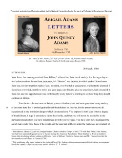
© Copyright 2026