
K Overgaard, T Sereghy, G Boysen, H Pedersen and N... 1992;23:1167-1173 doi: 10.1161/01.STR.23.8.1167
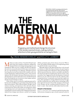
Reduction of infarct volume and mortality by thrombolysis in a rat embolic stroke
model.
K Overgaard, T Sereghy, G Boysen, H Pedersen and N H Diemer
Stroke. 1992;23:1167-1173
doi: 10.1161/01.STR.23.8.1167
Stroke is published by the American Heart Association, 7272 Greenville Avenue, Dallas, TX 75231
Copyright © 1992 American Heart Association, Inc. All rights reserved.
Print ISSN: 0039-2499. Online ISSN: 1524-4628
The online version of this article, along with updated information and services, is located on
the World Wide Web at:
http://stroke.ahajournals.org/content/23/8/1167
Permissions: Requests for permissions to reproduce figures, tables, or portions of articles originally
published in Stroke can be obtained via RightsLink, a service of the Copyright Clearance Center, not the
Editorial Office. Once the online version of the published article for which permission is being requested
is located, click Request Permissions in the middle column of the Web page under Services. Further
information about this process is available in the Permissions and Rights Question and Answer document.
Reprints: Information about reprints can be found online at:
http://www.lww.com/reprints
Subscriptions: Information about subscribing to Stroke is online at:
http://stroke.ahajournals.org//subscriptions/
Downloaded from http://stroke.ahajournals.org/ by guest on June 9, 2014
1167
Reduction of Infarct Volume and Mortality by
Thrombolysis in a Rat Embolic Stroke Model
Karsten Overgaard, MD; Tomas Sereghy, MD; Gudrun Boysen, MD, DMSc;
Hans Pedersen, MD; and Nils H. Diemer, MD, DMSc
Background and Purpose: Thrombolytic therapy with recombinant tissue plasminogen activator was
tested in a rat embolic stroke model.
Methods: The rat carotid territory was embolized with arterial-like microthrombi formed under
pressure. Hemispheric cerebral blood flow before and after embolization was measured by the intraarterial Xenon-133 injection method. Fifteen minutes after embolization, 24 rats were treated with 3
mg/kg or 10 mg/kg tissue plasminogen activator, and 27 were treated with saline. Carotid angiography
displayed the rate of occlusion of the cerebral arterial supply before and after treatment. Brains were fixed
and evaluated neuropathologically and infarct volume was measured.
Results: Cerebral bloodflowwas reduced 70-86% after embolization. The comparison of pretreatment
and posttreatment angiography showed significant (p=0.0005) reperfusion in the treated rats. Thrombolytic therapy significantly reduced the infarct volume from 55.1% to 24.4% of embolized hemisphere
volume (p=0.007) and increased the survival rate from 0.48 to 0.96 (p=0.0004). Fifty-three percent of the
embolized rats recanalized completely after thrombolytic treatment and developed almost no infarction
(median volume 2.8%), and all survived. No hemorrhagic complications were observed.
Conclusions: Early thrombolytic therapy induced recanalization and reduced mortality and infarct
volume after embolic stroke in this model. (Stroke 1992;23:1167-1174)
KEY WORDS • cerebral blood flow • cerebral ischemia • thrombolytic therapy • rats
R
ecently, thrombolytic therapy with recombinant
tissue plasminogen activator (rt-PA) in acute
,. myocardial infarction has been proven effective,1 and hopes for salvaging ischemic brain tissue in
the same way have arisen. In acute ischemic stroke, 75%
of all patients evaluated within the first 6 hours after
symptom onset have an occlusion of the cerebral arterial
supply.2 Probably more than 75% of these strokes are
caused by arterial occlusion because in some of the
cases recanalization could have taken place before
angiography, and the angiography itself cannot display
clinically relevant occlusion of small arteries and arterioles. Jorgensen and Torvik3 found thrombi or emboli
in relevant arteries by gross examination in 90-95% of
fatal large recent infarcts. In a few cases the occlusions
were caused by rupture of or bleeding into atheromatous plaques.4 In 21% of the cases a fresh thrombus was
superimposed on old organized thrombotic material,
and in another 19% of cases the ultrastructural composition of the occlusions could not be classified.56 It was
not stated which fraction of occlusions was caused by
From the Neurovascular Research Laboratory, Departments of
Neurology (K.O., T.S., G.B.) and Neuroradiology (H.P.), University Hospital of Copenhagen, Rigshospitalet; and the Cerebral
Ischemia Research Group, Institute of Neuropathology (N.H.D.),
University of Copenhagen, Denmark.
Address for reprints: Karsten Overgaard, Neurovascular Research Laboratory, Section 5463, Department of Neurology, Rigshospitalet, Blegdamsvej 9, DK-2100 Copenhagen, Denmark.
Supported by the Velux Foundation and by the Augustinus
Foundation.
Received November 1, 1991; accepted April 22, 1992.
fresh thrombi, consisting of fibrin intermingled with
blood cells; this is the type of occlusion most likely to be
dissolved by thrombolytic treatment. Occlusions of the
middle cerebral artery (MCA) were in almost all cases
embolic.3 We therefore developed a model of cerebral
embolization with arterial-like thrombi in rats7 that
resembled the human pathophysiological mechanism.
See Editorial Comment, p 1174
The outcome after an ischemic stroke is related to the
volume of infarction measured by computed tomographic scanning,8 and if a treatment achieves the goal
of reducing the infarct volume by even 15-20%, a
substantial amount of disability will be prevented.9 In
the present trial with rt-PA treatment of rat stroke, we
used reperfusion on angiography, volume of infarct, and
mortality as major end points. The appearance of
hemorrhagic complications after thrombolytic therapy
was also evaluated.
Materials and Methods
Our method of inducing embolic stroke in rats has
been reported previously.7 Sixty-four male SpragueDawley rats weighing 300-400 g were used. Anesthesia
was induced with 0.15 mg i.p. diazepam (Apozepam,
Apothekernes Laboratorium As., Oslo, Norway), 0.015
mg s.c. atropine, 0.8 mg i.m. fluanison, and 0.016 mg i.m.
fentanyl (Hypnorm, Pharmaceutica Beers, Belgium).
The anesthesia was prolonged when necessary with one
third of the initial dose. The body temperature was kept
between 37° and 38°C by rectal temperature monitoring
Downloaded from http://stroke.ahajournals.org/ by guest on June 9, 2014
1168
Stroke
Vol 23, No 8 August 1992
FIGURE 1. Left panel: Angiogram showing normal filling of arteries supplying the brain, angiographic score value 0. The
following arteries are identified: internal carotid artery (large thick arrow), anterior cerebral artery (small thick arrow), middle
cerebral artery (MCA) (large thin arrow), and MCA branches (small thin arrows). Right panel: Angiogram showing occlusion
(arrow) of main stem middle cerebral artery, angiographic score value 2.
and thermostat-controlled heating of the operating
table.
The right femoral artery and vein were catheterized
with a polyethylene PP 25 tube, and the arterial line was
filled with 0.5 ml saline with 5 units/ml heparin and
clamped. The venous line was kept patent by continuous
flow of saline at a rate of 0.5 ml/hr. Mean arterial blood
pressure and arterial Pao 2 , Paco 2 , and pH were measured twice (Radiometer ABL 2, Copenhagen). One
animal was excluded from the study because of Po2
saturation twice below 90%.
Preparation of the Emboli
A 1-ml insulin disposable syringe with a 28-gauge
(inner diameter, 0.164 mm) needle was filled with 50 fil
saline containing 2.5 units of thrombin (Topostasine,
Roche Laboratories, Nutley, N.J.). Then, in 44 rats, 150
fi\ arterial blood was drawn in another syringe of the
same type. After less than 20 seconds, the two syringes
were interconnected from tip to tip with a polyethylene
PP 10 tube, and the suspension was moved approximately 70 times from one syringe to the other during 3
minutes. The syringes were left standing for 30 minutes
until embolization. In another 20 animals, the emboli
solution was prepared in the same manner with 200 fi\
arterial blood instead of 150 /xl.
Carotid Operation Procedure
The right external carotid artery and its branches
were exposed and the pterygopalatine, thyroid, and
occipital arteries ligated. A polyethylene PP 25 catheter
was inserted through a transverse arteriotomy of the
external carotid artery with the tip 2 mm distal to the
bifurcation and fixed with ligatures; care was taken not
to injure the intima. Clotting in the catheter was
avoided by continuous flow of heparinized (5 units/ml)
saline through the line (the animals obtained up to 10
units of heparin during the entire procedure).
Embolization and Cerebral Blood
Flow (CBF) Measurements
Just before and after embolization, CBF was measured using an intracarotid bolus injection of 0.15-0.20
ml saline containing 5-10 mCi/ml Xenon-133 (Amersham). Clearance was recorded by external detection
with a collimated Nal(Th) crystal placed over the right
MCA area. The preembolic clearance was recorded as
the initial 15-second slope, then the preformed emboli
Downloaded from http://stroke.ahajournals.org/ by guest on June 9, 2014
Overgaard et al
Thrombolysis in a Rat Embolic Stroke Model
1169
film distance of 141.5 cm gave a linear magnification of
4.5. Angiograms were made on mammographic highresolution Kodak NRM-1 films and were evaluated in a
blinded manner by a neuroradiologist according to the
following score: 0, patent arteries; 1, distal MCA branch
occlusion; 2, main stem MCA occlusion; 3, internal
carotid artery occlusion (Figure 1).
suspension described above was gently injected into the
carotid catheter during the next 30 seconds. In the
following 15 seconds, the postembolic 133Xe efflux was
recorded. Both CBF values were calculated from the
slope of the clearance of 133Xe on a semilogarithmic plot
using the formula CBF (mlxlOO g-'xmirT'^Axln
10xD o xl00, where the blood partition coefficient for
the gray matter, A, is 0.87 ml/g and Do is the initial slope.
A peak value of around 2,000 cps assured that the
linearity of the logarithmic clearance curve was not
affected by low counting statistics.
Recombinant Tissue-Type Plasminogen Activator
(rt-PA) Administration
In the experimental group, eight rats embolized with
200-fil emboli suspension received 3 mg/kg rt-PA (Actilyse, Boehringer Ingelheim, Ridgefield, Conn.). Eight
rats embolized with 200-/xl emboli suspension and eight
embolized with 250 /xl emboli suspension received 10
mg/kg rt-PA. Intravenous infusion started 15 minutes
after embolization and was administered during 30 (3
mg/kg dose) or 45 (10 mg/kg dose) minutes using a
Harvard infusion pump. Twenty-seven control animals
received an equal amount of isotonic saline instead of
rt-PA. Thirteen died immediately after embolization
and consequently did not receive infusion. After the
second angiography, all animals received 10 ml i.p.
isotonic saline, and femoral and neck wounds were
closed after ligations of vessels. The anesthesia was
reverted with 0.1 mg i.m. naloxone (Narcanti, Du Pont
Pharmaceuticals, Wilmington, Del.). After recovery
from anesthesia, the rats neurological status was evaluated according to the method of Bederson et al.10 Rats
were held gently by the tail 1 meter above the floor and
observed for forelimb flexion. Normal rats extended
both forelimbs toward the floor and were assigned grade
0. Rats with flexion of the forelimb contralateral to the
injured hemisphere were graded 1, rats with reduced
resistance to lateral push toward the paretic side were
graded 2, and rats with spontaneous circling toward the
paretic side were graded 3.
The animals were then placed in an incubator with
75% humidity and a temperature of 28°C for approximately 10 hours. Animals unable to drink received
intraperitoneally 10 ml of equal parts of isotonic saline
and isotonic glucose 10 and 20 hours after embolization
to prevent dehydration.
Angiography
Immediately after and 2 hours after embolization,
angiography was performed via the carotid catheter by
bolus injection of 0.2 ml heparinized (5 units/ml) iohexol (Omnipaque, 300 mg I/ml, Nycomed, Denmark).
An x-ray tube (Philips SRO 03/100) with a small focus
spot 0.15 mm2 and a large focus spot 1.5 mm2 was used.
Exposure data were as follows: 70 kV, 14 mA, and 0.4
seconds. A focus-object distance of 31.5 cm and focus-
Preparation of Brain Tissue and Measurement of
Infarct Size
Four to 6 days after the operation, the rats were once
again graded according to the Bederson score and were
subsequently anesthetized and killed by cardiac perfusion fixation with a 4% phosphate buffered (pH 7.2)
formalin solution. In the perfusion-fixed rats and in
those that died before this procedure, the brains were
carefully removed, postfixed and dehydrated during 5
'•»
FIGURE 2. Photomicrograph of horizontal brain section
with middle cerebral artery and posterior cerebral artery area
infarction. Hematoxylin-eosin stain, original magnification,
TABLE 1.
Rats
Control
(«=27)
Treated
(«=23)
Blood Gas and Arterial Blood ]Pressure Values
Before embolization
MABP
98
(93-103)
99
(88-106)
PH
7.37
(7.33-7.40)
7.38
(7.34-7.41)
Pao2
88.0
(81.7-97.4)
89.6
(86.7-94.0)
Before second angiography
Paco2
48.5
(44.0-52.4)
46.3
(43.2-52.7)
MABP
pH
Pao 2
Paco2
94
(90-102)
98
(92-104)
7.39
(7.36-7.41)
7.37
(7.33-7.39)
92.3
(83.2-95.6)
87.6
(80.0-92.0)
43.9
(41.7-46.4)
44.4
(41.8-47.8)
All results are displayed as median values, with 25th and 75th percentiles shown in parentheses. Values of pH, Pao2, Paco2, and mean
arterial blood pressure (MABP) are expressed in mm Hg.
Downloaded from http://stroke.ahajournals.org/ by guest on June 9, 2014
1170
Stroke Vol 23, No 8 August 1992
FIGURE 3. Top panel: Micrograph of an emboli suspension
showing an elongated microembolus composed of fibrin (large
arrow) and platelets, a few erythrocytes (small arrow) and leukocytes (bended arrow). May-Grunwald-Giemsa stain. Original
magnification, X270. Bottom
panel: Cross section of middle
cerebral artery and embolus occluding its trunk (arrow). Due to
technical artefact during tissue
preparation, the embolus is not
occupying the whole luminal
space, as it probably did in vivo.
Hematoxylin-eosin stain. Original
magnification, X125.
days, embedded in paraffin, and cut into sections 4 /xm
section thick. In each brain, approximately 17 horizontal
sections with a distance of 0.4 mm between each were
obtained and stained with hematoxylin-eosin. Using a
Leitz TAS plus image analyzer (Figure 2), affected
hemisphere and infarct volumes were calculated as
areas multiplied with the distance between sections.
Infarct areas included areas with 100% neuronal death.
Results of infarct and hemisphere volumes were expressed as an average of two independent measurements performed without knowledge of the treatment
regimen.
Calculations and Statistical Analyses
Cerebral blood flow was measured twice, immediately
before and after embolization. Animal weight was also
obtained twice, at the induction of anesthesia and just
before perfusion fixation. Body weight reduction and
CBF reduction were calculated as the following ratio:
first—second value/first value.
Nonparametric statistical analyses of our data were
performed because all our data were either nominal or
could be placed on rank ordinal or ratio-interval scales
with no apparent normal distribution of values. MannWhitney U test was used for unpaired observations,
Wilcoxon matched-pairs for paired observations, Fisher's exact probability test for survival rate, and the
Spearman rank correlation test for paired ranked
observations.
Results
No differences were found in any parameters between
the groups of rats treated with 3 mg/kg and 10 mg/kg
rt-PA; therefore, the groups were pooled. Blood gas
values are shown in Table 1. No significant differences
between surviving, dying, treated, or control groups
were found. The animals hypoventilated slightly during
this anesthesia with spontaneous respiration. Thirteen
rats (10 of 43 embolized with 200 /xl suspension and
three of 20 embolized with 250 /id suspension) that died
Downloaded from http://stroke.ahajournals.org/ by guest on June 9, 2014
Overgaard et al
i
d
I
•a
a
P
I
H
<=
d
rt
en
"pa8
P
•5b
p 7
•a
3
u- c
CQ.9
°I
>n
—i
t—
oo
I
oo
p
oi
o oo
d
I
r- in
ON
p
d
p
o Si
in o\
-H"
oo
I
m
VO
>n
d
I
rt X,
3
1
II
3 M
.1
i
a
I!
•81
II
1171
immediately after embolization were excluded. The
emboli (Figure 3, top panel) were heterogenous in size
and shape, although the majority were elongated, irregularly outlined, less than 0.2 mm in cross diameter, and
resembled arterial "white" thrombi, rich in platelets
and fibrin, intermingled with a few erythrocytes and
leukocytes; emboli were present in many brain arteries
(Figure 3, bottom panel). Rats embolized with the
larger dose of emboli (Table 2) had larger CBF reduction after embolization (p=0.0025, Mann-Whitney
test). There was no difference in CBF reduction between treated and control rats. In all rats CBF reduction
after embolization was between 43% and 94% (total
range) of initial CBF, except for one animal in the
control group embolized with 200 fi\ of emboli with a
CBF reduction of only 15% (this animal was the only
control animal without infarction).
r
p
d
u
3
Thrombolysis in a Rat Embolic Stroke Model
00
Effect of Thrombolytic Therapy
The main results are displayed in Table 2. There was
no difference in the occlusion rate of 22 treated and 24
control rats comparing the first angiograms according to
the scale described in "Materials and Methods." Only
slight or nonsignificant spontaneous recanalization appeared in control animals, but highly significant recanalization was achieved by the thrombolytic therapy
(/7=0.0005 in 21 treated animals) assessed by comparing the pretreatment and posttreatment angiograms of
each animal. Comparing the posttreatment angiograms,
the patency rate was significantly higher in the rt-PA
treated groups than in the control groups (p=0.036,
Mann-Whitney test).
Recovering from anesthesia, the damaged animals
were lethargic, showing asymmetrical posture, flexion of
the forelimb contralateral to embolization, circling,
tilting of the head, and pallor of the eyeball. In a few
animals, episodes of motor seizures were observed. The
most severely damaged animals were unable to take
fluid and food in the postoperative period. As seen in
Table 2, the treatment increased the survival rate (from
0.48 in all controls to 0.96 in all treated animals;
p=0.0004, Fisher's exact test). Animals of the control
groups had severe clinical damage and obtained a high
Bederson score even 4-6 days after embolization or
died. The treated rats had a much better clinical recovery than control rats, comparing Bederson scores at day
5 (/?<0.00005, Mann-Whitney test) (dead animals were
not included in the calculation). All deaths were observed in the first 48 hours after ischemic event.
Neuropathological examination showed infarction in
all rats except in three treated and one control rat. The
right hemisphere ipsilateral to embolization was affected with loss of all cell types (neurons, astrocytes,
oligodendrocytes, and endothelial cells) and presence of
numerous macrophages, often with distended cytoplasm. There were no signs of inflammation. All infarcts
(Table 2) were anemic. Some of the infarcts were
irregularly shaped, and almost all infarcts were clearly
demarcated with a neuropathological borderzone of
only 5-10 cells in width, with sporadic neurons with
eosinophilic cytoplasm. In all animals with infarction,
the MCA area was involved without exception, the
region of the posterior cerebral artery was infarcted
with some variability, and the anterior cerebral artery
area was never infarcted. Brains of six dead control rats
Downloaded from http://stroke.ahajournals.org/ by guest on June 9, 2014
1172
Stroke
Vol 23, No 8 August 1992
were lost because of technical reasons (mostly, the
animals were found late after death with autolyzed
brains), and their hemisphere volume and infarct size
values were substituted by medians of the same values
obtained of all other dying animals from the same
group. The treatment reduced the infarct size from
43.7% of hemisphere volume to 11.3% (p=0.016) in the
group embolized with 200 fi\ emboli suspension and
from 66% to 27.5% (p=0.0006) in the group embolized
with 250 (xl emboli suspension (comparing all 23 treated
and 27 control rats, the significance rose to/?=0.0002, all
by Mann-Whitney tests). Treatment reduced edema
formation in the affected hemisphere: 23 treated rats
with a median embolized hemisphere volume of 265
mm3 were compared with 26 control rats with a median
embolized hemisphere volume of 399 mm3 (/>=0.0001,
Mann-Whitney test). If no substitution of infarct sizes
and embolized hemisphere volumes of dying animals
was performed (values shown in Table 2), both the
median infarct size and the significance of the rt-PAmediated reduction of infarct size were reduced in the
200-^1 embolized control group because some of the
presumed most heavily damaged and dead rats from
that group were not included in the calculation. The
significance of the difference between infarct volumes of
all 21 control (median infarct volume, 51.1%) and all 23
treated (median infarct volume, 24.4%) rats decreased
top=0.007.
Twenty-eight rats that on the second angiography had
patent arteries or only MCA branch occlusion developed smaller infarcts than 19 rats with more severe
occlusion (/?=0.0099, Mann-Whitney test). The overall
correlation between the occlusion rate on the posttreatment angiography and the infarct size of all 47 rats was
significant (p=0.026, Spearman test). In eight thrombolytic-treated animals embolized with 200 /xl suspension, recanalization was complete, with patent arteries
on the second angiography; infarct volume (median
2.8%) of those animals was significantly (p=0.04,
Mann-Whitney test) different from infarct volume (median 29%) in the seven nonreperfused treated animals,
which was not different from the controls.
There was a strong correlation between the clinical
score at day 5 and the infarct volume of the 35 surviving
rats (/?=0.0096) and among surviving rats in the treated
groups (p=0.003; both Spearman tests). The body
weight reduction (median 10.2%) correlated with the
infarct volume (p=0.0027, Spearman test). The total
range of the volume of the affected hemisphere in rats
that died was larger (378-429 mm3) than in survivors
(189-319 mm3) (/><0.00005, Mann-Whitney test).
Discussion
Our model demonstrates reduction of mortality by
thrombolytic therapy with rt-PA in an acute embolic
stroke model. The model documents arterial reperfusion resulting in a reduction of infarct volume as a direct
effect of thrombolytic treatment. The embolization is
caused by thrombotic clots7 resembling natural arterial
thrombi.
Doses of rt-PA in the range of 1-5 mg/kg in rabbit
models and 1.2-1.5 mg/kg in rat models have been
reported to reperfuse occluded cerebral arteries, proven
by in vivo12-18 and/or postmortem19 angiography, investigation of vessels at autopsy,20 autoradiography,21 clear-
ance of radioactivity from hemisphere embolized with
radiolabeled clots,22 and CBF measurements, 121923 although in some of these experiments usage of wholeblood clots with a high tendency to spontaneous recanalization7 could have lead to false conclusions in cases of
insufficient inclusion of nontreated control animals.
Recombinant t-PA in a dose of 0.3 mg/kg body weight
infused over 30 minutes failed to recanalize embolized
occluded intracranial arteries in a rat model.21 We used
a higher dose of rt-PA than previously reported by
others because rt-PA in rats probably has only 10% (in
rabbits, approximately 60%) of the efficacy of that in
humans.24-25 This placed our doses in the range of those
used in humans, ruling out the possibility that insufficient reperfusion and no limitation of infarction were
caused by insufficient dosage regimen of rt-PA.
The conclusions from the present study are that an
increase of the volume of emboli resulted in angiographically verified occlusion of larger arteries and
more pronounced CBF reduction after embolization.
Thrombolytic therapy reperfused occluded arteries, and
did so with more ease when the volume of emboli and
rate of angiographic occlusion were small.
The efficacy of thrombolytic therapy in stroke in
terms of limitation of infarct extent presently lacks
sufficient documentation. In animal models, neuropathologic microscopic examination of brain cells and
tissue with volumetric measurement of infarct sizes,
which may be considered one of the most reliable
parameters, was done by Zivin et al26-27 and Phillips et
al,15 who found no difference in infarct volume between
thrombolytic-treated and control animals. In the studies
of Benes et al,20 Chehrazi et al,13 and Bednar et al,23
possible infarcts were demonstrated by very early 2,3,5triphenyltetrazolium chloride staining, which early after
the ischemic event is not a reliable parameter of loss of
neurons but might indicate transient damage.28-29 These
problems have been solved in the present model.7
A number of papers demonstrate improvement in
different neurophysiological parameters such as neurological clinical score,18-26-27 decreased ratio of inorganic
phosphate/phosphocreatine measured by 31P spectroscopy,30 decreased lactate, pyruvate and water content of
tissue, improved electrocorticogram19 after rt-PA administered in doses in the range of 1-5 mg/kg starting
up to 4 hours after embolization in rabbit models1218,23,26,27,30 and 1.5-2 mg/kg starting up to 2 hours after
embolization in rat models.19-22
In the animals treated with rt-PA, apart from slight
oozing of blood during and shortly after operation, we
observed no hemorrhagic complications. Because this
phenomenon was rare in thrombolytic-treated as well as
in controls, this model is probably not sensitive to
hemorrhagic transformation of infarction. This is in
accordance with other investigators,18-31 even when such
a large dose of rt-PA was administered. Whether longer
delay between embolization and rt-PA infusion will
result in hemorrhagic complications remains to be
investigated.
We found in all animals that died before sacrifice a
massive enlargement of the injured hemisphere with
shift of the midline, and we considered that the animals
died of mass effect due to edema.
Our findings that animals with small occlusion or no
occlusion on the posttreatment angiography had smaller
Downloaded from http://stroke.ahajournals.org/ by guest on June 9, 2014
Overgaard et al
infarcts than the animals with more pronounced occlusion
and the fact that treated animals with complete recanalization were almost free of infarction supports the hypothesis that early reperfusion leads to limitation of the infarct
size. If the emboli had been easier to lyse, a larger
proportion of the animals might have recanalized completely with minimal brain damage. Our conclusions from
the present study are that the thrombolytic therapy is both
life saving and infarct reducing. The reduction of mortality
was more pronounced in the heavily embolized animals,
and we proved an infarct-limiting effect of the thrombolytic therapy that was more pronounced in the animals
embolized with a smaller volume of emboli.
Acknowledgments
Charlotte Wetterstein and Annette Fischer are thanked for
their excellent technical assistance and Michael Kragsholm for
setting up the x-ray equipment. Bent Larsen made a program
for personal computer-based registration of the experimental
data in dBase IV, version 1.1, and the necessary TurboPascal
programming for calculations and transferral of data to the
statistical program used, Medstat, version 2.11.
References
1. Wilcox RG, Olsson CG, Skene AM, von der Lippe G, Jensen G,
Hampton JR: Trial of tissue plasminogen activator for mortality reduction in acute myocardial infarction. Lancet 1988;2:
525-531
2. Fieschi C, Argentino C, Lenzi GL, Sacchetti ML, Toni D, Bozzao
L: Clinical and instrumental evaluation of patients with ischemic
stroke within the first six hours. Neurol Sci 1989;91:311-322
3. Jorgensen L, Torvik A: Ischaemic cerebrovascular diseases in an
autopsy series. J Neurol Sci 1966;3:490-509
4. Torvik A, Jorgensen L: Thrombotic and embolic occlusions of the
carotid arteries in an autopsy series. J Neurol Sci 1966;3:410-432
5. Torvik A, Jorgensen L: Thrombotic and embolic occlusions of the
carotid arteries in autopsy material. J Neurol Sci 1964;l:24-39
6. Jorgensen L, Torvik A: Ischaemic cerebrovascular diseases in an
autopsy series. / Neurol Sci 1969;9:285-320
7. Overgaard K, Sereghy T, Boysen G, Pedersen H, Hoyer S, Diemer
NH: A rat model of reproducible cerebral infarction using thrombotic blood clot emboli. J Cereb Blood Flow Metab 1992;12:484-490
8. Allen CMC: Predicting outcome after acute stroke: Role of computed tomography. Lancet 1985;2:464-465
9. Treatment for stroke? (editorial) Lancet 1991;337:1129-1131
10. Bederson JB, Pits LH, Tsun M, Nishimura MC, Davis RL, Bartkowski H: Rat middle cerebral artery occlusion: Evaluation of the
model and development of a neurologic examination. Stroke 1986;
17:472-476
11. Wulf RJ, Mertz ET: Studies on plasminogen: VIII. Species specificity of streptokinase. Can J Biochem 1969;47:927-932
12. Kissel P, Chehrazi B, Seibert JA, Wagner FC: Digital angiographic
quantification of blood flow dynamics in embolic stroke treated
with tissue-type plasminogen activator. J Neurosurg 1987;67:
399-405
Thrombolysis in a Rat Embolic Stroke Model
1173
13. Chehrazi BB, Seibert JA, Kissel P, Hein L, Brock JM: Evaluation
of recombinant tissue plasminogen activator in embolic stroke.
Neurosurgery 1989;24:355-360
14. Phillips DA, Davis MA, Fisher M: Selective embolization and clot
dissolution with tPA in the internal carotid artery circulation of the
rabbit. AJNR 1988;9:899-905
15. Phillips DA, Fisher M, Smith TW, Davis MA, Pang RHL: The
effects of a new tissue plasminogen activator analogue, Fb-Fb-CF,
on cerebral reperfusion in a rabbit embolic stroke model. Ann
Neurol 1989;25:281-285
16. Phillips DA, Fisher M, Smith TW, Davis MA: The safety and
angiographic efficacy of tissue plasminogen activator in a cerebral
embolization model. Ann Neurol 1988;23:391-394
17. Phillips DA, Fisher M, Davis MA, Smith TW, Pang RHL: Delayed
treatment with a t-PA analogue and streptokinase in a rabbit
embolic stroke model. Stroke 1990;21:602-605
18. Lyden PD, Zivin JA, Clark WA, Madden K, Sasse KC, Mazzarella
VA, Terry RD, Press GA: Tissue plasminogen activator-mediated
thrombolysis of cerebral emboli and its effect on hemorrhagic
infarction in rabbits. Neurology 1989;39:703-708
19. Papadopoulos SM, Chandler WF, Salamat MS, Topol EJ, Sackellares JC: Recombinant tissue-type plasminogen activator therapy
in acute thromboembolic stroke. J Neurosurg 1987;67:394-398
20. Benes V, Zabramski JM, Boston M, Puca A, Spetzler RF: Effect of
intra-arterial antifibrinolytic agents on autologous arterial emboli
in the cerebral circulation of rabbits. Stroke 1990;21:1594-1599
21. Penar PL, Greer CA: The effect of intravenous tissue-type plasminogen activator in a rat model of embolic cerebral ischemia. Yale
J Biol Med 1987;60:233-243
22. Hara T, Iwamoto M, Ogawa H, Yamamoto A, Tomikawa M:
Dissolution of emboli in rats with experimental cerebral thromboembolism by recombinant human tissue plasminogen activator.
Thromb Res 1990;59:703-712
23. Bednar MM, McAuliffe T, Raymond S, Gross CE: Tissue plasminogen activator reduces brain injury in a rabbit model of thromboembolic stroke. Stroke 1990;21:1705-1709
24. Collen D, Korninger C: Studies on the specific fibrinolytic effect of
human extrinsic (tissue-type) plasminogen activator in human
blood and in various animal species in vitro. Thromb Haemost
1981;46:561-565
25. Niewenhuizen W, Keyser J: Species specificity in the acceleration
of tissue-type plasminogen activator-mediated activation of plasminogens, by fibrinogen cyanogen bromide fragments. Thromb Res
1985;38:663-670
26. Zivin JA, Fisher M, DeGirolami U, Hemenway CC, Stashak JA:
Tissue plasminogen activator reduces neurological damage after
cerebral embolism. Science 1985;230:1289-1292
27. Zivin JA, Lyden PD, DeGirolami U, Kochhar A, Mazzarella V,
Hemenway CC, Johnston P: Reduction of neurologic damage after
experimental embolic stroke. Arch Neurol 1988;45:387-391
28. Cole DJ, Drummond JC, Ghazal EA, Shapiro HM: A reversible
component of cerebral injury as identified by the histochemical
stain 2,3,5 -triphenyltetrazolium chloride (TTC). Ada Neuropathol
1990;80:152-155
29. Bederson JA, Pitts LH, Germano SM, Nishimura MC, Davis RL,
Bartkowski HM: Evaluation of 2,3,5-triphenyltetrazolium chloride
as a stain for detection and quantification of experimental cerebral
infarction in rats. Stroke 1986;17:1304-1308
30. Lee BCP, Brock JM, Fan T, Seibert A, Moonen C, Kissel P,
Chehrazi B, Bradbury EM: 31P spectroscopy in thrombolytic treatment of experimental cerebral infarct. AJR 1989;152:623-628
31. Del Zoppo GJ, Copeland BR, Anderchek K, Hacke W, Koziol
JA: Hemorrhagic transformation following tissue plasminogen
activator in experimental cerebral infarction. Stroke 1990;21:
596-601
Downloaded from http://stroke.ahajournals.org/ by guest on June 9, 2014


© Copyright 2026