ABC
docz
Explore
Log in
Create new account
Download
Report
health and fitness
disease
cancer
Cervical screening in Australia 2012â2013
Cancer Screening and Control Services
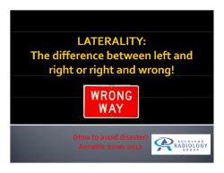
(How to avoid disaster) Annette Jones 2012
Preschool Screening
METHODS :
Genius Solutions, Inc. - Encounter Form / Superbill Sample TODAY’S CHARGES
Montana University System WellCheck Event Information How to Sign up:
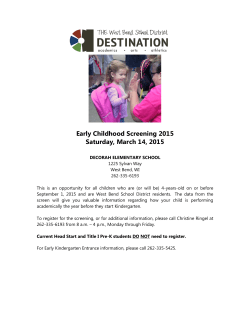
Early Childhood Screening - West Bend School District
Sex and the City GP Education
RADICULITIS SYNDROME
Kyphotic Deformities of the Cervical Spine
© Copyright 2026
About abcdocz
DMCA / GDPR
Report