
Michael K. McAdam, M.D. Anterior Cruciate Ligament Injury
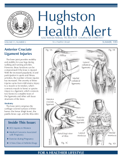
Michael K. McAdam, M.D.
Orthopedic Surgeon Specializing in Arthroscopy and Sports Medicine
Anterior Cruciate Ligament Injury
Injury to the anterior cruciate ligament (ACL) is common, especially in athletic individuals.
The overall incidence of ACL tear is about 1 in 3000. 1 70% of ACL injuries occur during a
sporting activity, especially those involving cutting or twisting, such as skiing, soccer, basketball,
or football.
When people tear their ACL it is usually a sudden event often accompanied by a “pop”,
and often a sense of the knee buckling or shifting. Swelling appears over the next few hours and
there is difficulty walking comfortably. The knee can feel unstable. Some patients may not
present immediately after the injury, but may come in for evaluation weeks, months, or even
years after the initial tear. They will usually describe episodes of the knee buckling or shifting,
often with recurrent swelling.
Physical examination if done either before muscle guarding has set in, or after it has
resolved, can usually diagnose a complete ACL tear. Lachman’s test is done by flexing the knee
to 30 degrees, and attempting to translate the tibia anteriorly, while holding the femur steady.
Translation of more than the opposite knee, especially if the endpoint feels spongy or soft, is
suggestive of an ACL tear.
Lachman’s Exam for ACL
Anterior drawer Exam
The anterior drawer test is done with the knee flexed to 90 degrees, and similar attempt
made to translate the tibia anteriorly. It is often helpful to sit on the patient’s foot while performing
the test in order to stabilize the leg. The pivot shift maneuver is the most specific test for ACL
tear, but requires a fair amount of practice to do accurately, and also requires a completely
relaxed patient. The pivot shift is done by internally rotating the lower leg with one hand at the
ankle, applying a valgus load to the proximal lateral tibia, and bringing the knee from flexion to
extension. A positive test will show a visible shifting of the tibia, often with a “clunk”, as the tibia
subluxes anterolaterally on the femur.
The medial and lateral collateral ligaments (MCL and LCL) should also be assessed for
injury with valgus and varus stress exams at 0 and 30 degrees of flexion. The PCL can be tested
with a posterior drawer (posterior translation of tibia with knee flexed to 90 degrees). Careful
neurovascular assessment should also be made, especially in the setting of a multi-ligament
injury.
Damage to the meniscus often accompanies an ACL tear. In the acute tear, the lateral
meniscus is most commonly injured. In patients with chronic ACL deficiency, the medial
meniscus is more commonly damaged, as it is a secondary stabilizer to anterior translation.
Careful palpation of the joint lines and McMurray’s test will usually elicit pain in the patient with a
meniscal tear.
McMurray’s Test: The leg is rotated while variably applying varus
and valgus loads in a flexed position. This may elicit a clunk or
merely pain at the joint line.
One should be especially cognizant of incarcerated bucket handle tears. In this type of
tear, the posterior horn of the meniscus has flipped anteriorly and become trapped there. This
patient will usually have a firm block to full extension. The Lachman and anterior drawer testing
may not have much anterior translation, even when a complete ACL tear exists, as the entrapped
meniscus will prevent this. A patient with a suspected acute bucket-handle tear should be given
crutches and kept non-weight bearing until an MRI and orthopedic evaluation can be performed.
Many bucket handle tears can be repaired, reducing the long-term risk of arthritis associated with
meniscus deficiency. This depends on the location of the tear and if there has been excessive
secondary damage to the meniscus, as well as the patient’s age.
Repaired tear of the medial meniscus
Imaging
Standard radiographs should be performed in any traumatic knee injury. While most ACL
tears will have only an effusion or hemarthrosis visible on the films, osteochondral injuries and
other fractures can occur with ACL tears, and can be spotted on routine x-rays. The Segond
fracture, or an avulsion fracture of the anterolateral capsule from the lateral tibial plateau, is
pathognomonic for an ACL tear, and may occasionally be seen. Tibial eminence avulsion
fractures may also indicate ACL injury.
Segond Fracture (red arrow) indicates ACL tear
MRI continues to be the noninvasive test of choice for diagnosis, both of the ACL tear
and of concomitant pathology. 80% of patients will have bone bruises seen on MRI, in addition to
the tear. These are seen in the posterolateral tibial plateau and the center of the lateral femoral
condyle, and are the result of the transient subluxation of the knee with impaction of the joint at
that location. Patients with bone bruising may have more pain, and greater difficulty
postoperatively than those without the bruising. Additionally, up to 65% of patients will develop
osteochondral damage at the site of the bone bruise within 5 years of the injury. 2
Normal ACL with arrow pointing to femoral origin
Torn ACL with absence of ligament at insertion
Treatment
When the ACL is completely torn it has no significant capability of repair and thus the
ligament function is lost. The resulting instability can only be treated two ways: appropriate
rehabilitation with activity modification or surgical reconstruction of the ACL. The word
reconstruction is used as rather than repair to indicate substitution of new tissue for the torn ACL.
Unfortunately, no technique of direct repair of the ACL has provided consistently successful
results.
The injury can be treated nonoperatively in lower demand patients. The quadriceps
undergoes significant weakening and atrophy in any knee injury, and must be strengthened.
Increasing the normal hamstring strength to supranormal levels can provide increased dynamic
stability to the anterior translation of the tibia, and help prevent instability symptoms. Despite
rehabilitation, 50-85% of patients will continue to note instability. It is important to note, that even
absent instability symptoms, the abnormal kinematics of the chronically ACL deficient knee do
lead to damage. In one study, up to 80% of knees x-rayed had developed osteophytes as a
precursor of arthritis. 3
The use of a derotation brace may supplement a nonoperative approach, but the brace
must be worn during all at-risk activity, and does not completely eliminate instability episodes.
Operative treatment of the ACL deficient knee involves replacing the torn ligament with
other tissue. Direct repair techniques have been attempted in multiple different ways in the past,
and unfortunately, have usually had very high failure rates. Therefore, reconstruction with tendon
grafts has become the treatment of choice. In most cases, this is done with arthroscopic
visualization of the knee for the reconstruction.
No matter where the graft comes from, the surgeon performing the reconstruction
attempts to place the new ligament through drill tunnels in the femur and tibia in the exact location
of the old ACL. A great deal of research has established a good consensus as to these locations.
The careful placement of these tunnels and graft will restore the stability of the knee, and allow
early range of motion (ROM) and weight-bearing.
Arthroscopic View of Normal ACL
Torn ACL
Grafts can be either autograft (from the patient), or allograft (from a cadaver). The most
popular graft for many years in ACL reconstruction has been the autograft bone-patellar tendonbone (BPTB) graft. A 10cm wide strip of the mid-third patellar tendon, along with a bone block
each from the central patella and the tibial tubercle are harvested for the reconstruction.
Proponents of this graft choice point to the advantages of bone to bone healing within the tunnels
as occurring earlier than soft tissue adherence to the tunnels. Downsides of the patellar tendon
autograft are numerous, however. Up to 30% of patients report persistent anterior knee pain and
difficulty kneeling or squatting after surgery. Quadriceps weakness is much more profound after
patellar tendon harvest compared with hamstrings. Although rare, patella fracture or patellar
tendon rupture can occur after harvest, and are devastating complications.
Hamstring autograft, most often the gracilis and semitendinosus has been gaining in
popularity over the last 10-15 years. Its proponents favor it over BPTB grafts due to the
decreased rates of anterior knee pain and quadriceps weakness, although weakness of the
hamstrings has certainly been noted. The initial graft strength of a quadrupled hamstring
autograft is twice that of the 50% more than the BPTB graft, and 100% more than the native ACL.
Here in the Northwest, it seems to be slightly more popular than BPTB grafts for these reasons.
The most common indication through the years for allografts has been for revision ACL
reconstruction, or multiple ligament reconstruction. The Achilles tendon, patellar tendon, and
hamstrings can all be used to reconstruct the ACL, and have been gaining in popularity as a
primary reconstruction method. It has the obvious advantages of no donor site morbidity, and
thus decreases initial postoperative pain and makes the initial rehabilitation faster. The graft can
be made as large as needed, and therefore even stronger than either autograft choice.
Disadvantages of allograft are the potential for disease transmission, both viral and bacterial.
The most current data report a risk of approximately 1 in 1.7 million chance of contracting
hepatitis C or HIV from a musculoskeletal allograft. The risk of bacterial transmission is small, but
statistically unknown due to a variety of factors. Incorporation of the allograft tissue may also
take slightly longer, but this doesn’t seem to be clinically significant.
Regardless of graft choice, any tissue that is placed for the reconstruction is devoid of
blood supply. It must go through a process called ligamentization, in which blood vessels grow
into the tissue, which is then broken down and rebuilt along the scaffold of the tendon graft. This
occurs without difficulty in 95% or more patients. It is a process that takes at minimum 3 months.
The graft reaches its nadir of strength around 6 weeks, and continues to mature and gain
strength over the next 12 months. It therefore is typical to hold patients out of cutting or pivoting
sports until 6 to 12 months postoperatively, dependent on the surgeon and graft type.
Reconstructed ACL
Rehabilitation
Regardless of the graft choice, the rehabilitation after an ACL is at least of equal
importance to the surgery itself. Regaining full range of motion and early and aggressive
strengthening of the quadriceps have been shown in multiple studies to be independent factors in
patient outcome after ACL reconstruction. Early work with physical therapy to these ends is often
beneficial, although some studies have shown good outcomes with physician directed selfrehabilitation. Most of the quadriceps strengthening should be done in a closed-chain fashion, or
in a way that keeps the foot planted on the floor or other platform, such as leg-press, squats, or
lunges.
Return to sports is dependent first on graft healing and the ligamentization process (see
above). After this, the patient’s quadriceps strength and balance and stability must be assessed
to determine if they are physically ready for their sport. Finally, psychological issues cannot be
underestimated. In professional athletes who have had ACL reconstruction, it is reported that it
takes more than a full season after surgery to feel that the knee is trustworthy again. This is
anecdotally true as well in recreational athletes, and many often feel hesitant to return for this
very reason. In actuality, the contralateral knee is up to 2 or 3 times more likely to have an ACL
tear than the reconstructed knee, which unfortunately usually doesn’t comfort the patient all that
much.
Special Issues: ACL tears in female athletes
Women are up to two to eight times more likely to have an ACL tear than men. This has
been attributed to slight differences in anatomy, coupled with disproportionately less muscle
strength gains during adolescence as compared with males. Less emphasis on strength training
in women’s athletics is also culpable. Much recent research has attempted to link hormonal
changes in knee laxity to incidence of ACL tears, as the ligaments do have receptors for estrogen
and progesterone. 4 Early studies showed some differences with more ruptures in the ovulatory
phase of the cycle, 5 and greater laxity has been shown during peak estrogen and progesterone
concentrations. 6 Regardless of the hormonal and other factors, the risk of ACL injury in women
can be lowered two to four fold through specific neuromuscular or plyometric training. One such
program is available at the website: http://www.aclprevent.com/pepprogram.htm. Institution of
such programs ideally should occur on an individual, team and league level, especially as greater
emphasis is placed on preventative medicine in the primary care setting.
Summary
While an ACL tear can be a frightening problem for a patient, early and proper
diagnosis, appropriate nonoperative or operative intervention and rehabilitation can very
successfully restore full function and return to activity. Early referral to a Sports
Medicine trained Orthopedic surgeon for management of the injury is the generally
accepted standard of care, but maintaining a knowledge base of the topic can help any
health care provider assist their patients in decision making. Recent efforts devoted to
prevention of ACL tears in female adolescent and adult athletes have been successful,
and these programs will hopefully become more widespread, preventing the need for
surgery.
1
Miyasaka KC, et al. The incidence of knee ligament injuries in the general population. Am J Knee Surg.
4:3-8,1991.
2
Johnson DL, et al. Articular cartilage changes seen with magnetic resonance imaging-detected bone
bruises associated with acute anterior cruciate ligament rupture. Am J Sports Med. 26:409-14, 1998.
3
Jacobsen K. Osteoarthrosis following insufficiency of the cruciate ligaments in man. Acta Orthop Scand.
48:520-6, 1977.
4
Liu SH, et al. Primary immunolocalization of estrogen and progesterone target cells in the human anterior
cruciate ligament. J Orthop Res. 14:526-33, 1996.
5
Juston LJ, Wojtys EM. Neuromuscular performance characteristics in elite female athletes. Am J Sports
Med. 24:427-436, 1996
6
Heitz NA, et al. Hormonal Changes Throughout the Menstrual Cycle and Increased Anterior Cruciate
Ligament Laxity in Females. J Athl Train. 34(2): 144-9, 1999.


© Copyright 2026