
Neutrophilic and Eosinophilic Dermatitis Caused by Contact Allergic Reaction
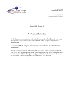
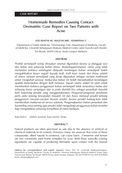
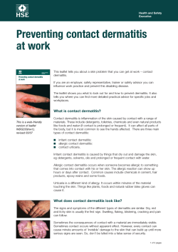
OBSERVATION
Neutrophilic and Eosinophilic Dermatitis
Caused by Contact Allergic Reaction
to Paraphenylenediamine in Hair Dye
Vincent Lo¨nngren, MScMed; Ewa Young, MD; Mecius Simanaitis, MD, PhD; Cecilia Svedman, MD, PhD
Background: Paraphenylenediamine (PPD) in hair dyes
can cause systemic as well as cutaneous allergic reactions such as neutrophilic and eosinophilic dermatitis.
The symptoms are often severe. The acute lesion is normally histologically indistinguishable from any eczematous reaction with marked spongiosis.
asthma. What is most remarkable about this case is the
histopathologic finding of neutrophilic cellulitis and a
marked neutrophilic infiltrate with variable spongiosis.
This unique finding was confirmed by histologic analysis of a patch test lesion specimen.
Conclusion: It is always important to consider contact
Observations: We report a case of allergic contact der-
matitis caused by the use of hair dye containing PPD that
developed in a patient who had been using the same hair
dye for many years. Her symptoms included scalp dermatitis and widespread skin lesions as well as lymphadenopathy and quite possibly dyspnea resembling
T
Author Affiliations:
Department of Dermatology,
Ska˚ne University Hospital,
Malmo¨ (Mr Lo¨nngren and
Dr Simanaitis), and Department
of Occupational and
Environmental Dermatology,
Lund University, Lund
(Drs Young and Svedman),
Sweden.
allergic dermatitis as a cause of dermatitis because of the
variable presentation of the disease, including unique histologic findings that do not fit the conventional picture,
as in the present case.
Arch Dermatol. 2012;148(11):1299-1301
HE POTENT SENSITIZER PARA-
phenylenediamine (PPD) is
a substance that is often
found in permanent hair
dyes.1 Contact allergy to
hair dyes is common among hairdressers
and consumers.2,3 As in any contact allergic dermatitis, the acute-stage lesions histologically resemble any eczematous reaction with marked spongiosis. The use of
PPD was banned in Sweden until 1992, but
the ban was lifted owing to legislative measures within the European Union. Paraphenylenediamine is well known to contribute to darker hair colors in permanent
hair dyes but can also be found in lighter
shades. When PPD is used in hair dyes, it
is mixed with oxidizing agents, such as hydrogen peroxide, and new, partly unknown substances are formed. Contact allergic reactions to these derivatives and
oxidation products of PPD are thought to
play a role in PPD-related contact allergy.4,5 Another risk of developing lifelong allergy to PPD, for children as well
as adults, is through temporary tattoos:
“black henna tattoos.” For such tattoos,
henna is mixed with PPD. Serious adverse reactions to PPD, including severe
facial and scalp dermatitis and asthma,
ARCH DERMATOL/ VOL 148 (NO. 11), NOV 2012
1299
have been reported as early as 1924.6 We
report a case that illustrates some of the
severe reactions that may arise from exposure to PPD in hair dyes and discuss
some of its unusual histologic and clinical characteristics.
REPORT OF A CASE
A 58-year-old woman presented with a
2-week history of painful and itchy lesions
on her chest, back, and neck (Figure 1 and
Figure 2). Her son recently had a case of
impetigo, and her family physician had suspected that she had the same condition. A
course of isoxazolyl penicillin had been
completed, without any effect on the lesions. The patient was otherwise healthy
except for chronic obstructive pulmonary disease and had no remarkable immunologic or dermatologic history. She
was afebrile, with only mild leukocytosis.
The lesions, which were nummular erythematous plaques with excoriations and
crusts, were located on the neck, the front
and back of the upper chest area, and the
scalp. A few lesions were found on the
arms, and 1 lesion was found on the right
ankle. A punch biopsy was performed, and
prednisolone therapy (40 mg/d) was ini-
WWW.ARCHDERMATOL.COM
©2012 American Medical Association. All rights reserved.
Downloaded From: https://jamanetwork.com/ on 09/09/2014
Author Affil
Department
Ska˚ne Unive
Malmo¨ (Mr L
Dr Simanaiti
of Occupatio
Environmen
Lund Univer
(Drs Young a
Sweden.
Figure 1. Back of the upper chest area and neck covered by nummular
erythematous plaques with excoriations and crusts.
Figure 2. The patient’s neck and ear covered with lesions.
A
B
C
D
Figure 3. Four photomicrographs of skin from the unusual lesions. A, Diffuse dermal infiltrate of neutrophils with variable epidermal spongiosis
(hematoxylin-eosin, original magnification ⫻200). B, Pronounced edema of the papillary dermis (Giemsa, original magnification ⫻200). C, Focal parakeratosis
(Giemsa, original magnification ⫻400). D, Admixture of eosinophils (Giemsa, original magnification ⫻400).
tiated. Histologic analysis revealed the presence of neutrophilic cellulitis and eosinophilic granulocytes. There
were remarkable dermal edema, variable spongiosis, and
only a few lymphocytes. A diagnosis of Sweet syndrome
was suggested, although the high frequency of eosinophilic granulocytes was difficult to explain (Figure 3).
The results of direct immunofluorescence microscopy
were negative.
Within a few days, the lesions had spread further, involving the perianal and genital regions. They had become increasingly wet and weeping, and topical treatment with betamethasone-clioquinol and potassium
ARCH DERMATOL/ VOL 148 (NO. 11), NOV 2012
1300
WWW.ARCHDERMATOL.COM
©2012 American Medical Association. All rights reserved.
Downloaded From: https://jamanetwork.com/ on 09/09/2014
permanganate was initiated. Bacterial culture revealed only
moderate growth of Staphylococcus aureus. Ten days after the first visit, there was no significant improvement.
Neutrophilic cutaneous lupus erythematosus was suspected because of the neutrophilic dermatitis in the upper part of the dermis, although there was no interface
dermatitis. Hydroxychloroquine sulfate therapy (400
mg/d) was initiated.
Incidentally, the patient developed an episode of dyspnea, which was treated at the emergency department.
The cause of the dyspnea was not entirely clear, but it
did not display the characteristics of a typical exacerbation of chronic obstructive pulmonary disease and lasted
only a few hours. Two weeks after the first visit, the lesions had finally started to fade, and a few days later, only
postinflammatory hyperpigmentation and some desquamation remained. The results of all immunoserologic tests
were negative, as were repeated attempts at direct immunofluorescence microscopy. All treatment except topical betamethasone was discontinued.
Surprisingly, a few days later, erythematous nummular lesions reappeared on the patient’s neck and forearms. It was noted that she had dyed her hair. She had
been using the same hair dye for many years. The distribution of the lesions raised the suspicion of allergic
contact dermatitis due to the use of hair colorants despite the atypical histologic findings. Epicutaneous patch
testing with the base line series and textile series showed
a contact allergic reaction to several allergens, most importantly a 3⫹ reaction to PPD and a positive reaction
to disperse orange 3. The patient was urged to discontinue the use of hair dye. A few months later, the lesions
returned in full force after she colored her hair again with
the same hair dye. This time, her occipital and cervical
lymph nodes were swollen and remained so for at least
2 months. Later, additional epicutaneous allergen testing revealed a reaction to a low concentration of PPD
(0.001%), among other substances. Considering the unusual histologic features of the previous specimen, a punch
biopsy was performed in the area of the positive reaction to PPD. The sample was found to be histologically
identical to the previous ones, quite unlike the conventional histologic appearance of eczema.
COMMENT
It is well known that PPD can cause unusually severe contact allergic reactions in susceptible individuals. In this case,
the lesions were not typical of contact allergic dermatitis
or baboon syndrome. The unusual clinical and histologic
appearance of the lesions initially misled us away from the
diagnosis of contact allergy. There have been some reports of erythema multiforme–like lesions in contact allergic dermatitis to PPD.7 However, the histologic appearance in our case was not that of erythema multiforme.
Instead, it was similar to neutrophilic dermatitis or Sweet
syndrome, apart from the pronounced eosinophilia. It did
not resemble a conventional eczematous reaction. The diagnosis was finally confirmed by the provocation with PPD
as well as by the results of patch testing, including those
of histologic examination of the patch test reaction site. It
is important to remember that a contact allergic reaction
to PPD can present with unusual clinical and histologic
features and to ask about hair dye use.
Accepted for Publication: June 15, 2012.
Correspondence: Vincent Lo¨nngren, MScMed, Department of Dermatology, Ska˚ne University Hospital, So¨dra
Fo¨rstadsgatan 101, 205 02 Malmo¨, Sweden (vincent
[email protected]).
Author Contributions: All authors had full access to all
the data in the study and take responsibility for the integrity of the data and the accuracy of the data analysis.
Study concept and design: Lo¨nngren, Young, Simanaitis,
and Svedman. Acquisition of data: Lo¨nngren, Young,
Simanaitis, and Svedman. Analysis and interpretation of
data: Lo¨nngren, Simanaitis, and Svedman. Drafting of the
manuscript: Lo¨nngren and Young. Critical revision of the
manuscript for important intellectual content: Lo¨nngren,
Young, Simanaitis, and Svedman. Administrative, technical, or material support: Svedman. Study supervision:
Simanaitis and Svedman.
Conflict of Interest Disclosures: None reported.
REFERENCES
1. Yazar K, Boman A, Lide´n C. Potent skin sensitizers in oxidative hair dye products
on the Swedish market. Contact Dermatitis. 2009;61(5):269-275.
2. Krasteva M, Bons B, Ryan C, Gerberick GF. Consumer allergy to oxidative hair coloring products: epidemiologic data in the literature. Dermatitis. 2009;20(3):123141.
3. Lind ML, Albin M, Brisman J, et al. Incidence of hand eczema in female Swedish
hairdressers. Occup Environ Med. 2007;64(3):191-195.
4. Basketter DA, Lide´n C. Further investigation of the prohapten concept: reactions
to benzene derivatives in man. Contact Dermatitis. 1992;27(2):90-97.
5. White JM, Kullavanijaya P, Duangdeeden I, et al. p-Phenylenediamine allergy: the
role of Bandrowski’s base. Clin Exp Allergy. 2006;36(10):1289-1293.
6. Nott HW. Systemic poisoning by hair dye. Br Med J. 1924;1(3297):421-422.
7. Balato A, Patruno C, Balato N, Gallo L, Ayala F. Erythema multiforme-like eruption
because of para-phenylenediamine. Contact Dermatitis. 2008;58(1):65-66.
ARCH DERMATOL/ VOL 148 (NO. 11), NOV 2012
1301
WWW.ARCHDERMATOL.COM
©2012 American Medical Association. All rights reserved.
Downloaded From: https://jamanetwork.com/ on 09/09/2014


© Copyright 2026