
Angiomatous hamartoma of the breast: a new entity Maria Daniela Podeanu
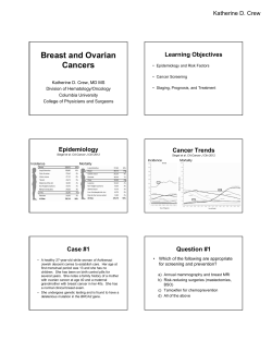
Revista Română de Medicină de Laborator Vol. 19, Nr. 2/4, Iunie 2011
199
Angiomatous hamartoma of the breast: a new entity
developing in pediatric patients
Hamartom mamar de tip angiomatos - entitate nou descrisă la copii
Maria Daniela Podeanu1*, Cristina Marian2, Constantin Copotoiu3,
Klara Brânzaniuc4, Simona Stolnicu2
1. Department of Radiology, University of Medicine and Pharmacy Tîrgu Mureş, România
2. Department of Pathology, University of Medicine and Pharmacy Tîrgu Mureş, România
3. Department of Surgery I, University of Medicine and Pharmacy Tîrgu Mureş, România
4. Department of Anatomy, University of Medicine and Pharmacy Tîrgu Mureş, România
Abstract
We report the case of a young prepuberal 11 year old patient who rapidly developed a breast hamartoma with a prominent angiomatous (vascular) component that was observed on both radiological and pathological examination, raising the possibility of other breast neoplasms. Only one case of vascular hamartoma of the
breast in a 5 year old girl has been previously reported in the English literature. We discuss the main differential
diagnostic problems and the possible histogenesis of this new entity.
Keywords: breast hamartoma, angiomatous component, immunohistochemistry
Rezumat
Raportăm cazul unei paciente prepuberale de 11 ani, care a dezvoltat rapid la nivelul glandei mamare
un hamartom ce prezintă o proeminentă componentă angiomatoasă (vasculară) identificată atât la examinarea
radiologică cât şi la cea patologică. Leziunea ridică suspiciunea unor alte neoplasme mamare. Doar un singur
alt caz de hamartom mamar la copii (o pacientă in vârstă de 5 ani) a mai fost descris până în prezent în literatura de specialitate publicată în limba engleză. In cadrul articolului discutăm principalele probleme de diagnostic
diferenţial şi histogeneza ale acestei noi entităţi.
Cuvinte cheie: hamartom mamar, componentă angiomatoasă, imunohistochimie
Introduction
Hamartoma of the breast was initially
defined as a malformation that resembles a neo*
plasm but in 1971 Arrigoni described it for the first
time as an encapsulated breast neoplasm that clinically and macroscopically resembled a fibroadenoma but, in contrast with this, on microscopic
Corresponding author:Maria Daniela Podeanu, Department of Radiology, University of Medicine and Pharmacy Targu Mures, Str. Gheorghe Marinescu Nr. 38, Telefon: 0265215551
Fax: 0265210407, E-mail: [email protected]
200
Revista Română de Medicină de Laborator Vol. 19, Nr. 2/4, Iunie 2011
examination it has a lobular arrangement of
glandular tissue and a fibrous stroma admixed with
fat tissue of various extension (1). However, the
World Health Organization (WHO) book from
2003 recognized the lesion as a tumor belonging to
the fibroepithelial category (2). Hamartoma subtypes such as fibroadenolipoma, myoid and chondrolipoma have already been described throughout
all ages (3,4). Only one case of vascular hamartoma of the breast in a 5 year old girl has been previously reported in literature (5). This is the second
case of a young prepuberal 11 year old girl who
presented a breast hamartoma with a prominent
vascular (angiomatous) component on both radiological and microscopic examination. We discuss
the main differential diagnostic problems and the
possible histogenesis of this new entity.
Case report
An 11 year old prepuberal patient presented to the Radiology and Imaging Department
with an asymmetry of the superior - exterior and
inferior - exterior right breast quadrants due to a
palpable painless mass noticed by her mother during the previous month. There was no family history of breast cancer. On clinical examination, the
tumor was approximately 40 mm in diameter,
freely movable on palpation, with rubbery consistency. There was no evidence of axillary
lymphadenopathy. Considering the patient's age,
the radiologist decided to perform ultrasound examination instead of mammography, that disclosed a 36 mm diameter solid mass with a macrolobulated well-defined contour, heterogeneous
hypoechoic pattern, some scattered microcystic
areas and numerous tubular channels with arterial
Doppler signal within the tumor (Figure 1, Figure
2). The ultrasound examination was neither characteristic for a fibroadenoma, nor for a malignant
tumor. Fine needle aspiration cytology (FNAC)
was performed and the aspirate was hypercellular
with a mixed population of cells including a large
number of epithelial and myoepithelial cell
groups, but also many stromal cells (of fibroblast-
ic and adipose type) suggesting a phyllodes tumor
and corresponding to C3 category (Figure 3). The
patient declined core biopsy and required surgery
due to fear of cancer and asymmetry of the breast.
Right lumpectomy was performed.
The surgical specimen measuring
40/35/20 mm contained a white nodule of 36/11
mm with hard consistency, well delineated contour
and small cystic spaces on the cut surface. On microscopic examination the tumor was surrounded
by a fibrous capsule and was composed of ducts
and acinar structures with lobular arrangement surrounded by fibrous hypocellular edematous stroma
and areas of adipose tissue (Figure 4, Figure 5).
The adipose tissue represented 20% of the stromal
component on all the examined slides. Some of the
mammary ducts were cystically dilated and some
others presented an epithelial gynecomastoid-like
proliferation within the lumen but lacking atypical
features (Figure 6). An inflammatory infiltrate
composed mainly of lymphocytes was observed.
Rare bundles of benign spindle looking cells of
myoid differentiation were encountered, but these
represented no more than 5-10% of the stroma.
More than 75% of the stroma contained
numerous vessels of small and large size that were
present on each of the 6 slides we have examined
from the tumor (Figure 7). Some of the vessels had
a round lumen, while others were compressed by
the surrounding stroma (Figure 8). Immunohistochemically, the vessels were diffusely positive for
specific markers as CD 31 (LAB VISION, Clone
JC/70A, dilution 1:25) and factor VIII (NOVOCASTRA, Clone NCL-VWF, dilution 1:150)
but also for Desmin (DAKO, Clone D 33, dilution
1:75) (Figure 9, Figure 10). The gynecomastoid-like proliferation within the ducts was positive for CK 5/6 (DAKO, Clone D5/16 B4, dilution
1:50) in a mosaic pattern. The morphology and immunohistochemical profile were consistent with an
angiomatous hamartoma with no evidence of malignancy. No other treatment was considered for
this patient since surgical resection margins were
free of tumor. Clinical and radiological follow-ups
were suggested.
Revista Română de Medicină de Laborator Vol. 19, Nr. 2/4, Iunie 2011
Figure 1. Sonographic examination: 36 mm
hipoechoic nodular well circumscribed lesion, with
macrolobulated contour and heterogeneous texture
Discussion
Breast hamartoma is a rare and underrecognized biphasic benign tumor, representing no more
that 0.1- 0.7% of all benign tumors, usually appearing in premenopausal women, but also described in
teenagers or in women over 60 years (6). Cases of
breast hamartoma have also been reported in male
or ectopic breast tissue (7, 8). It can grow rapidly or
slowly. In case there is a rapid growth and it reaches
a large diameter, it produces asymmetry and simulates a fibroadenoma on palpation. It can very rarely
be detected only mammographically, as a well demarcated density with mottled appearance due to tissue heterogeneity and surrounded by a radiolucent
halo that separates the tumor from the surrounding
breast parenchyma. The ultrasound examination
shows a sharp delineation of the tumor that contains
sonolucent fat and echogenic fibrous component
with a heterogeneous internal echo pattern (9). The
microscopic aspect of the breast hamartoma is still
poorly defined and overlapping and different attempts to subclassify the tumor according to the histological parameters into three categories (fibrous,
fatty and fibro fatty hamartoma) or four category
classification (encapsulated fibrocystic changes,
fibroadenoma with fibrous stroma, fibroadenomalike and circumscribed adenolipoma) have not been
widely accepted by the pathologists (10,11). Microscopic subtypes of breast hamartoma, such as
myoid, fibroadenolipoma and chondrolipoma have
201
Figure 2. Intratumoral vascularization: increased
central and peripheral arterial Doppler signal
been previously described and different focal
changes as cysts, simple epithelial hyperplasia, apocrine metaplasia, stromal giant cells have also been
reported (12, 13, 14). Only one case of a pediatric
patient with prominent vascular component within
the stroma has been published in literature so far (5).
In this paper, we report a second case of a young girl
with a breast hamartoma which grew rapidly and
had a prominent vascular component on both radiological and microscopic examination, suggesting
other benign or malignant breast tumors with different prognosis and management.
The clinical, sonographical and cytological
pattern were suggestive for a fibroadenoma or phyllodes tumor, especially due to the increase of cystic
spaces observed on sonographic examination, to the
diameter of the tumor at the presentation and to the
biphasic population of cells on the smear. However,
the prominent vascular Doppler signal was not consistent with either of the two previously mentioned
lesions. The macroscopic appearance (well - demarcated margins, white color and firm consistency)
was characteristic for a fibroadenoma. The cystic
spaces that were observed on the cut surface were
characteristic for focal phyllodes changes in a
fibroadenoma. On microscopy however, even
fibroadenoma may have very rare small areas of
adipous metaplasia, it does not have a lobular arrangement of the acini and ducts and loses the normal architecture of a breast tissue. Phyllodes tumor
may have small or large diameter and has been re-
202
Revista Română de Medicină de Laborator Vol. 19, Nr. 2/4, Iunie 2011
Figure 3. Citology( PAP stain): mixed population of
cells including a large number of epithelial and
myoepithelial cell groups but also many stromal cells
Figure 4. The tumor is surrounded by a fibrous
capsule(HE)
Figure 5. The tumor is composed of ducts and acinar
structures with a lobular arrangement surrounded
by fibrous hypocellular edematous stroma and areas
of adipose tissue (HE)
Figure 6. The mammary duct presents an epithelial
gynecomastoid-like proliferation within the lumen,
lacking atypical features (HE)
ported in pediatric population (15), but on microscopic examination there is an increase in the
stromal component (fibroblasts and adipose cells)
versus the epithelial one and numerous characteristic
cystic spaces can be seen, with stromal papillary
projections within the spaces. Areas of adipose tissue can be observed in a phyllodes tumor of benign
or malignant type. The fibrous stroma which sur-
rounds and extends between the individual lobules
and obliterates the usual interlobular specialized
loose stroma is characteristic to the hamartoma, but
can also be encountered in sclerosing lobular hyperplasia that is also a biphasic benign lesion which can
be associated with a classic fibroadenoma, but usually does not contain adipose tissue.
Prominent pseudovascular or true vas-
Revista Română de Medicină de Laborator Vol. 19, Nr. 2/4, Iunie 2011
203
Figure 7. More than 75% of the stroma contained
numerous vessels of small and large size (HE)
Figure 8. Some of the vessels had a round lumen and
others were compressed by the surrounding stroma (HE)
Figure 9. The vascular component is strongly and
diffusely positive to CD31
Figure 10. Factor VIII is positive in all the blood
vessels of the vascular component
cular component may be encountered in various
breast lesions. Pseudo-angiomatous stromal hyperplasia (PASH) changes can appear in a
breast hamartoma due to hormonal influence,
usually in premenopausal patients. In PASH,
anastomosing slit-like spaces delineated by
spindle cells of fibroblastic or myofibroblastic
type without atypia can be seen in a diffuse pattern, scattered throughout the tumor but the immunohistochemical examinations prove that
these slit-like spaces are not of vascular origin.
Other true vascular tumors that can involve the breast are: perilobular hemangiomas,
angiosarcomas and exceptionally lymphangiomas and angiomatosis. The perilobular hemangioma is usually found accidentally in a biopsy
specimen or on mastectomy performed for another type of breast lesion. It is composed of a
network of capillary-sized blood vessels that involve the lobular or perilobular stroma, but it is
a microscopic lesion that cannot be detected on
clinical or radiological examination. The an-
204
Revista Română de Medicină de Laborator Vol. 19, Nr. 2/4, Iunie 2011
giosarcoma is the malignant vascular tumor that
can also appear de novo in young patients, possibly due to hormonal stimulation or secondary
to irradiation or lymphedema after axillary
lymph node dissection for breast carcinoma in
elder patients. Macroscopically, it can appear as
single, but more frequently multiple reddish
nodules with poorly defined contour. For this
reason, clinical as well as radiological examination is able to distinguish the tumor from a vascular hamartoma. Microscopically, the atypical
endothelial cells that can either delineate vascular anastomosing channels or grow in solid
areas have the same immunohistochemical profile as vascular hamartoma, so differentiating
the two lesions is very important, since the angiosarcoma is associated with poor prognosis.
Regarding the histogenesis of the angiomatous hamartoma of the breast, the prominent vascular component cannot be attributed to
hormonal influence, since both this case and a
previously reported one developed in prepuberal patients, so it can be considered more as a
malformation than a neoplastic process. However, cases of breast hamartomas of other microscopic subtypes have been reported in association with pregnancy, so a hormonal influence
can be suspected in adult patients since vascular
breast tumors of benign or malignant potential
have been found to have a relationship with
hormonal influence in other cases (16).
Conclusions
Due to the rarity of these tumors and
the heterogeneous pattern on both radiological
and microscopic examination, hamartoma of
the breast is a difficult diagnosis for both the radiologist and pathologist. Especially when it is
associated with peculiar components, such as
muscle, cartilage or vessels, hamartoma of the
breast can raise the possibility of other more
common breast benign or malignant lesions
making the immunohistochemical examinations
very useful to distinguish between these types
of lesions. A prominent vascular component in
pediatric patients developing a hamartoma can
be considered more as a malformation than a
hormonal influence.
Acknowledgments. This paper is partially supported by the Sectoral Operational Programme Human Resources Development, financed from the European Social Fund and by the
Romanian Government under the contract number POSDRU/ 89/1.5/S/60782
Abbreviations
WHO – Worls Health Organization
PASH - Pseudo-angiomatous stromal hyperplasia
References:
1. Arrigoni M.G., Dockerty M.A., Judd E.S. The identification and treatment of mammary hamartomas. Surg.Gynecol.Obstet. 1971, 133: 577-582
2. Bellocq J. P., Magro G. Fibroepithelial tumours. In
Tavassoli F.A. and Devilee P., Tumours of the breast and
female genital organs, IARC Press, Lyon, 2003, 99-103
3. Rosen P.P. Fibroepithelial neoplasms. In Rosen P.P.,
Rosen’s Breast Pathology, Lippincott Williams & Wilkins,
Philadelphia, 2009, 187-230
4. Bayar S., Dusunceli E., Heper A.O., Guner R., Genc
V., Demirkazik A. Myoid hamartoma of the breast: a very
rare entity. The Breast Journal. 2010, 16 (1): 86-88
5. Deshmukh H., Prasad S., Patankar T. A giant vascular
hamartoma of the breast in a child. Journal of Postgraduate
Medicine, 1997, 43: 50-51
6. Tse G.M.K., Law B.K.B., Ma T.K.F., Chan A.B.W.,
Pang L.M., Chu W.C.W. et al. Hamartoma of the breast: a
clinicopathological review. J.Clin.Pathol. 2002, 55(12):
951-954
7. Ravakhah K., Javadi N., Simms R. Hamartoma of the
breast in a man: first case report. Breast J, 2001, 7(4): 266268
8. Dworak O., Reck T., Greskotter K.R., Kockerling F.
Hamartoma of an ectopic breast arising in the inguinal region. Histopathology, 1994, 24 (2): 169-171
9. Kopans D.B. Pathologic, mammographic and sonographic correlation. In Kopans D.B. Breast imaging, Lippincott - Raven, Boston, Massachusetts, 1998, 558- 560
10. McGuire L.I., Cohn D. Hamartoma of the breast.
Aust.N.Z.J.Surg., 1991, 61: 713-716
11. Jones M.W., Norris H.J., Wargotz E.S. Hamartomas
of the breast. Surg.Gynecol.Obstet., 1991, 173: 54-56
12. Fisher C.J., Hanby A.M., Robinson L. Mammary
Revista Română de Medicină de Laborator Vol. 19, Nr. 2/4, Iunie 2011
hamartoma - review of 35 cases. Histopathology, 1992,
20: 99-106
13. Daya D., Trus T., D’Souza T.J. Hamartoma of the
breast,
an
underrecognized
breast
lesion.
Am.J.Clin.Pathol., 1995, 103: 685-689
14. Agabiti S., Gurrera A., Amico P., Vasquez E., Magro
G.: Mammary hamartoma with atypical stromal cells: a
potential diagnostic dilema. Pathologica, 2007, 99(6): 434-
205
437
15. Amerson J.R. Cystosarcoma phyllodes in adolescent
females. A report of seven patients. Ann.Surg., 1970, 171:
849-853
16. Kotalwar M., Gadgil N.M., Ranadive N.U. Breast
hamartoma associated with pregnancy - a case report. Indian.J.Pathol.Microbiol. 2004, 47 (4): 551-553


© Copyright 2026