
On-line coupling of sequential injection extraction with restricted
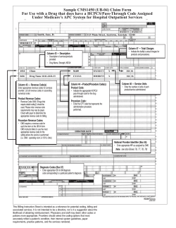
a n a l y t i c a c h i m i c a a c t a 6 0 0 ( 2 0 0 7 ) 122–128 available at www.sciencedirect.com journal homepage: www.elsevier.com/locate/aca On-line coupling of sequential injection extraction with restricted-access materials and post-column derivatization for sample clean-up and determination of propranolol in human plasma ˇ ınsky´ a,∗ , Hugo S. Serralheiro b , Petr Solich a , Dalibor Sat´ ´ b , Maria C.B.S.M. Montenegro b Alberto N. Araujo a Department of Analytical Chemistry, Faculty of Pharmacy, Charles University, Heyrovsk´eho 1203, ´ e 500 05, Czech Republic Hradec Kralov´ b Requimte, Departamento de Qu´ımica-F´ısica, Faculdade de Farmacia, ´ Universidade do Porto, R. An´ıbal Cunha 164, Porto 4070-047, Portugal a r t i c l e i n f o a b s t r a c t Article history: The presented paper deals with a new methodology for direct determination of propranolol Received 16 October 2006 in human plasma. The methodology described is based on sequential injection analy- Received in revised form sis technique (SIA) coupled with solid phase extraction (SPE) column based on restricted 6 February 2007 access materials (RAM). Special RAM column containing 30 m polymeric material—N- Accepted 10 February 2007 vinylacetamide copolymer was integrated into the sequential injection manifold. SIA–RAM Published on line 20 February 2007 system was used for selective retention of propranolol, while the plasma matrix components were eluted with two weak organic solutions to waste. Keywords: Due to the acid–basic and polarity properties of propranolol molecule and princi- Sequential injection analysis ples of reversed-phase chromatography, it was possible to retain propranolol on the Solid phase extraction N-vinylacetamide copolymer sorbent (Shodex MSpak PK-2A 30 m (2 mm × 10 mm)). Restricted access materials Centrifuged plasma samples were aspirated into the system and loaded onto the column Propranolol using acetonitrile–water (5:95, v/v), pH 11.00, adjusted by triethylamine. The analyte Human plasma was retained on the column while proteins contained in the sample were removed to Sample preparation waste. Interfering endogenous substances complicating detection were washed out by acetonitrile–water (15:85), pH 11.00 in the next step. The extracted analyte was eluted by means of tetrahydrofuran–water (25:75), pH 11.00 to the fluorescence detector (emission filter 385 nm). The whole procedure comprising sample pre-treatment, analyte detection and column reconditioning took about 15 min. The recoveries of propranolol from undiluted plasma were in the range 96.2–97.8% for three concentration levels of analyte. The proposed SIA–RAM method has been applied for direct determination of propranolol in human plasma. © 2007 Elsevier B.V. All rights reserved. ∗ Corresponding author. Tel.: +420 495067228; fax: +420 495067164. ˇ ınsky). ´ E-mail address: [email protected] (D. Sat´ 0003-2670/$ – see front matter © 2007 Elsevier B.V. All rights reserved. doi:10.1016/j.aca.2007.02.021 a n a l y t i c a c h i m i c a a c t a 6 0 0 ( 2 0 0 7 ) 122–128 1. 123 Introduction Sequential injection analysis (SIA) was introduced by Ruzicka and Marshall in 1990 [1] as a new generation in the development of flow injection technique. The principles upon which SIA is based, namely controlled partial dispersion and reproducible sample handling, are similar to those of the flow injection analysis (FIA). Characteristic advantages of SIA include its versatility, full computer compatibility, high sample throughput, and low sample and reagent consumption. However, in recent years, it has become apparent that the scope of SIA can be extended to encompass a variety of more complex, on-line sample-manipulation and pretreatment procedures. Then, the ports of the multi-position selection valve could be coupled to various units (e.g., reservoirs, detectors, pumps, reactors, separators, special cells, and other manifolds) [2]. One of the most widely used samplepre-treatment procedures in SIA is the automated solid-phase extraction (SPE). It employs an appropriate solid- or liquidextraction material attached to a suitable support, for which arrangement SIA is ideally suited. The extraction procedure accomplishes two goals: separation of the analyte from interfering species in the sample and preconcentration of the analyte to increase the sensitivity. The great advantage of SPE is that both organic compounds and inorganic species can be extracted. Depending on the nature of analyte and on required retention mechanism, different extraction materials can be used (e.g., hydrophilic ion-exchange resins, surface-modified beads, molecularly imprinted polymers and different types of hydrophobic polymers) [2]. In recent years, special SPE supports possessing restricted access properties have been developed to allow direct injection of untreated biological samples into on-line SPE liquid chromatography (LC) systems [3–5]. These sorbents called restricted access materials (RAMs), combine size exclusion of proteins (without destructive accumulation) and other macromolecular matrix components with the simultaneous enrichment of low-molecular analytes, which can be retained and extracted selectively. The low-molecular-mass analytes are retained by conventional retention mechanisms such as hydrophobic, ionic or affinity interactions at the inner surface of the sorbent particles. The access of proteins is prevented by a physical diffusion barrier or by a chemical barrier. In the majority of applications described in the literature, RAMs are used in on-line coupled-column SPE–LC systems. These systems require highly sophisticated apparatus, higher operating cost and expensive instrumentation (column switching systems, software control and two pumps). Coupling of SPE based on restricted access materials with SIA was described as low cost alternative for direct determination of drugs. First attempt to on-line coupling of RAM and SIA was performed by determination of potential antileucotrienic quinlucast in serum and it provided satisfactory results [6] that encouraged following studies of the hyphenation. The aim of presented work was to examine further possibilities of SIA–RAM system for direct analysis of propranolol in human plasma with fluorescence detection. Fig. 1 – Molecular structure of propranolol. Propranolol (structure shown in Fig. 1) was chosen as an analyte possessing fluorescence capability and representing polar and basic substances from the group of beta-blocking pharmaceuticals. Being a strong beta-adrenergic blocking drug, propranolol is widely used in clinical practice in the treatment of cardiac arrhythmia, hypertension, sinus tachycardia and angina pectoris [7]. It is also used in low activity sports, reducing cardiac frequency, contraction force and coronary flow [8]. Therefore, it has been included in the list of forbidden substances by International Olympic Committee [9]. Monitoring of propranolol in bio-fluids is important not only in the clinical practice but also in the field of doping control. Different techniques [10–16], including fluorimetry, HPLC, capillary electrophoresis and mass spectrometry, have been used to determine propranolol in commercial formulations or biological fluids. Practically, all previous methods operate in a flow or batch systems and require various tedious preliminary procedures such as pre-concentration in an organic solvent. Thus, in recent years new techniques to determine propranolol such as fluorescence optosensors [17], molecularly imprinted polymers [18] or ion selective PVC membrane electrodes [19] have been developed, but it is still necessary to further develop highly selective, simple, rapid and cheap procedures to determine propranolol in pharmaceutical preparations and bio-fluids. Method for direct determination of propranolol in human plasma comprising on-line sample preparation based on SIA–RAM hybrid technique has been proposed. Advantages and disadvantages of such a connection are reported in presented paper. 2. Experimental 2.1. Sequential injection system and RAM sorbents A commercially available instrument FIAlab® 3500 system (FIAlab® Instruments Inc., Bellevue, USA) with a syringe pump (syringe reservoir 5.0 mL) and an 8-port selection Cheminert valve (Valco Instrument Co., Houston, USA) was used. The manifold was equipped with fibre-optic fluorimetric detector PMT-FL (Ocean Optics Inc., Dunedin, USA) with UV light source D-1000-CE (Analytical Instrument Systems Inc., Flemington, USA). The fluorescence signal was scanned through secondary filter (385 nm, Edmund Industrie Optik, GmbH, Karlsruhe, Ger- 124 a n a l y t i c a c h i m i c a a c t a 6 0 0 ( 2 0 0 7 ) 122–128 Fig. 2 – Scheme of SIA–RAM system with post-column derivatization. many). The SIA system was equipped with flow rate variable peristaltic pump. The whole SIA system was controlled by the version of program FIAlab for Windows 98, WinFIA version 5.0. Flow lines were made of 0.75 mm i.d. PTFE tubing. On-line sample preparation was performed on RAM column ˚ pore size (Shodex, MS Pak PK-2A (30 m, 2 mm × 10 mm), 30 A Japan). The RAM column was placed between the selection valve and flow cell of the detector. A replaceable in-line filter (2–5 m, Merck) was installed ahead the column for protection. The next sorbent used in our study was LiChrospher® RP18 ADS (alkyl-diol silica) (25 m, 25 mm × 4 mm), from Merck (Germany). A scheme of the sequential injection extraction system with the RAM column and post-column derivatization is depicted in Fig. 2. 2.2. Reagents All chemicals used were of analytical grade quality. Propranolol and organic solvents were obtained from Sigma–Aldrich. Chemicals for buffer preparation were obtained from Merck, Germany. Millipore Milli-Q RG (Millipore s.r.o., Prague, Czech Republic) ultra pure water was used for preparing the solutions. Eluting solutions were degassed by helium before use. 2.3. Preparation of spiked human plasma and standard samples A stock solution of propranolol (1000 g mL−1 ) was prepared by dissolving the substance in methanol. The flask was stored in the refrigerator protected to light for 2 weeks without stability problems. Fresh working standard solutions were prepared daily by appropriate dilution of the stock solution in water to the concentration 20 g mL−1 . The sample of human plasma was spiked with stock solution of propranolol to get the final concentrations of propranolol (1, 5, 10 g mL−1 ) in undiluted plasma. The samples were spiked just before analysis, incubated for 1 h at 37 ◦ C and then centrifuged for 10 min at 1750 × g. The supernatant was used for the analysis. 2.4. Design of proposed SIA–RAM analytical procedure A procedure based on sequential aspiration of mobile phases of increasing content of organic modifier and their pro- pelling through the column was proposed. The analytical cycle involved four main steps: (1) loading the sample onto the column and removing proteinaceous ballast material; (2) washing the column and removing more polar interfering substances complicating detection; (3) elution and detection of the analyte; and (4) column reconditioning. The composition of mobile phases used for particular steps was optimised separately. First, the syringe pump was filled with loading mobile phase acetonitrile–water (5:95, v/v), pH 11.00 (adjusted by triethylamine) via the left position of the double position valve A. The sample (standard solution or spiked plasma solution, 50 L) was aspirated via port 5 of the selection valve B (switching the valve A to the right position) to the connecting tube leading from the middle port of the valve B to the pump. The sample was then propelled through port 8 of the selection valve B to the RAM column by reverse movement of the piston pump using flow rate 0.6 mL min−1 . Propranolol was extracted on the column while proteinaceous matrix of the sample was washed to waste. In the next step, washing mobile phase acetonitrile–water (15:85), pH 11.00 (adjusted by triethylamine) was aspirated via port 4 of the selection valve B and pushed through the column washing fluorescent interfering substances to waste (flow rate 1.2 mL min−1 ). Finally, eluting mobile phase tetrahydrofuran–water (25:75), pH 11.00 (adjusted by triethylamine) was aspirated via port 2 of the selection valve B and it was used for elution of extracted propranolol to the mixing coil for post column derivatization before fluorimetric detection (flow rate 1.2 mL min−1 ). The aspiration of derivatization reagent was carried out by peristaltic pump-flow rate 0.045 mL min−1 . It was necessary to wash the column with 80% acetonitrile (0.5 mL) and recondition it with the loading mobile phase (0.6 mL) (both flow rates 1.2 mL min−1 ) prior to aspiration of the next proteinaceous sample. The latter two steps were integrated at the beginning of the controlling program to ensure that the column will be prepared for the following cycle. The resulting signal was recorded in the form of peaks; the peak heights were calculated automatically by FIAlab® software and the data were stared by PC for subsequent processing. Each measuring cycle was carried out in triplicate and the mean peak height values were used for data evaluation. All measurements were performed at ambient temperature. a n a l y t i c a c h i m i c a a c t a 6 0 0 ( 2 0 0 7 ) 122–128 3. Results and discussion 3.1. Choice of the extraction column The experiments concerning a composition of mobile phases started searching the optimal solvent for final elution of propranolol. First experiments were carried out with LiChrospher® RP 18 ADS column. The optimum composition of eluting mobile phase was not found for this type of sorbent. The main problem of retention versus extraction of propranolol from LiChrospher® RP 18 ADS was in molecular structure of the drug. Propranolol shows relatively high lipophilicity coefficient (log P = 2.60) [20], which is suitable for sufficient retention on reversed phase C-18 sorbent. However, basic character of the propranolol molecule (pKa = 9.15) [20] resulted in a poor retention in working pH area of LiChrospher® RP 18 ADS column (pH 2–7). Moreover, the elution of propranolol from silica based RP 18 sorbent showed very strong peak tailing. The retention behaviour of propranolol was checked making different changes in the composition of the eluting mobile phase and observing the changes in retention time and shape of the peak. The tested organic modifiers were methanol, ethanol, acetonitril, propanol or tetrahydrofuran in different ratios with buffers in pH range from 2 to 7. The way to improve peak tailing on LiChrospher® RP 18 ADS sorbent was not found under the tested conditions. Peak shape of propranolol obtained on this sorbent could not be used for analytical evaluation. The value of peak asymmetry factor of propranolol was higher than 4.0. The main task of the following experiments was to obtain a symmetric peak of propranolol free of the noise of dead volume of the system. The next tested extraction column was polymeric material—Nvinylacetamide copolymer sorbent (Shodex MSpak PK-2A (30 m, 2 mm × 10 mm)). Compared to LiChrospher® RP 18 ADS column, Shodex MSpak PK-2A sorbent can be used in a wide pH range (pH 2–12). This sorbent was found suitable for the extraction of propranolol from human plasma. Extraction process was carried out at pH 11 regarding the acid–base properties of propranolol molecule. Symmetric peak of propranolol free of the noise of dead volume of the system was obtained during the elution step with optimal mobile phase tetrahydrofuran–water (25:75), pH 11.00 (adjusted by triethylamine). 3.2. Optimization of the steps of the sequential injection extraction A procedure based on sequential aspiration of different washing and eluting mobile phases and their propelling through the column was proposed. The priority of the study was to optimise the composition of all mobile phases to obtain a peak of the analyte that was differentiated both from the matrix of serum and from the disturbing peak of dead volume of mobile phase after the change of phases in the flow cell. The mobile phases used in particular steps influenced retention of the analyte and matrix during the whole procedure. To optimize their composition required a complex process. The optimization of the single steps discussed in the following text was based on finding the optimum composition of eluting mobile 125 phase and adapting the composition of other mobile phases to these conditions. Loading mobile phase is proposed for propelling of the sample to the column and subsequent elution of proteins from column. Content of organic phase is limited because of the risk of proteins denaturation and clogging the system. However, addition of low amount of organic solvent is recommended to effectively disrupt the drug and serum proteins interactions and to enhance the selectivity and sample clean up. Methanol–water (2:98, v/v) was tested but using this loading mobile phase for protein matrix elution resulted in slow and poor elution of proteinaceous ballast matrix. Finally, acetonitrile–water (5:95, v/v), pH 11.00, adjusted by triethylamine was employed as loading mobile phase with good results concerning the recovery of the analyte. Washing step using washing mobile phase had to be inserted between loading of the sample onto the column and elution of analyte to the detector in order to remove some interfering fluorescent substances to waste prior to analyte detection and quantitation. A compromise between sufficient strength of washing solution for removing the interfering components and low impact of this solution on the retention of propranolol during elution step had to be found. Acetonitrile–water (15:85, v/v) pH 11.00, adjusted by triethylamine was ascertained to be the optimal washing mobile phase that did not negatively influence retention of propranolol. Washing mobile phase of this composition enabled complete removing of fluorescent interferences from undiluted plasma. Complete sample clean-up procedure was achieved before elution of propranolol to detector. It was necessary to adjust the composition of eluting mobile phase to obtain convenient conditions for retention of propranolol at the column and its elution in form of a symmetric peak separated from the peak of dead volume after changing mobile phases in the flow cell. Due to the limited length of the Shodex MSpak column (10 mm) and high particles size (30 m) there was a tendency to peak broadening and tailing with lower content of organics in mobile phase. On the other hand, increase of the amount of organics in mobile phase led to co-elution of propranolol with the dead volume. Various types of organic modifiers (methanol, acetonitrile, isopropanol, tetrahydrofuran) in different ratios in water were tested. Triethylamine was added to the mobile phase in attempt to suppress the ionization of propranolol during the extraction process. Finally mobile phase tetrahydrofuran-water (25:75, v/v), pH 11.00 (adjusted by triethylamine) provided the best conditions for elution of analyte with respect to the retention time of propranolol, peak width and peak symmetry. Acetonitrile–water (80:20, v/v) was used for column cleanup and conditioning between the single analytical cycles. It was necessary to recondition the column with loading mobile phase prior to injection of the next sample to prevent denaturation of the plasma proteins in column. 3.3. Flow rates and volumes of the mobile phases in the particular steps of the procedure SIA is generally characterised by short time of analysis and high sample throughput. However, in case of SIA–RAM 126 a n a l y t i c a c h i m i c a a c t a 6 0 0 ( 2 0 0 7 ) 122–128 extraction the whole analytical procedure is more complex comprising sample pre-treatment and analyte quantitation and thus sample throughput is substantially reduced. Flow rates used with this technique are limited by back pressure of the RAM column, especially by using longer columns or lower particle size. Flow rate of 0.6 mL min−1 was proposed for the step of sample loading and for removing the macromolecular ballast to waste. Using this flow rate prevented overloading of the syringe pump and provided enough time for interactions of the analyte with sorbent. Volume of the loading mobile phase is a crucial parameter. To remove all proteinaceous matrix from the column prior to aspiration of washing mobile phase with higher content of organic phase (denaturating properties) is necessary. Contamination of the column with precipitated proteins would lead to an irreversible increase in back pressure and decrease in capacity and selectivity of the column. Removing of proteinaceous ballast was monitored using fluorimetric detector. The first step of the procedure (loading of sample and removing proteins from the column) was considered to be finished when the detector had reached the baseline. A total volume of 4.9 mL of loading mobile phase corresponding to time 490 s after injection was found to be sufficient for complete removing of proteinaceous matrix after injection of 50 L of undiluted plasma. The fluorescence signal after injection of 50 L of undiluted plasma did not reach the baseline within time ascertained for fractionation step, which indicates presence of some low-molecular interfering substances that have to be removed in the next step. The second washing mobile phase −4.0 mL of acetonitrile– water (15:85, v/v) pH 11.00 enabled complete removal of interfering substances without influencing the retention of the analyte. Flow rate was increased to 1.2 mL min−1 for this washing step, which was a compromise between the speed of analysis and washing efficiency. Elution mobile phase tetrahydrofuran–water (25:75, v/v), pH 11.00 was propelled through the column at flow rate of 1.4 mL min−1 without any problems concerning the back pressure. A higher value of flow rate also resulted in a narrower and higher peak of analyte comparing to lower flow rates. The lowest volume ensuring the complete elution of the analyte (5.0 mL) was applied. Flow rate 1.2 mL min−1 was used for column clean-up and conditioning between the single analytical cycles. 3.4. pH conditions and fluorescence Due to the acid–base properties of propranolol (pKa 9.15), it was necessary to increase pH of eluting mobile phase up to values at which ionisation of propranolol is suppressed and analyte is well adsorbed to N-vinylacetamide copolymer of the sorbent. Sufficient extraction of propranolol was achieved with eluting mobile phase at pH 11.00. High pH was crucial for shape of peak of the eluted analyte. pH of the loading and the washing mobile phase was adjusted to 11.00 for the same purpose. The effect of pH value on the fluorescence intensity was tested without RAM column in the wide range pH values 2–12. The solutions tested were 0.01 M HCl; phosphate buffers (0.05 M KH2 PO4 and 0.05 M K2 HPO4 ) and 0.01 M NaOH. These solutions were mixed with the zone of propranolol directly Fig. 3 – Effect of the flow rate of peristaltic pump and HCl concentration on fluorescence intensity of propranolol. in the system and they were transferred through the fluorescence detector. Significant increase in fluorescence signal of propranolol was observed in the acid area. Changing pH to alkaline area (pH 6 and higher) resulted in strong decrease in fluorescence signal about 90%. The solution of the mentioned problem was found in post column derivatization of propranolol. Main task of this procedure was to decrease pH of the elution zone of propranolol behind the column. T-point connection and mixing coil (15 cm length) where flow from the column was mixed with an acid solution from a peristaltic pump was used. Different concentrations of HCl solutions (0.001 M, 0.01 M and 0.1 M) were tested and 0.1 M HCl was found to be optimal. The flow rate of peristaltic pump was adjusted to 0.045 mL min−1 . There was necessary to find a compromise between the concentration of HCl and flow rate of the peristaltic pump. Higher flow rates of peristaltic pump resulted in dilution of sample zone. Therefore, the use of higher concentration of HCl with combination of lower flow rate of peristaltic pump showed a better sensitivity of the determination. The results of flow rates and HCl concentrations optimization are depicted in Fig. 3. 3.5. Validation The method was validated with respect to linearity, precision, accuracy, selectivity and sensitivity in order to evaluate the reliability of results provided by method. 3.5.1. Calibration and sensitivity The calibration curve was established by measuring the fluorescence signal of seven solutions of various concentrations of propranolol in plasma. The linear relation between fluorescence intensity and concentration of propranolol was found in the range 1.0–75 g mL−1 and was described by following equation: IF = (7506 ± 33)c + (80 632 ± 1183); (IF means intensity of fluorescence, c concentration of propranolol), the correlation coefficient being 0.9999. The limit of detection and limit of quantitation were calculated by comparison of three-fold (3) and 10-fold (10) a n a l y t i c a c h i m i c a a c t a 6 0 0 ( 2 0 0 7 ) 122–128 127 sible to carry out a sample throughput of four samples per hour. 4. Fig. 4 – Record of blank human plasma (A) and peak of propranolol (5 g mL−1 ) in undiluted plasma (B); 50 L of sample injection, tetrahydrofuran–water (25:75, v/v), pH 11.00, flow rate 1.4 mL min−1 , fluorescence detection. variation, respectively, of baseline noise and signals of plasma samples spiked with known concentrations of propranolol. The detection limit was 0.046 g mL−1 ; the limit of quantitation was estimated to be 0.15 g mL−1 for 50 L of undiluted plasma injection. The sensitivity of system was sufficient for real drug level monitoring of propranolol in human plasma of patients. 3.5.2. Precision Precision of proposed procedure was characterised by parameter of repeatability, which was calculated for eight consecutive measurements at three concentration levels 1, 5 and 10 g mL−1 of propranolol in spiked plasma samples. Relative standard deviations (R.S.D.) were 0.54%, 2.1% and 0.6%, respectively. The inter-day repeatability was determined at one concentration level 5 g mL−1 of propranolol standard water solution for three consecutive days. The result in form of R.S.D. was determined less than 4.0% (n = 8). 3.5.3. Accuracy Accuracy of the method was expressed as a parameter of recovery. The recovery was calculated at three concentration levels (1, 5 and 10 g mL−1 ) by comparison of responses of propranolol from spiked plasma samples with those found by injection of standard solutions at the same concentrations. The mean absolute recovery lay between 96.2 and 97.8% for propranolol and extraction efficiency was relatively constant over the range mentioned above (R.S.D. less than 5.2%). RAM column could be used repeatedly in the SIA system without loss of extraction efficiency. A hybrid SIA–RAM technique with post column derivatization and fluorescence detection of propranolol has been proposed. The RAM column can be used repeatedly and enables one to remove interfering substances and to determine propranolol directly in human plasma. The proposed SIA–RAM method involving sample preparation can be simply automated and offers the possibility of restriction of manual sample handling. Minimum manipulation with the biological sample results in improved precision and accuracy, shorter analysis time and lower costs per analysis. In contrast to HPLC with off-line SPE technique, SIA–RAM technique is based on non-continuous flow, which enables reduction of volumes of consumed reagents and produced waste. The method exhibits similar sensitivity as conventional separation techniques and requires less expensive instrumentation. SIA–RAM may well be a good alternative to more sophisticated techniques for the analysis of biological samples, controlling concentrations of pharmaceutical preparations and for screening of patient compliance or doping control. In the case of analysis of biological material it mostly enables to determine only the total amount of the original analyte and its metabolites, not the precise amounts of particular substances. Acknowledgments The authors gratefully acknowledge the financial support of the Grant Agency of Czech Republic, project no. GA CR 203/05/P164 and Czech Ministry of Education project MSM 0021620822. references [1] [2] [3] [4] [5] [6] 3.5.4. Selectivity The absence of interfering endogenous components of plasma during elution of propranolol is demonstrated in Fig. 4 (A). Propranolol contained in the plasma sample was adsorbed to the column while the interfering fluorescence substances were removed to waste. Analyte was then eluted from column separately and provided fluorescence signal, which could be used for determination of propranolol. Fig. 4 shows typical record obtained during elution step of blank plasma, and elution step by analysis of plasma spiked with propranolol (5 g mL−1 ). Washing of interfering components from column was achieved with two mobile phases, flow rates 0.6 mL min−1 and 1.2 mL min−1 , therefore it was pos- Conclusion [7] [8] [9] [10] [11] [12] [13] [14] J. Ruzicka, G.D. Marshall, Anal. Chim. Acta 237 (1990) 329. A. Economou, TrAC, Trends Anal. Chem. 24 (2005) 416. H. Hagestam, T.C. Pinkerton, Anal. Chem. 57 (1985) 2445. T.C. Pinkerton, J. Chromatogr. 544 (1991) 13. S. Souverain, S. Rudaz, J.-L. Veuthey, J. Chromatogr. B 801 (2004) 141. ˇ ınsky, ´ H. Sklenaˇ ´ rfova, ´ J. Huclova, ´ R. Karl´ıcek, ˇ D. Sat´ Analyst 128 (2003) 351. K. Parfitt (Ed.), Martindale: The Complete Drug Reference, Pharmaceutical Press, London, 1990. ´ ´ ´ J.A. Murillo-Pulgar´ın, A. Alan˜ on-Molina, P. Fernandez-L opez, Anal. Chim. Acta 370 (1998) 9. C. Rodr´ıguez Bueno, Dopaje, Mc-Graw Hill, Madrid, 1992. F. Belal, O.A. Al-Deeb, A.A. Al-Majed, E.A.R. Gad-Kariem, Farmaco 54 (1999) 700. K.A. Assi, B.J. Clark, K.D. Altria, Electrophoresis 20 (1999) 2723. A.J. Braza, P. Modamio, E.L. Marino, J. Chromatogr. B 738 (2000) 225. X.X. Bai, T.Y. You, H.W. Sun, X.R. Yang, E.K. Wang, Electroanalysis 12 (2000) 535. G.M. Hanna, F.E. Evans, J. Pharm. Biomed. 24 (2000) 189. 128 a n a l y t i c a c h i m i c a a c t a 6 0 0 ( 2 0 0 7 ) 122–128 [15] M.A. El-Ries, F.M. Abou Attia, S.A. Ibrahim, J. Pharm. Biomed. Anal. 24 (2000) 179. [16] M.E. Abdel-Hamid, Il Farmaco 55 (2000) 136. ´ ´ [17] J.F. Fernandez-S anchez, A. Segura Carretero, C. ´ ´ Cruces-Blanco, A. Fernandez-Guti errez, J. Pharm. Biomed. Anal. 31 (2003) 859. [18] W.M. Mullett, P. Mart´ın, J. Pawliszyn, Anal. Chem. 73 (2001) 2383. [19] H.Y. Aboul-Enein, X.X. Sun, Analusis 28 (2000) 855. [20] A. Detroyer, Y. Vander Heyden, S. Carda-Broch, M.C. Garc´ıa-Alvarez-Coque, D.L. Massart, J. Chromatogr. A 912 (2001) 211.


© Copyright 2026