
LRRK2, but not pathogenic mutants, protects against H2O2 stress
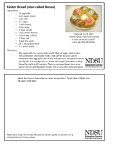
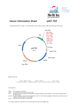
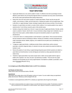
Biochimica et Biophysica Acta 1840 (2014) 2025–2031
Contents lists available at ScienceDirect
Biochimica et Biophysica Acta
journal homepage: www.elsevier.com/locate/bbagen
LRRK2, but not pathogenic mutants, protects against H2O2 stress
depending on mitochondrial function and endocytosis in a yeast model
Clara Pereira a,⁎, L. Miguel Martins b, Lucília Saraiva a,⁎
a
b
REQUIMTE, Laboratório de Microbiologia, Departamento de Ciências Biológicas, Faculdade de Farmácia, Universidade do Porto, Porto, Portugal
Cell Death Regulation Laboratory, MRC Toxicology Unit, Leicester, UK
a r t i c l e
i n f o
Article history:
Received 9 October 2013
Received in revised form 13 February 2014
Accepted 18 February 2014
Available online 24 February 2014
Keywords:
Saccharomyces cerevisiae
Parkinson's disease
LRRK2
Cellular dysfunction
Oxidative stress
a b s t r a c t
Background: Mutations in LRRK2 are the most common genetic cause of Parkinson's disease (PD). Studies in the
yeast Saccharomyces cerevisiae have provided valuable insights into the mechanisms of cellular dysfunction associated with the expression of faulty PD genes.
Methods: We developed a yeast model for full-length LRRK2 studies. We expressed wild-type (wt) LRRK2 and
mutations and evaluated their role during oxidative stress conditions. The involvement of mitochondria was
assessed by using rho-zero mutants and by evaluating reactive oxygen species (ROS) production and mitochondrial membrane potential by flow cytometry. The involvement of endocytosis was also studied by testing several
endocytic mutants and by following the vacuolar delivery of the probe FM4-64.
Results: Expression of LRRK2 in yeast was associated to increased hydrogen peroxide resistance. This phenotype,
which was dependent on mitochondrial function, was not observed for PD-mutants G2019S and R1441C or in the
absence of the kinase activity and the WD40 repeat domain. Expression of the pathogenic mutants stimulated
ROS production and increased mitochondrial membrane potential. For the PD-mutants, but not for wild-type
LRRK2, endocytic defects were also observed. Additionally, several endocytic proteins were required for
LRRK2-mediated protection against hydrogen peroxide.
Conclusions: Our results indicate that LRRK2 confers cellular protection during oxidative stress depending on
mitochondrial function and endocytosis.
General significance: Both the loss of capacity of LRRK2 pathogenic mutants to protect against oxidative stress and
their enhancement of dysfunction may be important for the development of PD during the aging process.
© 2014 Elsevier B.V. All rights reserved.
1. Introduction
Parkinson's disease (PD) is a fatal neurodegenerative disorder of the
central nervous system. Most of the PD cases are sporadic, although rare
familial forms of the disease have been linked to mutations in several
genes, providing research opportunities for pathogenic mechanisms [1].
Despite intensive research, the cause of PD remains obscure. Age is
apparently the greatest risk factor and many studies have implicated
mitochondrial dysfunction, oxidative stress, protein quality control,
autophagy and vesicular trafficking in PD pathogenesis or progression
[2]. Concerning mitochondrial dysfunction, extensive evidence has indicated a crucial role of this event in the pathogenesis of PD. Particularly, it
Abbreviations: H2O2, hydrogen peroxide; PD, Parkinson's disease; ROS, reactive oxygen species; S. cerevisiae, Saccharomyces cerevisiae; wt, wild-type; GFP, green fluorescent
protein; ΔΨm, mitochondrial membrane potential
⁎ Corresponding authors at: Laboratório de Microbiologia, Departamento de Ciências
Biológicas, Faculdade de Farmácia, Universidade do Porto, Rua Jorge Viterbo Ferreira,
228, 4050-313 Porto, Portugal. Tel.: +351 220428584; fax: +351 226 093 390.
E-mail addresses: [email protected] (C. Pereira), [email protected]
(L. Saraiva).
http://dx.doi.org/10.1016/j.bbagen.2014.02.015
0304-4165/© 2014 Elsevier B.V. All rights reserved.
was shown that mitochondrial toxins can cause Parkinsonism, and that
the activity of mitochondrial complex I is decreased in PD patients [3].
Also, several genes associated to familial PD are involved in mitochondrial function [4].
The most important genetic cause of both familial and a significant
proportion of apparently sporadic PD cases are autosomal-dominant
mutations in the leucine-rich repeat kinase 2 (LRRK2) gene [1,5].
LRRK2 has also been tentatively implicated in mitochondria dysfunction
and in the response to oxidative stress [6–9]. This is an attractive hypothesis as it suggests that different PD genes converge in a common
mitochondrial pathway. Yet, LRRK2 is a large and complex protein,
with multiple enzymatic and protein-interaction domains, whose biological function remains largely unknown. In the mammalian brain,
LRRK2 is present throughout the cytoplasm associated to various membranes and vesicular structures [10]. Also, a small percentage of LRRK2
was found associated to the mitochondrial outer membrane [11]. This
kinase has been implicated in several biological processes, including in
the endocytosis of synaptic vesicles [12,13], autophagy regulation [14],
and neurite outgrowth [15].
The LRRK2 domains include a leucine-rich repeat (LRR) domain, a
Roc (Ras of complex protein) domain related to the Ras-related GTPase
2026
C. Pereira et al. / Biochimica et Biophysica Acta 1840 (2014) 2025–2031
superfamily, a COR (C-terminal of Roc) domain, a serine/threonine
protein kinase domain, and a C-terminal WD40 repeats domain [16]. Although the presence of kinase and GTPase domains suggest a role of
LRRK2 in cellular signaling, the existence of multiple protein–protein
interaction domains, such as LRR, COR and WD40 repeats, suggests
that LRRK2 may also have a role in the assembly of protein complexes
[2,16]. The most common G2019S missense mutation lies in the kinase
domain, but disease-causing LRRK2 mutations are found almost throughout the protein [16].
Recently, relevant insights into the molecular mechanism of the PDrelated protein α-synuclein have been provided by the yeast model
system [17]. In fact, the study of human proteins in Saccharomyces
cerevisiae is attractive due to the simple genetic manipulation, overexpression and deletion screens (reviewed in [18]). Here, we report the
development of a yeast model of human LRRK2 expression to gain insights into the function and pathobiology associated with aberrant expression of this kinase.
2. Materials and methods
2.1. Vector construction for expression of wild-type (wt) and mutant forms
of LRRK2 in yeast
Human wt LRRK2 cDNA was obtained from a mammalian vector, generated and described in [11]. The LRRK2 construct was transferred to a
yeast expression vector using the Gateway cloning system (Invitrogen)
basically as reported [19]. The primers for cloning, listed in Table 1,
were designed for the LRRK2 gene from the published RNA sequence
(GenBank: AY792511) with the AttB1 and AttB2 overhangs for recombination cloning according to the supplier's instructions. The PCR cycling conditions consisted of an initial denaturation at 94 °C for 2 min,
10 cycles each consisting of 92 °C for 10 s, 53 °C for 30 s, and 68 °C for
8 min, followed by 20 cycles each consisting of 92 °C for 10 s, 53 °C
for 30 s, and 68 °C for 8 min + 10 s/cycle, with a final extension for
15 min at 68 °C. The directional gene transfer to the pAG423Gal-ccdBGFP expression vector (Addgene plasmid 14197; [20] was performed
via entry vector, using pDONR221 (Invitrogen) as donor plasmid, as described in the Gateway cloning protocol. For the construction of the
ΔWD40 mutant, the PCR amplification was performed as for the wt
LRRK2, but using a primer for the sequence just upstream of the
WD40 domain (Table 1). A LRRK2 containing the “triple kinase-dead”
construct [21] in a Gateway-compatible vector was kindly provided by
Dr. Mark Cookson and transferred to the pAG423Gal-ccdB-GFP vector
via pDONR221. LRRK2 patient-based mutations (G2019S and R1441C)
were introduced into pAG423Gal-LRRK2-GFP by PCR-based sitedirected mutagenesis [22] using the oligonucleotides listed in Table 1.
The PCR amplification was performed similarly as described before.
PCR products were digested with DpnI and amplified in Escherichia
coli NEB5a (New England Biolabs). All constructs were sequenced to ensure fidelity.
Table 1
Primers used in this study.
Purpose
Sequence
Cloning wt
LRRK2
Cloning
LRRK2
ΔWD40
Mutagenesis
G2019S
Mutagenesis
R1441C
GGGGACAAGTTTGTACAAAAAAGCAGGCTTAGAAGGAGATAGAACC
GGGGACCACTTTGTACAAGAAAGCTGGGTCCTCAACAGATGTTCGTCTCAT
GGGGACAAGTTTGTACAAAAAAGCAGGCTTAGAAGGAGATAGAACC
GGGGACCACTTTGTACAAGAAAGCTGGGTCGGGGCACAACCATATTCTT
CAAAGATTGCTGACTACAGCATTGCTCAGTACTGC
GCAGTACTGAGCAATGCTGTAGTCAGCAATCTTTG
CTTGGCTCTTCAATATAAAGGCTTGCGCTTCTTCTTC
GAAGAAGAAGCGCAAGCCTTTATATTGAAGAGCCAAG
2.2. Yeast strains, transformation and growth conditions
The S. cerevisiae strains used in this study are listed in Table 2. Cells
depleted of mitochondrial DNA (ρ0 cells) were generated from the
W303 strain by a two-step growth on Yeast Peptone Dextrose (YPD;
Difco) plates supplemented with ethidium bromide (40 μg/ml; Sigma)
[23]. The different strains were transformed with the vectors encoding
LRRK2 constructs/empty vector by the standard lithium acetate procedure [24]. Yeast cells were maintained and grown on selective minimal
medium with 2% (w/v) glucose (Sigma), 0.67% (w/v) Bacto-yeast nitrogen base w/o amino acids (Difco), and the required amino acids. For induction of protein expression, cells grown in selective minimal media
until exponential phase were diluted to 0.05 OD600 in selective induction media containing 0.05% (w/v) galactose (Sigma) plus 3% (w/v)
glycerol, instead of glucose, and incubated at 30 °C with shake
(200 rpm). For induction of protein expression in respiratory deficient
cells, a mixture of 0.05% galactose and 2% raffinose (Acros Organics)
was used.
All the experiments were performed with two independent clones
of the LRRK2-GFP constructs (wt and mutants).
2.3. Western blot analysis
Preparation of protein samples, SDS-PAGE and Western blots were
performed as previously described [25]. The primary antibodies,
mouse monoclonal against GFP (1:2500; Clontech) and yeast phosphoglycerate kinase (Pgk1p; 1:6000; Molecular Probes), were used followed by an anti-mouse horseradish-peroxidase-conjugated secondary
antibody (1:5000; Santa Cruz Biotechnology) and revealed by chemiluminescence (ECL, Amersham). Band intensities were quantified using
the Bio-Profil Bio-1D++ software (Vilber-Lourmat).
2.4. Viability assays
For oxidative stress experiments, exponential cultures (approximately 0.5 OD600) were treated with 1–5 mM H2O2 for 1 h with shaking
at 30 °C. Viability was assessed by colony-forming unit (CFU) counts
after 2 days incubation at 30 °C on Sabouraud Dextrose Agar (Difco)
plates, and expressed as percentage of time zero.
2.5. Endocytosis assay
Endocytosis was assessed using FM4-64 (N-(3-thiethylammoniumpropyl)-4-(p-diethyl-aminophenylhexatrienyl) pyridinium dibromide)
(Molecular Probes), basically as described [26]. Briefly, cells were incubated with 3 μg/ml FM4-64 along time at 30 °C and observed under an
Eclipse E400 fluorescence microscope (Nikon). Images were captured
with a Digital Sight Camera System (Nikon DS-5Mc) carrying built-in software for image acquisition (Nikon ACT-2U) and a semi-quantitative evaluation of cells with endocytic defects (lacking vacuolar staining) was
Table 2
Saccharomyces cerevisiae strains used in this study.
W303-1B
BY4741
ede1Δ
sla1Δ
end3Δ
end6Δ
bzz1Δ
vps21Δ
vps41Δ
ypt7Δ
Mata ura3-1 leu2–3, 112 his3–11,15
trp1-1 ade2-1 can1–100
Mat a; his3Δ 1; leu2Δ 0;
met15Δ 0; ura3Δ 0
By4741; YBL047c::kanMX4
By4741; YBL007c::kanMX4
By4741; YNL084c::kanMX4
By4741; YCR009c::kanMX4
By4741; YKL129c::kanMX4
By4741; YOR089c::kanMX4
By4741; YDR080w::kanMX4
By4741; YML001w::kanMX4
Lab collection
EUROSCARF collection
EUROSCARF collection
EUROSCARF collection
EUROSCARF collection
EUROSCARF collection
EUROSCARF collection
EUROSCARF collection
EUROSCARF collection
EUROSCARF collection
C. Pereira et al. / Biochimica et Biophysica Acta 1840 (2014) 2025–2031
estimated by counting at least 300 cells per sample in three independent
experiments.
2.6. Assessment of mitochondrial membrane potential (ΔΨm) and reactive
oxygen species (ROS) production
To assess ΔΨm, 5 × 10⁶ cells were incubated for 30 min at 30 °C
with 40 nM of 3,3-dihexyloacarbocynine iodide (DiOC6 (3)) (Molecular
Probes). To assess intracellular superoxide anion production, cells were
incubated with 5 μg/ml of dihydroethidium (DHE; Sigma) for 30 min at
30 °C. Sample analysis was performed in FACSCalibur (BD Biosciences)
flow cytometer using CellQuest software (BD Biosciences). Twenty
thousand cells were analyzed per sample.
3. Results
3.1. LRRK2 expression in yeast
To study human wt and mutant forms of LRRK2 in yeast, constructs
were tagged with a C-terminal green fluorescent protein (GFP) and
were expressed under the control of the regulatable GAL10 promoter.
Mutant forms of LRRK2 included the pathogenic mutations G2019S
and R1441C, and a LRRK2 mutant containing three point mutations
that disrupt kinase activity (3KD) [21]. A deletion in the WD40 domain
required for LRRK2 function and neurotoxicity (ΔWD40) was also studied [27,28] (Fig. 1A).
2027
Expression of wt and mutant forms of LRRK2-GFP in yeast was confirmed by Western analysis (Fig. 1B). As previously reported for native
LRRK2 [29], a high amount of LRRK2-GFP fusion forms were found as insoluble protein (accumulated between the stacking and separating gel),
while only a small fraction appeared as soluble protein (quantified in
Fig. 1C). The insoluble protein may be due to the formation of LRRK2GFP aggregates, as previously reported in several cell models for native
LRRK2 [9,30–32]. In fact, by fluorescence microscopy, diffuse cytoplasmic fluorescence and variable size inclusions (indistinguishable for the
distinct constructs) could be observed in yeast cells expressing the
LRRK2-GFP constructs, in contrast to the control yeast that only exhibited cytoplasmic fluorescence (Fig. 1D). Because we were using an inducible GAL promoter, and the amount of galactose used correlated
with expression levels, and consequent inclusion formation, we used a
lower percentage of galactose (0.05%; supplemented with 3% glycerol)
to minimize this effect.
As previously reported [29], no reproducible growth phenotype was
obtained with the LRRK2-GFP constructs (Fig. 1E).
3.2. Wt LRRK2 is associated with protection against H2O2 toxicity
There has been extensive evidence linking oxidative stress to both
the initiation and the progression of PD [33]. Several studies support a
role for LRRK2 in the response to oxidative stress, usually mimicked
by H2O2 exposure, but the outcome is controversial [7,34–37]. As such,
our yeast model system was used to address this relevant issue. For
Fig. 1. Expression of human wt and mutant forms of LRRK2 in yeast. (A) Schematic of the domain structure of human LRRK2 with the studied mutations indicated. ANK, ankyrin; LRR,
leucine-rich repeats; Roc, Ras of complex proteins; COR, C-terminal of Roc; and Kinase, mitogen-activated protein kinase kinase kinases. (B) Immunoblot analysis of cells expressing
LRRK2-GFP constructs. Pgk1p was used as loading control. (C) Quantification of protein expression (insoluble and soluble fractions). Fold change was quantified relative to Pgk1p.
(D) Fluorescence microscopy of yeast cells expressing GFP alone or LRRK2-GFP constructs. (E) Growth of control yeast (vector) and yeast expressing LRRK2 constructs in induction
media along time. Yeast cell growth was assessed by optical density measurements at 600 nm. Values are means ± SE (n = 4). The effect on growth was not significant; (P N 0.05),
two-way ANOVA.
2028
C. Pereira et al. / Biochimica et Biophysica Acta 1840 (2014) 2025–2031
that, cells expressing wt or mutant forms of LRRK2-GFP were exposed to
H2O2 for 1 h. Expression of wt LRRK2-GFP resulted in a significant resistance to oxidative stress. In fact, in the presence of wt LRRK2-GFP, about
20% and 14% increase in the percentage of cell survival was obtained for
3 mM and 5 mM, respectively, when compared to the control yeast
(Fig. 2A). On the contrary, for G2019S-GFP, R1441C-GFP, ΔWD40-GFP
and 3KD-GFP no significant differences in the percentage of cell survival
were observed (Fig. 2A). When 2% galactose was used to induce high
levels of expression, the higher inclusion formation was associated to
a loss of H2O2 resistance (not shown), supporting a role for soluble
LRRK2 in this resistance.
Because in recent years a connection of LRRK2 with mitochondria
has been suggested [2], we decided to determine whether functional mitochondria were determinant for LRRK2 response to oxidative stress in
yeast. For that, a ρ0 strain, which lacks mitochondrial genome, was
used. Since the ρ0 strains do not grow well in galactose alone, for induction of LRRK2 constructs expression, a combination of galactose and raffinose, a fermentable substrate, but not a mitochondrial function
repressor, was used. We confirmed that, in this culture medium, the
LRRK2-GFP resistance to H2O2 was not affected in the parental ρ+ strain
(Fig. 2B). Since ρ0 cells are more sensitive to H2O2, a lower concentration
of H2O2 (1 mM) was used in order to obtain a comparable percentage of
cell death to the wt respiratory competent cells. In the absence of functional mitochondria, wt and mutant forms of LRRK2-GFP exhibited a
similar percentage of cell survival to the control yeast (Fig. 2B). These
results indicate an involvement of the mitochondrial pathway in the
LRRK2 function. Based on this, markers of mitochondria dysfunction
were checked in the wt strain expressing LRRK2-GFP constructs in response to H2O2, particularly ROS production and ΔΨm. After 1 h treatment with 5 mM H2O2, a small decrease in ROS production for cells
expressing wt LRRK2-GFP was observed. On the contrary, stimulation
in ROS production was obtained for the mutants G2019S-GFP, R1441CGFP and ΔWD40-GFP (higher for the two former pathogenic mutants).
Interestingly, no effect on ROS production was observed for the 3KD mutant (Fig. 2C). The ΔΨm was also measured either in the absence and
presence of 5 mM H2O2. For untreated cells, a decrease in ΔΨm was observed for all LRRK2-GFP constructs, though more evident for the pathogenic mutants G2019S-GFP and R1441C-GFP, when compared to the
control vector (Fig. 2D; dark line). For H2O2-treated cells, though a typical depolarization was observed in the control vector, a hyperpolarization was obtained for the mutants G2019S-GFP and R1441C-GFP. For wt
LRRK2-GFP, ΔWD40-GFP and 3KD-GFP, an intermediate response
Fig. 2. Effect of wt and mutant forms of LRRK2 on cell survival upon H2O2 exposure. (A) Wt strain expressing LRRK2-GFP constructs or transformed with the empty vector (control) were
grown until approximately 0.4 OD600 followed by treatment with the indicated concentration of H2O2 for 1 h. Data are mean ± SE (n = 4). *P b 0.05, by Student's t-test compared to vector.
(B) Wt (ρ+) and derived ρ0 strains expressing LRRK2-GFP constructs or control were incubated in a combination of 0.05% galactose and 2% raffinose medium until approximately 0.4 OD600
followed by treatment with 1 mM H2O2 for 1 h. Cell survival was assessed by CFU counts; 100% survival corresponds to the number of CFU at time 0. Data are mean ± SE (n = 4). *P b 0.05,
by Student's t-test compared to Vector. Cellular dysfunction associated to H2O2 treatment in yeast expressing wt or mutant forms of LRRK2. (C) ROS production was assessed by monitoring
the increase in DHE fluorescence in untreated (black bars) or 5 mM H2O2-treated yeast cells (white bars) expressing LRRK2-GFP constructs and control yeast (vector) by flow cytometry.
Data are mean ± SE (n = 4). *P b 0.05, by Student's t-test compared to vector. (D) Mitochondrial depolarization was assessed by monitoring the DiOC6(3) fluorescence change in untreated
(dark line) and 5 mM H2O2-treated (light line) yeast cells expressing LRRK2-GFP constructs and control yeast (vector) by flow cytometry. Image shows monoparametric histograms of
DiOC6(3) fluorescence representing one of the four independent experiments.
C. Pereira et al. / Biochimica et Biophysica Acta 1840 (2014) 2025–2031
between the pathogenic mutants and the control vector was observed
(Fig. 2D; light line). The ΔΨm experiments were also performed in ρ0
cells without a functional respiratory chain. In these H2O2 treated cells,
unlike for ρ+ cells, the LRRK2 constructs exhibited a similar depolarization to the control vector (not shown). Together, these results suggest
that the pathogenic mutants G2019S-GFP and R1441C-GFP further exacerbate the mitochondrial dysfunction in response to H2O2 toxicity.
Moreover, since in ρ0 cells the mitochondrial respiratory chain is not
functional, these proteins may be acting on this complex.
3.3. LRRK2 function is dependent on the endocytic pathway
LRRK2 has been implicated in the modulation of synaptic vesicle
trafficking [12,32]. The GTPase domain may be particularly important
for this modulation since the expression of a LRRK2 fragment containing
the GTPase domain was shown to impair endocytosis in yeast [29]. We
decided to evaluate the effect of the full-length LRRK2 constructs on vesicular trafficking upon H2O2 treatment. For that, the internalization
of the membrane-binding fluorescent dye FM4-64 was monitored
after 2 h (when all the H2O2-untreated strains presented more than
2029
90% vacuolar staining) incubation. As expected, in untreated cells (vector or LRRK2-GFP constructs), FM4-64 was rapidly endocytosed and accumulated at the vacuolar membrane (not shown). Upon treatment
with 5 mM H2O2, a small percentage of control cells exhibited endocytic
defects characterized by the appearance of dot like structures (Fig. 3A,
green arrowhead; quantification in Fig. 3B and C) as described [38] in
addition to the large ring-like vacuolar staining (Fig. 3A, white arrowhead). In cells expressing G2019S-GFP or R1441C-GFP, the percentage
of cells with endocytic defects showed an increase of about 28% for
G2019S-GFP and of 20% for R1441C-GFP, when compared to the control
vector (Fig. 3B and C). In cells expressing wt LRRK2-GFP, ΔWD40-GFP or
3KD-GFP no major interference in endocytosis was observed, when
compared to the control vector (Fig. 3B and C).
Since the endocytic process is genetically well defined in yeast, to
assess the impact of vesicular trafficking on the cellular response to
LRRK2, a panel of null yeast mutants in defined steps of the endocytic
pathway was used (Fig. 3D). These included Ede1p (early immobile
phase), End3p, Sla1p and End6p/Rvs161 (Mid/late immobile phase),
Bzz1p (actin/mobile phase), Vps21p (vesicle transport), Vps41p and
Ypt7 (transport from late endosomes to the vacuole) [12,39].
Fig. 3. Effect of LRRK2 on endocytosis during H2O2 treatment. (A) Representative photomicrographs of 5 mM H2O2-treated cells stained with FM4-64. Bar, 15 μm. White arrowhead indicates normal cell with vacuolar staining, and green arrowhead indicates a cell with endocytic defects. (B) Quantitative expression of endocytic defects (lacking vacuolar staining), untreated (black bars) or after 2 h incubation with 5 mM H2O2 (white bars). Values are mean ± SE (n = 3). (C) Distribution of endocytic defects (cells with different amounts of FM4-64-stained
punctate structures). (D) Schematic of the tested endocytic mutants. (E) Stress resistance of endocytic mutant strains expressing wt LRRK2-GFP. Yeast cell growth was assessed by CFU
counts after 1 h incubation with 5 mM H2O2 in induction medium. Data are mean ± SE (n = 4). *P b 0.05, **P b 0.01, paired t-test.
2030
C. Pereira et al. / Biochimica et Biophysica Acta 1840 (2014) 2025–2031
After treatment with H2O2, protection provided by wt LRRK2-GFP
was strongly reduced when expressed in ede1Δ or end3Δ strains, and
moderately reduced when expressed in ypt7Δ (Fig. 3E). In the remaining endocytic yeast mutants (sla1Δ, bzz1Δ, vps41Δ, end6Δ, vps21Δ), protection provided by wt LRRK2-GFP was similar as when expressed in the
wt strain (Fig. 3E).
4. Discussion
In the present work, the yeast S. cerevisiae was used to study the full
length LRRK2 protein. We observed, as reported before, that the overexpression of wt LRRK2 or PD-associated mutants G2019S and R1441C was
not cytotoxic to yeast. However, unlike wt LRRK2, the PD-associated mutants were unable to protect cells against H2O2 toxicity. LRRK2 has been
associated with the protection against mitochondrial stressors and other
types of oxidative stress [7,34,36,37]. However, contradicting results, in
which LRRK2 exacerbates stress, have also been reported [35,40].
Based on our observations, these contradicting results may be attributed
to distinct experimental conditions. In fact, when a high percentage of
galactose was used (2%), leading to higher expression levels of LRRK2,
a small enhancement of H2O2 toxicity was also observed in our model
(not shown). The protective function we observed when expressing
LRRK2 at low levels, was in yeast, as reported for other cellular models
[37,41], dependent on the kinase activity. This reinforces the importance
of the LRRK2 catalytic domain in the resistance against stress, making the
inhibition of LRRK2 kinase as a therapeutic approach against PD to be
considered cautiously. Besides, in this work, the protective function of
another LRRK2 domain, the WD40 repeats, was also investigated. Our
results indicate that also this domain has a protective effect against
H2O2-induced oxidative stress.
As reported for other cellular systems [42], also in yeast the LRRK2mediated protection against H2O2-induced stress was lost for the pathogenic mutations G2019S and R1441C. This supports that deficits in oxidative stress resistance may be important in the development of PD.
The protective function of LRRK2 may be explained by a potential
role in the homeostasis of oxidative stress within the cells, by acting on
antioxidant proteins. In fact, this was suggested by the observation that
LRRK2 mutants decrease the mitochondrial peroxidase peroxiredoxin 3
(PRDX3)[43] and the glutathione s-transferase P1 (GSTP1)[44] activity,
contributing to mitochondrial dysfunction and increased oxidative damage. In accordance, we observed for the pathogenic mutants, in addition
to the loss-of-function, an increase in ROS production and also an increase in endocytic defects, which may be a consequence of increased
ROS. In cells with high energy demands, such as neurons, this enhancement of oxidative stress-induced dysfunction may be a relevant mechanism of pathogenicity. Oxidative stress is intimately linked with
mitochondrial dysfunction. A role for mitochondria in LRRK2 function
has already been suggested. Particularly, a small percentage of LRRK2
was found to be associated with the mitochondrial outer membrane
[11], and it was shown that LRRK2 protects against mitochondrial
stressors [6,42]. Additionally, it was recently reported that LRRK2 has a
role in regulating mitochondrial dynamics [45,46] and ΔΨm. In fact, it
was observed both in PD patients [8] and in vitro [47] that the G2019S
mutation reduces ΔΨm. These reports are in accordance with our findings in yeast. The protection provided by LRRK2 in yeast was significantly decreased in cells lacking functional mitochondria. In addition, in this
work, it was shown that G2019S and R1441C decreased ΔΨm in the absence of any stressor, which results in mitochondria hyperpolarization
instead of depolarization upon H2O2-induced stress. This hyperpolarization may be due to a stimulation of the respiratory chain activity, since in
ρ0 cells this effect is lost. Because in ρ0 yeast cells both respiration and
mitochondrial-nuclear communication (e.g., retrograde signaling) are
impaired [48], it will be interesting, in future work, to further ascertain
the contribution of these processes for LRRK2 toxicity.
The stress protection offered by LRRK2 was significantly decreased
in cells lacking several endocytic proteins. Taking into account the
number of tested endocytic proteins that were able to affect
the LRRK2-induced stress resistance, this pathway may have a major
role in the LRRK2 function. Recently, it was reported that diseaseassociated LRRK2 interacts with members of the dynamin GTPase superfamily, proteins with a role in the regulation of membrane dynamics,
important for both endocytosis and mitochondrial morphology, connecting these two processes [49]. Most of the proteins in the endocytic
pathway are evolutionary conserved in mammals. Ypt7 is the homologue of Rab7, recently described to physically interact with LRRK2 in
Caenorhabditis elegans [50] and Vps21 is the homologue of Rab5, described as a LRRK2 interactor, and suggested to co-regulate with
LRRK2 the endocytic pathway [12]. In our model, however, only Ypt7 affected LRRK2 function in the cellular stress response. The two proteins
with the most dramatic effects on wt LRRK2-mediated stress protection
were Ede1p and End3p that play a role in the initial phase of vesicle formation [39]. Ede1 and End3 have homology to human Eps15, an adaptor protein involved in the epidermal growth factor (EGF) receptor
(EGFR) endocytosis and trafficking. The Eps15 was the first identified
substrate of Parkin-mediated monoubiquitination [51]. Therefore, it
will be interesting to ascertain if Eps15 can also act as an LRRK2
substrate.
In conclusion, in the present work insights into the LRRK2 function
and the pathogenic mechanisms of PD-associated mutations were provided. Particularly, it was shown that mitochondrial function and endocytosis seem to be intersecting pathways in the LRRK2 function. This
yeast model may help to identify new associated/interacting proteins
therefore contributing to a deeper understanding of the role of LRRK2
in PD. Besides its use for genetic screens, this model may also be adapted
for chemical screening assays, based on the direct assessment of the
yeast cell growth.
Acknowledgments
We thank Dr. Mark Cookson for kindly providing the “triple kinasedead” LRRK2 construct. This work was supported by FCT (Fundação para
a Ciência e a Tecnologia) through REQUIMTE (PEst-C/EQB/LA0006/
2011) and by U. Porto/Santander Totta.
References
[1] C. Paisan-Ruiz, S. Jain, E.W. Evans, W.P. Gilks, J. Simon, M. van der Brug, A. Lopez de
Munain, S. Aparicio, A.M. Gil, N. Khan, J. Johnson, J.R. Martinez, D. Nicholl, I.M.
Carrera, A.S. Pena, R. de Silva, A. Lees, J.F. Marti-Masso, J. Perez-Tur, N.W. Wood,
A.B. Singleton, Cloning of the gene containing mutations that cause PARK8-linked
Parkinson's disease, Neuron 44 (2004) 595–600.
[2] E. Tsika, D.J. Moore, Mechanisms of LRRK2-mediated neurodegeneration, Curr.
Neurol. Neurosci. Rep. 12 (2012) 251–260.
[3] N. Exner, A.K. Lutz, C. Haass, K.F. Winklhofer, Mitochondrial dysfunction in
Parkinson's disease: molecular mechanisms and pathophysiological consequences,
EMBO J. 31 (2012) 3038–3062.
[4] Y. Sai, Z. Zou, K. Peng, Z. Dong, The Parkinson's disease-related genes act in mitochondrial homeostasis, Neurosci. Biobehav. Rev. 36 (2012) 2034–2043.
[5] A. Zimprich, S. Biskup, P. Leitner, P. Lichtner, M. Farrer, S. Lincoln, J. Kachergus, M.
Hulihan, R.J. Uitti, D.B. Calne, A.J. Stoessl, R.F. Pfeiffer, N. Patenge, I.C. Carbajal, P.
Vieregge, F. Asmus, B. Muller-Myhsok, D.W. Dickson, T. Meitinger, T.M. Strom, Z.K.
Wszolek, T. Gasser, Mutations in LRRK2 cause autosomal-dominant Parkinsonism
with pleomorphic pathology, Neuron 44 (2004) 601–607.
[6] S. Saha, M.D. Guillily, A. Ferree, J. Lanceta, D. Chan, J. Ghosh, C.H. Hsu, L. Segal, K.
Raghavan, K. Matsumoto, N. Hisamoto, T. Kuwahara, T. Iwatsubo, L. Moore, L.
Goldstein, M. Cookson, B. Wolozin, LRRK2 modulates vulnerability to mitochondrial
dysfunction in Caenorhabditis elegans, J. Neurosci. 29 (2009) 9210–9218.
[7] D. Wang, B. Tang, G. Zhao, Q. Pan, K. Xia, R. Bodmer, Z. Zhang, Dispensable role of
Drosophila ortholog of LRRK2 kinase activity in survival of dopaminergic neurons,
Mol. Neurodegener. 3 (2008) 3.
[8] H. Mortiboys, K.K. Johansen, J.O. Aasly, O. Bandmann, Mitochondrial impairment in
patients with Parkinson disease with the G2019S mutation in LRRK2, Neurology
75 (2010) 2017–2020.
[9] C.H. Ng, S.Z. Mok, C. Koh, X. Ouyang, M.L. Fivaz, E.K. Tan, V.L. Dawson, T.M. Dawson,
F. Yu, K.L. Lim, Parkin protects against LRRK2 G2019S mutant-induced dopaminergic
neurodegeneration in Drosophila, J. Neurosci. 29 (2009) 11257–11262.
[10] S. Biskup, D.J. Moore, F. Celsi, S. Higashi, A.B. West, S.A. Andrabi, K. Kurkinen, S.W. Yu,
J.M. Savitt, H.J. Waldvogel, R.L. Faull, P.C. Emson, R. Torp, O.P. Ottersen, T.M. Dawson,
V.L. Dawson, Localization of LRRK2 to membranous and vesicular structures in
mammalian brain, Ann. Neurol. 60 (2006) 557–569.
C. Pereira et al. / Biochimica et Biophysica Acta 1840 (2014) 2025–2031
[11] A.B. West, D.J. Moore, S. Biskup, A. Bugayenko, W.W. Smith, C.A. Ross, V.L. Dawson,
T.M. Dawson, Parkinson's disease-associated mutations in leucine-rich repeat kinase
2 augment kinase activity, Proc. Natl. Acad. Sci. U. S. A. 102 (2005) 16842–16847.
[12] N. Shin, H. Jeong, J. Kwon, H.Y. Heo, J.J. Kwon, H.J. Yun, C.H. Kim, B.S. Han, Y. Tong, J.
Shen, T. Hatano, N. Hattori, K.S. Kim, S. Chang, W. Seol, LRRK2 regulates synaptic
vesicle endocytosis, Exp. Cell Res. 314 (2008) 2055–2065.
[13] S. Matta, K. Van Kolen, R. da Cunha, G. van den Bogaart, W. Mandemakers, K.
Miskiewicz, P.J. De Bock, V.A. Morais, S. Vilain, D. Haddad, L. Delbroek, J. Swerts, L.
Chavez-Gutierrez, G. Esposito, G. Daneels, E. Karran, M. Holt, K. Gevaert, D.W.
Moechars, B. De Strooper, P. Verstreken, LRRK2 controls an EndoA phosphorylation
cycle in synaptic endocytosis, Neuron 75 (2012) 1008–1021.
[14] J. Alegre-Abarrategui, H. Christian, M.M.P. Lufino, R. Mutihac, L.L. Venda, O. Ansorge,
R. Wade-Martins, LRRK2 regulates autophagic activity and localizes to specific
membrane microdomains in a novel human genomic reporter cellular model,
Hum. Mol. Genet. 18 (2009) 4022–4034.
[15] D. MacLeod, J. Dowman, R. Hammond, T. Leete, K. Inoue, A. Abeliovich, The familial
Parkinsonism gene LRRK2 regulates neurite process morphology, Neuron 52 (2006)
587–593.
[16] I.F. Mata, W.J. Wedemeyer, M.J. Farrer, J.P. Taylor, K.A. Gallo, LRRK2 in Parkinson's
disease: protein domains and functional insights, Trends Neurosci. 29 (2006)
286–293.
[17] V. Khurana, S. Lindquist, Modelling neurodegeneration in Saccharomyces cerevisiae:
why cook with baker's yeast? Nat. Rev. Neurosci. 11 (2010) 436–449.
[18] C. Pereira, M. Leao, J. Soares, C. Bessa, L. Saraiva, New therapeutic strategies for cancer and neurodegeneration emerging from yeast cell-based systems, Curr. Pharm.
Des. 18 (2012) 4223–4235.
[19] J.L. Hartley, G.F. Temple, M.A. Brasch, DNA cloning using in vitro site-specific recombination, Genome Res. 10 (2000) 1788–1795.
[20] S. Alberti, A.D. Gitler, S. Lindquist, A suite of Gateway cloning vectors for highthroughput genetic analysis in Saccharomyces cerevisiae, Yeast 24 (2007) 913–919.
[21] E. Greggio, S. Jain, A. Kingsbury, R. Bandopadhyay, P. Lewis, A. Kaganovich, M.P. van
der Brug, A. Beilina, J. Blackinton, K.J. Thomas, R. Ahmad, D.W. Miller, S. Kesavapany,
A. Singleton, A. Lees, R.J. Harvey, K. Harvey, M.R. Cookson, Kinase activity is required
for the toxic effects of mutant LRRK2/dardarin, Neurobiol. Dis. 23 (2006) 329–341.
[22] M.P. Weiner, G.L. Costa, W. Schoettlin, J. Cline, E. Mathur, J.C. Bauer, Site-directed
mutagenesis of double-stranded DNA by the polymerase chain reaction, Gene 151
(1994) 119–123.
[23] T.D. Fox, L.S. Folley, J.J. Mulero, T.W. McMullin, P.E. Thorsness, L.O. Hedin, M.C.
Costanzo, Analysis and manipulation of yeast mitochondrial genes, Methods
Enzymol. 194 (1991) 149–165.
[24] R.D. Gietz, R.H. Schiestl, High-efficiency yeast transformation using the LiAc/SS carrier DNA/PEG method, Nat. Protoc. 2 (2007) 31–34.
[25] L. Saraiva, R.D. Silva, G. Pereira, J. Goncalves, M. Corte-Real, Specific modulation of
apoptosis and Bcl-xL phosphorylation in yeast by distinct mammalian protein kinase C isoforms, J. Cell Sci. 119 (2006) 3171–3181.
[26] T.A. Vida, S.D. Emr, A new vital stain for visualizing vacuolar membrane dynamics
and endocytosis in yeast, J. Cell Biol. 128 (1995) 779–792.
[27] N.D. Jorgensen, Y. Peng, C.C.Y. Ho, H.J. Rideout, D. Petrey, P. Liu, W.T. Dauer, The
WD40 domain is required for LRRK2 neurotoxicity, PLoS One 4 (2009) e8463.
[28] D. Sheng, D. Qu, K.H. Kwok, S.S. Ng, A.Y. Lim, S.S. Aw, C.W. Lee, W.K. Sung, E.K. Tan, T.
Lufkin, S. Jesuthasan, M. Sinnakaruppan, J. Liu, Deletion of the WD40 domain of
LRRK2 in Zebrafish causes Parkinsonism-like loss of neurons and locomotive defect,
PLoS Genet. 6 (2010) e1000914.
[29] Y. Xiong, C.E. Coombes, A. Kilaru, X. Li, A.D. Gitler, W.J. Bowers, V.L. Dawson, T.M.
Dawson, D.J. Moore, GTPase activity plays a key role in the pathobiology of LRRK2,
PLoS Genet. 6 (2010) e1000902.
[30] L.R. Kett, D. Boassa, C.C. Ho, H.J. Rideout, J. Hu, M. Terada, M. Ellisman, W.T. Dauer,
LRRK2 Parkinson disease mutations enhance its microtubule association, Hum.
Mol. Genet. 21 (2012) 890–899.
[31] W.W. Smith, Z. Pei, H. Jiang, D.J. Moore, Y. Liang, A.B. West, V.L. Dawson, T.M.
Dawson, C.A. Ross, Leucine-rich repeat kinase 2 (LRRK2) interacts with parkin,
and mutant LRRK2 induces neuronal degeneration, Proc. Natl. Acad. Sci. U. S. A.
102 (2005) 18676–18681.
2031
[32] G. Piccoli, S.B. Condliffe, M. Bauer, F. Giesert, K. Boldt, S. De Astis, A. Meixner, H.
Sarioglu, D.M. Vogt-Weisenhorn, W. Wurst, C.J. Gloeckner, M. Matteoli, C. Sala, M.
Ueffing, LRRK2 controls synaptic vesicle storage and mobilization within the
recycling pool, J. Neurosci. 31 (2011) 2225–2237.
[33] P. Jenner, Oxidative stress as a cause of Parkinson's disease, Acta Neurol. Scand.
Suppl. 136 (1991) 6–15.
[34] Y. Imai, S. Gehrke, H.Q. Wang, R. Takahashi, K. Hasegawa, E. Oota, B. Lu, Phosphorylation of 4E-BP by LRRK2 affects the maintenance of dopaminergic neurons in
Drosophila, EMBO J. 27 (2008) 2432–2443.
[35] A.B. West, D.J. Moore, C. Choi, S.A. Andrabi, X. Li, D. Dikeman, S. Biskup, Z. Zhang, K.L.
Lim, V.L. Dawson, T.M. Dawson, Parkinson's disease-associated mutations in LRRK2
link enhanced GTP-binding and kinase activities to neuronal toxicity, Hum. Mol.
Genet. 16 (2007) 223–232.
[36] E. Ohta, M. Kubo, F. Obata, Prevention of intracellular degradation of I2020T mutant
LRRK2 restores its protectivity against apoptosis, Biochem. Biophys. Res. Commun.
391 (2010) 242–247.
[37] A.K.F. Liou, R.K. Leak, L. Li, M.J. Zigmond, Wild-type LRRK2 but not its mutant attenuates stress-induced cell death via ERK pathway, Neurobiol. Dis. 32 (2008)
116–124.
[38] C. Pereira, C. Bessa, L. Saraiva, Endocytosis inhibition during H(2) O(2)-induced
apoptosis in yeast, FEMS Yeast Res. 12 (2012) 755–760.
[39] J. Lachmann, C. Ungermann, S. Engelbrecht-Vandre, Rab GTPases and tethering in
the yeast endocytic pathway, Small GTPases 2 (2011) 182–186.
[40] H.Y. Heo, J.M. Park, C.H. Kim, B.S. Han, K.S. Kim, W. Seol, LRRK2 enhances oxidative
stress-induced neurotoxicity via its kinase activity, Exp. Cell Res. 316 (2010)
649–656.
[41] D.C. Berwick, K. Harvey, LRRK2 functions as a Wnt signaling scaffold, bridging cytosolic proteins and membrane-localized LRP6, Hum. Mol. Genet. 21 (2012) 4966–4979.
[42] C.H. Hsu, D. Chan, E. Greggio, S. Saha, M.D. Guillily, A. Ferree, K. Raghavan, G.C. Shen,
L. Segal, H. Ryu, M.R. Cookson, B. Wolozin, MKK6 binds and regulates expression of
Parkinson's disease-related protein LRRK2, J. Neurochem. 112 (2010) 1593–1604.
[43] D.C. Angeles, B.H. Gan, L. Onstead, Y. Zhao, K.L. Lim, J. Dachsel, H. Melrose, M. Farrer,
Z.K. Wszolek, D.W. Dickson, E.K. Tan, Mutations in LRRK2 increase phosphorylation
of peroxiredoxin 3 exacerbating oxidative stress-induced neuronal death, Hum.
Mutat. 32 (2011) 1390–1397.
[44] J. Chen, A. Liou, L. Zhang, Z. Weng, Y. Gao, G. Cao, M.J. Zigmond, GST P1, a novel
downstream regulator of LRRK2, G2019S-induced neuronal cell death, Front. Biosci.
(Elite Ed.) 4 (2012) 2365–2377.
[45] X. Wang, M.H. Yan, H. Fujioka, J. Liu, A. Wilson-Delfosse, S.G. Chen, G. Perry, G.
Casadesus, X. Zhu, LRRK2 regulates mitochondrial dynamics and function through
direct interaction with DLP1, Hum. Mol. Genet. 21 (2012) 1931–1944.
[46] J. Niu, M. Yu, C. Wang, Z. Xu, Leucine-rich repeat kinase 2 disturbs mitochondrial dynamics via Dynamin-like protein, J. Neurochem. 122 (2012) 650–658.
[47] T.D. Papkovskaia, K.Y. Chau, F. Inesta-Vaquera, D.B. Papkovsky, D.G. Healy, K. Nishio,
J. Staddon, M.R. Duchen, J. Hardy, A.H. Schapira, J.M. Cooper, G2019S leucine-rich repeat kinase 2 causes uncoupling protein-mediated mitochondrial depolarization,
Hum. Mol. Genet. 21 (2012) 4201–4213.
[48] D.K. Woo, R.O. Poyton, The absence of a mitochondrial genome in rho0 yeast cells
extends lifespan independently of retrograde regulation, Exp. Gerontol. 44 (2009)
390–397.
[49] K. Stafa, E. Tsika, R. Moser, A. Musso, L. Glauser, A. Jones, S. Biskup, Y. Xiong, R.
Bandopadhyay, V.L. Dawson, T.M. Dawson, D.J. Moore, Functional interaction of
Parkinson's disease-associated LRRK2 with members of the dynamin GTPase superfamily, Hum. Mol. Genet. (2013), http://dx.doi.org/10.1093/hmg/ddt600 (Epub
ahead of print).
[50] M.W. Dodson, T. Zhang, C. Jiang, S. Chen, M. Guo, Roles of the Drosophila LRRK2
homolog in Rab7-dependent lysosomal positioning, Hum. Mol. Genet. 21 (2012)
1350–1363.
[51] L. Fallon, C.M. Belanger, A.T. Corera, M. Kontogiannea, E. Regan-Klapisz, F. Moreau, J.
Voortman, M. Haber, G. Rouleau, T. Thorarinsdottir, A. Brice, P.M. van Bergen En
Henegouwen, E.A. Fon, A regulated interaction with the UIM protein Eps15 implicates parkin in EGF receptor trafficking and PI(3)K-Akt signalling, Nat. Cell Biol. 8
(2006) 834–842.


© Copyright 2026