
2 How to Identify and Avoid Artifacts on DWI
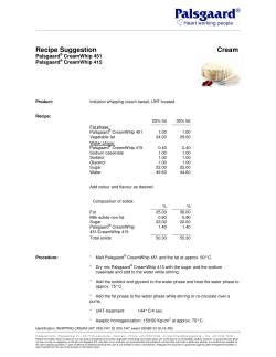
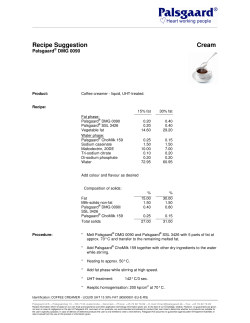
2
How to Identify and Avoid
Artifacts on DWI
Javier Sánchez-González
DWI is currently considered a cancer biomarker and
has a role in cancer detection and staging. DWI is also
able to depict early posttreatment changes in oncological lesions treated with vascular disruptive drugs and
useful for therapies that induce apoptosis. After these
treatments, cellular death and vascular changes occur
before changes in lesion size can be seen. Successful
treatment is reflected by increases in ADC values.
Rising ADC values with successful therapy have been
noted in several anatomic sites, including breast cancers, primary and metastatic cancers to the liver, primary sarcomas of bone, and in brain malignancies.
These new applications make necessary to control
the quality of DWI sequences which must be as accurate as possible for posterior quantitative analysis. In
order to analyze the technical issues that can affect the
quality of the diffusion images, it is necessary to
decompose the typical diffusion acquisition scheme
described in Chap. 1. This scheme is made up of two
different parts. The first part of the sequence corresponds to the preparation phase of the magnetization,
which is called the diffusion preparation part (Fig.
1.1.1). The second part is the readout scheme to acquire
the images and will be referred as the acquisition part
(Fig. 1.1.2). Although both parts are intimately related,
the effect of them in the final image can be separated as
well as their related artifacts.
J. Sánchez-González
Clinical Scientist, Philips Healthcare Iberia,
Madrid, Spain
e-mail: [email protected]
2.1
Optimization of Signal
to Noise Ratio
Since DWI is prone to have low SNR, it is necessary to
recover as much signal as possible. In order to increase
the SNR, it is desirable to reduce the effective TE of
the sequence to the minimum. The final TE of the diffusion sequence is affected by the total time of the diffusion preparation and the effective TE of the
acquisition part.
As it was commented on in Chap. 1, in order to
reduce the signal loss due to T2 effects, it is recommended to reduce the sequence time as much as possible (Fig. 1.3). In this sense, for a given b factor, it is
recommended to use the maximum available gradient
strength during the diffusion gradient lobes. In order
to obtain the maximum available gradient strength,
tetrahedral encoding or other simultaneous applications of gradient schemes (e.g., gradient overplus or
three-scan trace) can be also used. These techniques
do not use the diffusion-weighted gradients in pure
X, Y, and Z direction. On the contrary, new diffusion
directions are defined combining the maximum intensity of all the gradients at the same time. This
approach allows to obtaining a maximum gradient
strength that is the square root of three times higher
than the gradient strength in a single X, Y, or Z pure
direction. As a result, shorter effective TE can be
reached improving the total SNR of the sequence
(Fig. 2.1).
DWI normally has a low SNR especially for those
anatomies that require high b values (e.g., prostate). To
compensate this signal loss for high b values, it is desirable to increase the number of averages (Fig. 2.2). In
order to reduce the scan time, “state-of-the-art” scanners
A. Luna et al., Diffusion MRI Outside the Brain,
DOI 10.1007/978-3-642-21052-5_2, © Springer-Verlag Berlin Heidelberg 2012
17
18
2
b = 0 s/mm2
Overplus
How to Identify and Avoid Artifacts on DWI
Isotropic image b = 800 s/mm2
No overplus
Overplus
No overplus
SSh SE EPI diffusion
SENSE factor = 2
Acq Reso: 2.4 × 2.4 × 4.5 mm3
Fat suppression: SPIR
Scan time = 1:08
Effective TE = 56 ms (Overplus gradient strategy)
68 ms (Non overplus gradient strategy)
Fig. 2.1 SNR: gradient overplus use. In this figure, a pelvic
diffusion-weighted SS SE EPI using SPIR sequence with b values of 0 and 800 s/mm2 was acquired with and without gradient
overplus strategy in the same volunteer. It can be appreciated
that images acquired with overplus strategy have higher signal
compared with those acquired with nonoverplus strategy due to
the shorter TE. Notice increased depiction of the borders of the
right acetabulum in the acquisition with high b value using gradient overplus compared to the one without gradient overplus
(red arrows)
2.1
Optimization of Signal to Noise Ratio
Two
acquisitions
Three
acquisitions
b = 0 s/mm2
One
acquisition
19
Fig. 2.2 SNR: effect of the number of signal average. In this
figure, three liver DWI acquisitions in the same volunteer with
different number of averages are shown. As it is expected, it can
be noticed that SNR improves with the number of averages. An
improved SNR will provide a better estimation of the ADC. In
those cases where the gradients are not powerful enough or the
tissue has a very high diffusion coefficient, the best way to
increase the SNR is to increase the number of averages. The
main problem of this strategy is that it is very time consuming.
In order to reduce the scan time, modern scanners have the
option to perform a variable number of averages according to
the b value increasing the SNR just in those images with higher
b values that are more prone to have low signal. This strategy
makes feasible to maintain a good SNR in all the acquisitions
with different b values in a reasonable scan time
2
How to Identify and Avoid Artifacts on DWI
b = 500 s/mm2
20
SSh SE EPI diffusion
No triggered acquisition
Acq Reso: 3.0 × 3.0 × 7 mm3
Parallel factor = 2
Fat suppression: SPIR
Scan time = 49 s
Fig. 2.2 (continued)
has the capability to perform variable number of averages according to the b value performed, increasing the
SNR just in those images with high b values that are
prone to have low signal. This approach makes it possible to have a reasonable SNR in all the acquisitions
with different b values in a feasible scan time.
Fig. 2.3 Bandwidth: noise artifacts. A tool to improve the
SNR is to adjust the acquisition bandwidth of the sequence.
Schematic representations of the effect of the use of low and
high bandwidth (1 and 2 respectively) on signal intensity are
shown in the upper part of the figure. In both schemes, the blue
box represents the total amount of the acquired image while the
black line represents the noise level. In both cases, the total signal (area of the blue box) as well as the noise contamination is
equal. Taking into account the definition of white noise, the
same noise contamination is expected for all the bandwidth fre-
quencies. In the low bandwidth acquisition (2.3.1), the blue box
is spread in a narrow bandwidth obtaining a higher SNR compared with the high bandwidth acquisition (2.3.2). Besides,
using high bandwidth, the signal is spread in more frequencies,
including more noise in the acquired signal. Two series of a pelvic DWI with a b value of 500 s/mm2 using different bandwidth
frequencies in the same volunteer are shown in the lower part of
this figure. The images acquired with low bandwidth show a
higher SNR than those with high bandwidth (red arrows)
2.1
Optimization of Signal to Noise Ratio
Another degree of freedom to improve the SNR is to
change the received bandwidth during the acquisition
(Fig. 2.3). This sequence parameter has to be treated
21
carefully as lower bandwidth values increase the SNR
of the sequence but produce higher image distortion as
will be discussed in the distortion artifact section.
1
High bandwidth
Signal intensity
Signal intensity
Low bandwidth
Frequency
• SSh SE EPI diffusion
• Number of averages = 3
• b factor = 500 s/mm2
2
Frequency
• Acq Reso: 3.0 × 3.0 × 7 mm3
• Parallel acquisition factor = 3
• Effective TE in both cases = 55 ms
22
2.2
2
Geometrical Distortion Artifacts
On DWI, some distortion is usually appreciated due to
phase error accumulation during the EPI readout,
although the MR scanner is perfectly adjusted. These
geometric distortions can be reduced by increasing the
readout bandwidth. Increasing the readout bandwidth
has two effects. The first one is that the frequency
difference between two consecutive phase encoding
lines is higher, making that the phase error due to magnetic field inhomogeneities has less effect in the total
readout. Moreover, the second effect is that a faster
readout (equivalent to a higher bandwidth) leaves less
time to change the signal phase due to magnetic field
inhomogeneities producing less image distortion. For
body applications, the difference between two phase
encoding lines is typically between 1 and 2 kHz
(Fig. 2.4). However, a higher readout bandwidth also
increases the noise and Nyquist ghosting. Therefore,
bandwidth or echo spacing settings should be optimized (Fig. 2.3). Another way to reduce the accumulation of phase error during the readout is to use parallel
acquisition techniques (e.g., SENSE, GRAPPA, etc.).
These techniques skip the readout of phase encoding
steps that are compensated using the geometrical information of the coil sensitivity maps in the reconstruction process. As a consequence, this acquisition
strategy reduces the effective echo spacing and the
effect of image distortion (Fig. 2.5).
2.3
Motion Artifacts
Another problem involving the diffusion signal is the
macroscopic movement produced by respiratory and
cardiac motion which is critical in thoracic and abdominal acquisitions. In order to avoid these movements,
different strategies have been proposed. These strategies have been carefully studied in the liver. Kandpal
et al. demonstrated that respiratory triggered DWI
acquisitions showed higher SNR in normal liver and
higher CNR between normal liver and focal lesions
than breath-hold sequences. Kwee and colleagues
studied the effect of the heart motion on DWI of the
liver, showing a strong degradation of those images
acquired during the heart systole due to the effect of
the heart movement (Fig. 2.6). Therefore, motion control mechanisms are necessary to reduce these artifacts
(see Chap. 1 and Fig. 13.1).
2.4
How to Identify and Avoid Artifacts on DWI
Eddy Currents Artifacts
Eddy currents are generated by gradient switching producing changes in the static magnetic field. If the magnetic field variation produced by eddy currents
disappears between the time of the applied field gradient and the image readout, a spatially dependent change
in image phase with no discernible distortion will
result. Diffusion encoding normally relies on the attenuation of the image magnitude rather than in the phase
of the image. Therefore, a change in image phase of
DWI does not change the diffusion measurement as
long as the phase gradient per pixel is small. However,
when the eddy currents decay slowly, a residual magnetic field remains during the image readout. This field
behaves like an additional spatial encoding gradient
field causing distortions or shifting of the image
(Fig. 2.7).
From a technical point of view, eddy currents are
compensated changing the gradient waveform in such
a way that the final result is a very stable gradient on
time. This technique is called pre-emphasis.
2.5
Fat Suppression Artifacts
Fat signal produces many difficulties in the acquisition
of DWI in body applications, which are derived from
the 3.4 parts per million shifting of the precession frequency of the fat signal from the water one. This frequency difference produces a water-fat shift in the EPI
readout that can make the fat signal overlay in the studied region. Moreover, the contribution of the fat signal
to the image is more pronounced for high b values, due
to its very low diffusion coefficient. Under poor fat suppression conditions the combination of both effects
produces ghosting artifacts, which can produce an inadequate estimation of the ADC, due to the combination
of fat and tissue signal in the same voxel (Fig. 1.6).
Different strategies to reduce the fat contribution in
the final diffusion image were reviewed in Chap. 1.
When performing DWI over large FOVs on a 1.5-T
system, STIR may be more useful than other methods in achieving uniform fat suppression due to its
reduced sensitivity to magnetic field inhomogeneities.
Unfortunately, diffusion studies based on STIR
sequence show low SNR, making it necessary to
increase the number of averages to recover signal. For
targeted examinations to specific organs or anatomic
2.5
Fat Suppression Artifacts
23
Low bandwidth
1
High bandwidth
2
SSh SE EPI diffusion
Acq Reso: 3.0 × 3.0 × 7 mm3
Fat suppression: SPIR
Scan time = 44 s
Number of averages = 3
b factor = 500 s/mm2
Both acquisitions with gradient overplus strategy
Effective minimum TE in both cases = 71 ms (Low bandwidth)
49 ms (High bandwidth)
Fig. 2.4 Bandwidth: distortion artifacts. Two sets of DWI of
the liver in the same volunteer are shown. Series number 1 was
acquired with a low bandwidth (1,286 Hz per pixel) and series
number 2 with a high bandwidth (3,632 Hz per pixel). The
images of the series acquired with low bandwidth show strong
distortions, mainly in the anterior aspect of the liver, which were
minimized using high bandwidth. The increase of the bandwidth
also reduced the effective TE, which helps to compensate the
loss in SNR, due to a higher noise contamination proper of
higher frequencies. Although reducing the acquisition bandwidth improves the SNR, it can also affect the geometrical dis-
tortion of the images due to the EPI readout. Some distortion can
be expected on DWI due to phase error accumulation during the
EPI readout. From the acquisition point of view, these geometric
distortions can be reduced by increasing readout bandwidths.
Increasing the readout bandwidth has two effects: the frequency
difference between two consecutive phase encoding lines is
higher, making the phase error due to magnetic field inhomogeneities less important in the total readout; and a faster readout
(equivalent to a higher bandwidth) leaves a shorter time to
change the signal phase due to magnetic field inhomogeneities
producing less image distortion
24
2
Kx
How to Identify and Avoid Artifacts on DWI
Kx
Ky
Ky
No parallel imaging
1
Parallel imaging factor of 2
2
SSh SE EPI diffusion
Acq Reso: 3.0 × 3.0 × 7 mm3
Fat suppression: SPIR
b factor = 500 s/mm2
Both acquisitions with gradient overplus strategy
Effective TE minimum in both cases = 71 ms (No parallel imaging)
59 ms (Parallel imaging factor 2)
Fig. 2.5 Use of parallel imaging for distortion artifacts. Two
series of a liver DWI in the same volunteer are shown. Series
number 1 was acquired without parallel imaging and number 2
with a parallel factor of 2. This last sequence showed fewer artifacts in the anterior aspect of the liver, obtaining a more accurate
geometrical representation of the studied anatomy, than the one
without parallel imaging. As it was explained in Chap. 1, these
image acquisition strategies skip some phase encoding lines
replacing those non-acquired lines using the spatial information
of the sensitivity maps of surface phased array coils (see schemes
in the superior part of the figure). To skip some lines during the
acquisition means to reduce the phase error accumulation and the
associated image distortion
2.5
Fat Suppression Artifacts
b = 0 s/mm2
25
b = 500 s/mm2
SSh SE EPI diffusion in coronal orientation
Acq Reso: 4.0 × 4.0 × 10 mm3
Fat suppression: SPAIR
Scan time per dynamic = 1,300 ms
Number of dynamics = 10
b factor = 500 s/mm2 in foot-head direction
Fig. 2.6 Heart motion effects on DWI. In this figure, following the work of Kwee and colleagues, a dynamic DWI acquisition was performed in a volunteer under free breathing
conditions. The acquisition included ten dynamics obtained in
the coronal plane with two b values, that of 0 and 500 s/mm2
(series 1 and 2, respectively). A single diffusion direction was
acquired in foot-head direction for better evaluation of the
influence of the heart movement. All the dynamics of a central
slice of the acquisition using a b value of 0 s/mm2 are shown in
series number 1, and those acquired with b = 500 s/mm2 in the
series number 2. In all dynamics of series number 1, images are
equivalent. However, in series number 2, there are several artifacts in different dynamics. Yellow arrow points an area where
respiratory and heart movements completely destroy the signal
from the liver. Red arrow shows the dynamic with a better signal of the liver as it was acquired during expiration and heart
diastole. All the other images with b = 500 s/mm2 show different
areas of signal loss due to heart movement. ROIs surrounding
the shape of the liver in all dynamics were drawn for easier
visualization of the changes in signal
regions, the use of spectral spatial fat saturation techniques (e.g., SPIR or SPAIR) can be advantageous.
SPIR produces nice results in a reasonable scan time,
especially, on 1.5T magnets, due to the use of 120°
pulses in the suppression reducing the required inver-
sion time for zero cross of the fat signal. Conversely,
on 3T systems, SPAIR technique has several advantages derived from the more homogenous excitation
of the adiabatic pulses that reduce the effect of B1
inhomogeneities (dielectric or quadrupole artifacts).
26
2
1
2
3
4
How to Identify and Avoid Artifacts on DWI
SSh SE EPI diffusion
Acq Reso: 2.0 × 2.0 × 7 mm3
Fat suppression: SPIR
Scan time = 2:00 mn
Number of averages = 10
Maximum available gradient strength reaching an echo time = 49 ms
None gradient overplus strategy was applied to get X,Y and Z diffusion direction
b = 0 s/mm2(1) and 800 s/mm2 in phase, gradient and slice direction (2,3 and 4)
Fig. 2.7 Eddy currents artifacts. Different acquisitions of a
pelvic DWI study with a b value of 800 s/mm2 are shown, with
the diffusion encoding in the phase, frequency, and slice direction (images 2, 3 and 4 respectively). Several ROIs were drawn
in the b = 0 s/mm2 acquisition (image 1) and posteriorly overlaid
in the other acquisitions with different diffusion directions in
order to simplify the evaluation of image distortion. Red and
yellow arrows mark those regions where the diffusion images do
not perfectly fit with the b 0 image due to geometrical distortion
produced by the eddy currents influence during the acquisition
Unfortunately, the adiabatic pulses require a high
inversion time as these pulses need to excite a flip
angle of 180°. Besides, the SAR of these pulses is also
higher than that of the normal excitation pulses, requiring a longer TR in the sequence. Nowadays, the parallel excitation technology (Multi-Transmit) can also
provide a homogeneous B1 excitation, allowing a
uniform saturation using SPIR technique even in 3T
systems.
Another challenge for spectral fat saturation is the
magnetic field inhomogeneities especially in high
magnetic fields. In order to compensate this difficulty,
modern 3T systems are normally equipped with high
order shimming (normally until second order) for
2.5
Fat Suppression Artifacts
27
better compensation of magnetic field variation along
the high FOV used in body applications. Under poor
magnetic field homogeneity conditions, it is possible
to obtain two different effects. The first one is to have
Wrong position of the shimming box
suboptimal fat signal suppression as it was shown in
Fig. 1.6. The second effect is that some signal from the
studied organ can become saturated losing information
from those regions (Fig. 2.8).
Correct position of the shimming box
Water
Water
Fat saturation
pulse
Fat saturation
pulse
Fat
0 Hz
Fig. 2.8 Fat suppression artifacts: undesired tissue suppression. Fat suppression techniques can be divided into spectral and nonspectral selective techniques. In those techniques
like SPIR or SPAIR, a good shimming is required in order to
saturate properly just the spectral region of the fat signal. In
this sense, a reduced region is selected to improve the magnetic field shimming of the region of interest. This region has
to take into account the whole anatomy of interest or otherwise, the saturation pulse can destroy part of the signal from
the region of interest. This figure shows a liver DWI acquisi-
Fat
0 Hz
tion where the shimming box was placed excluding some part
of the right lobe of the liver (left column). As a result, the spectrum from a pixel in the right part of the liver is shifted due to
magnetic field inhomogeneity and the saturation pulse completely destroys the signal from that region. On the right column, the same DWI sequence, but with the shimming box
properly placed, shows how the saturation pulse only destroys
the fat signal, achieving a homogeneous fat suppression. The
spectrum of the same pixel in this case shows how tissue signal
is completely preserved
28
2
1
How to Identify and Avoid Artifacts on DWI
2
SSh SE EPI diffusion
SENSE factor = 2
Acq Reso: 3.0 × 3.0 × 7.0 mm3
b values = 800 acquired with gradient overplus
Respiratory triggered and SPIR fat suppression
Fig. 2.8 (continued)
2.6
Dielectric Shielding Artifacts
Although very high magnetic field scanners have several advantages, they also present some technical challenges to be overcome. As it was explained in the fat
suppression section, there is an inherent artifact associated to the 3T systems called dielectric artifact
(Fig. 2.9). This artifact produces a nonuniform excitation of the whole anatomy due to the interaction
between the radiofrequency wavelength of 3T systems
and the shape of the patient. Therefore, depending on
the patient shape, some regions may not be completely
excited producing a focal signal loss (Fig. 2.10).
2.7
Tips in DWI Sequence Design
for Body Applications
The image contrast at DWI relies on intrinsic differences in the water diffusion among tissues. Scanning
parameters must be optimized in order to increase SNR
and contrast to noise ratio (CNR). As previously
described, DWI is prone to motion and magnetic susceptibility artifacts since the majority of DWI are based
on EPI sequences. As a general rule, conventional DWI
has a limited spatial resolution. Therefore, it is important to find the optimum equilibrium between scan time
and spatial resolution. In order to increase the DWI
sequence quality, several rules should be followed,
which are a short resume of what has been detailed in
chapters 1 and 2:
• Use fat suppression techniques: The use of fat
suppression allows to increasing the dynamic range of
the DWI reducing the chemical shift–induced ghosting
artifacts. Although inversion-recovery approaches such
as STIR are useful for imaging large areas, the use of
chemical fat selective saturation is more appropriate
for smaller areas of interest due to their better SNR.
• Minimize T1 saturation: TR should be long enough to
avoid T1 saturation effects, which can result in falsely
low ADC values.
• Use short TE: This can be done by increasing the
gradient intensity in the gradient lobes, increasing
the bandwidth and using parallel imaging the bandwidth (up to a maximum of 1,500 MHz) and using
parallel imaging.
• Increase the number of acquisitions (NEX), because
the noise is disruptive and the signal is additive,
although it is time consuming.
• Decrease FOV to a minimum in the phase encoding
direction.
• Do not increase the resolution in plane to levels
where the noise increases significantly or image
quality decreases severely because it will decrease
the quality of ADC maps. Enlarging the FOV may
have a similar result.
• Trace approach/gradient overplus: The use of three
orthogonal motion-probing gradients to produce a
single diffusion direction allows us to improve the gradient strength by square root of three. Therefore, this
approach reduces the effective TE, increases the SNR
and minimizes susceptibility, EPI, or motion artifacts.
2.7
Tips in DWI Sequence Design for Body Applications
29
RF wave
RF send
RF receive
Body
anatomy
20–25 cm
1
None uniform
excitation
Standing
wave
b = 0 s/mm2
Low excitation due to
RF interaction with the
body
b = 900 s/mm2
2
SSh SE EPI diffusion
SENSE factor = 2
Acq Reso: 3.0 × 3.0 × 7.0 mm3
Effective TE = 59 ms
b values = 0.900 s/mm2 acquired with gradient overplus
Respiratory triggered
Fig. 2.9 Dielectric artifacts. Dielectric artifacts are typical of
3T magnets. The schemes in the first part of the figure summarize their origin. These artifacts produce a nonuniform excitation of the whole anatomy due to the interaction between the RF
excitation and the shape of the studied region producing a standing wave that can interact in a constructive and destructive manner. These interactions produce a nonuniform excitation of the
sample (2.9.1). This effect is particularly relevant in 3T systems
where the RF wavelength in the body is around 25 cm that fits
with patient diameter. Therefore, depending on the patient
shape, there are some regions that are not completely excited
producing signal loss and other regions that are overexcited producing hot spots of signal. In the second part of this figure
(2.9.2), a clinical example of a dielectric artifact in a liver DWI
sequence is shown. Notice the signal loss in the spine region that
reduces the signal in the spleen (red arrows) and in the posterior
part of the liver, for both b values (0 and 900 s/mm2). This signal
loss produces a lower SNR
30
2
Single-channel
excitation
1
How to Identify and Avoid Artifacts on DWI
Multi-channel
excitation
2
SSh SE EPI diffusion
Acq Reso: 2.6 × 2.6 × 6 mm3
Fat suppression: SPIR
Scan time = 20 s
Number of averages = 3
b factor = 500 s/mm2
Both acquisitions with gradient overplus strategy
Effective TE minimum in all cases = 60 ms (Parallel imaging factor 2.0)
Fig. 2.10 Single channel versus multichannel excitation. In
order to compensate the nonhomogeneous excitation of the
entire FOV due to dielectric artifacts in high magnetic fields, it
is necessary to look for new excitation strategies that allow a
better RF distribution. The best way to ensure a more homogeneous excitation is to share the excitation between different RF
excitation coils that can drive completely independent RF pulses
(different amplitude, phase, frequency, and waveform) that
allow an accurate excitation over the whole FOV, independently
of the patient anatomy. Nowadays, there are 3T systems that
allow excitation with completely independent RF excitation
sources as well as patient adaptive strategies that ensure a homogeneous excitation over the whole FOV independently of the
patient shape. This figure shows the results of two single breathhold DWI acquisitions of the same patient using a single-channel
(2.10.1) or multichannel acquisition strategies (2.10.2). Both
images were acquired with the same acquisition parameters and
displayed with the same window level and width for comparison. In these images, red arrows showed a dark signal region in
the spine in the single-channel excitation acquisition (2.10.1)
while a more homogeneous excitation is appreciated in the
whole FOV for multichannel excitation (2.10.2). Finally, yellow
arrows showed some fat artifacts in the single-channel excitation acquisition, that were not present in the multichannel acquisition, due to wrong excitation in the SPIR fat suppression
Further Reading
• SNR may be increased by using higher field strength
(3T magnets), reducing TE, applying higher
gradient power, using a short EPI train, and using
phase-array coils with more number of elements.
Further Reading
Hamstra D, Rehemtulla A, Ross BD (2007) Diffusion magnetic
resonance imaging: a biomarker for treatment response in
oncology. J Clin Oncol 25:4104–4109
Hayashida Y, Yakushiji T, Awai K et al (2006) Monitoring therapeutic responses of primary bone tumors by diffusionweighted image: initial results. Eur Radiol 16:2637–2643
Kamel IR, Reyes DK, Liapi E et al (2007) Functional MR imaging assessment of tumor response after 90Y microsphere
treatment in patients with unresectable hepatocellular carcinoma. J Vasc Interv Radiol 18:49–56
Kandpal H, Sharma R, Madhusudhan KS et al (2009) Respiratorytriggered versus breath-hold diffusion-weighted MRI of liver
lesions: comparison of image quality and apparent diffusion
coefficient values. Am J Roentgenol 192:915–922
King AD, Ahuja AT, Yeung DKW et al (2007) Malignant cervical lymphadenopathy: diagnostic accuracy of diffusionweighted MR imaging. Radiology 245:806–813
Kwee TC, Takahara T, Niwa T et al (2009) Influence of cardiac
motion on diffusion-weighted magnetic resonance imaging
of the liver. MAGMA 22:319–325
Mardor Y, Pfeffer R, Spiegelmann R et al (2003) Early detection
of response to radiation therapy in patients with brain malignancies using conventional and high b-value diffusionweighted magnetic resonance imaging. J Clin Oncol 21(6):
1094–1100
Merkle EM, Brian MD (2006) Abdominal MRI at 3.0T: the
basics revisited. Am J Roentgenol 186:1524–1532
31
Padhani AR, Liu G, Koh DM et al (2009) Diffusion-weighted
magnetic resonance imaging as a cancer biomarker: consensus and recommendations. Neoplasia 11(2):102–125
Patterson DM, Padhani AR, Collins DJ (2008) Technology insight:
water diffusion MRI – a potential new biomarker of response to
cancer therapy. Nat Clin Pract Oncol 5(4):220–233
Pickles MD, Gibbs P, Lowry M et al (2006) Diffusion changes
precede size reduction in neoadjuvant treatment of breast
cancer. Magn Reson Imaging 24:843–847
Shen SH, Chiou YY, Wang JH et al (2008) Diffusion-weighted
single-shot echo-planar imaging with parallel technique in
assessment of endometrial cancer. Am J Roentgenol 190(2):
481–488
Sumi M, Sakihama N, Sumi T et al (2003) Discrimination of
metastatic cervical lymph nodes with diffusion-weighted
MR imaging in patients with head and neck cancer. Am J
Neuroradiol 24:627–1634
Takahara T, Imai Y, Yamashita T et al (2004) Diffusion weighted
whole body imaging with background body signal suppression (DWIBS): technical improvement using free breathing,
STIR and high resolution 3D display. Radiat Med 22(4):
275–282
Theilmann RJ, Borders R, Trouard TP et al (2004) Changes in
water mobility measured by diffusion MRI predict response
of metastatic breast cancer to chemotherapy. Neoplasia
6:831–837
Thoeny HC, De Keyzer F, Vandecaveye V et al (2005) Effect of
vascular targeting agent in rat tumor model: dynamic contrast-enhanced versus diffusion-weighted MR imaging.
Radiology 237:492–499
Uhl M, Saueressig U, van Buiren M et al (2006) Osteosarcoma:
preliminary results of in vivo assessment of tumor necrosis
after chemotherapy with diffusion- and perfusionweighted
magnetic resonance imaging. Invest Radiol 41:618–623
Yankeelov TE, Lepage M, Chakravarthy A et al (2007)
Integration of quantitative DCE-MRI and ADC mapping to
monitor treatment response in human breast cancer: initial
results. Magn Reson Imaging 25:1–13
http://www.springer.com/978-3-642-21051-8


© Copyright 2026