
letters Structure of -lactam synthetase reveals how to
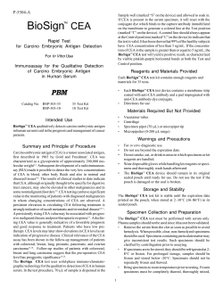
© 2001 Nature Publishing Group http://structbio.nature.com
letters
© 2001 Nature Publishing Group http://structbio.nature.com
Structure of β-lactam
synthetase reveals how to
synthesize antibiotics
instead of asparagine
Matthew T. Miller1, Brian O. Bachmann2,
Craig A. Townsend2 and Amy C. Rosenzweig1
1Departments of Biochemistry, Molecular Biology, and Cell Biology and
Chemistry, Northwestern University, Evanston, Illinois 60208, USA.
2Department of Chemistry, The Johns Hopkins University, Baltimore,
Maryland 21218, USA.
The enzyme β-lactam synthetase (β-LS) catalyzes the formation of the β-lactam ring in clavulanic acid, a clinically
important β-lactamase inhibitor. Whereas the penicillin
β-lactam ring is generated by isopenicillin N synthase (IPNS)
in the presence of ferrous ion and dioxygen, β-LS uses ATP
and Mg2+ as cofactors. According to sequence alignments,
β-LS is homologous to class B asparagine synthetases
(AS-Bs), ATP/Mg2+-dependent enzymes that convert aspartic
acid to asparagine. Here we report the first crystal structure
of a β-LS. The 1.95 Å resolution structure of Streptomyces
clavuligerus β-LS provides a fully resolved view of the active
site in which substrate, closely related ATP analog α,β-methyleneadenosine 5′-triphosphate (AMP-CPP) and a single
Mg2+ ion are present. A high degree of substrate preorganization is observed. Comparison to Escherichia coli AS-B reveals
the evolutionary changes that have taken place in β-LS that
impede interdomain reaction, which is essential in AS-B, and
that accommodate β-lactam formation. The structural data
provide the opportunity to alter the synthetic potential of
β-LS, perhaps leading to the creation of new β-lactamase
inhibitors and β-lactam antibiotics.
Antibiotic resistance is a serious and growing problem in
human health1,2. A principal bacterial resistance strategy is inactivation of β-lactam antibiotics by β-lactamases. These enzymes
hydrolyze front line antibiotics, such as the penicillins and
cephalosporins, destroying their ability to inhibit cell wall
biosynthesis1,3. Clavulanic acid, a weak antibiotic isolated from
Streptomyces clavuligerus, is a potent inhibitor of β-lactamases
and is used clinically in combination with amoxycillin and other
penicillins4,5. The key β-lactam ring of clavulanic acid is
generated by a recently characterized enzyme, β-lactam synthetase (β-LS)6–8, via a mechanism completely distinct from formation of the penicillin β-lactam ring (Fig. 1). For penicillin, the
enzyme isopenicillin N synthase (IPNS) catalyzes cyclization of a
tripeptide to the bicyclic penicillin nucleus in the presence of ferrous ion and dioxygen9,10. In contrast, β-LS converts N 2-(carboxyethyl)-L-arginine (CEA) to deoxyguanidinoproclavaminic
acid (DGPC) using ATP and Mg2+ (refs 6,7). A homologous
enzyme, CarA, is proposed to perform similar chemistry in the
biosynthesis of carbapenems11,12. These enzymes could be altered
by genetic methods to improve production of clinically useful
β-lactamase inhibitors or to biosynthesize new β-lactam antibiotics with enhanced resistance characteristics.
Although β-LS is an amide-synthesizing enzyme, its amino
acid sequence does not resemble tRNA synthetases or nonribosomal peptide synthetases but is similar to class B asparagine
synthetases (AS-Bs)13,14. These ATP/Mg2+-dependent enzymes,
which belong to the broader family of Ntn glutamine amidotransferases15, catalyze transfer of the amide nitrogen of glutamine to aspartic acid, producing asparagine (Fig. 1). AS-B from
Escherchia coli comprises two domains, each with an active site.
Its N-terminal glutaminase domain hydrolyzes glutamine to glutamic acid and ammonia, and the C-terminal synthetase domain
catalyzes the adenylation of aspartic acid and its subsequent
reaction with ammonia. An extended tunnel provides a conduit
for ammonia between the two sites16. β-LS has homology to both
domains but lacks the critical N-terminal Cys residue conserved
in all AS-Bs17. Both β-LS and AS-B mediate adenylation of an
amino acid, but, in contrast to AS-B, β-LS catalyzes an intramolecular rather than intermolecular amide bond formation.
The basis for the apparent close evolutionary relationship is not
clear. Detailed structural characterization of β-LS is essential to
exploit its potential in engineered antibiotic biosynthesis and to
understand the connection to AS-B. Here we report the 1.95 Å
resolution structure of Streptomyces clavuligerus β-LS in the
presence of substrate, CEA, and cofactors, close ATP analog α,
β-methyleneadenosine 5′-triphosphate (AMP-CPP) and a single
Mg2+ ion. In the crystal structure, these substrates are preorganized to favor the adenylation and β-lactamization reactions catalyzed by β-LS. In addition, comparison to E. coli AS-B reveals
Fig. 1 Reactions catalyzed by β-LS,
IPNS and AS-B. In clavulanic acid
biosynthesis,
N2-(carboxyethyl)L-arginine (CEA) is cyclized to
deoxyguanidinoproclavaminic acid
(DGPC) by β-LS. Subsequent chemical transformations by other
enzymes result in the formation
of clavulanic acid. In penicillin
biosynthesis, IPNS catalyzes the
cyclization of δ-(L-aminoadipoyl)L-cysteinyl-D-valine
(ACV)
to
isopenicillin N. In asparagine
biosynthesis,
glutamine
and
aspartic acid are converted to glutamic acid and asparagine.
684
nature structural biology • volume 8 number 8 • august 2001
© 2001 Nature Publishing Group http://structbio.nature.com
© 2001 Nature Publishing Group http://structbio.nature.com
letters
Fig. 2 Comparison of β-LS and AS-B. a, Stereo view of
β-LS. Residues 21–24, 165, 166 and 445–453 have not been
modeled. The bound CEA and AMP-CPP in the C-terminal
domain are shown as stick representations, and the Mg2+
ion is depicted as a yellow sphere. Regions that differ significantly from the corresponding regions in AS-B are colored magenta and labeled by amino acid residue number.
b, Stereo view of AS-B in the same orientation as β-LS in
(a). Regions that differ significantly between the two
enzymes are colored magenta. Glutamine present in the
N-terminal domain and AMP present in the C-terminal
domain are shown as stick representations. c, Surface representation of the β-LS active site color coded according to
electrostatic potential: red corresponds to –80 kT; white,
0 kT; and blue, +80 kT. CEA and AMP-CPP are shown as
stick representations, and the Mg2+ ion is obscured from
view. The negatively charged region surrounding the CEA
Arg side chain corresponds to Glu 382 and Asp 373.
d, Surface representation of the AS-B active site color
coded according to electrostatic potential: red corresponds to –80 kT; white, 0 kT; and blue, +80 kT. AMP is
shown as a stick representation, and a bound uranium ion
is shown as a yellow sphere. Negatively charged residues
in the active site include Asp 351, Glu 352 and Asp 384.
a
b
the evolutionary changes that have occurred in
β-LS to accommodate β-lactam formation and
provides new insight into the active site of AS-B.
Overall structure
c
β-LS comprises two distinct domains, an N-terminal domain including residues 1–210 and a C-terminal domain composed of residues 211–513 (Fig.
2a). The N-terminal domain consists of two
antiparallel β-sheets, one five-stranded and the
other six-stranded. Three short α-helices pack
against the six-stranded sheet. The outer surface of
the five-stranded sheet is covered by two loop
regions, the longer of which encompasses 28
residues and is preceded by a short α-helix. Several
residues in these loop regions are disordered and
could not be modeled (see Methods). The linker to
the C-terminal domain extends from the last strand
in the six-stranded sheet. The C-terminal domain, which houses
the active site, is composed of 11 α-helices surrounding a fivestranded parallel β-sheet. The active site is located in a cleft
formed by four β-strands and five α-helices. In both molecules
in the asymmetric unit, part of a loop covering the active site is
disordered. The interface between the two domains is quite
extensive, with ∼1,700 Å2 buried surface area per domain.
Interactions at this interface include several hydrophobic patches
and two salt bridges linking Glu 78 with Arg 343 and Lys 159
with Glu 317.
The two β-LS molecules in the asymmetric unit form a dimer
with a buried surface area of ∼1,350 Å2 per molecule. Two pairs of
hydrogen bonds separated by ∼30 Å constitute the main intermolecular interactions. In each dimer, Tyr 109 and His 112 from
one molecule interact with Glu 412 and Thr 368 from the second
molecule. The interface size is well within the range observed for
stable homodimers18, consistent with gel filtration and native gel
analyses indicating that β-LS is a dimer in solution at high concentrations (M.T.M. and A.C.R., unpublished data). The related
enzyme AS-B, which functions as a dimer, dimerizes using a completely different interface16. The two molecules in the β-LS asymmetric unit are very similar and can be superimposed with a root
mean square (r.m.s.) deviation of 0.7 Å for Cα atom coordinates.
The most prominent differences between the two β-LS
nature structural biology • volume 8 number 8 • august 2001
d
monomers in the asymmetric unit occur in residues 66–70 and
residues 454–468. These deviations are due to crystal packing
interactions. Residues 66–70 from both β-LS monomers are
involved in crystal lattice contacts, but the contacts are completely different for the two molecules. Residues 454–468 from only
one β-LS molecule participate in lattice interactions, accounting
for the observed differences in this region.
Evolutionary relationships
As anticipated by sequence alignments6, β-LS is structurally
similar to E. coli AS-B. The two enzymes share many secondary
and tertiary structural features, including distinct N- and C-terminal domains and a common overall fold (Figs 2, 3). However,
a number of changes consistent with the different functions of
the two enzymes are apparent. The N-terminal domain of β-LS
is substantially less organized than the corresponding glutaminase domain of AS-B (Fig. 2a,b). In AS-B, the critical N-terminal Cys residue, which was replaced with an Ala residue in the
crystal structure, is buried at the beginning of the first β-sheet
and positioned precisely to attack the γ-amide of the glutamine
substrate16. In β-LS, this residue is replaced with Phe and preceded by nine additional residues. These extra residues fill the
space occupied by the glutamine substrate in AS-B and are
accommodated by a large outward shift in the loop spanning
685
© 2001 Nature Publishing Group http://structbio.nature.com
letters
© 2001 Nature Publishing Group http://structbio.nature.com
Fig. 3 Structure-based sequence alignment of β-LS
and AS-B. The positions of the β-LS secondary structure elements are superimposed on the β-LS
sequence, and the positions of the AS-B secondary
structure elements are superimposed on the AS-B
sequence. Helices are shown as green cylinders,
and β-strands are shown as blue arrows. Residues
that are not observed in the crystal structures are
highlighted in red.
residues 52–61. In AS-B, the glutamine is stabilized by hydrogen bonding interactions
with Arg 50, Asn 75, Glu 77 and Asp 99. Only
two of these residues are conserved in β-LS.
The β-LS equivalent to AS-B Glu 77, Glu 78,
is hydrogen bonded to Arg 343, and the β-LS
equivalent to AS-B Asp 99, Asp 98, interacts
with the amide nitrogen of Ile 79. These
changes render the N-terminal domain of
β-LS chemically inactive, consistent with
kinetic experiments showing that catalysis is
unaffected by the addition of glutamine6.
Other major differences between the N-terminal domains of β-LS and AS-B are
observed in regions distant from the glutamine binding site. The β-LS β-strands spanning residues 145–154 and 195–202 are
shorter and longer, respectively, than their
AS-B counterparts and are tipped inward. In
addition, a large loop involving residues
163–183 is not conserved in AS-B, and the
somewhat disordered loop encompassing
β-LS residues 20–29 is a helix in AS-B.
The core regions of the C-terminal synthetase domains of β-LS and AS-B are very
similar, but a number of key differences are observed. In both
enzymes, the active site region is formed by four β-strands and
five α-helices (Fig. 2a,b). However, the β-LS substrate binding
cleft is relatively elongated (Fig. 2c,d). This difference in size is not
surprising because the β-LS binding site must accommodate
CEA, which is substantially longer (∼12 Å) than aspartic acid
(∼5 Å). The expansion of the active site is primarily due to the
presence of a loop at residues 377–390 rather than the helix at the
equivalent region in AS-B (Fig. 2a,b). In particular, residues
377–384 from β-LS are shifted away from the cleft, and most of
their bulky side chains, including Phe 377, Asp 378 and Asn 381,
point away from the active site. The space vacated by these
residues in β-LS is filled in AS-B by the side chains of Leu 380,
Tyr 383, Asp 384 and Arg 387. In AS-B, the visible part of the
C-terminus is located at the domain interface adjacent to the
helix containing these residues. The C-terminal region of β-LS
(residues 454–508), which ends in a surface helix, is completely
different and does not restrict the position of residues 377–384 in
a similar way. The two active sites in AS-B are connected by a
molecular tunnel16. Cavity analysis of the β-LS structure with the
program VOIDOO19 indicates that a similar tunnel is not present,
consistent with the absence of an N-terminal active site.
Furthermore, a number of highly conserved residues that line the
AS-B tunnel are not conserved in β-LS.
between two negatively charged residues, Glu 382 and Asp 373.
One CEA η-nitrogen is hydrogen bonded to the side chain of
Glu 382, and both η-nitrogens are linked to the carbonyl oxygen
of Asp 373 via a water molecule. The CEA Arg side chain is also
held in position by the side chain of Tyr 326. The α-carboxylate
of CEA is stabilized by interactions with the amide nitrogen of
Gly 349 and with a water molecule that is coordinated to the
Mg2+ ion. This water molecule is also hydrogen bonded to the
side chain of Asp 253, the carbonyl oxygen of Gly 347 and one of
the β-phosphate oxygens. The carboxyethyl group is not as well
ordered as the rest of the CEA molecule, with B-values of ∼60 Å2
as compared to ∼50 Å2 for the α-carboxylate and guanidino
groups and ∼40 Å2 for the AMP-CPP. Based on the visible electron density, the carboxyethyl group in monomer A was modeled in the eclipsed conformation, with the β-carboxylate and
α-amino groups approximately lined up with one another, consistent with the proposed chemical mechanism of intramolecular β-lactam formation7. The density in monomer B is less well
defined, and several conformations of the carboxyethyl group
are possible. The AMP-CPP–Mg2+ is very well defined in the
electron density map. It is bound in close proximity to the CEA,
with the α-phosphate presenting itself for attack by the β-carboxylate of CEA. The Mg2+ ion is coordinated by one side oxygen
each from Asp 253 and Asp 351; by the α-, β- and γ-phosphate
oxygens; and by a water molecule in a well-organized octahedral
The β-LS active site
geometry. The phosphate oxygens also interact with Lys 423,
The β-LS active site contains a full set of ligands, including the Lys 443 and the side chain hydroxyl groups of Ser 249 and
substrate CEA, the nonreactive ATP analog, AMP-CPP, and a Ser 254. The adenosine hydroxyl groups interact with the amide
Mg2+ ion (Fig. 4a–c). The guanidino group of CEA is situated nitrogens of Gly 347 and Tyr 348 and the carbonyl oxygen of
686
nature structural biology • volume 8 number 8 • august 2001
© 2001 Nature Publishing Group http://structbio.nature.com
© 2001 Nature Publishing Group http://structbio.nature.com
letters
a
c
b
d
e
Fig. 4 The active site. a, Stereo view of an Fo – Fc simulated annealing omit map contoured at 2 σ with the CEA, AMP-CPP and Mg2+ omitted. b, Stereo
view of the β-LS active site. Key residues that interact with the CEA, AMP-CPP and Mg2+ are shown. c, Schematic diagram of the β-LS active site showing hydrogen bonding interactions. d, Schematic diagram of Asp, AMP-CPP and Mg2+ modeled into the AS-B active site. Predicted hydrogen bonding
interactions and Mg2+ coordination environment are shown. e, Cyclization step in proposed chemical mechanism for β-LS. The position of the CEA
α-amino group in the crystal structure favors this β-lactamization reaction.
Val 247. The adenine nitrogens are hydrogen bonded to the carbonyl oxygen and amide nitrogen of Met 273.
The active site details revealed by the β-LS structure also provide new insight into the active sites of Ntn glutamine amidotransferases. In particular, the β-LS active site can be used to
model AMP-CPP–Mg2+ and Asp into the AS-B structure. One
AMP molecule and three uranyl ions were found in the AS-B
active site16. Notably, one of the uranyl ions occupies the same
position as the Mg2+ ion in β-LS and is also coordinated by two
Asp residues, Asp 238 and Asp 351. By placing Mg2+ in the position occupied by this uranyl ion and using the existing AMP as a
guide, Asp and AMP-CPP–Mg2+ were modeled into the E. coli
AS-B active site (Fig. 4d). In this model, the Asp is situated similarly to the CEA in β-LS, with its β-carboxylate adjacent to the
α-phosphate of AMP-CPP. The amino nitrogen is hydrogen
bonded to Asp 384, and the α-carboxylate oxygen interacts with
Arg 387. These two residues protrude into the AS-B active site
cleft, filling part of the region occupied by CEA in β-LS. A
BLAST20 analysis of the AS-B sequence reveals that both Asp 384
and Arg 387 are highly conserved in all AS-Bs. The aspartic acid
can be positioned so that it interacts with Lys 376 instead of
Arg 387, but this Lys residue is not conserved, suggesting that it is
less likely to play an important role in substrate positioning. The
nature structural biology • volume 8 number 8 • august 2001
model also predicts that Ser 234, Ser 239, Lys 429 and Lys 449
interact with the phosphate oxygens of the AMP-CPP. These findings may be useful in designing new inhibitors of AS-B, which is a
therapeutic target for acute lymphoblastic leukemia (ALL)21.
Mechanistic implications
The unreacted substrates in the β-LS active site are bound in a
geometry that favors both the adenylation and β-lactamization
reactions catalyzed by this enzyme. The proposed chemical
mechanism involves adenylation of the CEA β-carboxylate followed by cyclization via an oxoanion intermediate7 (Fig. 4e). The
β-carboxylate and α-amino group of CEA are positioned relative
to their respective electrophiles, the α-phosphate of AMP-CPP
and the hypothetical adenylate in a plausible near-attack conformation22. Notably, the α-amine is oriented at a favorable angle
with respect to the sp2 β-carboxylate, approaching the 110°
Bürgi-Dunitz trajectory23 for nucleophilic addition to a carbonyl
group. As the reaction progresses, the nascent adenylate carbonyl
can be bound in an oxoanion stabilizing Mg2+/H2O environment, orienting the adenylate and favoring formation of the
tetrahedral intermediate required for intramolecular closure of
the β-lactam bond (Fig. 4e). The striking preorganization of substrate molecules in the active site maximizes the energetic benefit
687
© 2001 Nature Publishing Group http://structbio.nature.com
letters
pH 7.5, and used to grow crystals for substrate soaking experiments.
© 2001 Nature Publishing Group http://structbio.nature.com
Table 1 Crystallographic statistics
Data collection and MAD phasing
λ11
Wavelength (Å)
0.9793
Resolution range (Å) 15–2.3
Observations
Unique
42,678
Total
485,821
Completeness2
96.4 (96.1)
Rsym3
0.066 (0.325)
% > 3 σ (I)
71.1 (46.9)
Rcullis4
0.73
Phasing power5
1.39
λ2
0.9791
15–2.3
λ3
0.9639
15–2.3
Substrate Complex
1.00
20–1.95
42,660
483,642
96.4 (96.1)
0.073 (0.336)
71.0 (31.4)
0.73
1.41
42,707
489,027
96.4 (96.7)
0.068 (0.326)
69.8 (31.1)
0.82
0.95
68,300
465,261
98.3 (98.1)
0.062 (0.231)
73.7 (35.5)
Refinement
Resolution (Å)
Number of reflections
R-factor6
Rfree7
Number of atoms
Protein, nonhydrogen
Nonprotein
R.m.s. deviations
Lengths (Å)
Angles (°)
Average B-value (Å2)
Substrate Complex
20–1.95
66,147
0.194
0.228
7,446
750
0.005
1.30
30.4
Crystallization and data collection. All
β-LS crystals were obtained by the hanging
drop vapor diffusion method at room temperature using a precipitant solution containing 7% (w/v) PEG 4000, 200 mM MgCl2,
7% (v/v) glycerol and 80 mM Tris, pH 8.0.
Crystals with average dimensions of 0.01 ×
0.05 × 0.20 mm3 appeared within 16 h and
were transferred to a cryosolution composed of 24% (w/v) PEG 4000, 260 mM
MgCl2, 20% (v/v) glycerol and 80 mM Tris,
pH 8.0. After 5 min, the crystals were
frozen directly in liquid nitrogen. The substrate CEA was synthesized by acid hydrolysis of deoxyguanidinoproclavaminic acid
(DGPC) as described25. Soaking experiments
were performed in the cryosolution supplemented with 5 mM each of CEA and the
ATP analog α,β-methyleneadenosine 5′triphosphate (AMP-CPP). After 1 h, the crystals were frozen in liquid nitrogen. The β-LS
crystals belong to space group P21 with unit
cell dimensions a = 61.0 Å, b = 98.2 Å, c =
81.3 Å and β = 91.27º. All data were collected using synchrotron radiation and
processed with DENZO and SCALEPACK26
(Table 1).
Structure determination. Data for MAD
phasing were collected at three wavelengths using the reverse beam technique
(Table 1). Selenium positions were determined with CNS27, which located 11 of 14
possible sites in the asymmetric unit. MAD
phasing and density modification with CNS27
yielded an interpretable electron density
map. The asymmetric unit contains two β-LS
molecules (labeled A and B) related by a
noncrystallographic two-fold axis. The program XtalView28 was used for model building, and the structure was
refined with CNS27 by iterative cycles of simulated annealing and
individual B-value refinement. Noncrystallographic symmetry
restraints were imposed until the final cycles of refinement to 2.3 Å
resolution. This model was then used as a starting model for refinement of the substrate structure (Table 1). The final model for the
substrate structure contains residues A4–A20, A25–A164,
A167–A444, A454–A508, B2–B21, B24–B444 and B450–B508; 622
water molecules; five glycerol molecules; two AMP-CPP molecules;
two CEA molecules; and two Mg2+ ions. No electron density was present for residues A21–A24, A165–A166, A445–A453, B22–B24 and
B445–B449. According to Ramachandran plots generated with
PROCHECK29, the model exhibits good geometry with 92.7% of the
residues in the most favored regions and all other residues in the
additionally allowed regions. Figures were generated with
MOLSCRIPT30, Raster3D31, GRASP32 and BOBSCRIPT33. Accessible surface area calculations were performed with the CCP4 program
AREAIMOL34.
All data sets were collected at the Dupont-Northwestern-Dow Collaborative Access Team (DNDCAT) beamline at the Advanced Photon Source using a 2K × 2K Mar CCD detector.
2Values in parentheses are for the highest resolution shell: λ1, λ2, λ3 = 2.38–2.30 Å; substrate =
2.02–1.95 Å.
3R
sym = Σ |Iobs – Iavg| / Σ Iobs , where the summation is over all reflections.
4R
27
cullis = lack of closure error / iso-ano difference (generalized Rcullis from CNS ).
5Phasing power = heavy atom structure factor / r.m.s. lack of closure error (statistics from CNS27).
6R-factor = Σ |F
obs – Fcalc| / Σ Fobs.
7For calculation of R
free, 8.8% of the reflections were reserved.
1
from the hydrolysis of ATP in the formation of the strained fourmembered ring. The structural features of β-LS that impede
attack by external nucleophiles coupled with the enlarged binding pocket provide an excellent starting point for protein engineering experiments to construct modified β-lactam antibiotics.
Methods
Purification of β-LS for crystallization. β-LS for initial crystallization trials and selenomethionine-substituted β-LS were purified as
described7. To generate the selenomethionine protein, the expression vector pBOB1 was transformed into E. coli B834(DE3), and transformed bacteria were grown in LeMaster media24 supplemented
with 50 mg l–1 selenomethionine. The gene for S. clavuligerus
β-LS was also cloned into a pET24a(+) vector, which was used to
transform E. coli BL21(DE3) cells. These transformed cells were
grown in a 12 l fermenter, and protein expression was induced with
0.5 mM IPTG at an optical density of 0.6–0.8. Cells were harvested
after 3 h, lysed with hen egg lysozyme and treated with DNase. After
centrifugation for 30 min at 12,000 × g, the supernatant was diluted
10-fold in 50 mM HEPES, pH 8.5, and loaded onto a Source 30Q column. A 0–330 mM KCl gradient was applied, and β-LS eluted at
∼25 mM KCl. Fractions containing β-LS were diluted 10-fold in 50 mM
HEPES, pH 7.5, loaded onto an arginine Sepharose column and eluted with a step gradient of 5 mM KCl followed by 200 mM KCl.
Fractions from the arginine Sepharose column were then applied to
a Blue Sepharose column. The protein, which did not bind to the column, was concentrated and further purified on Superdex 200 equilibrated in 50 mM HEPES, pH 7.5, containing 200 mM KCl. The final
purified material was concentrated to 20 mg ml–1 in 50 mM Tris,
688
Coordinates. The coordinates of β-LS have been deposited in the
Protein Data Bank (accession code 1JGT).
Acknowledgments
This work was supported by funds from the David and Lucile Packard Foundation
to A.C.R., by an NIH grant to C.A.T. and in part by an NIH training grant to
M.T.M. The DND-CAT Synchrotron Research Center at the Advanced Photon
Source is supported by the E.I. DuPont de Nemours & Co., The Dow Chemical
Company, the NSF and the State of Illinois.
Correspondence should be addressed to A.C.R. email: [email protected]
nature structural biology • volume 8 number 8 • august 2001
© 2001 Nature Publishing Group http://structbio.nature.com
letters
Received 2 April, 2001; accepted 7 June, 2001.
© 2001 Nature Publishing Group http://structbio.nature.com
1.
2.
3.
4.
5.
6.
7.
8.
9.
10.
11.
12.
13.
14.
15.
Neu, H.C. Science 257, 1064–1073 (1992).
Levy, S.B. N. Engl. J. Med. 338, 1376–1378 (1998).
Walsh, C. Nature 406, 775–781 (2000).
Baggaley, K.H., Brown, A.G. & Schofield, C.J. Nat. Prod. Rep. 14, 303–333 (1997).
Jensen, S.E. & Paradkar, A.S. Antonie van Leeuwenhoek 75, 125–133 (1999).
Bachmann, B.O., Li, R. & Townsend, C.A. Proc. Natl. Acad. Sci. USA 95, 9082–9086
(1998).
Bachmann, B.O. & Townsend, C.A. Biochemistry 39, 11187–11193 (2000).
McNaughton, H.J. et al. Chem. Commun. 21, 2325–2326 (1998).
Roach, P.L. et al. Nature 387, 827–830 (1997).
Burzlaff, N.I. et al. Nature 401, 721–724 (1999).
Li, R., Stapon, A., Blanchfield, J.T. & Townsend, C.A. J. Am. Chem. Soc. 122,
9296–9297 (2000).
McGowan, S.J., Bycroft, B.W. & Salmond, G.P.C. Trends Microbiol. 6, 203–208
(1998).
Scofield, M.A., Lewis, W.S. & Schuster, S.S. J. Biol. Chem. 265, 12895–12902 (1990).
Richards, N.G.J. & Schuster, S.M. Adv. Enzymol. Relat. Areas Mol. Biol. 72,
145–198 (1998).
Zalkin, H. & Smith, J.L. Adv. Enzymol. Relat. Areas Mol. Biol. 72, 87–143 (1998).
X-ray snapshots of serine
protease catalysis reveal a
tetrahedral intermediate
Rupert C. Wilmouth1,2, Karl Edman2,3, Richard Neutze3,4,
Penny A. Wright1, Ian J. Clifton1, Thomas R. Schneider5,
Christopher J. Schofield1 and Janos Hajdu2
1The Dyson Perrins Laboratory and Oxford Centre for Molecular Sciences,
University of Oxford, South Parks Road, Oxford OX1 3QY, UK. 2These
authors contributed equally to this paper. 3Department of Biochemistry,
Uppsala University, Box 576, Biomedical Centre, SE-75123 Uppsala, Sweden.
4Current address: Department of Molecular Biotechnology, Chalmers
University of Technology, P.O. Box 462, SE 40530 Göteborg, Sweden.
5Department
of Structural Chemistry, University of Göttingen,
Tammannstrasse 4, 37077 Göttingen, Germany.
Studies on the catalytic mechanism and inhibition of serine
proteases are widely used as paradigms for teaching enzyme
catalysis. Ground-breaking work on the structures of chymotrypsin and subtilisin led to the idea of a conserved catalytic
triad formed by the active site Ser, His and Asp residues. An
oxyanion hole, consisting of the peptide amide of the active site
serine and a neighbouring glycine, was identified, and hydrogen bonding in the oxyanion hole was suggested to stabilize the
two proposed tetrahedral intermediates on the catalytic pathway. Here we show electron density changes consistent with the
formation of a tetrahedral intermediate during the hydrolysis
of an acyl–enzyme complex formed between a natural
heptapeptide and elastase. No electron density for an
enzyme–product complex was observed. The structures also
suggest a mechanism for the synchronization of hydrolysis and
peptide release triggered by the conversion of the sp2
hybridized carbonyl carbon to an sp3 carbon in the tetrahedral
intermediate. This affects the location of the peptide in the
active site cleft, triggering the collapse of a hydrogen bonding
network between the peptide and the β-sheet of the active site.
Peptide hydrolysis catalyzed by serine proteases proceeds via
formation of an initial noncovalent enzyme–substrate complex. Nucleophilic attack by the active site Ser residue of the
peptide carbonyl results in the formation of a tetrahedral
oxyanion intermediate. Collapse of this intermediate results in
formation of an acyl–enzyme (ester) intermediate at the Ser
nature structural biology • volume 8 number 8 • august 2001
16. Larsen, T.M. et al. Biochem. 38, 16146–16157 (1999).
17. Boehlein, S.K., Richards, N.G.J. & Schuster, S.M. J. Biol. Chem. 269, 7450–7457
(1994).
18. Jones, S. & Thornton, J.M. Proc. Natl. Acad. Sci. USA 93, 13–20 (1996).
19. Kleywegt, G.J. & Jones, T.A. Acta Crystallogr. D 50, 178–185 (1994).
20. Altschul, S.F. et al. Nucleic Acids Res. 25, 3389–3402 (1997).
21. Chakrabarti, R. & Schuster, S.M. Int. J. Pediatr. Hematol. Oncol. 4, 597–611 (1997).
22. Bruice, T.C. & Benkovic, S.J. Biochemistry 39, 6267–6274 (2000).
23. Bürgi, H.B. & Dunitz, J.D. Acc. Chem. Res. 16, 153–161 (1983).
24. Hendrickson, W.A., Horton, J.R. & LeMaster, D.M. EMBO J. 9, 1665–1672 (1990).
25. Elson, S.W. et al. J. Chem. Soc. Chem. Commun. 15, 1212–1214 (1993).
26. Otwinowski, Z. & Minor, W. Methods Enzymol. 276, 307–326 (1997).
27. Brünger, A.T. et al. Acta Crystallogr. D 54, 905–921 (1998).
28. McRee, D.E. J. Struct. Biol. 125, 156–165 (1999).
29. Laskowski, R.A. J. Appl. Crystallogr. 26, 283–291 (1993).
30. Kraulis, P.J. J. Appl. Crystallogr. 24, 946–950 (1991).
31. Merritt, E.A. & Bacon, D.J. Methods Enzymol. 277, 505–524 (1997).
32. Nicholls, A., Sharp, K.A. & Honig, B. Proteins 11, 281–296 (1991).
33. Esnouf, R.M. J. Mol. Graph. Model. 15, 132–134 (1997).
34. Collaborative Computational Project, Number 4. Acta Crystallogr. D 50, 760–763
(1994).
residue and the release of the C-terminal product fragment. In
the second (deacylation) reaction, subsequent hydrolytic
attack by a water molecule of the ester carbonyl leads to the
second tetrahedral intermediate, which collapses to give the
N-terminal product fragment (Fig. 1). Here, we describe structures for the intermediates in this reaction trapped at liquid N2
temperature.
Spectroscopic observations using poor substrates and ‘partitioning’ studies1 provide evidence for the formation of an
acyl–intermediate during serine protease catalysis. Evidence for
the two high energy tetrahedral intermediates is less direct,
mostly from analogy with the proposed mechanism of amide
bond hydrolysis in small molecules2 and from the use of ‘transition state analog’ inhibitors, based upon Pauling’s theory of
enzyme catalysis3. Recently, the heptapeptide human β-casomorphin-7 (BCM7; YPFVEPI) was discovered to form a stable
acyl–enzyme intermediate with porcine pancreatic elastase
(PPE) at pH 5. The X-ray structure4 of the complex revealed the
heptapeptide bound in a productive manner as an antiparallel
β-strand, extending the β-sheet in the active site. The C-terminal
Ile residue of the peptide links via an ester bond to Ser 195 of the
catalytic triad (residue numbering follows PDB entry 1QNJ)5. A
water molecule (Wat 317) was hydrogen-bonded to the nearby
His 57. This was the first high resolution crystal structure for an
acyl–enzyme complex of a serine protease with a single naturally
occurring peptide bound in the active site (for other examples of
acyl–enzyme structures, see ref. 4).
Here, we describe high resolution crystal structures for cryogenically trapped intermediates in the hydrolysis of the
acyl–enzyme complex between β-casomorphin-7 and elastase.
The results provide the structural insights into the hydrolysis of a
serine protease peptidyl acyl–enzyme complex. Electron density
changes during this reaction are consistent with the formation of
a tetrahedral intermediate and suggest a mechanism for synchronizing hydrolysis and product release with peptide substrates in
serine proteses.
Reaction triggering and intermediate trapping
In order to characterize the structure of the putative tetrahedral intermediate of this deacylation pathway, we initiated
hydrolysis of the ester bond within crystals of the stable
PPE–BCM7 acyl–enzyme intermediate and undertook high
resolution X-ray analysis of the transient species (Fig. 1; Table
1). The reaction was triggered by immersing crystals of the stable PPE–BCM7 acyl–enzyme intermediate grown at pH 5 into
689


© Copyright 2026