
Why is epigenetics important in understanding Abstract
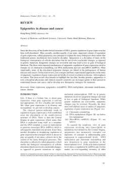
Oppermann Arthritis Research & Therapy 2013, 15:209
http://arthritis-research.com/content/15/2/209
REVIEW
Why is epigenetics important in understanding
the pathogenesis of inflammatory musculoskeletal
diseases?
Udo Oppermann*1,2
Abstract
In its widest sense, the term epigenetics describes
a range of mechanisms in genome function that
do not solely result from the DNA sequence itself.
These mechanisms comprise DNA and chromatin
modifications and their associated systems, as well
as the noncoding RNA machinery. The epigenetic
apparatus is essential for controlling normal
development and homeostasis, and also provides a
means for the organism to integrate and react upon
environmental cues. A multitude of functional studies
as well as systematic genome-wide mapping of
epigenetic marks and chromatin modifiers reveal the
importance of epigenomic mechanisms in human
pathologies, including inflammatory conditions and
musculoskeletal disease such as rheumatoid arthritis.
Collectively, these studies pave the way to identify
possible novel therapeutic intervention points and
to investigate the utility of drugs that interfere with
epigenetic signalling not only in cancer, but possibly
also in inflammatory and autoimmune diseases.
Introduction
Doubtlessly, the field of epigenetics has rapidly evolved
over the last decades – a quick literature survey shows 18
PubMed entries for 1975 to 1995, >400 entries for the
following 10 years and >2,000 entries from 2006 to 2010.
Importantly, the definition of epigenetics now extends
significantly from its initial meaning into other disciplines and encompasses wide research areas within
genetics, genomics, molecular biology and medicine
(including, for example, epidemiology and pathology)
*Correspondence: [email protected]
1
Botnar Research Center, NIHR Oxford Biomedical Research Unit, Oxford OX3 7LD,
UK
Full list of author information is available at the end of the article
© 2010 BioMed Central Ltd
© 2013 BioMed Central Ltd
(see Figure 1). The term epigenesis originally coined by
Waddington over 50 years ago was introduced in a
developmental biology context to describe how genotypes give rise to different phenotypes [1], a view that is
fundamentally different from the definition of ‘the
heritable transmission of phenotype without a change in
the underlying DNA sequence’ that is now widely in use.
Over the years, however, this interpretation of epigenetics
has found significant alterations – in fact, there now
appears to be no uniform consensus definition [2,3].
Whereas developmental biologists emphasise the transgenerational heritability aspect of epigenetics (that is, the
necessity to stably transmit epigenetic modifications to
achieve a phenotype), many scientists nowadays use the
term epigenetic in a less constrained manner. In this way
they relate almost any covalent chromatin modification
with underlying general events that are considered DNAtemplated processes and thus include transcription, DNA
repair or genome stability [4].
Irrespective of this semantic debate, this review aims to
describe the various major systems that modify chromatin
components as well as DNA to accomplish gene regulation and functional chromatin states. In this overview,
epigenetics is used in its widest sense – that is, epigenetics includes a discussion of DNA and chromatin
modifications as well as the area of noncoding RNA,
known to play key roles in imprinting, gene regulation
and silencing. The article proposes that a better
understanding of these epigenetic mechanisms and their
effects will lead to an appreciation of their potential roles
in musculoskeletal and inflammatory disease pathologies,
and, finally, might pave the way to novel possible
therapeutic intervention strategies.
What is the biochemical basis of epigenetics?
Chromatin is a highly organised and dynamic protein–
DNA complex consisting of DNA, histones and nonhistone proteins. Within this framework, epigenetic
mechanisms alter the accessibility of DNA by modification or rearrangement of nucleosomes, as well as through
a plethora of post-translational chemical modifications of
Oppermann Arthritis Research & Therapy 2013, 15:209
http://arthritis-research.com/content/15/2/209
Page 2 of 9
Figure 1. Impact of epigenetic research on understanding of
human disease and advancement towards novel therapeutic
principles. Epigenetics connects various disciplines such as genome
biology or genetics and will impact on clinical disciplines (see text for
details).
transcription factors or remodelling complexes to interact with their cognate binding sites within the regulatory
regions of genes, such as proximal promoters, enhancers
or silencers [7,9]. Modification systems (so-called writers
and erasers of chromatin marks) that covalently alter
specific residues of chromatin proteins play a pivotal role
in this process (Table 1). Equally important, the distinct
chromatin modifications or marks can act as beacons to
recruit specifically recognition domains and components
(readers) of transcriptional complexes, which thus serve
as the effectors of the modification. In this complex and
interdependent manner (defined as the histone code)
[11], chromatin modifying systems exert control of global
and local gene activation. In addition, chromatin capture
methods have revealed the critical importance of nuclear
architecture and long-range chromatin interactions in
regulation of concerted gene programmes [12] – this is
illustrated, for example, by the murine Th2 cytokine
locus where gene regions are folded into connected
dynamic DNA loop structures anchored by AT-rich
sequence binding proteins [13].
chromatin proteins such as histones and DNA itself (see
below). In addition to the intricate interactions that occur
between chromatin proteins and DNA, the noncoding
RNA machinery is included to be epigenetic – as part of
a complex network entangled with chromatin and DNA
modification systems, which alter and critically control
gene expression patterns during development,
homeostasis and disease [5,6].
Epigenomics – that is, the genome-wide study of epigenetics – is made feasible using recently developed nextgeneration sequencing platforms and, importantly, has
provided an insight into genome architecture that was
not anticipated by researchers a decade ago when completion of the first genome-sequencing projects was
accomplished. Following this development, the recent
large-scale chromatin profiling and interaction mapping
across many different cell types and their functional
states carried out by the ENCODE (Encyclopedia of
DNA Elements) consortium has already resulted in functional annotation of about 80% of the human genome, the
vast majority of which is nonprotein coding. This largescale collaborative project has revealed common regulatory elements, their functional interactions as well as
chromatin state dynamics leading to an unprecedented,
detailed view of genome biology [7-10] with clear
implications and novel avenues in understanding of
human disease (see below).
An important aspect in the epigenetic concept is that
the local chromatin structure is of critical importance –
for example, accessible chromatin (that is, as found in
euchromatin) allows gene-regulatory proteins such as
DNA methylation in an epigenetic context
Among the epigenetic mechanisms regulating gene
expression, DNA methylation is by far the most studied –
although, it is probably fair to say, still incompletely
understood. In vertebrate genomes, DNA methylation
mostly occurs at the 5’ position on cytosine bases and
largely in the context of CpG islands. This cytosine modification critically controls genome functions by silencing
of genes (see below), and has a function in controlling
centromeric stability and probably suppresses the expression and mobility of transposable elements [14]. Because
5-methylcytosine can be spontaneously deaminated (by
replacing nitrogen with oxygen) to thymidine, CpG sites
are frequently mutated and thus become rare in the
genome. Epigenetic changes of this type thus have the
potential to directly contribute to permanent genetic
mutations.
About 70 to 80% of annotated gene promoters are associated with CpG islands, which are usually unmethylated,
but a substantial amount of cytosine methylation is also
found in gene bodies and intergenic sequences, the function of which is beginning to emerge [15]. Importantly,
cell-type specific DNA methylation profiles appear to
vary more frequently at intergenic sequences compared
with annotated gene promoters [9]. These sites of differential methylation themselves might regulate the activity
of distant enhancers [16] or the transcription of noncoding RNAs and uncharacterised transcripts [17,18].
Methylation of CpG promoter sites is associated with
stable silencing of gene expression, and aberrant methylation patterns – for example, hypermethylation of tumour
suppressor genes or hypomethylation of oncogenes – are
Oppermann Arthritis Research & Therapy 2013, 15:209
http://arthritis-research.com/content/15/2/209
now recognised as hallmarks of cancer [19-23]. Silencing
through DNA methylation is achieved by preventing the
binding of distinct transcription factors, or by recruiting
methyl-binding proteins, thereby generating a repressed
chromatin environment. These patterns of DNA
methylation can be stably propagated during cell division,
which makes this process a paradigm for true epigenetic
regulation. Accordingly, these DNA modifications can
mediate long-lasting changes in gene expression even
when the initial triggering signal has disappeared.
DNA methylation patterns are known to be established
and modified in response to environmental factors by a
complex interplay of at least three independent DNA
methyltransferases, DNMT1, DNMT3A and DNMT3B
[24] – hence making DNA methylation a prime candidate
for linking environmental cues and disease. Interestingly,
a recent epigenome-wide DNA methylation study among
>300 rheumatoid arthritis (RA) patients identified several
differentially methylated regions within the MHC region,
suggesting a possible link between genetic predisposition
and epigenetic modification and function in RA [25].
DNA methylation patterns have long been known to
undergo significant changes during fertilisation and
embryogenesis, highlighting the existence of systems that
can revert and erase DNA methylation [24]. Once
established in differentiated cells, DNA methylation is
considered stable; however, recent studies reveal that it
appears to also be subject to demethylation (that is,
reversal of biological effect) in specific instances,
involving several incompletely characterised candidate
mechanisms (that is, methylcytosine hydroxylation, DNA
glycosylation, base excision repair and deaminases), all of
which have been shown to play important roles in
genome biology and disease (reviewed in [24]).
Histone modifications are important elements of
the epigenomic landscape
In addition to the modifications described above for
DNA, post-translational modifications of N-terminal,
unstructured tails of histone proteins have now been
recognised as key components in the regulation and
signalling of functional states of the epigenomic landscape. For example, trimethylated lysine 9 of histone 3
(H3K9me3) indicates heterochromatic or repetitive
regions, whereas H3K4me3 marks regulatory elements
associated with active promoters or transcription start
sites and H3K27me3 marks those for developmentally
repressed genes [9].
At present, several classes of histone modifications and
their respective enzymatic modification systems have
been identified (Table 1) [26]. Amongst their epigenetic
substrate marks, lysine and arginine modifications are
probably the best studied: acetylation and methylation of
lysine residues, as well as methylation of arginine [26-28].
Page 3 of 9
Whereas acetylation of histone tails is correlated with
gene activation [26], the influence of histone methylation
on regulating gene transcription depends on the exact
residue methylated and the number of added methyl
groups, both for arginine and lysine residues [28]. The
involvement of histone modifications in regulation of key
aspects in musculoskeletal biology – for example, in
inflammation [29-33] or differentiation [34-36] – has
recently been established. The best understood systems
of histone modifications that potentially allow transmission of stable heritable marks through cell divisions
comprise methylation of H3K9 (HP1, heterochromatin
establishment) and H3K27 and H3K4 (repression and
activation of genes through polycomb and trithorax
complexes, respectively) [37,38].
Importantly, histone modifications and DNA methylation act in concert with respect to gene regulation
because both activities are functionally linked [39]. One
should state that modifications of histone residues are the
best studied reactions, but constitute only the tip of the
iceberg of nuclear mechanisms regulating chromatin
function since many reader binding specificities or enzymatic activities have not yet been elucidated. Furthermore, many of the writers and erasers also modify other
chromatin-associated proteins such as key transcription
factors – including, for example, p53, retinoblastoma or
NF-κB [40-43] – and thus critically control gene transcription programmes and cell-fate decisions.
Noncoding RNAs contribute to epigenetic mechanisms
Over the last decade it has become apparent that the
nonprotein coding fraction of the human genome is of
critical importance for homeostasis and disease, as
discussed in greater detail elsewhere [5,6]. Those noncoding RNAs are currently divided into several classes
(transcribed ultraconserved regions, small nucleolar
RNAs, PIWI interacting RNAs, large intergenic noncoding RNAs, long noncoding RNAs and miRNAs) based
on their length, as well as their processing and effector
mechanisms [6]. Whereas the most studied class of
miRNAs are ~22 base long ribonucleotide sequences that
target complementary untranslated regions of mRNAs,
directing them for degradation in the RNA-induced
silencing complex, or regulate their translation, other
noncoding RNA types have different or less understood
mechanisms of action. Small nucleolar RNAs (60 to
300 bp size) are involved in ribosomal RNA modifications, the PIWI interacting RNAs (24 to 30 bp size)
interact with PIWI proteins that are critical for genome
stability regulation (for example, heterochromatin formation), and large intergenic RNAs and long noncoding
RNAs (>200 bp size) are found in chromatin complexes.
Several of the noncoding RNA classes are considered
part of the epigenetic machinery due to their critical
Oppermann Arthritis Research & Therapy 2013, 15:209
http://arthritis-research.com/content/15/2/209
Page 4 of 9
Table 1. Overview of major epigenetic DNA and chromatin modification systems
System
Abbreviation
Class
Substrate/mark
DNA methyltransferases
DNMT
Writer
Cytosine
Candidate systems: methylcytosine hydroxylation,
DNA glycosylation, base excision repair and
deaminases?
–
Eraser
5-methyl-C, 5-OH-methyl-C
Methyl-CpG binding domain
MBD
Reader
5-methyl-C
Histone lysine methyltransferases
KMT
Writer
Methylation (lysine)
Histone methyl-lysine demethylases
KDM
Eraser
Demethylation (methyl lysine)
Chromodomain, Tudor domain, MBT domain,
PWWP domain, PHD fingers, WD40 domain
–
Reader
Methyl lysine
Protein arginine methyltransferases
PRMT
Writer
Arginine methylation
Protein arginine deiminases
PADI
Eraser
deimination
Tudor domain
–
Reader
Methyl-arginine
Histone lysine acetyltransferases
KAT
Writer
Acetylation (lysine)
Histone lysine deacetylases
HDAC
Eraser
Deacetylation (lysine)
Bromodomain
–
Reader
Acetyl-lysine
DNA modifications
Histone modifications
For further details and nomenclature used, see [26,91].
involvement in epigenetic phenomena. For example, long
noncoding RNAs can recruit chromatin remodelling
complexes to specific loci, and are involved in DNA
methylation and other chromatin modifications. The importance of long noncoding RNAs is illustrated through
their complex interactions – for example, with the HOX
gene cluster, where hundreds of long noncoding RNAs
regulate in a specific temporal and spatial manner
chromatin accessibility and recruitment of histone modification systems and RNA polymerase. These noncoding
RNA–chromatin complexes are furthermore central to
X-chromosome inactivation and imprinting.
Much of the current work in this field has been directed
towards understanding of the miRNA system, and in
particular several of the miRNAs have been shown to
play key roles in disease [6]. However, the recurring
question for the cause or consequence relationships of
noncoding RNA systems is largely unanswered. Whereas
the involvement in cancer biology is well studied, their
role in other diseases such as inflammatory conditions
like RA is less understood and is only beginning to
emerge. Amongst the miRNAs, some such as miR21,
miR148a, miR155 or mi146a (and others) have been
linked to inflammatory disease and autoimmunity [44-48].
Importantly, polymorphisms in target regions (for
example, the 3’ UTR of IL-1 receptor-associated kinase 1)
of noncoding RNAs such as miR146 might contribute to
RA susceptibility [49], highlighting the interplay of
genetic and epigenetic mechanisms in disease. Taken
together, the field of noncoding RNAs is certainly in its
infancy, and future research will further clarify its role in
immunity and inflammation, and ultimately will have to
prove its therapeutic utility.
Reversibility of chromatin modification and
inheritance of phenotypes
The contemporary definition of epigenetics that describes
mechanisms to produce ‘stable, heritable phenotypes that
result from chromosomal changes without alteration in
DNA sequence’ implies a stably stored sort of memory at
a molecular level that is copied and maintained during
subsequent cell divisions and is independent of the
initiating stimulus.
In contrast to genetic lesions, epigenetic modifications
on DNA and histones are reversible, which is illustrated
by the activities of the various enzyme systems that are
operative in maintaining the epigenomic signatures (cf.
Table 1). For example, histone lysine acetyltransferases
are counteracted by histone lysine deacetylases (histone
deacetylases (HDACs)) in establishing histone acetylation
modifications at lysine residues in the N-terminal tails.
Similarly, histone lysine methyltransferases catalyse the
S-adenosylmethionine-dependent methylation of lysine
residues in histone and other chromatin proteins in a
sequence and methylation state-specific manner – these
marks can be removed by the recently discovered lysine
demethylases (formerly known as histone demethylases)
in establishing histone methylation modifications. These
opposing activities thus constitute a switch mechanism
between functional states – for example, changing
between the acetylated (active transcription) and trimethylated (repressed) state of H3K9 must involve the
Oppermann Arthritis Research & Therapy 2013, 15:209
http://arthritis-research.com/content/15/2/209
eraser activities described above. There is also no doubt
that active DNA demethylation plays a role, for example,
in myeloid cell development. Interestingly, a recent study
identified differentially methylated regions in postmitotic cells as shown in monocyte cultures differentiating to dendritic cell or macrophage populations [50].
Transmission of epigenetic and genetic states (for
example, DNA methylation) vary considerably, with an
error rate of 1 in 106 (DNA sequence) as compared with 1
in 103 (DNA modification) [51]. Consequently, epigenetic
signatures and marks differ fundamentally from genetic
lesions by showing a stochastic manifestation and often
incomplete distribution, and are in principle (at least
partially) reversible. Although much still needs to be
learned in terms of biological and clinical significance of
the reversible nature of these epigenetic modifications, it
does make the chromatin-modifying enzymes possible
therapeutic targets as discussed in some detail further
below.
How can epigenetics further our understanding of
human disease?
For most autoimmune diseases, genetic evidence from
monozygotic and dizygotic twin studies show concordance rates below 50%, suggesting that additional mechanisms exist which potentially link individual susceptibility
and environmental factors such as lifestyle (for example,
smoking or stress), infection or xenobiotic exposure [5255]. Genome-wide association studies (GWASs), for
example, have provided a wealth of possible genetic
factors contributing to the phenotypic diversity of syndromes such as RA and ankylosing spondylitis [56,57] .
Genes identified by searches for common genetic variants
associated with disease have been highly productive in
both RA and ankylosing spondylitis, and the effect of
targeting the products of such contributory genes may be
disproportionately greater than the apparent contribution
to syndrome susceptibility.
Furthermore, gene associations have thus far failed to
explain the heterogeneity of clinical features and response to targeted therapies across patient subgroups.
This concept of missing heritability might be (at least in
part) explained by several mechanisms such as unmapped
common variants, rare variants, gene–gene interaction
or, not unlikely, epigenetic mechanisms. Although genetic
mutations in the epigenetic machinery (that is, readers,
writers, erasers) occur – for instance, mutations in the
DNA methyltransferase DNMT3B in immunodeficiency/
centromeric instability/facial anomalies syndrome, or in
Rett syndrome showing mutations in the methyl-CpG
binding protein 2 – it is unlikely that monogenic lesions
in epigenetic effector mechanisms contribute significantly to complex multifactorial human autoimmune
disease such as RA. Many of the regions identified in
Page 5 of 9
GWAS do not coincide with coding regions, however, but
overlap with functional regulatory regions such as enhancers or transcription start sites identified in the
ENCODE project [7,9]. For example, 11 out of 57 SNPs
identified in RA GWASs overlap with transcription factor
binding sites such as NF-κB [9]. In addition, risk loci such
as the MHC cluster could be targeted by epigenetic
modification such as DNA methylation [25].
Epigenetics might also link environmental risk factors
with genetic variation. Importantly, the epigenome itself
is subject to environmental influences, as documented in
multiple instances [58-61], and thus could act in concert
with genetic variation to explain phenotypic variation
and plasticity [62,63].
Among chronic inflammatory diseases, RA has the
highest prevalence in the western world and is a chronic
and progressive inflammatory disease. In RA, for
example, the concordance of disease occurrence and
progression in identical twins is only 10%, clearly indicating that environmental and/or epigenetic factors are
involved both in induction (where smoking is the biggest
environmental risk) and progression of disease [64]. Of
note, a correlation between smoking and hypomethylation of a CpG motif in the IL-6 promoter and resulting
increased cytokine levels was established in a recent
study among RA and chronic periodontitis patients [65].
This correlation indicates that a causal environmental
disease trigger could indeed lead to a change in cytokine
profile, although the connecting epigenetic mechanism
in this relationship needs to be further defined.
The pathogenesis of disease in RA is attributed to the
production of proinflammatory cytokines from activated
cells that infiltrate the synovial tissues from the blood
(T cells, macrophages, plasma cells) together with resident cell types (fibroblasts and endothelium). Multiple
studies addressing chromatin and DNA modifications in
several autoimmune diseases (for reviews see [66-68])
have clearly shown that tissue-specific epigenetic modifications play a role in autoimmune disease. For example,
DNA methylation in RA is impaired in peripheral blood
mononuclear cells [69], and particularly in CD4+ T cells,
rendering them more autoreactive. This impairment has
been associated with decreased levels of DNA
methyltransferases in senescent CD4+CD28– T cells [70].
In RA peripheral blood mononuclear cells, demethylation of a single CpG in the IL-6 promoter region
increased the production of this proinflammatory cytokine [71]. In other autoimmune diseases such as systemic
lupus erythematosis, the correlation between DNA methylation and reactivity of CD4+ T cells was noted early and
led to the discovery of several key disease genes (reviewed
in [72]). Furthermore, RA synovial fibroblasts – that is,
the effector cells of joint and bone destruction in RA –
present an intrinsic aggressive behaviour even in the
Oppermann Arthritis Research & Therapy 2013, 15:209
http://arthritis-research.com/content/15/2/209
Page 6 of 9
Table 2. Epigenetic drugs or inhibitors targeting mechanisms in musculoskeletal disease
Target class/target
Reagent
(drug/chemical
probe/antisense)
Disease area
Mechanism
Histone demethylase, KDM6
subfamily of demethylases
GSK-J4
Inflammation,
autoimmunity
Suppression of proinflammatory
cytokine production, targeting of H3K27
demethylases
References
[31]
BET bromodomains
I-BET
Sepsis, inflammation
Inhibition of BET bromodomain interactions,
suppression of cytokine production
[84]
BET bromodomains
JQ1
Bone disease / multiple
myeloma
Inhibition of BET bromodomain interactions,
MYC targeting
[85]
Histone deacetylase (class 1 and II
HDACs)
HDAC inhibitors
Osteoarthritis, RA
[77,78,89,90]
miRNA (for example, miR146a,
miR155)
Antisense RNA
technologies
Autoimmunity, RA
[44-47,88]
BET, bromodomain and extraterminal; HDAC, histone deacetylase; I-BET, bromodomain and extraterminal bromodomain inhibitor; RA, rheumatoid arthritis.
absence of cells of the immune system or cytokines. Early
work suggested that the DNA of RA synovial fibroblasts
is partially hypomethylated, resulting in an activated
phenotype [73,74] – an obser vation that more recently
could be confirmed and expanded by showing cytokine
regulation of DNA methyltransferase expression, linked
to differentially methylated genes, and critical to RA
pathogenesis such as CHI3L1, CASP1, STAT3, MAP3K5,
MEFV and WISP3 [75,76]. Interestingly, epigenetic
inhibitor therapy appears to have therapeutic potential in
suppressing proliferation and aggressive phenotype of
synovial fibroblasts [77-79].
The effect of inhibition of DNA methyltransferases by
5-aza-deoxycytidine, procainamide or hydralazine on Tcell function, and the subsequent development of
systemic lupus erythematosis, underscores the importance of epigenetic modifications (in this case, DNA
methylation) in autoimmunity [80]. Furthermore, the
histone components of nucleosomes and anti-nucleosome antibody–nucleosome adducts have both been
implicated as severe immunostimulatory factors [81,82].
As demonstrated by the examples given above, the
characterisation of epigenomic modifications focusing on
post-translational histone modifications has started to
make significant advances in both the adaptive immune
system in T-cell differentiation and the innate immune
system in, for example, the regulation of TNF gene
expression in macrophages.
Interfering with chromatin modifications offers
novel possibilities in drug discovery
As discussed above, there are certainly good indicators
that epigenetic mechanisms do play a role in pathogenesis
and might even be targets for therapeutic intervention
(cf. Table 2) within the musculoskeletal disease arena,
which includes inflammatory conditions such as RA as
well as degenerative or malignant diseases such as osteoarthritis or bone cancers. The target classes identified in
these studies comprise well-established HDAC (including
clinically used) inhibitors or miRNAs, as well as novel
targets such as bromodomains, histone methyltransferases or histone demethylases.
Epigenetic target discovery in chronic inflammatory
diseases is expected to mirror the efforts currently
invested in epigenetic drug development in oncology.
This hypothesis is highlighted by the recent discovery
that selective and potent inhibitors can be developed
against a class of histone 3 lysine 27 (H3K27) demethylase
enzymes, which inhibit proinflammatory cytokine
production in lipopolysaccharide-stimulated primary
macrophages from healthy individuals or RA patients
[31]. This finding led to the discovery that parts of the
H3K4 and H3K27 methylation axis, which is regulated by
the opposition between Polycomb and Trithorax groups,
is inducible by lipopolysaccharide and regulated through
NF-κB pathways [29,30]. The inhibitor study is the first of
its kind, and a proof of concept that modulation of
chromatin modification systems is of potential therapeutic benefit in controlling proinflammatory mechanisms. In addition, the lipopolysaccharide response in
macrophages was recently discovered to require the
H3K4 methyltransferase Kmt2b [83], pointing to novel
opportunities to modulate inflammatory responses.
The compelling functional impact of epigenomic
modulation in the immune system has also recently been
demonstrated through the remarkable pharmacology
seen with bromodomain and extraterminal bromodomain inhibitor treatment in mouse models of bacterial
sepsis [84]. Inhibitors of this bromodomain and extraterminal class have been shown to critically regulate
effects of MYC and pTEFb transcriptional complexes
[84-86]. Interestingly, bromodomain and extraterminal
Oppermann Arthritis Research & Therapy 2013, 15:209
http://arthritis-research.com/content/15/2/209
bromodomain inhibitor suppresses the expression of a
subset of proinflammatory cytokines and chemokines
such as IL-1β, IL-6, IL-12α, CXCL9 and CCL12 [84].
Although some discrepancies remain with regards to the
specificity of the proinflammatory profiles that require
further investigation [87], the results clearly support the
notion that bromodomain proteins are key regulators of
the inflammatory response and constitute targets for
anti-inflammatory target discovery [87].
Consequently, these data also extend the disease
applications of anti-inflammatory bromodomain inhibitors into metabolic disorders such as obesity and insulin
resistance that have a strong inflammatory component.
Regarding other target classes, inhibition of HDACs has
been investigated using RNAi in RA demonstrating
critical functions of HDAC1 and HDAC2 in synovial
fibroblast proliferation and activity [88]. In addition,
HDAC inhibitors (for example, MS-275, Trichostatin A)
have shown therapeutic activity in inhibition of synovial
fibroblast proliferation [77,78] as well as in stress-induced
osteoarthritis models – for example, by inhibiting cyclic
tensile strain-induced expression of RUNX-2 and
ADAMTS-5 via the inhibition of mitogen-activated
protein kinase pathway activation in human chondrocytes [89,90].
Conclusion
The rise of epigenetics highlights the maturation of an
area, created half a century ago, which is still associated
with a somewhat blurred definition. Despite this uncertainty, epigenetics is now a dynamic discipline, driving
new technological advances as well as challenging and
revising traditional paradigms of biology. Through
epigenetics the classic genetic works are now seen in
different ways, and combined they help to understand the
roles and interplay of DNA, RNA, proteins, and
environment in inheritance and disease aetiology. The
epigenetics field is anticipated to contribute to
understanding of the complexities of genetic regulation,
cellular differentiation, embryology, aging and disease
but also to allow one to systematically explore novel
therapeutic avenues, ultimately leading to personalised
medicine.
For the foreseeable future, epigenetics will contribute
in at least two ways to the understanding of musculoskeletal disease. First, the systematic mapping of functional chromatin elements in combination with GWAS
outputs has generated a rich set of hypotheses to be
further tested in order to identify relevant pathways, and
to understand phenotypic variation and plasticity in
human disease. Secondly, epigenetic chemical biology
and drug discovery, although in its infancy, has already
resulted in identification of novel, possible targets in, for
example, inflammatory disease. Although much has to be
Page 7 of 9
learned in terms of mechanisms, therapeutic utility,
efficacy and safety of drugs targeting epigenetic modifiers
in inflammation, these novel approaches hold promise
for the future of drug discovery in inflammatory and
musculoskeletal disease.
This article is part of the series on Epigenetics and rheumatic diseases,
edited by Nan Shen. Other articles in this series can be found at
http://arthritis-research.com/series/epigenetics
Abbreviations
bp, base pair; GWAS, genome-wide association study; HDAC, histone
deacetylase; IL, interleukin; MHC, major histocompatibility complex; miRNA,
microRNA; NF, nuclear factor; RA, rheumatoid arthritis; RNAi, interfering RNA;
SNP, single nucleotide polymorphism; Th, T-helper; TNF, tumour necrosis
factor; UTR, untranslated region.
Competing interests
The author declares that he has no competing interests.
Acknowledgements
Funding in the author’s laboratory is obtained from BBSRC, OSCI,
GlaxoSmithKline and Bayer Healthcare. The Structural Genomics Consortium
is a registered charity (number 1097737) that receives funds from Abbott,
the Canadian Institutes for Health Research, the Canadian Foundation for
Innovation, Eli Lilly and Company, Genome Canada, GlaxoSmithKline, the
Ontario Ministry of Economic Development and Innovation, the Novartis
Research Foundation, Pfizer, and the Wellcome Trust. The author is indebted to
Peter Taylor for critical comments on the manuscript.
Author details
1
Botnar Research Center, NIHR Oxford Biomedical Research Unit, Oxford
OX3 7LD, UK. 2Structural Genomics Consortium, University of Oxford, Old Road
Campus, Roosevelt Drive, Oxford OX3 7DQ, UK.
Published: 3 April 2013
References
1. Waddington CH: The Strategy of the Genes. London: Allen & Unwin; 1957.
2. Bird A: Perceptions of epigenetics. Nature 2007, 447:396-398.
3. Ptashne M: On the use of the word ‘epigenetic’. Curr Biol 2007, 17:R233-R236.
4. Berger SL, Kouzarides T, Shiekhattar R, Shilatifard A: An operational definition
of epigenetics. Genes Dev 2009, 23:781-783.
5. Alexander RP, Fang G, Rozowsky J, Snyder M, Gerstein MB: Annotating noncoding regions of the genome. Nat Rev Genet 2010, 11:559-571.
6. Esteller M: Non-coding RNAs in human disease. Nat Rev Genet 2011, 12:861-874.
7. Ernst J, Kheradpour P, Mikkelsen TS, Shoresh N, Ward LD, Epstein CB, Zhang X,
Wang L, Issner R, Coyne M, Ku M, Durham T, Kellis M, Bernstein BE: Mapping
and analysis of chromatin state dynamics in nine human cell types. Nature
2011, 473:43-49.
8. Feinberg AP: Epigenomics reveals a functional genome anatomy and a
new approach to common disease. Nat Biotechnol 2010, 28:1049-1052.
9. Bernstein BE, Birney E, Dunham I, Green ED, Gunter C, Snyder M: An
integrated encyclopedia of DNA elements in the human genome. Nature
2012, 489:57-74.
10. Thurman RE, Rynes E, Humbert R, Vierstra J, Maurano MT, Haugen E, Sheffield
NC, Stergachis AB, Wang H, Vernot B, Garg K, John S, Sandstrom R, Bates D,
Boatman L, Canfield TK, Diegel M, Dunn D, Ebersol AK, Frum T, Giste E,
Johnson AK, Johnson EM, Kutyavin T, Lajoie B, Lee BK, Lee K, London D,
Lotakis D, Neph S, et al.: The accessible chromatin landscape of the human
genome. Nature 2012, 489:75-82.
11. Jenuwein T, Allis CD: Translating the histone code. Science 2001,
293:1074-1080.
12. Sanyal A, Lajoie BR, Jain G, Dekker J: The long-range interaction landscape
of gene promoters. Nature 2012, 489:109-113.
13. Cai S, Lee CC, Kohwi-Shigematsu T: SATB1 packages densely looped,
transcriptionally active chromatin for coordinated expression of cytokine
genes. Nat Genet 2006, 38:1278-1288.
Oppermann Arthritis Research & Therapy 2013, 15:209
http://arthritis-research.com/content/15/2/209
14. Jones PA: Functions of DNA methylation: islands, start sites, gene bodies
and beyond. Nat Rev Genet 2012, 13:484-492.
15. Deaton AM, Bird A: CpG islands and the regulation of transcription. Genes
Dev 2011, 25:1010-1022.
16. Stadler MB, Murr R, Burger L, Ivanek R, Lienert F, Scholer A, van Nimwegen E,
Wirbelauer C, Oakeley EJ, Gaidatzis D, Tiwari VK, Schubeler D: DNA-binding
factors shape the mouse methylome at distal regulatory regions. Nature
2011, 480:490-495.
17. Illingworth RS, Gruenewald-Schneider U, Webb S, Kerr AR, James KD, Turner
DJ, Smith C, Harrison DJ, Andrews R, Bird AP: Orphan CpG islands identify
numerous conserved promoters in the mammalian genome. PLoS Genet
2010, 6:e1001134.
18. Lujambio A, Portela A, Liz J, Melo SA, Rossi S, Spizzo R, Croce CM, Calin GA,
Esteller M: CpG island hypermethylation-associated silencing of noncoding RNAs transcribed from ultraconserved regions in human cancer.
Oncogene 2010, 29:6390-6401.
19. De Carvalho DD, Sharma S, You JS, Su SF, Taberlay PC, Kelly TK, Yang X, Liang
G, Jones PA: DNA methylation screening identifies driver epigenetic events
of cancer cell survival. Cancer Cell 2012, 21:655-667.
20. Taberlay PC, Jones PA: DNA methylation and cancer. Prog Drug Res 2011,
67:1-23.
21. Feinberg AP, Tycko B: The history of cancer epigenetics. Nat Rev Cancer 2004,
4:143-153.
22. Feinberg AP, Vogelstein B: Hypomethylation distinguishes genes of some
human cancers from their normal counterparts. Nature 1983, 301:89-92.
23. Greger V, Passarge E, Hopping W, Messmer E, Horsthemke B: Epigenetic
changes may contribute to the formation and spontaneous regression of
retinoblastoma. Hum Genet 1989, 83:155-158.
24. Auclair G, Weber M: Mechanisms of DNA methylation and demethylation
in mammals. Biochimie 2012, 94:2202-2211.
25. Liu Y, Aryee MJ, Padyukov L, Fallin MD, Hesselberg E, Runarsson A, Reinius L,
Acevedo N, Taub M, Ronninger M, Shchetynsky K, Scheynius A, Kere J,
Alfredsson L, Klareskog L, Ekstrom TJ, Feinberg AP: Epigenome-wide
association data implicate DNA methylation as an intermediary of genetic
risk in rheumatoid arthritis. Nat Biotechnol 2013, 31:142-147.
26. Dawson MA, Kouzarides T: Cancer epigenetics: from mechanism to therapy.
Cell 2012, 150:12-27.
27. Greer EL, Shi Y: Histone methylation: a dynamic mark in health, disease and
inheritance. Nat Rev Genet 2012, 13:343-357.
28. Ng SS, Yue WW, Oppermann U, Klose RJ: Dynamic protein methylation in
chromatin biology. Cell Mol Life Sci 2009, 66:407-422.
29. De Santa F, Narang V, Yap ZH, Tusi BK, Burgold T, Austenaa L, Bucci G,
Caganova M, Notarbartolo S, Casola S, Testa G, Sung WK, Wei CL, Natoli G:
Jmjd3 contributes to the control of gene expression in LPS-activated
macrophages. EMBO J 2009, 28:3341-3352.
30. De Santa F, Totaro MG, Prosperini E, Notarbartolo S, Testa G, Natoli G: The
histone H3 lysine-27 demethylase Jmjd3 links inflammation to inhibition
of polycomb-mediated gene silencing. Cell 2007, 130:1083-1094.
31. Kruidenier L, Chung CW, Cheng Z, Liddle J, Che K, Joberty G, Bantscheff M,
Bountra C, Bridges A, Diallo H, Eberhard D, Hutchinson S, Jones E, Katso R,
Leveridge M, Mander PK, Mosley J, Ramirez-Molina C, Rowland P, Schofield CJ,
Sheppard RJ, Smith JE, Swales C, Tanner R, Thomas P, Tumber A, Drewes G,
Oppermann U, Patel DJ, Lee K, et al.: A selective jumonji H3K27 demethylase
inhibitor modulates the proinflammatory macrophage response. Nature
2012, 488:404-408.
32. Natoli G: Specialized chromatin patterns in the control of inflammatory
gene expression. Curr Top Microbiol Immunol 2011, 349:61-72.
33. Stender JD, Pascual G, Liu W, Kaikkonen MU, Do K, Spann NJ, Boutros M,
Perrimon N, Rosenfeld MG, Glass CK: Control of proinflammatory gene
programs by regulated trimethylation and demethylation of histone
H4K20. Mol Cell 2012, 48:28-38.
34. Sinha KM, Yasuda H, Coombes MM, Dent SY, de Crombrugghe B: Regulation
of the osteoblast-specific transcription factor Osterix by NO66, a Jumonji
family histone demethylase. EMBO J 2009, 29:68-79.
35. Yasui T, Hirose J, Tsutsumi S, Nakamura K, Aburatani H, Tanaka S: Epigenetic
regulation of osteoclast differentiation: possible involvement of Jmjd3 in
the histone demethylation of Nfatc1. J Bone Miner Res 2011, 26:2665-2671.
36. Ye L, Fan Z, Yu B, Chang J, Al Hezaimi K, Zhou X, Park NH, Wang CY: Histone
demethylases KDM4B and KDM6B promotes osteogenic differentiation of
human MSCs. Cell Stem Cell 2012, 11:50-61.
37. Blackledge NP, Klose RJ: Histone lysine methylation: an epigenetic
Page 8 of 9
modification? Epigenomics 2010, 2:151-161.
38. Blomen VA, Boonstra J: Stable transmission of reversible modifications:
maintenance of epigenetic information through the cell cycle. Cell Mol Life
Sci 2010, 68:27-44.
39. Vaissiere T, Sawan C, Herceg Z: Epigenetic interplay between histone
modifications and DNA methylation in gene silencing. Mutat Res 2008,
659:40-48.
40. Carr SM, Munro S, Kessler B, Oppermann U, La Thangue NB: Interplay
between lysine methylation and Cdk phosphorylation in growth control
by the retinoblastoma protein. EMBO J 2010, 30:317-327.
41. Huang J, Sengupta R, Espejo AB, Lee MG, Dorsey JA, Richter M, Opravil S,
Shiekhattar R, Bedford MT, Jenuwein T, Berger SL: p53 is regulated by the
lysine demethylase LSD1. Nature 2007, 449:105-108.
42. Levy D, Kuo AJ, Chang Y, Schaefer U, Kitson C, Cheung P, Espejo A, Zee BM, Liu
CL, Tangsombatvisit S, Tennen RI, Kuo AY, Tanjing S, Cheung R, Chua KF, Utz PJ,
Shi X, Prinjha RK, Lee K, Garcia BA, Bedford MT, Tarakhovsky A, Cheng X,
Gozani O: Lysine methylation of the NF-κB subunit RelA by SETD6 couples
activity of the histone methyltransferase GLP at chromatin to tonic
repression of NF-κB signaling. Nat Immunol 2010, 12:29-36.
43. Lu T, Jackson MW, Wang B, Yang M, Chance MR, Miyagi M, Gudkov AV, Stark
GR: Regulation of NF-κB by NSD1/FBXL11-dependent reversible lysine
methylation of p65. Proc Natl Acad Sci U S A 2010, 107:46-51.
44. Ceribelli A, Nahid MA, Satoh M, Chan EK: MicroRNAs in rheumatoid arthritis.
FEBS Lett 2011, 585:3667-3674.
45. Chatzikyriakidou A, Voulgari PV, Georgiou I, Drosos AA: miRNAs and related
polymorphisms in rheumatoid arthritis susceptibility. Autoimmun Rev 2011,
11:636-641.
46. Kurowska-Stolarska M, Alivernini S, Ballantine LE, Asquith DL, Millar NL,
Gilchrist DS, Reilly J, Ierna M, Fraser AR, Stolarski B, McSharry C, Hueber AJ,
Baxter D, Hunter J, Gay S, Liew FY, McInnes IB: MicroRNA-155 as a
proinflammatory regulator in clinical and experimental arthritis. Proc Natl
Acad Sci U S A 2011, 108:11193-11198.
47. Pan W, Zhu S, Yuan M, Cui H, Wang L, Luo X, Li J, Zhou H, Tang Y, Shen N:
MicroRNA-21 and microRNA-148a contribute to DNA hypomethylation in
lupus CD4+ T cells by directly and indirectly targeting DNA
methyltransferase 1. J Immunol 2010, 184:6773-6781.
48. Baxter D, McInnes IB, Kurowska-Stolarska M: Novel regulatory mechanisms
in inflammatory arthritis: a role for microRNA. Immunol Cell Biol 2012,
90:288-292.
49. Chatzikyriakidou A, Voulgari PV, Georgiou I, Drosos AA: A polymorphism in
the 3’-UTR of interleukin-1 receptor-associated kinase (IRAK1), a target
gene of miR-146a, is associated with rheumatoid arthritis susceptibility.
Joint Bone Spine 2010, 77:411-413.
50. Klug M, Heinz S, Gebhard C, Schwarzfischer L, Krause SW, Andreesen R, Rehli
M: Active DNA demethylation in human postmitotic cells correlates with
activating histone modifications, but not transcription levels. Genome Biol
2010, 11:R63.
51. Ushijima T, Watanabe N, Okochi E, Kaneda A, Sugimura T, Miyamoto K:
Fidelity of the methylation pattern and its variation in the genome.
Genome Res 2003, 13:868-874.
52. Carson WF, Cavassani KA, Dou Y, Kunkel SL: Epigenetic regulation of immune
cell functions during post-septic immunosuppression. Epigenetics 2011,
6:273-283.
53. Javierre BM, Hernando H, Ballestar E: Environmental triggers and epigenetic
deregulation in autoimmune disease. Discov Med 2011, 12:535-545.
54. Lim U, Song MA: Dietary and lifestyle factors of DNA methylation. Methods
Mol Biol 2012, 863:359-376.
55. McEwen BS: Understanding the potency of stressful early life experiences
on brain and body function. Metabolism 2008, 57(Suppl 2):S11-S15.
56. Craddock N, Hurles ME, Cardin N, Pearson RD, Plagnol V, Robson S, Vukcevic
D, Barnes C, Conrad DF, Giannoulatou E, Holmes C, Marchini JL, Stirrups K,
Tobin MD, Wain LV, Yau C, Aerts J, Ahmad T, Andrews TD, Arbury H, Attwood
A, Auton A, Ball SG, Balmforth AJ, Barrett JC, Barroso I, Barton A, Bennett AJ,
Bhaskar S, Blaszczyk K, et al.: Genome-wide association study of CNVs in
16,000 cases of eight common diseases and 3,000 shared controls. Nature
2010, 464:713-720.
57. Evans DM, Spencer CC, Pointon JJ, Su Z, Harvey D, Kochan G, Oppermann U,
Dilthey A, Pirinen M, Stone MA, Appleton L, Moutsianas L, Leslie S,
Wordsworth T, Kenna TJ, Karaderi T, Thomas GP, Ward MM, Weisman MH,
Farrar C, Bradbury LA, Danoy P, Inman RD, Maksymowych W, Gladman D,
Rahman P, Morgan A, Marzo-Ortega H, Bowness P, Gaffney K, et al.:
Oppermann Arthritis Research & Therapy 2013, 15:209
http://arthritis-research.com/content/15/2/209
58.
59.
60.
61.
62.
63.
64.
65.
66.
67.
68.
69.
70.
71.
72.
73.
74.
75.
76.
77.
Interaction between ERAP1 and HLA-B27 in ankylosing spondylitis
implicates peptide handling in the mechanism for HLA-B27 in disease
susceptibility. Nat Genet 2011, 43:761-767.
Anway MD, Cupp AS, Uzumcu M, Skinner MK: Epigenetic transgenerational
actions of endocrine disruptors and male fertility. Science 2005,
308:1466-1469.
Sutherland JE, Costa M: Epigenetics and the environment. Ann N Y Acad Sci
2003, 983:151-160.
Waterland RA, Jirtle RL: Transposable elements: targets for early nutritional
effects on epigenetic gene regulation. Mol Cell Biol 2003, 23:5293-5300.
Weaver IC, Cervoni N, Champagne FA, D’Alessio AC, Sharma S, Seckl JR,
Dymov S, Szyf M, Meaney MJ: Epigenetic programming by maternal
behavior. Nat Neurosci 2004, 7:847-854.
Feinberg AP: Phenotypic plasticity and the epigenetics of human disease.
Nature 2007, 447:433-440.
Petronis A: Epigenetics as a unifying principle in the aetiology of complex
traits and diseases. Nature 2010, 465:721-727.
Klareskog L, Padyukov L, Alfredsson L: Smoking as a trigger for
inflammatory rheumatic diseases. Curr Opin Rheumatol 2007, 19:49-54.
Ishida K, Kobayashi T, Ito S, Komatsu Y, Yokoyama T, Okada M, Abe A,
Murasawa A, Yoshie H: Interleukin-6 gene promoter methylation in
rheumatoid arthritis and chronic periodontitis. J Periodontol 2012,
83:917-925.
Lopez-Pedrera C, Perez-Sanchez C, Ramos-Casals M, Santos-Gonzalez M,
Rodriguez-Ariza A, Cuadrado MJ: Cardiovascular risk in systemic
autoimmune diseases: epigenetic mechanisms of immune regulatory
functions. Clin Dev Immunol 2012, 2012:974648.
De Santis M, Selmi C: The therapeutic potential of epigenetics in
autoimmune diseases. Clin Rev Allergy Immunol 2012, 42:92-101.
Ospelt C, Reedquist KA, Gay S, Tak PP: Inflammatory memories: is
epigenetics the missing link to persistent stromal cell activation in
rheumatoid arthritis? Autoimmun Rev 2011, 10:519-524.
Liu CC, Fang TJ, Ou TT, Wu CC, Li RN, Lin YC, Lin CH, Tsai WC, Liu HW, Yen JH:
Global DNA methylation, DNMT1, and MBD2 in patients with rheumatoid
arthritis. Immunol Lett 2010, 135:96-99.
Liu Y, Chen Y, Richardson B: Decreased DNA methyltransferase levels
contribute to abnormal gene expression in ‘senescent’ CD4(+)CD28(–)
T cells. Clin Immunol 2009, 132:257-265.
Nile CJ, Read RC, Akil M, Duff GW, Wilson AG: Methylation status of a single
CpG site in the IL6 promoter is related to IL6 messenger RNA levels and
rheumatoid arthritis. Arthritis Rheum 2008, 58:2686-2693.
Zhang Y, Zhao M, Sawalha AH, Richardson B, Lu Q: Impaired DNA
methylation and its mechanisms in CD4(+)T cells of systemic lupus
erythematosus. J Autoimmun 2013, in press.
Karouzakis E, Gay RE, Gay S, Neidhart M: Epigenetic control in rheumatoid
arthritis synovial fibroblasts. Nat Rev Rheumatol 2009, 5:266-272.
Karouzakis E, Gay RE, Michel BA, Gay S, Neidhart M: DNA hypomethylation in
rheumatoid arthritis synovial fibroblasts. Arthritis Rheum 2009,
60:3613-3622.
Nakano K, Boyle DL, Firestein GS: Regulation of DNA methylation in
rheumatoid arthritis synoviocytes. J Immunol 2013, 190:1297-1303.
Nakano K, Whitaker JW, Boyle DL, Wang W, Firestein GS: DNA methylome
signature in rheumatoid arthritis. Ann Rheum Dis 2012, 72:110-117.
Choo QY, Ho PC, Tanaka Y, Lin HS: Histone deacetylase inhibitors MS-275
and SAHA induced growth arrest and suppressed lipopolysaccharidestimulated NF-κB p65 nuclear accumulation in human rheumatoid
arthritis synovial fibroblastic E11 cells. Rheumatology (Oxford) 2010,
49:1447-1460.
Page 9 of 9
78. Vojinovic J, Damjanov N: HDAC inhibition in rheumatoid arthritis and
juvenile idiopathic arthritis. Mol Med 2011, 17:397-403.
79. Vojinovic J, Damjanov N, D’Urzo C, Furlan A, Susic G, Pasic S, Iagaru N, Stefan
M, Dinarello CA: Safety and efficacy of an oral histone deacetylase inhibitor
in systemic-onset juvenile idiopathic arthritis. Arthritis Rheum 2011,
63:1452-1458.
80. Richardson B: DNA methylation and autoimmune disease. Clin Immunol
2003, 109:72-79.
81. Urbonaviciute V, Furnrohr BG, Meister S, Munoz L, Heyder P, De Marchis F,
Bianchi ME, Kirschning C, Wagner H, Manfredi AA, Kalden JR, Schett G,
Rovere-Querini P, Herrmann M, Voll RE: Induction of inflammatory and
immune responses by HMGB1-nucleosome complexes: implications for
the pathogenesis of SLE. J Exp Med 2008, 205:3007-3018.
82. Xu J, Zhang X, Pelayo R, Monestier M, Ammollo CT, Semeraro F, Taylor FB,
Esmon NL, Lupu F, Esmon CT: Extracellular histones are major mediators of
death in sepsis. Nat Med 2009, 15:1318-1321.
83. Austenaa L, Barozzi I, Chronowska A, Termanini A, Ostuni R, Prosperini E,
Stewart AF, Testa G, Natoli G: The histone methyltransferase Wbp7 controls
macrophage function through GPI glycolipid anchor synthesis. Immunity
2012, 36:572-585.
84. Nicodeme E, Jeffrey KL, Schaefer U, Beinke S, Dewell S, Chung CW,
Chandwani R, Marazzi I, Wilson P, Coste H, White J, Kirilovsky J, Rice CM, Lora
JM, Prinjha RK, Lee K, Tarakhovsky A: Suppression of inflammation by a
synthetic histone mimic. Nature 2010, 468:1119-1123.
85. Delmore JE, Issa GC, Lemieux ME, Rahl PB, Shi J, Jacobs HM, Kastritis E,
Gilpatrick T, Paranal RM, Qi J, Chesi M, Schinzel AC, McKeown MR, Heffernan
TP, Vakoc CR, Bergsagel PL, Ghobrial IM, Richardson PG, Young RA, Hahn WC,
Anderson KC, Kung AL, Bradner JE, Mitsiades CS: BET bromodomain
inhibition as a therapeutic strategy to target c-Myc. Cell 2011, 146:904-917.
86. Filippakopoulos P, Qi J, Picaud S, Shen Y, Smith WB, Fedorov O, Morse EM,
Keates T, Hickman TT, Felletar I, Philpott M, Munro S, McKeown MR, Wang Y,
Christie AL, West N, Cameron MJ, Schwartz B, Heightman TD, La Thangue N,
French CA, Wiest O, Kung AL, Knapp S, Bradner JE: Selective inhibition of BET
bromodomains. Nature 2010, 468:1067-1073.
87. Belkina AC, Denis GV: BET domain co-regulators in obesity, inflammation
and cancer. Nat Rev Cancer 2012, 12:465-477.
88. Horiuchi M, Morinobu A, Chin T, Sakai Y, Kurosaka M, Kumagai S: Expression
and function of histone deacetylases in rheumatoid arthritis synovial
fibroblasts. J Rheumatol 2009, 36:1580-1589.
89. Chen WP, Bao JP, Hu PF, Feng J, Wu LD: Alleviation of osteoarthritis by
Trichostatin A, a histone deacetylase inhibitor, in experimental
osteoarthritis. Mol Biol Rep 2010, 37:3967-3972.
90. Saito T, Nishida K, Furumatsu T, Yoshida A, Ozawa M, Ozaki T: Histone
deacetylase inhibitors suppress mechanical stress-induced expression of
RUNX-2 and ADAMTS-5 through the inhibition of the MAPK signaling
pathway in cultured human chondrocytes. Osteoarthritis Cartilage 2012,
21:165-174.
91. Allis CD, Berger SL, Cote J, Dent S, Jenuwien T, Kouzarides T, Pillus L, Reinberg
D, Shi Y, Shiekhattar R, Shilatifard A, Workman J, Zhang Y: New nomenclature
for chromatin-modifying enzymes. Cell 2007, 131:633-636.
doi:10.1186/ar4186
Cite this article as: Oppermann U: Why is epigenetics important in
understanding the pathogenesis of inflammatory musculoskeletal
diseases? Arthritis Research & Therapy 2013, 15:209.


© Copyright 2026