
Impact of epigenetics in the management of cardiovascular disease: a review
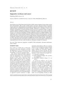
European Review for Medical and Pharmacological Sciences
2014; 18: 3097-3104
Impact of epigenetics in the management
of cardiovascular disease: a review
Y. CAO, L. LU, M. LIU, X.-C. LI1, R.-R. SUN, Y. ZHENG, P.-Y. ZHANG1
Graduate School, Nanjing University of Chinese Medicine, Nanjing, Jiangsu, China
1
Department of Cardiology, Xuzhou Central Hospital, Affiliated Xuzhou Hospital, Medical School of
Southeast University, Xuzhou Clinical Medical College of Nanjing University of Chinese Medicine,
Jiangsu, China
Y. Cao and L. Lu contributed equally to this work
Abstract. – Cardiovascular disease (CVD) is
the leading cause of death, irrespective of socioeconomic status, ethnic background and sex.
Despite the considerable progress in the treatment, the complex pathophysiology underlying
CVD is still not clear. In past few years, genetic
approaches including epigenetics and personalized medicine initiated a new way of treating
CVD. Epigenetics refers to the non-DNA sequence related heritable changes in gene expression and its role in understanding and treating coronary artery disease, heart failure, and
cardiac hypertrophy is currently recognized as
an important player. Histone acetylation, deactylation, DNA methylation and histone methylation are different mechanisms of epigenetic
modifications. Cardiac Hypertrophy is linked
with histone acetylation and the activity of histone acetyltransferases (HATs) has a positive
role in cardiac hypertrophy. Altered DNA methylation, miRNA activity have been shown to be
associated with atherosclerosis. It is documented that re-expression of certain fetal genes in
the adult heart contributes to the development
of heart failure syndrome, which is often associated with pathological cardiac remodeling comprising of changes in heart mass, size and
shape. Thus, it appears that approaches that
counteract epigenetic changes occurring in
CVD can prove to have significant therapeutic
impact. However, there are no major clinical
practice or therapeutics reports of epigenetics
contribution in CVD, even though deacetylase
inhibitors like trichostatin A were shown to have
some positive effects.
In this review we will present an overview of
various epigenetic mechanisms such as DNA
methylation, histone modifications, and microRNA-dependent mechanisms in CVD and the novel epigenetics-based therapeutic approaches.
Key Words:
Epigenetics, Cardiovascular disease, Histone,
mcroRNA.
Introduction
Cardiovascular disease (CVD) is the leading
cause of death worldwide despite significant disparities related to socioeconomic strata and
gender1. Several pathological conditions that afflict heart including myocardial infarction, cardiac
hypertrophy, myocardial ischemia, coronary artery
disease and hypertension, lead to heart failure
(HF)2,3. However, the complex pathophysiology
that leads to CVD is far from clear and alternate
mechanisms are constantly looked for in order to
provide better and collective explanations. Several
molecular and cellular mechanisms underlie the
development of HF that includes reprogramming
of expression of certain critical cardiac genes including downregulation of the alpha-myosin
heavy chain (2 -MHC) gene and sarcoplasmic
reticulum Ca+ATPase genes and reactivation of
specific fetal cardiac genes such as atrial natriuretic factor and brain natriuretic peptide4.
Over the past few years, advancements in the
areas of epigenetics and epigenomics have offered a new and different look at human diseases
and thus initiated a new era in genetic medicine5,6
by exploring the role of genetic heritability and
environmental interaction in disease pathology7.
Heritability of CVD can vary depending on sex
and environmental and lifestyle conditions. Genetic contribution to CVD can vary widely, as
much as 40% to 80%8 and environment-gene interactions (potentially mediated by epigenetics)
can affect the prevalence of CVD. Epigenetics
refers to mechanisms related to the regulation of
gene expression, which are chromatin-based but
not involving any DNA sequence changes. There
is significant experimental evidence suggesting
that epigenetic modifications serve as a cellular
Corresponding Author: Peiying Zhang, Ph.D; e-mail: [email protected]
3097
Y. Cao, L. Lu, M. Liu, X.-C. Li, R.-R. Sun, Y. Zheng, P.-Y. Zhang
memory of exposure in early life to unsuitable
environments. It has been noticed that even
among MZ twins, who are epigenetically indistinguishable in early life, environment and
lifestyles can greatly influence epigenetic patterns, with age. These differences affect singlecopy genes and repeat sequences. Anthropomorphic features, and vascular branching patterns are
few examples of how disease susceptibility might
vary between MZ twins9. Epigenetic modifications are reversible, vary between cell types, and
respond to endogenous and exogenous stimuli, as
well as environmental signals (Figure 1). Moreover, these modifications can elicit long-term
changes in gene transcription that potentially
lead to disease susceptibility in later life10.
Epigenetic Mechanisms and their
Implications in CVD
Epigenetics comprises of three distinct processes that are interrelated. These are (1) DNA base
modifications, (2) the histone modifications and
(3) RNA-based mechanisms (Figure 1)11.
DNA Base Modifications
Cytosine methylation is the major pathway
by which DNA is modified that is relevant for
epigenetic changes. DNA is made up of complementary pairs of the 4 nucleotides (guanine, cytosine, thymine, and adenine) running in various
sequences along two opposing DNA strands.
Cytosine methylation is conducted by DNA
methyltransferases (DNMTs), which transfer a
methyl group to the position-5 of the nitrogen
base cytosine ring. Approximately 0.6-1.5% of
the human genome is contributed by 5-methylcytosine (5 mC), even though its levels differ
between different tissue types12. Altered regulation of cytosine methylation has been linked to
tumour cell development and CVD13. Methylation of cytosines at promoter sites is associated
with long-term transcriptional repression of the
corresponding gene by directly blocking the
binding of specific transcription factors to their
cis-DNA elements. Even though this mechanism is relevant for transcriptional regulators
like Myc, AP2, hypoxia-inducible factor-1, and
Figure 1. The interrelation of three distinct processes: (1) DNA base modifications, (2) the histone modifications and (3)
RNA-based mechanisms.
3098
Impact of epigenetics in the management of cardiovascular disease
the insulator protein CTCF, DNA methylation
does not universally affect the binding of all
trans-factors (e.g., SP1)14.
Altered methylation patterns and extent of regulatory regions of genes is known to alter their
expression and correlate with failing hearts.
Three angiogenesis-related genes were found to
be differentially methylated, irrespective of the
etiology of HF. These are: hyper-methylation of
the 5’-regulatory region of platelet endothelial
cell adhesion molecule-1 and hypo-methylation
of the angiomotin like protein-2 is noted in failing hearts and this is associated with reduced expression of these genes. On the other hand, hypermethylation within the Rho-GTPase activating protein-24 gene in failing hearts is correlated
with elevated expression of this gene15. Moreover, a follow up study16 generated A genomewide DNA methylation map of human hearts and
showed significantly lowered global promoter
methylation of genes with increased expression
in failing hearts. Moreover, DNA methylation
can lead to hypermutability of distinct cardiac
genes, like the cardiac isoform of the myosin
binding protein C gene (Mybpc3), which has
higher level of exonic methylation of CpG sites
as compared to the skeletal muscle isoform17.
Clinical studies involving a follow up of elderly
subjects showed that hypomethylation of long interspersed nucleotide elements was associated
with higher incidence of ischemic heart disease
events18.
Associations have also been made between altered DNA methylation and atherosclerosis and
vascular inflammation. Deficiency of folic acid,
which is important factor in ‘One-carbon’ metabolism and as the carrier of methyl groups for
methylation reactions, has been epigenetically
linked to endothelial dysfunction and different
aspects of CVD, including coronary heart disease, anemia, atherosclerosis, and stroke19. Similarly, homocysteinuria has been associated with
the suppression of angiogenesis, VSMC proliferation, dyslipidemia, vascular oxidative stress, and
impairment of endothelial regeneration and function20. It has been suggested that dysregulation of
DNA methylation during different stages of embryonic growth might lead to congenital heart
disease, improperly functioning vascular tissue,
along with increased future risk of CVD as a result of the improper imprinting of genes, or inappropriate silencing of pluripotent or tissue-specific gene expression patterns21. Even at very early
stages of atherosclerosis, much before the
anatomical manifestation of the disease are noticed, hypomethylation could be detected22. Estrogen receptor (ESR1) and estrogen receptor
(ESR2), which are atheroprotective in vascular
smooth muscle cells, were found to be hypermethylated in human atherosclerosis23.
Cytosine Hydroxymethylation
Besides methylation, many genes, intergenic
regions, and repetitive elements are also hydroxymethylated in the heart, leading to altered expression of these genes24. For example, lowered
expression in association with hypermethylation
and a dysregulation of hydroxymethylation patterns of the gene coding for epidermal growth
factor receptor (EGFR), has been found to cause
abnormal valve differentiation in the heart, leading to aortic stenosis and left ventricular hypertrophy25.
Histone Modifications
Histones are the basic proteins around which
DNA is wound in chromatin and these proteins
make up nucleosomes. There are 8 core histone
proteins in nucleosomes: two dimers of
H2A/H2B and two dimers of H3/H4. Each core
histone has an amino-acid tail that can be modified. Posttranslational modifications to the N-terminals of these tails can modulate histone-DNA
interactions and are important players in epigenetic regulation of gene expression by influencing chromatin structure and controlling accessibility of transcriptional regulators to cis-DNA
binding elements14. Common modifications include lysine acetylation and methylation, arginine methylation, and serine phosphorylation26.
Histone acetylation is catalyzed by proteins
knows as histone acetyltransferases (HATs) and
histone deacetylation is carried out by HDACs.
Similarly, histone methylation levels are regulated by histone methyltransferases and histone
demethylases.
Some of the transcription factors, and their coactivators and repressors possess catalytic activity of either histone acetyaltion or deacetylation.
Histone acetylation at the lysine residues of the
histone tails results in chromatin de-condensation
and also provides a binding site for bromodomain proteins and transcriptional activators,
leading to transcriptional activation27. On the other hand, histone deacetylation causes reverse
changes, inducing chromatin condensation and
transcriptional repression28 (Figure 1). In experimental animal models, cardiac hypertrophy (CH)
3099
Y. Cao, L. Lu, M. Liu, X.-C. Li, R.-R. Sun, Y. Zheng, P.-Y. Zhang
has been linked with histone acetylation implicating both histone acetyltransferases (HATs)
and histone deacetylases (HDACs). It has been
shown that hypertrophic growth of cardiomyocytes could be induced by the overexpression of
transcriptional co-activators CREB binding protein (CRB) or p300, individually, in a manner
that is dependant on their HAT activity. Thus, inhibition of these co-activators could repress
phenylephrine-induced hypertrophic cell growth.
Overexpression of CBP or p300 with catalytic
activity, in cardiomyocytes, leads to hypertrophy,
whereas the corresponding mutant forms lacking
HAT activity did not produce such effects29.
The activity of HDACs has also been implicated in both pro- and anti-hypertrophic pathways,
resulting in conflicting data30 and further work is
needed to completely understand the role of
these enzymes in CH. The two classes of HDACs
were found to show opposing effects on cardiac
hypertrophy, with class I HDACs being pro-hypertrophic and class IIa HDACs being antihypertrophic31. Elevated expression of HDAC2 in cardiomyocytes mimics hypertrophic growth in an
Akt-dependent manner. Class I HDAC2 overexpressing transgenic mice were shown to develop
cardiac hypertrophy whereas HDAC2-null mice
were protected from pressure overload or isoproteranol induced cardiac hypertrophy32.
Histone methylation plays an important role in
the pathogenesis of CVD. In a genome-wide
study of histone methylation in heart, altered trimethylation of histones H3H4 and H3K9 has
been reported that in HF 33. Cardiac specific
knockout of Dot1L in mice, which catalyzes
H3K79 methylation in mammals, leads to a phenotype similar to dilated cardiomyopathy34. Dilated cardiomyopathy is a leading cause for HF
causing about one third of HF cases. A histone
trimethyl demethylase, JMJD2A35, is upregulated
in cardiomyopathy patients36 and cardiac-specific
overexpression of JMJD2A in mice causes extensive cardiac hypertrophy as compared to control
mice, following transverse aortic constriction.
However, jmjd2a-KO mice are protected from
cardiac stress induced by transverse aortic constriction36.
One of the important endothelial gene that is
relevant for angiogenesis and CVD that is regulated by the histone modifications is NOS3, that
codes for endothelial nitric oxide synthase
(eNOS), which forms NO from L-arginine in
blood vessels. Under normal physiological conditions, the NOS3 gene is transcriptionally active
3100
in endothelial cells, but repressed transcriptionally in VSMCs37. Under hypoxic conditions, there
is a substantial decrease in NOS3 transcription in
endothelial cells, probably due to eviction of histones, including those associated with transcriptional activation (acetyl H3K9, methyl H3K4,
acetyl H4K12), at the NOS3 proximal promoter
site38. Inhibition of histone deacetylation by trichostatin A (TSA) in vivo in mouse models, was
shown to prevent myocardial remodelling and to
reduce myocardial and serum tumour necrosis
factor alpha levels and to greatly improved functional myocardial recovery after myocardial infarction. HDAC inhibition also enhanced the formation of myocytes and microvessels in the
heart, in combination with increased cytokinesis.
These findings suggested that HDAC inhibition
can protect from heart failure by minimizing a
loss in myocardial performance after MI by promoting angiogenesis39.
RNA-based Mechanisms
Among the three general modes of epigenetic
regulation, RNA-based mechanisms are relatively the most recently described and much works
needs to be done to gain clear understanding of
these mechanisms. Current research focuses on
noncoding RNAs and small RNAs (i.e., RNAs
that do not code for any particular protein) that
function in the nucleus to regulate gene transcription. In particular, long noncoding RNAs (lncRNAs), that are longer than 200 nucleotides, play
an important role in epigenetic regulation40. Inside the nucleus RNA-based epigenetic regulation is mostly achieved by lncRNAs, whereas
small noncoding RNAs control posttranscriptional processes (translation and RNA degradation)
in the cytoplasm, even though some small noncoding RNAs were found to play a role in chromatin-based silencing41.
Micro-RNAs (miRNA) have emerged recently as important players of epigenetic regulators
of gene expression (Figure 1). MiRNA biogenesis involves formation of several thousands of
primary miRNA (pri-miRNA) through the RNA
polymerase II- mediated transcription of miRNA encoding regions. The hairpin-shaped transcript is processed by Drosha/DGCR8 complex
to produce precursor miRNA (pre-miRNA),
with a stem-loop structure of about 70-100 nucleotides in length. Then pre-miRNA is transported by exportin-5 from nucleus to the cytoplasm, where the enzyme Dicer cleaves the premiRNA into a short double-stranded RNA frag-
Impact of epigenetics in the management of cardiovascular disease
ment of 20-25 nucleotides long called miRNA:miRNA duplex. The miRNA is incorporated
into the RNA-induced silencing complex
(RISC), in which the miRNA is unwound and
split into two single strands, followed by the retention of the mature miRNA single strand, determining the formation of miRISC. The
miRISC complex recognizes and regulates specific target mRNA. MiRNAs are present in multiple copies and lead to translational suppression of target mRNA, by transcript degradation
and negative regulation of post-transcriptional
events. Key miRNAs regulate the expression of
several genes at the same time, and many miRNAs cooperatively regulate their target genes42.
More than a thousand different miRNAs have
been described in humans. Complete knowledge
of miRNAs biology in the cardiovascular system under physiological and pathological conditions appears to be important for CVD prevention, diagnosis and therapy43. It has been shown
that overexpression of miR-23a, miR-23b, miR24, miR-195 or miR-214 in neonatal cardiomyocytes induced CH, whereas overexpression of
miR-133 inhibited the phenotype44,45. MiRNAs
play important role in programming and modulating gene expression of different cell types
that participate in atherosclerosis. They mediate
inflammation, cholesterol influx, cellular differentiation and lipid uptake. Distinct expression
signatures of miRNAs in healthy and failing
hearts has been noted and the differential miRNA expression among failing hearts is dependent on the underlying HF etiology 46. Thus,
nearly 14 aortic stenosis-specific miRNAs were
found while a set of another eight miRNAs,
mainly expressed in a cardiomyopathic form of
HF, were noticed46.
The expression levels of miRNAs (Table I)
were found to vary with the progression of disease. Global miRNA profiles in a double transgenic mouse model, harboring mutations in both
the myosin heavy chain gene and the cardiac troponin I gene, resulting in severe HCM and premature mortality by age of 21 days, revealed stable downregulation of miR-1 and miR- 133 at
age of 10 and 16 days, suggesting a functional
role for these miRNAs throughout the progres-
Table I. Altered expressione of various miRNAs in cardiac abnormalities.
3101
Y. Cao, L. Lu, M. Liu, X.-C. Li, R.-R. Sun, Y. Zheng, P.-Y. Zhang
sion to HF. On the other hand, miR-31 levels increased at the end-stage of HF, suggesting a specific function for this miRNA during the final
phase of the disease47. MiRNA expression profiles (Table I) were shown to differ between
healthy and failing hearts, and that failing adult
hearts and fetal hearts display similar miRNA
profiles supporting the paradigm of reactivation
of a fetal gene program at the beginning or during the development of HF48.
ATP-Dependent Chromatin-Remodeling
ATP-dependent chromatin-remodeling complexes regulate gene expression by regulating nucleosome distribution on DNA, in ATP-dependent manner. Nucleosomal association with DNA
in turn controls the binding of transcription factors, which can bind to nucleosome core free
DNA regions. Chromatin-remodeling complexes
are divided into four families, viz., SWI/SNF,
ISWI, CHD, and INO80, based on their ATPase
subunits. Brahma-associated factor (BAF) complex, the mammalian ortholog of SWI/SNF complex, identified initially in Saccharomyces cerevisiae, consists of 14 BAF subunits encoded by
25 genes. These subunits can be assembled to
form a hundred different BAF complexes, which
have either brahma (Brm) or brahma-related gene
1 (Brg1) as the ATPase subunit and BAF complex is implicated in multiple processes, including heart and muscle development49. Brg1 deletion from endocardial cells during early stages of
development leads to morphological defects of
the heart due to deranged myocardial trabeculation50. Also, it has been shown that BAF180 subunit is necessary for normal coronary and heart
chamber development51.
Brg1 is expressed in embryonic heart but not
in adult cardiomyocytes. Brg1 is instrumental in
the switch from the expression of fetal myosin
heavy chain (i.e., b-MHC) to that of adult MHC
(a-MHC) isoform during cardiac hypertrophy.
However, Brg1 expression is induced by pro-hypertrophy stimuli; Brg1 then interacts with its
partners HDAC and PARP, forming complexes
that cause the pathological shift from adult to fetal MHC isoforms. Recent studies showed that
BRG1-KO mice are more resistant to a pro-hypertrophic stimuli, such as transverse aortic constriction. Also, expression level of Brg1 was
found to be correlated with the severity of cardiomyopathy in the hearts of hypertrophic cardiomyopathy patients52.
3102
Conclusions
Heart failure, a leading cause of morbidity and
mortality, is the final outcome of different pathological conditions of heart due to complex genetic predisposition and environmental factors, and
is accompanied by pathological cardiac remodeling, i.e., changes in heart mass, size, and shape.
Many recent studies support an important role of
epigenetics in the pathogenesis of heart failure
and cardiac hypertrophy. Despite significant research, still clear answers are not available for:
(1) the genomic regions that are regulated by epigenetic mechanisms; (2) the interactions and biological implications of the various epigenetic
mechanisms including DNA methylation, histone
modifications, chromatin remodeling and miRNAs; (4) the impact of epigenetic modifications
on the function of non-cardiomyocyte cells (e.g.,
cardiac fibroblasts and endothelial cells) of heart
tissue during heart failure; and (5) what is the interaction between environment (e.g., diet, smoking, stress) and epigenetic changes in the etiology of heart failure. One mechanism of gene expression regulation that has gained importance is
epigenetics. Counteracting the gene expression
changes occurring in heart failure could be a
therapeutic approach. Even though epigenetic
pathways have been therapeutically targeted for
cancer treatment, such efforts are still in infancy
for heart failure.
–––––––––––––––––-––––
Conflict of Interest
The Authors declare that there are no conflicts of interest.
References
1) ANONYMOUS. Cardiovascular health for all. Lancet
2013; 382: 572
2) DUYGU B, POELS EM, DA COSTA MARTINS PA. Genetics
and epigenetics of arrhythmia and heart failure.
Front Genet 2013; 4: 219.
3) PAPAIT R, CONDORELLI G. Epigenetics in heart failure. Ann N Y Acad Sci 2010; 1188: 159-164.
4) OLSON EN, BACKS J, MCKINSEY TA. Control of cardiac hypertrophy and heart failure by histone
acetylation/deacetylation. Novartis Found Symp
2006; 274: 3-12; discussion 13-19, 152-155,
272-156.
5) F EINBERG AP. Epigenomics reveals a functional
genome anatomy and a new approach to common disease. Nat Biotechnol 2010; 28: 10491052.
Impact of epigenetics in the management of cardiovascular disease
6) PORTELA A, ESTELLER M. Epigenetic modifications
and human disease. Nat Biotechnol 2010; 28:
1057-1068.
7) JAENISCH R, BIRD A. Epigenetic regulation of gene
expression: How the genome integrates intrinsic
and environmental signals. Nat Genet 2003; 33
Suppl: 245-254.
8) ELDER SJ, LICHTENSTEIN AH, PITTAS AG, ROBERTS SB,
FUSS PJ, GREENBERG AS, MCCRORY MA, BOUCHARD TJ,
JR., SALTZMAN E, NEALE MC. Genetic and environmental influences on factors associated with cardiovascular disease and the metabolic syndrome.
J Lipid Res 2009; 50: 1917-1926.
9) FRAGA MF, BALLESTAR E, PAZ MF, ROPERO S, SETIEN F,
BALLESTAR ML, HEINE-SUNER D, CIGUDOSA JC, URIOSTE
M, BENITEZ J, BOIX-CHORNET M, SANCHEZ-AGUILERA A,
LING C, CARLSSON E, POULSEN P, VAAG A, STEPHAN Z,
SPECTOR TD, WU YZ, PLASS C, ESTELLER M. Epigenetic
differences arise during the lifetime of monozygotic twins. Proc Natl Acad Sci U S A 2005; 102:
10604-10609.
10) BEEKMAN M, NEDERSTIGT C, SUCHIMAN HE, KREMER D,
VAN DER BREGGEN R, LAKENBERG N, ALEMAYEHU WG,
DE CRAEN AJ, WESTENDORP RG, BOOMSMA DI, DE GEUS
EJ, HOUWING-DUISTERMAAT JJ, HEIJMANS BT, SLAGBOOM
PE. Genome-wide association study (GWAS)identified disease risk alleles do not compromise
human longevity. Proc Natl Acad Sci U S A 2010;
107: 18046-18049.
11) WEBSTER AL, YAN MS, MARSDEN PA. Epigenetics and
cardiovascular disease. Can J Cardiol 2013; 29:
46-57.
12) LISTER R, PELIZZOLA M, DOWEN RH, HAWKINS RD, HON
G, TONTI-FILIPPINI J, NERY JR, LEE L, YE Z, NGO QM,
EDSALL L, ANTOSIEWICZ-BOURGET J, STEWART R, RUOTTI
V, MILLAR AH, THOMSON JA, REN B, ECKER JR. Human
DNA methylomes at base resolution show widespread epigenomic differences. Nature 2009; 462:
315-322.
13) KIKUYAMA M, TAKESHIMA H, KINOSHITA T, OKOCHI-TAKADA E, WAKABAYASHI M, AKASHI-TANAKA S, OGAWA T, SETO Y, USHIJIMA T. Development of a novel approach,
the epigenome-based outlier approach, to identify
tumor-suppressor genes silenced by aberrant
DNA methylation. Cancer Lett 2012; 322: 204212.
14) MATOUK CC, MARSDEN PA. Epigenetic regulation of
vascular endothelial gene expression. Circ Res
2008; 102: 873-887.
15) MOVASSAGH M, CHOY MK, GODDARD M, BENNETT MR,
DOWN TA, FOO RS. Differential DNA methylation
correlates with differential expression of angiogenic factors in human heart failure. PLoS One
2010; 5: e8564.
16) MOVASSAGH M, CHOY MK, KNOWLES DA, CORDEDDU L,
HAIDER S, DOWN T, SIGGENS L, VUJIC A, SIMEONI I, PENKETT C, GODDARD M, LIO P, BENNETT MR, FOO RS. Distinct epigenomic features in end-stage failing human hearts. Circulation 2011; 124: 2411-2422.
17) M EURS KM, K UAN M. Differential methylation of
CpG sites in two isoforms of myosin binding
18)
19)
20)
21)
22)
23)
24)
25)
26)
27)
28)
29)
protein C, an important hypertrophic cardiomyopathy gene. Environ Mol Mutagen 2011; 52:
161-164.
B ACCARELLI A, W RIGHT R, B OLLATI V, L ITONJUA A,
ZANOBETTI A, TARANTINI L, SPARROW D, VOKONAS P,
SCHWARTZ J. Ischemic heart disease and stroke in
relation to blood DNA methylation. Epidemiology
2010; 21: 819-828.
TALAULIKAR VS, ARULKUMARAN S. Folic acid in obstetric practice: a review. Obstet Gynecol Surv 2011;
66: 240-247.
YAN TT, LI Q, ZHANG XH, WU WK, SUN J, LI L, ZHANG
Q, TAN HM. Homocysteine impaired endothelial
function through compromised vascular endothelial growth factor/Akt/endothelial nitric oxide synthase signalling. Clin Exp Pharmacol Physiol
2010; 37: 1071-1077.
CEDAR H, BERGMAN Y. Programming of DNA methylation patterns. Annu Rev Biochem 2012; 81: 97117.
LUND G, ANDERSSON L, LAURIA M, LINDHOLM M, FRAGA
MF, VILLAR-GAREA A, BALLESTAR E, ESTELLER M, ZAINA S.
DNA methylation polymorphisms precede any
histological sign of atherosclerosis in mice lacking
apolipoprotein E. J Biol Chem 2004; 279: 2914729154.
KIM J, KIM JY, SONG KS, LEE YH, SEO JS, JELINEK J,
G OLDSCHMIDT-C LERMONT PJ, I SSA JP. Epigenetic
changes in estrogen receptor beta gene in atherosclerotic cardiovascular tissues and in-vitro vascular senescence. Biochim Biophys Acta 2007;
1772: 72-80.
KINNEY SM, CHIN HG, VAISVILA R, BITINAITE J, ZHENG Y,
ESTEVE PO, FENG S, STROUD H, JACOBSEN SE, PRADHAN
S. Tissue-specific distribution and dynamic
changes of 5-hydroxymethylcytosine in mammalian genomes. J Biol Chem 2011; 286: 2468524693.
BARRICK CJ, ROBERTS RB, ROJAS M, RAJAMANNAN NM,
SUITT CB, O'BRIEN KD, SMYTH SS, THREADGILL DW.
Reduced EGFR causes abnormal valvular differentiation leading to calcific aortic stenosis and left
ventricular hypertrophy in C57BL/6J but not
129S1/SvImJ mice. Am J Physiol Heart Circ
Physiol 2009; 297: H65-75.
LENNARTSSON A, EKWALL K. Histone modification patterns and epigenetic codes. Biochim Biophys Acta 2009; 1790: 863-868.
RODRIGUEZ JE, MCCUDDEN CR, WILLIS MS. Familial
hypertrophic cardiomyopathy: Basic concepts and
future molecular diagnostics. Clin Biochem 2009;
42: 755-765.
SHAHBAZIAN MD, GRUNSTEIN M. Functions of sitespecific histone acetylation and deacetylation. Annu Rev Biochem 2007; 76: 75-100.
GUSTERSON RJ, JAZRAWI E, ADCOCK IM, LATCHMAN DS.
The transcriptional co-activators CREB-binding
protein (CBP) and p300 play a critical role in cardiac hypertrophy that is dependent on their histone acetyltransferase activity. J Biol Chem 2003;
278: 6838-6847.
3103
Y. Cao, L. Lu, M. Liu, X.-C. Li, R.-R. Sun, Y. Zheng, P.-Y. Zhang
30) ABI KHALIL C. The emerging role of epigenetics in
cardiovascular disease. Ther Adv Chronic Dis
2014; 5: 178-187.
31) CHANG S, MCKINSEY TA, ZHANG CL, RICHARDSON JA,
HILL JA, OLSON EN. Histone deacetylases 5 and
9 govern responsiveness of the heart to a subset of stress signals and play redundant roles in
hear t development. Mol Cell Biol 2004; 24:
8467-8476.
32) TRIVEDI CM, LUO Y, YIN Z, ZHANG M, ZHU W, WANG T,
FLOSS T, GOETTLICHER M, NOPPINGER PR, WURST W,
F ERRARI VA, A BRAMS CS, G RUBER PJ, E PSTEIN JA.
Hdac2 regulates the cardiac hypertrophic response by modulating Gsk3 beta activity. Nat Med
2007; 13: 324-331.
33) KANEDA R, TAKADA S, YAMASHITA Y, CHOI YL, NONAKASARUKAWA M, SODA M, MISAWA Y, ISOMURA T, SHIMADA
K, M ANO H. Genome-wide histone methylation
profile for heart failure. Genes Cells 2009; 14: 6977.
34) NGUYEN AT, ZHANG Y. The diverse functions of Dot1
and H3K79 methylation. Genes Dev 2011; 25:
1345-1358.
35) KOOISTRA SM, HELIN K. Molecular mechanisms and
potential functions of histone demethylases. Nat
Rev Mol Cell Biol 2012; 13: 297-311.
36) ZHANG QJ, CHEN HZ, WANG L, LIU DP, HILL JA, LIU
ZP. The histone trimethyllysine demethylase
JMJD2A promotes cardiac hypertrophy in response to hypertrophic stimuli in mice. J Clin Invest 2011; 121: 2447-2456.
37) YAN MS, MATOUK CC, MARSDEN PA. Epigenetics of
the vascular endothelium. J Appl Physiol (1985)
2010; 109: 916-926.
38) HO JJ, MAN HS, MARSDEN PA. Nitric oxide signaling in hypoxia. J Mol Med (Berl) 2012; 90: 217231.
39) ZHANG L, QIN X, ZHAO Y, FAST L, ZHUANG S, LIU P,
CHENG G, ZHAO TC. Inhibition of histone deacetylases preserves myocardial performance and prevents cardiac remodeling through stimulation of
endogenous angiomyogenesis. J Pharmacol Exp
Ther 2012; 341: 285-293.
40) KHACHANE AN, HARRISON PM. Mining mammalian
transcript data for functional long non-coding
rnas. PLoS One 2010; 5: e10316.
3104
41) K AIKKONEN MU, L AM MT, G LASS CK. Non-coding
RNAs as regulators of gene expression and epigenetics. Cardiovasc Res 2011; 90: 430-440.
42) ESTELLER M. Non-coding RNAs in human disease.
Nat Rev Genet 2011; 12: 861-874.
43) BAUERSACHS J, THUM T. Biogenesis and regulation of
cardiovascular microRNAs. Circ Res 2011; 109:
334-347.
44) CARE A, CATALUCCI D, FELICETTI F, BONCI D, ADDARIO A,
GALLO P, BANG ML, SEGNALINI P, GU Y, DALTON ND,
ELIA L, LATRONICO MV, HOYDAL M, AUTORE C, RUSSO
MA, DORN GW, 2ND, ELLINGSEN O, RUIZ-LOZANO P,
PETERSON KL, CROCE CM, PESCHLE C, CONDORELLI G.
MicroRNA-133 controls cardiac hypertrophy. Nat
Med 2007; 13: 613-618.
45) LATRONICO MV, CONDORELLI G. MicroRNA and cardiac pathology. Nat Rev Cardiol 2009; 6: 419-429.
46) IKEDA S, KONG SW, LU J, BISPING E, ZHANG H, ALLEN
PD, GOLUB TR, PIESKE B, PU WT. Altered microRNA
expression in human heart disease. Physiol Genomics 2007; 31: 367-373.
47) BAGNALL RD, TSOUTSMAN T, SHEPHARD RE, RITCHIE W,
S EMSARIAN C. Global MicroRNA profiling of the
mouse ventricles during development of severe
hypertrophic cardiomyopathy and heart failure.
PLoS One 2012; 7: e44744.
48) BARRY SP, DAVIDSON SM, TOWNSEND PA. Molecular
regulation of cardiac hypertrophy. Int J Biochem
Cell Biol 2008; 40: 2023-2039.
49) HAN P, HANG CT, YANG J, CHANG CP. Chromatin remodeling in cardiovascular development and
physiology. Circ Res 2011; 108: 378-396.
50) STANKUNAS K, HANG CT, TSUN ZY, CHEN H, LEE NV, WU
JI, SHANG C, BAYLE JH, SHOU W, IRUELA-ARISPE ML,
CHANG CP. Endocardial Brg1 represses ADAMTS1
to maintain the microenvironment for myocardial
morphogenesis. Dev Cell 2008; 14: 298-311.
51) WANG Z, ZHAI W, RICHARDSON JA, OLSON EN, MENESES JJ, FIRPO MT, KANG C, SKARNES WC, TJIAN R. Polybromo protein BAF180 functions in mammalian
cardiac chamber maturation. Genes Dev 2004;
18: 3106-3116.
52) HANG CT, YANG J, HAN P, CHENG HL, SHANG C, ASHLEY E, ZHOU B, CHANG CP. Chromatin regulation by
Brg1 underlies heart muscle development and
disease. Nature 2010; 466: 62-67.


© Copyright 2026