
Managing Incidental Findings on Abdominal CT: White Paper of the
Managing Incidental Findings on
Abdominal CT: White Paper of the
ACR Incidental Findings Committee
Lincoln L. Berland, MDa, Stuart G. Silverman, MDb, Richard M. Gore, MDc,
William W. Mayo-Smith, MDd, Alec J. Megibow, MD, MPHe, Judy Yee, MDf,
James A. Brink, MDg, Mark E. Baker, MDh, Michael P. Federle, MDi,
W. Dennis Foley, MDj, Isaac R. Francis, MDk, Brian R. Herts, MDh,
Gary M. Israel, MDg, Glenn Krinsky, MDl, Joel F. Platt, MDk,
William P. Shuman, MDm, Andrew J. Taylor, MDn
As multidetector CT has come to play a more central role in medical care and as CT image quality has improved, there has
been an increase in the frequency of detecting “incidental findings,” defined as findings that are unrelated to the clinical
indication for the imaging examination performed. These “incidentalomas,” as they are also called, often confound
physicians and patients with how to manage them. Although it is known that most incidental findings are likely benign and
often have little or no clinical significance, the inclination to evaluate them is often driven by physician and patient
unwillingness to accept uncertainty, even given the rare possibility of an important diagnosis. The evaluation and surveillance of incidental findings have also been cited as among the causes for the increased utilization of cross-sectional imaging.
Indeed, incidental findings may be serious, and hence, when and how to evaluate them are unclear. The workup of
incidentalomas has varied widely by physician and region, and some standardization is desirable in light of the current need
to limit costs and reduce risk to patients. Subjecting a patient with an incidentaloma to unnecessary testing and treatment
can result in a potentially injurious and expensive cascade of tests and procedures. With the participation of other radiologic
organizations listed herein, the ACR formed the Incidental Findings Committee to derive a practical and medically
appropriate approach to managing incidental findings on CT scans of the abdomen and pelvis. The committee has used a
consensus method based on repeated reviews and revisions of this document and a collective review and interpretation of
relevant literature. This white paper provides guidance developed by this committee for addressing incidental findings in the
kidneys, liver, adrenal glands, and pancreas.
Key Words: Incidental findings, incidentaloma, pancreatic cyst, renal cyst, liver lesion, adrenal nodule
J Am Coll Radiol 2010;7:754-773. Copyright © 2010 American College of Radiology
FOREWORD
This white paper is meant not to comprehensively review
the interpretation and management of solid masses in each
organ system but to provide general guidance for managing
incidentally discovered masses, appreciating that individual
care will vary depending on each patient’s specific circumstances; the clinical environment, available resources; and the
judgment of the practitioner. Also, the term guidelines has not
a
Department of Radiology, University of Alabama at Birmingham, Birmingham, Alabama.
b
Department of Radiology, Brigham and Women’s Hospital, Boston, Massachusetts.
c
Department of Radiology, Evanston Hospital, Evanston, Illinois.
d
Department of Radiology, Brown University School of Medicine, Providence, Rhode Island.
e
Department of Radiology, NYU-Langone Medical Center, New York, New York.
f
Department of Radiology, University of California, San Francisco, San Francisco, California.
g
Department of Diagnostic Radiology, Yale University School of Medicine,
New Haven, Connecticut.
h
Department of Radiology, Cleveland Clinic, Cleveland, Ohio.
754
i
Department of Radiology, Stanford University Medical Center, Stanford,
California.
j
Department of Radiology, Medical College of Wisconsin, Milwaukee, Wisconsin.
k
Department of Radiology, University of Michigan, Ann Arbor, Michigan.
l
Radiology Associates of Ridgewood, PA, Waldwick, New Jersey.
m
Department of Radiology, University of Washington School of Medicine,
Seattle, Washington.
n
Department of Radiology, Virginia Commonwealth University Medical
Center, Richmond, Virginia.
Corresponding author and reprints: Lincoln L. Berland, MD, University of
Alabama at Birmingham, Department of Radiology, 619 S 19th Street, N348,
Birmingham, AL 35249-1900; e-mail: [email protected].
© 2010 American College of Radiology
0091-2182/10/$36.00 ● DOI 10.1016/j.jacr.2010.06.013
Berland et al/Managing Incidentalomas on Abdominal CT 755
been used in this white paper to avoid the implication that this
represents a component of the ACR Practice Guidelines and
Technical Standards (which represent official ACR policy, having undergone a rigorous drafting and review process culminating in approval by the ACR Council), or the ACR Appropriateness Criteria® (which use a formal consensus-building
approach using a modified Delphi technique). This white paper, which represents the collective experience of the Incidental
Findings Committee, using a less formal process of repeated
reviews and revisions of the draft document, does not represent
official ACR policy. For these reasons, this white paper should
not be used to establish the legal standard of care in any particular situation.
scribing findings related to specific medical conditions,
relatively little research has been devoted to understanding incidental findings. The most common reason to
pursue incidental findings is to differentiate benign from
potentially serious (including malignant) lesions. Although most incidental findings prove to be benign, their
discovery often leads to a cascade of testing that is costly,
provokes anxiety, exposes patients to radiation unnecessarily, and may even cause morbidity [15]. Articles describing criteria for detecting, categorizing, reporting,
and managing such findings have been inconsistent at
best and leave many unanswered questions [1,9-14].
PROJECT OBJECTIVES
INTRODUCTION
The objectives of this project were:
The rapid increase in the utilization of cross-sectional
imaging examinations over the past two decades, combined with the ongoing improvement in the spatial and
contrast resolution of these studies, has led to a marked
increase in the number of findings detected that are unrelated to the primary objectives of the examinations
[1-4]. An incidental finding, also known as an incidentaloma, may be defined as “an incidentally discovered
mass or lesion, detected by CT, MRI, or other imaging
modality performed for an unrelated reason” [5]. Although such findings are incidental to the primary purpose of the study, one analysis suggested, “Some research
and clinical activities are so prone to generating findings
not intentionally sought that it is disingenuous to term
them ‘unanticipated’ even if their precise nature cannot
be anticipated in advance” [6]. More important than the
definition is the action that each such finding invokes.
So, we are asked to consider, “What is the responsible use
of information that nobody asked for?” [7].
The burden of extra costs with incidental findings on
cross-sectional imaging has also raised concerns within
the government and third-party payers as medical imaging utilization and expenditures have risen. A recent example of this was seen in the May 2009 CMS noncoverage decision regarding screening CT colonography [8].
Although CT colonography focuses on detecting colorectal polyps to prevent colorectal carcinoma, an unenhanced, low–radiation dose CT scan of the lower chest,
entire abdomen, and pelvis contains clinically significant
incidental findings in 5% to 16% of asymptomatic patients [1,4,9-14], with a higher frequency in symptomatic patients [9,10,12-14]. The noncoverage decision by
CMS cited concern for the costs of evaluating extracolonic findings that are diagnostically indeterminate.
Other existing or developing technologies may face this
type of economic scrutiny as CMS and other third-party
payers become more focused on cost containment.
Although countless studies have been devoted to de-
●
●
●
to develop a consensus on sets of organ-specific imaging
features for some commonly affected organ systems
within the abdomen, which will lead to consistent definitions for, and identification of, incidental findings;
to develop medically appropriate approaches to managing
incidental findings that are diagnostically indeterminate; and
to address the differences between unenhanced, low–
radiation dose CT examinations and contrast-enhanced CT examinations using standard radiation
doses for detecting and managing incidental findings.
POTENTIAL BENEFICIAL OUTCOMES OF
THE PROJECT
Benefits anticipated from this effort included:
●
●
●
●
●
reducing risks to patients from additional unnecessary
examinations, including the risks of radiation and risks
associated with interventional procedures;
limiting the costs of managing incidental findings to
patients and the health care system;
achieving greater consistency in recognizing, reporting, and managing incidental findings, as a component
of formal quality improvement efforts;
providing guidance to radiologists who are concerned
about the risk for litigation for missing incidental findings that later prove to be clinically important; and
helping focus research efforts to lead to an evidencebased approach to incidental findings.
HISTORY OF THE PROJECT
Because of the increasing recognition of the problems
and opportunities of incidental findings, consideration
of a formal approach to these issues began within the
ACR in 2006. The Incidental Findings Committee was
formed under the auspices of the Body Imaging Commission of the ACR. After several meetings and conference calls, the concepts and objectives described above
756 Journal of the American College of Radiology/ Vol. 7 No. 10 October 2010
were formulated. The initial intent was to develop guidelines analogous to those produced by the Fleischner Society on pulmonary nodules [16] and the consensus conferences of the Society of Radiologists in Ultrasound on
thyroid nodules [17] and carotid imaging [18].
Because of the keen interest among groups both within
and outside the ACR, the committee’s participants were
recruited from members of the ACR, all of who were also
fellows or members of the Society of Computed Body Tomography and Magnetic Resonance, the Society of Gastrointestinal Radiologists, and the Society of Uroradiology.
Contacts from other groups within the ACR, including the
Colon Cancer Committee, the Appropriateness Criteria–
Adrenal Panel and the Appropriateness Criteria–GI Panel
(Liver Lesion Topic) also helped ensure the consistency of
the guidance produced among these groups.
CONSENSUS PROCESS
Expert radiologists in relevant organ systems were recruited to participate in the Incidental Findings Committee and its subcommittees. We plan to further review
and revise these recommendations periodically, on the
basis of comments and new research. Although the scope
of a project to address incidental findings on CT is large,
the committee decided to develop guidance for a limited
number of organ systems. Four subcommittees were established to address the largest number of incidental
findings within the abdomen, in the kidneys, liver, adrenal glands, and pancreas. A fifth subcommittee was
charged with attempting to ensure the use of common
terminology and a common format. The committee
elected to defer considering other incidental findings
arising in the abdomen and pelvis, such as ovarian
masses, splenic lesions, lymphadenopathy, and vascular
abnormalities, including arterial stenoses, abdominal
aortic aneurysms, and renal artery aneurysms. The membership of each subcommittee is listed in the Appendix.
Each subcommittee was tasked to develop organ-specific
guidance, which was initially formulated primarily by the
subcommittee chairs. When this was complete, these subsections were distributed to the subcommittee members for
further comments and discussion. Revisions of the entire
document were then distributed to the subcommittee
chairs, and multiple revisions ensued. Finally, the draft was
distributed to the entire Incidental Findings Committee for
additional review to achieve consensus and to arrive at a final
manuscript. Reviews by other ACR committees were also
integrated into drafts at appropriate points in the process.
To facilitate rapidly formulating and clearly communicating this guidance, and to provide convenient graphic summaries for easy reference, the committee decided to express
its recommendations in flowcharts and tables, buttressed
with explanatory text.
ELEMENTS OF THESE
RECOMMENDATIONS AND FLOWCHARTS
Certain subspecialties within radiology have addressed inconsistencies of documentation by creating structured reporting, such as the Breast Imaging Reporting and Data
System® classification [19]. In an analogous way, Zalis et al
[20], for the Working Group on Virtual Colonoscopy, proposed “C-RADS,” which includes an “E” classification system for extracolonic findings. Although this latter classification system has elements in common with these
recommendations, it is not included with them here.
In the flowcharts within this white paper, the algorithms
use yellow boxes for steps that involve data to affect management, such as categorization, demographics, history, and
the results of studies. Green boxes represent action steps,
such as performing a study, following up, or intervening
with a biopsy or surgery. Red boxes indicate that the evaluation process should stop, with no further action required,
because the lesion can be concluded to be benign.
CHALLENGES OF ADDRESSING
INCIDENTAL FINDINGS
One of the crucial obstacles to managing incidental findings cost-effectively is the unwillingness of many physicians to accept uncertainty even when the chance of a
serious diagnosis is extremely unlikely. This unwillingness is in part driven by a paucity of data, the lack of
clear-cut algorithms with regard to diagnostic and treatment strategies, fear of potential malpractice litigation,
and the desire of patients and their families to adhere to
the adage “better safe than sorry.” It may be difficult for
physicians or patients to appreciate at an intellectual or
emotional level that an incidental finding might not need
to undergo further examinations or follow-up. Not only
are further tests likely to yield a benign diagnosis, but
such testing could even lead to morbidity [15]. On the
other hand, an incidental finding could represent a serendipitous discovery of a serious diagnosis, such as a large
abdominal aortic aneurysm, and be potentially lifesaving;
hence the conundrum. The discussion of cost is also
burdened with strong opinions, with some believing that
cost should be no obstacle to reaching a comfortable level
of medical certainty for a positive or negative diagnosis
[21,22]. Others might argue that medical resources
should be best applied where they are known to be most
effective. However, there is strong scientific validation
for applying medical strategies that optimize results while
minimizing costs and applying “evidence-based” reasoning to medical decisions [21].
Unfortunately, information about the cost-effectiveness of pursuing incidental findings is largely lacking.
Therefore, achieving a consensus of experts, supported
by available literature, is a reasonable interim objective
Berland et al/Managing Incidentalomas on Abdominal CT 757
for this Incidental Findings Committee. However, there
are several reasons to hypothesize that a group of specialty
radiologists from academic institutions might be biased
toward the overuse of imaging studies. For example, the
culture of attempting to achieve diagnostic certainty
noted above may be more intense in an academic environment, partly because of the higher intensity of illness
seen there. Less experienced physicians in residency and
fellowship may be more inclined to depend on imaging
studies, with this inclination supported by attending
physicians wanting to enhance the teaching experience.
Also, academic institutions are more likely to have a
broad array of advanced imaging technologies, the use of
which is encouraged by the desire to perform research.
Additionally, academic experts are intensely focused in their
areas of interest and are keenly aware of the multitude of possible serious results from incidental findings, also potentially biasing their viewpoint. Therefore, in approaching incidental
findings in this way, there is a risk that rather than the results of
this project limiting the overuse of imaging, the detailed guidance generated from this project either might not affect such
overutilization or could even increase it. Our goal was not necessarily to reduce utilization (although we believe this is needed)
but rather to optimize utilization. In this way, only the appropriate incidental findings are evaluated further. These factors
were considered in designing these recommendations, especially regarding the guidance on the length and frequency of
follow-up studies for indeterminate lesions.
the length of the radiology report but might be inaccurate in a small percentage of situations;
3. Not reported at all. Particularly in the case of small
lesions, some would argue that such a finding is so
common and innocuous that it does not rise to the
level of an abnormality. Refraining from reporting
would be analogous to a nonradiologist physician not
mentioning an insignificant skin lesion on a physical
examination report. Because many patients and some
physicians become concerned about even minor findings, this would prevent any risk for further testing; or
4. Reported by stating that a definitive diagnosis cannot be
made, but there are no features to suggest a malignant
etiology, with one possible phrase being “indeterminate,
no malignant features.” This would leave the workup to
the discretion of the referring physician and perhaps the
patient. However, such a report leaves the referrer in a
quandary. This may lead to unnecessary testing, but it
would essentially acknowledge the limits of the examination and acknowledge that there are no evidencebased data to allow specific recommendations.
Option 1 was considered acceptable, but not necessarily preferred, by all members of the Incidental Findings
Committee. However, the committee could not reach a
consensus on all aspects of this subject, because various
members preferred, while others raised objections to each
of options 2, 3, and 4. Some members noted that reporting all incidental findings can be valuable if a patient has
a follow-up examination and only the report is available.
REPORTING CONSIDERATIONS
Some considerations are common to all organ systems.
One universal principle is to refer to available prior relevant imaging examinations when interpreting incidental
findings. Prior examinations need not be of precisely the
same type or modality but are useful if they include the
anatomic area in question, such as a chest CT scan that
includes the upper abdomen. Also, the approach to incidental findings should be placed in the context of the
individual patient’s situation. As an extreme but common example, the need to report or pursue incidental
findings may be unnecessary in patients with serious
medical comorbidities or limited life expectancy.
The wording of the radiology report is also controversial
and could fall into 4 categories. This can be illustrated
through the example of a renal mass that seems to be a
simple cyst on an unenhanced CT scan. Such a lesion could
be:
1. Described as a “low-attenuation mass statistically
likely to be a simple cyst” or a “low-attenuation mass
likely to be benign;”
2. Reported as a “renal cyst.” This contains the specific,
implicit recommendation to do nothing and limits
SCANNING TECHNIQUES
In the 4 organ-specific sections below (kidneys, liver,
adrenal glands, and pancreas), comments apply to standard–radiation dose examinations, whether performed
unenhanced or enhanced. However, low-dose unenhanced scans may be performed for CT colonography,
identifying urinary tract calculi and other applications.
We believe that incidental findings identified on such
low–radiation dose, unenhanced scans require special
considerations. These are separately addressed in an additional section following the 4 organ-specific sections.
KIDNEYS
Nature and Scope of the Problem
The literature regarding the approach to renal masses detected on renal mass–protocol CT or MRI is replete with
case series, retrospective analyses, and suggested clinical
guidelines that have been long accepted and are widely
adopted in clinical practice today [23-47]. A summary and
update of these guidelines, discussed in the context of an
758 Journal of the American College of Radiology/ Vol. 7 No. 10 October 2010
Table 1. Management recommendations for patients with incidental cystic renal masses
Appearance
Recommendation
Bosniak
Category
I†
II
IIF
III
IV
Imaging Features
Hairline-thin wall; no septa, calcifications, or solid
components; water attenuation; no
enhancement
Few hairline-thin septa with or without perceived
(not measurable) enhancement; fine
calcification or short segment of slightly
thickened calcification in the wall or septa;
homogeneously high-attenuating masses
(ⱕ3 cm) that are sharply marginated and do not
enhance
Multiple hairline-thin septa with or without
perceived (not measurable) enhancement,
minimal smooth thickening of wall or septa that
may show perceived (not measureable)
enhancement, calcification may be thick and
nodular but no measurable enhancement
present; no enhancing soft tissue components;
intrarenal nonenhancing high-attenuation renal
masses (⬎3 cm)
Thickened irregular or smooth walls or septa,
with measurable enhancement
Criteria of category III, but also containing
enhancing soft tissue components adjacent to
or separate from the wall or septa
General
Population
Ignore
Comorbidities or
Limited Life Expectancy
Ignore
Ignore
Ignore
Observeⴱ§
Observe§ or ignore储
Surgery‡
Surgery‡ or observe§
Surgery‡
Surgery‡ or observe§
Note: These recommendations are to be followed only if nonneoplastic causes of a renal mass (eg, infections) have been excluded;
see text for details. The recommendations are offered as general guidance and do not necessarily apply to all patients. Reprinted with
permission from Radiology 2008;249:16-31.
ⴱ
In selected patients (eg, young), early surgical intervention may be considered, particularly if a minimally invasive approach (eg,
laparoscopic partial nephrectomy) can be used.
†When a mass ⬍1 cm has the appearance of a simple cyst, further workup is not likely to yield useful information.
‡Surgical options include open or laparoscopic nephrectomy and partial nephrectomy; each provides a tissue diagnosis. Open,
laparoscopic, and percutaneous ablation may be considered when available, but biopsy would be needed to achieve a tissue
diagnosis. Long-term (5-year or 10-year) results of ablation are not yet known.
§Computed tomography or MRI at 6 and 12 months, then yearly for 5 years; the interval and duration of observation may be varied
(eg, longer intervals may be chosen if the mass is unchanged, longer duration may be chosen for greater assurance).
储Cystic masses ⱕ1.5 cm that are not clearly simple cysts or that cannot be characterized completely may not require further evaluation
in patients with comorbidities and in patients with limited life expectancy. Reprinted with permission from Radiology 2008;249:16-31.
incidental finding, has been recently detailed [48] and thus
is not entirely repeated in this white paper.
Detection and Characterization
A renal mass can be found incidentally, either as part of
an examination that allows the mass to be fully characterized or as part of an examination that does not allow
the mass to be evaluated fully. Many renal masses can be
characterized completely using ultrasound or contrast
material– enhanced CT; however, some renal masses
may require additional imaging [23-47]. Renal mass–
protocol CT or MRI examinations (scans obtained both
before and after intravenous contrast material) allow
most renal masses to be fully characterized. Renal masses
are divided into cystic and solid types, and recommendations are detailed for each and for both the general population and patients with comorbidities or limited life
expectancy (Tables 1-3). In general, the suggested management of renal masses begins first with ensuring that
the mass is not the result of a nonneoplastic condition
that can mimic a tumor. These conditions include
pseudotumors such as columns of Bertin, hypertrophied
tissue adjacent to scars, vascular anomalies and aneurysms, infarcts, and infections. Focal bacterial pyelonephritis commonly causes a masslike abnormality in the
kidney. Also, fat-containing angiomyolipomas should be
Berland et al/Managing Incidentalomas on Abdominal CT 759
Table 2. Management recommendations for incidental solid renal masses in patients in the general
population
Mass Size
Probable Diagnosis
Recommendation
Comment
Large (⬎3 cm)
Renal cell carcinoma† Surgery‡
Angiomyolipoma with minimal fat,
oncocytoma, other benign
neoplasms may be found at
surgery
Small (1-3 cm)
Renal cell carcinoma† Surgery‡
If hyperattenuating, and
homogenously enhancing,
consider MRI and percutaneous
biopsy to diagnose
angiomyolipoma with minimal
fat
Observe until 1 cm§ Thin (ⱕ3 mm) sections help
Very small (⬍1 cm) Renal cell carcinoma,
confirm enhancement
oncocytoma,
angiomyolipomaⴱ
Note: These recommendations are best followed after nonneoplastic causes of a renal mass (eg, infections) have been excluded; see
text for details. The recommendations are offered as general guidance and do not necessarily apply to all patients.
ⴱ
Benign entities are more likely in small renal masses than large ones.
†Provided there is no detectable fat by CT or MRI using protocols designed to evaluate renal masses.
‡Surgical options include open or laparoscopic nephrectomy and partial nephrectomy; both provide a tissue diagnosis. Open,
laparoscopic, and percutaneous ablation may be considered when available, but biopsy would be needed to achieve a tissue
diagnosis. Long-term (5-year or 10-year) results of ablation are not yet known.
§Computed tomography or MRI at 3 to 6 months, 12 months, and then yearly; the interval and duration of observation may be varied
(eg, shorter intervals if the mass is enlarging). Reprinted with permission from Radiology 2008;249:16-31.
excluded. With rare exceptions, a mass that contains fat,
particularly when not calcified, can be diagnosed as an
angiomyolipoma with confidence. The subsequent management then can be derived and is summarized in Tables 1 to 3. These tables are reconfigured in the form of
flowchart algorithms in Figures 1 and 2.
The approach to the cystic renal mass follows the
time-tested approach of Bosniak [23,25,27-32]. The tables and flowcharts are constructed so that both patients
in the general population and those with limited life
expectancy can be managed. In general, size is not a factor
in the Bosniak classification of cystic renal masses, because large cystic masses are often benign, and small ones
may be malignant. However, the smaller the mass, the
more likely it is benign. Therefore, the commonly encountered cystic-appearing renal mass that is too small to
evaluate all of its features, including its CT attenuation,
can be presumed to be benign if it does not display any
nonsimple features. In the green “action boxes” in the
flowcharts (Figures 1 and 2), observation with imaging,
also known as active surveillance [49,50], is recommended for indeterminate masses in Bosniak category
IIF and is also an option for masses in categories III and
IV in patients with limited life expectancy or comorbidities that would increase the risk of treatment.
There is no known interval of time that can be used to
diagnose an indeterminate renal mass with certainty, although 5 years has been suggested as a reasonable length
of time to diagnose an indeterminate renal mass as benign on the basis of the lack of morphologic change
[36,48]. Depending on the level of suspicion, and patient
and referrer comfort with observation, both the duration
and interval may be altered. As indicated in Tables 1 to
3 and the flowcharts (Figures 1 and 2), growth alone
cannot be used to definitively diagnose a mass (whether
solid or cystic) as malignant. Benign masses may grow,
and malignant ones may grow little, if at all [51,52].
Regarding the flowchart for cystic renal masses (Figure
1), both Bosniak category III and IV masses are managed
surgically; however, category IV masses have a greater
probability of malignancy than category III masses, and
management approaches other than resection carry more
risk. Because many Bosniak category III masses are malignant, surgery is recommended for the general population. Percutaneous biopsy of Bosniak category III renal
masses, although controversial, may be helpful, particularly in patients with comorbidities that would pose risk
to patients undergoing surgery [34,46]. If a definitive
malignant result can be obtained with biopsy, surgery
may be planned with confidence. For a benign biopsy
result to be useful, it should be both definitive and specific of a benign entity. Biopsy results that reveal nonspecific cells should be viewed with caution and cannot be
used alone to guide management. Because Bosniak category III masses typically contain few solid elements, it
may be difficult to both target and procure diagnostic
760 Journal of the American College of Radiology/ Vol. 7 No. 10 October 2010
Table 3. Management recommendations for incidental solid renal masses in patients with limited life
expectancy or comorbidities that increase the risk of treatment
Mass Size
Probable Diagnosis
Recommendation
Comment
Large (⬎3 cm)
Renal cell
Surgery‡ or observe
Angiomyolipoma with minimal fat,
carcinoma†
oncocytoma, other benign
neoplasms may be found at
surgery; biopsy can be used
preoperatively to confirm renal
cell carcinoma
Small (1-3 cm)
Renal cell
Surgery‡ or observe
If hyperattenuating, and
carcinoma†
homogenously enhancing,
consider MRI and percutaneous
biopsy to diagnose
angiomyolipoma with minimal
fat
Very small (⬍1 cm) Renal cell carcinoma, Observe until 1.5 cm§ Thin (ⱕ3 mm) sections help
oncocytoma,
confirm enhancement
angiomyolipomaⴱ
Note: These recommendations are best followed after nonneoplastic causes of a renal mass (eg, infections) have been excluded; see
text for details. The recommendations are offered as general guidance and do not necessarily apply to all patients.
ⴱ
Benign entities are more likely in small renal masses than large ones.
†Provided there is no detectable fat by CT or MRI using protocols designed to evaluate renal masses.
‡Surgical options include open or laparoscopic nephrectomy and partial nephrectomy; both provide a tissue diagnosis. Open,
laparoscopic, and percutaneous ablation may be considered when available, but biopsy would be needed to achieve a tissue
diagnosis. Long-term (5-year or 10-year) results of ablation are not yet known.
§Computed tomography or MRI at 3 to 6 months, 12 months, and then yearly; the interval of observation may be varied (eg, shorter
intervals if the mass is enlarging); the duration of observation may be individualized. Observation may be considered for a solid renal
mass of any size in a patient with limited life expectancy or comorbidities that increase the risk of treatment, particularly when the mass
is small. It may be safe to observe a solid renal mass beyond 1.5 cm, but there are insufficient data to provide definitive
recommendations on the risks and benefits of observation. Reprinted with permission from Radiology 2008;249:16-31.
tissue for biopsy, limiting the ability to achieve definitively benign or malignant results. However, even if a
confident diagnosis of a benign entity can be made in
these patients, observation is still warranted.
We define solid masses as those that contain little or no
fluid attenuating (⬍20 Hounsfield units [HU]) components and usually consist predominantly of enhancing tissue
(Tables 2 and 3, Figure 2). As described for cystic renal
masses, all solid masses should be evaluated first for features
suggesting a nonneoplastic etiology, such as focal bacterial
pyelonephritis or other conditions noted above. A thorough
search for fat cells using CT or MRI protocols designed to
evaluate renal masses should also be undertaken. Although
there are rare exceptions, fat-containing noncalcified renal
masses in adults can be diagnosed as benign angiomyolipomas with confidence [48]. The subsequent approach to a
solid renal mass is then predicated mostly on size. Although
there is no single feature of a renal mass that can be used to
predict its biologic behavior accurately, size is a reasonable
and practical approach. In general, large (⬎3 cm) solid renal
masses are likely malignant; similarly, the smaller a solid
mass, the more likely it is benign. In addition, a small renal
cell carcinoma is more likely to be low grade and indolent
behaving than a larger one [53]. Therefore, we have sug-
gested that solid masses ⬍1 cm be observed [48]. This
approach is further supported by the difficulty of confirming that masses of this size are enhancing and are therefore
solid. Partial volume effects can mimic enhancement. Thus,
the use of thin-section (ⱕ3 mm) CT and MRI is advised
when both evaluating and observing such small masses.
However, there are rare cases of aggressively behaving small
renal cell carcinomas, even those ⬍1 cm. Therefore, observation is not completely without risk [54].
Solid renal masses between 1 and 3 cm can be characterized as enhancing with confidence. Unlike masses ⬍1 cm,
these masses are large enough to be targeted for percutaneous biopsy. Although still somewhat controversial, in some
patients, biopsy can be used to provide a definitive diagnosis
of oncocytoma and angiomyolipoma, the two most common benign neoplasms found after surgical resection of a
solid renal mass [53,55]. Because an angiomyolipoma with
minimal fat typically presents as a hyperdense, T2-hypointense, homogeneously enhancing mass, MRI, with or without CT, can be used to identify such masses and lead to
percutaneous biopsy [41,47,48]. Although oncocytomas
are typically homogeneously enhancing masses and may
display a central scar, these features may also be found in
oncocytic renal cell carcinomas. Therefore, specific recom-
Berland et al/Managing Incidentalomas on Abdominal CT 761
Incidental Cystic Renal Mass 1
Detected on CT
Bosniak I or II
Bosniak IIF
Benign
no further follow-up 2
Bosniak III or IV
General population
Limited life expectancy or
co-morbidities 7
General population
Limited life expectancy or
co-morbidities 7
CT or MRI at 6 and 12 mo,
then yearly for 5 yrs. 3, 4
If follow-up appropriate,
CT or MRI at 6 and 12 mo,
then yearly for 5 yrs. 3, 8
Surgery 6
If follow-up appropriate,
CT or MRI at 6 and 12 mo,
then yearly for 5 yrs. 3, 9
LEGEND
Further action based on
change, life expectancy and
co-morbidities
No morphologic change
Morphologic change 5
Morphologic change 5
Benign
no further follow-up
Surgery 6
Surgery, follow-up or no
further follow-up based on
life expectancy and
co-morbidities
1 These recommendations are to be followed only if
non-neoplastic causes of a renal mass (e.g., infections)
have been excluded; see Ref. 48 for details. The
recommendations are offered as general guidance and
do not necessarily apply to all patients. See Table 1 for
detailed description of Bosniak Classification.
2 When a mass smaller than 1 cm has the appearance of
a simple cyst, further work-up is not likely to yield
useful information.
3 Interval and duration of observation may be varied (e.g.,
longer intervals may be chosen if the mass is unchanged;
longer duration may be chosen for greater assurance).
4 In selected patients (e.g., young), early surgical
intervention may be considered, particularly if a
minimally invasive approach (e.g., laparoscopic partial
nephrectomy) can be utilized.
5 Morphologic change refers to change in feature
characteristics, such as number of septations or their
thickness. Growth should be noted, but by itself does
not indicate malignancy.
6 Surgical options include open or laparoscopic
nephrectomy and partial nephrectomy; each provides a
tissue diagnosis. Open, laparoscopic, and percutaneous
ablation may be considered where available, but biopsy
would be needed to achieve a tissue diagnosis. Long-term
(5- or 10-year) results of ablation are not yet known.
7 Limited life expectancy and co-morbidities that increase
the risk of treatment.
8 Cystic masses 1.5 cm or smaller that are not clearly
simple cysts or that cannot be characterized completely
may not require further evaluation in patients with
co-morbidities and in patients with limited life
expectancy.
9 Percutaneous biopsy of Bosniak Category III masses
may be considered, but may not be diagnostic.
Fig 1. Flowchart for incidental cystic renal mass detected on CT.
mendations as to which masses should undergo percutaneous biopsy cannot be made.
LIVER
Nature and Scope of the Problem
Recent advances in multidetector CT, MRI, ultrasound
and 2-[18F]fluoro-2-deoxyglucose PET have led to the
detection of incidental hepatic masses in both the oncology and nononcology patient population that in the past
remained undiscovered. This has engendered a management dilemma that is particularly pertinent to oncology
patients, in whom any hepatic mass, clinical or subclinical, warrants attention. At autopsy, as many as 52% of
noncancer patients have benign hepatic lesions, and liver
metastases are found in as many as 36% of patients dying
with cancer [56]. Key questions to answer include the
following: (1) Does the hepatic incidentaloma put the
patient at risk for an adverse outcome? (2) Can a primary
or metastatic malignancy be accurately and confidently
differentiated from a benign incidentaloma? and (3) If a
benign lesion, might it still require surgical intervention,
such as resecting a hepatic adenoma to prevent rupture?
Implications of Imaging and Clinical
Features
Strategies for optimizing the management of these lesions are only beginning to emerge in terms of deciding
which of these incidental liver masses may not need further evaluation, which may simply be monitored over
time, and which require more aggressive workup. Preoperative percutaneous biopsy may minimize diagnostic
error but is associated with a postprocedural morbidity of
2.0% to 4.8% and mortality of 0.05% [57-59].
The Incidental Findings Committee’s guidance for
managing liver incidental findings is illustrated in Figure
3. Managing incidental liver lesions depends on the
probable importance of the mass. This is assessed both by
the appearance of the mass and the level of risk that each
patient has for developing important liver masses. Important liver masses are not limited to malignancies. For
example, a benign hepatic adenoma might require surgical intervention. These categories are defined as follows:
762 Journal of the American College of Radiology/ Vol. 7 No. 10 October 2010
Incidental Solid Renal Mass 1
Detected on CT
<1 cm 2
1-3 cm 5
General population
Limited life expectancy and
co-morbidities 3
Follow-up until 1 cm:
CT or MRI at 3-6 mo and 12
mo, then yearly 4
Follow-up until 1.5 cm:
CT or MRI at 3-6 mo and 12
mo, then yearly 4
>3 cm 9
General population
Limited life expectancy or
co-morbidities 3
Surgery 7
Surgery 7, 10
General population
LEGEND
Hyperattenuating,
homogeneously enhancing:
consider MRI, biopsy 6
1 These recommendations are to be followed only if
non-neoplastic causes of a renal mass (e.g., infections
and fat-containing angiomyolipomas) have been
excluded; see Ref. 48 for details. The recommendations
are offered as general guidance and do not necessarily
apply to all patients.
2 Differential diagnosis includes renal cell carcinoma,
oncocytoma, angiomyolipoma. Benign entities are more
likely in small renal masses than large ones.
3 Limited life expectancy and co-morbidities that
increase the risk of treatment.
4 Interval and duration of observation may be varied
(e.g., shorter interval if the mass is enlarging).
Follow-up 8
Limited life expectancy and
co-morbidities 3
Surgery 7
Follow-up 8
5 Probable diagnosis renal cell carcinoma, provided there
is no detectable fat at CT or MRI using protocols
designed to evaluate renal masses.
6 If hyperattenuating and homogeneously enhancing,
consider MRI and percutaneous biopsy to diagnose
angiomyolipoma with minimal fat.
7 Surgical options include open or laparoscopic
nephrectomy and partial nephrectomy; both provide a
tissue diagnosis. Open, laparoscopic, and percutaneous
ablation may be considered where available, but biopsy
would be needed to achieve a tissue diagnosis.
Long-term (5- or 10-year) results of ablation are not
yet known.
8 Observation may be considered for a solid renal mass
of any size in a patient with limited life expectancy or
co-morbidities that increase the risk of treatment,
particularly when the mass is small. It may be safe to
observe a solid renal mass beyond 1.5 cm; however,
there are insufficient data to provide definitive
recommendations on the risks and benefits of
observation. Thin (≤3 mm) sections help confirm
enhancement.
9 Probable diagnosis renal cell carcinoma.
Angiomyolipoma with minimal fat, oncocytoma, and
other benign neoplasms may be found at surgery.
10 Percutaneous biopsy can be utilized preoperatively to
confirm renal cell carcinoma.
Fig 2. Flowchart for incidental solid renal mass detected on CT.
1. Low-risk individuals: Young patients (aged ⱕ40 years),
with no malignancies, hepatic dysfunction, hepatic malignant risk factors, or symptoms attributable to the liver.
2. Average-risk individuals: Patients aged ⬎40 years, with no
known malignancies, hepatic dysfunction, hepatic malignant risk factors, or symptoms attributable to the liver.
3. High-risk individuals: Patients with known primary
malignancies with a propensity to metastasize to the
liver, cirrhosis, or other hepatic risk factors. Hepatic
risk factors include hepatitis, chronic active hepatitis, sclerosing cholangitis, primary biliary cirrhosis,
hemochromatosis, hemosiderosis, hepatic dysfunction, and long-term oral contraceptive use.
ADRENAL GLANDS
Nature and Scope of the Problem
An incidental adrenal mass, often referred to as an adrenal incidentaloma, is defined as an adrenal mass (ⱖ1 cm)
discovered incidentally on a cross-sectional imaging examination performed for another reason. Incidental adrenal masses are very common, estimated to occur in
approximately 3% to 7% of the adult population [60-
63]. The most frequent pathology for an incidentally
discovered adrenal mass is a nonhyperfunctioning adenoma [64]. It was shown in one study that the overwhelming majority of incidentally discovered adrenal
masses are benign in patients with no known malignancies [65]. Statistics indicate that given the high prevalence of nonhyperfunctioning adrenal adenomas in the
general population, an incidentally discovered adrenal
mass in an oncology patient is most likely benign. However, the adrenal gland is also a common site for metastases and, somewhat less commonly, primary adrenal
tumors, including pheochromocytomas, aldosteronomas, and adrenal cortical carcinomas.
The goal of imaging when an incidental adrenal
mass is discovered is to differentiate a benign “leavealone” mass (eg, nonhyperfunctioning tumor, myelolipoma, hemorrhage, cyst) from a mass that warrants
treatment (eg, metastasis, pheochromocytoma, adrenal cortical carcinoma). From an imaging perspective,
an optimal algorithm should be used to diagnose both
leave-alone masses and masses that need treatment,
using as few tests as possible. The adrenal flowchart
(Figure 4) and recommendations described here at-
Berland et al/Managing Incidentalomas on Abdominal CT 763
A
Incidental Liver Mass
Detected on CT
<0.5 cm
0.5-1.5 cm
Low or average risk 1, 2
High risk 3
Benign, no further
follow-up
Follow-up 4
Low attenuation, benign
imaging features 5
Low attenuation,
suspicious imaging
features 7
Any risk level 1, 2, 3
Any risk level 1, 2, 3
Benign, no further
follow-up 6
Follow-up 4
>1.5 cm
Flash filling
(robustly enhancing)
Low or average risk 1, 2
High risk 3
Benign, no further
follow-up 8, 9
Evaluate 7
Low attenuation,
benign imaging
features 5
Low attenuation,
suspicious imaging
features 6
Benign, no further
follow-up 6
LEGEND
B
Follow-up 4
Flash filling
(robustly enhancing)
Low risk 1
Average risk 2
High risk 3
Follow-up 4
Evaluate 7
Biopsy, core preferred
Benign diagnostic
imaging features 8, 9
Benign, evaluate if possible
FNH, adenoma 8, 9
No benign diagnostic
imaging features 10
Follow-up 4,
evaluate 7 or biopsy,
core preferred
Incidental Liver Mass
Detected on CT
1 Low risk individuals:Young patient (≤ 40 years
old), with no known malignancy, hepatic
dysfunction, hepatic malignant risk factors, or
symptoms attributable to the liver.
2 Average risk individuals: Patient >40 years old,
with no known malignancy, hepatic dysfunction,
abnormal liver function tests or hepatic
malignant risk factors or symptoms
attributable to the liver.
3 High risk individuals: Known primary
malignancy with a propensity to metastasize to
the liver, cirrhosis, and/or other hepatic risk
factors. Hepatic risk factors include hepatitis,
chronic active hepatitis, sclerosing cholangitis,
primary biliary cirrhosis, hemochromatosis,
hemosiderosis, oral contraceptive use, anabolic
steroid use.
4 Follow-up CT or MRI in 6 months. May need
more frequent follow-up in some situations,
such as a cirrhotic patient who is a liver
transplant candidate.
5 Benign imaging features: Typical hemangioma
(see below), sharply marginated, homogeneous
low attenuation (up to about 20 HU), no
enhancement. May have sharp, but irregular
margins.
6 Benign low attenuation masses: Cyst,
hemangioma, hamartoma,Von Meyenberg
complex (bile duct hamartomas).
7 Suspicious imaging features: Ill-defined margins,
enhancement (more than about 20 HU),
heterogeneous, enlargement. To evaluate,
prefer multiphasic MRI.
8 Hemangioma features: Nodular discontinuous
peripheral enhancement with progressive
enlargement of enhancing foci on subsequent
phases. Nodule isodense with vessels, not
parenchyma.
9 Small robustly enhancing lesion in average risk,
young patient: hemangioma, focal nodular
hyperplasia (FNH), transient hepatic
attenuation difference (THAD) flow artifact,
and in average risk, older patient: hemangioma,
THAD flow artifact. Other possible diagnoses:
adenoma, arterio-venous malformation (AVM),
nodular regenerative hyperplasia.
Differentiation of FNH from adenoma
important especially if larger than 4 cm and
subcapsular.
10 Hepatocellular or common metastatic
enhancing malignancy: islet cell,
neuroendocrine, carcinoid, renal cell
carcinoma, melanoma, choriocarcinoma,
sarcoma, breast, some pancreatic lesions.
Fig 3. Flowchart for incidental liver mass detected on CT.
tempt to do both. The algorithm reflects the most
commonly encountered imaging scenarios. However,
it is important to note that there are exceptions to
some of the recommendations depending on individual patients’ presentations and histories. As noted in
other sections of this white paper, if a patient has
limited life expectancy or severe comorbidities,
workup of an incidentally discovered adrenal mass
may not be appropriate. Readers are also directed to a
recent comprehensive review on this topic [66].
764 Journal of the American College of Radiology/ Vol. 7 No. 10 October 2010
Incidental Adrenal Mass (≥1 cm)
Detected on CT or MR
Imaging features are diagnostic
Imaging features not diagnostic
>4 cm
Myelolipoma, ca ++
= benign, no F/U
HU ≤10 or ↓ signal on CS-MR
= adenoma1
Prior imaging
1−4 cm
No history of cancer:
consider resection2
No prior imaging
History of cancer
No prior imaging
No history of cancer
Stable ≥1 year
Lesion enlarging
Benign1
Concerning for malignancy
Consider biopsy or resection2
Benign imaging features3:
Presume benign1, consider 12
month F/U CT or MR
Suspicious imaging features4
LEGEND
2
3
4
If patient has clinical signs or symptoms of adrenal
hyperfunction, consider biochemical evaluation
Consider biochemical testing to exclude pheochromocytoma
Benign imaging features = homogeneous, low density,
smooth margins
Suspicious imaging features = heterogeneous, necrosis,
irregular margins
APW = Absolute Percentage Washout
RPW = Relative Percentage Washout
CS-MR = Chemical Shift MRI
F/U = Follow-up
HU = Hounsfield Unit
↓ = decreased
Consider PET or below
Unenhanced CT or CS-MR
HU ≤10 or ↓ signal on
CS-MR = adenoma1
1
History of cancer:
consider PET or biopsy2
HU >10 or no ↓ signal on
CS-MR
Adrenal washout CT
No enhancement (≤10 HU)
= cyst or hemorrhage
APW / RPW ≥60/40%
APW / RPW <60/40%
Benign, no F/U
Adenoma1
Biopsy if appropriate2 or
consider CS-MR if not done
Fig 4. Flowchart for incidental adrenal mass detected on CT or MR.
Imaging Characterization and Workup
Algorithm
If an adrenal mass has diagnostic features of a benign
lesion such as a myelolipoma (presence of macroscopic
fat) or cyst (simple cyst-appearing without enhancement), no additional workup or follow-up imaging is
needed. If the lesion is 1 to 4 cm and has a density of ⱕ10
HU on CT or signal loss compared with the spleen on
out-of-phase images of a chemical-shift MRI (CS-MRI)
examination, it is almost always diagnostic of a lipid-rich
adenoma [67-72]. If diagnostic imaging features are not
present but the adrenal mass has been stable for ⱖ1 year,
it is likely benign [66].
If a patient has no history of cancer, there are no prior
examinations, and the mass has benign imaging features
(low density, homogeneous with smooth margins), one
may consider a follow-up unenhanced CT or CS-MRI
examination in 12 months. However, if there are suspicious imaging features on contrast-enhanced CT, such as
necrosis, heterogeneous density, or irregular margins,
one could proceed with an unenhanced CT or CS-MRI
examination. If these do not confirm that the lesion is
a lipid-rich adenoma, adrenal washout CT with 15minute delayed imaging to calculate contrast material
washout may be helpful [73-75].
In patients with histories of cancer and adrenal masses,
if the imaging features are not diagnostic and there is no
prior imaging to confirm stability, one may consider
unenhanced CT, CS-MRI, or PET imaging [76]. If the
mass cannot be diagnosed as a lipid-rich adenoma, adrenal washout CT may be helpful. In patients with no
histories of cancer and adrenal masses ⬎4 cm, one may
consider resection.
Adenomas typically enhance rapidly using either iodinated contrast material or gadolinium chelates and also
display rapid washout [74]. Although metastases generally enhance rapidly, their washout is more prolonged.
Using CT, absolute percentage washout values are calculated using the formula (enhanced HU ⫺ 15-minute
delayed HU)/(enhanced HU ⫺ unenhanced HU) ⫻
100. A value of ⱖ60% is diagnostic of an adenoma.
Relative percentage washout is used when an unen-
Berland et al/Managing Incidentalomas on Abdominal CT 765
hanced CT value is not available and the enhanced values
are compared with 15-minute delayed scans. Relative
percentage washout is calculated using the formula (enhanced HU ⫺ 15-minute delayed HU)/enhanced HU ⫻
100; a value of ⬎40% is diagnostic for an adenoma
[73-75]. Adrenal washout CT was used successfully to
distinguish adenomas from nonadenomas in 160 of 166
adrenal masses with 98% sensitivity and 92% specificity [73].
Recent advances in imaging characterization with
CT, MRI, and PET have decreased the need for image-guided percutaneous biopsies to characterize adrenal masses [77]. However, if an adrenal mass is enlarging, it may be prudent to proceed to percutaneous
adrenal biopsy or surgical resection. In an oncology
patient, a new adrenal mass in a patient with known
metastases elsewhere is most likely another metastasis.
However, an isolated adrenal mass could be benign or
malignant. If the mass cannot be characterized as an
adenoma using CT, MRI, or PET, a biopsy may be
appropriate. If there are signs or symptoms of pheochromocytoma, it may be prudent to obtain plasmafractionated metanephrine and normetanephrine levels before biopsy [78].
Imaging examinations are useful to separate adrenal
adenomas from other masses but cannot be used to distinguish hyperfunctioning adenomas from nonhyperfunctioning adenomas. One approach would be to rely
on history and physical examination to determine which
patients should undergo biochemical testing for hyperfunctioning adrenal neoplasms. Some endocrinologists
recommend excluding an occult, asymptomatic hyperfunctioning neoplasm in all adrenal incidentalomas
[60,62,63]. This approach would be costly and is not
routinely performed by many physicians. Regarding the
radiology report, when an adenoma can be diagnosed
with imaging, we suggest stating, “Findings consistent
with a benign adenoma. If there are clinical signs or
symptoms of adrenal hyperfunction, biochemical evaluation may be appropriate.”
PANCREAS
Nature and Scope of the Problem
The frequency of detection of pancreatic cysts by CT scanning is reported between 1.2% [79] and 2.6% [80]. For
MRI, the reported frequency is significantly higher, at
19.9% of MRI examinations [81]. Because pancreatic cysts
are quite prevalent, a practicing radiologist may see several
for every 100 abdominal imaging cases performed.
Cystic pancreatic tumors are most often frankly benign or low-grade indolent neoplasms. In one study that
included asymptomatic patients with pancreatic cysts in
whom there was operative correlation, 17% of asymp-
tomatic cysts were serous cystadenomas, 28% were mucinous cystic neoplasms, 27% were intraductal papillary
mucinous neoplasms (IPMNs), 2.5% were ductal adenocarcinomas, and 3.8% were pseudocysts [82]. Intraductal papillary mucinous neoplasms were the most common cystic neoplasm when both symptomatic and
asymptomatic patients were evaluated. In another series,
39% of IPMNs were incidentally detected, and 50% of
IPMNs were side branch or branch duct IPMNs with a
5-year risk for developing high-grade dysplasia or invasive carcinoma of 15% [83].
Mucinous cystic masses, namely IPMNs and mucinous cystic neoplasms, have a well-established malignant
potential likened to an adenoma-carcinoma sequence
[84]. Because of this malignant potential, it has become
increasingly difficult for radiologists evaluating individual cases to know how to frame the report to help guide
appropriate management. We believe that the guidance below will help in the evaluation and reporting of the
majority of these lesions. These recommendations are
also summarized in Figure 5.
Detection and Characterization
This discussion is limited to unexpected pancreatic cysts
in asymptomatic patients. Asymptomatic patients have
no clinical or laboratory indication directly referable to
the pancreas, including but not limited to hyperamylasemia, recent-onset diabetes, severe epigastric pain,
weight loss, or jaundice. The most frequently detected
cyst is ⬍10 mm in size [85]. Cysts of this size are particularly prevalent on MRI. Imaging will not be able to
characterize these lesions. The question of appropriate
follow-up is subsequently addressed.
There is ample literature to support the nonsurgical management of pancreatic cysts ⬍3 cm that do not display
“worrisome features” [86-90]. Some recommend 2.5 cm as
a maximal diameter for nonsurgical management [91].
Worrisome features include larger size, presence of mural
nodules, dilation of the common bile duct, involvement of
the main pancreatic duct, and lymphadenopathy [92-96].
Studies of patients in whom cysts have been resected or
aspirated find that malignancy or premalignancy does not
correlate with cyst size alone. These studies suggest that
mucinous lesions of any size are premalignant [97,98].
However, in a series of 170 of 539 patients who underwent
operative resection of pancreatic cysts, no invasive cancers
were found in mucinous cysts ⬍3 cm [90].
Nevertheless, establishing a cyst as mucinous is important because of their higher risk for the presence or future
development of malignancy. Morphologic features that
aid in diagnosis of a mucinous tumor include (1) the
presence or absence of septae (mucinous cystic neoplasms
generally are multilocular, with large cysts), (2) the position of calcification (mucinous cystic neoplasms typically
766 Journal of the American College of Radiology/ Vol. 7 No. 10 October 2010
Asymptomatic 1 Patient with Incidental Pancreatic Cystic Mass
Detected on CT, MRI (with or without contrast) or US
<2 cm
2-3 cm
Single follow-up in
1 yr, preferably MRI 2
Imaging characterization,
preferably MRI/MRCP 3
Stable
Uncharacterized
cystic mass
BD-IPMN
Serous cystadenoma
Follow-up yearly
Follow-up every
6 mo for 2 years 4
Follow-up every
2 yr
Serous cystadenoma
Uncharacterized
cystic mass or other
cystic neoplasm
Consider resection
when ≥ 4 cm
Cyst aspiration
Resect, depending
on co-morbidities
and risk
LEGEND
Benign, no further
follow-up
Growth
>3 cm
1 Signs and symptoms include
hyperamylasemia, recent onset diabetes,
severe epigastric pain, weight loss,
steatorrhea or jaundice.
2 Consider decreasing interval if younger,
omitting with limited life expectancy.
Recommend limited T2-weighted MRI for
routine follow-ups.
3 Recommend pancreas-dedicated MRI
with MRCP.
4 If no growth after 2 years, follow yearly. If
growth OR suspicious features develop,
consider resection.
5 BD-IPMN = branch duct intraductal
papillary mucinous neoplasm.
Fig 5. Flow chart for an asymptomatic patient with an incidental pancreatic cystic mass detected on CT, MRI
(with or without contrast), or ultrasound (US). MRCP ⫽ MR cholangiopancreatography.
have peripheral calcification, whereas serous tumors have
central calcification), (3) location within the pancreas,
and (4) the presence of main pancreatic duct involvement [99-102]. Mucinous cystic tumors can be suspected when a cyst is present in the tail of the pancreas in
a perimenopausal woman [84]. The presence or absence
of direct communication with the main pancreatic duct
must be established to distinguish a mucinous cystic tumor (with relatively high malignant potential) from a
branch duct IPMN (with relatively low malignant potential). Three-dimensional imaging with either MRI or CT
can address this question. Conversely, serous cystadenoma characteristically displays variably dense radial septae in a honeycombed or spongiform pattern and central
calcification. The more peripheral cysts are larger than
the more central cysts.
A simple but useful imaging-based classification system differentiates pancreatic cystic masses into 4 morphologic types: (1) unilocular (pseudocysts, mucinous
cystic neoplasms, lymphoepithelial cysts, small IPMNs,
and small serous tumors), (2) microcystic (serous cystadenomas and lymphoepithelial cysts), (3) macrocystic
(mucinous cystic neoplasms, oligocystic serous tumors,
and IPMNs) and (4) cysts with solid components (solidappearing serous tumors, solid pseudopapillary neoplasms, and cystic islet cell tumors) [103].
Implications of Imaging and Clinical
Features
Most incidental cysts can be detected on routine abdominal studies. However, if a cyst needs to be characterized,
it is recommended that a diagnosis of a specific cyst type
not be made unless the patient undergoes a dedicated
“pancreas-style” study. For multidetector CT, this would
require a dual-phase contrast-enhanced acquisition in
both pancreatic and portal venous phases using a narrow
detector configuration. Thin-section images should be
available on a workstation that can perform 3-D analysis.
Magnetic resonance imaging should be performed at
1.5 T. Phased-array torso coils enhance signal and parallel imaging increases speed and improves resolution. The
study should include sequences that display in-phase and
out-of-phase T1, T2 (preferably with fat suppression),
and 3-D, fat-saturated, gradient-echo T1 gadoliniumenhanced sequences in pancreatic, portal, and equilibrium phases. Additionally, MR cholangiopancreatogra-
Berland et al/Managing Incidentalomas on Abdominal CT 767
phy is necessary. Current MRI scanners have respiratory
triggered 3-D sequences for MR cholangiopancreatography [104]. Secretin administration may facilitate visualization of the communication of a cyst with the main
pancreatic duct [105]. By consensus, the Incidental
Findings Committee suggests dedicated MRI as the imaging procedure of choice to characterize a pancreatic
cyst. This reflects the superior contrast resolution of
MRI, facilitating the recognition of septae, nodules, and
duct communication [106].
The pretest likelihood that a given lesion in an individual patient is a malignant neoplasm is of paramount
consideration when deciding on management. Controversy exists between using dedicated imaging or an attempt at aspiration of a cyst under endoscopic ultrasound
guidance. Most often, this decision will be made on the
basis of the size of the cyst, location within the pancreas,
accessibility to the endoscopic ultrasound approach, and
expertise of the endosonographer. A carcinoembryonic
antigen level in the aspirate of 192 ng/mL has a high
specificity for discriminating mucinous from nonmucinous cysts, demonstrating higher accuracy than cyst morphology [107]. Amylase levels of ⬍250 U/L exclude
pseudocysts. There is a high degree of overlap between
the values obtained at aspiration [108]. Recent reports
have documented the development of ductal adenocarcinoma in a remote site in the pancreas from an IPMN
[109,110]. Many believe that the presence of a mucinous
lesion is a signal of increased risk for pancreatic neoplasm
anywhere within the gland.
The consensus of the Incidental Findings Committee
is that if surgery is contemplated, aspiration of a pancreatic cyst ⱕ3 cm should be attempted. It is a widely held
opinion, shared by this committee, that cysts ⬍1.5 cm
need not be immediately characterized, whereas it is appropriate to characterize other cysts, depending on comorbid conditions and life expectancy.
Imaging surveillance of pancreatic cystic neoplasms is
controversial. However, emerging consensus suggests
that selective nonoperative management in patients with
incidental pancreatic cysts is appropriate [96,111,112].
In a series of 369 of 539 patients with a mean radiographic follow-up period of 24 months (range, 1-172
months), 8% developed changes that prompted resection. Malignancies were present in 38% [90]. In a retrospective case series of 79 patients with long-term followup, either 5 years by imaging or 8 years clinically,
diagnosed with small (ⱕ2 cm), simple pancreatic cysts
on sonography or CT from 1985 to 1996 were reviewed.
Of the 22 patients with radiologic follow-up, 59% had
cysts that remained unchanged or became smaller (mean
size, 8 mm; mean follow-up period, 9 years), and 41%
had cysts that enlarged, from a mean of 14 mm to a mean
of 26 mm (mean follow-up period, 8 years). Of the 27
patients with clinical or questionnaire follow-up (mean
follow-up period, 10 years), none developed symptomatic pancreatic disease. Twenty-three percent died within
8 years without adequate radiologic follow-up, none of
pancreas-related causes [113]. Another series of 90 patients with incidental cysts with a mean follow-up period
of 48 months revealed malignancy in 1 patient 7 years
from diagnosis [86]. The frequency of cancer in surgically resected cysts ⬍3 cm has been reported as 19%
(including symptomatic and asymptomatic patients)
[114], but when only truly incidental cysts are evaluated,
the frequency is reported as only 3.5% [82].
A follow-up examination must clearly establish the
stability of a cyst. Therefore, patients should be advised
to undergo serial imaging at facilities with protocols for
dedicated pancreatic imaging. Although there is no clear
consensus among pancreatic experts regarding the optimal imaging test for follow-up of pancreatic cysts, a limited MRI examination relying exclusively on T2weighted unenhanced acquisitions has been proposed as
a practical follow-up strategy [115].
Careful evaluation of the imaging findings is directed at
inspecting the lesion for changes in the thickness of the wall,
mural irregularities, or frank solid nodules. For branch duct
IPMNs, the adjacent main pancreatic duct diameter should
be recorded. The lesion should be carefully measured with
slice number and series appearing in the report, and electronic calipers should be placed on the exact image used to
determine the diameters. Currently, there is no consensus
on defining what increment of growth is important. It is
well known that the precision of manual measurement is
inversely related to the lesion diameter. Thus, it may be
difficult to determine if the reported growth of a small lesion
is true growth or measurement error.
As of this writing, there is no universally accepted follow-up protocol. Most proposed programs are based on the
Sendai criteria that arose from a consensus conference addressing the management and follow-up of mucinous pancreatic cysts. Cysts ⬍1 cm are followed yearly, cysts between
1 and 3 cm are sent for further imaging (endoscopic ultrasound or MRI) looking for septae and mural nodules, and
simple cysts are followed at 6-month intervals for 2 years
and then yearly. If they grow above 3 cm or develop any
worrisome features, patients are considered candidates for
resection [88]. In contradistinction, a recommendation derived from reviewing 166 cysts with a mean size of 2 cm in
150 patients revealed that 89% showed no growth over 2
years. The only predictor of cyst growth was the presence of
mural nodules. This study suggested no follow-up until 2
years after detection [85]. In the Incidental Findings Committee’s recommendations, cysts ⬍2 cm may be followed at
1-year intervals, and if there is no growth, follow-up ceases if
the patient remains asymptomatic. A cyst that is ⱖ3 cm is
considered a surgical lesion unless it is a serous cystadenoma
768 Journal of the American College of Radiology/ Vol. 7 No. 10 October 2010
or if patient comorbidities preclude benefit from resection.
A cyst between 2 and 3 cm may be characterized and followed semiannually if mucinous, yearly if uncharacterized,
and every 2 years if it is a serous cystadenoma.
Serous cystadenoma is a benign lesion. However, studies
have clearly documented that these lesions may grow.
Therefore, some recommend resecting serous cystadenomas
⬎4 cm regardless of the presence of symptoms [116], or in
symptomatic patients regardless of size [90]. Solid pseudopapillary epithelial neoplasm is a low-grade malignancy that
can present with cystic-appearing components. The majority are found in young women. They frequently contain
peripheral calcification and variable content (most characteristically hemorrhages) within the cysts. Solid pseudopapillary epithelial neoplasm lesions should undergo resection.
Summary
The Incidental Findings Committee recommends the
following for managing incidental pancreatic cysts:
1. Surgery should be considered for patients with cysts
⬎3 cm.
a. If the lesion is a serous cystadenoma, surgery is
deferred until the cyst is ⬎4 cm.
b. Solid pseudopapillary epithelial neoplasm tumors
should be resected.
c. Patient factors ultimately determine the appropriateness of surgical treatment.
2. Patients with simple (not containing any solid elements) cysts ⬍3 cm can be followed.
a. Attempts should be made to characterize all cysts
ⱖ2 cm at the time of detection. Magnetic resonance imaging is the imaging procedure of choice.
b. Cyst aspiration is strongly advised before any surgery
is undertaken in a patient with a cyst of this size.
c. Cysts ⬍2 cm can be followed less frequently than
those between 2 and 3 cm.
d. Avoid characterizing cysts ⬍1.5 to 2 cm unless
absolutely characteristic.
3. The presence of symptoms is a critical factor in deciding appropriate therapy.
a. The frequency of malignancy in small cysts is significantly higher in symptomatic patients.
SPECIAL CONSIDERATIONS FOR
LOW-DOSE UNENHANCED CT
Because of the advent of screening CT examinations such
as CT colonography and heightened concern about radiation exposure, low-dose unenhanced CT examinations
of the abdomen are increasing in use. The management
of incidental findings discovered either on such examinations or on conventional-dose unenhanced examinations
is controversial, and there are different challenges. Low-
dose techniques will increase image noise but should not
change the mean HU values to determine adrenal mass
density. The following sections describe organ-specific
approaches for these types of examinations that may vary
from those described above.
Kidneys
The management of a renal mass detected on an unenhanced CT scan is controversial. To the best of our
knowledge, no studies have addressed how best to manage non-fat-containing renal masses detected with unenhanced CT, and thus, these recommendations reflect our
opinions on the basis of our experience and understanding of the prevalence and natural history of such findings.
Furthermore, other than angiomyolipomas, renal masses
detected incidentally on unenhanced CT scans often
cannot be accurately characterized.
Our experience suggests that when a renal mass seems to
be a simple cyst on an unenhanced CT scan, the chance that
the mass is benign is extremely high. However, careful evaluation of the mass’s features is important. To be considered
a probable simple cyst on unenhanced CT, the mass should
be well marginated, contain contents that are homogeneous, and water attenuation (0-20 HU), and display no
septa, wall thickening, calcification (unless minimal, thin
calcification within the wall), or nodularity. If any of these
latter features is present, a renal mass–protocol CT or MRI
would be needed to diagnose the mass with complete confidence. Sonography may be helpful, but in some cases it
may not be definitive.
To our knowledge, no studies in the literature have specifically addressed the likelihood of cancers in lesions that
seem to be simple cysts on unenhanced CT. Furthermore,
when low-dose CT techniques are used, nonsimple (and
potentially malignant) features that otherwise would be detected with standard-dose CT may not be detectable. As a
theoretical example, the heterogeneity of a renal cell carcinoma may be incorrectly attributed to noise of low-dose CT
and undergo no further evaluation or follow-up. Also, some
simple cysts may not appear homogeneous, because of
noise, so differentiating heterogeneity sometimes encountered on low-dose CT from a heterogeneous solid mass may
be difficult. Hence, although the possibility of misinterpreting a renal cancer as a simple cyst exists, it is well understood
that the technical factors used to perform an examination
affect sensitivity and specificity.
The Incidental Findings Committee recommends the
following for low-dose unenhanced CT examinations for
renal masses:
1. It may be appropriate to interpret incidental renal
masses as simple cysts unless suspicious features noted
above are convincingly present. The argument for
adopting this approach is even stronger when consid-
Berland et al/Managing Incidentalomas on Abdominal CT 769
ering small (⬍3 cm) masses, particularly those ⬍1
cm. The smaller the mass (even when solid), the more
likely it is benign. Furthermore, masses ⬍1 cm may
not be able to be fully characterized, even if renal
mass–protocol CT or MRI was performed. Although
this represents a consensus opinion of the committee,
no data are yet available to support this approach.
2. If a renal mass is small (⬍3 cm), homogeneous, and
⬎70 HU, recent data suggest that the mass can be
confidently diagnosed as a benign hyperattenuating
cyst (Bosniak category II) [43].
Liver
The recommendations in the flowchart in Figure 3 apply
to low-dose unenhanced procedures as well as standard–
radiation dose enhanced examinations.
The Incidental Findings Committee recommends the
following for low-dose unenhanced CT examinations for
liver masses:
1. In low-risk and average-risk patients, sharply marginated, low-attenuation (⬍20 HU) solitary or multiple
masses may typically not need further evaluation.
2. Small, solitary masses ⱕ1.5 cm that are not cystic and
are discovered on unenhanced or standard-dose or
low-dose scans in low-risk and average-risk patients
may typically not need further evaluation.
Adrenal Glands
The low-dose unenhanced technique is less sensitive for
determining the internal architecture and heterogeneity
of an adrenal mass than contrast-enhanced CT with a
standard radiation exposure. We are not aware of any
helpful literature addressing the topic of adrenal mass
characterization on low-dose unenhanced CT examinations. Therefore, these recommendations represent the
consensus opinion on the basis of the clinical experience
of the committee members.
The Incidental Findings Committee recommends the
following for low-dose unenhanced CT examinations for
adrenal masses:
1. Because attenuation should not be altered by a lowdose technique, if the mean attenuation of an adrenal
mass is ⱕ10 HU on a low-dose CT examination, one
may conclude that the adrenal mass is likely to be a
benign adenoma.
2. If a lesion is ⬎10 HU and 1 to 4 cm in an asymptomatic patient without cancer, 1-year follow-up CT or
MRI may be considered, if no prior studies for comparison are available. Prior examinations that show
stability for ⱖ1 year can eliminate the need for further
workup, so every effort should be made to obtain
prior CT or MRI examinations in these situations.
3. For adrenal masses ⬎4 cm, dedicated adrenal MRI or
CT should be considered to further characterize.
Pancreas
The recommendations shown in the pancreatic flowchart
(Figure 5) also apply to low-dose unenhanced CT examinations. The importance of comparison with prior CT or
MRI examinations cannot be overemphasized to potentially
avoid further workup. Specifically, for lesions ⬍2 cm, stability over ⱖ1 year is highly suggestive of a benign lesion
and may eliminate the need for follow-up imaging.
FUTURE COMMITTEE OBJECTIVES
The Incidental Findings Committee hopes that these
recommendations will become widely applied and will
search for additional methods to disseminate them. The
committee also expects to refine and adapt these recommendations and to develop additional guidance for other
types of incidental findings. To advance the scientific
evidence regarding incidental findings, the committee
recommends that the concepts, terminology, and parameters discussed in this paper become the basis for future
research, to help the results of such research be more
easily applied within a common framework.
CONCLUSIONS
Incidental findings on imaging during daily practice have
grown in number related to the rapid increase in the utilization
of CT and to its improved image quality. These incidental
findings potentially lead to increased risk to the patient and cost
from additional procedures. Underscoring concern among
physicians is the fear that failure to report incidental findings
and recommend follow-up will place radiologists in jeopardy
for malpractice litigation, should a lesion eventually lead to a
life-threatening health problem.
This effort within the ACR, conducted by the Incidental Findings Committee, attempts to systematically
describe a variety of the most common potential incidental findings on abdominal CT and provide detailed recommendations to assist practicing radiologists in making
informed decisions about reporting such masses and
advising their referring clinicians and patients about
whether and how these should be managed. This is part
of an ongoing project to develop, refine, and disseminate
information about incidental findings and to collect information and support research about them.
ACKNOWLEDGMENTS
To N. Reed Dunnick, MD, who supported and helped
establish the Committee within the Body Imaging Commission of the ACR and to David Kurth of the ACR staff,
770 Journal of the American College of Radiology/ Vol. 7 No. 10 October 2010
who helped support and coordinate the activities that led
to this manuscript.
REFERENCES
1. Pickhardt PJ, Hanson ME, Vanness DJ, et al. Unsuspected extracolonic
findings at screening CT colonography: clinical and economic impact.
Radiology 2008;249:151-9.
2. Bovio S, Cataldi A, Reimondo G, et al. Prevalence of adrenal incidentaloma in a contemporary computerized tomography series. J Endocrinol
Invest 2006;29:298-302.
3. Wagner SC, Morrison WB, Carrino JA, Schweitzer ME, Nothnagel H.
Picture archiving and communication system: effect on reporting of
incidental findings. Radiology 2002;225:500-5.
4. Yee J, Kumar NN, Godara S, et al. Extracolonic abnormalities discovered incidentally at CT colonography in a male population. Radiology
2005;236:519-26.
5. Incidentaloma. Available at: http://medical-dictionary.thefreedictionary.
com/Incidental⫹finding. Accessed May 20, 2010.
6. Parker LS. The future of incidental findings: should they be viewed as
benefits? J Law Med Ethics 2008;36:341-51.
7. Fletcher RH, Pignone M. Extracolonic findings with computed tomographic colonography: asset or liability? Arch Intern Med 2008;168:
685-6.
20. Zalis ME, Barish MA, Choi JR, et al. CT colonography reporting and
data system: a consensus proposal. Radiology 2005;236:3-9.
21. Elstein AS. On the origins and development of evidence-based medicine
and medical decision making. Inflamm Res 2004;53(suppl):S184-9.
22. Wolf SM, Lawrenz FP, Nelson CA, et al. Managing incidental findings
in human subjects research: analysis and recommendations. J Law Med
Ethics 2008;36:219-48.
23. Birnbaum BA, Bosniak MA, Megibow AJ, Lubat E, Gordon RB. Observations on the growth of renal neoplasms. Radiology 1990;176:695-701.
24. Birnbaum BA, Hindman N, Lee J, Babb JS. Renal cyst pseudoenhancement: influence of multidetector CT reconstruction algorithm and scanner type in phantom model. Radiology 2007;244:767-75.
25. Bosniak M. Problematic renal masses. In: RSNA categorical course in
diagnostic radiology: genitourinary radiology. Oak Brook, Ill: Radiological Society of North America; 1994:183-91.
26. Bosniak MA. The current radiological approach to renal cysts. Radiology 1986;158:1-10.
27. Bosniak MA. The small (less than or equal to 3.0 cm) renal parenchymal
tumor: detection, diagnosis, and controversies. Radiology 1991;179:
307-17.
28. Bosniak MA. Diagnosis and management of patients with complicated
cystic lesions of the kidney. AJR Am J Roentgenol 1997;169:819-21.
29. Bosniak MA. Should we biopsy complex cystic renal masses (Bosniak
category III)? AJR Am J Roentgenol 2003;181:1425-6.
8. Centers for Medicare and Medicaid Services. Decision memo for screening computed tomography colonography (CTC) for colorectal cancer
(CAG-00396N). Baltimore, Md: Centers for Medicare and Medicaid
Services.
30. Bosniak MA, Birnbaum BA, Krinsky GA, Waisman J. Small renal parenchymal neoplasms: further observations on growth. Radiology 1995;
197:589-97.
9. Berland LL. Incidental extracolonic findings on CT colonography: the
impending deluge and its implications. J Am Coll Radiol 2009;6:14-20.
31. Bosniak MA, Megibow AJ, Hulnick DH, Horii S, Raghavendra BN. CT
diagnosis of renal angiomyolipoma: the importance of detecting small
amounts of fat. AJR Am J Roentgenol 1988;151:497-501.
10. Hara AK, Johnson CD, MacCarty RL, Welch TJ. Incidental extracolonic findings at CT colonography. Radiology 2000;215:353-7.
11. Hassan C, Pickhardt PJ, Laghi A, et al. Computed tomographic
colonography to screen for colorectal cancer, extracolonic cancer, and
aortic aneurysm: model simulation with cost-effectiveness analysis. Arch
Intern Med 2008;168:696-705.
12. Hellstrom M, Svensson MH, Lasson A. Extracolonic and incidental
findings on CT colonography (virtual colonoscopy). AJR Am J Roentgenol 2004;182:631-8.
13. Xiong T, McEvoy K, Morton DG, Halligan S, Lilford RJ. Resources and
costs associated with incidental extracolonic findings from CT colonography: a study in a symptomatic population. Br J Radiol 2006;79:948-61.
14. Xiong T, Richardson M, Woodroffe R, Halligan S, Morton D, Lilford
RJ. Incidental lesions found on CT colonography: their nature and
frequency. Br J Radiol 2005;78:22-9.
15. Casarella WJ. A patient’s viewpoint on a current controversy. Radiology
2002;224:927.
16. MacMahon H, Austin JH, Gamsu G, et al. Guidelines for management
of small pulmonary nodules detected on CT scans: a statement from the
Fleischner Society. Radiology 2005;237:395-400.
32. Bosniak MA, Rofsky NM. Problems in the detection and characterization of small renal masses. Radiology 1996;198:638-41.
33. Curry NS, Cochran ST, Bissada NK. Cystic renal masses: accurate
Bosniak classification requires adequate renal CT. AJR Am J Roentgenol
2000;175:339-42.
34. Harisinghani MG, Maher MM, Gervais DA, et al. Incidence of malignancy in complex cystic renal masses (Bosniak category III): should
imaging-guided biopsy precede surgery? AJR Am J Roentgenol 2003;
180:755-8.
35. Hartman DS, Weatherby E III, Laskin WB, Brody JM, Corse W, Baluch
JD. Cystic renal cell carcinoma: CT findings simulating a benign hyperdense cyst. AJR Am J Roentgenol 1992;159:1235-7.
36. Israel GM, Bosniak MA. Follow-up CT of moderately complex cystic
lesions of the kidney (Bosniak category IIF). AJR Am J Roentgenol
2003;181:627-33.
37. Israel GM, Bosniak MA. Calcification in cystic renal masses: is it important in diagnosis? Radiology 2003;226:47-52.
38. Israel GM, Bosniak MA. How I do it: evaluating renal masses. Radiology
2005;236:441-50.
17. Frates MC, Benson CB, Charboneau JW, et al. Management of thyroid
nodules detected at US: Society of Radiologists in Ultrasound consensus
conference statement. Radiology 2005;237:794-800.
39. Israel GM, Hindman N, Bosniak MA. Evaluation of cystic renal masses:
comparison of CT and MR imaging by using the Bosniak classification
system. Radiology 2004;231:365-71.
18. Grant EG, Benson CB, Moneta GL, et al. Carotid artery stenosis: grayscale and Doppler US diagnosis—Society of Radiologists in Ultrasound
Consensus Conference. Radiology 2003;229:340-6.
40. Israel GM, Hindman N, Hecht E, Krinsky G. The use of opposed-phase
chemical shift MRI in the diagnosis of renal angiomyolipomas. AJR
Am J Roentgenol 2005;184:1868-72.
19. D’Orsi CJ, Bassett LW, Berg WA, et al. Breast Imaging Reporting and
Data System: ACR BI-RADS-Mammography. 4th ed. Reston, Va:
American College of Radiology; 2003.
41. Jinzaki M, McTavish JD, Zou KH, Judy PF, Silverman SG. Evaluation
of small (ⱕ 3 cm) renal masses with MDCT: benefits of thin overlapping
reconstructions. AJR Am J Roentgenol 2004;183:223-8.
Berland et al/Managing Incidentalomas on Abdominal CT 771
42. Jinzaki M, Tanimoto A, Narimatsu Y, et al. Angiomyolipoma: imaging
findings in lesions with minimal fat. Radiology 1997;205:497-502.
43. Jonisch AI, Rubinowitz AN, Mutalik PG, Israel GM. Can high-attenuation renal cysts be differentiated from renal cell carcinoma at unenhanced CT? Radiology 2007;243:445-50.
62. Grumbach MM, Biller BM, Braunstein GD, et al. Management of the
clinically inapparent adrenal mass (“incidentaloma”). Ann Intern Med
2003;138:424-9.
63. Young WF Jr. Clinical practice. The incidentally discovered adrenal
mass. N Engl J Med 2007;356:601-10.
44. Kim JK, Park SY, Shon JH, Cho KS. Angiomyolipoma with minimal
fat: differentiation from renal cell carcinoma at biphasic helical CT.
Radiology 2004;230:677-84.
64. Mansmann G, Lau J, Balk E, Rothberg M, Miyachi Y, Bornstein SR.
The clinically inapparent adrenal mass: update in diagnosis and management. Endocr Rev 2004;25:309-40.
45. Silverman S, Gan, YU, Mortele, KJ, Tuncali, K, Cibas, ES. Renal mass
biopsy in the new millennium: an important diagnostic procedure. In:
RSNA categorical course in diagnostic radiology: genitourinary radiology. Chicago, Ill: Radiological Society of North America; 2006:219-36.
65. Song JH, Chaudhry FS, Mayo-Smith WW. The incidental adrenal mass
on CT: prevalence of adrenal disease in 1,049 consecutive adrenal masses
in patients with no known malignancy. AJR Am J Roentgenol 2008;
190:1163-8.
46. Silverman SG, Gan YU, Mortele KJ, Tuncali K, Cibas ES. Renal masses
in the adult patient: the role of percutaneous biopsy. Radiology 2006;
240:6-22.
66. Boland GW, Blake MA, Hahn PF, Mayo-Smith WW. Incidental adrenal lesions: principles, techniques, and algorithms for imaging characterization. Radiology 2008;249:756-75.
47. Silverman SG, Mortele KJ, Tuncali K, Jinzaki M, Cibas ES. Hyperattenuating renal masses: etiologies, pathogenesis, and imaging evaluation.
Radiographics 2007;27:1131-43.
67. Boland GW, Lee MJ, Gazelle GS, Halpern EF, McNicholas MM, Mueller PR. Characterization of adrenal masses using unenhanced CT: an
analysis of the CT literature. AJR Am J Roentgenol 1998;171:201-4.
48. Silverman SG, Israel GM, Herts BR, Richie JP. Management of the
incidental renal mass. Radiology 2008;249:16-31.
68. Fujiyoshi F, Nakajo M, Fukukura Y, Tsuchimochi S. Characterization
of adrenal tumors by chemical shift fast low-angle shot MR imaging:
comparison of four methods of quantitative evaluation. AJR Am J
Roentgenol 2003;180:1649-57.
49. Campbell SC, Novick AC, Belldegrun A, et al. Guideline for management of the clinical T1 renal mass. J Urol 2009;182:1271-9.
50. American Urological Association. Guideline for management of the
clinical stage 1 renal mass. Available at: http://www.auanet.org/content/
media/renalmass09.pdf?CFID⫽2980781&CFTOKEN⫽53338937&
jsessionid⫽84305943038372e237f85a47377557304155. Accessed
May 15, 2010.
51. Kassouf W, Aprikian AG, Laplante M, Tanguay S. Natural history of
renal masses followed expectantly. J Urol 2004;171:111-3.
52. Kunkle DA, Crispen PL, Chen DY, Greenberg RE, Uzzo RG. Enhancing renal masses with zero net growth during active surveillance. J Urol
2007;177:849-53.
53. Thompson RH, Kurta JM, Kaag M, et al. Tumor size is associated with
malignant potential in renal cell carcinoma cases. J Urol 2009;181:
2033-6.
69. Israel GM, Korobkin M, Wang C, Hecht EN, Krinsky GA. Comparison
of unenhanced CT and chemical shift MRI in evaluating lipid-rich
adrenal adenomas. AJR Am J Roentgenol 2004;183:215-9.
70. Korobkin M, Giordano TJ, Brodeur FJ, et al. Adrenal adenomas: relationship between histologic lipid and CT and MR findings. Radiology
1996;200:743-7.
71. Lee MJ, Hahn PF, Papanicolaou N, et al. Benign and malignant adrenal
masses: CT distinction with attenuation coefficients, size, and observer
analysis. Radiology 1991;179:415-8.
72. Mayo-Smith WW, Lee MJ, McNicholas MM, Hahn PF, Boland GW,
Saini S. Characterization of adrenal masses (⬍5 cm) by use of chemical
shift MR imaging: observer performance versus quantitative measures.
AJR Am J Roentgenol 1995;165:91-5.
54. Nguyen MM, Gill IS. Effect of renal cancer size on the prevalence of
metastasis at diagnosis and mortality. J Urol 2009;181:1020-7.
73. Caoili EM, Korobkin M, Francis IR, et al. Adrenal masses: characterization with combined unenhanced and delayed enhanced CT. Radiology
2002;222:629-33.
55. Frank I, Blute ML, Cheville JC, Lohse CM, Weaver AL, Zincke H. Solid
renal tumors: an analysis of pathological features related to tumor size.
J Urol 2003;170:2217-20.
74. Korobkin M, Brodeur FJ, Francis IR, Quint LE, Dunnick NR, Londy F.
CT time-attenuation washout curves of adrenal adenomas and nonadenomas. AJR Am J Roentgenol 1998;170:747-52.
56. Washington K. Masses of the liver. In: Odze R, Goldblum JR, eds.
Surgical pathology of the GI tract, liver, biliary tract and pancreas. 2nd
ed. New York: Elsevier; 2009:657-789.
75. Pena CS, Boland GW, Hahn PF, Lee MJ, Mueller PR. Characterization
of indeterminate (lipid-poor) adrenal masses: use of washout characteristics at contrast-enhanced CT. Radiology 2000;217:798-802.
57. Padia SA, Baker ME, Schaeffer CJ, et al. Safety and efficacy of sonographic-guided random real-time core needle biopsy of the liver. J Clin
Ultrasound 2009;37:138-43.
76. Boland GW, Blake MA, Holalkere NS, Hahn PF. PET/CT for the
characterization of adrenal masses in patients with cancer: qualitative
versus quantitative accuracy in 150 consecutive patients. AJR Am J
Roentgenol 2009;192:956-62.
58. Appelbaum L, Kane RA, Kruskal JB, Romero J, Sosna J. Focal hepatic
lesions: US-guided biopsy-lessons from review of cytologic and pathologic examination results. Radiology 2009;250:453-8.
59. Whitmire LF, Galambos JT, Phillips VM, et al. Imaging guided percutaneous hepatic biopsy: diagnostic accuracy and safety. J Clin Gastroenterol 1985;7:511-5.
60. NIH state-of-the-science statement on management of the clinically
inapparent adrenal mass (“incidentaloma”). NIH Consens State Sci
Statements 2002;19:1-25.
61. Choyke PL. ACR Appropriateness Criteria® on incidentally discovered
adrenal mass. J Am Coll Radiol 2006;3:498-504.
77. Paulsen SD, Nghiem HV, Korobkin M, Caoili EM, Higgins EJ. Changing role of imaging-guided percutaneous biopsy of adrenal masses: evaluation of 50 adrenal biopsies. AJR Am J Roentgenol 2004;182:1033-7.
78. Silverman SG, Mueller PR, Pinkney LP, Koenker RM, Seltzer SE.
Predictive value of image-guided adrenal biopsy: analysis of results of
101 biopsies. Radiology 1993;187:715-8.
79. Spinelli KS, Fromwiller TE, Daniel RA, et al. Cystic pancreatic neoplasms: observe or operate. Ann Surg 2004;239:651-9.
80. Laffan TA, Horton KM, Klein AP, et al. Prevalence of unsuspected
pancreatic cysts on MDCT. AJR Am J Roentgenol 2008;191:802-7.
772 Journal of the American College of Radiology/ Vol. 7 No. 10 October 2010
81. Zhang XM, Mitchell DG, Dohke M, Holland GA, Parker L. Pancreatic
cysts: depiction on single-shot fast spin-echo MR images. Radiology
2002;223:547-53.
82. Fernández-del Castillo C, Targarona J, Thayer SP, Rattner DW, Brugge
WR, Warshaw AL. Incidental pancreatic cysts: clinicopathologic characteristics and comparison with symptomatic patients. Arch Surg 2003;
138:427-34.
83. Levy P, Jouannaud V, O’Toole D, et al. Natural history of intraductal
papillary mucinous tumors of the pancreas: actuarial risk of malignancy.
Clin Gastroenterol Hepatol 2006;4:460-8.
102. Procacci C, Carbognin G, Accordini S, et al. CT features of malignant
mucinous cystic tumors of the pancreas. Eur Radiol 2001;11:1626-30.
103. Sahani DV, Kadavigere R, Saokar A, Fernández-del Castillo C, Brugge WR,
Hahn PF. Cystic pancreatic lesions: a simple imaging-based classification
system for guiding management. Radiographics 2005;25:1471-84.
104. Sodickson A, Mortele KJ, Barish MA, Zou KH, Thibodeau S, Tempany
CM. Three-dimensional fast-recovery fast spin-echo MRCP: comparison with two-dimensional single-shot fast spin-echo techniques. Radiology 2006;238:549-59.
84. Adsay NV. Cystic neoplasia of the pancreas: pathology and biology. J
Gastrointest Surg 2008;12:401-4.
105. Carbognin G, Pinali L, Girardi V, Casarin A, Mansueto G, Mucelli RP.
Collateral branches IPMTs: secretin-enhanced MRCP. Abdom Imaging
2007;32:374-80.
85. Das A, Wells CD, Nguyen CC. Incidental cystic neoplasms of pancreas:
what is the optimal interval of imaging surveillance? Am J Gastroenterol
2008;103:1657-62.
106. Waters JA, Schmidt CM, Pinchot JW, et al. CT vs MRCP: optimal classification of IPMN type and extent. J Gastrointest Surg 2008;12:101-9.
86. Lahav M, Maor Y, Avidan B, Novis B, Bar-Meir S. Nonsurgical management of asymptomatic incidental pancreatic cysts. Clin Gastroenterol Hepatol 2007;5:813-7.
87. Sahani DV, Saokar A, Hahn PF, Brugge WR, Fernández-Del Castillo C.
Pancreatic cysts 3 cm or smaller: how aggressive should treatment be?
Radiology 2006;238:912-9.
88. Tanaka M, Chari S, Adsay V, et al. International consensus guidelines
for management of intraductal papillary mucinous neoplasms and mucinous cystic neoplasms of the pancreas. Pancreatology 2006;6:17-32.
107. Brugge WR, Lewandrowski K, Lee-Lewandrowski E, et al. Diagnosis of
pancreatic cystic neoplasms: a report of the cooperative pancreatic cyst
study. Gastroenterology 2004;126:1330-6.
108. van der Waaij LA, van Dullemen HM, Porte RJ. Cyst fluid analysis in
the differential diagnosis of pancreatic cystic lesions: a pooled analysis.
Gastrointest Endosc 2005;62:383-9.
109. Tada M, Kawabe T, Arizumi M, et al. Pancreatic cancer in patients with
pancreatic cystic lesions: a prospective study in 197 patients. Clin Gastroenterol Hepatol 2006;4:1265-70.
89. Walsh RM, Vogt DP, Henderson JM, et al. Natural history of indeterminate pancreatic cysts. Surgery 2005;138:665-71.
110. Uehara H, Nakaizumi A, Ishikawa O, et al. Development of ductal
carcinoma of the pancreas during follow-up of branch duct intraductal
papillary mucinous neoplasm of the pancreas. Gut 2008;57:1561-5.
90. Allen PJ, D’Angelica M, Gonen M, et al. A selective approach to the
resection of cystic lesions of the pancreas: results from 539 consecutive
patients. Ann Surg 2006;244:572-82.
111. Edirimanne S, Connor SJ. Incidental pancreatic cystic lesions. World
J Surg 2008;32:2028-37.
91. Gomez D, Rahman SH, Wong LF, Verbeke CS, Menon KV. Predictors
of malignant potential of cystic lesions of the pancreas. Eur J Surg Oncol
2008;34:876-82.
112. Bassi C, Sarr MG, Lillemoe KD, Reber HA. Natural history of intraductal papillary mucinous neoplasms (IPMN): current evidence and
implications for management. J Gastrointest Surg 2008;12:645-50.
92. Lee SH, Shin CM, Park JK, et al. Outcomes of cystic lesions in the
pancreas after extended follow-up. Dig Dis Sci 2007;52:2653-9.
113. Handrich SJ, Hough DM, Fletcher JG, Sarr MG. The natural history of
the incidentally discovered small simple pancreatic cyst: long-term follow-up and clinical implications. AJR Am J Roentgenol 2005;184:20-3.
93. Salvia R, Crippa S, Falconi M, et al. Branch-duct intraductal papillary
mucinous neoplasms of the pancreas: to operate or not to operate? Gut
2007;56:1086-90.
94. Bassi C, Crippa S, Salvia R. Intraductal papillary mucinous neoplasms
(IPMNs): is it time to (sometimes) spare the knife? Gut 2008;57:287-9.
95. Javle M, Shah P, Yu J, et al. Cystic pancreatic tumors (CPT): predictors
of malignant behavior. J Surg Oncol 2007;95:221-8.
96. Brounts LR, Lehmann RK, Causey MW, Sebesta JA, Brown TA. Natural course and outcome of cystic lesions in the pancreas. Am J Surg
2009;197:619-23.
97. Goh BK, Tan YM, Cheow PC, et al. Cystic lesions of the pancreas: an
appraisal of an aggressive resectional policy adopted at a single institution during 15 years. Am J Surg 2006;192:148-54.
98. Walsh RM, Vogt DP, Henderson JM, et al. Management of suspected
pancreatic cystic neoplasms based on cyst size. Surgery 2008;144:
677-85.
99. Procacci C, Graziani R, Bicego E, et al. Serous cystadenoma of the
pancreas: report of 30 cases with emphasis on the imaging findings.
J Comput Assist Tomogr 1997;21:373-82.
100. Procacci C, Biasiutti C, Carbognin G, et al. Characterization of cystic
tumors of the pancreas: CT accuracy. J Comput Assist Tomogr 1999;
23:906-12.
101. Rautou PE, Levy P, Vullierme MP, et al. Morphologic changes in
branch duct intraductal papillary mucinous neoplasms of the pancreas: a
midterm follow-up study. Clin Gastroenterol Hepatol 2008;6:807-14.
114. Lee CJ, Scheiman J, Anderson MA, et al. Risk of malignancy in resected
cystic tumors of the pancreas ⬍ or ⫽3 cm in size: is it safe to observe
asymptomatic patients? A multi-institutional report. J Gastrointest Surg
2008;12:234-42.
115. Macari M, Lee T, Kim S, et al. Is gadolinium necessary for MRI follow-up evaluation of cystic lesions in the pancreas? Preliminary results.
AJR Am J Roentgenol 2009;192:159-64.
116. Tseng JF, Warshaw AL, Sahani DV, Lauwers GY, Rattner DW, Fernández-del Castillo C. Serous cystadenoma of the pancreas: tumor growth
rates and recommendations for treatment. Ann Surg 2005;242:413-21.
APPENDIX
Committee Members
Incidental Findings Committee: Lincoln L. Berland,
MD, chair.
Kidney Subcommittee: Stuart G. Silverman, MD,
chair, Joel F. Platt, MD, Brian R. Herts, MD, and
Gary M. Israel, MD.
Liver Subcommittee: Richard M. Gore, MD, chair,
Michael P. Federle, MD, Mark E. Baker, MD, and
W. Dennis Foley, MD.
Berland et al/Managing Incidentalomas on Abdominal CT 773
Adrenal Gland Subcommittee: William W. MayoSmith, MD, chair, Joel F. Platt, MD, Brian R.
Herts, MD, Glenn A. Krinsky, MD, and Isaac R.
Francis, MD.
Pancreas Subcommittee: Alec J. Megibow, MD,
MPH, chair, Richard M. Gore, MD, Mark E.
Baker, MD, and Andrew J. Taylor, MD.
Lexicon Subcommittee: Lincoln L. Berland, MD,
chair, William P. Shuman, MD, Stuart G. Silverman, MD, and Alec J. Megibow, MD, MPH.
ACR Colon Colonography Committee: Judy Yee,
MD, chair.
Ex Officio: James A. Brink, MD, chair, Body Imaging
Commission.
To access the article and take the exam, log in to www.acr.org and click on the CME icon
located next to the JACR cover. Follow the instructions and answer 3 questions to
complete the requirement for CME. Claim the credit and print your CME certificate
online. Note: CME for ACR members is free, however you will need to click on the “Buy Now”
button and proceed through the shopping cart process in order to receive the credit.
Accreditation Statement
The American Roentgen Ray Society is accredited by the Accreditation Council on
Continuing Medical Education to provide continuing medical education for physicians.
Designation Statement
The American Roentgen Ray Society designates this educational activity for a maximum of
1 AMA PRA Category 1 Credit(S)™. Physicians should only claim credit commensurate
with the extent of their participation in the activity.
The American Medical Association has determined that physicians not licensed in the U.S.
who participate in this CME activity are eligible for AMA PRA Category 1 Credit(S)™.
The American Roentgen Ray Society is a strategic partner of the American College of
Radiology.
Faculty Disclosure Statements
The faculty members listed below have indicated that they have no relevant financial relationships or potential conflicts of interest related to the material presented, and they do not discuss
the use of a medical device or pharmaceutical that is classified by the FDA as investigational for
the intended use, or that is “off-label,” e.g., a use not described on the product’s label.
● Richard M. Gore, MD
● William W. Mayo-Smith, MD
●
Alec J. Megibow, MD, MPH
● James A. Brink, MD
● Michael P. Federle, MD
● Issac R. Francis, MD
● Brian R. Herts, MD
● Gary M. Israel, MD
● Glenn Krinsky, MD
● Joel F. Platt, MD
● Andrew J. Taylor, MD
The faculty members listed below have disclosed the following financial relationship and there are no
conflicts of interest related to their material, and they do not discuss the use of a medical device or
pharmaceutical that is classified by the FDA as investigational for the intended use, or that is “off-label,”:
e.g., a use not described in the product’s label.
● Lincoln L. Berland, MD - Consultant, Nuance, Inc.
● Stuart G. Silverman, MD - Book Royalties, Lippincott, Williams, & Wilkins
● Judy Yee, MD - Research Grant, GE Healthcare
● Mark E. Baker, MD - Informal Consultant on Volumen, Bracco; Research Agreement, Siemen’s
Medical Systems
● W. Dennis Foley, MD - Medical Advisory Board Honorarium, GE Healthcare
● William P. Shuman, MD - Clinical Research Grant, GE Healthcare


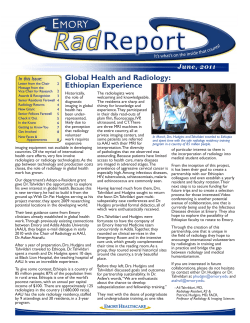


© Copyright 2026