
Diagnostic Imaging Prep Guidelines Table of Contents Next Page
Table of Contents
Next Page
Diagnostic Imaging
Prep Guidelines
Previous Page
Table of Contents
Next Page
IV CONTRAST REQUIREMENTS
IV Contrast Requirements
All patients requiring IV contrast must be screened prior to
receiving contrast!
For patients that are diabetic, or have kidney disease, or have
lupus and/or other collagen vascular diseases, a BUN and
Creatininte must be available, performed within the 30 dyas
prior to the exam date. These results are needed before the
patient is injected with contrast media. Any patient 70 or older
must have a BUN and creatinine done within the last 30 days.
NOTE: If the patient is taking Glucaphage, Glucovance,
Avandament, Metaglip or any form of Metformin for diabetes,
these medications MUST be discontinued for 48 hours after
being given the IV contrast. It is recommended by the
manufacturers that the medication also be stopped for 48 hours
PRIOR to the exam.
We are ONLY able to access MMHS lab results. If the labs were
performed elsewhere, a written report must be sent with patient
or faxed to the Imaging Department prior to the CT exam date.
Contraindications for IV Contrast:
• Elevated BUN/Creatinine, ie Cr >=1.7
• Severe allergy to iodine (history of anaphylactic reaction)
• Sickle cell crisis
• Multiple Myeloma
• Pheochromocytoma
If the patient is allergic to Iodine, they must follow the
premedication protocol per MMHS Radiology Department
Contrast for MRI
Patients with end-stage renal failure MUST be evaluated
PRIOR to administration of contrast and may require dialysis if
contrast is administered.
Radiology Department Protocol for
Premedication for IV Contrast
•
•
•
•
50 mg Prednisone PO 13 hours prior to exam
50 mg Prednisone PO 7 hours prior to exam
50 mg Prednisone PO 1 hours prior to exam
50 mg Benadryl PO 1 hours prior to exam
Diagnostic Imaging Prep Guidelines
1
Previous Page
Table of Contents
This page intentionally left blank
2
Diagnostic Imaging Prep Guidelines
Next Page
I M P O R TA N T P H O N E N U M B E R S
Previous Page
Table of Contents
Diagnostic Scheduling
(772) 288-5817
Active Fax Scheduling
(772) 223-4514
MRI Scheduling
(772) 221-7226
Next Page
Interventional Procedure
Scheduling
(772) 223-5945 ext. 3137
Diagnostic Scheduling
Martin Memorial Diagnostic Center
2396 S.E. Ocean Blvd., Stuart, FL 34996
Main Phone: 223-4959 • Fax: 223-2876
St. Lucie West
1095 St. Lucie West Blvd., Port St. Lucie, FL 34986
Main Phone: 223-5945, ext. 5502 • Fax: 785-5543
Martin Memorial Medical Center
200 S.E. Hospital Ave., Stuart, FL 34994
Main Phone: 223-5945, ext. 3120 • Fax: 223-5908
Martin Memorial Medical Center Cardio and
Neurodiagnostics
Main Phone: 285-5882 • Fax: 223-5948
Martin Memorial Hospital South
2100 S.E. Salerno Road, Stuart, FL 34997
Main Phone: 223-5714 • Fax: 223-5702
MRI Scheduling
Martin Memorial Medical Center MRI
625 Riverside Dr., Stuart, FL 34994
Main Phone: 221-7226 • Fax: 223-5933
St. Lucie West MRI
1095 St. Lucie West Blvd., Suite 224, Port St. Lucie, FL
34986
Main Phone: 785-5567 • Fax: 785-5568
South MRI
2150 S.E. Salerno Road, Suite 104, Stuart, FL 34997
Main Phone: 223-5767 • Fax: 223-5787
Diagnostic Imaging Prep Guidelines
3
Previous Page
Table of Contents
This page intentionally left blank
4
Diagnostic Imaging Prep Guidelines
Next Page
Previous Page
Next Page
TABLE OF
CONTENTS
Diagnostic Imaging Prep Guidelines
5
Previous Page
Table of Contents
This page intentionally left blank
6
Diagnostic Imaging Prep Guidelines
Next Page
Previous Page
Next Page
IV CONTRAST REQUIREMENTS . . . . . . . . . . . . .1
SCHEDULING INFORMATION . . . . . . . . . . . . . . 3
MANAGED CARE LIST . . . . . . . . . . . . . . . . . . . 11
TA B L E O F CO N T E N T S
FLUOROSCOPY . . . . . . . . . . . . . . . . . . . . . . . . 13
Call (772) 288-5817 to schedule
Esophagram (Barium Swallow) . . . . . . . . . . . . . . . . . . . . . . . . . . . 15
Swallowing Evaluation Study (cookie swallow) . . . . . . . . . . . 15
Upper GI Series . . . . . . . . . . . . . . . . . . . . . . . . . . . . . . . . . . . . . . . . . . 16
Small Bowel Series . . . . . . . . . . . . . . . . . . . . . . . . . . . . . . . . . . . . . . . 16
Contrast Enema. . . . . . . . . . . . . . . . . . . . . . . . . . . . . . . . . . . . . . . . . . 17
Fistulagram . . . . . . . . . . . . . . . . . . . . . . . . . . . . . . . . . . . . . . . . . . . . . . 18
Sniff Test - Chest Fluoroscopy . . . . . . . . . . . . . . . . . . . . . . . . . . . 18
Cystogram / Voiding Cystourethrogram (VCUG) . . . . . . . . . . 19
Retrograde Urethrogram (Male) . . . . . . . . . . . . . . . . . . . . . . . . . 20
Hysterosalpingogram (HSG). . . . . . . . . . . . . . . . . . . . . . . . . . . . . . 20
Intravenous Pyelogram – IVP . . . . . . . . . . . . . . . . . . . . . . . . . . . . . 21
INVASIVE FLUOROSCOPY
PROCEDURES . . . . . . . . . . . . . . . . . . . . . . . . 22
Call (772) 223-5945, ext. 3137 to schedule
Arthrogram . . . . . . . . . . . . . . . . . . . . . . . . . . . . . . . . . . . . . . . . . . . . . . 22
Extremity Venogram – Unilateral or Bilateral. . . . . . . . . . . . . . 23
Catheter Placement Check . . . . . . . . . . . . . . . . . . . . . . . . . . . . . . . 23
Myelogram . . . . . . . . . . . . . . . . . . . . . . . . . . . . . . . . . . . . . . . . . . . . . . 24
Cisternogram . . . . . . . . . . . . . . . . . . . . . . . . . . . . . . . . . . . . . . . . . . . . 25
Lumbar Puncture under Fluoroscopy . . . . . . . . . . . . . . . . . . . . 25
T-Tube Cholangiogram. . . . . . . . . . . . . . . . . . . . . . . . . . . . . . . . . . . 26
MAMMOGRAPHY . . . . . . . . . . . . . . . . . . . . . . 27
Call (772) 288-5817 to schedule
Mammography – unilateral or Bilateral . . . . . . . . . . . . . . . . . . . 29
COMPUTED TOMOGRAPHY CT . . . . . . . . . . 31
Call (772) 288-5817 to schedule
CT of the Head (without contrast or with and without) . . . 33
CT of the Orbits . . . . . . . . . . . . . . . . . . . . . . . . . . . . . . . . . . . . . . . . . . 34
CT of the Paranasal Sinuses . . . . . . . . . . . . . . . . . . . . . . . . . . . . . . 34
CT of the Temporal Bones . . . . . . . . . . . . . . . . . . . . . . . . . . . . . . . . 35
CT of the Neck Soft Tissues . . . . . . . . . . . . . . . . . . . . . . . . . . . . . . . 36
CT of the Cervical Spine . . . . . . . . . . . . . . . . . . . . . . . . . . . . . . . . . . 36
Diagnostic Imaging Prep Guidelines
7
Previous Page
Next Page
CT of the Thoracic Spine . . . . . . . . . . . . . . . . . . . . . . . . . . . . . . . . . 37
CT of the Lumbar Spine . . . . . . . . . . . . . . . . . . . . . . . . . . . . . . . . . . 38
CT of the Chest . . . . . . . . . . . . . . . . . . . . . . . . . . . . . . . . . . . . . . . . . . 38
CT of the Abdomen . . . . . . . . . . . . . . . . . . . . . . . . . . . . . . . . . . . . . . 40
CT of the Pelvis. . . . . . . . . . . . . . . . . . . . . . . . . . . . . . . . . . . . . . . . . . . 41
CT of the Extremities . . . . . . . . . . . . . . . . . . . . . . . . . . . . . . . . . . . . . 42
TA B L E O F CO N T E N T S
INTERVENTIONAL CT . . . . . . . . . . . . . . . . . 42
Call (772) 223-5945, ext. 3137 to schedule
CT Guided Biopsy of Drainage. . . . . . . . . . . . . . . . . . . . . . . . . . . . 42
CT Guided Facet Block . . . . . . . . . . . . . . . . . . . . . . . . . . . . . . . . . . . 44
CT Guided Bone Marrow Biopsy. . . . . . . . . . . . . . . . . . . . . . . . . . 44
ULTRASOUND . . . . . . . . . . . . . . . . . . . . . . . . . 45
Call (772) 288-5817 to schedule
Ultrasound of the Abdomen . . . . . . . . . . . . . . . . . . . . . . . . . . . . . 47
Ultrasound of the Neonatal Brain. . . . . . . . . . . . . . . . . . . . . . . . . 48
Ultrasound of the Baby Hips . . . . . . . . . . . . . . . . . . . . . . . . . . . . . 48
Ultrasound of the Baby Spine . . . . . . . . . . . . . . . . . . . . . . . . . . . . 49
Ultrasound of the Breast—Unilateral or Bilateral. . . . . . . . . . 49
Ultrasound of the Pregnancy. . . . . . . . . . . . . . . . . . . . . . . . . . . . . 50
Ultrasound of the Pregnancy Limited . . . . . . . . . . . . . . . . . . . . 50
Ultrasound of the Pelvis . . . . . . . . . . . . . . . . . . . . . . . . . . . . . . . . . . 51
Ultrasound of the Prostate . . . . . . . . . . . . . . . . . . . . . . . . . . . . . . . 51
Ultrasound of the Thyroid . . . . . . . . . . . . . . . . . . . . . . . . . . . . . . . . 52
Ultrasound of the Renal Artery Doppler . . . . . . . . . . . . . . . . . . 52
Ultrasound of the Evaluation of “TIPS”—Liver Doppler. . . . 53
Ultrasound of the Testicular (Scrotal) . . . . . . . . . . . . . . . . . . . . . 53
Ultrasound of the Chest . . . . . . . . . . . . . . . . . . . . . . . . . . . . . . . . . . 54
INTERVENTIONAL ULTRASOUND . . . . . . 55
Call (772) 223-5945, ext. 3137 to schedule
Ultrasound of the Chest with Thoracentesis . . . . . . . . . . . . . . 55
Ultrasound Guided Paracentesis . . . . . . . . . . . . . . . . . . . . . . . . . 56
Ultrasound Guided Biopsy . . . . . . . . . . . . . . . . . . . . . . . . . . . . . . . 56
NUCLEAR MEDICINE . . . . . . . . . . . . . . . . . . . 57
Call (772) 288-5817 to schedule
Nuclear Medicine Whole Body Bone Scan . . . . . . . . . . . . . . . . 59
Nuclear Medicine Triple Phase Bone Scan . . . . . . . . . . . . . . . . 60
Nuclear Medicine GI Bleeding Study . . . . . . . . . . . . . . . . . . . . . 60
Nuclear Medicine Gallium Scan . . . . . . . . . . . . . . . . . . . . . . . . . . 61
Nuclear Medicine Gastric Emptying . . . . . . . . . . . . . . . . . . . . . . 61
Nuclear Medicine Hepatobiliary/Pipida Scan . . . . . . . . . . . . . 62
8
Diagnostic Imaging Prep Guidelines
TA B L E O F CO N T E N T S
Previous Page
Next Page
Nuclear Medicine Liver/Spleen Scan. . . . . . . . . . . . . . . . . . . . . . 63
Nuclear Medicine Tumor Localization—
Sentinel Node Study . . . . . . . . . . . . . . . . . . . . . . . . . . . . . . . . . . . 63
Nuclear Medicine Ventilation and Perfusion Lung Scan . . . 64
Nuclear Medicine Split Perfusion Lung Scan . . . . . . . . . . . . . . 64
Nuclear Medicine Muga Scan . . . . . . . . . . . . . . . . . . . . . . . . . . . . 65
Nuclear Medicine Renal Scan. . . . . . . . . . . . . . . . . . . . . . . . . . . . . 66
Nuclear Medicine Thyroid Scan. . . . . . . . . . . . . . . . . . . . . . . . . . . 66
Nuclear Medicine Thyroid Uptake and Scan . . . . . . . . . . . . . . 67
Nuclear Medicine Parathyroid Scan. . . . . . . . . . . . . . . . . . . . . . . 68
Nuclear Medicine Cisternogram. . . . . . . . . . . . . . . . . . . . . . . . . . 68
Nuclear Medicine Octreotide Scan . . . . . . . . . . . . . . . . . . . . . . . 69
Nuclear Medicine Prostascint Scan . . . . . . . . . . . . . . . . . . . . . . . 69
ANGIOGRAPHY . . . . . . . . . . . . . . . . . . . . . . . 71
Call (722) 223-5945, ext. 3137 to schedule
Arteriograms or Angiograms . . . . . . . . . . . . . . . . . . . . . . . . . . . . . 73
CARDIOVASCULAR PROCEDURES . . . . . . 75
Call (772) 288-5817 to schedule
Transesophageal Echocardiogram (TEE). . . . . . . . . . . . . . . . . . 77
2-D Echocardiogram with Doppler and Color Flow . . . . . . . 78
Carotid Duplex Color Flow Imaging . . . . . . . . . . . . . . . . . . . . . . 78
Arterial Evaluation of Lower and Upper Extremities (Pulse
Volume Recordings “PVR”) . . . . . . . . . . . . . . . . . . . . . . . . . . . . . 79
Venous B-Scan . . . . . . . . . . . . . . . . . . . . . . . . . . . . . . . . . . . . . . . . . . . 80
A-V Access Graft Imaging . . . . . . . . . . . . . . . . . . . . . . . . . . . . . . . . 80
Arterial Graft Assessment . . . . . . . . . . . . . . . . . . . . . . . . . . . . . . . . 81
Electrocardiogram (EKG) . . . . . . . . . . . . . . . . . . . . . . . . . . . . . . . . . 82
Holter Monitoring. . . . . . . . . . . . . . . . . . . . . . . . . . . . . . . . . . . . . . . . 82
Patient Initiated Event Monitor (PIM) . . . . . . . . . . . . . . . . . . . . . 84
Telephonic Pacemaker Checks . . . . . . . . . . . . . . . . . . . . . . . . . . . 85
NEURODIAGNOSTIC PROCEDURES . . . . . 87
Call (772) 288-5817 to schedule
Electroencephlogram (EEG) . . . . . . . . . . . . . . . . . . . . . . . . . . . . . . 89
Ambulatory Electroencephlogram (EEG) . . . . . . . . . . . . . . . . . 90
Sleep Deprived Electroencephlogram (EEG) . . . . . . . . . . . . . . 91
Visual Evoked Potential (VEP). . . . . . . . . . . . . . . . . . . . . . . . . . . . . 92
Brainstem Auditory Evoked Potential (BAEP) . . . . . . . . . . . . . 93
Electromyography (EMG) . . . . . . . . . . . . . . . . . . . . . . . . . . . . . . . . 94
Diagnostic Imaging Prep Guidelines
9
Previous Page
Next Page
TA B L E O F CO N T E N T S
STRESS TEXT (GXT) . . . . . . . . . . . . . . . . . . . 94
Call (772) 288-5817 to schedule
10
Stress Test Listing . . . . . . . . . . . . . . . . . . . . . . . . . . . . . . . . . . . . . . . . 94
Regular Stress Test (Exercise only,
without nuclear imaging) . . . . . . . . . . . . . . . . . . . . . . . . . . . . . . 95
Cardiolite Stress Test
(Exercise with nuclear imaging) . . . . . . . . . . . . . . . . . . . . . . . . 96
Persantine Cardiolite Stress Test
(Pharmacological Stress test, pt. is laying down,
with nuclear imaging.) . . . . . . . . . . . . . . . . . . . . . . . . . . . . . . . . . 97
Adenosine Cardiolite Stress Test
(Pharmacological Stress test, pt. is laying down,
with nuclear imaging.) . . . . . . . . . . . . . . . . . . . . . . . . . . . . . . . . . 98
Dobutamine Cardiolite Stress Test
(Pharmacological Stress test, pt. is laying down,
with nuclear imaging.) . . . . . . . . . . . . . . . . . . . . . . . . . . . . . . . . . 99
Dobutamine Stress Echo Test
(Pharmacological Stress test with 2-D Echo.
Pt is lying down, without nuclear imaging.) . . . . . . . . . . . 100
Stress Echo Test (Exercise with 2-D Echo-Imaging,
without nuclear imaging.) . . . . . . . . . . . . . . . . . . . . . . . . . . . . 101
PET/CT . . . . . . . . . . . . . . . . . . . . . . . . . . . . . . 103
Call (772) 288-5893 to schedule
PET/CT . . . . . . . . . . . . . . . . . . . . . . . . . . . . . . . . . . . . . . . . . . . . . . . . . 105
MAGNETIC RESONANCE IMAGING . . . . . 107
Call (772) 221-7226 to schedule
MRI/MRA/MRCP . . . . . . . . . . . . . . . . . . . . . . . . . . . . . . . . . . . . . . . . 109
CATHETERIZATION LAB . . . . . . . . . . . . . . . 111
Call (772) 223-5919 to schedule
Cath Lab. . . . . . . . . . . . . . . . . . . . . . . . . . . . . . . . . . . . . . . . . . . . . . . . 113
Diagnostic Imaging Prep Guidelines
Previous Page
Back to Table of Contents
Next Page
MANAGED CARE LIST
MARTIN MEMORIAL HEALTH SYSTEMS
DIAGNOSTIC CENTER CONTRACTS
Aetna US HealthCare (PPO, MC - No HMO)
BC/BS - PPS/PPC/NETWORKBLUE
BC/BS-Health Options (HMO)
Beech Street (PPO & WC - Network)
Choice Managed Networks (WC)
CIGNA (PPO/POS/HMO)
Corvel/CorCare (W/C)
Coventry (PPO-Includes First Health & Southcare)
ECN/WebTPA PPO & EPO
Evercare (United Healthcare Medicare Special Needs Plan)
Evolutions Healthcare Systems, Inc.
(PPO & WC - Network)
First Health (PPO & WC - Network)
First National Bank (TPA: CoreSource, Inc.)
Florida Health Administrators (PPO - Network)
GreatWest Healthcare
HealthEase
(Medicaid HMO/ Healthy Kids Program:Martin County ONLY)
Heritage/Summit Healthcare (W/C)
Humana Health Care Plans
Hygeia Travel Health (PPO - Network)
Interplan Health Group/Accountable (PPO Network)
Marriott Casualty (WC)
Metcare Health Plans, Inc. (Medicare HMO)
MetraComp (WC)
(Partnership with Focus HealthCare effective 6/6/02)
Multiplan (PPO Network)
Private Healthcare Systems (PPO - Network)
Protegrity Services (WC)
Provider Management Group Network (PMG) (WC)
Prudential (PRUNET)
QRS Managed Care Services (WC)
Secure Horizons (United Healthcare Medicare PPO)
SouthCare (PPO - Network)
United HealthCare (PPO/POS/EPO/HMO)
Diagnostic Imaging Prep Guidelines
11
Previous Page
Back to Table of Contents
This page intentionally left blank
12
Diagnostic Imaging Prep Guidelines
Next Page
Previous Page
Back to Table of Contents
Next Page
FLUOROSCOPY
SECTION
AND
INVASIVE
FLUOROSCOPY
PROCEDURES
Call (772) 288-5817 to schedule
Diagnostic Imaging Prep Guidelines
13
Previous Page
Back to Table of Contents
This page intentionally left blank
14
Diagnostic Imaging Prep Guidelines
Next Page
Previous Page
Back to Table of Contents
Next Page
FLUOROSCOPY . . . . . . . . . . . .
ESOPHAGRAM (BARIUM SWALLOW)
Description
Images of the esophagus are taken while a patient swallows a barium drink.
Indications
Swallowing difficulty, dysphagia, suspected stricture
Prep
Patients should have nothing by mouth for 2 hours prior to the exam.
Length
Minimum 20 minutes.
Facility
Martin Memorial Medical Center
Martin Memorial Hospital South
Martin Memorial St. Lucie West Diagnostic
East Ocean Diagnostic Center
SWALLOWING EVALUATION STUDY (COOKIE
SWALLOW)
Description
Images are taken during a speech therapy evaluation. The patient chews and
swallows barium and/or food of various consistencies, and occasionally an
inert pill.
Indications
To evaluate problems of chewing, swallowing, or aspiration; often on stroke
patients.
Prep
No prep necessary.
Length
Minimum 30 minutes.
Facility
Martin Memorial Medical Center
Appointment must be made in conjunction with Speech Pathology.
Please page (772) 335-6432.
Diagnostic Imaging Prep Guidelines
15
Previous Page
Back to Table of Contents
Next Page
UPPER GI SERIES
Description
The patient drinks a contrast media, (Barium or Gastrografin) and images
of the esophagus and stomach are taken (may be performed with Small
Bowel Series)
Indications
To look for reflux, strictures, ulcers, source of GI bleeding, upper
abdominal pain.
Prep
Nothing by mouth after midnight. No chewing gum, cigarette smoking, or
tooth brushing on morning of exam. May take medications with a small
amount of water 4 hours prior to exam.
Length
Minimum 30 minutes
Facility
Martin Memorial Medical Center
Martin Memorial Hospital South
Martin Memorial St. Lucie West Diagnostic
East Ocean Diagnostic Center
SMALL BOWEL SERIES
Description
Patient drinks a contrast media (barium) and, images are taken serially as
the barium passes through the small intestines.
Prep
Nothing by mouth after midnight prior to test.
Length
Minimum 1 hour (exam may take up to 5 or more hours depending on the
transit of the barium through the small intestine. Avg. – 2 hours)
Facility
Martin Memorial Medical Center
Martin Memorial Hospital South
Martin Memorial St. Lucie West Diagnostic
East Ocean Diagnostic Center
16
Diagnostic Imaging Prep Guidelines
Previous Page
Back to Table of Contents
Next Page
CONTRAST ENEMA
1. Air contrast w/ Barium (ACBE)
2. Barium Enema (BE)
3. Gastrografin Enema
Description
An enema of contrast media is performed and images of the colon are
taken.
Indications
Anemia, GI bleed, polyps, colon cancer screening, constipation, abdominal
pain, diarrhea, diverticulosis
Notes: The best study is an Air Contrast Barium Enema (ACBE). This is
the test of choice unless the patient can not move enough (roll over) to
have this test, and then a single contrast Barium enema is done. The
air and barium outline any tumors or polyps in the colon. A bowel
prep is REQUIRED for this type of enema.
A Gastrografin enema is performed in cases of suspected obstruction,
perferation or cases of constipation. Gastrografin enemas are
occasionally done WITHOUT any prep.
Prep
Two days before the exam
* Eat a clear liquid diet, which includes:
• Water, clear broth, bouillon, fruit juices with no pulp, soft drinks,
black coffee, tea, honey, sugar, jello, clear hard candy. You may have as
much of these liquids as desired until the time of the exam. The fluid
prevents your body from becoming dehydrated and aids in cleaning
your bowel for the exam. No milk products or cream except skim milk.
Day before the Exam
• Continue clear liquid diet. Patient to stay well hydrated.
• Drink one 10 oz bottle of magnesium citrate at 12:00 noon followed
with one full glass of water and continue liquid diet.
• At 5:00 pm, have a liquid meal followed by four (4) Dulcolax tablets
(Bisacodyl). Swallow tablets whole with a full glass of water. DO NOT
chew tablets. Continue clear liquid diet.
Day of Exam
• Breakfast should consist of clear fruit juice, coffee or tea.
Diagnostic Imaging Prep Guidelines
17
Previous Page
Back to Table of Contents
Next Page
Length
Minimum 1 hour.
Facility
Martin Memorial Medical Center
Martin Memorial Hospital South
Martin Memorial St. Lucie West Diagnostic
East Ocean Diagnostic Center
FISTULAGRAM
Description
Contrast media is injected into the sinus or fistulous tract through a
catheter under fluoroscopy for evaluation.
Indications
stoma evaluation, fistulas to skin from bowel or bladder
Prep
No prep necessary.
Length
Minimum 1 hour
Facility
Martin Memorial Medical Center
Martin Memorial Hospital South
SNIFF TEST — CHEST FLUOROSCOPY
Description
Evaluation of the diaphragm movement under fluoroscopy.
Indications
Recurrent infections or possible diaphragm paralysis
Prep
No prep necessary
Length
Minimum 30 minutes
Facility
Martin Memorial Medical Center
Martin Memorial Hospital South
Martin Memorial St. Lucie West Diagnostic
East Ocean Diagnostic Center
18
Diagnostic Imaging Prep Guidelines
Previous Page
Back to Table of Contents
Next Page
CYSTOGRAM/VOIDING CYSTOURETHROGRAM
(VCUG)
Description
A catheter is inserted through the urethra into the bladder, and the bladder
is filled with contrast, and images are then taken of the bladder. When a
VOIDING study is ordered, the patient will empty their bladder
DURING the procedure under fluoroscopy.
Indications
Recurrent UTIs; difficulty voiding; bladder leakage, hydronephrosis
Prep
No prep necessary.
Length
Minimum 45 minutes
Facility
Martin Memorial Medical Center
Martin Memorial Hospital South
Children under the age of 12 must be scheduled at the Medical Center
only, and the catheter will be placed by Pediatrics. Child must not be
medicated prior to exam.
Diagnostic Imaging Prep Guidelines
19
Previous Page
Back to Table of Contents
Next Page
RETROGRADE URETHROGRAM (MALE)
Description
A catheter is inserted into the tip of the penis and the penile urethra is
evaluated under fluoroscopy.
Prep
None
Length
Minimum 45 minutes
Facility
Martin Memorial Medical Center
Martin Memorial Hospital South
HYSTEROSALPINGOGRAM (HSG)
Description
A Pelvic exam is performed and a speculum placed in the vagina. Contrast
media is injected into the uterus and the fallopian tubes through a catheter
to evaluate patency of the fallopian tubes and to outline the uterine cavity.
Indications
Infertility
Prep
ONLY performed on days 7 through 10 days after last day of menstrual
cycle.
Length
Minimum 30 minutes
Facility
Martin Memorial Medical Center
Martin Memorial Hospital South
Note: Patient should bring a panty liner for after the examination.
20
Diagnostic Imaging Prep Guidelines
Previous Page
Back to Table of Contents
Next Page
INTRAVENOUS PYELOGRAM (IVP)
Description
Evaluation of the kidneys, ureters, and bladder with IV contrast injection
and serial imaging of the abdomen.
Indications
Kidney stones, blood in urine, infections, hydronephrosis
Prep
The Day before the Exam
• Patient should pick-up prep from Imaging department at any Martin
Memorial facility or purchase at any local pharmacy.
• At 2:00 pm take two (2) Dulcolax (Bisacodyl) laxative tablets by
mouth.
• Drink 10 oz bottle of Magnesium Citrate at 6:00 pm
• Patient should not eat or drink after midnight
Day of Exam
• No eating or drinking the morning of the exam.
• Patients should take all scheduled a.m. medications with a small
amount of water.
Length
Minimum 1 hour
Facility
Martin Memorial Medical Center
Martin Memorial Hospital South
Martin Memorial St. Lucie West Diagnostic
East Ocean Diagnostic Center
SEE STANDARD IV CONTRAST PROTOCOL.
Diagnostic Imaging Prep Guidelines
21
Previous Page
Back to Table of Contents
Next Page
INVASIVE FLUOROSCOPY
PROCEDURES . . . . . . . . . . . . .
Scheduling through Interventional Radiology Coordinator
Call (772) 223-5945, ext. 3137 to schedule
ARTHROGRAM
May be performed with an ASPIRATION or INJECTION
Description
Contrast media is injected under fluoroscopic guidance into a specific joint
to evaluate the joint space, i.e., knee, shoulder, hip, ankle, wrist, etc.
An ASPIRATION will be performed when requested if there is fluid
present and infection or Gout is suspected. A PRESCRIPTION for lab
orders should be brought to the exam as well.
An INJECTION of steroid and anesthetic can be placed in the joint at
this time as well, if ordered by the requesting MD (typically an
Orthopedist). If an injection is to be performed, it MUST BE noted on the
prescription.
Indications
Joint pain to evaluate for tears, arthritis, loose bodies, infection, gout
Prep
No prep necessary
Length
Minimum 1 hour.
Facility
Martin Memorial Medical Center
Martin Memorial Hospital South
22
Diagnostic Imaging Prep Guidelines
Previous Page
Back to Table of Contents
Next Page
EXTREMITY VENOGRAM — UNILATERAL or
BILATERAL
Description
Intravenous contrast media is injected into a peripheral vein of the
extremity (usually the leg), and images of the extremity are taken to look
for clots in the venous system. This exam is only done after a Doppler has
been done and is inconclusive.
Indications
Detection of blood clots; swelling.
Prep
Nothing to eat or drink 1 hour prior to test
Length
Minimum 1 hour
Facility
Martin Memorial Medical Center (Fluoroscopy suite or Interventional
Suite)
Martin Memorial Hospital South
SEE STANDARD IV CONTRAST PROTOCOL.
CATHETER PLACEMENT CHECK
Description
Patient is given an injection of contrast media into an existing catheter to
check placement under fluoroscopy.
Prep
No prep necessary. If the catheter is UNDER the skin, it must be accessed
prior to the exam.
Length
Minimum 30 minutes
Facility
Martin Memorial Medical Center (Fluoroscopy suite or Interventional
Suite)
Martin Memorial Hospital South
SEE STANDARD IV CONTRAST PROTOCOL.
Diagnostic Imaging Prep Guidelines
23
Previous Page
Back to Table of Contents
Next Page
MYELOGRAM (ordering physician typically a
Neurologist or Neurosurgeon)
•
•
•
Cervical
Thoracic
Lumbar
Description
Contrast media is injected into the spinal canal, for the evaluation of the
spinal cord, nerve root branches and/or disc herniation.
Prep
Nothing to drink or eat after midnight.
Length
Minimum 30-60 minutes – then usually followed by CT which will be
additional imaging time
Facility
Martin Memorial Medical Center
Martin Memorial Hospital South
Attention physician offices:
• Patient must stop taking Plavix 7 days prior to exam.
• Patient must stop taking Coumadin 7 days prior to exam.
• Patient must stop taking Aspirin, minimum 5 days prior to exam.
Lab: All patients must have PT and PTT drawn within 7 days prior to
exam.
24
Diagnostic Imaging Prep Guidelines
Previous Page
Back to Table of Contents
Next Page
CISTERNOGRAM
Description
The injection of the radionuclide for a Cisternogram is performed in
fluoroscopy the same as for a Myelogram above.
Prep
Nothing to drink or eat after midnight.
Length
Minimum 30-60 minutes – then usually followed by CT which will be
additional imaging time
Facility
Martin Memorial Medical Center
Martin Memorial Hospital South
Attention physician offices:
• Patient must stop taking Plavix 7 days prior to exam.
• Patient must stop taking Coumadin 7 days prior to exam.
• Patient must stop taking Aspirin, minimum 5 days prior to exam.
Lab: All patients must have PT and PTT drawn within 7 days prior to
exam.
SEE STANDARD IV CONTRAST PROTOCOL.
LUMBAR PUNCTURE UNDER FLUOROSCOPY
Description
Cerebrospinal fluid is drained from around the spinal cord from the lower
back. The needle is placed in the fluoroscopy suite and the fluid can be sent
to the lab for testing.
Indications
Sometimes performed to measure intracranial pressure, to look for
meningitis, or to relieve hydrocephalus, or to evaluate for Multiple Sclerosis
or other central nervous system diseases. The ordering physician MUST
provide a prescription or orders for what tests should be performed on the
obtained fluid.
Prep
Nothing to drink or eat after midnight.
Length
Minimum 30-60 minutes – then usually followed by CT which will be
additional imaging time
Diagnostic Imaging Prep Guidelines
25
Previous Page
Back to Table of Contents
Next Page
Facility
Martin Memorial Medical Center
Martin Memorial Hospital South
Attention physician offices:
• Patient must stop taking Plavix 7 days prior to exam.
• Patient must stop taking Coumadin 7 days prior to exam.
• Patient must stop taking Aspirin, minimum 5 days prior to exam.
Lab: All patients must have PT and PTT drawn within 7 days prior to
exam.
T-TUBE CHOLANGIOGRAM
Description
Contrast media is injected into an existing biliary T-Tube to check
placement under fluoroscopy.
Prep
No prep necessary
Length
Minimum 30 minutes
Facility
Martin Memorial Medical Center (Fluoroscopy suite or Interventional
Suite)
Martin Memorial Hospital South
26
Diagnostic Imaging Prep Guidelines
Previous Page
Back to Table of Contents
Next Page
MAMMOGRAPHY
Call (772) 288-5817 to schedule
Diagnostic Imaging Prep Guidelines
27
Previous Page
Back to Table of Contents
This page intentionally left blank
28
Diagnostic Imaging Prep Guidelines
Next Page
Previous Page
Back to Table of Contents
Next Page
MAMMOGRAPHY . . . . . . . . . .
Call (772) 288-5817 to schedule
MAMMOGRAPHY /UNILATERAL/BILATERAL
Description
A series of x-ray images of the breasts, performed to detect abnormalities.
•
•
•
•
Screening: Routine exam which can only be bilateral.
Diagnostic Unilateral: One breast is imaged for an area of concern, or
because the other breast is absent.
Diagnostic Bilateral: Both breasts are imaged because of clinical or
physical exam findings. Images are evaluated by the reading radiologist
before the patient leaves to ensure complete evaluation of the area of
concern
Male mammogram: Always bilateral and diagnostic based on clinical
exam.
Prep
Do not use powder, lotions or deodorant prior to exam. Patient may want
to take Tylenol or Advil 30 minutes prior to exam, if approved by their
physician, to help with discomfort. Patient should wear two piece clothing
that can easily be removed from the waist up.
Length
Minimum 30 minutes
Facility
Martin Memorial Hospital South
Martin Memorial St. Lucie West Diagnostic
East Ocean Diagnostic Center
Scheduler
* ask the patient if they have breast implants, if “yes,” schedule TWO
time slots.
• If the patient has had any prior mammograms performed outside of
Martin Memorial Health Systems, please have the patient send or
bring their films to the appointment, as they will be needed for
comparison to read the current exam.
• The patient MUST have their prescription at the time of their
appointment.
NOTE: DIGITAL MAMMO has NO differences in prep or compression time versus standard mammography.
Diagnostic Imaging Prep Guidelines
29
Previous Page
Back to Table of Contents
This page intentionally left blank
30
Diagnostic Imaging Prep Guidelines
Next Page
Previous Page
Back to Table of Contents
Next Page
COMPUTED
TOMOGRAPHY
(CT)
Call (772) 288-5817 to schedule
Diagnostic Imaging Prep Guidelines
31
Previous Page
Back to Table of Contents
This page intentionally left blank
32
Diagnostic Imaging Prep Guidelines
Next Page
Previous Page
Back to Table of Contents
Next Page
COMPUTEDTOMOGRAPHY
(CT)
Call (772) 288-5817 to schedule
GENERAL:
A CT or CAT SCAN is a machine which uses a moving x-ray tube to
generate images that look like slices through the patient’s body. The center
of the hole is where the imaging takes place. These images can be
manipulated into 3-Dimensional pictures if a high enough resolution is
obtained.
CT of the HEAD — Without contrast or With &
Without contrast
Description
Thin axial images obtained through the brain and skull. Contrast is added
if there is suspicion of a mass, aneurysm, metastatic disease, or infection.
Prep
Clear liquids 4 hours prior to exam if receiving IV contrast.
Length
Minimum 30 minutes
Facility
Martin Memorial Medical Center
Martin Memorial Hospital South
Martin Memorial St. Lucie West Diagnostic
East Ocean Diagnostic Center
SEE STANDARD IV CONTRAST PROTOCOL.
Diagnostic Imaging Prep Guidelines
33
Previous Page
Back to Table of Contents
Next Page
CT of the ORBITS — WITH and/or WITHOUT
CONTRAST
Description
Axial CT imaging is performed through the eyes usually before and after
giving IV contrast. Coronal images may also be taken if the patient can lie
on their stomach. For fracture or trauma evaluation NO contrast is used.
Prep
Clear liquids 4 hours prior to exam if receiving IV contrast.
Length
Minimum 30 minutes
Facility:
Martin Memorial Medical Center
Martin Memorial Hospital South
Martin Memorial St. Lucie West Diagnostic
East Ocean Diagnostic Center
SEE STANDARD IV CONTRAST PROTOCOL.
CT of the PARANASAL SINUSES
•
•
•
Routine
Limited
Complete with contrast (ordered by ENT for Cancer evaluation)
Description
•
•
•
The LIMITED study is a quick low radiation study priced
comparable to a Sinus X-ray series, but more detailed, without
evaluating all of the area of the sinuses.
The ROUTINE exam evaluates the sinuses completely, and creates
images in two planes.
The COMPLETE is ordered typically by an ENT for evaluation of a
mass, tumor, or in preparation for surgery, and will usually be
performed WITH and WITHOUT contrast.
Prep
Clear liquids 4 hours prior to exam if receiving IV CONTRAST.
Length
Minimum 30 minutes
34
Diagnostic Imaging Prep Guidelines
Previous Page
Back to Table of Contents
Next Page
Facility:
Martin Memorial Medical Center
Martin Memorial Hospital South
Martin Memorial St. Lucie West Diagnostic
East Ocean Diagnostic Center
SEE STANDARD IV CONTRAST PROTOCOL.
CT of the TEMPORAL BONES
Description
Axial and Coronal CT imaging is performed through the temporal bones
at the level of the ears.
Indication
Hearing loss, ear pain, cholesteatoma
Prep
Clear liquids 4 hours prior to exam.
Length
Minimum 30 minutes
Facility:
Martin Memorial Medical Center
Martin Memorial Hospital South
Martin Memorial St. Lucie West Diagnostic
East Ocean Diagnostic Center
SEE STANDARD IV CONTRAST PROTOCOL.
Diagnostic Imaging Prep Guidelines
35
Previous Page
Back to Table of Contents
Next Page
CT of the NECK SOFT TISSUES
Description
CT imaging is performed through the neck from the level of the ears to the
upper chest. This test is much less sensitive if not performed with IV
CONTRAST, and the STANDARD study is done WITH IV
CONTRAST.
Indication
Neck swelling, mass, adenopathy, voice changes or vocal cord paralysis,
cheek or gland swelling, nose, throat or neck cancer.
Prep
Clear liquids 4 hours prior to exam if IV CONTRAST is ordered.
Length
Minimum 30 minutes
Facility:
Martin Memorial Medical Center
Martin Memorial Hospital South
Martin Memorial St. Lucie West Diagnostic
East Ocean Diagnostic Center
SEE STANDARD IV CONTRAST PROTOCOL.
Note: This study is NOT directed towards the spine or vertebrae, and if
a spinal cord problem is suspected, a CT of the Cervical Spine should be
ordered.
CT of the CERVICAL SPINE — WITH Coronal and
Sagittal reformats (standard)
Same study POST MYELOGRAM (North/South only)
Description
Axial CT images are obtained through the spine from the skull base to the
top of the chest and the images are also processed into sagittal and coronal
slices.
Indication
Neck pain, radiculopathy, bone lesions in spine (usually in patients who can
NOT have an MRI)
Prep
No prep necessary.
36
Diagnostic Imaging Prep Guidelines
Previous Page
Back to Table of Contents
Next Page
Length
Minimum 30 minutes
Facility
Martin Memorial Medical Center
Martin Memorial Hospital South
Martin Memorial St. Lucie West Diagnostic
East Ocean Diagnostic Center
Note: This study looks primarily at the spinal cord and vertebral bodies
and does NOT cover the entire neck and soft tissues.
If the patient has had any previous spine imaging done outside Martin
Memorial Health Systems, such as plain x-rays, MRI, myelograms or
bone scans, please have the patient send or bring their films to the
appointment. These images will be needed for comparison.
CT of the THORACIC SPINE — WITH Coronal and
Sagittal reformats (standard)
Same study POST MYELOGRAM (North/South only)
Description
Axial CT images are obtained through the spine from the neck to the mid
back and the images are also processed into sagittal and coronal slices.
Indication
Back pain, radiculopathy, bone lesions in spine
Prep
No prep necessary.
Length
Minimum 30 minutes
Facility:
Martin Memorial Medical Center
Martin Memorial Hospital South
Martin Memorial St. Lucie West Diagnostic
East Ocean Diagnostic Center
If the patient has had any previous spine imaging done outside Martin
Memorial Health Systems, such as plain x-rays, MRI, myelograms or
bone scans, please have the patient send or bring their films to the
appointment. These images will be needed for comparison.
Diagnostic Imaging Prep Guidelines
37
Previous Page
Back to Table of Contents
Next Page
CT of the LUMBAR SPINE — WITH Coronal and
Sagittal reformats (standard)
Same study POST MYELOGRAM (North/South only)
Description
Axial CT images are obtained through the spine from the mid back to the
top of the buttocks and the images are also processed into sagittal and
coronal slices.
Indication
Back pain, radiculopathy, bone lesions in spine (usually in patients who can
NOT have an MRI)
Prep
No prep necessary.
Length
Minimum 30 minutes
Facility
Martin Memorial Medical Center
Martin Memorial Hospital South
Martin Memorial St. Lucie West Diagnostic
East Ocean Diagnostic Center
If the patient has had any previous spine imaging done outside Martin
Memorial Health Systems, such as plain x-rays, MRI, myelograms or
bone scans, please have the patient send or bring their films to the
appointment. These images will be needed for comparison.
CT of the CHEST
•
•
•
•
WITHOUT CONTRAST
WITH IV CONTRAST
PE (Pulmonary Embolus) Protocol (specialty – requires IV
CONTRAST)
High Resolution (ordered by a Pulmonologist, usually with a
routine CT)
Description
Cross-sectional CT images are taken through the chest, from the neck to
below the diaphragm.
38
Diagnostic Imaging Prep Guidelines
Previous Page
Back to Table of Contents
Next Page
Indications
Respiratory symptoms, abnormal chest x-ray, nodule, fibrosis, COPD,
smoking history, bronchitis, heart failure, chest pain, infection, cancer.
The PE PROTOCOL is specifically targeted to the pulmonary artery tree
and evaluates for a possible pulmonary embolus. CONTRAST is
MANDATORY for this study as it is the only way to identify the emboli.
The study uses a greater amount of radiation to the chest, and does not
cover the entire area that is covered in a routine chest CT and should only
be used in patients where there is a HIGH SUSPICION for PE. Spiral CT
is NOT an acceptable order as all chest CTs are performed spirally.
High Resolution CT is used to evaluate interstitial diseases, alveolar
changes, fibrosis and bronchiectasis, and is added to a routine CT in
specific cases.
Prep
Clear liquids after midnight and NPO 4 hours prior to exam.
Length
Minimum 30 minutes.
Facility
Martin Memorial Medical Center
Martin Memorial Hospital South
Martin Memorial St. Lucie West Diagnostic
East Ocean Diagnostic Center
SEE STANDARD IV CONTRAST PROTOCOL.
Notes: The standard evaluation of the Chest is performed WITH IV
CONTRAST. The contrast helps to evaluate the complex mediastinal
vessels and lymph nodes, and better evaluates the aorta and pulmonary
vessels. Contrast also helps increase sensitivity in infections and evaluation of pleural effusions.
Diagnostic Imaging Prep Guidelines
39
Previous Page
Back to Table of Contents
Next Page
CT of the ABDOMEN
•
•
•
•
•
•
WITH CONTRAST
WITHOUT CONTRAST
WITH AND WITHOUT CONTRAST
AAA — Abdominal Aortic Aneurysm
Stone Protocol
Hematuria Protocol (only ordered by Urologists)
NOTES: The standard Abdominal CT is performed with ORAL contrast to opacify the bowel loops, and with IV CONTRAST to enhance
the detail of the liver and solid organs, to assess the kidneys and their
function, and to evaluate for inflammation, infection, vascular compromise, aneurysm, trauma, leakage and metastatic disease.
Description
The patient will lie down in the CT scanner while images are taken of the
abdomen, from the diaphragm to the iliac crests of the pelvis.
ORAL CONTRAST will be given to ALL abdomen studies EXCEPT
the specialty protocols (Stone, AAA, and Hematuria). These specialty
protocols will ALWAYS be performed with a CT PELVIS of the same
type.
IV CONTRAST is given for the WITH CONTRAST, AAA and
Hematuria protocols.
Prep
Clear liquids after midnight and NPO 4 hours prior to exam.
Length
Minimum 30 minutes
Facility
Martin Memorial Medical Center
Martin Memorial Hospital South
Martin Memorial St. Lucie West Diagnostic
East Ocean Diagnostic Center
SEE STANDARD IV CONTRAST PROTOCOL.
40
Diagnostic Imaging Prep Guidelines
Previous Page
Back to Table of Contents
Next Page
CT of the PELVIS
•
•
WITH CONTRAST
WITHOUT CONTRAST
Description
The patient will lie down in the CT scanner while images are taken of the
pelvis, from the iliac crests through the upper thigh. The patient will be
asked to drink oral contrast 2 1/2 to 3 hours before scan. For the WITH
CONTRAST exam, they will also be given an injection of IV
CONTRAST during the scan.
Notes: Usually ordered WITH a CT abdomen. The standard CT of the
Pelvis is with ORAL and IV CONTRAST. May be ordered alone in
work ups for pelvic pain or prostate cancer.
Prep
Clear liquids after midnight and NPO 4 hours prior to exam.
Length
Minimum 30 minutes
Facility:
Martin Memorial Medical Center
Martin Memorial Hospital South
Martin Memorial St. Lucie West Diagnostic
East Ocean Diagnostic Center
SEE STANDARD IV CONTRAST PROTOCOL.
Diagnostic Imaging Prep Guidelines
41
Previous Page
Back to Table of Contents
Next Page
CT of the EXTREMITIES
Description
CT imaging can be performed of any joint or bone as needed. Each
segment will be imaged separately. Some joints will be images with
additional techniques. A CT Arthrogram will require an injection of
contrast prior to the CT study, and is used in the shoulder and knee to
evaluate patients who can not have an MRI. If an infection is suspected, and
an MRI can not be obtained, imaging of the extremity will be performed
with IV CONTRAST.
Indication
Bone or joint pain, fracture or surgical planning, suspected infection, tumor
or mass
Prep
Clear liquids 4 hours prior to exam if receiving IV CONTRAST.
Length
Minimum 30 minutes
Facility
Martin Memorial Medical Center
Martin Memorial Hospital South
Martin Memorial St. Lucie West Diagnostic
East Ocean Diagnostic Center
SEE STANDARD IV CONTRAST PROTOCOL.
INTERVENTIONAL CT . . . . . .
Call (772) 223-5945, ext. 3137 to schedule
CT GUIDED BIOPSY or DRAINAGE
The CT scanner allows for visualization of deep structures, allowing biopsy
of tumors that can not be clearly felt of localized superficially. CT also
allows visualization of adjacent structures such as bowel or blood vessels
that should be avoided during biopsy.
42
Diagnostic Imaging Prep Guidelines
Previous Page
Back to Table of Contents
Next Page
Before any biopsy can be scheduled, a lesion must be known to exist, and
an interventional radiologist MUST APPROVE the case as one that can
be performed safely. The patient must provide all outside images to the
radiology staff through Interventional scheduling at x3137 for
evaluation. Most imaging studies performed in MMHS can be reviewed on
PACS.
Procedure
The type of biopsy will be determined by the radiologist performing the
procedure. The patient will be placed on the CT table in a position to
provide easy access to the lesion (tumor). The CT will localize the lesion
and the needle as the procedure is performed.
Patients will be monitored by a nurse during the procedure, will receive
local anesthetic, and MAY require additional sedation and pain control.
Prep
Clear liquids 4 hours prior to exam.
Length
Minimum 1 1/2 hours
Facility
Martin Memorial Medical Center
Martin Memorial Hospital South
Attention physician offices:
• Patient must stop taking Plavix 7 days prior to exam.
• Patient must stop taking Coumadin at least 3 days prior to exam.
• Patient must stop taking Aspirin, minimum 5 days prior to exam.
Lab: All patients must have PT and PTT drawn within 7 days prior to
exam.
We are ONLY able to access MMHS lab results. If labs taken elsewhere,
written report must be sent with patient or faxed to Imaging Department
prior to exam date.
Diagnostic Imaging Prep Guidelines
43
Previous Page
Back to Table of Contents
Next Page
CT GUIDED FACET BLOCK
Description
CT imaging is performed over the segment of the spine to be treated. An
anesthetic and steroid cocktail is injected into the facet joint affected.
Prep
Clear liquids 4 hours prior to exam.
Length
Minimum 1 1/2 hour
Facility:
Martin Memorial Medical Center
Note: Must be ordered by neurosurgeon, neurologist or an orthopedic
surgeon and must be scheduled through Interventional Radiology at
x3137. Prior imaging studies must be reviewed by the performing radiologist before the procedure can be scheduled.
CT GUIDED BONE MARROW BIOPSY
Description
CT imaging is performed over the lower pelvis with the patient on their
stomach. The bones of the pelvis are localized, and the skin anesthetized,
and a biopsy needle is used to obtain a marrow smear, fluid and core
specimen.
Prep
Clear liquids 4 hours prior to exam.
Length
Minimum 1 1/2 hour
Facility
Martin Memorial Medical Center
Martin Memorial Hospital South
Note: Must be scheduled through Interventional Radiology
at ext. 3137.
44
Diagnostic Imaging Prep Guidelines
Previous Page
Back to Table of Contents
Next Page
ULTRASOUND
Call (772) 288-5817 to schedule
Diagnostic Imaging Prep Guidelines
45
Previous Page
Back to Table of Contents
This page intentionally left blank
46
Diagnostic Imaging Prep Guidelines
Next Page
Previous Page
Back to Table of Contents
Next Page
ULTRASOUND . . . . . . . . . . . . .
Call (772) 288-5817 to schedule
GENERAL:
An Ultrasound probe produces high frequency sound waves, which can not
be felt or heard and have no radiation, to generate multiple images of soft
tissues. The probe is moved over the surface of the skin using a water
soluble gel, and images or organs, and assessment of blood flow can be
obtained. Fat in the skin decreases the sensitivity and penetration of the
sound waves. Gas in the bowel distorts the sound waves and obstructs
visualization. Ultrasound can not image through solid bones.
ULTRASOUND of the ABDOMEN
•
•
Complete (standard)
Limited
• Renal
• Right Upper Quadrant
• Retroperitoneal (AAA))
• Pylorus (Pyloric Stenosis)
COMPLETE: The major organs of the abdomen are evaluated including
the liver, gallbladder, pancreas, spleen, kidneys, inferior vena cava and
Aorta.
RENAL: The kidneys ONLY are evaluated.
RUQ: The structures of the Right Upper Quadrant are evaluated,
excluding the spleen and left kidney and Aorta, compared to a complete
abdomen.
Retroperitoneal: Usually performed for evaluation of the Aorta and
kidneys for an aneurysm or a suspected retroperitoneal hematoma or
abscess.
Pylorus: Performed on very young infants to evaluate for obstruction of
the stomach. Infants must be NPO for 2 hours prior to Pylorus US exam.
Prep
Nothing by mouth after midnight (for Complete and RUQ only)
Diagnostic Imaging Prep Guidelines
47
Previous Page
Back to Table of Contents
Next Page
Length
Minimum 30 minutes
Facility
Martin Memorial Medical Center
Martin Memorial Hospital South
Martin Memorial St. Lucie West Diagnostic
East Ocean Diagnostic Center
May take medications with small amount of water.
ULTRASOUND of the NEONATAL BRAIN
Description
The imaging probe is placed over a front of the head for evaluation of
structure, position and development of the neonatal brain.
Prep
No prep necessary.
Length
Minimum 30 minutes
Facility
Martin Memorial Medical Center
ULTRASOUND — BABY HIPS
Description
The imaging probe is placed over a neonates hips for evaluation of
structure, position and mobility. Only infants LESS THAN 5 month of
age can be evaluated with this technique.
Prep
No prep necessary.
Length
Minimum 45 minutes
Facility
Martin Memorial Medical Center
Martin Memorial Hospital South
48
Diagnostic Imaging Prep Guidelines
Previous Page
Back to Table of Contents
Next Page
ULTRASOUND — BABY SPINE
Description
The imaging probe is placed over a neonates lower spine for evaluation of
structure, position and mobility of the lower spinal cord, in cases of
suspected tethered cord or for evaluation of a sacral dimple. Typically
performed on very young infants less than SIX months of age, as older than
this age, the cord may NOT be visible by US.
Prep
No prep necessary.
Length
Minimum 45 minutes
Facility
Martin Memorial Medical Center
Martin Memorial Hospital South
East Ocean Diagnostic Center
ULTRASOUND of the BREAST (Unilateral or
Bilateral)
Description
The Ultrasound probe is placed directly on the breast to identify any
unusual masses. Specifically used to differentiate between solid and cystic
lesions.
Prep
No prep necessary.
Length
Minimum 30 minutes
Facility
Martin Memorial Medical Center
Martin Memorial Hospital South
Martin Memorial St. Lucie West Diagnostic
East Ocean Diagnostic Center
If patient had a previous mammogram, breast Ultrasound, or breast MRI
done outside Martin Memorial Health Systems, the patient MUST send or
bring their films to the appointment. These images will need to be reviewed
to ensure accuracy of the study.
Not performed as a screening exam.
Diagnostic Imaging Prep Guidelines
49
Previous Page
Back to Table of Contents
Next Page
ULTRASOUND of PREGNANCY
Description
The Ultrasound probe is placed directly on the mother’s abdomen to
evaluate the fetal anatomy, amniotic fluid, heart rate, cervix, placenta and
fetal presentation.
Prep
Drink 32 oz of water one hour prior to exam. A full urinary bladder is
needed.
Length
Minimum 60 minutes
Facility
Martin Memorial Medical Center
Martin Memorial Hospital South
Martin Memorial St. Lucie West Diagnostic
East Ocean Diagnostic Center
ULTRASOUND of PREGNANCY — LIMITED
Description: The Ultrasound probe is placed directly on the mother’s
abdomen to evaluate amniotic fluid index, size and dates, cervical length
and placenta location.
Prep
Drink 32 oz of water one hour prior to exam. A full urinary bladder is
needed.
Length
Minimum 30 minutes
Facility
Martin Memorial Medical Center
Martin Memorial Hospital South
Martin Memorial St. Lucie West Diagnostic
East Ocean Diagnostic Center
50
Diagnostic Imaging Prep Guidelines
Previous Page
Back to Table of Contents
Next Page
ULTRASOUND of the PELVIS
Description
The Ultrasound probe is placed directly on the pelvis to evaluate the pelvic
contents. Male - the bladder and prostate gland are evaluated. Female – the
bladder, uterus, and ovaries are evaluated (FEMALE exam may require
placement of the probe inside the vagina for better evaluation of internal
structures).
Indications
•
•
Male – Prostate enlargement, voiding problems, stones, UTI
Female – Bleeding, pain, mass, infection, UTI, urinary problems
Prep
Drink 32 oz. of water one hour prior to exam. A full urinary bladder is
needed.
Length
Minimum 45 minutes
Facility
Martin Memorial Medical Center
Martin Memorial Hospital South
Martin Memorial St. Lucie West Diagnostic
East Ocean Diagnostic Center
Same prep is required for male and female
ULTRASOUND of the PROSTATE
Description
The Ultrasound probe is placed inside the rectum to evaluate the prostate
gland.
Prep
Fleets enema 2 hours prior to exam.
Length
Minimum 30 minutes
Facility
East Ocean Diagnostic Center
Diagnostic Imaging Prep Guidelines
51
Previous Page
Back to Table of Contents
Next Page
ULTRASOUND of the THYROID
Description
The Ultrasound probe is placed directly on the lower neck to evaluate the
thyroid gland.
Prep
No prep necessary.
Length
Minimum 30 minutes
Facility
Martin Memorial Medical Center
Martin Memorial Hospital South
Martin Memorial St. Lucie West Diagnostic
East Ocean Diagnostic Center
No neck jewelry can be worn during the exam.
ULTRASOUND — RENAL ARTERY DOPPLER
Description
The Ultrasound probe is placed directly on the abdomen to evaluate the
kidneys. Doppler studies measure the speed and direction of flow in the
renal arteries and in the kidneys.
Indication
Hypertension, suspected Renal Artery Stenosis
Prep
Nothing by mouth after midnight. Patient to take over the counter anti-gas
medication 2 hours prior to exam.
Length
Minimum 60 minutes
Facility
Martin Memorial Medical Center
Martin Memorial Hospital South
52
Diagnostic Imaging Prep Guidelines
Previous Page
Back to Table of Contents
Next Page
ULTRASOUND EVALUATION OF “TIPS” — LIVER
DOPPLER
Description
The Ultrasound probe is placed directly on the abdomen to evaluate the
blood flow into and from the liver, or to evaluate a venous shunt in a patient
who has had a TIPS procedure performed. Doppler studies measure the
speed and direction of flow in the hepatic veins, portal veins, IVC, and
hepatic artery.
Prep
The patient should be NPO for four hours prior to the exam, and should
take over the counter anti-gas medication 2 hours prior to exam.
Length
Minimum 60 minutes
Facility
Martin Memorial Medical Center
Martin Memorial Hospital South
Martin Memorial St. Lucie West Diagnostic
East Ocean Diagnostic Center
ULTRASOUND — TESTICULAR (SCROTAL)
Description
The Ultrasound probe is placed directly on the scrotum to evaluate the
testes, and scrotal contents. May include Doppler imaging to assess blood
flow to each testis.
Indication
Scrotal pain, mass, suspected torsion, infection, swelling, hernia
Prep
No prep necessary.
Length
Minimum 30 minutes
Facility
Martin Memorial Medical Center
Martin Memorial Hospital South
Martin Memorial St. Lucie West Diagnostic
East Ocean Diagnostic Center
Diagnostic Imaging Prep Guidelines
53
Previous Page
Back to Table of Contents
Next Page
ULTRASOUND of the CHEST
Description
The Ultrasound probe is placed directly on the chest from the back to
determine the presence and volume of pleural effusions. This area may be
marked for possible thoracentesis (drainage) either by the Radiologist or
the Pulmonologist.
Prep
No prep necessary.
Length
Minimum 30 minutes
Facility
Martin Memorial Medical Center
Martin Memorial Hospital South
Martin Memorial St. Lucie West Diagnostic
East Ocean Diagnostic Center
54
Diagnostic Imaging Prep Guidelines
Previous Page
Back to Table of Contents
Next Page
INTERVENTIONAL
ULTRASOUND . . . . . . . . . . . . .
Call (772) 223-5945, ext. 3137 to schedule
ULTRASOUND of the CHEST with THORACENTESIS
Description
The Ultrasound probe is placed directly on the chest from the back to
determine the presence and volume of pleural effusions. This area may be
marked for possible thoracentesis (drainage) either by the Radiologist or
the Pulmonologist.
Prep
PT and PTT required prior to procedure, must be less than 30 days old.
Length
•
•
Minimum 30 minutes for chest ultrasound
Minimum 60 minutes for thoracentesis procedure
Facility
Martin Memorial Medical Center
Martin Memorial Hospital South
Note: If scheduled by a Pulmonologist, times must be coordinated with
US and the MD
ULTRASOUND guided PARACENTESIS
Description
The Ultrasound probe is placed directly on the abdomen to determine the
Diagnostic Imaging Prep Guidelines
55
Previous Page
Back to Table of Contents
Next Page
presence and volume of ascites (abdominal fluid). The area will be marked
for possible paracentesis (drainage) by the Radiologist if there is felt to be
enough fluid present.
Prep
PT and PTT are required prior to the procedure, and must be less than
30 days old.
Length:
•
•
Minimum 30 minutes for abdominal ultrasound
Minimum 60 minutes for paracentesis procedure
Facility
Martin Memorial Medical Center
Martin Memorial Hospital South
ULTRASOUND GUIDED BIOPSY
Description
The Ultrasound probe is placed directly on the area being biopsied
(thyroid gland or breast are typical). The Ultrasound can show the lesion
and shows the location of the biopsy needle helping the physician guide the
needle to a specific site.
Length:
Minimum 90 minutes
Facility
Martin Memorial Medical Center
Must be scheduled through Interventional Scheduling (x3137)
Labs may be necessary prior to test, PT-PTT (depending on site of biopsy)
56
Diagnostic Imaging Prep Guidelines
Previous Page
Back to Table of Contents
Next Page
NUCLEAR
MEDICINE
Call (772) 288-5817 to schedule
Diagnostic Imaging Prep Guidelines
57
Previous Page
Back to Table of Contents
This page intentionally left blank
58
Diagnostic Imaging Prep Guidelines
Next Page
Previous Page
Back to Table of Contents
Next Page
NUCLEAR MEDICINE
Call (772) 288-5817 to schedule
GENERAL
Nuclear Medicine cameras can image small amounts or radioactive
compounds called tracers that are injected into the body. The tracers are
attached to specific compounds to image specific internal functions, such as
liver function, bone growth, thyroid uptake, and blood flow. The cameras
used identify the sites and amount of activity in the body to determine
anatomy and function.
NUCLEAR MEDICINE WHOLE BODY BONE SCAN
Description
A radioactive bone-seeking material is injected into a vein where it enters
the bloodstream and is carried to the bone. The patient is encouraged to
drink fluids between injection and scan. Imaging is done a minimum of 2
hours after injection. Usually the entire body is imaged, and extra views
(images) may be taken to provide as much information as possible about a
specific area.
Indication
Cancer, bone pain, abnormal calcium or enzyme levels, parathyroid
problems
Prep
No prep necessary.
Length
Patient receives injection (approximately 30 minutes) and returns in 2-3
hours. Imaging takes approximately 1 hour after patient returns.
Facility
Martin Memorial Medical Center
Martin Memorial Hospital South
Martin Memorial St. Lucie West Diagnostic
Diagnostic Imaging Prep Guidelines
59
Previous Page
Back to Table of Contents
Next Page
NUCLEAR MEDICINE TRIPLE PHASE BONE SCAN
Description
A radioactive bone-seeking material is injected into a vein where it enters
the bloodstream and is carried to the bone. Imaging is done during the
injection, over an area of suspected infection, and shortly thereafter, and of
the whole body after a minimum of 2 hours delay.
Indication
Osteomyelitis (infection of bone), Raynaud’s disease, Reflex Sympathetic
Dystrophy (RSD)
Prep
No prep necessary.
Length
Patient gets injected and returns in 2-3 hours. Test takes approx. 1 hour
after patient returns and maybe longer if extra Image are needed.
Facility
Martin Memorial Medical Center
Martin Memorial Hospital South
Martin Memorial St. Lucie West Diagnostic
NUCLEAR MEDICINE GI BLEEDING STUDY
Description
Blood is drawn from the patient. The blood sample is then mixed with a
radioactive tracer that attaches to the red blood cells. 20 minutes later the
patient will be re-injected with their own tracer labeled blood, and images
will be taken for at least 1 hour. Delayed images can be taken up to 24 hours
later.
Indication
Suspected or active GI bleed.
Prep
No prep necessary.
Length
Minimum 3-4 hours.
Facility
Martin Memorial Medical Center
Martin Memorial Hospital South
Martin Memorial St. Lucie West Diagnostic
60
Diagnostic Imaging Prep Guidelines
Previous Page
Back to Table of Contents
Next Page
NUCLEAR MEDICINE GALLIUM SCAN
Description
The patient is injected with a tracer which localizes in areas of
inflammation or infection, and then is imaged to look for infection or
abscesses Depending on the diagnosis and the initial images, the patient
may be scanned at 24, 48, and 72 hours post injection.
Indication
Fever of unknown origin, suspected abscess, Hodgkin’s disease, tumor
staging, or localization, Sarcoid.
Prep
If needed, the patient will be given a bowel prep kit and clear liquid diet by
the Nuclear Medicine Department.
Length
Minimum 45 minutes for 3 consecutive days or as radiologist orders.
Facility
Martin Memorial Medical Center
Martin Memorial Hospital South
Martin Memorial St. Lucie West Diagnostic
NUCLEAR MEDICINE GASTRIC EMPTYING
Description
The patient will eat a small amount of oatmeal with a radiotracer present.
The meal fills the stomach and imaging determines the rate at which food
leaves the stomach.
Indication
Gastroparesis, suspected gastric outlet obstruction
Prep
Nothing by mouth after midnight.
Length
Minimum 2 hours.
Facility
Martin Memorial Medical Center
Martin Memorial Hospital South
Diagnostic Imaging Prep Guidelines
61
Previous Page
Back to Table of Contents
Next Page
NUCLEAR MEDICINE HEPATOBILIARY/PIPIDA
SCAN (HIDA SCAN)
Description
The patient is injected with a tracer that is processed in the liver and
excreted into the bile ducts. Images are taken over the liver and abdomen to
look for obstructions of the biliary tract, and assess liver function.
Indications
Cholecystitis, gallbladder or upper abdomen pain,
Prep
Nothing by mouth after midnight. May take meds with a sip of water. Hold
Demerol or any morphine based medication for 6 hours prior to exam, as it
interferes with gall bladder visualization.
GALL BLADDER EJECTION FRACTION — in addition, the same
exam can be used with a drug, Cholecystokinin (CCK), to force the
gallbladder to contract, and the amount of excretion can be measured.
(only done at MMMC and MMHS)
Length
Minimum 3 hours.
Facility
Martin Memorial Medical Center
Martin Memorial Hospital South
Martin Memorial St. Lucie West Diagnostic
62
Diagnostic Imaging Prep Guidelines
Previous Page
Back to Table of Contents
Next Page
NUCLEAR MEDICINE LIVER/SPLEEN SCAN
Description
A tracer which localizes to the liver, spleen, and blood-producing marrow is
injected into a vein, and images are taken 15 minutes later. This study
evaluates the liver and spleen and marrow function.
Prep
No barium for 48 hours before exam.
Length
Minimum 1 hour.
Facility
Martin Memorial Medical Center
Martin Memorial Hospital South
Martin Memorial St. Lucie West Diagnostic
NUCLEAR MEDICINE TUMOR LOCALIZATION —
SENTINEL NODE STUDY
Description
A tracer which follows the drainage around a tumor is injected around the
tumor and images are obtained to localize “sentinel” lymph nodes.
Indications
ONLY scheduled with surgery schedule for same day, typically for
melanoma or breast cancer.
Length
Minimum 1 hour.
Facility
Martin Memorial Medical Center
Diagnostic Imaging Prep Guidelines
63
Previous Page
Back to Table of Contents
Next Page
NUCLEAR MEDICINE VENTILATION AND
PERFUSION LUNG SCAN
Description
The patient is asked to breathe a air which contains a tracer evaluating the
lung ventilation. When this is completed, the patient is injected with
particles of albumin labeled with a tracer, which are carried by the blood to
the lungs. Images are then taken over the chest to assess perfusion of the
lungs.
Indication
Suspected pulmonary embolus.
Prep
Must have chest x-ray within 24 hours prior to exam or can be done at
time of exam if none available.
Length
Minimum 1 hour.
Facility
Martin Memorial Medical Center
Martin Memorial Hospital South
NUCLEAR MEDICINE SPLIT PERFUSION LUNG
SCAN
Description
The patient is injected with particles of albumin labeled with a tracer,
which are carried by the blood to the lungs. Images are then taken over the
chest to assess perfusion of the lungs, and calculations are performed over
each segment of the lungs.
Indication
Evaluation of function prior to surgery.
Length
Minimum 1 hour.
Facility
Martin Memorial Medical Center
Martin Memorial Hospital South
Martin Memorial St. Lucie West Diagnostic
64
Diagnostic Imaging Prep Guidelines
Previous Page
Back to Table of Contents
Next Page
NUCLEAR MEDICINE MUGA SCAN
Description
The patient’s blood is labeled to evaluate the function and output of the
heart. Imaging is performed over the chest while an EKG records the
activity of the heart.
Indication
Determine ejection fraction of left ventricle for chemotherapy or post MI
Prep
No prep necessary.
Length
Minimum 1 hour.
Facility
Martin Memorial Medical Center
Martin Memorial Hospital South
Martin Memorial St. Lucie West Diagnostic
Diagnostic Imaging Prep Guidelines
65
Previous Page
Back to Table of Contents
Next Page
NUCLEAR MEDICINE RENAL SCAN
Description
The patient is injected with tracer for visualization of the kidneys. Imaging
is performed over the back during injection, and after to assess blood flow
to the kidneys, and their output and function.
Indications
Renal failure, acute or chronic; Hypertension.
WITH LASIX — additional imaging can be performed after Lasix is given
to assess hydronephrosis (only done at MMMC and MMHS)
WITH CAPTOPRIL —imaging can be performed with a Captopril
administration in cases of suspected renal artery stenosis. (only done at
MMMC) The patient MUST be off of ACE inhibitors at least 48 hours
prior to test, and must be NPO for 4 hours prior.
Prep
No prep necessary. (except Captopril, see above)
Length
Minimum 45 minutes.
Facility
Martin Memorial Medical Center
Martin Memorial Hospital South
Martin Memorial St. Lucie West Diagnostic
NUCLEAR MEDICINE THYROID SCAN
Description
The patient receives a tracer injection into a vein, and 15 minutes post
injection the thyroid gland is imaged.
Prep
No prep necessary.
Length
Minimum 40 minutes.
Facility
Martin Memorial Medical Center
Martin Memorial Hospital South
Martin Memorial St. Lucie West Diagnostic
66
Diagnostic Imaging Prep Guidelines
Previous Page
Back to Table of Contents
Next Page
NUCLEAR MEDICINE THYROID UPTAKE AND SCAN
This is a 2 day exam.
Description
Day 1
A small amount of radioactive iodine (I-123) is given by mouth as a
capsule.
Day 2
The thyroid function is measured with a thyroid probe. The patient is
injected with a radioactive tracer and imaging takes place 5 – 10
minutes later.
Approximate Length
2 Day Procedure
Day 1: 15 minutes;
Day 2: 45 Minutes to 1 hour
Prep
•
•
No thyroid medications for 6 weeks prior to test.
No IV contrast in the 4 – 6 weeks prior to the exam
48 Hours prior to test:
• No seafood (primarily shellfish)
• No strawberries
• No Chinese food - soy sauce
• No antihistamines
• No vitamins containing iodine.
• No iodized salt.
Facility
Martin Memorial Medical Center
Martin Memorial Hospital South
Martin Memorial St. Lucie West Diagnostic
Diagnostic Imaging Prep Guidelines
67
Previous Page
Back to Table of Contents
Next Page
NUCLEAR MEDICINE PARATHYROID SCAN
Description
The patient receives a tracer injection into a vein, and the neck is imaged
immediately and after a delay to assess for a parathyroid adenoma.
Prep
No prep necessary.
Length
Minimum 40 minutes.
Facility
Martin Memorial Medical Center
NUCLEAR MEDICINE CISTERNOGRAM
Description
The patient must come to the fluoroscopy suite at the Main Hospital and a
Lumbar puncture is performed under fluoroscopy to access the spinal fluid.
A radiotracer is injected into the fluid, and a series of images are then
obtained with a nuclear medicine camera, after four hours, and on
sequential days, up to 3 days following, based on each days images.
Only scheduled on Monday and Tuesday and must be scheduled in both
fluoro and nuclear medicine.
Length
Minimum 5 hours (initial visit only), approximately 30 minutes for followup
Facility
Martin Memorial Medical Center
Attention physician offices
• Patient must stop taking Plavix 7 days prior to exam.
• Patient must stop taking Coumadin 3 days prior to exam.
• Patient must stop taking Aspirin, minimum 5 days prior to exam.
Lab: All patients must have PT and PTT drawn within 7 days prior to
exam.
68
Diagnostic Imaging Prep Guidelines
Previous Page
Back to Table of Contents
Next Page
NUCLEAR MEDICINE OCTREOTIDE SCAN
Description
The patient receives a tracer injection into a vein, and the abdomen is
imaged after a delay to assess for tumors in the endocrine family, such as
carcinoid, and pancreatic tumors.
Prep
No prep necessary.
Length
The initial dosing visit requires approximately 30 minutes. Images will be
taken at 24 and possibly 48 hours.
Facility
Martin Memorial Medical Center
MUST be scheduled through Lynn Hageman, ext.1540
NUCLEAR MEDICINE PROSTASCINT SCAN
Description
The patient receives a tracer injection into a vein, and the pelvis is imaged
immediately and after a delay to assess for prostate cancer recurrence after
surgery.
Note: ONLY performed on patients who have had TOTAL surgical
prostate removal.
Prep
Bowel prep necessary.
Length
The initial dosing visit requires approximately 30 minutes. Images will be
taken at 24 and possibly 48 hours and 72 hours.
Facility
Martin Memorial Medical Center
MUST be scheduled through Lynn Hageman, ext.1540
Diagnostic Imaging Prep Guidelines
69
Previous Page
Back to Table of Contents
This page intentionally left blank
70
Diagnostic Imaging Prep Guidelines
Next Page
Previous Page
Back to Table of Contents
Next Page
ANGIOGRAPHY
Call (772) 223-5945, ext. 3137 to
schedule
Diagnostic Imaging Prep Guidelines
71
Previous Page
Back to Table of Contents
This page intentionally left blank
72
Diagnostic Imaging Prep Guidelines
Next Page
Previous Page
Back to Table of Contents
Next Page
ANGIOGRAPHY . . . . . . . . . . . .
Call (772) 223-5945, ext. 3137 to schedule
ARTERIOGRAMS or ANGIOGRAMS
Schedule with Interventional Radiology at ext. 3137.
When scheduling an outpatient arteriogram. Please inform the patient of
the following information prior to his arrival for the exam:
1. If the patient usually takes insulin in the morning, tell the patient to
hold his/her insulin and breakfast until after the arteriogram. Check
with the physician regarding insulin dose. If the patient takes an oral
hypoglycemic agent instead of insulin, give them the same instructions.
2. Instruct the patient to take all blood pressure or cardiac medication
they usually take in the morning, with the exception of Aspirin and
blood thinners (heparin, and/or Coumadin). If the patient is taking
antihypertensive medication, stress the importance of taking those as
prescribed by their doctor.
3. The patient will need a driver and can NOT drive following the
procedure
4. Mention that if the approach (of the arteriogram site) is to be brachial,
that the patient will have one arm incapacitated with an arm board and
an ace wrap for at least two to four hours following the procedure.
5. Patients may be required to stay for monitoring up to six hours
following their procedure.
Attention physician offices
• Patient must stop taking Plavix 7 days prior to exam.
• Patient must stop taking Coumadin 7 days prior to exam.
• Patient must stop taking Aspirin, minimum 5 days prior to exam.
Lab: All patients must have PT and PTT drawn within 7 days prior to
exam.
Please See Standard IV Contrast requirements.
Diagnostic Imaging Prep Guidelines
73
Previous Page
Back to Table of Contents
This page intentionally left blank
74
Diagnostic Imaging Prep Guidelines
Next Page
Previous Page
Back to Table of Contents
Next Page
CARDIOVASCULAR
PROCEDURES
Call (772) 288-5817 to schedule
Diagnostic Imaging Prep Guidelines
75
Previous Page
Back to Table of Contents
This page intentionally left blank
76
Diagnostic Imaging Prep Guidelines
Next Page
Previous Page
Back to Table of Contents
Next Page
CARDIOVASCULAR
PROCEDURES . . . . . . . . . . . . .
Call (772) 288-5817 to schedule
TRANSESOPHAGEAL ECHOCARDIOGRAM (TEE)
Description
This test involves the patient swallowing a small, flexible, probe (which
sends off sound waves). A Cardiologist uses the probe to take images of the
heart from inside the esophagus. During the procedure, the patient will be
sleeping, and breathing on their own, and will have no awareness of the
pictures being taken. The procedure will take place in a recovery room
setting. The patient will be placed on a monitor which records heart rate,
blood pressure, and oxygen saturation.
Prep
Nothing to eat or drink after midnight.
Length
Approximately 5-6 hours for admission, assessment, procedure, and
recovery time.
Facility
Only at the main medical centers.
Martin Memorial Medical Center
Martin Memorial Hospital South
*PATIENT WILL NEED A DRIVER. The patient will be scheduled at
either North or South Hospital. It is expected that the patient arrive in
the Outpatient Department on the day of the procedure, two (2) hours
prior to the actual starting time of the procedure. This will allow sufficient time for the staff to complete the admission process, start an IV
line, and draw lab work and/or give medications, if necessary. It is
important that the patient has a driver to take them home, as the patient
will be receiving light anesthesia during the procedure.
The patient will generally be sent home with their driver approximately 2
hours after procedure time.
Diagnostic Imaging Prep Guidelines
77
Previous Page
Back to Table of Contents
Next Page
2-D ECHOCARDIOGRAM WITH DOPPLER AND
COLOR FLOW
Description
A non-invasive ultrasound examination of the heart. It provides the
cardiologist with a view of the valves, walls, chamber size, and movement of
the heart. A Doppler and Color Flow study aids in the visualization of
blood flow irregularities.
Indication
Doppler may be used to determine if valves are leaking, or to determine if
there is a congenital heart problem.
Prep
No prep necessary.
Length
Approximately 1 hour.
Facility
Martin Memorial Medical Center
Martin Memorial Hospital South
Martin Memorial St. Lucie West Diagnostic
Ensure that ordering Physician wants Doppler and Color Flow study, as
this will be charged separately.
CAROTID DUPLEX COLOR FLOW IMAGING
Description
This is an ultrasound exam of the carotid arteries used to visualize
artherosclerotic plaque that may be present. The patient will lie on a
stretcher in a darkened room. A technologist moves the transducer up and
down the sides of the neck obtaining images of the arteries and the blood
flow through the arteries.
Indication
Doppler spectral analysis is performed to measure blood flow frequency
that assists the physician in determining the severity of disease.
Prep
No prep.
78
Diagnostic Imaging Prep Guidelines
Previous Page
Back to Table of Contents
Next Page
Length
Approximately 45 minutes to 1 hour.
Facility
Martin Memorial Medical Center
Martin Memorial Hospital South
Martin Memorial St. Lucie West Diagnostic
ARTERIAL EVALUATION OF LOWER AND UPPER
EXTREMETIES (Pulse Volume Recordings “PVR”)
Description
The patient will lie on a stretcher for this procedure. Blood pressure cuffs
are applied to either the upper or lower extremities (depending on what is
being evaluated) to measure blood flow and pressure. If the patient is being
evaluated for lower extremities then they might be asked to walk on a
treadmill for a few minutes.
Indication
This study enables the Physician to evaluate circulation in the legs and
arms.
Prep
No prep.
Length
Approximately 45 minutes to 1 hour.
Facility
Martin Memorial Medical Center
Martin Memorial Hospital South
Martin Memorial St. Lucie West Diagnostic
Diagnostic Imaging Prep Guidelines
79
Previous Page
Back to Table of Contents
Next Page
VENOUS B-SCAN
Description
This is an ultrasound examination to evaluate venous circulation.
The patient will lie on a stretcher in a darkened room. The technologist
moves the transducer up and down the extremity looking at the vessels and
measuring the blood flow. The patient may be asked to change positions.
The images are recorded on videotape or digitally captured so the physician
may review the images later.
Indication
To visualize blood clots and/or check for phlebitis.
Prep
No prep.
Length
Approximately 1 hour.
Facility
Martin Memorial Medical Center
Martin Memorial Hospital South
Martin Memorial St. Lucie West Diagnostic
Specify “bilateral” or “unilateral” study.
If “unilateral” study, specify “right” or “left”.
A-V ACCESS GRAFT IMAGING
Description
This is color-flow, ultrasound imaging of an arterial venous access graft used
for dialysis. The technologist moves the transducer up and down the
extremity looking at the vessels and measuring the blood flow.
The patient may be asked to change positions.
Indication
To evaluate if there is any narrowing of the graft, or if there is an out-flow
obstruction which may shorten the life of the graft.
Prep
No prep.
80
Diagnostic Imaging Prep Guidelines
Previous Page
Back to Table of Contents
Next Page
Length
Approximately 45 minutes to 1 hour.
Facility
Martin Memorial Medical Center
Martin Memorial Hospital South
ARTERIAL GRAFT ASSESSMENT
Description
This is an ultrasound examination to evaluate lower or upper extremity
arterial bypass graft. The patient will lie on a stretcher in a darkened room.
The patient will be covered while the area is being studied. To improve the
quality of the pictures, a colorless gel is applied to the area where the
transducer will be placed. This may feel cool and moist, but the gel will be
wiped off at the end of the test.
The technologist moves the transducer up and down the extremity looking
at the vessels and measuring the blood flow. The patient may be asked to
change positions.
The images are recorded on videotape or digitally captured so the physician
may review the images later.
Indication
It assists in determining the graft’s patency and hemodynamics, and also
helps identify correctable lesions.
Prep
No prep.
Length
Approximately 45 minutes to 1 hour.
Facility
Martin Memorial Medical Center
Martin Memorial Hospital South
Martin Memorial St. Lucie West Diagnostic
Diagnostic Imaging Prep Guidelines
81
Previous Page
Back to Table of Contents
Next Page
ELECTROCARDIOGRAM (EKG)
Indication
To determine abnormal heart rhythms.
Description
This is a study that interprets the electrical activity of the heart. The patient
will lie down for this procedure. The technician will place several electrodes
(small sticky patches) on the patient’s chest. Men may need to have areas of
their chest shaved, to ensure that the electrodes stay in place.
The technician will enter the patient’s information into the machine, ask
the patient to hold real still for a minute. An EKG will print out and then
the technician will remove the electrodes off the patient.
Prep
No prep.
Length
Approximately 30 minutes.
Facility
Martin Memorial Medical Center
Martin Memorial Hospital South
Martin Memorial St. Lucie West Diagnostic
East Ocean Diagnostic Center
HOLTER MONITORING
Indication
To diagnose abnormal heart rhythms.
Description
Holter monitoring is a continuous recording of your heart rhythm, usually
for 24 hours, while you go about your usual daily activities.
Electrodes are attached to your chest and connected by wires to the
recorder. It is very important that all electrodes remain attached for the
entire recording period.
The technician will give the patient written instructions to take home. The
patient with keep the recorder with them at all times for the duration of the
test. The patient can do anything they would normally do, except take a
bath or shower.
82
Diagnostic Imaging Prep Guidelines
Previous Page
Back to Table of Contents
Next Page
The patient will carry a diary in which they will enter their activities, any
symptoms them may experience during the recording period, and the time
at which their symptom occurred.
At the end of the recording, the display goes blank. The patient may take
the electrodes off, put wires, recorder and diary into a bag and return to
where it was put on.
Several areas on the chest will be cleansed with alcohol, rubbed with
abrasive paper and abrasive gel, to ensure good electrode contact. Men may
need to have areas of their chest shaved.
The patient can do anything they would normally do, except take a bath or
shower.
Prep
Wear a loose-fitting blouse or shirt, with the buttons in the front.
Length
Approximately 30 minutes.
Facility
Martin Memorial Medical Center
Martin Memorial Hospital South
Martin Memorial Diagnostic Center
Martin Memorial St. Lucie West Diagnostic
IMPORTANT!!! Patient should be informed that Medicomp is an outsourced company that will be scanning the information that is recorded.
Medicomp will bill the patient’s insurance separately for this service. If
the patient’s insurance does not cover this, Medicomp will bill the
patient separately for this service.
Diagnostic Imaging Prep Guidelines
83
Previous Page
Back to Table of Contents
Next Page
PATIENT INITIATED EVENT MONITOR (PIM)
Description
This recorder has the capability to record the patient’s EKG for short
periods of time and enables the patient to transmit this information over
the phone to a monitoring company, who will alert the physician if there
are any abnormal rhythms. The technician will go through with the patient
the process of attaching the monitor and call ing in when feeling symptoms.
Prep
Wear a loose fitting blouse or shirt, with the buttons in the front.
Length
Approximately 30 minutes.
Facility
Martin Memorial Medical Center
Martin Memorial Hospital South
Martin Memorial St. Lucie West Diagnostic
IMPORTANT!!! Patient should be informed that Medicomp is an outsourced company that will be monitoring the patient’s transmissions.
Medicomp will bill the patient’s insurance company separately for this
service. If the patient’s insurance does not cover this, Medicomp will bill
the patient separately for monitoring from the hook up and physician
interpretation.
84
Diagnostic Imaging Prep Guidelines
Previous Page
Back to Table of Contents
Next Page
TELEPHONIC PACEMAKER CHECKS
Description
This study determines if a patient’s pacemaker is functioning correctly.
Patient will initially come in to receive a transmitter and be scheduled to
call into the hospital and using the transmitter, over the phone allow the
technician to check the parameters of their pacemaker. The technician will
give the patient directions over the phone.
Pacemaker checks are covered by Medicare and other health insurance.
MMMC will file all insurances. The patient will be billed initially for a
phone check. Separate physician interpretation charge is also covered by
Medicare. It will be the patient’s responsibility to obtain from their
physician yearly a new prescription for their phone checks.
Prep
No prep.
Length
Approximately 30 minutes.
Facility
Martin Memorial Medical Center
Diagnostic Imaging Prep Guidelines
85
Previous Page
Back to Table of Contents
This page intentionally left blank
86
Diagnostic Imaging Prep Guidelines
Next Page
Previous Page
Back to Table of Contents
Next Page
NEURODIAGNOSTIC
PROCEDURES
Call (772) 288-5817 to schedule
Diagnostic Imaging Prep Guidelines
87
Previous Page
Back to Table of Contents
This page intentionally left blank
88
Diagnostic Imaging Prep Guidelines
Next Page
Previous Page
Back to Table of Contents
Next Page
NEUROGIAGNOSTIC
PROCEDURES . . . . . . . . . . . . .
Call (772) 288-5817 to schedule
ELECTROENCEPHLOGRAM (EEG)
Description
An EEG records the electrical activity of the brain. Electrodes are placed
on the patient’s scalp and other areas of the body. You may be asked to do
certain things during the recording, such as breathe deeply and rapidly for
several minutes or be exposed to a bright flickering light.
Indication
To assist physicians in the diagnosis of a variety of neurological problems.
Prep
•
•
•
You will need to wash your hair the night before the test. Do not use
any oils, sprays, or conditioner on your hair before the test.
Your health care provider may want to discontinue some medications
before the test. Do not change or stop medications without first
contacting your health care provider.
You should avoid all foods containing caffeine for 8 hours before the
test.
Length
Approximately 1 hour.
Facility
Martin Memorial Medical Center
Martin Memorial Hospital South
Diagnostic Imaging Prep Guidelines
89
Previous Page
Back to Table of Contents
Next Page
AMBULATORY ELECTROENCEPHLOGRAM (EEG)
Description
An EEG records the electrical activity of the brain. Electrodes are placed on
the patient’s scalp and other areas of the body. In ambulatory EEG
monitoring, the person is able to move around, and the test allows for
prolonged reading of electrical activity in the brain. Fewer electrodes are
attached to the person, and the person carries a small, mobile recording
unit. The recording may last for a full day or more, and the person is
allowed to leave the testing facility.
Indication
To assist physicians in the diagnosis of a variety of neurological problems.
Prep
•
•
•
You will need to wash your hair the night before the test. Do not use
any oils, sprays, or conditioner on your hair before the test.
Your health care provider may want to discontinue some medications
before the test. Do not change or stop medications without first
contacting your health care provider.
You should avoid all foods containing caffeine for 8 hours before the
test.
Length
Approximately 1-2 hours to set up.
Facility
Martin Memorial Medical Center
Martin Memorial Hospital South
Patient returns 24 — 48 hours later to have monitor removed.
90
Diagnostic Imaging Prep Guidelines
Previous Page
Back to Table of Contents
Next Page
SLEEP DEPRIVED ELECTROENCEPHLOGRAM (EEG)
Description
An EEG records the electrical activity of the brain.
The patient will lie on a stretcher for this procedure. Electrodes are then
placed on the patient’s scalp and other areas of the body. For a sleep
deprived EEG, the patient will be awakened at 2:00 am and then kept
awake for the rest of the night. The patient is brought to the EEG
Department at about 7:30 am and will have a routine EEG for which they
must try to stay awake. Then the technician will make the patient
comfortable so that they are able to fall back asleep. Then a sleep tracing is
recorded while they sleep.
Indication
EEG’s assist physicians in the diagnosis of a variety of neurological
problems.
Prep
•
•
•
You will need to wash your hair the night before the test. Do not use
any oils, sprays, or conditioner on your hair before the test.
Your health care provider may want to discontinue some medications
before the test. Do not change or stop medications without first
contacting your health care provider.
You should avoid all foods containing caffeine or sugar for 8 hours
before the test.
Length
Approximately 2 hours.
Facility
Martin Memorial Medical Center
Martin Memorial Hospital South
Diagnostic Imaging Prep Guidelines
91
Previous Page
Back to Table of Contents
Next Page
VISUAL EVOKED POTENTIAL (VEP)
Description
A VEP is a recording of electrical activity from the brain, spinal nerves and
sensory receptors. The patient will be sitting in a chair for this procedure.
Electrodes are placed on the patient’s scalp. Once the electrodes are placed,
an eye patch is placed over one eye. The room is then darkened, and the
patient will then look on a checkerboard image on a screen, with their
unpatched eye. The same procedure will be done to the other eye. A
computer records the neurological responses to stimuli which are plotted
on a graph and interpreted by a physician.
Indication
Evoked potentials are helpful in evaluating a number of different
neurological problems.
Prep
•
•
•
You will need to wash your hair the night before the test. Do not use
any oils, sprays, or conditioner on your hair before the test.
Your health care provider may want to discontinue some medications
before the test. Do not change or stop medications without first
contacting your health care provider.
You should avoid all foods containing caffeine for 8 hours before the
test.
Length
Approximately 30 minutes.
Facility
Martin Memorial Medical Center
Martin Memorial Hospital South
Patient needs to ensure that they bring eyeglasses if needed.
92
Diagnostic Imaging Prep Guidelines
Previous Page
Back to Table of Contents
Next Page
BRAINSTEM AUDITORY EVOKED POTENTIAL
(BAEP)
Description
A BAEP is a recording of electrical activity from the brain, spinal nerves
and sensory receptors. The patient will be sitting in a chair, or lying on a
stretcher for this procedure. Electrodes are then placed on the patient’s
scalp. Once the electrodes are placed, earphones or ear plugs are placed
over each ear. A click or a swishing sound is generated in one ear at a time.
The stimulus (click or swish) will continue for a few minutes, producing a
response in the brain that is picked up by the electrodes on the head.
A computer records the neurological responses to stimuli which are plotted
on a graph and interpreted by a physician. There is no pain associated with
this test.
Indication
Evoked potentials are helpful in evaluating a number of different
neurological problems.
Prep
•
•
•
You will need to wash your hair the night before the test. Do not use
any oils, sprays, or conditioner on your hair before the test.
Your health care provider may want to discontinue some medications
before the test. Do not change or stop medications without first
contacting your health care provider.
You should avoid all foods containing caffeine for 8 hours before the
test.
Length
Approximately 45 minutes.
Facility
Martin Memorial Medical Center
Martin Memorial Hospital South
Diagnostic Imaging Prep Guidelines
93
Previous Page
Back to Table of Contents
Next Page
ELECTROMYOGRAPHY (EMG)
Description
The patient will lie on a stretcher for this procedure in an operating room
setting. A physician inserts a needle into a muscle and electrodes are placed
on the surface. The electrical activity of the muscle is examined during
insertion of the needle, at rest, and during contraction.
Indication
An EMG is used to detect denervation and myopathies.
Prep
No body lotion.
Length
Approximately 1 hour.
Facility
Martin Memorial Medical Center.
STRESS TEST (GXT) . . . . . . . .
Purpose
To aid in the diagnosis of coronary artery disease, evaluate the severity of
coronary artery disease, and/or evaluate the effectiveness of drug, exercise,
surgical, or other forms of therapy on patients with known coronary artery
disease.
•
•
•
•
•
•
•
94
Regular Stress Test (Exercise only, without nuclear imaging.)
Cardiolite Stress Test (Exercise with nuclear imaging.)
Persantine Cardiolite Stress Test (Pharmacological Stress test, pt. is
lying down, with nuclear imaging.)
Adenosine Cardiolite Stress Test (Pharmacological Stress test, pt. is
lying down, with nuclear imaging.)
Dobutamine Cardiolite Stress Test (Pharmacological Stress test. Pt is
lying down, with nuclear imaging.)
Dobutamine Stress Echo Test (Pharmacological Stress test with 2-D
Echo. Pt is lying down, without nuclear imaging.)
Stress Echo Test (Exercise with 2-D Echo-Imaging, without nuclear
imaging.)
Diagnostic Imaging Prep Guidelines
Previous Page
Back to Table of Contents
Next Page
REGULAR STRESS TEST
Description
The patient will be exercising to the best of their ability on a motor-driven
treadmill, (in some instances, the physician may use a stationary, Airdyne
bicycle instead). The patient will be wearing an EKG monitor with
electrodes attached. In order to achieve the desired heart rate, the test
begins with treadmill at a constant speed and elevation for a predetermined period of time. The treadmill may then increase in speed
and/or elevation, as indicated by the Cardiologist. The test will be
terminated earlier if the patient and/or cardiologist indicate that it is
necessary to do so. Reasons for termination include, but are not limited to
anginal pain, inappropriate breathlessness, faintness, dizziness,
lightheadedness, confusion, leg pain (claudication) or the patient request
the need for the test to be terminated.
Prep
Nothing to eat or drink after midnight. Please wear comfortable walking
shoes and comfortable clothing.
Length
Approximately 1 hour.
Facility
Martin Memorial Medical Center
Martin Memorial St. Lucie West Diagnostic
Martin Memorial Hospital South
Do not take a hot shower or bath two hours after this test.
Diagnostic Imaging Prep Guidelines
95
Previous Page
Back to Table of Contents
Next Page
CARDIOLITE STRESS TEST
Description
This is a two-day test. On day one: The patient will be exercising to the
best of their ability on a motor-driven treadmill, (In some instances, the
physician may use a stationary, Airdyne bicycle instead). The patient will be
wearing an EKG monitor with electrodes attached. An IV will be inserted,
and a solution of sterile Normal Saline will be started at a very slow rate. In
order to achieve the desired heart rate, the test begins with treadmill at a
constant speed and elevation for a pre-determined period of time. The
treadmill may then increase in speed and/or elevation, as indicated by the
Cardiologist. The test will be terminated earlier if the patient and/or
cardiologist indicate that it is necessary to do so. Reasons for termination
include, but are not limited to anginal pain, inappropriate breathlessness,
faintness, dizziness, lightheadedness, confusion, leg pain (claudication) or
patient requests the need for the test to be terminated.
When the desired heart rate is achieved, or when the Cardiologist
determines it necessary, the nuclear technician will inject a small amount of
radioactive tracer, called Cardiolite, into the prepared IV site. The
Cardiolite is slightly radioactive, and enables pictures to be taken of the
heart. The pictures taken on Day 1 will be called “Exercise pictures”. The
Cardiolite is absorbed through the patient’s system, and is quickly
eliminated.
After exercising, it is necessary for the Cardiolite to circulate in the patient’s
system. How long this will take will be individually determined, but
generally speaking it takes approximately 30 - 45 minutes to circulate.
During this “waiting period”, the patient is encouraged to have something
to eat or drink, as long as the patient is not having any other procedure that
requires them to remain fasting.
The Nuclear images are taken in the Nuclear Medicine Department. The
patient will be asked to lie flat on a table, where the nuclear camera will take
pictures over the chest area.
After the nuclear images are taken, someone from the Nuclear Medicine
Department will instruct the patient as to when they need to return on Day
2 for another set of pictures, called the “resting pictures.” The images will be
compared with each other to determine if there are any changes.
Prep
Nothing to eat or drink after midnight. Please wear comfortable walking
shoes and comfortable clothing.
96
Diagnostic Imaging Prep Guidelines
Previous Page
Back to Table of Contents
Next Page
Length
Approximately 3 hours.
Facility
Martin Memorial Medical Center
Martin Memorial St. Lucie West Diagnostic
Martin Memorial Hospital South
Do not take a hot shower or bath two hours after this test.
PERSANTINE CARDIOLITE STRESS TEST
Description
This is a two-day test. On day one: The patient will be lying on a stretcher
throughout the test. The patient will be wearing an EKG monitor with
electrodes attached. An IV will be inserted, and a solution of sterile Normal
Saline will be started at a very slow rate. During this 15 minute, timed
procedure, a medication called Persantine will be infused into this sterile
saline line to achieve a desired dilation effect.
When the Cardiologist determines it necessary, the nuclear technician will
inject a small amount of radioactive tracer, called Cardiolite, into the
prepared IV site. The Cardiolite is slightly radioactive, and enables pictures
to be taken of the heart. The pictures taken on Day 1 will be called
“Exercise pictures”. The Cardiolite is absorbed through the patient’s system,
and is quickly eliminated.
If the patient is having discomfort, a reversal agent called Aminophylline is
administered. which in essence “turns it off ”.
The procedure continues with EKG, HR and Blood Pressure monitoring,
and this is called the “Recovery Phase”.
The Nuclear images are taken in the Nuclear Medicine Department. The
patient will be asked to lie flat on a table, where the nuclear camera will
take pictures over the chest area.
After the nuclear images are taken, someone from the Nuclear Medicine
Department will instruct the patient as to when they need to return on
Day 2 for another set of pictures, called the “resting pictures”. The images
will be compared with each other to determine if there are any changes.
Prep
Make certain that patient is not on Aminophylline, Aminophyllinecontaining medications, or long acting Theophylline compounds 24 hours
prior to procedure. Nothing to eat or drink after midnight, especially no
Diagnostic Imaging Prep Guidelines
97
Previous Page
Back to Table of Contents
Next Page
coffee, tea, caffeinated or decaffeinated beverages of any kind. No
chocolate.
Length
Approximately 3 hours.
Facility
Martin Memorial Medical Center
Martin Memorial Hospital South
Martin Memorial St. Lucie West Diagnostic
*ALLERGY TO PERSANTINE IS AN ABSOLUTE CONTRAINDICATION.
Do not take a hot shower or bath two hours after this test.
ADENOSINE CARDIOLITE STRESS TEST
This test must be ordered by a cardiologist
Description
This is a two-day test. On day one: The patient will be lying on a stretcher
throughout the test. The patient will be wearing an EKG monitor with
electrodes attached. An IV will be inserted, and a solution of sterile Normal
Saline will be started at a very slow rate. During this 15 minute, timed
procedure, a medication called Adenocard will be infused into this sterile
saline line to achieve a desired dilation effect.
After the Adenocard is started, the nuclear technician will inject a small
amount of radioactive tracer, called Cardiolite, into the prepared IV site.
The Cardiolite is slightly radioactive, and enables pictures to be taken of the
heart. The pictures taken on Day 1 will be called “Exercise pictures”. The
Cardiolite is absorbed through the patient’s system, and is quickly
eliminated.
If the patient is having discomfort, a reversal agent called Aminophylline
may be administered, in essence “turns it off ”. It cannot eliminate
symptoms encountered prior to procedure.
The procedure continues with an EKG, HR and Blood Pressure
monitoring, and this is called the “Recovery Phase”.
The Nuclear images are taken in the Nuclear Medicine Department. The
patient will be asked to lie flat on a table, where the nuclear camera will take
pictures over the chest area.
98
Diagnostic Imaging Prep Guidelines
Previous Page
Back to Table of Contents
Next Page
After the nuclear images are taken, someone from the Nuclear Medicine
Department will instruct the patient as to when they need to return on
Day 2 for another set of pictures, called the “resting pictures”. The images
will be compared with each other to determine if there are any changes.
Prep
Make certain that patient is not on Aminophylline, Aminophyllinecontaining medication, or long acting Theophylline compounds 24 hours
prior to procedure. Nothing to eat or drink after midnight, especially no
coffee, tea, caffeinated or decaffeinated beverages of any kind. No
chocolate.
Length
Approximately 3 hours.
Facility
Martin Memorial Medical Center
Martin Memorial Hospital South
Martin Memorial St. Lucie West Diagnostic
Do not take a hot shower or bath two hours after this test.
DOBUTAMINE CARDIOLITE STRESS TEST
Description
This is a two-day test. On day one: The patient will be lying on a stretcher
throughout the test. The patient will be wearing an EKG monitor with
electrodes attached. An IV will be inserted, and a solution of sterile Normal
Saline will be started at a very slow rate. During the procedure, a
medication called Dobutamine will be infused into this sterile saline line
to achieve an exertional effect. The patient may feel sensations as though he
/she is exercising, e.g. faster / pounding heart rate.
When the desired heart rate is achieved, or when the Cardiologist
determines it necessary, the nuclear technician will inject a small amount of
radioactive tracer, called Cardiolite, into the prepared IV site. The
Cardiolite is slightly radioactive, and enables pictures to be taken of the
heart. The pictures taken on Day 1 will be called “Exercise pictures”. The
Cardiolite is absorbed through the patient’s system, and is quickly
eliminated.
When the Dobutamine infusion has been stopped, the patient will remain
in the Cardiology Department for a few minutes to take EKG, HR and
Blood Pressure readings. This is called the “Recovery Phase” the procedure.
Diagnostic Imaging Prep Guidelines
99
Previous Page
Back to Table of Contents
Next Page
The Nuclear images are taken in the Nuclear Medicine Department.
The patient will be asked to lie flat on a table, where the nuclear camera will
take pictures over the chest area.
After the nuclear images are taken, someone from the Nuclear Medicine
Department will instruct the patient as to when then need to return on
Day 2 for another set of pictures, called the “resting pictures”. The images
will be compared with each other to determine if there are any changes.
Prep
Nothing to eat or drink after midnight.
Length
Approximately 3 hours.
Facility
Martin Memorial Medical Center
Martin Memorial Hospital South
Do not take a hot shower or bath two hours after this test.
DOBUTAMINE STRESS ECHO TEST
This test must be ordered by a cardiologist
Description
The patient will be lying on a stretcher throughout the test. The patient will
be wearing an EKG monitor with electrodes attached. An IV will be
inserted, and a solution of sterile Normal Saline will be started at a very
slow rate. During the procedure, a medication called Dobutamine will be
infused into this sterile saline line to achieve an exertional effect. The
patient may feel sensations as though he /she is exercising, e.g. faster /
pounding heart rate.
Prior to the Dobutamine infusion, A 2-D Echocardiogram will be
performed to obtained preliminary, or “resting” echo images. This study is
an ultrasound examination of the heart. It provides the cardiologist moving
images that shows any part of the heart muscle that does not move properly
which indicates that part is not receiving enough blood because of a
blocked or narrowed artery.
100
Diagnostic Imaging Prep Guidelines
Previous Page
Back to Table of Contents
Next Page
After 2-D Echo images are taken, the Dobutamine infusion begins. When
the desired heart rate is achieved, another set of 2D Echo images are taken
during peak exercise, and are called “exercise images”. These images are
compared to the resting images to see if there are any changes.
When the Dobutamine infusion has been stopped, the patient will remain
in the Cardiology Department for a few minutes to take EKG, HR and
Blood Pressure readings. This is called the “Recovery Phase” of the
procedure. Afterwards, the patient will be permitted to go home.
Prep
Nothing to eat or drink after midnight.
Length
Approximately 1-2 hours.
Facility
Martin Memorial Medical Center
Martin Memorial Hospital South
Do not take a hot shower or bath two hours after this test.
STRESS ECHO TEST
Description
The patient will be exercising to the best of their ability on a motor-driven
treadmill. The patient will be wearing an EKG monitor with electrodes
attached. In order to achieve the desired heart rate, the test begins with
treadmill at a constant speed and elevation for a pre-determined period of
time. The treadmill may then increase in speed and/or elevation, as
indicated by the physician. The test will be terminated earlier if the patient
and/or physician indicate that it is necessary to do so. Reasons for
termination include, but are not limited to patient requests the need for the
test to be terminated, anginal pain, inappropriate breathlessness, faintness,
dizziness, lightheadedness, confusion or leg pain (claudication)
Prior To Exercising
A 2-D Echocardiogram will be performed to obtain preliminary or
“resting” echo images. This study is an ultrasound examination of the heart.
It provides the cardiologist moving images of the heart to look at any parts
that do not move properly which could indicate that part of the heart
muscle may not be receiving enough blood because of blocked or narrowed
artery.
Diagnostic Imaging Prep Guidelines
101
Previous Page
Back to Table of Contents
Next Page
The patient will then begin the exercise portion of the procedure. When
the desired heart rate is achieved, the patient will transfer from the
treadmill to the stretcher where another set of 2D Echo images are taken.
These peak exercise images are called “exercise images”. These images are
then compared to the resting images to see if there are any changes.
After the procedure, the patient will remain in the Cardiology Department
for a few minutes to take EKG, HR and Blood Pressure readings. This is
called the “Recovery Phase” of the procedure. Afterwards, the patient will
be permitted to go home.
Prep
Nothing to eat or drink after midnight.
Length
Approximately 1-2 hours.
Facility
Martin Memorial Medical Center
Martin Memorial Hospital South
Martin Memorial St. Lucie West Diagnostic
Do not take a hot shower or bath two hours after this test.
102
Diagnostic Imaging Prep Guidelines
Previous Page
Back to Table of Contents
Next Page
PET/CT
Call (772) 288-5817 to schedule
Diagnostic Imaging Prep Guidelines
103
Previous Page
Back to Table of Contents
This page intentionally left blank
104
Diagnostic Imaging Prep Guidelines
Next Page
Previous Page
Back to Table of Contents
Next Page
PET/CT
Call (772) 288-5817 to schedule
Description
Positron Emission Tomography is a diagnostic imaging procedure which is
used for early diagnosis, staging, treatment planning and monitoring of
cancer. It can also be used for evaluation of brain function, and even for
cardiac viability. It is based on monitoring labeled sugar molecules within
the patient and looking at the activity of cells and groups of cells. Imaging
is done lying down on a machine that looks just like a CT, only slightly
longer. The patient will be on a movable table that slides into a large square
machine with a central circular hole. Patients will be lying down on the
table for about 20-30 minutes for the images to be acquired.
When the patient arrives in Radiology, an IV will be started and the
patient’s blood sugar level must be checked. The Glucose (blood sugar)
level must be under 200, or we will NOT be able to perform the study. The
patient will be injected with the radiopharmaceutical, called FDG. The
patient is made comfortable in an easy chair in a private waiting room so
that the body’s natural absorption of the sugar may begin. This takes
approximately 1 hour. The patient is encouraged to rest and refrain from
ANY activity during this time to obtain the best results. Patients may listen
to music on a CD player, but cannot read during the 1 hour preparation
time.
Preparation (these are CRITICAL to performing a good test):
Day before test:
* Drink plenty of water
* Eat
• Beef, chicken or fish (protein)
• Vegetables (no potatoes or yams – AVOID starch)
• Tossed Salad (no carrots or tomatoes – these contain sugars)
• LOW carbohydrate diet (no pasta, rice, bread, potatoes, etc)
• Avoid sweets, desserts, fresh fruit; fast, fried, or breaded food;
milk, fruit juice or any sugar drink; wine, beer or spirits.
Diagnostic Imaging Prep Guidelines
105
Previous Page
Back to Table of Contents
Next Page
Day of test:
• Do not eat at least 6 hrs prior to appointment. Continue drinking
plenty of water. Diabetics should take their diabetes control
medication up to 6 hours PRIOR to their appointment.
If medications are a part of their daily routine, have them check with their
physician. Most of the time they CAN take their medication, UNLESS
eating a meal is part of the regimen.
On RARE occasions a patient may want medication for anxiety; a
prescription for this medication must be obtained and filled from their
physician BEFORE the exam. Bring this medication with you to your
appointment. Show it to the technician who will advise you of the best time
to take the medication.
Normally, the imaging portion of the test will take from 25 to 30 minutes.
If a whole body is indicated (such as for melanoma) it will take between 30
and 45 minutes. If a whole body scan is indicated, please schedule at 12:30
PM or book 2 consecutive appointments.
Patient needs to arrive 45 minutes before their appointment time.
Any prior films or CD’s must be made available for comparison.
106
Diagnostic Imaging Prep Guidelines
Previous Page
Back to Table of Contents
Next Page
MAGNETIC
RESONANCE
IMAGING
Call (772) 221-7226 to schedule
Diagnostic Imaging Prep Guidelines
107
Previous Page
Back to Table of Contents
This page intentionally left blank
108
Diagnostic Imaging Prep Guidelines
Next Page
Previous Page
Back to Table of Contents
Next Page
MRI/MRA/MRCP . . . . . . . . . . .
Call (772) 221-7226 to schedule
MRI/MRA/MRCP
Description
Magnetic Resonance Imaging uses radiofrequency waves and a strong
magnetic field to provide clear and detailed pictures of internal organs and
tissues. MRI’s enable the detection of abnormalities that might not be seen
with other imaging methods.
Patients will be called in advance for pre-screening.
Day of the exam
Patients are comfortably placed on the table and moved partially into the
scanner by the technologist and/or nurse. The patient is not exposed to any
radiation. MRI’s are non-invasive, but on site nurses are available to
administer IV medication for claustrophobic patients that have a driver.
Indication
MR imaging and MR angiography are state of the art in the detection,
diagnosis, and treatment of acute stroke and vascular disease. MR imaging
is the most sensitive exam for spinal and joint problems and is a powerful
tool for locating tumors and diagnosing many types of cancer.
Prep
•
•
No prep necessary for MRI and MRA patients
NPO for 4 hours prior to appointment time for MRCP patients only
Length
Average 15 to 45 Minutes
Facility
Martin Memorial Medical Center (behind the Emergency Room)
Martin Memorial Medical Office Building (next to the South Hospital)
Martin Memorial St. Lucie West
No patients with a pacemaker, cochlear implants, or that have had a
stapes implant.
CONTRAST FOR MRI: MRI contrast does NOT contain iodine, but
patients with end-stage renal failure MUST be evaluated PRIOR to
administration of contrast and may require dialysis if contrast is
administered.
Diagnostic Imaging Prep Guidelines
109
Previous Page
Back to Table of Contents
This page intentionally left blank
110
Diagnostic Imaging Prep Guidelines
Next Page
Previous Page
Back to Table of Contents
Next Page
CARDIAC
CATHETERIZATION
Call (772) 288-5882 to schedule
Diagnostic Imaging Prep Guidelines
111
Previous Page
Back to Table of Contents
This page intentionally left blank
112
Diagnostic Imaging Prep Guidelines
Next Page
Previous Page
Back to Table of Contents
Next Page
CARDIAC
CATHETERIZATION . . . . . . . .
Call (772) 223-5919 to schedule
Cardiac Catherization
When scheduling a cardiac catherization, please inform the patient of the
following information prior to his or her arrival for the exam:
1. Please DO NOT eat or drink after midnight the night before the
procedure.
2. Please take your usual morning medications ( including aspirin and
plavix ) the morning of the procedure with a sip of water.
3. If you are a diabetic, DO NOT TAKE ANY ORAL DIABETIC
MEDICATIONS. If you take insulin injections, only take HALF of
your usual long-acting insulin dose (NPH) and take NO regular
insulin.
4. If you are taking Glucophage or Coumadin, inform your Cardiolist
immediately.
5. If you have a history of allergy (hives, rash, swelling, shortness of
breath) to x-ray dye, fish, shellfish, or iodine, inform your cardiologist
immediately.
6. Wear loose-fitting clothing so you will be comfortable on the way
home.
7. Please make arrangements for someone to drive you home.
Facility
Martin Memorial Medical Center
Please see Standard IV Contrast requirements.
Diagnostic Imaging Prep Guidelines
113
Previous Page
Back to Table of Contents
Stuart • St. Lucie West • Port St. Lucie • Jensen Beach • Palm City • Hobe Sound
MEDICAL CENTER 200 S.E. Hospital Ave. • Stuart, FL 34994
(772) 287-5200 • www.mmhs.com


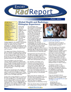

© Copyright 2026