
Cancer Imaging: Lung and Breast Carcinomas
FM-P370468.qxd
13:08:07
10:43 AM
Page i
Cancer Imaging:
Lung and Breast Carcinomas
FM-P370468.qxd
13:08:07
10:43 AM
Page ii
FM-P370468.qxd
13:08:07
10:43 AM
Page iii
Cancer Imaging:
Lung and Breast
Carcinomas
Volume 1
Edited by
M. A. Hayat
Distinguished Professor
Department of Biological Sciences
Kean University
Union, New Jersey
AMSTERDAM • BOSTON • HEIDELBERG • LONDON
NEW YORK • OXFORD • PARIS • SAN DIEGO
SAN FRANCISCO • SINGAPORE • SYDNEY • TOKYO
Academic Press is an imprint of Elsevier
FM-P370468.qxd
13:08:07
10:43 AM
Page iv
Elsevier Academic Press
300 Corporate Drive, Suite 400, Burlington, MA 01803, USA
525 B Street, Suite 1900, San Diego, California 92101-4495, USA
84 Theobald’s Road, London WC1X 8RR, UK
This book is printed on acid-free paper.
Copyright © 2008 Elsevier Inc. All rights reserved.
No part of this publication may be reproduced or transmitted in any form or by any means,
electronic or mechanical, including photocopy, recording, or any information storage and retrieval
system, without permission in writing from the publisher.
Permissions may be sought directly from Elsevier’s Science & Technology
Rights Department in Oxford, UK: phone: (+44) 1865 843830, fax: (+44) 1865 853333, e-mail:
[email protected]. You may also complete your request on-line via the Elsevier homepage (http://elsevier.com), by selecting “Customer Support” and then “Obtaining Permissions.”
Library of Congress Cataloging-in-Publication Data
Cancer imaging : lung and breast carcinomas / editor, M.A. Hayat.
p. ; cm.
Includes bibliographical references and index.
ISBN-13: 978-0-12-370468-9 (hardcover : alk. paper)
ISBN-10: 0-12-370468-5 (hardcover : alk. paper) 1. Lungs—Cancer—Imaging.
2. Breast—Cancer—Imaging, I. Hayat, M. A., 1940[DNLM: 1. Carcinoma, Non-Small-Cell Lung—diagnosis. 2. Breast Neoplasms—diagnosis.
3. Carcinoma, Lobular—diagnosis. 4. Diagnostic Imaging—methods. 5. Lung Neoplasms—
diagnosis. WF 658 C21442008]
RC280.L8C34 2008
616.994240754—dc22
2007002969
British Library Cataloguing in Publication Data
A catalogue record for this book is available from the British Library
ISBN: 978-0-12-370468-9
For all information on all Academic Press publications
visit our Web site at www.academicpress.com
Printed in China
07 08 09 10
11
9
8
7
6
5
4
3
2
1
FM-P370468.qxd
13:08:07
10:43 AM
Page v
To
The men and women involved in the odyssey of deciphering the complexity of
cancer initiation, progression, and metastasis, its diagnosis, cure, and hopefully its prevention.
FM-P370468.qxd
13:08:07
10:43 AM
Page vi
FM-P370468.qxd
13:08:07
10:43 AM
Page vii
Contents of Volume 1
Contents of Volume 2
Contributors
xxix
Preface
xxxix
Selected Glossary
xli
Introduction
xlix
Part I
1.1
xxiii
1.2
Synthesis of 18F-fluoromisonidazole
Tracer for Positron Emission
Tomography
15
Ganghua Tang
Introduction
15
Methods
16
Results and Discussion
References
21
Instrumentation
Strategies for Imaging Biology in
Cancer and Other Diseases
3
1.3
Radiation Hormesis
19
23
Philipp Mayer-Kuckuk and Debabrata Banerjee
Rekha D. Jhamnani and Mannudeep K. Kalra
Introduction
3
Imaging Strategies
4
Conferring Imaging Visibility
Peptides, Proteins, and Probes
Imaging Reporter Genes
Imaging Modalities
7
Preclinical Applications
7
Gene Transcription
7
Ribonucleic Acid Biology
Protein Biology
8
Imaging Strategies for
Clinical Applications
10
Receptors and Cell-Surface
Targets
10
Enzyme Activities
11
Transporters
11
Cell Death
11
Acknowledgments
12
References
12
Introduction
Hormesis
Mechanisms
Animal Studies
Human Studies
Controversy
References
4
4
5
8
Part II
2.1
23
23
24
24
24
25
26
General Imaging
Applications
Molecular Imaging in Early
Therapy Monitoring
29
Susanne Klutmann and Alexander Stahl
Introduction
29
The Place of Early Therapy Monitoring
in the Management of Cancer
29
vii
FM-P370468.qxd
13:08:07
10:43 AM
Page viii
viii
Contents of Volume 1
What Can be Expected from Positron
Emission Tomography Imaging?
30
F-18-FDG in Therapy Monitoring
30
Monitoring Neoadjuvant Therapy
31
Therapy Monitoring in Non-Small Cell
Lung Cancer (NSCLC)
31
Therapy Monitoring in Non-Hodgkin’s
Lymphoma (NHL)
32
Therapy Monitoring in Carcinomas of the
Esophagus, Esophagogastric Junction,
and Stomach
33
General Aspects of Early Therapy
Monitoring with FDG-Positron
Emission Tomography
33
Specific Aspects
34
Procedural Aspects
35
Colorectal Cancer
35
References
36
2.2
Positron Emission Tomography in
Medicine: An Overview
39
Abbas Alavi and Steve S. Huang
2.4
Introduction
39
Positron Emission Tomography
in Oncology
39
Positron Emission Tomography in
Lung and Breast Cancer
41
Positron Emission Tomography
in Brain Imaging
42
Positron Emission Tomography
in Cardiac Imaging
43
Positron Emission Tomography in
Infection and Inflammation
43
Cell Proliferation Agents
43
Hypoxia Positron Emission
Tomography Imaging
44
Peptide and Protein Positron Emission
Tomography Tracers
44
References
44
2.3
Fluoroscopy
53
Radiation Quality
54
Tube Potential
54
Filtration
54
Scattered Radiation
55
Optimization of Technique
in Fluoroscopy
56
Computed Tomography
56
Computed Tomography
Scanners
56
Radiation Dose and Image
Quality
57
Computed Tomography Dose
Assessment
58
Radionuclide Imaging
58
Imaging Technique
59
Radiation Dose and Image
Quality
59
Conclusions
60
References
61
Radiation Dose and Image
Quality
45
Colin J. Martin and David G. Sutton
Introduction
45
Radiation Dose
46
Image Quality
48
X-ray Beam Interactions
Radiographic Imaging
49
50
Contrast Agents for Magnetic
Resonance Imaging:
An Overview
63
Alan Jasanoff
Introduction
63
Relaxation Agents
64
Basic Principles of Relaxation
Contrast
64
Determinants of Inner Sphere
Relaxivity
65
Determinants of Outer Sphere
Relaxivity
66
Characteristics of T1 Agents
67
Characteristics of T2 Agents
67
Advances in the Design
of Relaxation Agents
69
Chemical Exchange-dependent
Saturation Transfer Agents
70
The CEST Effect
70
CEST Agents and Applications
72
Nonproton Contrast Agents
73
Direct Detection of Nuclei Other
Than Protons
73
19
F and 13C Imaging Agents
73
Hyperpolarization Techniques
75
Conclusions
75
References
76
FM-P370468.qxd
13:08:07
10:43 AM
Page ix
Contents of Volume 1
2.5
ix
Whole-body Computed Tomography
Screening
79
Considerations on Screening
Programs
91
18
F-fluorodeoxyglucose-Positron
Emission Tomography
92
Negative Tumors
92
Radiation Protection
92
References
93
Lincoln L. Berland and Nancy W. Berland
Introduction
79
What Is Whole-body Computed Tomography
Screening?
79
How Is it Done? Standards, Protocols,
and Informed Consent
80
What Is Found on Whole-body
Computed Tomography Screening?
80
Renal Cell Carcinoma
80
Abdominal Aortic Aneurysm
81
Ovarian Carcinoma
81
Other Findings on Whole-body
Computed Tomography Screening
81
Liver Lesions
81
Adrenal Lesions
81
Other Miscellaneous Conditions
82
Risks and Costs of Positive Results
82
Risks of Positive Results
82
Radiation
82
Costs of Positive Results
83
Analyzing the Rationale of Whole-body
Computed Tomography Screening
83
Analogies to Existing Screening
Practices
83
Distrust of Authority and
Self-empowerment
84
Is Proof of Value Necessary?
84
Is Whole-body Computed Tomography
Screening Truly Screening?
85
Psychological Implications
86
Variability of Rate of Positive Results
86
Enhancement of Radiology’s
Role in Medicine
87
Entrepreneurial Value of Screening
87
References
88
2.6
Whole-body 18F-fluorodeoxyglucosePositron Emission Tomography:
Is It Valuable for Health
Screening?
89
2.7
Staging Solid Tumors with
F-fluorodeoxyglucose-Positron
Emission Tomography/Computed
Tomography
95
18
Gerald Antoch and Andreas Bockisch
Introduction
95
PET/CT Imaging Protocols for Staging
Solid Tumors
96
Staging Solid Tumors with
FDG-PET/CT
96
T-stage
97
N-stage
98
M-stage
100
References
102
2.8
Laser Doppler Perfusion Imaging:
Clinical Diagnosis
103
E. Y-K Ng, S. C. Fok, and Julie Richardson
Introduction
103
Review of Laser Doppler
Perfusion Imaging
104
Some Past and Recent LDPI
Applications
106
Potential Integration of LDPI
in Cancer Diagnosis
110
Conclusions
112
Acknowledgment
112
References
112
2.9
Dynamic Sonographic Tissue
Perfusion Measurement
with the PixelFlux Method
Matthias Weckesser and Otmar Schober
Thomas Scholbach, Jakob Scholbach,
and Ercole Di Martino
Introduction
89
Current Positron Emission Tomography
Screening Programs
91
Introduction
115
Tumor Perfusion Evaluation—State
of the Art
115
115
FM-P370468.qxd
13:08:07
10:43 AM
Page x
x
Contents of Volume 1
Dynamic Tissue Perfusion Measurement
(PixelFlux)
116
Preconditions
117
Workflow
117
Procedure
117
Output
117
Use of Contrast Enhancers
118
Application
118
PixelFlux Application in
Oncology
118
Evaluation of PixelFlux Results
119
Comparison of Results with Other
Techniques
123
Conclusions and Outlook
123
References
124
Physiological Imaging
Molecular Imaging
Conclusions
156
References
156
Part III
3.1
Lung Carcinoma
The Role of Imaging
in Lung Cancer
163
Clifton F. Mountain and Kay E. Hermes
Introduction
163
The International System for Staging
Lung Cancer
163
Stage Groups and Survival
Patterns
164
The Role of Imaging in Lung
Cancer Staging
165
Imaging for Primary Tumor
Evaluation
165
Imaging for Evaluation of Regional
Lymph Nodes
167
Imaging for Evaluation of
Distant Metastasis
167
Restaging
168
Implications of Imaging for Lung
Cancer Screening
168
Conclusions
169
References
169
2.10 Immuno-Positron Emission
Tomography
127
Lars R. Perk, Gerard W. M. Visser, and
Guus A. M. S. van Dongen
Introduction
127
Diagnostic and Therapeutic Applications of
Monoclonal Antibodies
128
Therapy Planning with
Monoclonal Antibodies
128
Immuno-PET: Imaging and
Quantification
129
Clinical PET Imaging Systems
130
Positron Emitters for Immuno-Pet
130
Experience with Preclinical
Immuno-Pet
131
Experience with Clinical
Immuno-Pet
133
Acknowledgments
136
References
136
144
148
3.2
Lung Cancer Staging: Integrated
F-fluorodeoxyglucose-Positron
Emission Tomography/Computed
Tomography and Computed
Tomography Alone
171
18
Kyung Soo Lee
2.11 Role of Imaging Biomarkers
in Drug Development
139
Janet C. Miller, A. Gregory Sorensen,
and Homer H. Pien
Introduction
139
Biomarkers and Surrogate
Markers
140
Imaging Biomarkers
Anatomic Imaging
140
142
Introduction
171
Results Obtained by Previous
Studies
172
T-Staging
172
N-Staging
172
M-Staging
173
Problems and Their Solutions
Potential Advancements
References
175
174
175
FM-P370468.qxd
13:08:07
10:43 AM
Page xi
Contents of Volume 1
3.3
xi
Computed Tomography Screening
for Lung Cancer
177
T-staging
192
Chest Wall Invasion
193
Invasion of Fissures and
Diaphragm
194
Invasion of Mediastinum
194
N-staging
194
M-staging
195
Assessment of Response to Treatment
and Tumor Recurrence
195
Virtual Bronchoscopy
196
Conclusions
196
References
196
Claudia I. Henschke, Rowena Yip, Matthew D.
Cham, and David F. Yankelevitz
Introduction
177
Prior Screening Studies
177
Memorial Sloan-Kettering Cancer Center
(MSKCC) and Johns Hopkins Medical
Institution (JHMI) Studies
178
Mayo Lung Project (MLP)
178
Czechoslovakia Study
178
Recommendations and Controversy Resulting
from Prior Studies
178
The Early Lung Cancer Action
Project Paradigm for Evalution
of Screening
179
The Early Lung Cancer Action
Project
180
Computed Tomography Screening
in Japan
181
The New York Early Lung
Cancer Action Project
181
International Conferences on Screening
for Lung Cancer
181
International Early Lung Cancer
Action Program
182
National Cancer Institute
Conferences
182
Performance of Computed Tomography
Screening for Lung Cancer
182
Updated Recommendations
Regarding Screening
185
Problems Identified in Performing Randomized
Screening Trials
185
References
188
3.5
Tae Sung Kim
Intoduction
199
Pleomorphic Carcinoma
of the Lung
199
References
202
3.6
Lung Cancer: Role of Multislice
Computed Tomography
191
Suzanne Matthews and Sameh K. Morcos
Introduction
191
Multislice Computed Tomography Technique
for Diagnosis and Staging
of Bronchogenic Carcinoma
192
Scanning Protocol
192
Imaging Protocol
192
Multislice Computed Tomography
Staging of Bronchogenic
Carcinoma
192
Lung Cancer: Low-dose Helical
Computed Tomography
203
Yoshiyuki Abe, Masato Nakamura, Yuichi Ozeki,
Kikuo Machida, and Toshiro Ogata
Introduction
203
Materials and Methods
Results
204
Discussion
205
References
206
3.7
3.4
Surgically Resected Pleomorphic
Lung Carcinoma: Computed
Tomography
199
204
Lung Cancer: Computer-aided
Diagnosis with Computed
Tomography
209
Yoshiyuki Abe, Katsumi Tamura, Ikuko Sakata, Jiro
Ishida, Masayoshi Nagata, Masato Nakamura,
Kikuo Machida, and Toshiro Ogata
Intoduction
209
Materials and Methods
Results
211
Discussion
212
Conclusions
213
References
213
210
FM-P370468.qxd
13:08:07
10:43 AM
Page xii
xii
3.8
Contents of Volume 1
Stereotactic Radiotherapy for
Non-small Cell Lung Carcinoma:
Computed Tomography
215
Peculiarity of Radiation Injury of the Lung
after Stereotactic Radiotherapy
222
Appearance Time of Radiation Injury
of the Lung after Stereotactic
Radiotherapy
223
Summary of Computed Tomography
Findings of Radiation Injury of the Lung after
Stereotactic Radiotherapy
224
Computed Tomography Evaluation of the
Tumor Response and Progression
225
Tumor Response
225
Local Recurrence
225
Cases of Computed Tomography Findings
after Stereotactic Radiotherapy
226
Guidelines for Quality Control of Computed
Tomography Images
226
Future Direction
227
Image Quality of Cone Beam
Computed Tomography
227
Megavoltage Computed
Tomography
227
Helical Tomotherapy
227
Imaging Supplement for Computed
Tomography for Evaluating Tumor
Malignancy and Extension
228
References
229
Hiroshi Onishi, Atsushi Nambu, Tomoki Kimura,
and Yasushi Nagata
Introduction
215
Definition of Stereotactic
Radiotherapy
216
Clinical Status of Stereotactic Radiotherapy for
Early-Stage Lung Carcinoma
216
The Significance of Computed
Tomography Imaging for Stereotactic
Radiotherapy
216
Utility of Computed Tomography for
Radiotherapy Treatment Planning of
Stereotactic Radiotherapy for
Lung Carcinoma
217
Definition of Target Volumes with Computed
Tomography Images
217
Radiologic-Pathologic Correlation of Stage I
Lung Carcinoma
218
Usefulness of Thin-section Computed
Tomography in the Evaluation
of Lung Carcinoma
218
Attenuation of Lung Carcinoma
218
Solid Attenuation
218
Ground-glass Opacity
218
Borders Characteristics
219
Spicula and Pleural Indentation
219
Growth Patterns of Lung
Carcinoma
219
Limits of Computed Tomography
for Evaluating Lung Tumors
220
Management of Respiratory Motion
of the Target during Irradiation
220
Simulation Using Slow-scan Computed
Tomography for Free or Suppressed
Breathing Technique
220
Three-dimensional Stereotactic
Repositioning of the Isocenter
during Irradiation
221
Computed Tomography-Linear Accelerator
(Linac) Unit
221
Cone Beam Computed
Tomography
221
Evaluation of the Treatment Effect and
Differentiation between Inflammatory Change
and a Recurrent Mass
222
3.9
Thin-section Computed Tomography
Correlates with Clinical Outcome
in Patients with Mucin-producing
Adenocarcinoma of the Lung
231
Ukihide Tateishi, Testuo Maeda, and Yasuaki Arai
Introduction
231
Materials and Methods
Results
233
Discussion
234
Acknowledgments
References
235
232
235
3.10 Non-small Cell Lung Carcinoma:
18
F-fluorodeoxyglucose-Positron
Emission Tomography
237
Rodney J. Hicks and Robert E. Ware
Introduction
237
Role of FDG-PET on Diagnosing
Lung Cancer
238
FM-P370468.qxd
13:08:07
10:43 AM
Page xiii
Contents of Volume 1
Preoperative PET Staging of
Non-small Cell Lung Cancer
239
Evaluation of Distant Metastasis (M)
Stage
240
Evaluation of Intrathoracic Lymph
Node (N) Stage
240
Evaluation of Tumor (T) Stage
241
Impact of Staging FDG-PET
on Patient Management
241
Role of PET in Therapeutic Response
Assessment in NSCLC
243
Use of FDG-PET for Restaging
Following Definitive Treatment
of NSCLC
244
A Philosophical Perspective on
the Quantitative Analysis of FDG
Uptake in NSCLC
244
Use of Hybrid PET-CT Images in
Staging
246
Conclusions
246
References
246
3.11 Evaluating Positron Emission
Tomography in Non-small Cell Lung
Cancer: Moving Beyond Accuracy
to Outcome
249
Harm van Tinteren, Otto S. Hoekstra,
Carin A. Uyl-de Groot, and Maarten Boers
Introduction
249
Diagnostic Accuracy of Positron Emission
Tomography in Non-small Cell
Lung Cancer
250
The Framework
251
Literature Analysis
251
Exploiting Clinical Data Obtained Prior
to Introducing a New Test
251
Decision Modeling
252
Clinical-Value Studies
252
Randomized Controlled
Trials
253
Economic Evaluation
254
Before and After
Implementation
254
Conclusions
255
References
255
xiii
3.12 Non-small Cell Lung Cancer:
False-positive Results with
18
F-fluorodeoxyglucose-Positron
Emission Tomography
257
Siroos Mirzaei, Helmut Prosch, Peter Knoll,
and Gerhard Mostbeck
Introduction
257
Physiological High Uptake of 18F-FDG
in Different Tissues
258
Head and Central Nervous
System
258
Neck
258
Chest
258
Abdomen
258
Urinary Tract
258
Breast
259
Skeletal Muscle
259
Focal Uptake of FDG Due to
Benign Disease
259
High Metabolic Activity after
Treatment
261
Focal Uptake of FDG
Due to Artifacts
262
Focal Uptake of FDG Due to Artifacts
by New PET Devices
262
Conclusions
263
References
263
3.13 Oxygen-enhanced Proton Magnetic
Resonance Imaging of the Human
Lung
267
Eberhard D. Pracht, Johannes F. T. Arnold, Nicole
Seiberlich, Markus Kotas, Michael Flentje, and
Peter M. Jakob
Introduction
267
Respiratory Physiology
268
Theory of Oxygen-enhanced
Imaging
269
T1-Relaxation in the Human
Lung
269
Influence of Oxygen and the Oxygen
Transfer Function (OTF)
270
T2*Relaxation in the Human
Lung
274
Oxygen-enhanced Imaging in Volunteers
and Patients
276
FM-P370468.qxd
13:08:07
10:43 AM
Page xiv
xiv
Contents of Volume 1
Pathogenesis of Lung Cancer
in Idiopathic Pulmonary Fibrosis
296
Clinical Features
296
Chest Radiograph
296
Computed Tomography
and High-resolution Computed Tomography
Findings
296
References
298
Improvement of Imaging
Technique
276
Studies in Patients and Correlation with
Physiologic Parameters
277
Conclusions
277
References
278
3.14 Detection of Pulmonary Gene Transfer
Using Iodide-124/Positron Emission
Tomogrpahy
281
Part IV Breast Carcinoma
Frederick E. Domann and Gang Niu
Introduction
281
Pulmonary Applications of Gene
Therapy
281
Gene Therapy for Inherited
Lung Diseases
282
Cystic Fibrosis
282
Alpha-1 Anitrypsin Deficiency
283
Gene Therapy for Lung Cancer
283
Gene Delivery Vehicles and Vectors
283
Retroviruses
284
Adenoviruses
284
Adeno-associated Viruses (AAV)
284
Nonviral Liposomal Vectors
284
Molecular Imaging of Pulmonary
Gene Transfer
284
Reporter Gene Systems
285
Herpes Simplex Virus-1 Thymidine
Kinase (HSV1-TK)
286
Sodium Iodide Symporter
287
Considerations in PET Imaging of Pulmonary
Gene Transfer
288
Gene Transfer Barriers
288
Iodine-124 as Imaging Agent
288
Relationship between PET Signal and
Reporter Gene Expression
289
Resolution and Sensitivity of PET
290
Acknowledgements
290
References
290
3.15 Lung Cancer with Idiopathic
Pulmonary Fibrosis: High-resolution
Computed Tomography
295
Kazuma Kishi and Atsuko Kurosaki
Introduction
295
Prevalence of Lung Cancer
in Idiopathic Pulmonary Fibrosis
295
4.1
Categorization of Mammographic
Density for Breast Cancer: Clinical
Significance
301
Mariko Morishita, Akira Ohtsuru, Ichiro Isomoto,
and Shunichi Yamashita
Introduction
301
Breast Density by Mammography
301
Clinical Applications of Breastdensity Category
303
Analysis of Patient Characteristics
by Breast-density Category
303
Steroid Receptor Status and
Breast-density Category
303
Comparison of Nottingham Prognostic
Index Scores in Breast-density
Categories
304
Patient Prognosis and
Breast-density Category
304
Analytic Considerations
304
References
305
4.2
Breast Tumor Classification
and Visualization with
Machine-learning
Approaches
309
Tim W. Nattkemper, Andreas Degenhard,
and Thorsten Twellmann
Introduction
309
The Contribution of Machine Learning
and Artificial Neural
Networks
310
State-of-the-Art Approaches to Dynamic
Contrast-enhanced Magnetic
Resonance Visualization
311
Learning Algorithms
312
Learning Clusters
312
FM-P370468.qxd
13:08:07
10:43 AM
Page xv
Contents of Volume 1
xv
Realization of the Phase-contrast Technique in
Mammography
341
Design of Digital Image Acquisition
and Output
341
Magnification-demagnification Effect
in Digital Mammography
342
Sharpness
342
Image Noise
343
Improvement of Image Quality by the
Magnification-demagnification
Effect
343
Improvement of Image Sharpness
in Digital Full-field PCM
343
Clinical Images
344
Clinical Experience
345
Future Development
345
Acknowledgements
347
References
347
Human Experts versus Computer
Algorithms
314
Monitoring Tumor Development
317
Supervised Learning Algorithms
318
Supervised Detection and
Segmentation of Lesions
319
Supervised Classification
of Lesions
319
Summary and Outlook
321
Acknowledgements
321
References
321
4.3
Mass Detection Scheme for Digitized
Mammography
325
Bin Zheng
Introduction
325
Basic Architecture of Mass
Detection Schemes
325
Computer-aided Detection Schemes
Based on a Single Image
325
Computer-aided Detection Schemes
Based on multi-image
329
Evaluation and Application of Commercial
Computer-aided Detection Systems
331
New Developments in Mass
Detection Schemes
332
Improvement of Computer-aided Detection
Performance
333
Improvement of Reproducibility of Computeraided Detection Schemes
333
Interactive Computer-aided
Detection Systems
334
References
336
4.4
Full-field Digital Phase-contrast
Mammography
339
Toyohiko Tanaka, Chika Honda, Satoru Matsuo,
and Tomonori Gido
Introduction
339
Historical Background of the Phase-contrast
Technique
340
Absorption Contrast and Phase
Contrast
340
Edge Effect Due to Phase
Contrast
341
4.5
Full-field Digital Mammography
versus Film-screen
Mammography
349
Arne Fischmann
Introduction and Historical
Perspective
349
Physical Performance of Digital
Compared to Film-screen
Mammography
350
Phantom Studies Comparing Full-field
Digital Mammography and
Film-screen Mammography
350
Simulated Microcalcifications
351
Clinical or Diagnostic
Digital Mammography
353
Full-field Digital Mammography and
Film-screen Mammography in
Screening
354
Oslo I and II Studies
355
Digital Mammography Imaging
Screening Trial
355
Financial Considerations
of Digital Mammography
356
Radiation Dose Considerations
356
References
357
FM-P370468.qxd
13:08:07
10:43 AM
Page xvi
xvi
4.6
Contents of Volume 1
Use of Contrast-enhanced Magnetic
Resonance Imaging for Detecting
Invasive Lobular Carcinoma
359
Results and Discussion
Conclusions
380
References
381
377
Carla Boetes and Ritse M. Mann
Introduction
359
Incidence
359
Presentation
359
Pathology
360
Mammography
360
Ultrasound
360
Goal of MRI in the Assessment of Invasive
Lobular Carcinoma
361
Magnetic Resonance
Imaging
361
Dynamic Sequences in Breast Magnetic
Resonance Imaging
362
False-Negative Imaging on Magnetic
Resonance Imaging
363
Conclusions
364
References
364
4.7
Axillary Lymph Node Status in Breast
Cancer: Pinhole Collimator Single–
Photon Emission Computed
Tomography
367
Giuseppe Madeddu, Orazio Schillaci, and Angela
Spanu
Introduction
367
99m
Tc-tetrofosmin Pinhole–Single Photon
Emission Computed
Tomography
369
Method
369
Results and Discussion
369
Conclusions
371
References
372
4.8
Detection of Small-size Primary Breast
Cancer: 99mTc-tetrofosmin Single
Photon Emission Computed
Tomography
375
Angela Spanu, Orazio Schillaci, and Giuseppe
Madeddu
Introduction
375
The Planar and SPECT Scintimammography
Method
376
4.9
Microcalcification in Breast
Lesions: Radiography and
Histopathology
383
Arne Fischmann
Introduction
383
Histopathology
383
Detection
384
Classification of Breast
Calcifications
385
Systematic Classification
Breast Imaging-Reporting
and Data System
386
Work-up of Breast
Calcifications
389
Summary
391
References
391
385
4.10 Benign and Malignant
Breast Lesions: Doppler
Sonography
393
José Luís del Cura
Introduction
393
Doppler Ultrasound Technique
in Breast Diseases
394
Breast Doppler Limitations
394
Differentiation of Benign and Malignant
Solid Breast Lesions
395
Tumor Vessel Identification
395
Quantitative Criteria
395
Semiquantitative Criteria
396
Breast Cancer Prognosis
397
Assessment of Lymph Node
Involvement
397
Recurrence versus Scar
in Operated Patients
398
Treatment Monitoring
398
Conclusions
398
References
399
FM-P370468.qxd
13:08:07
10:43 AM
Page xvii
Contents of Volume 1
xvii
4.11 Response to Neoadjuvant Treatment
in Patients with Locally Advanced
Breast Cancer: Color-Doppler
Ultrasound Contrast
Medium (Levovist)
401
Paolo Vallone
Introduction
401
Materials and Methods
Results
402
Discussion
402
References
406
401
4.12 Magnetic Resonance Spectroscopy of
Breast Cancer: Current Techniques
and Clinical Applications
407
Sina Meisamy, Patrick J. Bolan, and Michael
Garwood
Introduction
407
Background
407
The “Choline Peak”
407
Why Is tCho Elevated
in Cancer?
408
Technique
408
Tumor Localization
408
Technical Issues
408
Respiratory Artifact
409
Quantification
409
How Reliable Is the tCho
Measurement?
410
Clinical Applications
410
Diagnosis
410
Sample Diagnostic Cases
411
Therapeutic Monitoring with
Early Feedback
412
Sample Therapeutic Monitoring
Cases
413
Acknowledgement
414
References
414
4.13 Breast Scintigraphy
417
Orazio Schillaci, Angela Spanu, and Giuseppe
Madeddu
Introduction
417
Breast Scintigraphy
417
Planar Method
417
Planar Results
418
Single Photon Emission Computed
Tomography Method
418
Single Photon Emission Computed
Tomography Results
419
Dedicated Imaging Systems
419
Methods
419
Results
420
Clinical Indications of Breast Scintigraphy
or Scintimammography
421
References
421
4.14 Primary Breast Cancer: False-negative
and False-positive Bone
Scintigraphy
423
Hatice Mirac Binnaz Demirkan and Hatice Durak
Introduction
423
Search Strategy and Selection
Criteria
423
Procedures and Technical Aspects
of Bone Scan
423
Clinical Applications in Breast
Cancer
427
Pitfalls of Bone Scan Encountered in Breast
Cancer Patients and their Solutions with
Potential Advances
429
References
431
4.15 Improved Sensitivity and Specificity
of Breast Cancer Thermography
435
E. Y-K. Ng
Introduction
435
Image Analysis Tools
436
Thermography
436
Artificial Neural Networks
437
Backpropagation
437
Radial Basis Function Network
438
Biostatistical Methods
438
Data Acqusition
439
Procedures for Thermal
Imaging
439
Designed Integrated Approach
440
Step 1: Linear Regression
440
Step 2: ANN RBFN/BFN
440
Step 3: ROC Analysis
441
FM-P370468.qxd
13:08:07
10:43 AM
Page xviii
xviii
Contents of Volume 1
Results and Discussion
441
Summarized Results for Step 1:
Linear Regression
441
Selected Results for Step 2:
ANN RBFN/BPN
441
Selected Results (with Area > 0.85)
for Step 3: ROC Analysis
441
Conclusions and Future
Trends
442
Acknowledgements
443
References
443
4.16 Optical Mammography
445
Sergio Fantini and Paola Taroni
Introduction
445
Sources of Intrinsic Optical
Contrast in Breast Tissue
446
Principles of Optical
Mammography
447
Continuous-wave Approaches:
Dynamic Measurements and Spectral
Information
448
Time-resolved Approaches
448
Interpretation of Optical
Mammograms
450
Prospects of Optical
Mammography
452
Acknowledgements
453
References
453
4.17 Digital Mammography
455
John M. Lewin
Introduction
455
Technical Advantages of Digital
Mammography
455
Technologies Used for Digital
Mammography
456
Clinical Advantages of Digital
Mammography
456
Advanced Applications
of Digital Mammography
Tomosynthesis
457
Contrast-enhanced Digital
Mammography
458
References
458
4.18 Screening for Breast Cancer
in Women with a Familial or
Genetic Predisposition: Magnetic
Resonance Imaging versus
Mammography
459
Mieke Kriege, Cecile T. M. Brekelmans,
and Jan G. M. Klijn
Introduction
459
Magnetic Resonance Imaging
Screening Studies
461
Results
461
Discussion
462
References
463
4.19 Mammographic Screening:
Impact on Survival
465
James S. Michaelson
Introduction
465
Why Screening Works
465
Screening Effectiveness
465
Cancers Become More Lethal
as they Increase in Size
466
Present and Future Life-saving Impact
of Screening
466
Life-saving Potential of Screening
Tumor Size and Survival
467
False-positives
468
How is Screening Actually Used
Present Status of Breast Cancer
Screening
469
References
470
467
468
4.20 False-positive Mammography
Examinations
473
Pamela S. Ganschow and Joann G. Elmore
457
Introduction
473
Definitions
473
Current Estimates of False-positive
Rates in the United States and
International Guidelines
474
Cumulative False-positive Rates
Predictors of False-positive
Mammograms
475
Patients factors
476
Radiologist Factors
478
475
FM-P370468.qxd
13:08:07
10:43 AM
Page xix
Contents of Volume 1
xix
Facility and System Factors
479
Predicting the Cumulative Risk
of False-positive Mammograms
480
Significance of False-positive
Mammography Examination
480
Recall Rates in the United States
versus Other Countries
481
Efforts to Reduce False-positive
Mammograms and to Better
Deal with Expected False-Positive
Screenings
483
Acknowledgment
483
References
483
4.21 Breast Dose in Thoracic Computed
Tomography
487
Eric N. C. Milne
Introduction
487
Methodology
488
Results
488
Discussion
489
Cancer Risks
489
Computed Tomography of
the Breast
490
Reducing Radiation Dose
490
Imaging without Using Ionizing
Radiation
490
Optical Imaging
491
Ultrasound
491
Magnetic Resonance Imaging
491
References
492
4.22 Absorbed Dose Measurement in
Mammography
493
Marianne C. Aznar and Bengt Å. Hemdal
Introduction
493
Estimation of Absorbed Dose
to the Breast
494
Concepts and Quantities Used
From Measurement to
Dose Estimate
496
Dose Limits and Diagnostic
Reference Levels
497
Dosimeters for Indirect Measurements
Ionization chambers
498
Semiconductors
498
494
Dosimeters for Direct in vivo
Measurements
498
Thermoluminescence Detectors
Novel in vivo Techniques
Summary and Conclusions
References
500
499
499
500
4.23 Metastatic Choriocarcinoma to the
Breast: Mammography and Color
Doppler Ultrasound
503
Naveen Kalra and Vijaynadh Ojili
Introduction
503
Mammography
504
Ultrasonography and
Color Doppler
504
Tissue Diagnosis
506
References
507
4.24 Detection and Characterization
of Breast Lesions: Color-coded
Signal Intensity Curve Software
for Magnetic Resonance–based
Breast Imaging
509
Federica Pediconi, Fiorella Altomari, Luigi
Carotenuto, Simona Padula, Carlo Catalano,
and Roberto Passariello
Introduction
509
Computer-aided Diagnosis: Features
and Applications
511
Computer-aided Detection for Breast Magnetic
Resonance Imaging
512
Characterization Algorithm
514
Registration Algorithm
514
Conclusions
515
References
517
4.25 Detection of Breast Malignancy:
Different Magnetic Resonance
Imaging Modalities
519
Wei Huang and Luminita A. Tudorica
498
Introduction
519
Major Breast Imaging
Modalities
519
Breast Dynamic Contrast-enhanced Magnetic
Resonance Imaging
520
FM-P370468.qxd
13:08:07
10:43 AM
Page xx
xx
Contents of Volume 1
Subjective Assessment
520
Empirical Quantitative
Characterization
521
Analytical Pharmacokinetic
Modeling
522
1
Breast H Magnetic
Resonance Spectroscopy
523
Breast T2*-Weighted Perfusion Magnetic
Resonance Imaging
525
References
526
4.26 Breast Lesions: Computerized
Analysis of Magnetic Resonance
Imaging
529
Kenneth G. A. Gilhuijs
Introduction
529
Mechanisms of Functional Imaging using
Magnetic Resonance Imaging
530
Interpretation of Contrast-enhanced Magnetic
Resonance Imaging
531
Reduction of Motion Artifacts
532
Computerized Extraction of
Temporal Features
533
Computerized Extraction of
the Region of Interest
534
Computerized Extraction
of Morphological Features
535
Computerized Classification
of Features of Enhancement
536
Current Status and Future Role
of Computerized Analysis of Breast
Magnetic Resonance Imaging
537
References
538
4.27 Optical Imaging Techniques
for Breast Cancer
539
Outlook
References
544
544
4.28 Magnetic Resonance Imaging:
Measurements of Breast Tumor
Volume and Vascularity for
Monitoring Response to
Treatment
547
Savannah C. Partridge
Introduction
547
Magnetic Resonance Imaging
of the Breast
547
Neoadjuvant Treatment
547
Magnetic Resonance Imaging to
Monitor Treatment Response
548
Measuring Changes in Tumor
Size with Treatment
548
Measuring Changes in Tumor
Vascularity with Treatment
548
Imaging Considerations
548
Magnetic Resonance Imaging
Acquisition
548
Imaging Postprocessing
549
Assessing Treatment Response
550
Changes in Tumor Volume Predict
Recurrence-free Survival (RFS)
550
Vascular Changes with
Treatment
550
Conclusions
551
Acknowledgements
552
References
552
4.29 Defining Advanced Breast Cancer:
18
F-fluorodeoxyglucose-Positron
Emission Tomography
555
Alexander Wall and Christoph Bremer
William B. Eubank
Introduction
539
Tomographic Imaging
540
Nonspecific Contrast Agents (Perfusion-type
Contrast Agents)
540
Fluorochromes with Molecular
Specificity
542
Smart Probes
542
Targeted Probes
542
Multimodality Probes
543
Introduction
555
Positron Emission Tomography
Principles
556
Positron Emission Tomography
Instrumentation
556
Fluorodeoxyglucose (FDG)
556
Axillary Node Staging
557
Detection of Locoregional
and Distant Recurrences
557
FM-P370468.qxd
17:08:07
06:32 AM
Page xxi
Contents of Volume 1
xxi
Dynamic Infrared Imaging
573
Mechanism for Breast Cancer
Detection with Dynamic Infrared
575
Applications of Dynamic Infrared
Imaging
576
Pitfalls of Dynamic Infrared
Imaging
577
Conclusion: The Future of Infrared
and Dynamic Infrared Imaging
for Breast Cancer Detection
578
Acknowledgements
579
References
579
Locoregional Recurrences
557
Intrathoracic Lymphatic
Recurrences
558
Distant Metastases
559
Response to Therapy
560
Impact of FDG-PET on
Patient Management
561
Beyond FDG: Future Applications of
PET to Breast Cancer
562
Estrogen Receptor Imaging
562
References
563
4.30 Leiomyoma of the Breast Parenchyma:
Mammographic, Sonographic, and
Histopathologic Features
567
4.32 Phyllodes Breast Tumors: Magnetic
Resonance Imaging
581
Aysin Pourbagher and M. Ali Pourbagher
Aimée B. Herzog and Susanne Wurdinger
Introduction
567
Mammographic Appearance
Sonographic Appearance
References
570
Introduction
581
Magnetic Resonance Imaging
Morphology
582
Signal Intensity
582
Contrast-enhancement
Characteristics
583
Guidelines
583
References
584
568
568
4.31 Detection of Breast Cancer: Dynamic
Infrared Imaging
571
Terry M. Button
Introduction: Infrared and
its Detection
571
History of Infrared for Breast
Cancer Detection
572
Index
585
582
FM-P370468.qxd
13:08:07
10:43 AM
Page xxii
FM-P370468.qxd
13:08:07
10:43 AM
Page xxiii
Contents of Volume 2
1.9
Part I: Instrumentation
1.1
Proton Computed
Tomography
3
Gadolinium-Based Contrast Media
Used in Magnetic Resonance
Imaging: An Overview
81
1.2
Multidetector Computed
Tomography
17
1.10 Molecular Imaging of Cancer with
Superparamagnetic Iron-Oxide
Nanoparticles
85
1.3
Megavoltage Computed Tomography
Imaging
27
1.11 Adverse Reactions to Iodinated
Contrast Media
97
1.4
Integrated Set-3000G/X Positron
Emission Tomography Scanner
1.5
Part II:
37
High-Resolution Magic Angle
Spinning Magnetic Resonance
Spectroscopy
45
General Imaging
Applications
2.1
The Accuracy of Diagnostic
Radiology
109
Spatial Dependency of Noise and Its
Correlation among Various Imaging
Modalities
53
2.2
Diffraction-Enhanced Imaging:
Applications to Medicine
119
1.7
Computed Tomography Scan Methods
Account for Respiratory Motion in
Lung Cancer
61
2.3
Role of Imaging in Drug
Development
127
1.8
Respiratory Motion Artifact Using
Positron Emission Tomography/
Computed Tomography
69
2.4
Characterization of Multiple
Aspects of Tumor Physiology by
Multitracer Positron Emission
Tomography
141
1.6
xxiii
FM-P370468.qxd
17:08:07
06:32 AM
Page xxiv
xxiv
Contents of Volume 2
2.5
Whole-Body Magnetic Resonance
Imaging in Patients with
Metastases
155
2.15 In Vivo Molecular Imaging in
Oncology: Principles of Reporter
Gene Expression Imaging
249
2.6
Whole-Body Imaging in Oncology:
Positron Emission Tomography/
Computed Tomography
(PET/CT)
161
2.16 Medical Radiation-Induced
Cancer
261
2.7
Whole-Body Cancer Imaging:
Simple Image Fusion with Positron
Emission Tomography/Computed
Tomography
169
2.8
Whole Body Tumor Imaging:
O-[11C]Methyl-L-Tyrosine/Positron
Emission Tomography
175
2.9
Tumor Proliferation 2-[11C]-Thymidine
Positron Emission Tomography
181
2.10
18
F-Fluorodeoxyglucose-Positron
Emission Tomography in Oncology:
Advantages and Limitations
193
Part III: Applications to
Specific Cancers
Adrenal Cortical Carcinoma
3.1
Adrenal Lesions: Role of Computed
Tomography, Magnetic Resonance
Imaging, 18F-FluorodeoxyglucosePositron Emission Tomography,
and Positron Emission Tomography/
Computed Tomography
269
3.2
Hemangioendothelioma: Whole-Body
Technetium-99m Red Blood Cell
Imaging-Magnetic Resonance
Imaging
281
Bone Cancer
3.3
Malignant Bone Involvement:
Assessment Using Positron
Emission Tomography/Computed
Tomography
291
3.4
Bone Metastasis: Single Photon
Emission Computed
Tomography/Computed
Tomography
305
2.11 Positron Emission Tomography
Imaging of Tumor Hypoxia and
Angiogenesis: Imaging Biology and
Guiding Therapy
201
2.12 Noninvasive Determination of
Angiogenesis: Molecular Targets
and Tracer Development
211
2.13 Gross Tumor Volume and Clinical
Target Volume: Anatomical Computed
Tomography and Functional
FDG-PET
225
2.14 Post-Treatment Changes in Tumor
Microenvironment: Dynamic
Contrast-Enhanced and DiffusionWeighted Magnetic Resonance
Imaging
235
3.5
Bone Cancer: Comparison of
F-Fluorodeoxyglucose-Positron
Emission Tomography with Single
Photon Emission Computed
Tomography
311
18
3.6
Bone Metastasis in Endemic
Nasopharyngeal Carcinoma:
18
F-Fluorodeoxyglucose-Positron
Emission Tomography
321
FM-P370468.qxd
13:08:07
10:43 AM
Page xxv
Contents of Volume 2
Colorectal Cancer
3.7
3.8
3.9
Colorectal Polyps: Magnetic Resonance
Colonography
327
Early Bile Duct Carcinoma: Ultrasound,
Computed Tomography,
Cholangiography, and Magnetic
Resonance Cholangiography
335
Incidental Extracolonic Lesions:
Computed Tomography
339
3.10 Colorectal Cancer: Magnetic Resonance
Imaging–Cellular and Molecular
Imaging
355
3.11 Potential New Staging Perspectives
in Colorectal Cancer: Whole-Body
PET/CT-Colonography
365
Esophageal Cancer
3.12 Thoracic Esophageal Cancer: Interstitial
Magnetic Lymphography Using
Superparamagnetic Iron
373
3.13 Esophageal Cancer: Comparison of
18
F-Fluoro-3-Deoxy-3-L-fluorothymidinePositron Emission Tomography with
18
F-Fluorodeoxyglucose-Positron
Emission Tomography
379
Gastrointestinal Cancer
3.14 Gastrointestinal Stromal Tumors:
Positron Emission Tomography and
Contrast-Enhanced Helical Computed
Tomography
385
3.15 Gastrointestinal Stromal Tumor:
Computed Tomography
391
3.16 Gastrointestinal Lipomas: Computed
Tomography
395
xxv
3.17 Computed Tomography in Peritoneal
Surface Malignancy
399
3.18 Gastrointestinal Tumors: Computed
Tomography/Endoscopic
Ultrasonography
407
3.19 Magnetic Resonance
Cholangiopancreatography
413
Head and Neck Cancer
3.20 Occult Primary Head and Neck
Carcinoma: Role of Positron Emission
Tomography Imaging
423
3.21 Benign and Malignant Nodes in
the Neck: Magnetic Resonance
Microimaging
431
3.22 Oral Squamous Cell Carcinoma:
Comparison of Computed
Tomography with Magnetic
Resonance Imaging
437
Kidney Cancer
3.23
Computed Tomography in Renal Cell
Carcinoma
445
3.24
Renal Cell Carcinoma Subtypes:
Multislice Computed
Tomography
457
3.25
Renal Impairment: Comparison
of Noncontrast Computed
Tomography, Magnetic Resonance
Urography, and Combined
Abdominal Radiography/
Ultrasonography
467
3.26 Renal Lesions: Magnetic
Resonance Diffusion-Weighted
Imaging
475
FM-P370468.qxd
13:08:07
10:43 AM
Page xxvi
xxvi
Lymphoma
Contents of Volume 2
3.37
3.27 Malignant Lymphoma:
18
F-Fluorodeoxyglucose-Positron
Emission Tomography
485
Melanoma
3.28 Malignant Melanoma: Positron
Emission Tomography
491
3.38 Pancreatic Islet Cell Tumors:
Endoscopic Ultrasonography
Myeloma
3.31 Nasopharyngeal Carcinoma for
Staging and Restaging with
18
F-FDG-PET/CT
513
Parotid Gland Tumors: Advanced
Imaging Technologies
563
Pituitary Tumors
3.40
Nasopharyngeal Cancer
3.30 Nasopharyngeal Carcinoma:
18
F-Fluorodeoxyglucose-Positron
Emission Tomography
505
Pituitary Macroadenomas:
Intraoperative Magnetic
Resonance Imaging and Endonasal
Endoscopy
575
Penile Cancer
3.41
Penile Cancer Staging:
F-Fluorodeoxyglucose-Positron
Emission Tomography/Computed
Tomography
581
18
Ovarian Cancer
Peripheral Nerve Sheath Tumors
3.32 Ovarian Sex Cord Stromal Tumors:
Computed Tomography and Magnetic
Resonance Imaging
523
3.42
3.33 Malignant Germ Cell Tumors:
Computed Tomography and Magnetic
Resonance Imaging
527
3.34 Ovarian Small Round Cell Tumors:
Magnetic Resonance Imaging
533
3.35 Ovarian Borderline Serous Surface
Papillary Tumor: Magnetic Resonance
Imaging
537
Pancreatic Cancer
3.36 Chronic Pancreatitis versus Pancreatic
Cancer: Positron Emission
Tomography
541
557
Parotid Gland Cancer
3.39
3.29 Multiple Myeloma: Scintigraphy
Using Technetium-99m-2Methoxyisobutylisonitrile
499
Pancreatic Cancer: P-[123I]
Iodo-L-Phenylalanine Single Photon
Emission Tomography for Tumor
Imaging
547
Malignant Peripheral Nerve Sheath
Tumors: [18F]FluorodeoxyglucosePositron Emission
Tomography
587
Prostate Cancer
3.43
Prostate Cancer: 11C-Choline-Positron
Emission Tomography
595
3.44
Metabolic Characterization of Prostate
Cancer: Magnetic Resonance
Spectroscopy
603
3.45
Prostate Cancer: Diffusion-Weighted
Imaging
615
3.46 Prostatic Secretory Protein of 94 Amino
Acids, Gene-Directed Transgenic
Prostate Cancer: Three-dimensional
Ultrasound Microimaging
619
FM-P370468.qxd
13:08:07
10:43 AM
Page xxvii
Contents of Volume 2
3.47
Prostate Cancer within the Transition
Zone: Gadolinium-Enhanced Magnetic
Resonance Imaging
629
3.48
Rectal Wall Invasion of Locally
Advanced Prostate Cancer:
Comparison of Magnetic Resonance
Imaging with Transrectal
Ultrasound
637
3.49
Local Staging of Prostate Cancer
Using Endorectal Coil Magnetic
Resonance Imaging
641
xxvii
Spleen Cancer
3.53
Thyroid Cancer
3.54
Thyroid Cancer: 18F-FDG-Positron
Emission Tomography
687
3.55
Thyroid Cancer: 18F-Fluoro-2-Deoxy-DGlucose-Positron Emission
Tomography (An Overview)
699
3.56
Diagnosis of Thyroid Cancer in
Children: Value of Power Doppler
Ultrasound
707
Sarcoma
3.50
3.51
Extremity Sarcoma: Positron Emission
Tomography
655
Retroperitoneal Synovial Sarcoma:
Color-Doppler Ultrasound, Computed
Tomography, and Magnetic
Resonance Imaging
667
Thymoma Cancer
3.52
Thymoma: Computed
Tomography
671
Hemolymphangiomatosis of the
Spleen: Conventional Diagnostic
Imaging and Magnetic Resonance
Imaging With Superparamagnetic
Contrast Agent
681
FM-P370468.qxd
13:08:07
10:43 AM
Page xxviii
FM-P370468.qxd
13:08:07
10:43 AM
Page xxix
Contributors
Abe, Yoshiyuki
Tokorozawa PET Diagnostic Imaging Clinic
Higashisumiyoshi 7-5
Tokorozawa City
359-1124 Saitama Prefecture
Japan
Alavi, Abbas
Division of Nuclear Medicine
Department of Radiology
Hospital of the University of Pennsylvania
110 Donner Building
3400 Spruce Street
Philadelphia, PA 19104
Arnold, Johannes F. T.
Department of Physics
EP 5 (Biophysics)
University of Wuerzburg
Am Hubland
D-97074 Wuerzburg
Germany
Aznar, Marianne C.
Department of Radiation Physics
Copenhagen University Hospital
Blegdamsvej 9
2100 Copenhagen
Denmark
Altomari, Fiorella
Department of Radiological Sciences
University of Rome “La Sapienza”
V.Ie Regina Elena, 324
00161 Rome
Italy
Banerjee, Debabrata
Medicine and Pharmacology
Cancer Institute of New Jersey, RWJMS/UMDNJ
195 Little Albany Road
New Brunswick, NJ 08903
Antoch, Gerald
Department of Diagnostic and Interventional Radiology
and Neuro-Radiology
University Hospital Essen
Hufelanstrasse 55
45122 Essen
Germany
Berland, Lincoln L.
Department of Radiology
UAB Hospital
619 South 19th Street
University of Alabama
Birmingham, AL 35249
Arai, Yasuaki
Division of Diagnostic Radiology and Nuclear Medicine
National Cancer Center Hospital
5-1-1, Tsukiji Chuo-ku
104-0045 Tokyo
Japan
Berland, Nancy W.
Department of Radiology
UAB Hospital
619 South 19th Street
University of Alabama
Birmingham, AL 35249
xxix
FM-P370468.qxd
13:08:07
10:43 AM
Page xxx
xxx
Bockisch, Andreas
Department of Diagnostic and Interventional Radiology
and Neuro-Radiology
University Hospital Essen
Hufelanstrasse 55
45122 Essen
Germany
Boers, Maarten
Department of Clinical Epidemiology and Biostatics
VU University Medical Center, PK 6Z 185
P.O. Box 7057
1007 MB Amsterdam
The Netherlands
Boetes, Carla
430 Department of Radiology
University Medical Center
Nijmegen P.O. Box 9101
6500 HB Nijmegen
The Netherlands
Bolan, Patrick J.
Department of Radiology
Center for Magnetic Resonance Research
University of Minnesota
2021 6th Street SE
Minneapolis, MN 55455
Brekelmans, Cecile T. M.
Department of Medical Oncology
Erasmus Medical Center
Daniel den Hoed Cancer Center
Groene Hilledijk 301
3075 EA Rotterdam
The Netherlands
Bremer, Christoph
Department of Clinical Radiology
University Hospital Münster
Albert-Schweitzer-Str. 33,
D-48129 Münster
Germany
Button, Terry M.
Department of Radiology
State University of New York
Stony Brook, NY 11794
Contributors
Carotenuto, Luigi
Department of Radiological Sciences
University of Rome “La Sapienza”
V.Ie Regina Elena, 324
00161 Rome
Italy
Catalano, Carlo
Department of Radiological Sciences
University of Rome “La Sapienza”
V.Ie Regina Elena, 324
00161 Rome
Italy
Cham, Matthew D.
Department of Radiology
New York Presbyterian Hospital
Weill Cornell Medical Center
Weill Medical College
Cornell University
525 East 68th Street
New York, NY 10021
Degenhard, Andreas
Applied Neuroinformatics Group
Faculty of Technology
Bielefeld University
P.O. Box 10031
D-33501 Bielefeld
Germany
del Cura, José Luis
Servicio de Radiodiagnostico
Hospital de Basurto
Avenida Montevideo 18
48013 Bilbao
Spain
Demirkan, Hatice Mirac Binnaz
Division of Medical Oncology
Department of Internal Medicine
Dokuz Eylul University
Medical School
Inciralti
35340 Izmir
Turkey
FM-P370468.qxd
13:08:07
10:43 AM
Page xxxi
Contributors
xxxi
Di Martino, Ercole
Ear, Nose & Throat Hospital at
Diakonissenkrankenhaus Bremen
Gröpelinger Heerstraβe 406–408
D-28239 Bremen
Germany
Flentje, Michael
Department of Physics, EP5
Uni Wuerzburg
Am Hubland
97074 Wuerzburg
Germany
Domann, Frederick E.
Associate Professor of Radiation Oncology
Free Radical & Radiation Biology Program
The University of Iowa
Iowa City, IA 52242
Fok, S. C.
Department of Mechanical Engineering
The Petroleum Institute
Abu Dhabi
United Arab Emirates
Durak, Hatice
Department of Nuclear Medicine
Dokuz Eylul University
Medical School
Inciralti
35340 Izmir
Turkey
Ganschow, Pamela S.
Rush University Medical College
Chicago, IL 60612
and
Cook County Hospital
Chicago, IL 60612
Elmore, Joann G.
General Internal Medicine
Harborview Medical Center
325 Ninth Avenue
Box 359780
Seattle, WA 98104
Eubank, William B.
Department of Radiology
Puget Sound Veterans Affairs
Health Care System
1660 South Columbian Way
S-113-RAD
Seattle, WA 98108
Fantini, Sergio
Tufts University
Department of Biomedical Engineering
4 Colby Street
Medford, MA 02155
Fischmann, Arne
Department of Medical Radiology
University Hospital Basel
Petersgraben 4
4031 Basel
Switzerland
Garwood, Michael
Department of Radiology
Center for Magnetic Resonance Research
University of Minnesota
2021 6th Street SE
Minneapolis, MN 55455
Gido, Tomonori
R & D Center
Konica Minolta Medical & Graphic. Inc.
2970 Ishikawa-machi
Hachioji-shi
192-8505 Tokyo
Japan
Gilhuijs, Kenneth G. A.
Department of Radiology
Netherlands Cancer Institute
Antoni van Leeuwenhoek Hospital
Plesmanlaan 121
1066 CX Amsterdam
The Netherlands
Hemdal, Bengt Å.
Department of Radiation Physics
Malmö University Hospital
SE-205 02 Malmö
Sweden
FM-P370468.qxd
13:08:07
10:43 AM
Page xxxii
xxxii
Henschke, Claudia I.
Department of Radiology
New York Presbyterian Hospital
Weill Cornell Medical Center
Weill Medical College
Cornell University
525 East 68th Street
New York, NY 10021
Hermes, Kay E.
Clifton F. Mountain Foundation for
Research & Education in Lung Cancer
2028 Albans Road
Houston, TX 77005
Contributors
Huang, Wei
Department of Medical Physics
Memorial Sloan-Kettering Cancer Center
1275 York Avenue
New York, NY 10021
Ishida, Jiro
Tokorozawa PET Diagnostic
Imaging Clinic
Higashisumiyoshi 7-5
Tokorozawa City
359-1124 Saitama Prefecture
Japan
Herzog, Aimée B.
Department of Gynaecologic Radiology
Institute for Diagnostic and Interventional Radiology
Friedrich-Schiller-University
Bachstrasse 18
D-07740 Jena
Germany
Isomoto, Ichiro
Department of Radiological Sciences
Unit of Translational Medicine
Nagasaki University Graduate School
of Biomedical Sciences
1-12-4 Sakamoto, Nagasaki City
852-8523 Nagasaki
Japan
Hicks, Rodney J.
Center for Molecular Imaging
The Peter MacCallum Cancer Center
12 Cathedral Place
East Melbourne, Victoria 3002
Australia
Jakob, Peter M.
Department of Physics, EP 5
Uni Wuerzburg
Am Hubland
97074 Wuerzburg
Germany
Hoekstra, Otto S.
Department of Nuclear Medicine
VU University Medical Center
De Boelelaan 1117
1081 HV Amsterdam
The Netherlands
Jasanoff, Alan
Department of Nuclear Science & Engineering
& Brain & Cognitive Sciences
Francis Bitter Magnet Laboratory
Department of Brain & Cognitive Sciences
Massachusetts Institute of Technology
77 Massachusetts Avenue
Cambridge, MA 02139
Honda, Chika
R & D Center
Konica Minolta Medical & Graphic, Inc.
2970 Ishikawa-machi
Hachioji-shi
192-8505 Tokyo
Japan
Huang, Steve S.
Division of Nuclear Medicine
Department of Radiology
Hospital of the University of Pennsylvania
110 Donner Building
3400 Spruce Street
Philadelphia, PA 19104
Jhamnani, Rekha D.
Department of Radiology
Emory University School of Medicine
1364 Clifton Road NE
Atlanta, GA 30322
Kalra, Mannudeep K.
Department of Radiology
Emory University School of Medicine
1364 Clifton Road NE
Atlanta, GA 30322
FM-P370468.qxd
13:08:07
10:43 AM
Page xxxiii
Contributors
xxxiii
Kalra, Naveen
Department of Radiodiagnosis
Postgraduate Institute of Medical Education and
Research, PGIMER
Sector 12, Chandigarh
India
Kotas, Markus
Department of Radiation Oncology
Uni Wuerzburg
Am Hubland
97074 Wuerzburg
Germany
Kim, Tae Sung
The Department of Radiology and
Center for Imaging Science
Samsung Medical Center
Sungkyunkwan University School of Medicine
50 Ilwon-dong, Gangnam-gu
135-710 Seoul
Republic of Korea
Kriege, Mieke
Department of Medical Oncology
Erasmus Medical Center
Daniel den Hoed Cancer Center
Groene Hilledijk 301 3075 EA
Rotterdam
The Netherlands
Kimura, Tomoki
Department of Radiology
School of Medicine
Kagawa University
1750-1 Ikenobe
Miki-cho, Kita-gun
761-0793 Kagawa
Japan
Kishi, Kazuma
Toranomon Hospital
Department of Clinical Oncology
2-2-2 Toranomon Hospital
Minato-ku
105-8470 Tokyo
Japan
Klijn, Jan G. M.
Department of Medical Oncology
Erasmus Medical Center
Daniel den Hoed Cancer Center
Groene Hilledijk 301
3075 EA Rotterdam
The Netherlands
Klutmann, Susanne
Department of Nuclear Medicine
University Hospital Hamburg-Eppendorf
Martinistrasse 52
D-20246 Hamburg
Germany
Knoll, Peter
Wilhelminenspital
Department of Nuclear Medicine
Montleartstr. 37
A-1171 Vienna
Austria
Kurosaki, Atsuko
Toranomon Hospital
Department of Diagnostic Radiology
2-2-2 Toranomon Hospital
Minato-ku
105-8470 Tokyo
Japan
Lee, Kyung Soo
Department of Radiology
Samsung Medical Center
Sungkyunkwan University School of Medicine
50 Ilwon-dong
Kangnam-ku
135-710 Seoul
Korea
Lewin, John M.
Diversified Radiology of Colorodo, P.C.
938 Bannock Street, Suite 300
Denver, CO 80204
Machida, Kikuo
Tokorozawa PET Diagnostic Imaging Clinic
Higashisumiyoshi 7-5
Tokorozawa City
359-1124 Saitama Prefecture
Japan
Madeddu, Giuseppe
Department of Nuclear Medicine
University of Sassari
Viale San Pietro 8
07100 Sassari
Italy
FM-P370468.qxd
13:08:07
10:43 AM
Page xxxiv
xxxiv
Maeda, Testuo
Division of Diagnostic Radiology and Nuclear Medicine
National Cancer Center Hospital
5-1-1, Tsukiji, Chuo-ku
104-0045 Tokyo
Japan
Mann, Ritse M.
Department of Radiology (667)
Radboud University Nijmegen Medical Center
Geertgrooteplein 10
P.O. Box 9101
6500 HB Nijmegen
The Netherlands
Martin, Colin J.
Health Physics
Department of Clinical Physics and Bio-Engineering
West House (Ground Floor)
Gartnavel Royal Hospital
1055 Great Western Road
G12 OXH Glasgow
Scotland
United Kingdom
Matthews, Suzanne
Department of Diagnostic Imaging
Northern General Hospital
Herries Road
S5 7AU Sheffield
United Kingdom
Matsuo, Satoru
Shiga University of Medical Science
Department of Radiology
Tsukinowa-cho, Seta, Otsu
520-2192 Shiga
Japan
Mayer-Kuckuk, Philipp
Hospital for Special Surgery
535E. 70th Street
New York, NY 10021
Meisamy, Sina
Department of Radiology
Center for Magnetic Resonance Research
University of Minnesota
2021 6th Street SE
Minneapolis, MN 55455
Contributors
Michaelson, James S.
Department of Pathology
Harvard Medical School
Division of Surgical Oncology
Yawkey Building 7th Floor
Massachusetts General Hospital
55 Fruit Street
Boston, MA 02114
Miller, Janet C.
Center for Biomarkers in Imaging
Department of Radiology
Massachusetts General Hospital
175 Cambridge Street
Mail Code CPZ 175-200
Boston, MA 02114
Milne, Eric N. C.
Department of Clinical Research
Imaging Diagnostic Systems
6531 NW 18th Court
Plantation, FL 33313
Mirzaei, Siroos
Wilhelminenspital
Department of Nuclear Medicine
Montleartstr. 37
A-1171 Vienna
Austria
Morcos, Sameh K.
Department of Diagnostic Imaging
Northern General Hospital
Herries Road
S5 7AU Sheffield
United Kingdom
Morishita, Mariko
Department of Molecular Medicine
Atomic Bomb Disease Institute
Nagasaki University Graduate School
of Biomedical Sciences
1-12-4 Sakamoto, Nagasaki City
852-8523 Nagasaki
Japan
Mostbeck, Gerhard
Otto Wagner Hospital
Department of Radiology
Sanatoriumsstrasse 2
1140 Vienna
Austria
FM-P370468.qxd
13:08:07
10:43 AM
Page xxxv
Contributors
Mountain, Clifton F.
Division of Cardiothoracic Surgery
The University of California at San Diego
1150 Silverado Dr., Suite 110
La Jolla, CA 92037
Nagata, Masayoshi
Iruma Heart Hospital
Koyata 1258-1
Iruma City
358-0026 Saitama
Japan
Nagata, Yasushi
Department of Therapeutic Radiology and Oncology
Kyoto University Graduate School of Medicine
606-8501 Kyoto
Japan
Nakamura, Masato
Department of Pathology
Tokai University School of Medicine
Namiki 3-1
Tokorozawa City
359-8513 Saitama
Japan
Nambu, Atsushi
Department of Radiology
University of Yamanashi
1110 Shimokato Tamaho-cho Nakakoma-gun
409-3898 Yamanashi
Japan
Nattkemper, Tim W.
Applied Neuroinformatics Group
Faculty of Technology
Bielefeld University
P.O. Box 10031
D-33501 Bielefeld
Germany
Ng, E.Y-K
School of Mechanical & Aerospace Engineering
College of Engineering
Nanyang Technological University
50 Nanyang Avenue
Singapore 639798
Niu, Gang
Department of Radiology
Molecular Imaging Program at Stanford
Stanford University
1201 Welch Road
Stanford, CA 94041
xxxv
Ogata, Toshiro
Department of Surgery II
National Defense Medical College
Namiki 3-2
Tokorozawa 359-8513
359-1124 Saitama
Japan
Ohtsuru, Akira
Department of Molecular Medicine
Atomic Bomb Disease Institute
Nagasaki University Graduate School of Biomedical
Sciences
1-12-4 Sakamoto, Nagasaki City
852-8523 Nagasaki
Japan
OJili, Vijaynath
Department of Radiology
Emory University School of Medicine
1364 Clifton Road NE
Atlanta, GA 30322
Onishi, Hiroshi
Department of Radiation Oncology
University of Yamanashi
1110 Shimokato Tamaho-cho Nakakoma-gun
409-3898 Yamanashi
Japan
Ozeki, Yuichi
Department of Surgery II
National Defense Medical College
Namiki 3-1
Tokorozawa City
359-8513 Saitama
Japan
Padula, Simona
Department of Radiological Sciences
University of Rome “La Sapienza”
V.Ie Regina Elena, 324
00161 Rome
Italy
Partridge, Savannah C.
Department of Radiology
University of Washington
Seattle Cancer Care Alliance
825 Eastlake Avenue East
G3-200, P.O. Box 19023
Seattle, WA 98109
FM-P370468.qxd
13:08:07
10:43 AM
Page xxxvi
xxxvi
Contributors
Passariello, Roberto
Department of Radiological Sciences
University of Rome “La Sapienza”
V.Ie Regina Elena, 324
00161 Rome
Italy
Prosch, Helmut
Otto Wagner Hospital
Department of Radiology
Sanatoriumsstrasse 2
1140 Vienna
Austria
Pediconi, Federica
Department of Radiological Sciences
University of Rome “La Sapienza”
V.Ie Regina Elena, 324
00161 Rome
Italy
Richardson, Julie
Moor Instruments, Ltd.
Millwey, Axminster
13 5HU Devon, Essex
United Kingdom
Perk, Lars R.
Section of Tumor Biology
Department of Otolaryngology/
Head and Neck Surgery
Vrije Universiteit
University Medical Center
De Boelelaan 1117
1081 HV Amsterdam
The Netherlands
Pien, Homer H.
Center for Biomarkers in Imaging
Radiology Department
Massachusetts General Hospital
149 13th Street, Bldg 36, Rm. 2301
Charlestown, MA 02129
Pourbagher, Aysin
Department of Radiology
Baskent University and Medical Research Center
Dadaloglu Mah 39 sk
Yuregir, Adana 01250
Turkey
Pourbagher, M. Ali
Department of Radiology
Baskent University and Medical Research Center
Dadaloglu Mah 39 sk
Yuregir, Adana 01250
Turkey
Pracht, Eberhard D.
Department of Physics, EP 5
Uni Wuerzburg
Am Hubland
97074 Wuerzburg
Germany
Sakata, Ikuko
Tokorozawa PET Diagnostic Imaging Clinic
Higashisumiyoshi 7-5
Tokorozawa City
359-1124 Saitama Prefecture
Japan
Schillaci, Orazio
Department of Biopathology and Diagnostic Imaging
University Tor Vergata
00195 Rome
Italy
Schober, Otmar
Klinik und Polikllinik für Nuklearmedizin
Universitätsklinikum Münster
Albert-Schweitzer-Str. 33
D-48149 Münster
Germany
Scholbach, Jakob
University of Leizpig
Faculty of Mathematics and Computer Science
Augustusplatz 10/11
D-04109 Leipzig
Germany
Scholbach, Thomas
“St. Georg” Municipal Children’s Hospital
Delitzscher Str. 144
D-04129 Leipzig
Germany
Seirberlich, Nicole
Department of Physics
EP 5 (Biophysics)
University of Wuerzburg
Am Hubland
D-97074 Wuerzburg
Germany
FM-P370468.qxd
13:08:07
10:43 AM
Page xxxvii
Contributors
Sorensen, A. Gregory
Center for Biomarkers in Imaging
Department of Radiology
Massachusetts General Hospital
Boston, MA 02114
Spanu, Angela
Department of Nuclear Medicine
University of Sassari
Viale San Pietro 8
07100 Sassari
Italy
Stahl, Alexander
Department of Nuclear Medicine
Technical University of Munich
Klinikum rechts der Isar
Ismaninger Strasse 22
81675 München
Germany
Sutton, David G.
Medical Physics Department
Ninewells Hospital and Medical School
DD1 9SY Dundee
Scotland
United Kingdom
Tamura, Katsumi
Tokorozawa PET Diagnostic Imaging Clinic
Higashisumiyoshi 7-5
Tokorozawa City
359-1124 Saitama Prefecture
Japan
Tanaka, Toyohiko
Shiga University of Medical Science
Department of Radiology
520-2192 Shiga
Japan
Tang, Ganghua
PET Center and Nuclear Medicine Department
Nang Fang Hospital
Southern Medical University
51055 Guangzhou
China
xxxvii
Taroni, Paola
INFM-Dipartimento di Fisica and IFN-CNR
Politecnico di Milano
Piazza Leonardo da Vinci 32
I-20133 Milano
Italy
Tateishi, Ukihide
Division of Diagnostic Radiology and Nuclear Medicine
National Cancer Center Hospital
5-1-1, Tsukiji, Chuo-ku
104-0045 Tokyo
Japan
Tudorica, Luminita A.
Department of Radiology
State University of New York
Stony Brook, NY 11794
Twellmann, Thorsten
Department of Biomedical Engineering
Eindhoven University of Technology
P. O. Box 513
NL-5600 MB Eindhoven
The Netherlands
Uyl-de Groot, Carin A.
Institute for Medical Technology Assessment iMTA
Erasmus MC (University Medical Centre Rotterdam)
P. O. Box 1738
3000 DR Rotterdam
The Netherlands
Vallone, Paolo
Department of Diagnostic and Interventional Radiology
National Cancer Institute
80131 Naples
Italy
van Dongen, Guus A. M. S.
Department of Otolaryngology/Head and Neck Surgery
Vrije University Medical Center
De Boelelaan 1117
1081 HV Amsterdam
The Netherlands
van Tinteren, Harm
Comprehensive Cancer Center Amsterdam
Plesmanlaan 125
1066 CX Amsterdam
The Netherlands
FM-P370468.qxd
13:08:07
10:43 AM
Page xxxviii
xxxviii
Visser, Gerard W. M.
Nuclear Medicine & PET Research
Vrije Universiteit University Medical Center
De Boelelaan 1117, P. O. Box 7057
1007 MB Amsterdam
The Netherlands
Wall, Alexander
Department of Clinical Radiology
University Hospital Muenster
Albert-Schweitzer-Str.33
D-48129 Münster
Germany
Ware, Robert E.
The Center for Molecular Imaging
The Peter MacCallum Cancer Center
12 Cathedral Place
East Melbourne, Victoria 3002
Australia
Weckesser, Matthias
Klinik und Polikllinik für Nuklearmedizin
Universitätsklinikum Münster
Albert-Schweitzer-Str. 33
D-48149 Münster
Germany
Wurdinger, Susanne
Department of Gynaecologic Radiology
Institute for Diagnostic and Interventional Radiology
Friedrich-Schiller-University
Bachstrasse 18
D-07740 Jena
Germany
Contributors
Yamashita, Shunichi
Department of Molecular Medicine
Atomic Bomb Disease Institute
Nagasaki University Graduate School of Biomedical
Sciences
1-12-4 Sakamoto, Nagasaki City
852-8523 Nagasaki
Japan
Yankelevitz, David F.
Department of Radiology
New York Presbyterian Hospital
Weill Cornell Medical Center
Weill Medical College
Cornell University
525 East 68th Street
New York, NY 10021
Yip, Rowena
Department of Radiology
New York Presbyterian Hospital
Weill Cornell Medical Center
Weill Medical College
Cornell University
525 East 68th Street
New York, NY 10021
Zheng, Bin
Imaging Research Center
Department of Radiology
University of Pittsburgh, School of Medicine
300 Halket Street, Suite 4200
Pittsburgh, PA 15213
FM-P370468.qxd
13:08:07
10:43 AM
Page xxxix
Preface
The primary objective of this series, CANCER IMAGING, is to present the readers with the most up-to-date
instrumentation, general applications, as well as specific applications of imaging technology to diagnose
various cancers. The present work concentrates on the
application of this technology to the diagnosis of lung
and breast carcinomas, the two major worldwide
cancers.
In this work we discuss strategies for imaging cancer
and clinical applications of this technology and explain
the role of molecular imaging in early therapy monitoring. In addition, we cover the following topics: synthesis and use of different contrast agents, especially the
most commonly used tracer, 18F-fluorodeoxyglucose
(FDG), in conjunction with imaging modalities (e.g.,
magnetic resonance imaging [MRI]); the advantages
and limitations of the tracers; and rational and protocol
details of whole-body screening with computed tomography (CT), positron emission tomography (PET), and
PET/CT. We also describe the role of imaging in drug
development, gene therapy, and therapy with monoclonal antibodies; for example, we describe the advantages of preclinical immuno-PET. The advantages of
hybrid modalities, such as PET/CT (e.g., in lung cancer), are also presented.
The importance of the use of imaging for clinical diagnosis is presented in detail, and the relationship between
radiation dose and image quality is discussed. In addition, we present detailed methods for absorbed X-ray
dose measurement in mammography to avoid the risk of
radiation-induced carcinogenesis.
We also detail lung cancer screening, staging, and
diagnosing, applying different imaging modalities and
point out false-negative and false-positive images potentially encountered in various body parts when imaged.
Imaging modalities, including high-resolution CT, thin-
section CT, computer-aided diagnosis with CT, low-dose
helical CT, and FDG-PET/CT, used for staging and diagnosing lung cancer (e.g., non-small cell lung carcinoma),
are discussed.
Details of a large number of imaging modalities,
including optical mammography, digital mammography, contrast-enhanced MRI, SPECT, color-Doppler
sonography, and sonography, used for diagnosing breast
cancer are presented. Other topics include use of imaging for diagnosing invasive lobular carcinoma, density
of breast carcinoma, axillary lymph node status in
breast cancer, small-size primary breast cancer, and
microcalcification in breast lesions. The role of
Doppler sonography in differentiating benign and
malignant solid breast tumors is discussed, as are breast
scintigraphy and magnetic resonance spectroscopy of
breast cancer. Use of MRI for monitoring response to
breast cancer treatment is explained. Other methods
such as dynamic infrared imaging and thermography
are also presented.
This work consists of 61 chapters and has been developed through the efforts of 132 authors and coauthors
representing 17 countries. The high quality of each manuscript made my work as the editor an easy one. Strictly
uniform style of manuscript writing has been accomplished. The results are presented in the form of both
black-and-white and color images that are appropriately
labeled.
I am indebted to the contributors for their promptness in accepting my suggestions, and appreciate their
dedication and hard work in sharing their knowledge
with the readers. Each chapter provides unique individual, practical knowledge based on the expertise of the
authors. The chapters contain the most up-to-date information, and it is my hope that the book will be published expeditiously.
xxxix
FM-P370468.qxd
13:08:07
10:43 AM
Page xl
xl
I am thankful to the Board of Trustees and Dr. Dawood
Farahi for recognizing the importance of scholarship in an
institution of higher education, and for providing resources
for completing this project. I appreciate the cooperation
shown by Phil Carpenter (developmental editor), and I am
Preface
grateful to Betsy Mathew for her help in preparing this
volume.
September 2006
M. A. Hayat
FM-P370468.qxd
13:08:07
10:43 AM
Page xli
Selected Glossary
ABI: Analyzer-based imaging
ATR:
Ablation: Consists of the removal of a body part or the
destruction of its function.
BCT: Breast-conserving therapy
BOLD: Blood oxygen level dependent
ACF: Autocorrelation function
ACS:
Attenuated total reflection
BPAS-MR:
resonance
Autocalibrating signals
Basic parallel anatomic scanning–magnetic
ADC: Apparent diffusion coefficient; a measure of the
mean-square displacement of an ensemble of molecules
within a unit of time.
Brachyradiotherapy: Radiotherapy in which the source of
radiation is placed close to the surface of the body or within
a body cavity.
Adenocarcinoma: A malignant neoplasm of epithelial cells
in a glandular or glandlike pattern.
CAD: Computer-aided detection
Adenoma: A benign epithelial neoplasm in which the
tumor cells form glands or glandlike structures; does not
infiltrate or invade adjacent tissues.
Cancer Chemoprevention: The prevention of cancer or
treatment of identifiable precancers; histopathologic or
molecular intraepithelial neoplasia.
Adjuvant: Additional therapy given to enhance or extend
the primary therapy’s effect, as in chemotherapy’s addition
to a surgical regimen; a treatment added to a curative treatment to prevent recurrence of clinical cancer from microscopic residual disease.
Carcinoma: Various types of malignant neoplasm arising
from epithelial cells, mainly glandular (adenocarcinoma) or
squamous cell. Carcinoma is the most common cancer and
displays uncontrolled cellular proliferation, anaplasia, and
invasion of other tissues, spreading to distant sites by metastasis. The origin of carcinoma in both sexes is in skin, and in
the prostate in men and in the breast in women. The most frequent carcinoma in both sexes is bronchogenic carcinoma.
CADx: Computer-aided diagnosis
Algorithm: A systematic process consisting of an ordered
sequence of steps, each step depending on the outcome of
the previous one; a step-by-step protocol management of a
health-care problem.
CBCT: Cone beam computed tomography
Antibody (immunoglobulin): A protein produced by Blymphocytes that recognizes a particular foreign antigenic
determinant and facilitates clearance of that antigen; antigens can also be carbohydrates and even DNA.
CCA: Conical correlation analysis
APTI: Amide proton transfer imaging
CDUS: Color-Doppler ultrasonography
CCDC: Charge-coupled device camera
CDMAM: Contrast detectability mammography phantom
xli
FM-P370468.qxd
13:08:07
10:43 AM
Page xlii
xlii
Selected Glossary
CECT: Contrast-enhanced computed tomography
CED: Convection-enhanced drug delivery
CE-MRA: Contrast-enhanced
angiography
magnetic
resonance
CW-NMRI: Continuous-wave nuclear magnetic resonance imaging
DBT: Digital breast tomosynthesis
DBTM: Digital breast tomosynthesis mammography
CEUS: Contrast-enhanced ultrasound
DCIS: Ductal carcinoma in situ
CHARMED: Composite hindred and restricted model of
diffusion
DEDM: Dual-energy digital mammography
CIMS: Chemical imaging mass spectrometry provides
both the chemical information of a mass spectrometer and
the spatial organization of each component on a surface,
including biological surfaces.
DEPT: Distortionless enhancement by polarization
enhancement
CISS: Constructive interference in a steady state
Clinical Guidelines: Statements aimed at assisting clinicians in making decisions regarding treatment for specific
conditions. They are systematically developed, evidencebased, and clinically workable statements that aim to provide
consistent and high-quality care for patients. From the perspective of litigation, the key question has been whether
guidelines can be admitted as evidence of the standard of
expected practice, or whether this should be regarded as
hearsay. Guidelines may be admissible as evidence in the
United States if qualified as authoritative material or as a
learned treatise, although judges may objectively scrutinize
the motivation and rationale behind guidelines before
accepting their evidential value. The reason for this scrutiny
is the inability of guidelines to address all the uncertainties
inherent in clinical practice. However, clinical guidelines
should form a vital part of clinical governance.
CMRI:
Cardiac magnetic resonance imaging
CMT: Continuously moving table
CNB: Core needle biopsy
COX:
Cyclooxygenase
CR: Computed radiography
CSI:
CTA:
Chemical shift imaging
Computed tomography arteriography
CTC: Computed tomography colonography
CTDI: Computed tomography dose index
CTF:
Computed tomography fluoroscopy
DEI: Diffraction-enhanced imaging
DFM:
Dipolar field microscopy
DFS: Disease-free survival
Diagnosis: The differentiation of malignant from benign
disease or of a particular malignant disease from others. A
tumor marker that helps in diagnosis may be helpful in identifying the most effective treatment plan.
DOP:
Depth of penetration
DOT: Diffuse optical tomography
DPI:
Doppler perfusion index
DSA:
Digital subtraction angiography
DSC:
Dynamic susceptibility contrast
DSE: Dobutamine stress echocardiography
DSR:
Dynamic spatial reconstructor
DTI: Diffusion tensor imaging
DT-MRI: Diffusion tensor magnetic resonance imaging
DVH: Dose volume histogram
DWI:
Diffusion-weighted imaging
DW-MRI: Diffusion-weighted magnetic resonance imaging
EBCT: Electron beam computed tomography
EBCTA: Electron beam computed tomographic angiography
EBP: Evidence-based practice
EBRT: External beam radiotherapy
EBT: Electron beam tomography
ECD: Electrochemical detector
ECGI: Electrocardiographic imaging
CTHA: Computed tomography hepatic arteriography
ECR: Equivalent cross-relaxation rate
CTLM: Computed tomography laser mammography
ECRI: Equivalent cross-relaxation rate imaging
CTP:
Computed tomography portography
CTV: Clinical target volume
ECS: Echocontrast cystosonography
EEG/MEG: Electro- and magnetoencephalography
FM-P370468.qxd
13:08:07
10:43 AM
Page xliii
Selected Glossary
EFG: Electric field gradient
Epidermal growth factor receptor
EGFR:
xliii
FPT: Fast Padè transform
FRET: Fluorescence resonance energy transfer
EPI:
Echo-planar imaging
FSCT: Fast-scan computed tomography
EPI:
Echo-portal imaging
FSE:
EPID: Electronic portal imaging device
Fast-spin echo
3D T2-weighted fast-spin echo
3D FSE:
EPR:
Electron paramagnetic resonance
FSEI: Fast-spin echo imaging
EPR:
Enhanced permeation and retention
FTIR: Fourier transform infrared resonance
EPSI: Echo-planar spectroscopic imaging
ERUS: Endorectal ultrasound
ESFT: Ewing sarcoma family of tumors
EUS: Endoscopic ultrasonography
FA:
Fractional anisotropy
FBP: Filter back projection
FDG:
18
F-fluoro-2-deoxy-D-glucose
FDPM: Frequency-domain photon migration
FDTD: Finite difference time domain
FFDM: Full-field digital mammography
Gallium-68 (68Ga): A positron-emitting cyclotronindependent radionuclide with a short half-life of 68 min
Gastritis: The inflammation, especially mucosal, of the
stomach
Gd-DTPA:
Gd-diethylenetriaminepentaacetic acid
Gene Therapy: A therapy in which a gene(s) or genetransducer cells are introduced to a patient’s body for a therapeutic or gene-making purpose. Gene therapy by definition
is not necessarily a molecular targeting therapy, but the reason for the high expectations is that the new mechanisms of
cancer cell targeting can be integrated into therapy.
GIST: Gastrointestinal stromal tumor
FFT: Fast Fourier transform
GRAPPA: Generalized autocalibrating partially parallel
acquisition
FIGO: Federation of International Gynecology and
Obstetrics
HCC:
FISP:
Fast imaging with steady precession
FLAIR: Fluid attenuation inversion recovery
FLASH: Fast low-angle shot
Fluorescence lifetime imaging microscopy
FLIM:
Functional lumen imaging probe
FLIP:
Hepatocellular carcinoma
HCT: Helical computed tomography
HDR:
High-dose rate
HGGT: High-grade glial tumor
HIPAA:
Act
HPLC:
Health Insurance Portability and Account Ability
High-performance liquid chromatography
18
FLT: [ F] 3′-deoxy-3′-fluorothymidine
fMRI: Functional magnetic resonance imaging
FMISO:
18
F-fluoromisonidazole
HRCT: High-resolution computed tomography
HRMAA: High-resolution melting amplicon analysis;
used primarily to screen for mutationally activated proteins.
FMT: Fluorescence-mediated tomography
HT: Helical tomography
FNAB: Fine-needle aspiration biopsy
ICRP: International
Protection
FNH: Focal nodular hyperplasia
fNIRS:
Functional near-infrared spectroscopy
FOV: Field of view
FOX:
Field of excitation
FPA:
Focal plane array
FPI: Flat-panel imager
FPI: Fluorescent protein imaging
Commission
on
Radiological
IGRT: Image-guided radiotherapy
Immunotherapy: Involves delivering therapeutic agents
conjugated to monoclonal antibodies that bind to the antigens at the surface of cancer cells. Ideal antigens for
immunotherapy should be strongly and uniformly expressed
on the external surface of the plasma membrane of all cancer cells. Many solid neoplasms often demonstrate regional
variation in the phenotypic expression of antigens. These
FM-P370468.qxd
13:08:07
10:43 AM
Page xliv
xliv
Selected Glossary
regional differences in the immunophenotypic profile
within the same tumor are referred to as intratumoral heterogeneity. Therapeutic agents that have been used include
radioisotopes, toxins, cytokines, and immunologic cells.
IMRT: Intensity-modulated radiation therapy; a special
form of CFRT (conformal radiotherapy). IMRT is the delivery of radiation to the patient via fields that have nonuniform
radiation fluence. However, it is fluence, not intensity, that is
modulated.
IOC:
Intraoperative cholangiogram
IRFSE: Inversion recovery fast-spin echo
IRSE: Inversion recovery spin echo
IVM:
Intravital microscopy
LDR: Low-dose rate
LGGT: Low-grade glial tumor
LINAC:
Linear accelerator
LRRT: Locoregional radiotherapy
LSI: Laser speckle imaging
LSS: Light-scattering spectroscopy
Lymph: The intracellular tissue fluid that circulates
through the lymphatic vessels.
Lymphadenopathy: The enlargement of the lymph nodes.
Lymph nodes: Small secondary lymphoid organs containing populations of lymphocytes, macrophages, and dendric
cells that serve as sites of filtration of foreign antigens and
activation of lymphocytes.
Lymphoma: A cancer of lymphoid cells that tend to proliferate as solid tumors.
MADD: Maximum allowed dose difference
Malignant: Tumors that have the capacity to invade and
alter the normal tissue.
MALT: Mucosa-associated lymphoid tissue
MBF:
Myocardial blood flow
MCE: Myocardial contrast echocardiography
MCMLI: Multicolumn multiline interpolation
MCR-ALS:
least square
Multivariate curve resolution-alternating
MDCT: Multidetector row computed tomography
MDEFT: Modified driven equilibrium Fourier transform
Mediastinoscopy: An invasive procedure used for staging
mediastinal lymph node metastases, which has a sensitivity
of ~ 90%.
MEG:
Magnetoencephalography
MEMRI:
imaging
Manganese-enhanced magnetic resonance
Metastasis: Initially tumor growth is confined to the
original tissue of origin, but eventually the mass grows
sufficiently large to push through the basement membrane
and invade other tissues. When some cells loose adhesiveness, they are free to be picked up by lymph and carried to
lymph nodes and/or may invade capillaries and enter blood
circulation. If the migrant cells can escape host defenses and
continue to grow in the new location, a metastasis is established. Approximately more than half of cancers have metastasized by the time of diagnosis. Usually it is the metastasis
that kills the person rather than the primary (original) tumor.
Metastasis itself is a multistep process. The cancer must
breakthrough any surrounding covering (capsule) and
invade the neighboring (surrounding) tissue. Cancer cells
must separate from the main mass and be picked up by the
lymphatic or vascular circulation. The circulating cancer
cells must lodge in another tissue. Cancer cells traveling
through the lymphatic system must lodge in a lymph node.
Cancer cells in vascular circulation must adhere to the
endothelial cells and pass through the blood vessel wall into
the tissue. For cancer cells to grow, they must establish a
blood supply to bring oxygen and nutrients; this usually
involves angiogenesis factors. All of these events must occur
before host defenses can kill migrating cancer cells.
If host defenses are to be able to attack and kill malignant
cells, they must be able to distinguish between cancer and normal cells. In other words, there must be immunogens on cancer cells not found on normal cells. In the case of virally
induced cancer circulating cells, viral antigens are often
expressed, and such cancer cells can be killed by mechanisms
similar to those for virally infected tissue. Some cancers do
express antigens specific for those cancers (tumor-specific
antigens), and such antigens are not expressed by normal cells.
Although metastasis is known to be the principal cause of
death in individuals with cancer, its molecular basis is poorly
understood. To explore the molecular difference between
human primary tumors and metastases, gene expression profiles of adenocarcinoma metastases of multiple tumor types
have been compared with unmatched primary adenocarcinomas. A gene-expression signature that distinguished primary
from metastatic adenocarcinomas was found. More importantly, it was found that a subset of primary tumors resembles
metastatic tumors with respect to this gene-expression signature. The results of this study differ from most other earlier
studies in that the metastatic potential of human tumors is
encoded in the bulk of a primary tumor. In contrast, some
earlier studies suggest that most primary tumor cells have
low metastatic potential, and cells within large primary
tumors rarely acquire metastatic capacity through somatic
mutation. The emerging notion is that the clinical outcome of
FM-P370468.qxd
13:08:07
10:43 AM
Page xlv
Selected Glossary
xlv
individuals with cancer can be predicted using the gene profiles of primary tumors at diagnosis.
MIP: Maximum-intensity projection
NEC: Noise-equivalent quanta describes the equivalent
number of quanta or counts required by an ideal imaging
system to produce the same noise characteristics as does an
actual system that is degraded by noise.
MITS: Matrix inversion tomosynthesis
NECR: Noise-equivalent counting rate
Molecular Genetics: A subdivision of the science of
genetics, involving how genetic information is encoded
within the DNA and how the cell’s biochemical processes
translate the genetic information into the phenotype.
Neoplasia: Pathologic process that causes the formation
and growth of an abnormal tissue.
Molecular Imaging: The in vivo characterization and measurement of biological processes at the cellular and molecular
levels. In other words, in contrast to conventional diagnostic
imaging, molecular imaging probes the molecular abnormalities that are the basis of disease, including cancer, rather than
image the end effects of these molecular alterations.
Monitoring: Repeated assessment if there are early
relapses or other signs of disease activity or progression. If
early relapse of the disease is identified, a change in patient
management will be considered, which may lead to a favorable outcome for the patient.
MPR: Multiplanar reconstruction
Magnetic resonance angiography
MRA:
Magnetic resonance arthrography
Magnetic resonance cholangiopancreatography
MRDSA: Magnetic
angiography
resonance
Near-infrared optical imaging
NIOI:
NIR:
Near-infrared
NIRL: Near-infrared light
NIRS: Near-infrared spectroscopy
Nuclear magnetic resonance
NMR:
NPV: Negative predictive values
Number of signal averages
NSA:
NTCP: Normal tissue complication probability
OAP: Oblique axial plane
MRA:
MRCP:
Neoplasm: An abnormal tissue that grows by cellular proliferation faster than normal and continues to grow.
digital
subtraction
MRE: Magnetic resonance elastography
MREIT: Magnetic resonance electrical impedance
tomography
MRS: Magnetic resonance spectroscopy analyzes specific
atomic nuclei and their compounds using the phenomenon
of magnetic resonance and chemical shift. This method provides information on the metabolism of organs and cells,
biochemical changes, and quantitative analysis of compounds in humans, with no harm to the body.
OCT: Optical coherence tomography is based on imaging
probes inserted into a body lumen or directly into a soft tissue through thin catheters. Such catheter-based OCT has
been developed primarily for gastrointestinal and intravascular imaging.
ODT: Optical diffusion tomography
ODT: Optical Doppler tomography
OGTT: Oral glucose tolerance test
OHR: Optimized head and neck reconstruction
OIS:
Optical imaging spectroscopy
OOSCC:
Oropharyngeal squamous cell carcinoma
OPET: Optical positron emission tomography
OPSI:
Orthogonal polarization spectral imaging
MRSI: Multivoxel magnetic resonance spectroscopic
imaging
OPT: Optical projection tomography
MSCT: Multislice computed tomography
OSEM:
MTC: Magnetization transfer contrast
Palliative: Reducing the severity of a disease; denotes the
alleviation of symptoms without curing the underlying
disease.
MTD: Maximum tolerated dose
MTI: Microwave tomographic imaging; provides quantitative maps of tissue dielectric properties that may correlate
with tissue functional information.
MVCT: Megavoltage computed tomography
NDD:
Normalized dose difference
PAM:
Ordered subsets expectation maximization
Photoacoustic mammoscope
Pancreatitis: Inflammation of the pancreas can be caused
by alcoholism, endocrine diseases, hereditary, viral, parasitic, allergic, immunologic, pregnancy, drug effects, and
abdominal injury.
FM-P370468.qxd
13:08:07
10:43 AM
Page xlvi
xlvi
Selected Glossary
PAT: Parallel acquisition technique
Sarcoma: A connective tissue neoplasm that is usually
highly malignant; formed by proliferation of mesodermal
cells.
PAT: Photoacoustic tomography
PCA:
Principal component analysis
Sarcomatoid: A neoplasm that resembles a sarcoma.
PCM: Phase-contrast mammography
Spatial compound imaging
SCI:
Proliferating cell nuclear antigen
PCNA:
PCT: Perfusion computed tomography
PDT: Photodynamic therapy is a promising treatment for
accessible tumors; localized to the tumor tissue by using
photosensitive drugs, which may lead to tumor regression or
even death.
PEDRI: Proton electron double-resonance imaging
Phase-contrast X-ray Imaging: Utilizes refractive index
variations (phase information) in addition to conventional
absorption information with conventional X-ray absorption
techniques. Phase-contrast images can be recorded with a
significantly lower dose than conventional images.
Scintigraphy: A diagnostic method consisting of the
administration of a radionuclide having an affinity for the
organ or tissue of interest, followed by photographic recording of the distribution of the radioactivity with a stationary
or scanning external scintillation camera.
Screening: The application of a test to detect disease in a
population of individuals who do not show any symptoms of
their disease. The objective of screening is to detect disease
at an early stage, when curative treatment is more effective.
SEA: Single echo acquisition
Sensitivity encoding
SENSE:
SFRT: Stereotactic fractionated radiation therapy
phMRI: Pharmacological MRI; a technique that can be
used to monitor the neurophysiological effects of central
nervous system–active drugs.
SIMS:
PI: Parallel imaging
SLN: Sentinel lymph node
PIT: Parallel imaging technique
SLNB:
Pharmacologic magnetic resonance imaging
PMRI:
PPI: Partially parallel imaging
PPILS: Partially
sensitivities
parallel
imaging
with
localized
PPV: Positive predictive values
Prognosis: The prediction of how well or how poorly a
patient is likely to fare in terms of response to therapy,
relapse, survival time, or other outcome measures.
PSI: Probabilistic similarity index
PTC:
Percutaneous transhepatic cholangiogram
RCT: Randomized controlled trial
RECIST:
Response evaluation criteria in solid tumors
Secondary ion mass spectrometry
SLEPI: Spin-locked echo planar imaging
Sentinel lymph node biopsy
SMART: Simultaneous modulated accelerated radiation
therapy
Simultaneous acquisition of spatial-harmonics
SMASH:
SMRI:
Stereotactic magnetic resonance imaging
SNB: Sentinel node biopsy
SNR:
Signal-to-noise ratio
Specificity: The capacity for discrimination between antigenic determinants by antibody or lymphocyte receptor.
SPECT: Single photon emission computed tomography; a
cross-sectional, quantitative functional imaging modality in
routine use in oncology for the initial staging of the cancer.
SPI:
Single-point imaging
Superparamagnetic iron oxide
RET: Resonance energy transfer
SPIO:
RF: Radiofrequency
Sporadic: A multilocal genocopy, occurring irregularly is
a disease occurring only rarely without regularity; extreme
variability in the expression of a gene.
RGCT: Respiratory-gated computed tomography scanning
ROCM: Receiver operating characteristic method
ROI: Region of interest
SPRITE: Single-point
enhancement
RP: Radical prostatectomy
SSCT: Slow-scan computed tomography
SAR:
Specific absorption ratio/rate
SS-NMR:
ramped
imaging
with
Solid-state nuclear magnetic resonance
T1
FM-P370468.qxd
13:08:07
10:43 AM
Page xlvii
Selected Glossary
STAT: Signal transducers and activators of transcription
STEAM: Stimulated echo acquisition mode
STIR: Short T1 inversion recovery
STRAFI: Stray field imaging
SURLAS: Scanning ultrasound reflector linear array system
SUV: Standard uptake value
SWI: Stiffness-weighted image
SWR: Standardized whole-body reconstruction
TACE:
Transarterial chemoembolization
TACT: Tuned aperture computed tomography
TCP:
Tumor control probability
TDI: Tissue Doppler imaging
TEE:
Transesophageal echocardiography
TEM: Transverse electromagnetic
TERUS: Tracked endorectal ultrasound
TESO: Time-efficient slice ordering
TGC:
Time gain compensation
THI: Tissue harmonic imaging is a grayscale ultrasound
mode that can provide images of higher quality than conventional sonography by using information from harmonics.
xlvii
Harmonics are generated by nonlinear wave propagation of
ultrasound in tissue.
Tomography: The making of a radiographic image of a
selected plane by means of reciprocal linear or curved
motion of the X-ray tube and film cassette; images of all
other planes are out of focus and blurred.
Two-pulse phase-modulated
TPPM:
TRAIL:
Two reduced acquisitions interleaved
Transmit SENSE: Transmit sensitivity encoding
TTE:
Transthorasic echocardiography
Tumor Microenvironment: The interaction between
epithelial tumors and their stroma, including fibroblasts,
blood vessels, and extracellular matrix. This definition is
extended to interactions between potential tumor cells and
immediately surrounding cells of the same tissue type.
TVDT: Tumor volume doubling time
UHF:
US:
USP:
Ultrahigh magnetic field
Ultrasonography
United Sates pharmacopoeia
VEGFR: Vascular endothelial growth factor receptor
XeCT: Xenon-enhanced computed tomography
XRF:
X-ray fluoroscopy
FM-P370468.qxd
13:08:07
10:43 AM
Page xlviii
FM-P370468.qxd
13:08:07
10:43 AM
Page xlix
Introduction
M. A. Hayat
In spite of the massive efforts being made worldwide to
understand molecular genetics and epigenetic factors
responsible for the initiation and progression of cancer,
the statistics on this malignancy have remained enormously negative; the following data testify to this unfortunate human condition. There are more than 100 types of
cancers that can inflict any part of the body. In 2005, 7.6
million people died of cancer, which makes up 13% of
the 58 million deaths worldwide. Approximately 1.3 million people in the United States are diagnosed each year
with cancer, with an estimated 564,830 deaths in the year
2006. However, the hopeful sign is that since 1990, cancer mortality rates have been declining in the United
States by ~ 10% each year.
The most common types of cancer that kill men worldwide are (in order of frequency) lung, stomach, liver, colorectal, and esophagus; in the case of women, the order of
frequency is breast, lung, stomach, colorectal, and cervical
(World Health Organization). Tobacco use is the single
largest preventable cause of cancer in the world.
Approximately 40% of cancer can be prevented, mainly by
not using tobacco and avoiding excessive intake of alcoholic beverages, having a healthy diet, being physically
active, and preventing infections that may cause cancer.
It is well established that cancer is the result of cumulative mutations in proto-oncogenes and tumor suppressor genes. The aberrations in the status of the
functionality of the normal gene, which contribute to
human cancer depending on the tissue type, are derived
from overactive and/or deregulated oncogenes. Such
genes become activated due to alteration in one or both
alleles, or from tumor suppressor genes whose functionality is eliminated when both alleles are damaged or lost.
In other words, cancer is a complex disease that develops
as a result of reversible or irreversible damage to critical
genes in a multistep process involving the accumulation
of genetic and epigenetic alterations. Such alterations
lead to losses or abnormal functioning of genes affecting
processes that maintain or regulate orderly, normal cell
function, resulting in the phenotypic manifestation of
specific types of cancer. In fact, such mutations lead to
the initiation and progression of malignancy and metastasis. The cells that have acquired the initial gatekeeper
alterations undergo localized evolution at increments to
convert the tumor cells to become aggressive in their ability to proliferate as well as to invade and spread to distant
sites.
One of the major challenges in studying the genetics
and epigenetics of cancer to identify markers or targets for
diagnosis, prognosis, and therapy is the inconsistency in
their identity and profiles observed in different specimens
for a similar type of tumor. In order to expedite further
understanding of cancer development, the National
Cancer Institute has developed a research agenda consisting of nine scientific areas: cancer etiology, tumor
immunology, tumor microenvironment, tumor stem cells,
cell response to DNA damage, cancer susceptibility, epigenetic mechanisms, inflammation, and integrative cancer
biology. Epidemiologic studies have shown that chronic
inflammation predisposes to various types of cancers,
xlix
FM-P370468.qxd
13:08:07
10:43 AM
Page l
l
including liver, colon, and prostate. It is also known that
an inflammatory component is present in the microenvironment of most neoplastic tissues.
Cancer cure in general is difficult because the initial
invasive processes during cancer development remain
largely undetected. Cancer is deleterious because of, as
stated earlier, the systemic dissemination of the disease
and the development of metastasis. At the initial invasive
stage, primary invasive cancer cells remain as individual
cells or groups of a small number of cells. This event is
ephemeral because rapidly or eventually (depending on
the type of cancer) these cells become invasive. The
invaded normal cells are modified in response to the
invading cancer cells, resulting in the establishment of
secondary cancer or metastasis.
Imaging plays an important role not only in the
pretherapeutic assessment but also in monitoring the
tumor after therapy. Imaging technology is continuing to
progress at a rapid pace, providing important diagnostic
and therapeutic information. Regardless of the speed of
future advancements in imaging technologies, it is
already accepted that many difficult, intriguing medical
problems that could not previously be addressed, can now
be solved. Exciting times are ahead in the investigation of
anatomical, molecular, and functional aspects of cancer
development and patient management, including diagnostic and therapeutic assessment and interventions, and
even in potentially life-saving consequences. The ultimate goal is early detection and prevention of cancer and
other diseases.
Deciphering the complexity of cancer diagnosis, therapy, and prognosis would not be possible without the
wide spectrum of imaging technologies developed during
the last three decades. Presently, the detection, surgical
management, radiation planning, chemotherapeutic
assessment, and follow-up evaluation of patients with
cancer and other diseases are highly dependent on imaging technologies. These technologies include mammography, ultrasound and Doppler imaging, magnetic
resonance imaging (MRI), computed tomography (CT),
positron emission tomography (PET), single photon
emission computed tomography (SPECT), electron paramagnetic resonance imaging, microwave imaging, and
their variations and combinations. The applications,
advantages, and limitations of most of the aforementioned
and
other
imaging
modalities
are
discussed here and in forthcoming works in Volume 2 of
this series. It is difficult to imagine where disciplines of
determination of cancer risk and intervention, cancer
diagnosis and treatment, radiation dose determination,
and therapy assessment would be without these tools.
These modalities are also beginning to be used for drug
development and gene therapy. Briefly, the importance of
prior laboratory prediction of individual drug response
Introduction
cannot be overestimated. Drug sensitivity is determined
by multiple genes. The complexity of mechanisms
involved in drug sensitivity becomes apparent considering
that gene expression profiles in response to drug exposure
vary considerably among individuals even for the same
drug or regimen. Therefore, determination of predictive
marker genes for drug sensitivity is urgently needed,
which will allow us to predict therapeutic response to
drugs. This approach will identify patients who would
benefit from a specific antitumoral drug.
Due to imaging’s ability to quantitate drug properties
in vivo and the aforementioned reasons, this technology
is playing an important role in the development of effective drugs. Drug discovery and development are accelerating owing to rapid synthesis of potential drugs and
development of high throughput in vitro tests. In fact,
imaging is beginning to be used in all phases of drug discovery and development. A detailed discussion of such
development is presented by Janet C. Miller and her colleagues in this work.
Tumor gene therapy is beginning to be tested in clinical settings. To achieve the full potential of this therapy,
the efficiency of this method needs to be assessed as early
as possible. Imaging methods, such as magnetic resonance imaging and magnetic resonance spectroscopy, can
assess early treatment response. In fact, these methods
are capable of assessing different stages of therapy from
transgene delivery to final tumor eradication. This subject is discussed by Frederich E. Domann in lung cancer.
One of the important advancements in imaging instrumentation is the development of hybrid modalities.
Multimodality image-guided diagnosis and therapy
means acquisition and interpretation of multiple imaging
modalities in order to attempt medical intervention. One
of the hybrid modalities is PET/CT, which is considered
to be a road map for imaging studies in oncology, with
CT adding anatomical details to functional details provided by PET. This single-schedule examination provides
many clinical and workflow benefits. This combination
makes CT almost a standard part of a PET purchase.
Although PET/CT continues to be in routine clinical use,
the application of SPECT/CT is progressing. Some of the
other combined imaging approaches that facilitate accurate spatial and temporal registration of information are
also discussed in Volume 2 of this series.
As is true in the case of molecular genetics, imaging,
especially molecular imaging, is revolutionizing the
understanding of cancer. Imaging technology has
advanced to diagnose not only at the whole-body, organ,
tissue, and cell levels, but also at the molecular level
in vivo. A relatively recent advance consists of molecular
imaging that provides information on cellular processes
and molecular pathways in vivo. Molecular imaging provides insight into the molecular mechanisms underlying
FM-P370468.qxd
13:08:07
10:43 AM
Page li
Introduction
carcinogenesis by chemical, physical, and biological
agents and into inherited susceptibility to cancer.
Although anatomic imaging is in common use, functional imaging has the advantage of discerning the underlying biochemical pathways that confirm the presence or
absence of pathology. In other words, molecular imaging
advances our understanding of biology and medicine
through noninvasive in vivo investigation of molecular
events involved in normal and pathologic processes. In
addition, the importance of molecular imaging in early
diagnosis and assessment of therapy becomes apparent
considering that alterations in molecular processes during
treatment generally precede anatomical changes. Positron
emission tomography is ideally suited to detect changes
in molecular processes resulting, for example, from
chemotherapy. The transition of molecular imaging from
the animal-imaging environment to the clinical human
environment is progressing rapidly. An effort has been
made in this work to integrate molecular imaging into
clinical radiology. It is not an exaggeration to say that we
are observing the practice of medical imaging in an era of
molecular medicine. In other words, we have entered
molecular medicine from molecular imaging. Structure,
function, and biochemistry have become fused in some
areas of clinical practice. Spatial resolution obtained with
imaging has progressed, as stated earlier, from whole
body to organs, from tissues to cells, and to molecules. It
is possible now to study not only biological states but also
biochemical processes that precede a state or phenotype
at a specific point in time.
A number of tracers (radionuclides) are in use; the following data indicate the application of different tracers in
the descending order of their use: 18F, 11C, 99mTc, and 131I.
However, 18F remains the dominant tracer. Only two of
these tracers are summarized here; these and other tracers
are discussed in detail in some other chapters in this and
Volume 2 of this series.
18
F-fluorodeoxyglucose (FDG) is the most common
tracer used in imaging for cancer diagnosis. It is a
radioactively labeled glucose analog that can be localized
accurately due to its emission of positrons. This tracer is
essentially a radiopharmaceutical agent that provides the
ability for imaging glucose metabolism that closely mimics endogenous molecules. The usefulness of FDG is due
to the evidence that tumor cells have increased levels of
glucose metabolism and rapid cell proliferation. The
increased glucose uptake observed in malignant cells is
attributed to either an increase in the transcription and
translation of glucose transport proteins in tumor cells or
to hexokinase activity. Because of this characteristic,
FDG uptake is elevated in malignant cells compared with
normal cells. As a result, neoplasms are reliably distinguishable from surrounding tissues on FDG-PET imaging. Quantification of FDG uptake is also useful to
li
determine if the tissue or organ involved is benign or
malignant. The tracer is also crucial in determining the
therapeutic response and prognosis. High sensitivity and
high-negative predictive value have led to its important
role in cancer diagnosis. Thus, the application of this
tracer in the diagnosis of lung and breast cancers is discussed extensively in this work.
In contrast to anatomic imaging modalities such as CT
and MRI, FDG-PET imaging is based on metabolism and
tissue perfusion. In other words, this protocol is a functional imaging modality that characterizes different tissues in the body according to glucose metabolism. It is
well known, as mentioned earlier, that most malignant
cells require a high glucose uptake because of increased
glycolysis. This is the reason to use FDG-PET as the
screening tool.
Positron emission tomography using FDG is a noninvasive imaging modality for assessment of glucose
metabolism in a variety of malignancies, including adenocarcinomas. This protocol is widely used as a marker in
many oncology and nuclear medicine centers throughout
the world for the diagnosing and staging of cancer. The
medium half-life of fluorine-18 (~ 110 min) makes it ideally suitable to accomplish these goals.
In clinical practice, FDG-PET scanning is used for
staging tumors, detecting tumor recurrence, monitoring
efficacy of therapy, and differentiating malignant from
nonmalignant tissues. This system is more accurate than
CT alone in the detection of both primary tumors and distant metastases. It is interesting to find out whether
increased uptake of FDG precedes the onset of malignancy or whether it follows the initiation of cancer development. If the former is true, then tissue abnormalities
associated with cancer can be determined at the premalignant dysplasia stage. This advantage allows early diagnosis of cancer and screening of high-risk groups. The
question still remains as to the nature of the mechanism
responsible for initiating the accumulation of FDG in premalignant abnormal cells.
It should be noted, however, that FDG is also taken up
by normal tissues as well as in inflamed conditions.
Nevertheless, there is an increasing uptake of FDG with
time, for example, in breast malignancies, whereas such
uptake in inflammatory lesions and normal breast tissues
decreases with time. In any case, it is necessary to be
aware of normal variants, artifacts, and other causes of
false-positive results. Another PET tracer is 3-deoxy-318
F-fluorothymidine (18F-FLT), which is thought to be a
superior predictor, for example, of brain tumor progression and survival when compared with FDG.
FLT is a thymidine analog developed for imaging
tumor proliferation with PET. To quantitatively assess
images, the blood activities of FLT and its glucoronidated
metabolites can be measured and its kinetics analyzed.
FM-P370468.qxd
13:08:07
10:43 AM
Page lii
lii
Dynamic measurements of FLT retention can be used to
calculate metabolic rates using a limited set of specimens
and correction for metabolites measured in a single specimen obtained in 1 hour.
Thymidine analogs have the advantage of being readily labeled with 18F, have limited metabolism in vivo,
and are trapped in the DNA synthetic pathways. The
mechanism of trapping is via monophosphorylation to
[18F]-FLT-phosphate catalyzed by the cytosolic enzyme
thymidine kinase 1, which being cell-cycle regulated,
provides a surrogate measure of cells in the S phase of
the cell cycle. Thus, changes in [18F]-FLT uptake can be
correlated directly with cell proliferation, although FLT
is not incorporated into DNA. Although this protocol
has high detection sensitivity, some limitations in specificity, such as the difficulty of distinguishing between
proliferating tumor cells and inflammation, have been
reported. FLT-PET can be further developed as a generic
pharmacodynamic readout for early quantitative imaging of drug-induced changes in cell proliferation
in vivo.
It is well established that early diagnosis is key to a cancer “cure.” Prognosis is highly dependent on the stage of
the disease. Thus, a simple and reliable screening method
would be of tremendous advantage. For example, mammography and CT colonography have established a niche
for imaging in cancer screening. Imaging techniques in
clinical practice are used for staging tumors, detecting
tumor recurrence, monitoring efficacy of therapy, and differentiating between malignant and benign tissues.
Unfortunately, however, there are no or only a few associated early symptoms of some cancer types. Pancreatic and
ovarian cancers and lymphoma are examples of malignancies difficult to diagnose at an early stage.
It cannot be overemphasized that careful training and
thoughtful use of imaging technology undeniably
enhance patient care. It is worth remembering that imaging information by itself is of limited value; it is the careful interpretation and thoughtful application of what we
have seen that gives our data significance. This technology enhances physical examination, can image the disease process, and can bring to light new issues in patient
Introduction
management with greater clarity than patient history and
conventional examination alone. Medical or surgical
treatment plans can be modified according to the information extracted from imaging.
It is estimated that imaging testing will cost more than
$100 billion in the year 2006 in the United States. This
staggering amount of money will be spent because imaging is also a defensive medicine. Nevertheless, excessive
use of imaging will exact a heavy toll on available monetary resources.
Although the question of whether exposure to medical
radiation increases the incidence of cancer in the general
population is controversial, it is known that repeated
exposure, for example, to mammography and computed
tomography screening (depending on the radiation dose)
is harmful to the patient. Therefore, imaging modalities
that introduce radiation should be used only when necessary. In this work, Eric Milne points out the risk of developing breast cancer after repeated thoracic and/or
abdominal CTs; this subject is also discussed by James S.
Michaelson, and Pamela S. Ganschow and Joann G.
Elmore in their chapters in this work. In addition, the
European Union has adopted a directive cautioning
against occupational exposure to electromagnetic fields
produced by MRI; it applies only to workers, however,
not to patients.
Four major topics discussed in this work are imaging
instrumentation, imaging applications, and imaging of
lung and breast carcinomas. Although cancer therapy is
not our main subject, the crucial role of imaging in selecting the type of therapy and its posttreatment assessment
is discussed. The major emphasis in this work is on cancer imaging; however, differentiation between benign
tumors and malignant tumors is also covered. Continued
investment of time and expertise by researchers worldwide has contributed significantly to a greater understanding of the cancer process. In most cases, the
methodologies presented were either introduced or
refined by the authors and routinely used in their clinical
facilities. Some of the new topics that are at an experimental stage are also included for further testing and
refinement.


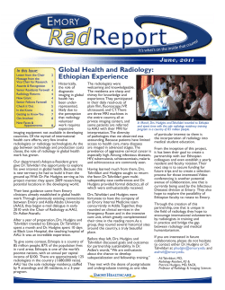
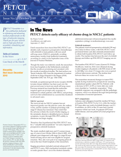


© Copyright 2026