
Evidence of activation of the protein C pathway during acute... damage induced by Mediterranean spotted fever
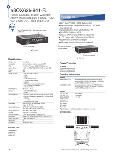
From www.bloodjournal.org by guest on October 28, 2014. For personal use only.
1991 78: 416-422
Evidence of activation of the protein C pathway during acute vascular
damage induced by Mediterranean spotted fever
V Vicente, F Espana, D Tabernero, A Estelles, J Aznar, S Hendl and JH Griffin
Updated information and services can be found at:
http://www.bloodjournal.org/content/78/2/416.full.html
Articles on similar topics can be found in the following Blood collections
Information about reproducing this article in parts or in its entirety may be found online at:
http://www.bloodjournal.org/site/misc/rights.xhtml#repub_requests
Information about ordering reprints may be found online at:
http://www.bloodjournal.org/site/misc/rights.xhtml#reprints
Information about subscriptions and ASH membership may be found online at:
http://www.bloodjournal.org/site/subscriptions/index.xhtml
Blood (print ISSN 0006-4971, online ISSN 1528-0020), is published weekly by the American
Society of Hematology, 2021 L St, NW, Suite 900, Washington DC 20036.
Copyright 2011 by The American Society of Hematology; all rights reserved.
From www.bloodjournal.org by guest on October 28, 2014. For personal use only.
Evidence of Activation of the Protein C Pathway During Acute Vascular Damage
Induced by Mediterranean Spotted Fever
By Vicente Vicente, Francisco Espatia, Dolores Tabernero, Amparo Estelles, Justo Aznar,
Sylvia Hendl, and John H. Griffin
Mediterranean spotted fever (MSF) is a rickettsiosis that
induces widespread microvascular injury. To obtain quantitative information on the in vivo activation and inactivation of
the protein C system during the acute phase of endothelial
damage, several components of the protein C pathway were
studied in 28 MSF patients. Upon admission (day l), patients
showed clear evidence of endothelial damage as reflected by
the significant decrease in the ratio VIII:C/vWF:Ag (0.36f
0.14,mean SD) compared with normals (0.98f 0.14),and
clinical and laboratory signs of hemostatic alterations such
as decreased platelet count, positive fibrinogedfibrin degradation products, and increased thr0mbin:antithrombin-Ill
complex levels. Antigenic protein C (72% -C 18%) and protein
C inhibitor (PCI) (41% % 20%) were significantly decreased
(P < ,001).Complexes of activated protein C (APC) with PCI
or with %-antitrypsin (a,AT) and of plasma kallikrein with PCI
(KK:PCI) were measured using sandwich enzyme-linked immunosorbent assays. APC:qAT complex levels were increased in patients at day 1 (27 k 13 ng/mL) compared with
controls (72 2 ng/mL), and APC:PCI and KK:PCI complexes,
which were not detectable in any of the controls, were
present in 57% and 75% of the 28 MSF patients, with mean
levels of 1 1 k 5 and 46 ? 16 ng/mL, respectively. After
remission of the disease (day 30). a trend toward normal
values in the majority of the parameters studied was found.
This study shows that, in the course of endothelial injury,
MSF patients experience a generalized activation of the
protein C pathway, resulting in consumption of protein C and
PCI, and in the appearance of APC:inhibitor complexes.
Moreover, these data provide the evidence that KK:PCI
circulating complexes occur in vivo.
o 1991by The American Society of Hematology.
M
which these cells participate is the protein C anticoagulant
pathway. The cell surface possesses a thrombin-binding
protein, thrombomodulin (TM). Thrombin bound to TM
initiates protein C activation on endothelial surfaces.6
Activated protein C (APC) then catalyzes the proteolytic
inactivation of factors Va and VIIIa with its cofactor
protein S. APC has also been reported to have profibrinolytic activity, possibly by neutralizing plasminogen activator inhibitors.’.’ In vivo studies have shown that APC is
antithrombotic in a baboon model of platelet-dependent
thrombosis8and in a dog model of venous thrombosis.’
APC is inhibited by a heparin-dependent plasma inhibitor named protein C inhibitor (PCI),’” which has been
purified and chara~terized’.’’.’~
and the sequence of its
cDNA reported.13Plasma contains another major inhibitor
of APC that is heparin-inde~endent,’~
which has been
shown to be a,-antitrypsin (a,AT).I5 We demonstrated that
APC forms complexes in vivo with each of these inhibitors
in several clinical situations where activation of coagulation
has oc~urred,’~.’’
and have suggested that the measurement
of APC complexes may provide sensitive parameters for
specific detection of activation of the clotting and protein C
pathways.
As indicated, any injury of the endothelial cells that
affects the appropriate expression of the protein C system
could potentially lead to a hypercoagulable state and result
in thrombotic complications. The acute phase of MSF
fulfills the criteria of endothelial cell perturbation that may
lead to such a state.
This study was undertaken to obtain qualitative and
quantitative information on the activation and inactivation
of protein C in MSF and its distribution between its known
inhibitors, PCI and a,AT, during the acute and subacute
phases of this vascular infectious disease.
*
EDITERRANEAN spotted fever (MSF) is a rickettsiosis belonging to the spotted fever group of infections and is caused by tick-borne Rickettsia conorii. MSF
shares with other rickettsiosis one dominant feature, widespread microvascular injury.’ Rickettsiae infect human
endothelial cells, proliferate intracellularly by binary fission, and spread contiguously to infect numerous adjacent
endothelial cells.’ Rickettsial vasculitis, in fact, comprises
endothelial injury (swelling and necrosis) and the immune
and phagocytic host responses (lymphocytes and macrop h a g e ~ ) .The
~ pathophysiologic effects of vascular injury
include increased vascular permeability, edema, hypovolemia, and activation of humoral inflammatory and coagulation mechanisms.4 As discussed previously: endothelial
damage could be the principal mechanism responsible for
triggering the hypercoagulation state that occurs in the
acute phase of MSF.
Intact vascular endothelium provides several anticoagulant mechanisms for the maintenance of blood fluidity and
the prevention of thrombosis. One of the several systems in
From the Department of Medicine, School of Medicine, Murcia, and
Research Center and Department of Clinical Pathology, Hospital “La
Fe, ” Valencia, Spain; and Committee on Vascular Biology and
Department of Molecular and Experimental Medicine, Research
Institute of Scripps Clinic, La Jolla, CA.
Submitted December 24,1990; accepted March 19, 1991.
Supported by research grants from Fondo de Investigaciones Sanitarias de la Seguridad Social (9010586) and from CICYT (No. PA
85-0347), Spain, and from the National Institutes of Health (HL31950).
Address reprint requests to Francisco Esparia, PhD, Hospital “La
Fe, ” Centro de Investigacibn, Av. Campanar, 21, 46009 Valencia,
Spain.
The publication costs of this article were defrayed in part by page
charge payment. This article must therefore be hereby marked
“advertisement” in accordance with 18 U.S.C. section 1734 solely to
indicate this fact.
0 1991 by The American Socieiy of Hematology.
0006-4971191l7802-0OI7$3.00/0
416
MATERIALS AND METHODS
Patient data. Twenty-eight consecutively admitted patients (age
range 5 to 86 years, mean 59) from the 1988-1989 epidemic in
Salamanca (Spain) and 30 normal subjects (age range 18 to 65
years, mean 39) were examined. Diagnosis of MSF was confirmed
Blood, Vol 78, No 2 (July 15). 1991: pp416-422
From www.bloodjournal.org by guest on October 28, 2014. For personal use only.
417
PROTEIN C ACTIVATION IN SPOlTED FEVER
by clinical findings and by serologically measuring titters of
antibody specific for this illness using indirect immunofluorescence? Blood samples were obtained on admission, ie, during the
acute phase of the disease (day 1) and 30 days after the start of
treatment (day 30). The elapsed time from the onset of symptoms
(fever) to diagnosis ranged between 3 and 12 days (mean, 7 days).
In the acute phase of the disease, two patients presented clinical
signs and evident symptoms of deep vein thrombosis in their legs.
The diagnosis was confirmed by ascending venography. However,
all patients recovered favorably and did not exhibit any other
clinical complications.
Informed consent was obtained from all patients and control
subjects before blood collection.
Blood samples. Blood collection was made by venipuncture
with a 19-gauge needle. Nine parts of blood were added to plastic
tubes containing one part of 0.13 mol/L trisodium citrate with or
without 50 mmol/L benzamidine. Blood samples were rapidly
centrifuged at 2,50Cg for 20 minutes. The plasma was used fresh or
snap-frozen in small portions and stored at -70°C until the assays
were performed in series (less than 6 months). Pooled normal
human plasma was made from 24 healthy donors and used as a
reference.
Blood samples for determination of protein C antigen, total and
free protein S, PCI antigen, antithrombin I11 (AT-HI), a,AT,
C4b-binding protein, and enzyme:inhibitor complexes were collected in citrate containing 50 mmol/L benzamidine. Protein S
functional activity and protein C amidolytic activity were determined in samples collected with citrate alone.
Quantitative assay of the protein C system components. Protein C
antigen and amidolytic activity,’*anticoagulant protein S,I9 and PCI
antigen and activitp were assayed as indicated before. Total and
free protein S antigen were assayed by enzyme-linked immunosorbent assays (ELlSAs), as indicated by Deutz-Terlouw et a1,” using
Complexes of APC
specific anti-protein S polyclonal antib~dies.’~.’’
with PCI (APCPCI)20or with a,AT (APC:a,AT),” and complexes
of plasma kallikrein (KK) with PCI (KK:PCI)’ were determined by
using sandwich ELISAs, as reported. Briefly, microplates were
coated with monoclonal antibody (MoAb) to protein C (for
APC:PCI and APCa,AT complexes) or to PCI (for KK:PCl
complexes), and complexes were detected with peroxidase-labeled
polyclonal antibodies to either PCI or a,AT or KK, respectively.
Reference curves were constructed with known amounts of in vitro
preformed complexes. The detection limit of the assays in plasma
was 1,3, and 15 ng/mL, respectively.
Determination of other blood coagulation components. Platelets
were counted with an electronic hematology analyzer (Hemalog-8;
Technicon, Tarritown, NY). Factor VI11 procoagulant activity
(V1II:C) was assayed in a one-stage test using platelet-poor plasma
(PPP) from a patient with severe hemophilia A as substrate, as
previously reported? Quantitative immunologic determinations of
von Willebrand factor antigen (vWF:Ag), AT-111, a,AT, and C4bbinding protein were performed by a electroimmunoassay techniquex using rabbit antisera to these proteins (Behringwerke,
Marburg, Germany). Factors 11, VII, IX, X, XI, and XI1 were
assayed in one-stage tests using PPP obtained from patients
congenitally deficient in the respective factors as substrate (Behringwerke), as previously reported? Fibrinogen was assayed by the
Clauss methodz5and fibrin degradation products (FDP) by agglutination of particles coated with specific antibodies (Diagnostica
Stago, Asnieres, France). Complexes of thrombin with AT-I11
(T:AT-111) were assayed by a sandwich ELISA (Enzygnost-TAT,
Behringwerke AG) that uses a polyclonal antibody specific for
neoantigenic determinants on thrombin as the capture antibody
and peroxidase-labeled polyclonal antibody to AT-I11 as the tag.
Statistical analysis. A normal (Gaussian) distribution of levels
of each parameter in each group was checked using the KolmogorovSmirnov test. Variations in parameter levels in patient groups
compared with levels in the normal control group were investigated
using the Student’s t-test with two-tailedPfor significance. Comparisons between patients upon admission (day 1) and 30 days later
were calculated with the Student’s t-paired test. All assay variables
are represented by their mean and standard deviation.
RESULTS
Coagulationparameters. Table 1 shows the mean values
of several blood coagulation parameters studied in 30
healthy subjects and in 28 MSF patients. Compared with
the control group, MSF patients in the acute phase of
disease (day 1) showed a significant decrease in platelet
count and VIII:C/vWF:Ag ratio (P < .001) and a significant increase in vWF:Ag and fibrinogen (P < .OOl). At the
time of diagnosis (day l), nine patients were detected as
FDP positive ( > 10 pglmL). After 30 days of treatment (day
30), a significant increase in platelet number was observed
when compared with the values at day 1 (P < .OOl).
VIII:C/vWF:Ag ratio increased significantly (P < .001) at
day 30 when compared with that at day 1, but it remained
decreased compared with normal values (P < .Ol). vWFAg
decreased during the course of the disease, but it remained
increased compared with normal values (P < .001). Coagulation factors 11,VII, IX, X, XI, XII, and prekallikrein were
in the normal range both before and after treatment.
Table 2 shows the mean values of several components of
the protein C pathway and other related parameters studied in normal subjects and in MSF patients. Compared with
the control group, MSF patients in the acute phase of
disease (day 1) showed a significant decrease in protein C
amidolytic activity and antigen, anticoagulant protein S
activity, and PCI antigen (P < .001). In contrast, free
protein S antigen and AT-I11 levels at day 1 were normal.
At day 1, a significant increase in total protein S antigen,
Table 1. Coagulation Parameters for 28 Patients With MSF During
the Acute Phase of the Disease (day 1) and After Remission (day 30)
and 30 Healthy Subjects
Patients (n = 28)
Day 1
Day 30
Platelet count (109/L)
vWF:Ag (%)
VIII:C/vWF:Ag ratio
Fibrinogen g/L
FDP positive ( > 10 pg/mL)Z
Factor II (YO)
Factor VI1 (%)
Factor IX (YO)
Factor X 1%)
Factor XI (%)
Factor XI1 (%)
Prekallikrein 1%)
Healthy
Subjects
(n = 80)
264 f 70t
189 2 82’
301 f 61
550 f 310* 239 2 131t 106 f 21
0.36 f 0.14’ 0.68 -c 0.25t 0.98 r 0.14
462 ? 161* 401 ? 104
278 2 49
9
1
0
106 -c 16
108 f 16
102 f 20
9 8 f 17
100 f 16
101 f 16
109 ? 22
102 f 17
98 f 20
101 2 16
9 6 2 19
9 9 2 19
108 f 19
9 9 2 15
103 L 16
96 f 20
103 18
100 -c 20
98 ? 18
95 21
101 ? 16
*
*
Data for all parameters except FDP positive are expressed a s mean f
SD.
* P < ,001 with respect to healthy subjects.
t P < ,001 with respect to patients at day 1.
*Number of patients with FDP positive.
From www.bloodjournal.org by guest on October 28, 2014. For personal use only.
VICENTE ET AL
418
Table 2. Protein C Pathway Components and Other Related
Parameters in 28 Patients With MSF During the Acute Phase of the
Disease (day 1) and After Remission (day 30) and 30 Healthy
Normal Subjects
Patients In = 28)
Day 1
Day 30
Protein C
antigen (%)
amidolytic (%)
Protein C inhibitor
antigen (%)
activity (%)
a,AT (Yo)
Protein S
total (%I
free (YO)
anticoagulant (%)
C4b-binding protein (%)
AT-Ill (Yo)
T:AT-Ill complex (ngimL)
Healthy
Subjects
(n = 30)
68 f 19'
60 f 18*
102 * 19t
108 f 20t
99 f 20
103 k 19
41 20*
37 f 20'
159 f 38*
88 f 19t
85 f 19t
107 * 23t
99 f 21
97 f 19
102 2 16
119 f 19*
97 f 31
72 2 23*
135 31*
96 f 24
94 2 21
99 f 30
87 2 23t
116 f 23$
103 f 18
4 f 3t
9 5 2 12
942 19
91 f 21
102 f 18
101 2 12
221
*
1 1 f?*
lution of the disease and, on day 30, only eight patients had
detectable APC:PCI complexes, ranging from 3 to 8 ng/mL.
All healthy subjects had detectable levels of APC:a,AT
complexes, ranging from 3 to 12 ng/mL, with a mean value
of 7 ng/mL (Fig 1A). As seen in Fig lA, MSF patients at day
1 had significantly increased levels of APC:a,AT complex
(mean 27 2 13 ng/mL, range 8 to 53 ng/mL) as compared
with controls (P < .001). APC:a,AT complex levels were
significantly decreased on day 30 (mean 14 5 7 ng/mL,
range 3 to 37 ng/mL) compared with day 1 (P < .001), but
remained significantly increased (P < .001) compared with
normal controls (Fig 1A).
PCI is a good inhibitor of KK in in vitro s t ~ d i e s . ' ~ ~ * ~
Because this enzyme may be formed in the course of
infection and vascular damage?'.'' we investigated the
presence of KK:PCI complexes in MSF patients. Detectable KK:PCI complex levels ( < 15 ng/mL) were not found
t
Data for all parameters are expressed as mean f SD.
*P < ,001with respect to healthy subjects.
tP < ,001with respect to patients at day 1.
SP < .01 with respect to patients at day 1.
C4b-binding protein, and a,AT (P < .001) was observed in
MSF patients compared with healthy subjects. After 30
days of treatment (day 30), a significant increase in protein
C antigen and activity was observed when compared with
the values at day 1 (P < .001). Although PCI antigen and
activity levels increased significantly (P < .001) at day 30
when compared with those at day 1, these values remained
slightly decreased compared with normal values (P < .05).
a,AT, total protein S, and C4b-binding protein decreased
steadily towards normal values as the disease progressed
(Table 2). AT-I11 and free protein S did not show any
significant change during the evolution of the disease.
Although AT-I11 levels were normal on day 1, a significant increase (P < ,001) in T:AT-I11 complexes could be
detected. However, a trend of TAT-I11 towards normal
values was seen on day 30 (Table 2).
At the time of diagnosis (day 1),two patients had deep
vein thrombosis, but there were no relevant laboratory
differences compared with the other 26 patients (data not
shown).
Quantification of protein C activation and inactivation.
Because of the remarkable decrease in protein C activity
and its specific inhibitor, PCI, which suggests activation of
the protein C system, we determined the levels of the
complexes of APC with two physiologic inhibitors, PCI and
a,AT, as well as of KK:PCI complexes. Figure 1 shows the
results obtained. All healthy subjects had levels of APC:PCI
complex below the detection limit of the assay used ( < 3
ng/mL) (Fig 1B). On the other hand, in the acute phase of
the disease at day 1, 16 of the 28 patients studied had
detectable APC:PCI complex levels, ranging from 4 to 32
ng/mL, and a mean value of 11 ng/mL (Fig 1B). These
complexes decreased significantly (P < .001) with the evo-
00
0
I
00
1
0
20
0
B
0
21
00
8
000
00
0
0
1
0
60
00
t
MI t
L
00
20
C
0
00
00
1
0'
CONTROLS
00
00
0
w
0
0
to
,DAY 1
DAV 30
PATIENTS
,
I
Fig 1. Levels of (A) APC:a,AT, (B) APC:PCI, and (C) KK:PCI complexes in normal healthy controls and MSF patients on admission (day
1) and after remission of the disease (day 30). The continuous line
indicates the detection limit of the complex assay.
From www.bloodjournal.org by guest on October 28, 2014. For personal use only.
PROTEIN C ACTIVATION IN SPOTTED FEVER
in the healthy normal subjects, whereas 21 of the 28 MSF
patients had detectable levels of KK:PCI (mean 46 2 16
ng/mL, range 21 to 86 ng/mL) (Fig 1C) during the acute
phase of the disease (day 1). After remission of the disease
(day 30), only five patients had detectable KK:PCI complex
levels (range 24 to 33 ng/mL) (Fig 1C).
Correlation between APC:inhibitor complexes and F A T-111
complexes. Figure 2 shows the correlation between APC:
a,AT and T:AT-I11 complex levels (A), APC:PCI and
T:AT-I11complex levels (B), and APC:a,ATand APC:PCI
complex levels (C) in MSF patients during the acute phase
of the disease at day 1. There was a positive correlation
(r = 504, P < .005) between APC:a,AT complex levels
and TAT-I11 complex levels (Fig 2A). However, no significant correlation was observed between T:AT-I11and APC:
PCI complex levels (r = .138,P > S ) (Fig 2B) nor between
APC:PCI levels and APC:a,AT levels (r = .309, P > . l )
(Fig 2C).
Correlation between KK:PCI complexes and APCinhibitor
and EAT-111 complexes. Figure 3 shows the correlation
between the KK:PCI complex levels and APC:a,AT (A),
APCPCI (B), or T:AT-I11 complex levels in acute MSF
patients (n = 28) at day 1. There was a positive correlation
(r = .438, P < .05) between KK:PCI complex levels and
APC:a,ATcomplex levels and between KK:PCI and APC:
PCI complex levels ( r = ,459, P < .05) but not between
KEPCI and T:AT-I11complex levels ( r = .314, P > .1).
Normalization ofAPC:@T complex levels. Because a,AT
is an acute phase reactant and is elevated in MSF patients
(Table 2), we “normalized” the values for APC:a,AT by
dividing the observed level of APC:a,AT by the observed
level of a,AT, and then calculated the correlation of the
other complexes measured versus the “normalized” APC:
a,AT levels. Again, a positive correlation was found between “normalized” APC:a,AT levels and T:AT-I11
(r = 3 1 , P < .01) and between “normalized” APC:a,AT
and KK:PCI levels (r = S10, P < .Ol), and no significant
correlation was found between “normalized” APC:a,AT
and APC:PCI levels (r = .281,P > .1). Although there was
a significant increase in the a,AT levels in MSF patients at
admission (Table 2), no correlation was found between the
levels of APC:a,ATand those of a,AT (data not shown).
419
60
4
E
50
\
m
E
3tl
4
B0
40
30
0
20
10
0
4
E
\
m
C
------- 7
.em...).
0
10
.
20
30
30
r:0.309
”
20
w
x
W
4
8
0
10
k
PU
Fig 2. (A) Correlationbetween the levels of APC:a,ATcomplex and
T:AT-Ill complex in MSF patients at day 1. (B) Correlationbetween the
levels of APC:PCI complex and T:AT-Ill complex in MSF patients at
day 1. (C) Correlation between the levels of APC:PCI complex and
APC:a,AT complex in MSF patients at day 1. For calculation of the
coefficients of correlation, APC:PCI complex levels below the detection limit [ < 3 ng/mL) were consideredto be 1 ng/mL.
0
1:ATIII COMPLEXES, ng/mL
DISCUSSION
In the acute phase of MSF, widespread inflammatory
microvasculitis is a dominant feature.’ A pronounced decrease in the VIII:C/vWF:Ag ratio secondary to endothelial
damage and laboratory and clinical indications of activation
of the hemostatic system, such as a significantly reduced
numbers of circulating platelets and decreased protein C
..
.. .
.
. .
.
.
; pa0.1
.
. .. .
/
.
.
APCa,AT COMPLEXES, ng/mL
C
From www.bloodjournal.org by guest on October 28, 2014. For personal use only.
420
VICENTE ET AL
'= 0.438
3~0.05
..
A
.
0
.
.. . . .
0
0
B
r=0.459
P c 0.05
.
0
0
0
.
0..
0
.
rz0.314 ; PaO.1
0
.. . .
0
8
0
0
0
C
e
20
40
.
60
80
100
KK:PCI COYPLEXES, nOlmL
Fig 3. (A) Correlation between the levels of KK:PCI complex and
APC:a,AT complex in MSF patients at day 1. (6) Correlation between
the levels of KK:PCI and APC:PCI complexes in acute MSF patients.
(C) Correlation between the levels of KK:PCI and T:AT-Ill complexes
in MSF patients at day 1. For calculation of the coefficients of
correlation, APC:PCI complex levels below the detection limit ( < 3
ng/mL) were considered to be 1 ng/mL and KK:PCI complex levels
below the detection limit ( < 1 5 ng/mL) were considered to be 10
ng/mL.
levels, were found in this study, and FDP was detected in
some patients as well as clinical evidence of deep vein
thrombosis in two of them. On the other hand, and as
previously r e p ~ r t e dcoagulation
,~
factors 11, VU, IX, X, XI,
and XI1 did not show significant variations, and fibrinogen
levels were strikingly elevated, reflecting the behavior of
fibrinogen as an acute-phase reactant (Table 1). This
finding suggests that the abnormalities in the hemostatic
system seen in MSF patients are due to local endothelial
alterations and are not secondary to a disseminated intravascular coagulation (DIC). In fact, after rickettsial infection
of endothelial cells, fourfold increased platelet attachment
to the vessel wallzyand signs of platelet activation3' have
been demonstrated.
In the presence of an endothelial cell surface protein,
TM, the rate of protein C activation increases approximately 20,OOO-f0ld.~'Thus, the small amounts of thrombin
generated by the platelet activation that occur in the acute
phase of rickettsial infection might accumulate on the
endothelial cell surface, and the resulting thrombin-TM
complex formed could trigger the activation of the anticoagulant protein C system. In fact, during the acute phase of
MSF, activation of protein C system is directly demonstrated by the reduced protein C and PCI levels as well as by
the appearance or increase of circulating APC:PCI and
APC:a,AT complexes.
During the acute phase of the disease all patients had
detectable levels of APC:a,AT whereas only 16 of the 28
patients had moderate levels of APC:PCI complex. Moreover, there was a positive correlation between the APC:
a,AT complex levels and the T:AT-I11 complex levels
(r = ,604,P < .005) (Fig 2A), which suggests that protein C
activation and complexation increase parallel to thrombin
generation. However, there was no significant correlation
between APC:PCI and T:AT-111 levels (Fig 2B) nor between the levels of APC:a,AT and the levels of APC:PCI
(Fig 2C).
The relative proportion of APC:PCI and APC:a,AT
complex levels seen here in MSF patients coincides with
that found in patients with thrombotic disease,'"'' but
contrasts with that detected in vitro following addition of
purified APC to citrated normal
In that in vitro
study we observed that the two types of complexes are
formed at approximately the same rate. The APC complex
levels seen here in MSF patients also contrast with in vivo
studies in a baboon thrombotic model in which infusion of
APC into baboons generated APC:PCI complex levels
twice as high as the APC:a,AT levels immediately following
the APC i n f ~ s i o n . ~A' .plausible
~~
explanation for the higher
APC:a,AT complex/APC:PCI complex ratio seen in MSF
patients as well as in patients with thrombotic disease is
that the APC:PCI complexes are cleared faster than the
A P C q A T complexes. Indeed, we recently found that
during APC infusions into baboons, APC:PCI complexes
were cleared about three times more rapidly than APC:
a,AT complexes, with a t1,*of about 40 minutes versus 140
minutes, respectively." Another possibility is that enzymes
generated during inflammatory vascular damage, such as
thrombin and KK, could complex to and/or cleave
pCI,7 ll.lZ.23.26 thus reducing PCI levels, thereby making a,AT
the major inhibitor of APC. In fact, in this study we found
that 21 of the 28 MSF patients in the acute phase of the
disease had detectable circulating levels of KK:PCI complex, whereas this complex was not detected in any of the
From www.bloodjournal.org by guest on October 28, 2014. For personal use only.
PROTEIN C ACTIVATION IN SPOTTED FEVER
42 1
healthy normal subjects studied. These data provide the
first evidence for in vivo occurrence of KKPCI complexes.
This evidence suggests that during the acute phase of
vascular damage, or during any other situation in which KK
is generated, this enzyme could also modulate the in vivo
activity of PCI. Plasma prekallikrein activation in rickettsiosis has been demonstrated.28xMR a o e t alMstudied volunteers
in whom Rocky Mountain spotted fever developed after
challenge with Rickettsia rickettsii, and observed an increase
in plasma C1-inhibitor:KK complexes from 40 ng/mL before challenge to about 300 ng/mL 24 hours after the onset
of the illness.
Twenty-seven of the 28 patients studied showed, a t
admission, APC:a,AT complex levels higher than TAT-I11
complex levels, with ratios of AE’C.a,AT levels t o that of
T:AT-I11 ranging from 1.1 to 7.0. TAT-I11 (tin = 10 to 15
minutes)” is probably cleared from the circulation more
rapidly than APC:a,AT complex, and this difference in
half-life would explain the lower T:AT-I11levels detected in
MSF patients.
Several observations suggest that APC may act beneficially a t an early step in the chain of events involved in
inflammation-induced c~agulopathy.~’
It has been shown
that APC prevents the coagulopathy as well as the fatal
outcome associated with Escherichia coli-induced shock in a
baboon model of ~ e p t i c e m i aand
’ ~ that APC inhibits plateletdependent thrombosis in a baboon arterial model.’ A s
indicated here, vasculitis seems to b e an important trigger
for activation of t h e anticoagulant protein C system. Initially, t h e activation of protein C could be considered a
defense mechanism preventing local thrombosis, and this
could be the reason for the absence of occlusive thrombosis
a t the level of the inflammatory vascular injury in fulminant
rickettsiosis.)6 One could speculate that when the vascular
injury is more severe, it may lead to inadequate production
of APC that then cannot prevent t h e severe hemostatic
alterations seen in the severe forms of MSF.
In conclusion,our study performed in 28 MSF patients in
the acute phase of the disease where a widespread microvascular injury (vasculitis) is a dominant feature has demonstrated the activation of the protein C anticoagulant system,
suggesting that this pathway is intimately involved in vivo in
the interaction of the coagulation and inflammatory system.
These results also suggest t h e interest in screening the
protein C system in clinical situations where the inflammatory process in a variety of vasculitides is involved.
REFERENCES
1. Walker DH: Pathology and pathogenesis of the vasculotropic
Rickettsiosis, in Walker DH (ed): Biology of Rickettsial Diseases
(vol 1). Boca Raton, FL, CRC, 1988, p 115
2. Wisseman C: Selected observations on rickettsiae and their
host cells, in Kazar J (ed): Rickettsiae and Rickettsial Diseases.
Bratislava, Czechoslavakia,Publishing House of the Slovak Academy of Sciences, 1985, p 167
3. Montenegro MR, Mansueto S, Hegarty BC, Walker DH: The
histology of “taches noires” of boutonneuse fever and demonstration of Rickettsia conorii in them by immunofluorescence. Virchows Arch [A] 400:309,1983
4. Ruiz R, Martin AM, Herrero JI, Muiioz VM, Mateos, A, Sanz
F, Becares M, Querol R, Portugal J: Fiebre botonosa mediterranea. Andisis clinic0 y diagnbstico de 50 enfermos. Enf Infecc
(Barcelona) 1:16, 1983
5. Vicente V, Alberca I, Ruiz R, Herrero I, Gonzalez R,
Portugal J: Coagulation abnormalities in patients with Mediterranean spotted fever. J Infect Dis 153:128,1986
6. Esmon CT: The roles of protein C and thrombomodulin in
the regulation of blood coagulation.J Biol Chem 2644743,1989
7. Espaiia F, Berrettini M, Griffin JH: Purification and characterization of plasma protein C inhibitor. Thromb Res 55:369, 1989
8. Gruber A, Griffin JH, Harker LA, Hanson SR: Inhibition of
platelet-dependent thrombus formation by human activated protein C in a primate model. Blood 73:639,1989
9. Emerick SC, Maruyama H, Yan SB, Long GL, Harms CS,
Marks CA, Mattler LE, Huss C, Comp PC, Esmon NL, Esmon CT,
Bang NU: Preclinical pharmacology of activated protein C, in
Holcenberg JC, Winkenhale J (eds): The Pharmacology and
Toxicology of Proteins. New York, NY, Liss, 1987, p 351
10. Marlar RA,Griffin JH: Deficiency of protein C inhibitor in
combined factor VNIII deficiency disease. J Clin Invest 66:1186,
1980
11. Suzuki K, Nishioka J, Hashimoto S: Protein C inhibitor:
Purification from human plasma and characterization.J Biol Chem
258:163,1983
12. Suzuki K, Nishioka J, Kusumoto H, Hashimoto S: Mechanism of inhibition of activated protein C by protein C inhibitor. J
Biochem 95:187,1984
13. Suzuki K, Deyashiki Y, Nishioka J, Kurachi K, Akiras M,
Yamamoto S, Hashimoto S: Characterization of a cDNA for
human protein C inhibitor. J Biol Chem 262:611,1987
14. Heeb MJ, Espaiia F, Griffin JH: Inhibition and complexation of activated protein C by two major inhibitors in plasma.
Blood 73:446,1989
15. Heeb MJ, Griffin JH: Physiologic inhibition of human
activated protein C by alpha 1-antitrypsin.J Biol Chem 263:11613,
1988
16. Tabernero D, Espaiia F, Vicente V, Estell6s A, Gilabert J,
Aznar J: Protein C inhibitor and other componentsof the protein C
pathway in patients with acute deep vein thrombosis during
heparin treatment. Thromb Haemost 63:380,1990
17. Espaiia F, Vicente V, Tabernero D, Scharrer I, Griffin JH:
Determination of plasma protein C inhibitor and of two activated
protein C-inhibitor complexes in normals and in patients with
intravascular coagulation and thrombotic disease. Thromb Res
59:593, 1990
18. Espaiia F, Estell6s A, Aznar J, Gilabert J: Assay of protein C
in human plasma: Comparison of amidolytic, coagulation and
immunochemicalassays. Thromb Res 44:771,1986
19. Fernandez JA, Estell6s A, Gilabert J, Espaiia F, Aznar J:
Functional and immunologic protein S in normal pregnant women
and in full term newborns. Thromb Haemost 61:474,1989
20. Espaiia F, Griffin JH: Determination of functional and
antigen protein C inhibitor and its complexes with activated
protein C in plasma by ELISA’s. Thromb Res 55:671,1989
21. Deutz-TerlouwPP, Ballering L, van Wijngaarden A, Bertha
RM: Two ELISA’s for measurement of protein S, and their use in
the laboratory diagnosis of protein S deficiency. Clin Chim Acta
186:321,1989
22. Gilabert J, Fernandez JA, Espaiia F, Aznar J, Estellts A:
Physiological coagulation inhibitors (protein S , protein C and
From www.bloodjournal.org by guest on October 28, 2014. For personal use only.
422
antithrombin 111) in severe preeclamptic states and in users of oral
contraceptives. Thromb Res 49:319,1988
23. Espafia F, Estellts A, Griffin JH, Aznar J: Interaction of
plasma kallikrein with protein C inhibitor in purified mixtures and
in plasma. Thromb Haemost 65:46,1991
24. Laurel1 CB: Quantitative estimation of proteins by electrophoresis in agarose gel containing antibodies. Anal Biochem 15:45,
1966
25. Clauss A: Gerinnungsphysiologischeschenellmethode zur
Bestimmung des fibrinogens. Acta Haematol (Basel) 17:237, 1957
26. Espaiia F, Griffin JH: Plasma protein C inhibitor (PCI)
inhibits procoagulant and profibrinolytic enzymes. Blood 70:401,
1987 (abstr)
27. Colman RW, Edelman R, Scott CF, Gilman RH: Plasma
kallikrein activation and inhibition during typhoid fever. J Clin
Invest 61:287, 1978
28. Yamada T, Harber P, Pettit GW, Wing DA, Oster CN:
Activation of the kallikrein-kinin system in Rocky Mountain
Spotted Fever. Ann Intern Med 88:764,1978
29. Silverman DJ: Adherence of platelets to human endothelial
cells infected by Rickettsia rickettsii. J Infect Dis 153:694,1986
30. Rao AK, Schapira M, Clements ML, Niewiarowski S,
Budzynski AZ, Schmaier AH, Harpel PC, Blackwelder WC,
VICENTE ET AL
Scherrer JR, Sobel E, Colman RW: A prospective study of
platelets and plasma proteolytic systems during the early stages of
Rocky Mountain Spotted Fever. N Engl J Med 318:1021, 1988
31. Esmon NL, Esmon CT: Protein C and the endothelium.
Semin Thromb Haemost 14:210,1988
32. Espana F, Gruber A, Kelly AB, Hanson SR, Harker LA,
Griffin JH: Complexation of activated protein C (APC) with two
physiologic plasma inhibitors. Circulation 78:318,1988(abstr)
33. Espafia F, Gruber A, Heeb MJ, Hanson SR, Harker LA,
Griffin JH: In vivo and in vitro complexes of activated protein C
with two inhibitors in baboon. Blood 77:1754,1991
34. J@rgensenLN, Lind B, Hauch 0, Leffers A, Abrecht-Beste
E, Konradsen LAG: Thrombin-antithrombin 111-complex and
fibrin degradation products in plasma: Surgery and postoperative
deep venous thrombosis. Thromb Res 59:69,1990
35. Taylor FB, Chang A, Esmon CT, D’Angelo A, ViganoD’Angelo S, Blick KE: Protein C prevents the coagulopathic and
lethal effects of Escherichia coli infusion in the baboon. J Clin
Invest 79:918,1987
36. Walker DH, Hawkin HL, Hudson P: Fulminant Rocky
Mountain fever. Its pathologic characteristics associated with
glucose-6-phosphate dehydrogenase deficiency. Arch Pathol Lab
Med 107:121,1983


© Copyright 2026