
ITRAX Core Scanner: Multi-Function Scanner for Rock & Sediment Cores
This file is meant for on-screen viewing only
If you would like to have printed copies, please contact us
We support our customers worldwide, also in Chinese
A firsthand choice for detailed rock and sediment core investigations
Offers XRF, radiographic and optical scanning of variable resolution
Whole and slabbed cores can be scanned in short time
Sample information is obtained non-destructively
Straightforward operation and reliable analyses
I TRAX
CORESCANNER
UNIQUE MULTI-FUNCTION SCANNER
FOR CORE EXAMINATIONS
Some Itrax Corescanner features:
•
•
•
•
•
•
•
XRF scanner for sediment cores, rock cores and other flat samples
Offers XRF multi-element analysis of Al to U including Rare Earths
The XRF can analyze down to the PPM level for most metals
Digital radiography for improved sample interpretation
Scanning, digital RGB line camera for sample imaging
Can analyze one meter of core in minutes
Accepts flat surface samples as well as those with varying height
Sediment core scanning offers many paleoclimate proxies
• Biogenic silica
• Provenance studies
• Oxic/anoxic conditions
• Reconstruction of past conductivity from
element ratios in carbonate sediments
• Grain size related fractionation effects
• Tefra layers and detrital clay
• Metal pollution signatures
• Estimation of past weathering, leaching and
erosion intensities and primary productivity
• Shallow water aragonite formation
• Sub-mm scale analysis and counting of fine
laminations.
• Sediment grading
• Paleo-redox conditions
• Seasonal changes and temperature
estimates
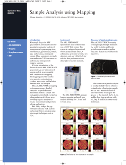
Camera technology that sees more
High
quality
sample
images are essential for
a core scanner, and the
Itrax offers superb digital
RGB images with down
to 50 microns pixels. Not
only are the images clear
and detailed, but they
can also reveal much
more than the eye. With
digital image processing,
also minute changes
become clear, offering
the user a new dimension for looking into the
samples. This new technique combines well with
the radiography and XRF
to disclose the sample
secrets. These images
show an example of how
a sediment image (left)
can be enhanced (right)
to reveal increased detail
that can help in an understanding of the sedimentary processes.
and more should come
X-ray radiography adds confidence
Radiographic images can help in verifying
whether an XRF peak is to be interpreted as
a e.g. a grain or a layer. The x-ray images
show distribution of chemical structures that
can be invisible by eye and optical cameras.
The integration of radiography and XRF into
a single instrument guarantees that radiographic data match the positioning of XRF.
This matching is often critical, for instance to
reveal migration of elements in relation to
sedimentary layers.
An example of
combined XRF and radiographic data is
shown in image 1. to the right (Sr/Ca ratio
overlaid on a magnified radiographic image
of a lake sediment).
Slit-free XRF offers advantage
ITRAX offers downcore resolution ranging from
centimetre scale and down to 100 microns,
using a system setup that is unique. With this,
the detection limits, speed of analysis and
repeatability are of cutting edge class and are
maintained regardless of the selected resolution,
unlike the standard XRF slit solution.
Image 1.
e 1.
Some Itrax Corescanner features
continued :
• Produces data with high accuracy and reproducibility
• Offers best available analytical sensitivity and precision per time unit
• Scans with any step size from centimeter down to 0.1 millimeter
• Shows the same superior detection limits at any step size
• Analyzes non-destructively and without sample contact
• Optical image enhancement for improved sample information
• Reliable construction and quick service minimizes instrument downtime
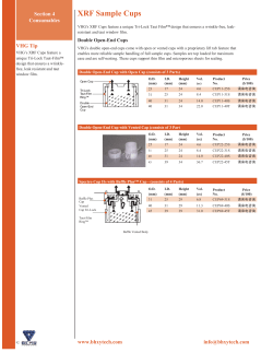
Rock core analysis
The high capacity and precision of Itrax
Ni
Zr
allows for multi element scanning of large
Pb
volumes of rock cores to provide sample
average data, as used in e.g. research and
metals and oil prospecting and production.
This 27 cm long core section was analyzed using 0.2 mm
The technique can also show element distristep and 30 s counting time per point.
bution down to sub-millimeter resolution.
This is exemplified by results obtained on this paleoproterozoic laminated carbonaceous shale, rich
in pyrite. Please note the Zirconium variations in individual beds tracking the grain-size changes, and
enrichments of Nickel and Lead in the thick pyrite layer.
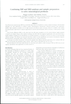
Unsurpassed speed combined with superb reproducibility
The Itrax can analyze as fast as 1 second per
point at any down core resolution, providing
reasonable precison. At 10 seconds per point
you can reach high precision. Please note
that this diagram shows two scans per
element, so a total of 8 graphs are plotted!
The high speed and high data quality make
Itrax a really high capacity instrument.
Two scans per element !
Reproducibility values for 15 sec. analysis:
Al shows a typical reproducibility of <4% (1 S.D.) at 10% Al.
Si shows a typical reproducibility of <2% (1 S.D.) at 10% Si.
Ca shows a typical reproducibility of <1% (1 S.D.) at 1% Ca.
Ba shows a typical reproducibility of <7% (1 S.D.) at 0.01% Ba.
Table 1. Typical reproducibility (Cr tube, 15 seconds
anaysis).
{
{
Ca run1
Ca run 2
Si run 1
Si run 2
{
{
Al run 1
Al run 2
Ba run 1
Ba run 2
Figure 1. This diagram shows Ca, Si, Al, and Ba profiles from two subsequent sediment scans. Each element is
represented by two graphs. Time was 15 seconds per point, clay rich lake sediment (arbitrary scale). Sample
kindly provided by Prof E. Bard, CEREGE, Aix-en-Provence, France.
Core Scanner specifications
Instrument reliability
XRF (X-ray Fluorescence) element analysis for determination of Al and heavier
The Itrax corescanner is a proven workhorse. Most of the
elements. Mg can be analyzed in dry samples. The X-ray beam size is 20x0.2 or
installations are in use several thousand of hours per year, many
20x0.1 millimeter, with 0.2/0.1 mm in the sample length direction. Full cover of
on a 24-7 basis. In spite of this heavy workload, these
larger steps is achieved by scanning along each step of the sample with the beam
while measuring.
The XRF detector offers < 133 eV FWHM resolution for Mn Kα. Normal count-rate
input when analyzing is up to 80.000 counts per sec.
Light element analysis enhancement by (helium-free) vacuum system for best
sensitivity, contact free analysis, and lowest running cost.
Digital X-ray radiography with 16 bit image format and variable lateral resolution
down to 20x20 micrometers pixels. The image width is 20 millimeters. Sample
variations as small as ~1%, or lower. Sample thickness limitations apply, depending on matrix composition.
The X-ray source is a 2.2 kW/60 kV high power X-ray tube with Cr anode as
standard (also Mo and others available). Time to switch tube is about 10 minutes.
Low cost x-ray tube with 3-4.000 hours expected life time.
Optical, digital RGB line camera with 3x2000 pixels high color definition and 50
instruments have very little down-time. This has three reasons;
one being a reliable construction, the others are the effective
service offered by Cox and our Internet support, which is a quick
way to get answers. Our users can confirm this (a complete
customers list is available through Cox).
The Internet support with shared computer desktop view allows
for very fast and efficient problem solving. The service staff at
Cox see what the user sees, whether on the computer screen or
through a web camera. The first contact with someone in our
service team can often be made within minutes, leading to
immediate help when in doubt. Support is given in English as
well as in Chinese (Mandarine).
microns pixel size. Pixel binning for higher resolution. Offers up to 900 true
grayscale steps. Digital contrast enhancement for improved sample detail
visibility.
Add-ons
XRF, radiographic, and optical measurements are non-destructive and are
performed without contact to the sample surface. The sample surface height
profile is measured along the sample, and adjusted for during analysis for best
results. This allows for flat samples as well as samples with somewhat varying
height to be analyzed with good results. Magnetic susceptibility measurement
(optional equipment) is done in contact with the sample surface.
Measurements can be done with, or without, a plastic film covering the sample
surface.
Available optional equipment include but are not limited to:
Magnetic susceptibility sensor, Bartington MS2E type.
On-board ship and mobile installations.
We also offer service contracts, Internet support contracts,
Reproducibility Testing contracts, Service training for
technicians, etc.
Maximum core length is 1.75 meters as standard.
Sample thickness range is 22-56 millimeters for whole cores as standard. For
split sediment cores it is 30-60 millimeters thickness, i.e. 60-120 millimeters full
circle as standard. We can provide a modified Itrax that accepts spilt cores with
diameters from 40 up to 150 mm on request. Slabs and U-channels, wood
Detection limits
samples, and flat slices of other sample types can also be analyzed. An
U-channel holder is available.
Scan time: Down to ~0.5 s/point for radiography, and ~1.5s/point for XRF, which
refers to total time per point including analysis, as well as overhead time for
sample re-positioning, data storage, etc. The overhead time between measurements is ~0.5 sec. for XRF/~0.2 sec for radiography.
Software:
Itrax Navigator software for instrument operation with intuitive
graphical user interface. Sets of analytical parameters can be stored and
re-called for fast set-up of standardized analyses. XRF data are available as
spectra, and as peak areas or element concentrations in table format. Raw
analytical data from each point are stored as spectra for quality assurance
reasons. Data can be exported to e.g. Excel. Calibration for quantification is
based on standards and fundamental parameters. The ReDiCore software for
data display and evaluation greatly simplifies data interpretation, as well as image
and graph production, applying a copy-and-paste function.
Itrax is delivered with a one year warranty including Internet support. The
warranty time can be extended to up to three years.
The Itrax Core Scanner dimensions are ~4500x820x1570 millimeters LxWxH
with a weight of about 1000 kilos. Different voltages and frequencies can be
applied.
The Itrax is a complete plug-and-play delivery with all that you need, including
hardware and software, computers, UPS, training, Internet support, etc.
The instrument fulfils radiation safety requirements with interlock safety switches,
and it is completely safe to work close to the instrument without limitations.
Cox Analytical Systems
Östergårdsgatan 7
S-43153 Mölndal, Sweden
www.coxsys.se
[email protected]
phone +46 31 708 3660
Element
Mg
Al
Si
P
S
Cl
K
Ca
Ti
Mn
Fe
Br
Sr
Ba
Pb
Cr tube (ppm)
11000
1000
250
96
47
27
9
6
3
130
25
12
15
20
24
Mo tube (ppm)
7700
1950
890
245
112
36
24
14
7
7
5
5
43
13
The detection limits of the Itrax corescanner are highly competitive.
A great advantage for high resolution work is that they apply
regardless of selected analytical resolution. The elements that can
be detected range from Mg or Al and all the way up to U (Uranium).
This list contains only a short selection of elements, contact us for
a complete list. The values are all based on a 100 second analysis
and refer to a clay matrix. Detection limits for two different x-ray
tubes are listed, where Cr is the standard tube and the Mo tube
applies when quantification of heavier elements are in focus. All
elements listed are determined simultaneously and in one analysis
for each tube type, without further restrictions, changes or special
settings. The elements that are most commonly detected in an
unpolluted, clay based sediment sample are Al, Si, S, Cl, K, Ca, Ti,
Cr, Mn, Fe, Ni, Cu, Zn, Br, Rb, Sr, Zr, Ba, and Pb.


© Copyright 2026