
Document 438362
IN THIS ISSUE
Comparative Dermatology
Cultivated Mammalian Cells
Editorial Staff
\V.
ALBERT SULLIVA/\, JR.,
M.D.
Editor
EIVlKD HOFF, JR.
Managing Editor
ELLEN
Y.
SIEGELMAN
Copy Editor
Board of Editors
\V. FRAWLEY
'WESLEY W. SPINK, :\I.D.
LEE VVATTE:'\BERG, .\1.D.
GEHARD
Administrative Sponsors
University of Minnesota Hospitals
RAY \1. A~IUEHG, Director
Minnesota Medical Foundation
AmmLD LAZAHOW, \I.D., President
COHRI]'; H. HOD(;SO:\"
\1.D., Vice-President
N. L. GAULT, JH., \I.D., Secretary-Treasurer
EI\IND HOFF, JR., Executire Director
University of Minnesota Medical School
O. :\[EREDITH \VILSO]';, President
University of Minnesota
ROBERT B. HOWARD, :\I.D., Dean
College of :\[eclieal Sciences
N. L. GAULT, JH., \I.D.
H. \lEAD CAVERT, \I.D.
\VILLIAM FLEESO]';, :\I.D.
RICHARD M. \lAGRAW, \I.D.
Assistant Deans
Minnesota Medical Alumni Association
CHAHLES J. BECK, M.D., President
NEIL \1. PALM, \I.D., Vice-President
JA~IES C. \lANKEY, :\I.D.,
Vice-President
ROBEHT H. \lONAHAN, M.D., Secretary
DUAKE C. OLSO:", \1.D., Treasurer
UNIVERSITY OF MINNESOTA
Medical Bulletin
Official Publication of
UNIVERSITY OF MINNESOTA HOSPITALS
MINNESOTA MEDICAL FOUNDATION
MINNESOTA MEDICAL ALUMNI ASSOCIATION
Circulatio1! this
VOLUME XXXIII
;SSIU':
J)('('('IIt1WI'
a,()()()
I.9fJ J
NUMBEH :1
CONTENTS
STAFF MEETING REPORTS
A COlllpamtive Study of
Callille Derllllltology
MILTON
OIlKIN,
ll,//IU/ll
IIlld
M.n.
110
Species-Specificity and "Transformation"
Plte1101IIe1JOn of Cultivllted lIfalllllllllilill Cells
K.
GIo:IIII.\II11 BIIANIl, M.n,
120
MEDICAL SCHOOL NEWS
1:10
MEDICAL ALUMNI REUNION
1.1R
ALUMNI NOTES
1,19
Published monthly from October to June at Minneapolis, Minn. Editorial offices:
1342 Mayo Memorial, University of Minnesota, Minneapolis 14, Minnesota.
Sf'cond Class Postage Paid at Minneanolis. Minnesota.
Staff Meeting Report
A Comparative Study of Human
and Canine DermatologyOf
!\1i1ton Orkin, !\i.D.t
"Bt'tWt't'1l animal and human tlH'uicilH' tht'H" is no dividing line~
nor should there be. The ohjt'ct is dift't'rt'llt, hut thp t'xpt'rit'llct'
ohtaim·d constitutt-'s the hasis of all medicine."
HU"Ol.t' VIR""0W (IH21-1902)
oInimals have often been used for experimental production of disease but seldom for observation of spontaneous disorders. The broad purpose of the present study is to define and
characterize spontaneous skin diseases of the dog and to correlate these with human counterparts, when they exist. No sllch
systematic comparison of skin diseases of man and another species has been made previously. The more specific purposes are:
( 1) to accumulate fundamental knowledge
of canine dermatoses and tumors, both for
the intrinsic value of this information and
for its usefulness in future research; (2) to
compare canine and human dermatoses and
tumors and to experiment on analogous canine lesions so as to permit application of
the information to both species; and (3) to
seek for common patterns of disease behavior despite species differences.!
This delineation of canine diseases is
based upon intensive study of 292 dogs
spontaneously afflicted with cutaneous tu:Milton Orkin
mors or dermatoses or both. The initial
phase of the project consisted of review and study of comparative (dog and man) cutaneolls anatomy, biochemistry, and physi{)logy.~ This information is not included in the present com°Presented at the Staff I\:Ipf'ting of the U nivt'fsity of :rvlil11H-'sota Hospitals Oil
October 20. 1961
tThe study which forms the hasis of this report was undertaken in conjunctioll
with Dr. Robert :M. Schwartzman, formerly Instructor, Collegp of Veterinary
~fedicine, University of ~1innesota; presently Assistant Profpssor. Schoo] of
Veterinary Medicine, University of Pennsylvania.
:j:Clinical Instnlctor. Division of Dt-'rmatology
110
TIlE MEDICAL BULLETIN
munication; emphasis here is placed on interesting clinical and
histologic aspects of comparative dermatology.
A.
CUTANEOUS TUJ\foI\S
Spontancous tumors are more common in dogs than in any
other animal species. Approximately one-third of canine neoplasms originate from skin and subcutaneous tissue,:] visceral
carcinomas being relatively uncommon.
Our initial series consisted of 100 cutaneous tumors collected
from 90 dogs. There was slight preponderance of ectodermal
over mesodermal neoplasms. Iu addition, the incidence of sarcoma approximated that of carcinoma, while in human beings a
higher proportion of carcinoma is noted among skin tumors.
In order to assess the age, breed, and sex distribution among
dogs in the original tumor series, we found it necessary to correlate the observed cases with a general canine population. The
"sample normal population" consisted of the first 1,000 dogs
examined for any condition at the University of Minnesota Veterinary Clinic during 19.56.
Age Distributioll. Age distribution of the affected dogs differed grossly from that of the sample normal population. In this
tumor-bearing population very few animals under one year of
age were affected (human equivalent-1.5 years). The vast majority of maliglul1lt conditions occurred in animals nine years of
age and older (H. E.-.52 years). The marked proclivity toward
malignant disease among older dogs has been widely reported.
Breed Distribution. There was no tendencv for cutaneous
neoplasia in general to be associated with any' major breed of
dog in the sample normal population. There is, however, an
apparent tendency for breeds to be afHicted by specific types of
tumors and dermatoses, and tlll'se will 1)(' mentioned under
specific entities.
Sex Distrihutioll. The incidcm'(' of cutan('ous tumors was indepeudent of the sex of thc canines of the sample normal population.
SlJecific Entities
1) Transmissible Reticulum-Cell Tumor. This uniqu(', and
fairly common neoplasm (17 per cent of original series) is characterized bv the ease with which it can be transmitted to other
dogs, by rl'atural or artificial means. 4 Despite their sometimes
sarcomatous appearance clinically, and its malignant aspect histologically, the vast majority of these tumors spontaneously regress. The eellular appearance and total architecture of the
111
THE MEDICA L BULLETIN
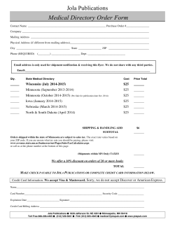
Fi~. 1. Transmissihle Reticulum-Cell Tumor-reticulum l'dls
with several mitotic: figures indistinguishable from Illullan n'ticulllm-cell lymphomH. lIematoxylin and eosin; X 242..'5
hlmor arc microscopically indistinguishable from those of rcticulum-cell lymphoma of man (Fig. 1). Clinical and experimental
data snggest the presence of a specific tumor cell antibodv in
animals recovering from this neoplasm.
.
No specifically comparable tumor exists in man, although
human kerntoacanthoma shares the following facets of host response: (1) Both are essentially benign conditions in which
malignancy is simulated histologically, i.e., reticulum-cell sarcoma simulated in transmissible reticulum-cell tumor, and squamons-cell carcinoma in keratoacanthoma; (2) freljUent spontaneous healing OCCIll'S in both; amI (3) relatlive immunity is
achieved in both conditions after involution of the tumor. .
2) Mastocytoma. This entity accounts for 1.'3 per cent of
cutaneous canine tllmors,5 a predilection for mastocytoma being
evident in Boston terriers and hoxers. It is not clear whether
the condition represents a sarcoma, systemic hyperplasia, reticuloendotheliosis, or systemic inflammatory prot;ess. Clinical involvement of regional lymph nodes, spleen, and liver, is fairly
common. Whether or not this implies metastases or autochthoo
nous involvement of normally present mast cells cannot be stated. Canine mastocytoma and human urticaria pigmentosa (systemic form) yield very similar autopsy findings.
112
THE MEDICAL BULLETIN
Pathologic diagnosis of atypical cascs re<lllires the use of a
metachromatic stain. Such cases tend to simulate reticulum-cell
lymphoma or Hodgkin's disease in humans.
3) Pigllle/lted Nevils allli Malignant Melanoma. Pigmented
nevi and malignaut cntaneons melanomas are relatively common
in man and dog.1: Although in the dog the clinical- evolution of
pigmented nevi is incompletely delineated, the disorder appears
comparable to its human counterpart. Malignant melanoma is
clinically comparahle in hoth species, and is often associated
with Widespread metastases amI poor prognosis (Fig. 2). Darkly
pigmented dogs are more susceptible to malignant melanoma,
whilc lightly pigmentcd humans are more likely to cxhibit this
disorder.
Histopatbnlogil'allry, calliue bCllign and maliglliJnt melanocytie tumors have rare junctional nests (with the exception of oral
lesions) as comparcd to many human !csions, which have frequcnt junctional nests.
Interestillgly, malignant melanoma has been produced cxpcrimentaJly in the dog with the use of tar, even though dogs
are refractory to incluetion of the usual tar cancer (spinocellnlar) .7.S
4) Senile Se!Jacc.:o/ls Hyperplasia (Adenoma). Tllcsc cntmlC-
Fig. 2. Canine Malignant Melanoma-ulcerated,
black, nodular lesion of lower lip
113
THE MEDICAL BULLETIN
Fig. 3. Canine Verruca-digitate, rough growth
ous tumors are common in both species and appear to be comparable in morphology and topography, and in age at which
they occur. The condition usually affects male dogs. Canine and
human lesions are indistinguishahle histopathologically; both
show dermal hyperplasia of mature sebaceous glands.
5) EpUiemUll Cysts. These tumors are common in man and
dog. Sebaceous cysts, however, are uncommon in both species.
6) Verruca Vulgaris. This condition occurs in all species.
The tumors are common in dogs under two years of age (H. E.24 years) or less. They tend to predominate on fhe mucous
membranes of the mouth, tongue, and lips, then being referred
to as Canine Oral Papillomatosis. Warts are clinically and histologically comparahle in dog and man (Fig. 3). The virus of
each is apparently species-spedfic; interspecies h'ansmission has
not been satisfactorily accol1lplishedY
7) Derlllatofib-roma. These lesions are clinically and histopathologically comparable in dog and man. In both species the
tumors are common and benign, usually consisting of single lesions composed microscopicallv of mature fibroblasts (with
spindle-shaped nuclei) admixed with young collagenous fihrils
and bundles.
8) Fibrosarcoma. Canine fibrosarcomas appear to be analogous, histologically and clinicalJy, to the poorly differentiated
114
THE MEDICAL BULLETIN
group of human fibrosarcomas. Both species show a remarkable
capacity for invasion (by the tumor) of contiguous structures.
Visceral metastases usual'ly occur late after repeated local recurrences.
9) Neurofibroma. Individual canine neurofibromas are clinically comparable to, and histologically indistinguishable from,
their human counterparts. The complex syndrome of human
multiple neurofibromatosis (von Recklinghausen's disease) has
not been described fully in the dog.
B. CANINE DERMATOSES
These disorders are common, accounting for approximately
25 per cent of sma.ll animal practice.
1) Sehorrheic Dermatitis. In both species seborrheic dermatitis occurs with characteristic yellow lesions, having greasy
scales and crusts (Fig. 4). The disease tends to affect patientsboth human and canine-from puberty onward. Sites of predilection, morphology of individual lesions, and potentiality for an
exfoliative phase are observed in both species. Also common to
both is the tendency to secondary infection (same organisms),
eczematization, and lichenification. In both species seborrheic
determatitis involves endocrine activity in complex ways.
2) Cutaneous Pollinosis. The canine condition has many fea-
Fig. 4. Canine Seborrheic Dermatitis-yellow patches
with greasy scales and alopecia
115
TilE ]\[EDICAL BULLETIN
hires in COlllmon with lllunan ragweed coutad dermatitis, which
has heen related to sensitivitv to the oleoresin fractiou of the
plant. In both species the recurrence of the pruritic disease
parallels the season of ragweed pollinatiou, namely, from early
August until shortly after the first frost. No definite statement of
comparability can be made until more intensive skin testing of
afflicted dogs is accomplished with ragweed oleoresin (patch
tests) and protein (seratch and and intradermal) fractions.
3) Locali;::;ed Neurodermatitis.
a. Acral Pruritic Nodule. This appears comparahle to the
hypertrophic or nodular variant of localized ueurodermatitis of
man. The ahseuce of lichenification iu the canine lesious is prohahly a manifestation of terrain, since similar traumata (prolonged licking, ruhbing, or scratching) in the lateral flank, axilla,
thigh, or abdomen, often result in lichenification microscopically
and macroscopically similar to that of man. Similarly, in man
localized neurodermatitis nla~' present eczematization rather than
licheuification iu areas of thiu skin, and may he seen in sharply
demarcated plaques on the palms and soles. lo In hoth species,
patients (usually adults) frelJuently confine self-inflicted trauma
to relatively small segments of the body surface. Many of the
"emotional" facets of this disorder in human patients have their
couuterparts in similarly afflicted canines: pruritus is out of
proportion to the presenting dermatitis, symptoms are likely to
develop when the patient is inactive, and certain accessible
scratch sites are often utilized (ankle and lower third of the leg
commonly).
b. Axilwry Interlrigo. In the dog one form of this conditiou appears comparable to localized neurodermatitis in man.
In both species lichenified, thickeued, pigmented patches arc
ohserved in adults.
c. Lichenificatum of the Fhlllks. Lichenified p!<HJues in
dogs are morphologically and histologically comparable to those
in the human condition (Fig..'5).
4) Hypothyroidism. Facets shared hy hoth specit's include:
similar relative age of patients, elevated cholesterol, decreased
hasal metabolic rate, and some clinical signs, e.g., lethargy, irregular menses, and frequent weight gain. Dermatologic changes
shared include coarseness, dryness, and scaliness of skin (seborrhea-like at times), and varying degrees of alopecia. Non-pitting
edema significant in some human cases is not prominent in the
dog.
S) Delllodectic Mange. The causal organism, Demodex fol116
THE MEDICAL BULLETIN
/
Fig. 5. Canine Lichenification of the FlanksIichenified pigmented plaque
liculorum, may inhabit hair follicles in many species: man, dog,
cat, cow, horse, mouse, etc. The mite of one animal is virtually
indistinguishable from that of another. In canines, the condition
occurs as a localized or generalized, subacute or chronic, nonpruritic dermatosis of young animals. It is characterized by
patchy alopecia or pyoderma or both. The hair follicle may undergo destruction and thus allow the entrance of mites and
bacteria into the corium. The canine mite has been isolated in
blood, viscera, lymph glands, and feces; it tends to flourish in
seborrheic tissue.
In man, as opposed to the dog, the entire life cycle of the
mite occurs within the pilosebaceous apparatus. No convincing
evidence exists that Demodex folliculorum contributes, either
directly or indirectly, to the pathogenesis of any human disease.
6) North American Bl&tomycosis. The dog is the domestic
animal most commonly affiicted with this condition. As in man,
canines with this disorder are characterized clinically by fever
and debility, cough, dyspnea, and cutaneous lesions (e.g., cutaneous ulcers, furuncles, and subcutaneous abcesses). The organisms which infect dog and man are apparently identical;
neither has been isolated from the soil. The condition does not
spread from man to man, and no instance has been reported
involving transmission from dog to man. Little is known about
117
THE MEDICAL BULLETIN
the epidemiology of this condition. We have reported on a
canine and a human case of Northern American blastomvcosis
which appeared to be related epidemiologically.ll Both' cases
occurred in a small communitv in northern Minnesota, where
the dog was owned by friends 'of the patient. The canine infection preceded the human infection by three months, and both
ended fatally. The possibility of dog-to-man transmission of this
disease was suggested but not proved.
C01\CLUSI01\
Dermatoses and tumors discussed in the preceding sections
may not be biologically identical in dog and man, because of
obvious species dissimilarities. Nevertheless, a number of the
dermatoses and tumors discussed can be considered analogousi.e., they are similar in function and appearance, if not in origin
or development. Such conditions offer a unique opportunity for
study of disease biology as it occurs spontaneously in an available mammal.
REFERENCES
1. Schwartzman, R. M., and Orkin, M.: A Comparative Study of
Skin Diseases of Dog and Man, Springfield, Chas. C Thomas Co.,
1962 (in press).
2. Schwartzman, R. M., and Orkin, M.: A Comparative Study of
Canine and Human Dermatology-General Introduction, AJ..I.A.
Arch. Dermat. 78:630, 1958.
3. Cotchin, E.: Further Observations on Neoplasms in Dogs, with
Particular Reference to Site of Origin and Malignancy, Brit. Vet.
J. 110:218, 1954.
4. Orkin, M., and Schwartzman, R. M.: A Comparative Study of
Canine and Human Dermatology, II: Cutaneous Tumors; Introduction and Discussion of Transmissible Reticulum-Cell Tumor,
A.M.A. Arch. Dermat. 81 ::347, 1960.
5. Orkin, ~L, and Schwartzman, R. M.: A Comparative Study of
Canine and Human Dermatology, II: Cutaneous Tumors; The
~last Cell and Canine Mastocytoma, J. Invest. Dermat. 32:451,
1959.
6. Orkin, M., and Schwartzman, R. M.: Comparative Study of
Canine and Human Dermatology, II: Cutaneous Tumors; Pigmented Nevi and Malignant Melanoma, A.M.A. Arch. Dermat.
84:227, 1961.
7. Passey, R. D.: Experimental Tar Tumors in Dogs, J. Path. &
Bact. 47:,'349, 1938.
8. Seelig, M. G., and Cooper, Z. K.: A Review of the Recent Literature of Tar Cancer, Am. J. Cancer 17 :589, 1933.
9. Findlay, G. M., in Andrewes, F. W., et. al: A System of Bacteri118
1
THE MEDICAL BULLETIN
ology (Vol. 7), London, His Majpsty's Stationery
10. Shaffer, B., and Beerman, H.: Lichen Chronicus
Its Variants, Arch. Dermat. & Syph. 64:,340, 19.'51.
II. Schwartzman, R. M.; Fusaro, R. M., and Orkin, M.:
of North American Blastomycosis: Possible Case of
From Dog to Human, J.A.l\I.A. 171 :21il.'5, 19,')9.
Office, 19,30.
Simplex and
Transmission
Transmission
119
Staff Meeting Report
Species-Specificity and ~~Transformation"
Phenomenon of Cultivated Mammalian Cells Of
K. Gerhard Brand, M.D.t
:Jor a number of years we have been interested in immunologic studies on cultivated mammalian cells. In fact, the
experimental work underlying this report was begun in 19,57,
under the guidance of the late Dr. J. T. Syverton.
The purpose of the present report is to relate those of our
findings that appear to have some theoretical as well as practical importance. We have been concerned
with the following problems:
I) Mammalian cells when cultivated
in vitro lose or change progressively many
of their original in vivo characteristics such
as morphologic appearance, functional capacities, virus susceptibilities, chromosomal
composition, and even certain antigenic
specificities. Availability of well defined reference cell strains is, therefore, highly desirable.§ Accordingly, we sought to determine whether or not cell strains can be
effectively characterized immunologically
K, Gerhard Brand
with special reference to species, individual,
or organ of cell strain origin.
2) A striking feature of established cell strains is their capacity for continuous and indefinite propagation. The obvious
analogy to the growth of malignant tumors had occasioned
speculation as to whether or not established cell cultures have
(iThis paper was prpsellted ~t the Staff Meeting of thE' University of ~Iinnpsota
Hospitals on Nov. 17. 1961.
tAidt'd by grants from thp National Foundation, National Cancpr Institute, and
American CancPT Societv.
:j:Associate Professor. De!;artment of Microhiology
§A national "CpU Culture Collt"'ction" is pTPsently lwing pstahlished for those
purposes.
Acknowledgments: I acknowledge gratefully many opportunities for discussion
with the latf' Dr. J. T. Syverton-discussions that never failed to produce valHahlt' suggestions. I also wish to thank ~frs. Josephine Brumhaugh, St'nior Technician, Departnwnt of Microhiolo~y. for hf'r tt>chnieal help.
120
I
I
I
I
;
I
il
Ii
I
Iii'
~
Ii
Ii
THE MEDICAL BULLETIN
malignant properties or potentialities. This possibility precludes
the use of established human cell strains for the production of
viral vaccines such as Salk's polio vaccine. It is obvious that
cancer-biological studies, including immunologic investigations,
on tissue and cell cultures of normal and malignant origin may
furnish theoretically and practically valuable information, especially regarding cellular indices of malignancy.
3) Since 19.57, observations of cell transformation or alteration have attracted much interest. This phenomenon may best
be explained by a description of what had happened with the
prototype of one class of cell strains. The ERK -1 strain 1 was
derived from embryonic rabbit kidney. The primary culture, as
usually happens, faded away after five passages. In one culture
bottle, however, there appeared a single colony of cells which
looked morphologically different, grew very rapidly, and could
easily and continuously be subcultivated. These particular cells
were found-amazingly-to be fully susceptible to poliovirus infection, although rabbit cells do not normally have such susceptibility. This discovery was considered to have utmost practical
importance, since mass cultivation of poliovirus for vaccine pro c
duction in a non-primate established cell strain now appeared
feasible. But was ERK-l still a real rabbit cell strain? Genetic
hypotheses adduced to explain the phenomenon x were challenged by the assumption of either cell contamination or "mixup" of cell cultures. In view of the history of ERK-l and other
strains, it was thought that the primary cultures, while slowly
dying away, were outgrown after contamination with cells of an
established laboratorv cell strain. Clarification of this controversial matter was con~idered an urgent necessity. Immunologic
identification or at least classification of cell strains appeared
to he a promising approach.
EXPERIMENTAL ApPROACH
1) We decided to concentrate our main efforts on the demonstration of species-specific antigens in cells. Such a method,
we felt, would facilitate cell characterization. Moreover, it promised to help clarify the cell transformation phenomenon: If cell
"transformation" did indeed result from cell contamination, then
contaminating cells appeared to belong to established cell strains
predominantly employed in tissue culture laboratories, e. g., the
human HeLa or the mouse "L" cell strains. It was to be expected, therefore, that most "transformed" cell strains independent
of their original species could he of either human or mouse
species.
121
THE MEDICAL BULLETIN
2) Detection of species changes in transformed cell strains
would not necessarily disprove the genetic hypothesis. If this
concept was true, however, lability and variability of cellular
species-specificity should be demonstrable under controlled experimental conditions. Findings to the contrary, proving speciesspecificity to be a stable and permanent characteristic of cells,
would strongly favor the contamination hypothesis. With regard
to general classification purposes, stability of cellular speciesspecificity would be the prerequisite for a permanent species
label of cell strains. Investigations in this direction, therefore,
wen' considered essential.
DEVELOPMENT OF A SPEC!ES-SPEC[FIl:
HEMAGGLUTINATION
TEST1,r,
It soon became clear from our work as well as from publications of other authors that conventional serologic procedures such as precipitation, agglutination, and complement
fixation, were difficult to apply to immunologiC studies on cultured cells. The multipliCity of cellular antigens was so great
that direct reactions between cellular material and corresponding antisera were too complex to yield sufficiently specific results for satisfactory evaluation. Not even speCies-specificity
expressed itself unequivocally. Moreover, latent microbial contaminants of cells or culture media, such as pleuro-pneumonialike organisms,1I were found to overlap cell-specific reactions considerably. We realized that it would be essential to develop
a method able to match specifically those various obstacles in
cell-immunology. Among several methodological possibilities, an
application of the hemagglutination techni(lue appeared to be
promising. Accordingly, cultured cells under investigation were
injected into guinea pigs or other laboratory animals. Anti-cell
sera were tested for the presence of hemagglutinins against
erythrocytes of various species. The species of erythrocytes positively agglutinated were found to correspond to the species of
the cultured cells as used for immunization. The main feature
of this method was the nonidentity of immunizing antigen (cultured cells) and indicator test antigen (erythrocytes). Furthermore, the antigenic composition of the erythrocyte membrane
was assumed to be much simpler than that of a complex cell.
Therefore, we expected that it might be pOSSible to define precisely the specificity of this type of hemagglutination reaction.
Study of the literature revealed several reports on incidental
findings of hemagglutinins in anti-cell sera, and others. No
attempts had been made, hmvever, to determine the exact specificitv.
122
!
TilE MEDll:AL BULLETIN
TESTS OF SPECIFICITY"'s
Alter the technique was basically established, optimal test
conditions Wl're worked out by tryiug various modifications.
I n order to prove the species-specificity of the reaction several
experiments were designed:
1) Blood group substances may be present in cultured eells
to evoke blood group antibodies. Sinee eertain blood groups are
known to overlap speeies, the possibility of interference had to
be exduded:
a) Antisera against various human cell strains were checked
in agglutination tests against human erythrocytes of every possible blood group or blood group combination, induding Rhfactor. No significant differences in titers were observed.
b) The same antisera were absorbed with blood group 0,
Hh negative erythrocytes. This resulted in elimination of hemagglutinins for erythrocytes of any blood group.
We concluded that hemagglutinins in anti-cell sera were
definitely not blood group antibodies.
2) Pre-immunization sera usually contained low-titered
"natural" heterophile hem agglutinins. it therefore appeared advisable not to rely on absolute titers of post-immunization sera
in order to evaluate immunization effects, but rather to ealculate increase of hemagglutinins by comparing pre- and postimmunization sera in simultaneous titrations.
The following experiment was undertaken to differentiate
natural heterophile from cell-indueed hemagglutinins : Pre- and
postimmunization sera were absorbed a) with homologous erythrocvtes, and b) with cultured cells. Ervthrocytes were observed
to ;emove hemagglutinins from both pi-e- ami postimmunization
sera. Cells, however, removed only cell-indueed hemagglutinins
from their homologous postimmunization sera. From this observation the following conclusions were drawn: a) Cell-induced
hemagglutinins were distinguishable from natural heterophile
hemagglutinins; b) Cultured cells did not contain demonstrable
heterophile components related to heterophile antigens of erythrocyte membranes; c) Post-immunization sera contained mainly
cell-induced hemagglutinins with the pre-immunization level of
heterophile antibodies unchanged .
.3 ) Crucial experiments to verify positively tIll' species-specificity of the reaction were performed as follows: Antisera against
cells of various species such as man, monkey, rabbit, mouse,
hamster, swine, and calf were titrated in hemagglutination tests
against erythrocytes from man, monkey, rabbit, mouse, hamster,
123
TIlE l\IEDICAL BULLETIN
swine, calf, sheep, horse, cat, dog, chicken, duck, and other
species. Results obtained were clearly species-specific, with only
one exception: species-overlapping reactions were regularly recorded between man and monkey, but only occasionally (and
almost never in high titers) among the other species tested.
4) In order to distinguish human and monkey species, cross
absorption procedures had to be carried out: antisera against
human and monkey cells were absorbed with a)' human and
b) monkey cells. The species-specific antibody fractions were
observed to be removed only by cells of the same species. In
other words, human cells did not reduce significantly the hemagglutination titer in anti-monkey serum for monkey erythrocytes, and vice versa. The strict species-specificity of this absorption method was thought to be a valuable (in certain instances, even necessary) addition to a routine procedure of
species-determination on cultivated cells. Therefore, in comparative studies the techni(jue was improved and simplified.
SPECIES DETERMINATION ON THANSFORl\IED Al'\D
NON-TRANSFORl\lED CELL STRAINS~·il
The problem of cell transformation was investigated by determining the actual species of 17 transformed and 14 nontransformed established cell strains besides numerous primary cell
cultures of various species to serve as controls. The method included: a) demonstration of species-specific hemagglutinins
in sera of animals after injection of cells under investigation,
and b) absorption of species-specific hemagglutinins from anticell sera by control cells of known species. The following results
were obtained: Twelve transformed cell strains which reportedlv were derived from monkey, rabbit, swine, calf, hamster,
or "duck definitely belonged to 'human species. The remaining
five transformed cell strains, reportedly derived from man or
monkey, definitely were mouse cells. As for the 14 nontransformed cell strains, no discrepancy was found between species
diagnosis and species of stated origin.
The interpretation of these results, therefore, favored the cell
contamination hypothesis rather than the genetic concepts of
cell transformation, i.e.: 1) Though transformed cell strains had
originated from many different species they were found to be
related to only two species, human or mouse; 2) established
human and mouse cell strains are the most probable sources of
possible contaminations; 3) in individual laboratories multiple
cell transformation occurred either to mouse or to human species; 4) there was no indication of possible inductive mechanisms
124
THE MEDICAL BULLETIN
which might explain these changes. In particular, culture media
reportedly did not contain human or mouse specific substances
before or during phases of transformation. However, as discussed earlier, the significance of our results largely depended
on two factors: a) the stability and permanence of the speciesspecific hemagglutinogen in cultured cells; b) the sensitivity
of our method. The following experiments were concerned with
these factors.
STABILITY OF CELLULAR SPECIES-SPECIFICITy 10
Various approaches were taken in investigating the stability
of species-specific antigens:
1) Comparative species determinations were made on original tissues and primary cultures, as well as on established "parent" cell strains and their clonal variants. In addition, sublines
of the HeLa strain were kept through some one hundred passages on media containing various heterologous serum supplements such as calf or horse serum. In no instance was the original species-specificity influenced, lost, overlapped, or changed.
2) Similar results were obtained with the mouse L strain.
This strain was originated as early as in 1943 from normal
mouse skin. I I After treatment with carcinogens, cells resulted
which produced malignancies in mice. This alteration was reflected by marked chromosomal changes to an extent that the
L cell-specific idiogram was readily distinguishable from a normal mouse cell idiogram. Nevertheless, parent and clonal L cell
strains were still found to carry their mouse-specific antigen
labels.
3) HeLa cells were subjected to various treatments, such as
X-irradiation, heating up to 80° C., repeated freezing and thawing, and partial trypsinization. Species-specific antigenicity remained qualitatively unchanged.
We concluded that species-specific hemagglutinins in cultured cells are remarkably stable. It appeared highly unlikely
that under controlled natural conditions the species label of cells
can easily be lost or changed. Cell contamination was therefore
declared the most convincing explanation of the cell transformation phenomenon.
SENSITIVITY OF METHOD 5 . iJ
In order to determine the sensitivity of this method, the following experiments were performed: •
1) An artificial mixture of less than 10 rabbit cells per 10,000
human cells was used for immunization of guinea pigs. The
125
THE ]\(EDICAL BULLETIN
sera were found to contain both human and rabbit hemagglutinins, suggesting that the method possessed a high degree of
sensitivity.
2) Animals were pre-immunized with the transformcd rabbit
ERK-I cell strain which actually had been diagnosed as of
human species. After about four weeks the animals were divided
into two groups and hyperimmunized with a) known human
cells or b) known rabbit cells. In animals hyperimmunized with
human cells, a secondary type of antibody response occurred
as the titer of human hemagglutinins rose rapidly within a few
days. In animals hyperimmunized with rabbit cells, a primary
type of response with slow increase of rabbit hemagglutinins
at the end of the second week was observed, indicating no presensitization to rabbit antigens. Control animals showed convincingly that presensitization with minute amounts of rabbit
cell antigen would have been detected in this type of "crosshyperimmunization" experiment.
WOHK OF OTIlEIIS
Hesults and conelusions described in the foregoing account
have been supported by several authors.! ~ Their work was based
in part on immunologic techni<!ues as well as transplantation
experiments, mixed cell agglutination, and application of fluorescent antibodies. Strong support has come from chromosomal
studies. Clausen];\ in our department and other authors have
been able to obtain unequivocal species-specific results which
are in agreement with those obtained by immunologic techni<!ues. Identification of cell strains by determination of the
spectrum of viral susceptibilities has been used by several investigators. However, these characteristics are biologically too
variable to justify decisive conclusions. H
TilE PHESENT PROHLE:\I; CELL CONTAI\IlNATlUN
WITHI!' SPECIES
Undoubtedly, contaminations of cell cultures with established cells of a different species have occurred with remarkable
ease and fre<luency. This observation would seem to justify the
expectation that certain "transformed" cell strains, though unchanged in species, might nevertheless stem from contamination
with established cells of the same species. This possibility can
be suspected especially for some transformed human cell strains,
since established human cells evidently represent the most likely
source of contamination. Examples of contamination of mouse
cell cultures with mouse L-cells have already been demonstrated,15 based on strain-specific chromosome markers of L-celllines.
126
TIlE 1\IEDICAL BULLETIN
Prelimillary studies designed to attack the probklll by imnlUIlOlogic means are reportedly under way in several laboratories, 1;,
as well as in this department.
THE NATUHE OF SPECIES-SPECiFIC HEMAGGLUTlNOt;ENS
IN CELLS 1",17
Investigations to be described in the eOlleluding part of this
report were concerned with problems of theoretical interest. The
first series of experiments aimed at investigating the chemical
nature of species-specific hemagglutinogens in cells. Whereas a
direct analysis appeared difficult, an indirect approach involving
investigation of species-specific erythrocyte receptors turned out
to be fruitful. Erythrocytes were treated with trypsin, papain,
or periodate. Treated and untreated erythrocytes were comparatively evaluated in agglutination and absorption tests with anticell, blood-group specific, and heterophile hemagglutinating sera.
The following results were obtained:
a) Hemagglutinatioll titers of anti-eel I sera decreased when
erythrocytes were treated with trypsin or papain, but remailled
unaffected when erythrocytes were treated with periodate. With
anti-A control sent and heterophile pre-immunization sera, the
pattern was reversed: erythrocyte treatment with trypsin or
papain caused increase, and use of periodate caused decrease
of hemagglutination titers.
b) Absorption of anti-cell sera with ulltreated or periodatetreated erythrocytes eliminated species-specific hemagglutinins,
while absorption with trypsin- or papain-treated erythrocytes was
relatively ineffective. Again, opposite results were observed with
anti-A and heterophile sera.
It was concluded that heterophile alld blood group reactions
take place on receptor components of carbohydrate nature.
Species-specific antibodies in anti-cell sera, however, appeared
to be directed against proteinaceous receptor substances of erythrocvtes. Hence, it was inferred that the evocative cellular antige~s also are proteins. Detailed analyses of our results suggested
further that the species-specific erythrocyte weeptor substance
consists of at least two antigenetically distinct components with
differing susceptibilities to enzymatic treatment.
THE LOCATION OF SPECIES-SPECIFIC HEMAGGLUTINOGENS IN CELLS 1"
Attempts to locate species-specific hemagglutinogens in cells
were carried out in the following way: Cells were mechanically
disrupted, and the nuclear, mitochondrial, microsomal, and sol127
THE MEDICAL BULLETIN
uble fractions were separated. The cell fractions were completely homogenized and then examined for their capacity to
inhibit homologous species-specific hemagglutination, and for
their protein content. (The principle of the species-specific hemagglutination inhibition test is that cellular fractions are admixed
in various dilutions to the reaction between anti-cell serum and
homologous erythrocytes. The highest dilution of cellular material preventing species-specific hemagglutination represents the
"titer.") It was found that every cell fraction was capable of
inhibiting species-specific hemagglutination. Inhibition titers and
protein contents appeared to have a close quantitative correlation. Nuclear fractions possessed the weakest inhibiting power,
corresponding to their low protein content. It was concluded
that proteins throughout the cell may be carriers of speciesspecific hemagglutinogenic activity.
Comparative experiments have been carried out with cells
which were pre-treated with papain in order to remove peripheral parts of cellular cytoplasm. It was clearly demonstrated
that cellular species-specific hemagglutinogens are e<lually distributed throughout the cytoplasm without accumulation in the
cell surface region.
SUMMARY
1) An immunologic method was developed for species determination of cultivated mammalian cells.
2) The method was proved to possess high degrees of sensitivity and specificity.
3) Species-specificity was demonstrated to be a stable characteristic of cells.
4) Classification of cell cultures according to species is,
therefore, considered a valuable aid for "Cell Bank" purposes.
5) Cell "transformation" with resultant species change is
postulated as being caused by cell contamination rather than
by a genetic process.
6) Species-specific receptors of erythrocytes as well as hemagglutinogens in cells are proteins.
7) Proteins located throughout the cell, including the nucleus, carry species-specific hemagglutinogenic activity.
REFERENCES
1. Westwood, J. C. N.; MacPherson, I. A.; and Titmuss, D. H. J.:
Transformation of Normal Cells in Tissue Culture: Its Significance Relative to Malignancy and Virus Vaccine Production,
Brit. J. Exper. Path. 38:138-154, 1957.
128
!
TIlE MEDICAL BULLETIN
2. Sheffield, F. \V. and Churl:her, G. ~I.: The Serial Propagation
of Poliomyelitis Viruses in Cells Derived from Rabbit Embryo
Kidney, Brit. J. Exper. Path. :38: 1.5.5-1.59, 1957.
:3. Westwood, J. C. N. and Titmuss, D. H. J.: Transformation in
Tissue Culture Cell Lines: The Possible Genetic Mel:hanism,
Brit. J. Exper. Path ..38:.587-600, 1957.
4. Brand, K. G. and Syverton, J. T.: Hemagglutination Test for
Species Specifkity of Cultivated Mammalian Cells, Proe. Am.
Assoe. Caneer Res. 3:8-9, 1959.
.5. Brand, K. G. and Syverton, J. T.: Immunology of Cultivated
Mammalian Cells: 1. Speeies Speeifkity Determined by Hemagglutination, J. Nat. Canl:er Inst. 24:1007-1019,1960.
6. Coriell, L. L.; Tall, M. G.; and Gaskill, H.: Common Antigens
in Tissue Culture Cell Lines, Scienl:e 128: UJ8-199, 19.58.
7. Lumsden, C. E.: Effel:ts of Antibodies on Cells in Tissue Culture, Anat. Rel:. 124:493, 1956.
8. Brand, K. G. and Syverton, .I. T.: Species Determination of Cultivated Mammalian Cells, Proc. Am. Assoc. Caneer Res. ,3 :97,
1960.
9. Brand, K. G. and Syverton, .J. T.: Results of Species-Speeific
Hemagglutination Tests on "Transformed," Non-transformed, and
Primary Cell Cultures, J. Nat. Cancer Inst. In Press.
10. Brand, K. G.: To be published.
11. Earle, \V. R.: Changes Induced in a Strain of Fibroblasts from
a Strain C3H Mouse by the Action of 20-Methylcholanthrene
(Prelimimlry Report), J. Nat. Cancer Inst. 3: 555-.5.58, 194.3.
12. Brand, K. G.: "Transformed" or "Altered" Cell Strains Proven
or Suspected to Stem from Cell Contamination, Mammalian
Chromosomes Newsletter, Univ. of Texas, No.6, 1961.
13. Clausen, J. J.: .J. Nat. Cancer Inst., In Press.
14. Defendi, V.; Billingham, R. E.; Silvers, \V. K.; and Moorhead,
P.: Immunological and Karyological Criteria for Identification of
Cell Lines, J. Nat. Caneer Inst. 2.5:,359-:385, 1960.
15. Rothfels, K. H.; AxelnHI, A. A.; Siminovitch, L.; MeCulioeh, E.
A.; and Parker, R. C.: The Origin of Altered Cell Lines from
Mouse, Monkev and Man, as Indieated bv Chromosome aml
Transplantatio; Studies, Can'ld. Cancer Con( 3: 189-214, 19.59.
16. Syverton Memorial Symposium: "Preservation, Characterization
and Supply of Certified Cultures," Detroit 1961, J. Nat. Caneer
In5t. In Press.
17. Brand, K. G.; and Syverton, J. T.: Erythrocyte Receptors for
Species Specific Hemagglutinins Evoked by Cultivated Cells, Fed.
Proe. 20 (Part 1): 151, 1961.
129
Medical School News
$8 MILLION IN RESEARCH CRANTS AWARDED
A dozen major grants for medical research have been 1"('ccived recently by the Medical School and its individual faculty
members, underlining again the national reputation enjoyed lry
the University of Minnesota in the field of medical research and
experimelltatlon.
In recent months more than $8.100,000 was awarded to
finance the following proje<:ts:
• AU. S. Public Health Service grant
of more than $3.1 million was committed to
establish a seven year program in heart disease research. Major part of the program
will be the establishment of a cardiovascular dinkal research center, with new
equipment and up to two dozen new professors, assistant professors, research professors, and technicians. The "center" will be
integrated within existing departments of
the Medical School already engaged in heart
research. Dr. Robert A. Good, Professor of
Pediatrics, will be seientific director.
ROBEllT
A.
COOD
• A seven-year U.S.P.H.S. grant for $200,000 for continued
research in rheumatic fever and rheumatic heart disease. Dr.
Lewis ,.y. ,.yannamaker, Amerkan Heart Association career investigator and Professor of Pediatrics, is directing the project.
• A National Science Foundation grant of $20,000 to cover
two years' research on "Ion Fluxes in Heart Musde" by Dr. Victor Lorber, American Hcart Assoeiation carecr investigator and
Professor of Physiology.
• A $20,786 U.S.P.H.S. grant to Dr. Lloyd D. MacLcan,
Associate Professor of Surgery, for study of "Distribution of
Blood Flow to the Heart."
• A $16,170 U.S.P.H.S. grant to Dr. Naip Tuna, Assistant
Professor of Medieinc, for "Fattv Acid Metabolism amI Atherosclcrosis" studies.
'
• A $1.5,907 U.S.P.H.S. grant for "Electron Microscopy 01
Human Skin and Cutaneous Tumors" by Dr. Alvin S. Zelickson.
Instructor in Medicine.
THE MEDICAL BULLETIN
• Dr. William G. Kubicek, Professor of Physical Medicine
and a University colleague received a $75,869 research contract
from the U.S. Air Force for a medical electronic project aimed
at developing a rapid, convenient method of measlll'ing cardiac
OlltpUt.
• A U.S.P.H.S. grant of $438,07] to help build and equip
psychiatric, neurologic, and pediatric research facilities iu Diehl
Hall, the University's new bio-medical lihrary.
• A seven-year grant totaling $418,950 from U.S.P.H.S. for
long range basic research into oxidative phosphorylation. Dr.
Paul D. Boyer, Professor of Physiological Chemistry, will conduct a study of the production by the body cells of a new substance called adenosine triphosphatl~, uSNI withiu the body for
m;lllY vital cellular fUllctions.
• Establishment of a major rehal'Jilitation-research-training center by the Office
of Vocational Rehabilitation at the University of Minnesota under a $2.5 million grant
covering five years. Director of the center
will be Dr. Frederic J. Kottke, Professor
and Head of the Department of Physical
Medicine. Much of the research will be
conducted at the Sister Elizabeth Kennv
nehahilitation Institute.
.
• A U.S.P.H.S. grant of $1.2 million
for five years to establish a neurological
F . .I. KOTTKJ':
research center for the study of cerebrovascubr disease. Dr. Maynard M. Cohen,
Professor of Neurology and director of the
center, said the grant will enable the Uuiversity to nearly triple the amount of neurological studies previously done in basic research into strokes, mental deterioration,
and involvements of the nervous system.
Mt\YNA1UJ
M.
COHEN
• Research grants totalling $129,6.')1
were received from the American Cancer
Society. Recipients were Dr. Norman B.
Ackerman, Dr. John J. Bittner, Dr. Edgar
L. Makowski, Dr. Carlos Martinez, and Dr.
Samuel Schwartz.
131
Medical Foundation News
N. L. GAULT ELECTED SECRETARYTREASURER OF MEDICAL FOUNDAnON
Trustees of the Minnesota Medical Foundation have elected
Dr. N. L. Gault. Jr., to the post of Secretary-Treasurer. An Assistant Dean of the Medical School, Dr. Gault will serve a oneyear term succeeding Dr. John A. Anderson, now on sabbatical'
leave in Sweden. Dr. Gault formerly served in the same capacity
for the Foundation.
i\Ieeting October 2,5, the Trustees also
accepted the trusteeship and administration of a fund to be raised by the Minneapolis Society of Internal Medicine. Using
funds contributed by members and friends,
the Society will sponsor an annual research
award of $.500.00 to be given for the most
outstanding research achievement by a physician in graduate clinical training in any
clinical department of the Medical School,
including the departments of Pathology and
Laboratorv Medicine.
N. J..
GAlJl.T, .TH.
The award will he deSignated as the "W'atson Award," in
honor of Dr. Cecil J. "Vatson, Professor and Head of the Department of Internal l\'ledicine at the University, and selection
of the recipient will be a function of the Medical School administration, in cooperation with the Society.
The Society will sponsor the award from current funds each
year, with the objective of ultimately endowing the award,
according to Dr. Alvin Schultz, president. The award is aimed
at strengthening the research program of the Medical School by
recognizing and rewarding physicians in graduate clinical training who actively pursue medical research as part of their medical education.
Dr. Arnold Lazarow, preSident of the Foundation, said the
Foundation was pleased to serve as Trustee of the "Vatson
Award fund, as part of its objective of advanCing medical
education and research at the University of Minnesota .
.1:32
THE MEDICAL BULLETIN
HERMAN E. DRILL RECEIVES
OUTSTANDING ACHIEVEMENT AWARD
"Don't forfeit what you stand for in Medicine, out of Apathy
or by Default!"
With those words, Dr. Herman E. Drill, Hopkins, Minn., accepted the University of Minnesota's Outstanding Achievement
Award at special ceremonies held Oct. 27, 1961. The 58-yemold physician, cited for "noted professional attainment," hecame
the 30th Medical School graduate to receive
the honor. The gold medal and citation were
presented by Dr. Charles W. Mayo, RochesI
ter, Minn., who has since been elected chair.~\
man of the University's Board of Regents.
Only 350 of the University's 160,000 de,
I
)... -f
gree-holders have been named recipients of
the Outstanding Achievement Awnrd.
,
Before a Medical Alumni Association
",..
~
homecoming reunion throng which included
Mrs. Drill and sons Frederick (Med. '56),
and David (Med. '59), Dr. Drill declared it
a privilege to be counted as an alumnus of
Ih:nM.'l< E. DRILL
the Medical School. "I have been proud of
my preceptors and peers for the many, and often great, contributions they have made to the advancement of medicine and
science," he said. "From the day I graduated I always have
felt an obligation and sense of duty to the University, and
especialiv to the Medical School. Medicine has been good to
me; the community I have lived in has been good to me; and
mv friends in the Medical Alumni Association and the Medical
School have been good t.o me. I am happy to share th'is honor
with them."
Dr. Drill, a graduate of the Class of 1928, is a past president
of the Minnesota Medical Alumni Association and the Minnesota Medical Foundation. He has served for several veal's as
president of the Hennepin County Tuberculosis Ass~ciation,
and Hopkins city health officer. He is especially recognized for
his concern for all aspects of medical education and practice in
Minnesota, and in the nation.
]33
TUE MEDICAL BULLETIN
GEORGE E. FAHR PROGRAM ANNOUNCED
Dr. Howard B. Sprague, Brookline,
Mass., one of America's leading cardiologists, will be the main speaker at the banquet climaxing Dr. George Fahr's 80th
Birthday Testimonial Jannary 27th.
Dr. Sprague's address, titled "Dr. Georgt'
E. Fahr and His Era," will highlight a formal dinner at the Minneapolis Cluh, and
will follow the presentation of an oil portrait of Dr. Fain, commissioned and painted
for the observ(1nce. Dr. Robert B. Howard,
Dean of Medical Sciences, will accept the
portrait for the University. It will he hung
at the Iv{edical School in recognition of
Dr. F'ahr's 40-vear teaching career at the
institution.
Hundreds of his friends, former students, amI colleagnes are participating in
the ohservance. A committee headed bv
Dr. A. C. Kerkhof has planned the event,
which includes a fnll d(1V of seientific sessiclIls at the Mayo Amlitorium, featuring
fifteen papers written and presented h~,
fonner students of Dr. Fahr.
Contributions are still welcome and mav
he made payable and sent to the Minnesot~1
Medical Foundation, 1:342 l"{avo Memorial
Gr;:ORcn: E. F.\J1H
Bnilding, University of Minnesota, l\'linneapolis ] 4, Minn. Donors of $30.00 or more
will receive a hard-cover fC51schrift containing all papers presented at the meeting,
as well as Dr. Fahr's life storv, anrl a full
color reproduction of the pailiting. Dinner
reservations shonld be made with Dr. Kerkhof, 60] Medical Arts Bnilding, MinneapoliS 2, Minn.
Dr. Spragnc> is a past president of thc'
Massachusetts and American Heart Association. He is a former lecturer in medicine
at Harv;u-d Medical Scboo!' from which II(>
gradnated in 1922.
A. C. KEI\KIIOI'
1:34
THE MEDICAL BULLETIN
SCIENTIFIC PROGRAM
Mavo Memorial Auditorium
University of Min:1esota
MORNING SESSION - 9:00
'1.111.
O. L. NOHMAN NELSON, M.D., Chairman
1. Hypothyroid I-lecrt Disease
DAvID L. FINGERMAN, ,\1.D. I\linneapolis, Minn.
2. Relatir;e Hypoglycemia (A Clinical Rer:iew of 3.50 Cases)
MARTIN C. BUEHLEH, M.D., Dallas, Texas
:3. Colorado Tick Fever: A Stl:dy of the Natural History of the "iI'!/8
CAHL M. EKLUND, 11/I.D., Hamilton, Montana
4. Life Insurance and Medical Research
HARHY E. UNGERLElDEH, M.D., New York, N. Y.
5. Clinical Value of Left Axis Der:iation in the Electrocardiogl'llm:
A Renaissance
HowAHD B. BURCHELL, M.D., Roehester, Minn.
6. Surgical Treatment of Stokes-Adams Syndrome by Pacemaker
Implantation
C. \YALTON LILLEHEI, M.D., Minneapolis, Minn.
7. The Urobilin Problem - A Semi-historical Suney
CECIL
J.
\YATSON, M.D., Minneapolis, Minn.
AFTERNOON SESSION - 2:00 p.m.
REUBEN BEHl\IAN, M.D., Chairman
8. The Problem of Immunological Deficiency
ROBERT A. GOOD, M.D., Minneapolis, Minn.
9. The Effect of Fibrinolytic Agents on Experimental Myocardial
Infarction
JOHN S. LADUE, M.D., New York, N. Y.
10. Simultaneous Measurements of Work, Heart Production, and
Oxygen Consumption by the 'Working Mammalian Heart
MAURICE B. VISSCHER, J\LD., Minneapolis, Minn.
11. Our Teachers
OWEN H. \VANGENSTEEN, M.D., Minneapolis, .\Iinn.
12. Surgically Correctible Hypertension of Renal Origin, with Case
Reports
RICHAHD L. VARCO, I\1.D., Minneapolis, Minn.
I.'3. The Correlations of Dermatology and Cardiology
ROBEHT R. KIERLAl\D, M.D., Roehester, Minn.
14. The Dilemma of the Full-Time Instructor in Medicine
GEORGE N. AAGAAHD, M.D., Seattle, \Yash.
IS. A Wonderful Year
CHAHLES E. REA, M.D., St. Paul, Minn.
Medical Alulnni News
Dr. Virgil J. P. Lundquist, (second from right) showed guests ot the
Medicol Alumni Associotion Homecoming Reunion the slide pictures of
the Medical Student Center Project scheduled to be built next year at
the University of Minnesota Medical School. Or. Lundquist is choirman
of the project. Onlookers (left to right) include Dr. H. E. Drill (Med.
'28), Dr. Robert B. Howard (Med. '43), Dean of the College of Medical
Sciences, Dr. Chorles W. Mayo, Choirman, Boord of Regents, University
of Minnesota, Mr. Eivind Hoff, Jr., Executive Secretary, Minnesota
Medical Foundation, Dr. Lundquist, and Dr. Sheldon M. Lagaard, President, Minnesoto Medical Alumni Association.
\IEDICAL ALUMNI IN HOMECOMING FETE
:\lembers of the Minnesota Medical Alumni Association held
their Annual i\'leeting, Dinner-Dance and Homecoming Reunion
Oct. 2.7, 1961, at the Radisson Hotel, Minneapolis. One humlred fift~, one alumni and guests attended, including two dozen
members of Class of 1936, University of Minnesota Medical
School, who were honored on the 2.5th anniversary of their
grad ua tion.
.
1:36
THE MEDICAL l1t I LLETIN
Dr. Charles ,. Beek (Med. '40), St. Paul, and Dr. Charles
Haberle (Mee!' '4.5), Dr. John W. LaBree (Mee!' '40), and Dr.
James C. Mankey (Mee!' "43), all of Minneapolis, were elected
to three year terms on the Board of Trustees.
At a suhsequent meeting of the Board on Nov. 21, Dr.
Charles J. Beck was elected President of the Minnesota Medical
Alumni 'Association. Other new officers are Dr. Neil Palm, St.
Paul, (Meel. '.50), Jst Vice President; Dr. James C. Mankey,
Minneapolis, (Med. '4:3), 2nd Vice President; Dr. Rohert Hugh
Monahan, St. Paul (Med. '42), Secretary; and Dr. Duane C.
Olson, Minneapolis, (Med. '37), Treasur~'r.
The Silver Aniversarv ohservanee for the Class of U:J37 will
he held Friday, Octohet: J9, 1962, at the Radisson Hotel, Minneapolis. Co-chairmen of the event are Dr. Lyle Hay and Dr.
Lloyd F. Sherman, Minneapolis.
The annual Senior-Alumnus luncheon will he held Mav 3,
J 962, in Coffman Memorial Union at the University,
MEDICAL SCHOOL ENROLLMENT
A total of ,527 studellts were emoIled at the Medical
School of the University of Minnesota during the FaIl
Quarter, 1961, including a fuIl quota of 14.5 freshmen. In
only one other year in the Medical School's 73-year history
have there heen this manv freshmen students.
There were 12.5 sophomores, 1.3,5 jUlliors, ami 122
seniors included in the student hodv, which is exceeded
in size hy only seven of the 86 U.S. medical schools. Helatively fewer of the nation's college graduates have gone
into medical training since World War II. Minnesota, however, has eounted an increase in applications for its freshman class each year since 19,58, and plans to accommodate a freshman class of 1,50 students in the near future.
1.'37
Student News
The Medical School Studeut Council has been organized
for 1961-62. Thomas Crowley, a senior, is president, Quentin
Anderson. a senior, is vice president, and Allen Larson, a junior,
is Secretary-Treasurer. i\'Iemhers-at-Large include Keith Burnes,
senior; James H. Quakenbush and Bruce Jensen, juniors; \ViIlimn B. Torp, Eugene Bagley, Donald Gines, Dick Siebert,
Ralph Bergstrom, and Robert Van Tassel, sophomores; and John
Barry. Alexander Janes, and James Good. freshmen.
DR. JONAS SALK GIVES A.a.A. LECTURE
Dr. Jonas Salk, discoverer of the polio vaccine which bears his name,
was guest lecturer of Alpha Omego Alpha, honorary medical fraternity,
at the University of Minnesota Nov. 16. The distinguished University of
Pi"sburgh researcher and professor is shown (center) with Charles
Drage, president, and Margaret Grunnet, secretary, of the Minnesoto
Chapter, A.O.A. He lectured on "Humanitie. from the Viewpoint of 0
Biologi.t," before a capacity audience at the Mayo Auditorium, and
wa. an honored guest at the A.O.A. initiation banquet that evening.
Dr. W. Albert Sullivan, Jr., Associate Professor of Surgery, was elected
a faculty member of A.O.A., and twenty medical students were initiated
as members. They are: Joseph S. Emond, Jr., Paul F. Eng5lrom, Stanley
A. Gall, Bruce D. Howard, Roger J. Jackman, Woldemar G. Johanson,
Eugene S. LaPlante, Ronald L. Logemann, Douglas Mair, Paul E. Mer·
tens, Lawrence B. Pearson, Laurence S. Rivkin, Albert H. Roth, Barbara
Williamson Goksen, Richard A. Willson, Robert C. Wood, Charles W.
Decker, Dennis D. Jacobsen, H. Thomas Hobday, and James H. House .
.1:38
I Alumni Notes I
• 1912
Ralph T. Knight received the Distinguished Service Award
of the American Society of Anesthesiologists on Oct. 26th in
Los Angeles, Calif. He headed the University of Minnesota's
Department of Anesthesiology from its inception in 1920 until
his retirement in 1954. The Ralph T. Knight Anesthesiology Research Laboratories in Diehl Hall at the University of Minnesota Medical Center were dedicated in his honor h;st Februarv.
• 1918
Lester D. Powell has been promoted to the rank of full professor in the Department of Medicine at the State University of
Iowa, Iowa City, Iowa.
• 1925
Ejvind P. Fenger was appointed Minnesota state director of
tuberculosis services. Dr. Fenger, 64, has been associated with
the Glen Lake (Minn.) sanatorium for the past 35 years, and is
a cUnical assistant professor of medicine at the Medical School.
• 1927
Sumner S. Cohen, veteran tuberculosis
fighter, was appointed medical director of
the nursing home and tuberculosis treatment center, which is being transferred
from Walker, Minn. to Glen Lake, Minn.
Dr. Cohen will also continue the private
practice of medicine in Minneapolis.
SU:\INER
S.
COHEN
• 1928
Herman E. Drill, Hopkins, Minn. general
practitioner, was reelected president of the
Hennepin County (Minneapolis) Tubercu-
losis Associatioll.
• 1929
E. G. Hubin was elected president of the Medical Staff of
Pine County Memorial Hospital, Sandstone, Minn.
• 1935
Clifford O. Erickson of Minneapolis was elected president of
the Minnesota Psychiatric Society.
139
TIlE j\lEDICAL BULLETIN
•
1936
George N. Aagaard, Dean and Professor
of Medicine at the University of \Vashington School of Medicine, Seattle, Wash.,
has been appointed a member of the National Advisorv Heart Council bv Dr. LIIther L. Terr~~, surgeon generai of U. S.
P. H. S. He will serve a four vear term. He
is l'urrent president of the Association of
American Medical Colleges.
(;EOUG1:: N. AAGAAHU
HJ37
Lyle J. Hay is serving as president of till'
Minnesota Division, American Cancer Societv. Newlv elected
Directors of the Division include Dr. \N. F: Nordm:\Jl, Mora
(Med. '29), Dr. l'vlelvin B. Sin~'kin, Minneapolis (Med. '3.5), Dr.
N. Logan Leven, St. Paul (Med. '27), and Dr. Frederick Owens,
Jr., St. Paul.
•
•
HJ43
John B. Moyer, Duluth cardiologist, was
elected president of the Minnesota Heart
Association. He will serve until September,
1962.
Col. John P. Stapp, associatcd with the
Aerospace Medieine program at Brooks Air
Force Base, Texas, was a main speaker at
the fifth Stapp Automotive Crash ilnd Field
Demonstration conference held Sept. J4-16
at the Universitv of Minnesota. The annual
JOliN' B. 1\IOYI'.;1\
conferellce, originated b~' and named after
Col. Stapp, is designed to stud~! means of reducing automobile
deaths and injuries through improved safety engineering. Col.
Stapp is a holder of the University of Minnesota's Outstanding
Achicvenlent Award.
•
1944
John I. Cue, chief of pathology at Millneapolis General Hospital, was named a diplomate in forensic pathology by the
American Board of Pathology. He is the first pathologist in
Minnesota to pass the tests in legal pathology recently instituted
b~' the Board. He became a diplomate in general pathology in
19.50.
1,10
THE MEDICAL BULLETIN
•
1946
Harold O. Perry, a member of the Mayo Clinic staff since
19.'53, was awarded the bronze medal of the Southern Minnesota Medical Association on Sept. II. His report on "Ichthyosis
and Loss of Hair Following the Administration of Triparonal
(MER-29), was jndged I)f'st given at tIlt' Association's 196 I
annnal meeting.
•
1953
Arnold M. Berg of Hosean, Minn. announces his association
in practice at tIl(' Hosean Clinic with Dr. John J. Cisvold.
•
1954
Richard H. Meyer is now associated at Faribanlt, M inn. with
his father, Dr. Paul F. Mever (Meel. '22), and brother, Dr.
Robert F. Mever (Meel. '47) in the practice of n1Pdicine and
snrger~·.
•
1956
Jack E. Wall is now associated with Dr. H. T. Seashorc and
Dr. K. VV. Teich at Duluth, Minn. in the practice of Obstetrics
and Cynecologv. He completed a three-~'ear residency at Universit~· Hospitals and tIlt' Fargo, N.D. Clinic' befon' moving to
DlIlnth.
Lt. Abel R. Ellingson is now in dntv as a medical officer at
the Naval Hospital, National Naval Medical Center, Retllt'sda,
Md.
•
1957
John E. Mulvahill has begun the private practice of psychiatry at 824 Manluette Bank Building, Minneapolis, Minn. He is
also on the clinical teaching staff in psychiatry at Minneapolis
Ceneral Hospital, and is serving as a consnltant at Hastings
(Minn.) State Hospital.
•
19.'5R
Lt. Roger A. Meyer has rec('iv('d his disdlarg(' from th('
Medical Corps of the U. S. Nav\'.
Floyd J. Swenson will begin a residenc~' in orthopedic snrg('rv on fan. I, ] 962 at the Mavo Clinic. He has been associated
with tl;e Lenont-Peterson r:li,;ic in Cook, Minn.
•
1960
Richard G. Rowe has ('nt('r('d into practic(' in Littl('fork,
Minn., in association with Dr. H. A. MacDonald (Mcd. '46).
141
THE MEDICAL BULLETIN
Roger D. Morse has joined his father in practice at LeRoy.
Minn.
James Knapp has become associated with the Detroit Lakes,
(Minn.) Clinic.
Louis A. Vontver is now with the U. S. Air Force Medical
Corps in the Pacific area. His address is Capt. Louis A. Vontver,
U. S. A. F. Hospital, Detachment .'5, A.P.O. 99, San Francisco,
Calif.
Memorial Gifts
Memorial gifts to the Minnesota Medical Foundation
have been received recently in memory of:
Mr. Ray J. Quinlivan
St. Cloud, Minn.
Mr. Isaac Moskowitz
Los Angeles, Calif.
Dr. Ben Sommers
St. Paul, Minn.
Mrs. Clinton T. Johnson
St. Paul, Minn.
Memorial contributions are a practical means of honoring the memory of a friend or loved one, while helping the
Minnesota Medical Foundation in the advancement of
medical education and research. Appropriate acknowledgements are promptly sent to both donor and family of the
deceased.
142
THE MEDICAL BULLETIN
ALUMNI DEATHS
• 1903
"Dr. Frederick C. Schuldt, St. Paul, died August 17, 1961 at
the age of 8.5. He had retired in 194.5 after practicing thirty
years, and was a past president of the Ramsev County (St. Paul)
Medical Societv.
•
1905
Dr. Joseph Patrick Kane died June 16, 1961 at the VA Hospital in Palo Alto, Calif. He fonnerl~' practiced in Tacoma,
Wash., where he was a past president of the Pierce County
Medical Societv. A veteran of \Vorld \Var I, Dr. Kane died
of cancer at thi· age of 88 ~"ears.
•
1923
Dr. Richard Stanley Ahrens, Hot Springs, Ark., died Jul~" 1,
1961 of a cerebro-vascular accident. He was 67 veal'S old, and a
practicing psvchiatrist.
"
• 192.')
Dr. John Dordal, Sacred Heart, Minn., died Julv 20, 1961
after an illness of six months. He was 71 vears old. "Dr. Dordal
was a member of man v medical societies and active in comnlllnitv affairs in Sacred Heart.
•
1937
Dr. Ben Sommers, wideh- known St. Paul, Minn. cardiologist,
collapsed and died of a heart attack on October 18, 1961. At
the time of his death he was teaching a Medical School class
in cardiology at Ancker Hospital. Dr. Sommers was ,'51 years
old, and ,I diplomate of the Anwriean Board of Internal Medicine.
•
1939
Dr. Russell George Barnes, Jr., Medford, Ore., died Jul~' 1:3,
1961 of cancer at the age of 46 years. He was a member of
the American Acaclemv of C,eneral Practice, and veteran of
World \Var n.
•
1942
Dr. Max Marcus Tenen of Downev, Calif. died Mav 11, 1961
at the age of 4.5 years. He had interned at Minneapolis General Hospital, and was a resident physician there and at the
Universih" Hospitals.
SUPPORT THE
MEDICAL STUDENT CENTER PROJECT
DETAf:n AND MAlT. '''TTII YOUR CONTRIBUTION TODAY
(Date)
o
Hen> is mv gift of
o
Please accept
o
Bill me.
$
my pledge of
to the MEDICAL
STUDENT CENTER PROJECT
$~~---~ to the MEDICAL
STUDENT CENTER PROJECT
CLASS OF
NAME
AnDRESS
CITY
ZONE
STATE
Make Checks Payable to: MEDICAL STUDENT CENTER PROJECT
Mail to: MEDICAL STUDENT CENTER PROJECT
Northwestern Bank Building
Minneapolis 2, Minn.
The Medical Student Center Project is a special account of the Minnesota Medical
Alumni Association, Inc. Gifts to the project are designated Wholly to use by the
University of Minnesota and are deductible fOT tax purposes.
144
Coming Events
University of Minnesota Medical School
List of Continuation Courses for Physicians
University of Minnesota
Center for Continuation Study
1962
All Year
Cancer Detection for Gcneral Physicians
January 2-6
Intermediate Electrocardiography for
General Physicians and Specialists
February 12-14
Pediatric Neurology
March 5-7
Anesthesia for General Physicians
March 16-17
Treatment of Traumatic Injuries
April 12-14
Otolar~ngology for General Physicians
April 16-18
Internal Medicine for Internists
April 26-28
Surgery for Surgeons
April 3D-May 2
Gynecology for General Physicians
Mav 7-9
Ophthalmology for Specialists
May 14-18
Proctology for General Physicians
May 31-June 2
Psychiatry for General Physicians
The University of Minnesota reserves the right to change this schedule
without notification.
Courses are held at the Center for Continuation Study or the Mayo
Memorial Auditorium on the campus of the University of Minnesota.
Usual tuition fees are $30 for a two-day course, $50 for a three-day
course, and $7.5 for a one-week course.
Specific announcements arc sent Ollt abollt two months prior to each
course to all members of the Minnesota State "-Iedical Association and
to any physicians who re(luest information for a specific conrse. For
further information write to:
DIHECTOR
DEPT. OF CONTINUATION "-IEUICAL EDUCATION
THE ~IEUICAL CENTER
UNIVEHSITY OF "-lI1':NESOTA
MIN1':EAPOLIS
14,
.\lINNESOTA
." D
·0 '0
D..
..
.
..
C
C) . :
.E~
~ ,
0. .~
..
..
11
U
"0
Q.
D
..
c:
""C .:
8~
..
Vl
D
A Word About
Memorial Gifts
The Minnesota Medical Foundation welcomes
your memorial contributions when an appropriate
occasion arises. Memorial gifts serve the living
and pay thoughtful tribute to the memory of a
friend or relative.
The Foundation will promptly acknowledge
your gifts to both the donor and the family of
the deceased. The gift will help finance the
Foundation's program for the advancement of
medical education and research. The Medical
School at the University of Minnesota will be
the direct beneficiary.
Gifts should be sent to the Minnesota Medical
Foundation, 1342 Mayo Memorial, University of
Minnesota, Minneapolis 14, Minn.


© Copyright 2026