
Bladder Cancer Detection by Fluores- cence In Situ Hybridization (FISH)
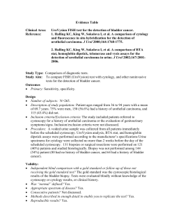
MLabs Spectrum
A supplement to the MLabs Handbook
October 2008
Volume 22, Number 3
Bladder Cancer Detection by Fluorescence In Situ Hybridization (FISH)
Rajal B. Shah, M.D., Associate Professor of Pathology and Urology, and Bryan Betz, Ph.D., Technical
Director of MLabs Molecular Diagnostics Laboratory
E
ach year in the United States about 55,000-60,000 new cases
of urothelial carcinoma, the most prevalent form of bladder
cancer, occurs with about 12,500 deaths due to this disease.
The natural history of urothelial carcinoma is defined by tumor
recurrence and progression. Low-grade papillary tumors tend to
recur with small risk of progression, while high grade tumors are
characterized by both high risk of recurrence and progression to
invasive cancer. Early detection of bladder cancer at its presentation as well as during its subsequent long term follow up is a key to
the long term survival of patients affected by this cancer. Due to its
enigmatic natural history, bladder cancer is one of the most expensive cancers to diagnose and treat.
Assays for Urothelial Carcinoma Detection
Currently there are many non-invasive tests available that can assist
cystoscopy examination to improve the sensitivity and specificity of
bladder cancer detection, specifically for flat urothelial carcinoma
and upper tract disease, which can be missed by traditional cystoscopy examination. Cytology is a widely utilized traditional adjunct
to tissue diagnosis for detection and monitoring of bladder cancer.
Cytology examination provides high specificity but low sensitivity,
particularly for the detection of low-grade bladder tumors. The
suboptimal sensitivity of urine cytology prompted development of
new tests with improved sensitivity for bladder cancer detection.
Antigen based methods (BTA-Stat, Immunocyt, and NMP22) and
the cytogentic/molecular FISH-based method (UroVysion™) offer
alternative non-invasive modalities to supplement cystoscopy examination. Tests employing antigen-based methods can improve
sensitivity, but typically lack high specificity. UroVysion™ FISH is
unique in that it detects bladder cancer at the genetic level. Since
this test simultaneously assesses multiple genetic targets within an
individual cell, both sensitivity and specificity of bladder cancer detection is high. FISH is especially useful in patients with “atypical”
or “equivocal” urine cytology as a reflex test to improve bladder
cancer detection.
DEPARTMENT OF PATHOLOGY
in this issue
1 Bladder Cancer Detection by Fluorescence In Situ Hybridiza-
tion (FISH) 4 Spotlight on Rajal B. Shah, M.D.
5 Test Updates
New Tests
• Clear Cell Sarcoma Translocation Assay
• HER2 Amplification by FISH
• KRAS Mutation Detection
Test Methodology, Reference Range, and Specimen Handling Changes
• Aldosterone, Urine
• Calcitonin
• Carotene
• Chromogranin A
• Dilute Russell’s Viper Venom Test
• Glucagon, Plasma
• Pituitary Tumor Marker, Alpha Subunit
• Serotonin Release Assay
• Stool, Aerobic Culture
• TB Gold QuantiFeron
• VGKC and GAD65 Testing
• Vitamin B1, Whole Blood
• Vitamin D 1,25-dihydroxy
Discontinued Tests
• Cytomegalovirus Cultures & Shell Vials
• Rubella Virus Antibody, IgM
8 MLabs Q & A
MLabs News
UroVysion™ FISH Test for Urothelial Carcinoma Detection
FISH is a molecular technique that utilizes fluorescently labeled
DNA probes to detect genetic abnormalities within individual
cells. Urothelial carcinoma cells easily shed into urine and are
characterized by a high frequency of aneuploidy (abnormal
chromosome number). The UroVysion™ FISH assay directly
detects these chromosomally abnormal cells.
UroVysion™ employs a four-color, four-probe mix to detect
specific regions of chromosomes 3, 7, 9, and 17 within individual cells that are shed into urine. The centromeric (CEP)
probes 3, 7, and 17 aim to detect aneuploidy (gain) of these
chromosomes, while the 9p21 locus specific probe (LSI) aims
to detect loss of this specific region of chromosome 9. These
chromosomal alterations are commonly associated with bladder cancer. The use of this multi-probe cocktail increases both
the sensitivity and specificity of the assay over single or dual
probes.
UroVysion™ FISH is typically used in conjunction with cystoscopy as a:
• Monitoring tool to detect bladder cancer recurrence.
• Diagnostic tool for high-risk hematuria patients.
It is in general unaffected by BCG therapy, instrumentation,
benign prostate enlargement (BPH), hematuria, infection or
inflammatory conditions.
How does UroVysion™ fare in comparison to traditional cytology and other commonly utilized non-invasive
assays?
Demonstrated Sensitivity (by Stage) and Specificity of Different Tests
pTa
pTis
pT1-T4
Overall
Sensitivity
Specificity
UroVysion™
65%
100%
95%
81%
96%
Cytology
47%
78%
60%
58%
98%
BTA-Stat
63%
94%
94%
78%
74%
Immunocyt
96%
80%
86%
86%
79%
60-70%
60-80%
NMP22
(pTa=non-invasive papillary tumor, Tis= in situ carcinoma, T1= lamina propria invasion, T2=
muscularis propria invasion, T3= extra vesicle tumor, T4= invasion of adjacent organs). [1-3]
Based on these and other studies, FISH has demonstrated a high degree of sensitivity and specificity for detection of urothelial carcinoma.
2 U-M Department of Pathology
FIGURE 1: UroVysion™ FISH. (A) Normal result.
Each of the colored fluorescent probes exhibit
the normal value of 2 signals. Chromosomes 3, 7,
17, and the 9p21 locus are indicated by red, green,
aqua, and gold, respectively. The cell nucleus is
counterstained blue with DAPI, a DNA-specific
stain. (B) Abnormal result. This cell demonstrates
polysomy (more than two copies) of chromosomes
3, 7, and 17. Note also the characteristic nuclear
enlargement and mottled appearance of the DAPI
counterstain, both of which are abnormal cytologic features associated with neoplastic cells.
How is a positive test defined?
The UroVysion™ assay is performed by first processing
urine to isolate cells and to prepare them onto slides.
The fluorescently labeled DNA probes are then hybridized to the chromosomes within the cells. After two
washes, the cell nuclei are counterstained with DAPI,
a DNA-specific stain that fluoresces blue. Slides are
now coverslipped and scored on a fluorescent microscope that is equipped with filters to allow visualization
of the DAPI counterstain and the fluorescent probes
(red, green, aqua, and gold for CEP 3, 7, 17, and LSI
9p21, respectively). During scoring, the entire slide
is scanned for morphologically abnormal nuclei by
DAPI stain (nuclear enlargement, irregularity, and/or
mottled appearance). Once identified, each of the individual probe signals is enumerated within these cells.
If no morphologically abnormal cells are present, then
nuclei representing the most abnormal are scored.
Normal cells have 2 signals of each of the four probes,
indicating the presence of the normal 2 copies of each
chromosome (Figure 1A). Tumor cells from urothelial carcinoma patients most frequently present with
polysomy (more than 2 copies) of chromosomes 3, 7,
and/or 17. Polysomic cells are rarely if ever seen in
normal value studies and therefore the finding of these
in urine even in small numbers is virtually pathognomic for the presence of tumor. Polysomy is seen in
over 90% of urothelial carcinoma cases. Isolated homozygous deletion of 9p21, or isolated trisomy (3 copies) of a single chromosome (3, 7, or 17), also occur
in urothelial carcinoma but are relatively uncommon.
A case is considered positive for malignancy if ≥4 cells
exhibit polysomy of two or more chromosomes (3, 7,
and/or 17). This abnormality presents as a gain (3 or
more signals) of multiple chromosomes within the same
cell (Figure 1B). A second criterion for a positive test
result is homozygous 9p21 deletion (0 signals of the
LSI 9p21 probe) in ≥12 cells. These cutoffs were determined by the test manufacturer for the best combination of sensitivity and specificity. Trisomy of a single
chromosome may be considered positive when 10 or
more cells exhibit this change. For positive cases the
percent abnormal cells is calculated which may help estimate tumor burden. This is determined by scoring 100
consecutive urothelial cells for abnormal FISH values.
What is the clinical significance of a positive test without cystoscopically evident
abnormality?
A positive FISH test in the absence of tumor evidence by
cystoscopy or other modalities can pose a diagnostic and
management dilemma. Such cases are often referred to
as “anticipatory positive”. The significance and management of such cases remains controversial. However, this
scenario has been associated with a high likelihood of disease recurrence or presence of disease in upper tract that
may not have been visualized by traditional diagnostic modalities [4]. In our experience these patients often need
extensive cystoscopic examination with biopsy sampling
that also includes upper tract to locate malignant cells.
What are the limitations of the UroVysion™
test?
UroVysion™ testing provides no additional value in patients with positive cytology. A positive
UroVysion™ result in the absence of other evidence of bladder cancer may indicate other genitourinary related cancers (ureter, urethra, renal, or
prostate), or metastatic cancer involving the genitourinary tract. Non-neoplastic urothelial cells
may rarely exhibit tetrasomy by FISH (4 copies of
each of the four probes). This may be observed in
reactive cells, umbrella cells, or cells in the G2/M
phase of the cell cycle. If ≥4 cells exhibit tetrasomy, a case can be considered positive since these
cells fulfill the criteria for polysomy. Thus, when
tetrasomy is identified, this finding is specifically
noted and this result should be interpreted with
caution. However, this finding can be significant,
particularly when high numbers of tetraploid cells
are present (≥10), since tumors can also be tetraploid or near-tetraploid.
Specimen requirements
The UroVysion™ FISH assay is performed on
voided urine specimens. Greater than 33 mL of
urine is recommended for optimal number of
cells, however lower volumes may be adequate if
there is a minimum 25 cells available for evaluation. Mix voided urine 2:1 (v:v) with preservative
(PreservCyt or Carbowax). If urine is not shipped
immediately after collection, refrigerate and ship
via overnight courier within 24 hours.
MLabs Spectrum 3
Spotlight on
Rajal B. Shah, M.D.
Associate Professor of Pathology and Urology
Director, Genitourinar y Pathology Ser vice
Co-Director, Prostate SPORE Tissue Core
Dr. Shah received his medical degree from Gujarat University, India and subsequent post graduate residency training in anatomic and clinical pathology from
the Gujarat Cancer
& Research Institute, India and St.
John’s Hospital and
Medical Center in
Detroit, Michigan.
He completed a
two year fellowship
in Urologic/Surgical Pathology at the
University of Michigan and joined the
faculty of the University of Michigan,
Department of Pathology as an Assistant Professor of
Pathology and Urology in 2001. He was promoted to the rank of Associate Professor in 2007.
Dr. Shah currently oversees the section of Urologic Pathology and also serves as fellowship
director for the subspecialty training program
in Urologic Pathology. He has broad clinical and translational research experience in
the field of urologic cancers with over 70 peer
reviewed publications in reputed journals.
Dr. Shah’s research focuses on translational
application of prognostic and diagnostic biomarkers from bench to clinical practice, morphological and molecular classification of urologic cancers, and understanding outcome of
urologic cancers. He has served as a director
of the NIH funded U-M Prostate Specialized
Program of Research Excellence (SPORE) tissue banking section. Currently, he serves as codirector of the NIH funded prostate SPORE tissue
core and principal pathologist for this program.
4 U-M Department of Pathology
Summary
UroVysion™ offers a high degree of both sensitivity and specificity in urothelial carcinoma detection. The test has been FDA approved for monitoring tumor recurrence in patients with a history
of bladder cancer, and as an aid for bladder carcinoma diagnosis in patients with hematuria. This
assay offers the opportunity to improve the clinical management of patients with urothelial carcinoma. A negative FISH result increases the confidence for safe extension of cystoscopy intervals,
while a positive FISH result warrants a through
investigation and close follow-up with re-biopsy.
Select References
1. Halling, K.C., et al., A comparison of BTA stat,
hemoglobin dipstick, telomerase and Vysis
UroVysion assays for the detection of urothelial carcinoma in urine. J Urol, 2002. 167(5): p.
2001-6.
2. Halling, K.C., et al., A comparison of cytology
and fluorescence in situ hybridization for the
detection of urothelial carcinoma. J Urol, 2000.
164(5): p. 1768-75.
3. Lokeshwar, V.B., et al., Bladder tumor markers
beyond cytology: International Consensus Panel on bladder tumor markers. Urology, 2005.
66(6 Suppl 1): p. 35-63.
4. Yoder, B.J., et al., Reflex UroVysion testing of
bladder cancer surveillance patients with equivocal or negative urine cytology: a prospective
study with focus on the natural history of anticipatory positive findings. Am J Clin Pathol,
2007. 127(2): p. 295-301.
how to send a specimen
UroVysion™ testing will be offered through MLabs in
the near future; testing is currently forwarded to Mayo
Medical Laboratories.
For assistance 24 hours per day, 7 days per week,
call MLabs at 800-862-7284 or visit our web site at
www.mlabs.umich.edu.
Test Updates
New Tests
clear cell sarcoma translocation assay
The MLabs Molecular Diagnostics Laboratory began
performing a Clear Cell Sarcoma Translocation assay effective July 1, 2008.
Greater than 90% of clear cell sarcomas harbor the reciprocal chromosomal translocation t(12;22)(q13;q12).
This rearrangement joins the EWSR1 and ATF1 genes
and leads to expression of EWSR1/ATF1 fusion transcripts. This test detects three EWSR1/ATF1 fusion
transcript types: Type 1 (EWSR1 exon 8/ATF1 exon
4), Type 2 (EWSR1 exon 7/ATF1 exon 5), and Type 3
(EWSR1 exon 10/ATF1 exon 5) which collectively account for almost all t(12;22)-bearing clear cell sarcoma
cases. Testing for EWSR1/ATF1 fusion is a useful diagnostic adjunct in the differential diagnosis of clear cell
sarcoma, since malignant melanoma is not associated
with this chimeric transcript.
Collection Instructions: Send fresh, frozen, or formalinfixed paraffin-embedded tissue. Fresh tissue should be
sent on a piece of gauze in saline, or in RPMI, within 16
hours of collection; refrigerate. Frozen tissue should
be stored at -80 degrees C; do not allow to thaw at any
time. Paraffin-embedded tissue should be stored at
room temperature. Please include any pertinent clinical history.
her2 amplification by fish
The MLabs Molecular Diagnostics Laboratory began
performing HER2 Amplification by FISH effective July
1, 2008.
This test detects amplification of the HER2 gene via
fluorescence in situ hybridization (FISH) in formalinfixed, paraffin-embedded breast cancer tissue specimens. FISH is performed using PathVysion (Abbott
Molecular, Inc) probes to the HER2 locus (17q11.2-q12)
and the chromosome 17 centromere (D17Z1). Results
are reported as a HER2:D17Z1 copy number ratio and
interpreted according to ASCO/CAP guideline recommendations. HER2 amplification status can be a useful
adjunct indicator of prognosis, and can assist in the selection of patients for whom Herceptin (Trastuzumab)
treatment is being considered. This test can also rule-out
or confirm gene amplification in cases previously evaluated by HER2 immunohistochemistry (IHC), especially
those with equivocal (2+) IHC results.
Collection Instructions: Submit a formalin-fixed, paraffin block of the breast cancer tissue; store at room temperature. Please provide a pathology report with each
specimen, including the type of fixative and time of fixation (if known).
kras mutation detection
Effective October 1, 2008, the MLabs Molecular Diagnostics Laboratory will begin performing KRAS Mutation Detection by Polymerase Chain Reaction (PCR) followed by sequencing analysis to detect KRAS mutation
if present.
Activating mutations in the KRAS gene occur in approximately 20% of lung adenocarcinomas, 30-40% of
colorectal carcinomas, and a variety of other human
cancers. Mutations are single nucleotide substitutions,
occurring most frequently within codon 12 or 13. These
have been associated with a limited clinical response to
epidermal growth factor receptor (EGFR) targeted therapies in lung and colorectal cancers, and may indicate
prognosis. This DNA sequencing test will detect all mutations within codons 12, 13, and 61 of the KRAS gene
from formalin-fixed paraffin-embedded tissue blocks.
Specimens should contain an adequate proportion of
tumor (>40%) to ensure mutation detection.
Collection Instructions: Send fresh, frozen, or formalinfixed paraffin-embedded tissue containing greater than
40% tumor. Fresh tissue should be sent on a piece of
gauze in saline, or in RPMI, within 24 hours of collection; refrigerate. Frozen tissue should be stored at -80
degrees C; do not allow to thaw at any time. Paraffin-embedded tissue should be stored at room temperature.
Test Methodology,
Reference Range, and
Specimen Handling Changes
aldosterone, urine
Due to the low volume of requests, the MLabs Chemistry
Laboratory will discontinue performing Aldosterone,
Urine. Effective October 1, 2008, requests for this test
will be sent to Mayo Medical Laboratories:
Collection Instructions: Collect 24 hour urine specimen.
Add 25 mL of 50% acetic acid (15 mL) for pediatric patients (<5 years) as a preservative prior to start of collection. Aliquot 11 mL (minimum 6 mL) into a plastic
urine container and refrigerate. Record total 24 hour
urine volume and collection dates/times on request
form.
MLabs Spectrum 5
calcitonin
pituitary tumor marker, alpha subunit
Effective August 19, 2008, Mayo Medical Laboratories
revised the reference range for the Calcitonin assay to
include all age groups for both males and females:
Effective June 17, 2008, Mayo Medical Laboratories
changed their test methodology for the Alpha-Subunit
Pituitary Tumor Marker (Alpha-Subunit Pituitary Glycoprotein Hormones) to immunochemiluminescent assay
(ICMA).
Reference Range: BASAL: Males: <16 pg/mL; Females:
<8 pg/mL. PEAK CALCIUM INFUSION: Males: < or
=130 pg/mL; Females: < or =90 pg/mL.
carotene
Effective July 21, 2008, there was a change to the specimen collection and handling requirements for Warde
Medical Laboratory’s Carotene assay. This test is now a
“strict frozen”.
Collection Instructions: Collect blood in a red top or SST
tube from a fasting patient (12 hour fast). Patient should
not consume any alcohol for 24 hours prior to collection
of specimen. Centrifuge, aliquot serum into plastic vial
and freeze immediately. Protect specimen from light.
chromogranin a
Effective July 28, 2008, refrigerated specimens are no
longer acceptable for the Chromogranin A assay. This
change was made to avoid multiple freeze/thaw cycles
prior to analysis of the specimen.
Collection Instructions: Collect specimen in a red top
tube. Centrifuge, aliquot serum into a plastic vial and
freeze.
dilute russell’s viper venom test
The reference range for the Dilute Russell’s Viper Venom Test (DRVVT) changed as follows, effetive June 20,
2008. Note that the DRVVT is a component of the Lupus Anticoagulant Screen panel.
Reference Range: DRVVT <41 seconds; DRVVT Ratio
<1.2; LA CONFIRM negative.
glucagon, plasma
Effective June 19, 2008, Mayo Medical Laboratories
changed their test methodology for the Glucagon, Plasma assay to extraction followed by immunoassay.
Reference Range: < or = 80 pg/mL
6 U-M Department of Pathology
Reference Range: Pediatric: age < or = 5 days: < or = 50
ng/mL; age 5 days - < 3 months: < or = 10 ng/mL; age
3 months - < 2 years: < or = 1.2 ng/mL; age 2 years – puberty onset: < or = 1.2 ng/mL; Tanner II – IV: < or = 1.2
ng/mL; Adults: Males: < or = 0.5 ng/mL; Premenopausal females: < or = 1.2 ng/mL; Postmenopausal females:
< or = 1.8 ng/mL.
Collection Instructions: Collect specimen in a red top or
green top tube. Centrifuge, aliquot 2 mL (minimum 0.3
mL) of serum or plasma into a plastic vial and refrigerate.
serotonin release assay
Effective August 11, 2008, the Serotonin Release assay is
sent to BloodCenter of Wisconsin.
Collection Instructions: Collect specimen in a red top
tube. Centrifuge, aliquot 5 mL of serum into a plastic
vial and refrigerate. Citrated or ACD plasma is acceptable if serum is not available.
stool, aerobic culture
Effective June 18, 2008, all Stool Culture specimens
are screened for Shiga-like toxin (SLT) at an additional charge. Previously, Shiga-like toxin testing was performed only during the summer months, if Hemolytic
Uremic Syndrome was suspected, or if the stool was
bloody.
tb gold quantiferon
Effective August 11, 2008, the MLabs Chemical Pathology Laboratory has changed the collection method for
the QuantiFeron - TB Gold test.
The new system utilizes three special collection tubes
provided by the manufacturer: gray top (NIL), red top
(TB antigen), and purple top (Mitogen). Collection kits
available from MLabs consist of an appropriate transport
bag, the three collection tubes, and instructions for collecting the QuantiFeron samples.
vgkc and gad65 testing
Discontinued Tests
Effective June 18, 2008, Voltage-Gated Potassium Channel (VGKC) and Glutamic Acid Decarboxylase (GAD65)
Autoantibody testing have been added to the following
Mayo Medical Laboratory Panels:
cytomegalovirus Cultures & shell vials
• VGKC and GAD65 have been added as reflex components of the Myasthenia Gravis Evaluation, Adult.
• VGKC and GAD65 are included as part of the Myasthenia Gravis Evaluation, Thymoma.
• VGKC is included as part of the Paraneoplastic Autoantibody Evaluation, Serum.
vitamin b1, whole blood
Effective July 10, 2008, Vitamin B1, Whole Blood, is sent
to Mayo Medical Laboratories.
Collection Instructions: Collect specimen in a green
top tube (sodium heparin) following an overnight (12
hour) fast. Freeze and send 5 mL intact whole blood
specimen. Protect specimen from light. Do not freeze
glass vacutainer tubes; collect in plastic vacutainer tube
or transfer blood to a plastic tube prior to freezing.
Reference Range: 80 - 150 nmol/L.
vitamin d 1,25-dihydroxy
Mayo Medical Laboratories changed their test methodology for Vitamin D 1,25-dihydroxy to cartridge extraction/LC-MS/MS effective June 2, 2008.
Collection Instructions: Collect blood in a red top or
SST tube from a fasting patient (4 hour fast). Centrifuge, aliquot 1.5 mL of serum into a plastic vial, and refrigerate.
There will be a reference range change for this assay effective October 14, 2008:
Reference Range: <16 years: 24 – 86 pg/mL; Males >=16
years: 18 – 64 pg/mL; Females >=16 years: 18 – 78 pg/
mL. Normal patients who have increased exposure to
sunlight may have values above the normal range.
The MLabs Virology Laboratory no longer performs Cytomegalovirus Cultures or Shell Vials on blood or bone
marrow specimens. Cytomegalovirus DNA by PCR,
Quantitative, is performed in place of culture for blood
specimens.
rubella virus antibody, igm
Effective July 1, 2008, Mayo Medical Laboratories has
discontinued Rubella Virus IgM Antibody testing.
MLabs and Mayo Medical Laboratories will no longer
forward specimens for rubella IgM-class antibody testing. If acute rubella or Congenital Rubella Syndrome
(CRS) is suspected, it is recommended to communicate
with your state health laboratory or the CDC to evaluate
your patient. Michigan clients may contact the MDCH
Bureau of Epidemiology at 517-335-8165 for assistance
in evaluation of your patient.
In 2005, the Centers for Disease Control and Prevention
(CDC) announced the absence of endemic transmission
of rubella in the United States, largely due to successful
vaccination programs. Fewer than 10 cases of rubella
were reported in 2004, and the majority of these cases
occurred in individuals born outside the United States.
The incidence of congenital rubella syndrome (CRS)
has also been significantly reduced, with a total of 4 cases
of CRS being reported to the CDC during 2001-2004.
Given the low prevalence of rubella in the United States,
routine serologic testing for IgM-class antibodies to this
virus may yield false positive results, which can negatively
impact patient care. This is especially problematic when
rubella IgM testing is included in the routine screening
of asymptomatic, pregnant women.
Rubella Virus Antibody, IgG will continue to be available
for the determination of immune status to rubella.
While 1, 25-dihydroxy vitamin D is the most potent
vitamin D metabolite, levels of the 25-OH forms of vitamin D more accurately reflect the body’s vitamin D
stores. Consequently, Vitamin D 25-hydroxy (order code
25HD) is the preferred initial test for assessing vitamin D
status. However, in the presence of renal disease, 1, 25dihydroxy vitamin D levels may be needed to adequately
assess vitamin D status.
MLabs Spectrum 7
For additional clarification
concerning any of the information contained in this Spectrum,
please contact the MLabs Client
Services Center at 734-936-2598
(local) or 800-862-7284.
Address correspondence to:
MLabs Spectrum
PO Box 976
Ann Arbor, MI 48106-0976
To keep our Spectrum circulation
records accurate and up to date,
please send any name or address
changes, corrections, additions
or deletions to the address listed
above. Thank you.
Executive Officers of the University of Michigan
Health System: Robert P. Kelch, Executive Vice
President for Medical Affairs; James O. Woolliscroft, Dean, U-M Medical School; Douglas
Strong, Chief Executive Officer, U-M Hospitals and
Health Centers; Kathleen Potempa, Dean, School
of Nursing.
The Regents of the University of Michigan: Julia
Donovan Darlow, Laurence B. Deitch, Olivia P.
Maynard, Rebecca McGowan, Andrea Fischer
Newman, Andrew C. Richner, S. Martin Taylor,
Katherine E. White, Mary Sue Coleman (ex officio).
Nondiscrimination Policy Statement
The University of Michigan, as an equal opportunity/affirmative action employer, complies
with all applicable federal and state laws regarding
nondiscrimination and affirmative action, including
Title IX of the Education Amendments of 1972 and
Section 504 of the Rehabilitation Act of 1973. The
University of Michigan is committed to a policy of
nondiscrimination and equal opportunity for all persons regardless of race, sex, color, religion, creed,
national origin or ancestry, age, marital status,
sexual orientation, gender identity, gender expression, disability, or Vietnam-era veteran status in
employment, educational programs and activities,
and admissions. Inquiries or complaints may be
addressed to the Senior Director for Institutional
Equity and Title IX/Section 504 Coordinator, Office
of Institutional Equity, 2072 Administrative Services
Building, Ann Arbor, Michigan 48109-1432, 734763-0235, TTY 734-647-1388. For other University of
Michigan information call 734-764-1817.
©2008 The Regents of the University of Michigan.
3/08/900A
8 U-M Department of Pathology
MLabs Q & A
When doing Employee Health Screens for Rubella, Rubeola, Mumps and
Varicella, are titers necessary when the screen is positive?
Question answered by Duane Newton, Ph.D., Director of MLabs Microbiology and Virology Laboratories
The term “titer” when applied to serology testing pertains to the quantitative level of
antibody present in a sample that is directed against the target of interest. Historically,
detection of antibodies directed against Rubella virus, Mumps virus, Rubeola virus, etc.
was performed using manual, labor-intensive methods (such as immunofluorescence or
hemagglutination assays). By performing serial dilutions of the patient’s serum sample,
a quantitative value, or titer, could be reported using these methods. Virtually all clinical serology testing being performed currently has migrated to some type of instrument
performing enzyme immunoassays (EIA) for qualitative detection of antibodies. Although this provides only an answer of “antibody detected” or “antibody not detected,”
the benefits of using these methods on automated instruments allows for increased
throughput, improved sensitivity and specificity, and shorter turnaround-time for reporting results. The thresholds for positivity for these EIAs have also been correlated to
quantitative titers determined using the conventional methods so that the positive and
negative qualitative results are clinically appropriate. MLabs often receives requests to
test for “antibody titers,” which is most often due to the carryover in historical terminology used for performing serology testing rather than a true clinical need or desire to
determine quantitative antibody levels. Qualitative testing performed by MLabs for the
viruses mentioned above do not require additional follow-up testing using quantitative
methods because 1) for negative samples, quantitative testing will not increase the sensitivity, and 2) for positive samples, the threshold for positivity correlates with clinically
relevant (i.e., protective) levels of antibodies, so quantitative testing will not provide any
additional information.
MLabs News
u-m department of pathology news
Congratulations are to be extended to Thomas M. Annesley, Ph.D., Professor and Director, Drug Analysis and Toxicology. Dr. Annesley received the “Presidential Citation of
the National Academy of Clinical Biochemistry” - the elected academy of the American
Association of Clinical Chemistry (AACC) - and the inaugural award for “Outstanding
Contribution to Clinical Mass Spectrometry”.
Suzanne H. Butch, MA, MT(ASCP)SBB, Administrative Manager of the Blood Bank
& Transfusion Service was installed as president of the honorary fraternity Alpha Mu
Tau. The mission of Alpha Mu Tau is to recognize persons who have made outstanding
professional contributions to the field of clinical laboratory science and to enhance the
profession by providing scholarships to support educational endeavors.
Jeffrey Myers, M.D., Dan Visscher, M.D., and Ul Balis, M.D. are prominently featured
in the July 2008 issue of CAP Today’s story “Informatics to the rescue in fixing ‘blunt
end’ AP errors”. The article highlights a number of initiatives in Anatomic Pathology including work flow redesign, hand off procedures, critical values reporting management
and other informatics initiatives to improve efficiency and patient safety.


© Copyright 2026