
Tyrosine phosphorylation of transcriptional coactivator
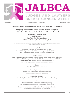
The FASEB Journal article fj.10-169136. Published online June 3, 2011. The FASEB Journal • Research Communication Tyrosine phosphorylation of transcriptional coactivator WW-domain binding protein 2 regulates estrogen receptor ␣ function in breast cancer via the Wnt pathway Shen Kiat Lim,*,† Magali Orhant-Prioux,† Weiyi Toy,*,† Kah Yap Tan,*,† and Yoon Pin Lim*,†,‡,§,1 *Department of Biochemistry, Yong Loo Lin School of Medicine, †Cancer Science Institute, and ‡ Department of Biological Sciences, Faculty of Science, National University of Singapore, Singapore; and §Bioinformatics Institute, Agency for Science and Technology, Singapore ABSTRACT WW-binding protein 2 (WBP2) has been demonstrated in different studies to be a tyrosine kinase substrate, to activate estrogen receptor ␣ (ER␣)/progesterone receptor (PR) transcription, and to play a role in breast cancer. However, the role of WBP2 tyrosine phosphorylation in regulating ER␣ function and breast cancer biology is unknown. Here, we established WBP2 as a tyrosine phosphorylation target of estrogen signaling via EGFR crosstalk. Using dominant-negative, constitutively active mutants, RNAi, and pharmacological studies, we demonstrated that phosphorylation of WBP2 at Tyr192 and Tyr231 could be regulated by c-Src and c-Yes kinases. We further showed that abrogating WBP2 phosphorylation impaired >60% of ER␣ reporter activity, putatively by blocking nuclear entry of WBP2 and its interaction with ER␣. Compared to vector control, overexpression of WBP2 and its phospho-mimic mutant in MCF7 cells resulted in larger tumors in mice, induced loss of cell-cell adhesion, and enhanced cell proliferation, anchorageindependent growth, migration, and invasion in both estrogen-dependent and -independent manners, events of which could be substantially abolished by overexpression of the phosphorylation-defective mutant. Hormone independence of cells expressing WBP2 phospho-mimic mutant was associated with heightened ER␣ and Wnt reporter activities. Wnt/-catenin inhibitor FH535 blocked phospho-WBP2-mediated cancer cell growth more pronouncedly than tamoxifen and fulvestrant, in part by reducing the expression of ER␣. Wnt pathway is likely to be a critical component in WBP2-mediated breast cancer biology.—Lim, S. K., Orhant-Prioux, M., Toy, W., Tan, K. Y., Lim, Y. P. Tyrosine phosphorylation of transcriptional coactivator WW-domain binding protein 2 regulates estrogen receptor ␣ function in breast cancer via the Wnt pathway. FASEB J. 25, 000 – 000 (2011). www.fasebj.org Key Words: WBP2 䡠 ER␣ 䡠 breast cancer 䡠 EGFR 䡠 -catenin 䡠 crosstalk Potentiation of gene transcription by nuclear hormone receptors involves interplay with coactivators and the basal transcriptional machinery through protein0892-6638/11/0025-0001 © FASEB protein interactions (1, 2). Coactivators can act as transcriptional adaptors that mediate formation of transcriptional complexes or modify chromatin through histone acetyl transferases (HATs) or nucleosome remodeling machineries. Some of the known nuclear hormone receptor coactivators include p160 family members of coactivators, SRC-1, SRC-2[TIF-2/GRIP-1/NCoA-2], SRC3[pCIP/ACTR/AIB-1/RAC-3/TRAM-1], NRIF-3, E6-AP, and WBP2. Because of their pleiotropic roles, it is not surprising that transcriptional coactivators are emerging as a group of proteins increasingly implicated in cancer development (3–5). Transcriptional coactivators are often regulated by post-translational modifications, such as phosphorylation. Phosphorylation of specific members of the SRC/ p160 family of proteins had been reported to enhance their nuclear localization (6), inhibit their interactions with non-nuclear receptor (NR) activators (7), or stimulate their intrinsic coactivator activities (8). Phosphorylation had also been shown to regulate protein stability and activity of AIB1 and PGC-1 (9, 10). Furthermore, phosphorylation of NRIF3 via Pak1 promotes estrogen receptor ␣ (ER␣) transactivation through increased ER␣-NRIF3 interaction (5). WW domain binding protein (WBP2) is a transcriptional coactivator demonstrated to selectively enhance ER␣/progesterone receptor (PR) transactivation via hormone-dependent ER␣/PR-WBP2 interaction and recruitment of WBP2 to hormone-responsive elements (11). WBP2 contains an intrinsic activation domain. One of its 3 polyproline (PPXY) motifs, PY3, is essential for activating ER␣/PR function. Its coactivator activity could be further enhanced by Yes kinase-associated protein (YAP), a protein with transforming properties in mammary epithelial cells (12) that also regulates 1 Correspondence: Department of Biochemistry, Yong Loo Lin School of Medicine, National University of Singapore, MD4A, 02-05, 8 Medical Dr., Singapore 117597. E-mail: [email protected] doi: 10.1096/fj.10-169136 This article includes supplemental data. Please visit http:// www.fasebj.org to obtain this information. 1 transcription factors such as p73 (13), Runx2 (14), TEAD/TEF (15), and ErbB4 (16). Applying phosphoproteomics on an isogenic MCF10AT model of breast cancer progression, we previously showed that breast cancer cells have higher level of tyrosinephosphorylated WBP2 than normal breast mammary epithelial cells (17). Because of the lack of commercial antibodies at that time, we coexpressed V5-tagged WBP2 with EGFR and subsequently showed that WBP2 was an authentic target of EGFR signaling. In this study, we hypothesized that EGFR-mediated tyrosine phosphorylation of WBP2 plays a role in regulating ER␣ function and breast cancer biology. Therefore, we attempted to delineate the signaling pathways for the EGFR-mediated tyrosine phosphorylation of WBP2 and to study the effect of WBP2 phosphorylation on its coactivator activity. The role of WBP2 and its tyrosine phosphorylation on ER␣-positive breast cancer biology and the underlying mechanisms were also investigated. following were kind gifts: Src-WT, constitutively active (CA)Y529F, and dominant-negative (DN)-K295M-pSGT were from Sara Courtneidge (European Molecular Biology Laboratory, Heidelberg, Germany); Yes-WT and CA-Y537F were from Marius Sudol (Weis Center for Research, Danville, PA, USA); 2XERE-TATA-Firefly Luc and 2XPRE-TATA-Firefly Luc were from Dean P. Edwards (Baylor College of Medicine, Houston, TX, USA); and TOPFlash reporter plasmid was from Yoshiaki Ito (National University of Singapore, Singapore). pRL-TK was purchased from Promega (Madison, WI, USA). siRNA sequences Yes1 siRNA (5⬘-UUCUCCUACAAGAAUAUUAGCAGCC-3⬘), v-Src siRNA (5⬘-GCCUCUCAGUGUCUGACUUCGACAA3 ⬘ ) , L u c i f e r a s e - G L 2 s i R N A (5⬘-CGUACGCGGAAUACUUCGA-3⬘), WBP2 siRNA (5⬘-AGCAUCCGCUGUCCGAACUCAAUGG-3⬘), and WBP2 UTR-siRNA (5⬘-CAGGAACUAGCAUUGUGGGACAUUA-3⬘) were obtained from Invitrogen. Cell culture, ligand/drug treatments, and lysis MATERIALS AND METHODS Antibodies Through NeoMPS, Inc. (Strasbourg, France), we generated in-house polyclonal antibodies against WBP2 based on a 17-aa (N⬘-NDMKNVPEAFKGTKKGT-C⬘) peptide sequence reported in another study (11), which were affinity purified and stringently validated via comparative immunoblotting with preimmune serum, in the presence of WBP2-specific and control peptides, reciprocal immunoprecipitation of exogenously expressed tagged WBP2 protein, and immunoblotting with anti-tag and anti-WBP2 antibodies (data not shown). Anti-WBP2 mouse monoclonal antibodies were purchased from Abnova Corp. (Taipei, Taiwan); anti-PY20-HRP, antiEGFR, anti--catenin, and anti-Yes mouse monoclonal antibodies were obtained from BD Biosciences (San Diego, CA, USA); anti-actin-HRP, anti-ER␣, anti-phospho-ER␣-S118, antiSrc, anti-cyclin B1, anti-c-Myc, anti-p21, anti-p16, anti-p27, anti-MMP2, anti-ZO-2 rabbit polyclonal, anti-ER␣ and anticyclin D1 mouse monoclonal, anti-GADPH, and anti-vimentin goat polyclonal antibodies were obtained from Santa Cruz Biotechnology (Santa Cruz, CA, USA); anti-phospho-SrcY416, anti-EGFR, anti-YAP1, anti-Histone 2A, anti-E-cadherin, anti-Wnt3a, anti-GSK, anti-phospho-GSK-S9, anti-Bcl2, antiAkt and anti-pERK1/2-Thr202/Tyr204 rabbit polyclonal, anti-pAkt-S473, and anti-ERK1/2 mouse monoclonal antibodies were obtained from Cell Signaling Technology Inc. (Danvers, MA, USA); anti-V5 mouse monoclonal and anti-V5-HRP antibodies were obtained from Invitrogen (Carlsbad, CA, USA); anti-Yes rabbit polyclonal antibodies were obtained from Millipore Corp. (Billerica, MA, USA); anti-phosphoFAK1-Y397 rabbit polyclonal antibodies were obtained from Abcam (Cambridge, UK); and anti-mouse, anti-rabbit, and anti-goat horseradish peroxidase (HRP) conjugates were purchased from Sigma-Aldrich (St. Louis, MO, USA). Plasmids and reporters cDNA for wild-type (WT) WBP2 (Origene, Rockville, MD, USA) was subcloned into pcDNA6.2-Directional-TOPO vector and pCEP4 (Invitrogen). All mutations were generated using the QuikChange II XL Site-Directed Mutagenesis Kit (Stratagene; Agilent Technologies, Santa Clara, CA, USA). The 2 Vol. 25 September 2011 MCF7, MDA-MB231, T47D, HeLa, and MCF10A cells were from American Type Culture Collection (Manassas, VA, USA). MCF7, HeLa, and MDA-MB231 cells were maintained in RPMI1640 (Sigma) containing 10% FBS (Thermo Scientific Hyclone, South Logan, UT, USA) and 100U penicillin/ streptomycin (Invitrogen). T47D cells were maintained in RPMI1640 containing 10% FBS, 10 g/ml insulin (Sigma) and 100 U penicillin/streptomycin. MCF10A cells were maintained in DMEM/F12 (Sigma) containing 5% horse serum with additives previously described (17). For all experiments involving estrogen(E2)/progesterone (P4) (Sigma), MCF7 and T47D cells were cultured in Phenol-Red Free RPMI1640 (Sigma) containing 5% charcoal-dextran stripped FBS (Thermo Scientific Hyclone) for ⱖ2 d before stimulation. EGF was from Millipore; Wnt3a ligand was from R&D Systems (Minneapolis, MN, USA); tamoxifen, fulvestrant, and FH535 were from Sigma; and Iressa and AZD0530 were from AstraZeneca Singapore Pte. Ltd. (Singapore). Fresh hormone/drug was replenished every day for treatment that lasted for ⬎1 d. Cell lysis was performed as reported previously (18). Transient and stable transfection For transient transfection, cells were seeded at 70 – 80% confluency in a 60-mm dish in antibiotic-free medium 1 d before transfection and transfected with 4 g plasmid DNA or 50 –100 nM siRNA and 12 l Lipofectamine 2000 (Invitrogen), according to the manufacturer’s instructions. Cells were harvested at 24 – 48 h post-transfection. For reverse transfection, cells were transfected while they were still in suspension (i.e., after trypsinization and prior to plating). For stable transfection, MCF7 or MCF10A cells were transfected with pCEP4 vector, WBP2-WT, WBP2-Y192-231E, WBP2-Y192231D, and WBP2-Y192-231F/pCEP4. At 48 h post-transfection, cells were exposed to hygromycin (Invitrogen; 250 g/ml for MCF7 and 50 ng/ml for MCF10A) for 3 wk and screened for WBP2 protein expression. Selected clones were pooled and maintained with same selection pressure. WBP2 expression was checked periodically. Immunoprecipitation and immunoblotting Immunoprecipitation and immunoblotting were carried out as described previously (19). For coimmunoprecipitation, The FASEB Journal 䡠 www.fasebj.org LIM ET AL. cells were lysed in ice-cold buffer (50 mM Tris, pH 7.5; 150 mM NaCl; 1 mM EDTA; 10% glycerol; 0.5% Nonidet-P40; 0.5% Triton X-100; 50 mM NaF; 1⫻ protease inhibitor; and 1 mM Na3VO4) and incubated on ice for 15 min for complete lysis. Prior to immunoprecipitation, 500-1000 g of lysates was precleared by incubation with 50 l of 50% slurry of mouse/ rabbit-IgG agarose (Sigma) for 1 h at 4°C. Meanwhile, an equivalent amount of anti-mouse/rabbit-IgG agarose was blocked with 5% BSA overnight, 4°C. Precleared lysates were then incubated with 1–2 g antibodies overnight at 4°C. Immune complexes were then captured by incubation with preblocked anti-mouse/rabbit-IgG agarose for 2 h at 4°C. Immunoprecipitates were then washed extensively 4 times, 5 min each, with buffer (50 mM Tris, pH 7.5; 150 mM NaCl; 1 mM EDTA; 10% glycerol; 0.5% Nonidet P-40; 0.5% Triton X-100; and 1 mM Na3VO4). Subcellular fractionation The subcellular fractionation was carried out according to a protocol by Dr. Richard Pattern (Tufts-New England Medical Centre, Boston, MA, USA; http://www.abcam.com/index.html? pageconfig⫽resource&rid⫽11473). Briefly, cells on plates were rinsed once with ice-cold PBS and scraped in ice-cold hypotonic lysis buffer (20 mM HEPES, pH 7.4; 10 mM KCl; 2 mM MgCl2; 1 mM EDTA; 1 mM EGTA; 1 mM DTT; 1⫻ protease inhibitor cocktail; 50 mM NaF; and 1 mM sodium orthovanadate) and incubated on ice for 1 h. The lysate was passed through a 25-gauge needle 15 times. The nuclear pellet was centrifuged out at 3000 rpm for 5 min and washed twice with hypotonic lysis buffer before resuspending and lysing in nuclear lysis buffer (NID lysis buffer with 10% glycerol and 1% SDS). The nuclear lysate was then sonicated briefly. The supernatant was centrifuged at 8000 rpm for 5 min. The resulting supernatant was the cytosolic and membrane fraction. This supernatant was further centrifuged at 40,000 rpm for 1 h. The resulting supernatant was collected as the cytosolic fraction. Dual luciferase reporter assay The Dual-Luciferase reporter assay system (Promega) was used for sequential measurement of firefly and Renilla luciferase activities, according to the manufacturer’s instructions. Quantification of luciferase activities was carried out using a luminometer (Sirius; Berthold-DS, Pforzheim, Germany). Briefly, transfected cells in 24-well plates were rinsed once with ice-cold PBS and lysed in 1⫻ passive lysis buffer (PLB) for 15 min at room temperature. The crude lysate was first incubated with luciferase assay substrate (LAR II) to measure the firefly luciferase activity, followed by incubation with Stop and Glo substrate to measure the Renilla luciferase activity. Immunofluorescence Cells were grown on coverslips in 6-well plates. Following various treatments, cells were fixed with 4% paraformaldehyde at room temperature and permeabilized with 0.5% Triton-X-100 in PBS. After blocking with 5% BSA for 1 h, the cells were incubated with primary antibodies overnight (antiER␣ mouse monoclonal, 1:100; anti-E-cadherin rabbit polyclonal, 1:100; anti ZO-2 rabbit polyclonal, 1:100). This was followed by secondary antibodies conjugated to Alex Fluor 488 (Molecular Probes; Invitrogen) at 1:1000 dilution for 1 h. The nuclei of the cells were counterstained with 4,6-diamidino-2-phenylindole (DAPI) at 1:10,000 dilution for 1 min. Coverslips containing the cells were then mounted onto glass slides with the addition of Prolong antifade reagent WBP2 REGULATES ER␣ IN BREAST CANCER VIA Wnt (Molecular Probes; Invitrogen) and sealed with Eukitt quickhardening mounting medium (Fluka; Sigma). Analyses were made using the Carl Zeiss Axioplan 2 fluorescence microscope (Carl Zeiss Imaging, Göttingen, Germany) with a ⫻100 oil-immersion objective. The Axiovision Rel4.6 software (Carl Zeiss) was used to capture and analyze the images. In vitro cell-based assays Cell proliferation was measured using the CellTiter 96 Aqueous One Solution Non-Radioactive (MTS) Cell Proliferation Assay Reagent (Promega). Briefly, 5000 cells/well were seeded in triplicate in 100 l culture medium in a 96-well plate on d 0, and cell growth was monitored daily until d 4. MTS reagent (20 l) was added to each well and mixed by swirling the plate. After 1 h, the absorbance was measured on a plate reader (Tecan Group Ltd, Mannedorf, Switzerland) at 490 nm. The anchorage-independent growth was assayed using CytoSelect 96-well cell transformation colorimetric assay kit (Cell Biolabs, San Diego, CA, USA). Briefly, 50 l of base agar matrix was added in the bottom of each well of a 96-well plate. When the base agar was solidified, 75 l of cell suspension/soft agar matrix containing 5000 cells was layered on top, followed by 50 l of culture medium. After 10 d of incubation at 37°C, the agar matrix was solubilized, and cells were detected using 3-(4,5-dimethyl-2-thizolyl)-2,5-diphenyl2H-tetrazolium bromide (MTT) solution. Absorbance was measured at 570/630 nm. For scratch assay, cells were seeded onto 6-well plates and grown until a confluent monolayer was obtained. A wound was incised in the cell monolayer with a p200 pipette tip. The cells were washed once with growth medium to remove the cell debris and to smooth the edge of the scratch, and the medium was replaced with fresh growth medium. The cells were incubated at 37°C, and their migration into the scratch area was monitored up to 24 h. Using a phase-contrast microscope, images of the scratch were captured in the same field at 0, 8, 16, and 24 h after scratching. The relative width of the scratch was measured quantitatively using Photoshop 5.5 (Adobe Systems, San Jose, CA, USA). The extent of gap closure was determined as the rate of cell migration. For chemotaxis assay, 2 ⫻ 105 cells were serum starved overnight and seeded in serum-free medium in the top chambers of a 96-transwell plate with polycarbonate membrane chambers (8-m pore size, Cell Biolabs), and medium containing 10% FBS was added to the bottom chambers as the chemoattractant. After 2 h incubation, top nonmigratory cells were removed; bottom migrated cells were first dissociated from the membrane, and then lysed and quantified using CyQuant GR fluorescent dye (Invitrogen) at 480 nm/520 nm. For invasion assay, 2 ⫻ 105 cells were serum starved overnight and seeded in serum-free medium in the top chambers of a 96-transwell plate with polycarbonate membrane chambers coated with a uniform layer of basement membrane matrix (8-m pore size, Cell Biolabs), and medium containing 10% FBS was added to the bottom chambers. After 24 h incubation, top noninvasive cells were removed; bottom invaded cells were first dissociated from the membrane, and then lysed and quantified using CyQuant GR fluorescent dye at 480 nm/520 nm. Tumor xenograft studies in nude mice Female nude mice (4 to 6 wk old) were inoculated s.c. in the hind flanks with 5 ⫻ 106 MCF7 cells stably transfected with pCEP4, WBP2-WT, WBP2-Y192-231E, or WBP2-Y192-231F, suspended in 100 l of Matrigel (BD Biosciences). Tumor development was monitored every 3– 4 d until d 32. Two measurements were made, one at d 22 and the other at d 32. 3 Statistical analysis For analysis of statistical difference between control and experimental groups, the paired 2-sided Student’s t test was performed. Values of P ⱕ 0.05 were considered to indicate statistically significant differences. SPSS 13.0 software (SPSS, Chicago, IL, USA) was used for the statistical calculation of IC50. In anticipating that tumor volume was non-normally distributed, statistical differences in the incidence of tumorigenicity between cell types in the mouse xenograft model were assessed by a repeated-measures analytical tool, the population-averaged generalized estimating equations (PAGEEs) (20), with ␥ distribution and identity link function. All statistical tests were performed at 5% level of significance. Data were analyzed with Stata 11 (Stata Corp., College Station, TX, USA). All statistical tests were performed at 5% level of significance. RESULTS E2 and P4 induced tyrosine phosphorylation of WBP2 at Tyr192 and Tyr231 via EGFR crosstalk Crosstalk between E2/P4 and EGFR signaling is well reported (21–23). Since we have previously demonstrated exogenously expressed WBP2 to be tyrosine phosphorylated following EGFR activation, we investigated whether E2/P4 could induce endogenous WBP2 tyrosine phosphorylation through this crosstalk. Cells were treated with E2 or P4 for 24 h since this was the time point used by the group that reported on the role of WBP2 as a coactivator for E2 and P4 receptors (11). Another reason was the fact that E2/P4 crosstalk with EGF signaling has been attributed, in part, to upregulation of EGFR ligands and subsequent autocrine activation of EGFR (24). As shown in Fig. 1A, WBP2 was tyrosine phosphorylated on E2 or P4 stimulation in MCF7 cells, albeit weaker compared to EGF stimulation. Significant abolishment of E2/P4-induced WBP2 phosphorylation by Iressa indicated that crosstalk with EGFR plays a role in E2/P4-mediated WBP2 phosphorylation. Next, we sought to map the EGF-dependent tyrosine phosphorylation sites on WBP2. Seventeen potential tyrosine phosphorylation sites predicted by various software (Netphos2.0, www.cbs.dtu.dk/services/NetPhos/; HPRD Phosphomotif Finder, www.hprd.org/PhosphoMotif_ finder; and NetphosKd, www.cbs.dtu.dk/services/NetPhosK/) were individually mutated to phenylalanine. WBP2 or Y3 F mutant was cotransfected with EGFR into HeLa cells, which were then treated with EGF. All Y3F mutants, except Y192F and Y231F, were tyrosine phosphorylated to the same extent as WT WBP2 on EGF stimulation (Fig. 1B). Near-complete abolishment of WBP2 tyrosine phosphorylation was observed when both Figure 1. E2- and P4-induced tyrosine phosphorylation of WBP2 at Tyr192 and Tyr231 via EGFR crosstalk. A) Serum-starved MCF7 cells were stimulated with 50 ng/ml EGF (E) for 5 min, or hormone-stripped cells were stimulated with 10 nM E2 or 100 nM P4 for 24 h with or without 10 M Iressa (Ir) pretreatment for 1 h. B) HeLa cells were cotransfected with EGFR- and V5-tagged WBP2 or individual Y3 F mutants. C) HeLa cells were cotransfected with EGFR- and V5-tagged WBP2, individual single-mutant (Y192F, Y231F, Y253F), or double-mutant (Y192-231F). B, C) At 24 h post-transfection, cells were serum starved overnight and stimulated with 50 ng/ml EGF for 5 min. D) MCF7 cells were transfected with V5-tagged WT-WBP2 or Y192-231F mutant. At 24 h post-transfection, serum-starved MCF7 cells were stimulated with 50 ng/ml EGF for 5 min, or hormone-stripped cells were stimulated with 10 nM E2 or 100 nM P4 for 24 h. Whole-cell lysates were used for immunoprecipitation (IP)/immunoblot (IB) analysis with antibodies indicated. O, untreated/vehicle-treated. 4 Vol. 25 September 2011 The FASEB Journal 䡠 www.fasebj.org LIM ET AL. Y192 and Y231 were mutated (Fig. 1C). To ascertain whether the same two sites were tyrosine phosphorylated in response to E2/P4 stimulation, MCF7 cells transfected with WBP2 or Y192/231F double mutant were treated with E2/P4, and their tyrosine phosphorylation status was compared. Mutation of both Y192 and Y231 almost completely abolished the E2/P4-induced tyrosine phosphorylation of WBP2 (Fig. 1D). c-Src and c-Yes regulate WBP2 tyrosine phosphorylation Src family kinases are key intracellular signal transducers and regulators of EGFR in breast cancer (25). There are 9 members, but only c-Src, c-Yes, and Fyn are dominantly expressed in a wide variety of cell types, whereas the other members are expressed primarily in hematopoietic cells (26). In addition, c-Yes had been reported to interact with YAP (27), which, in turn, had been demonstrated to interact with WBP2 (28, 29). Therefore, we asked whether c-Src and c-Yes play a role in EGF-induced WBP2 tyrosine phosphorylation. Disruption of c-Src activity by AZD0530, a highly selective, dual-specific Src/Abl kinase inhibitor (Fig. 2A) and DN c-Src-K295M (Fig. 2B) abolished EGF-induced tyrosine phosphorylation of WBP2 in HeLa cells. It is noted from Fig. 2A, B that c-Src inhibition also resulted in reduction of EGFR activation (EGFR autophosphoryla- tion). Hence, it is possible that the negative effect of c-Src inhibition on WBP2 phosphorylation might be mediated partly through its inhibitory effect on EGFR activation. This is not surprising, since c-Src mediated phosphorylation of EGFR had been reported to increase EGFR activity (30). Therefore, c-Src could potentially act both upstream and downstream of EGFR during WBP2 tyrosine phosphorylation. Overexpression of WT or CA c-Src-Y529F resulted in tyrosine phosphorylation of WBP2, albeit in an EGF-independent manner (Fig. 2C, left panels). c-Src-induced WBP2 phosphorylation was mediated largely through Tyr192 and Tyr231 (Fig. 2C, right panels). Unlike c-Src, WT and CA-Y357F-c-Yes-mediated WBP2 tyrosine phosphorylation was EGF dependent, although the basal and EGF-induced WBP2 tyrosine phosphorylation was higher for CA compared to WT c-Yes (Fig. 3A). It is noted that c-Yes-mediated WBP2 phosphorylation was less remarkable as compared to that mediated by EGFR. Synergistic potentiation of WBP2 phosphorylation by coexpression of c-Yes with EGFR was also EGF inducible. Hence, the role of c-Yes in WBP2 phosphorylation was dependent on EGFR activation. As in the case of c-Src, c-Yes regulated phosphorylation of WBP2 at Tyr192 and Tyr231. The roles of c-Yes and c-Src in WBP2 phosphorylation were further addressed by siRNA studies. EGFinduced WBP2 tyrosine phosphorylation was almost completely abolished by c-Yes or c-Src siRNA knock- Figure 2. Regulation of WBP2 tyrosine phosphorylation by c-Src. A) HeLa cells were cotransfected with EGFR- and V5-tagged WBP2. At 24 h post-transfection, cells were serum starved and stimulated with 50 ng/ml EGF for 5 min with or without 1 h pretreatment with 10 M Iressa or 1M AZD0530. B) HeLa cells were cotransfected with EGFR- and V5-tagged WBP2 in the absence or presence of Src-DN-K295M overexpression. C) Left to right: HeLa cells were cotransfected with WBP2; WBP2 and EGFR; WBP2 and WT-Src; WBP2 and CA-SrcY529F; WBP2, EGFR, and WT-Src; WBP2, EGFR, and CA-Src; and Y192-231F mutant, EGFR, and WT-Src. B, C) At 24 h post-transfection, cells were serum starved overnight and stimulated with 50 ng/ml EGF for 5 min. Whole-cell lysates were used for IP/IB analysis with antibodies indicated. WBP2 REGULATES ER␣ IN BREAST CANCER VIA Wnt 5 Figure 3. Regulation of WBP2 tyrosine phosphorylation by c-Yes. A) Left to right: HeLa cells were cotransfected with WBP2; WBP2/Y192-231F mutant and EGFR; WBP2/Y192-231F mutant and WT-Yes; WBP2/Y192-231F mutant, EGFR, and WT-Yes; WBP2/Y192-231F mutant and CA-Yes-Y537F; and WBP2/Y192-231F, EGFR mutant, and CA-Yes. B) HeLa cells were cotransfected with V5-tagged WBP2 and either negative control siRNA, c-Yes siRNA, or c-Src siRNA. At 24 h post-transfection, cells were serum starved overnight and stimulated with 50 ng/ml EGF for 5 min. Asterisks indicate position of tyrosine phosphorylated immunoprecipitated WBP2. Whole-cell lysates were used for IP/IB analysis with antibodies indicated. down in HeLa (Fig. 3B) and MCF7 cells (Supplemental Fig. S1A). The EGF-induced tyrosine phosphorylation signal of WBP2 in control siRNA-transfected cells was not as strong as previous experiments because EGFR was not cotransfected with WBP2. Collectively, both c-Yes and c-Src play a role in EGF-induced tyrosine phosphorylation of WBP2, but on the basis of their differential dependence on EGFR activation, c-Yes is more likely to act downstream of EGFR, while c-Src may act indirectly via EGFR. Tyrosine phosphorylation of WBP2 potentiates its coactivation function in ER␣ activity via regulation of its nuclear entry and interaction with ER␣ As phosphorylation is well known to regulate the activity of transcriptional coactivators, we examined whether tyrosine phosphorylation of WBP2 modulates its reported coactivator function in ER␣/PR transactivation. As shown in Fig. 4A (top panel), MCF7 cells stably expressing WBP2 and Y192-231F mutant displayed higher basal ER␣ reporter activity compared to vector control that could be enhanced following E2 but not EGF treatment (Supplemental Fig. S1B), suggesting that ER␣ transcriptional complex formation is induced predominantly by E2. The fold activation of ER reporter enhanced by the presence of WBP2 here (10fold) is similar to that previously reported (12-fold; ref. 11). Relative to vector and compared to WBP2, Y192231F mutant displayed ⬃60% loss of coactivator activity on E2 stimulation. To eliminate the possible interference from endogenous WBP2, endogenous WBP2 expression was silenced using siRNA against 5⬘ UTR sequences and ER␣ reporter assays repeated in Y192231F-expressing cells. No further decrease in ER␣ transactivation was observed (data not shown). This indicates that Y192-231F mutation did not exert a 6 Vol. 25 September 2011 dominant-negative effect, and tyrosine phosphorylation of WBP2 is not absolutely required for its coactivation of ER␣ activity. The difference in the potential between WT and Y192-231F in ER␣ transactivation was subsequently confirmed by differential expression of an E2-responsive target gene, cyclin D1 (Supplemental Fig. S1C). We also studied the effect of Y192-231F mutation on PR reporter activity in T47D cells, which are more responsive to P4 stimulation than MCF7 cells. As shown in Fig. 4A (bottom panel), Y192-231F mutation significantly (⬃50%, relative to vector and compared to WBP2) impaired the PR coactivator function of WBP2 during P4 stimulation. In all cases, E2/P4-induced coactivator activities of WBP2 could be abolished by Iressa, pointing to the role of EGFR-mediated tyrosine phosphorylation in regulating the ER␣/PR-coactivating function of WBP2. While phosphorylation of Y192 and Y231 were important, they were not absolutely required for steroid hormone-dependent transcriptional control. Owing to its transcription regulatory role, WBP2 should localize to the nucleus at some time point to carry out its function. As shown in Fig. 4B, WBP2 was localized predominantly to the cytoplasm at basal state, while increased nuclear WBP2 could be observed on E2 stimulation of T47D cells. Next, we wanted to know whether nuclear localization of WBP2 might be regulated by WBP2 phosphorylation. To this end, we took the cytosolic and nuclear fractions, conducted immunoprecipitation of WBP2, and examined its tyrosine phosphorylation status. As shown in Fig. 4B, only the nuclear-localized WBP2 was tyrosine phosphorylated. Similar observations were made when T47D and MDAMB-231 cells were stimulated with EGF (Supplemental Fig. S2A, B). These pointed to a close association of The FASEB Journal 䡠 www.fasebj.org LIM ET AL. Figure 4. Tyrosine phosphorylation of WBP2 potentiates its coactivation function in ER␣ activity via regulation of its nuclear entry and interaction with ER␣. A) Hormone-stripped MCF7 or T47D cells were cotransfected with vector, WBP2, or Y192-231F mutant and ERE/PRE-luciferase reporter constructs. Cells were either left alone or stimulated with 10 nM E2/100 nM P4 for 24 h in the absence or presence of 1 h pretreatment with 10 M Iressa (Ir). ER␣ (top panel)- and PR (bottom panel)-transactivated luciferase activities in various conditions were then assayed analyzed relative to vehicle-treated vector control. **P ⬍ 0.01, ***P ⬍ 0.001; Student’s t test (2-tailed). B, C) T47D cells (B) were hormone stripped and stimulated with 10 nM E2 for the indicated time (0 –24 h); WBP2- or Y192-231F mutant-transfected T47D cells (C) were hormone stripped, followed by 24 h stimulation with 10 nM E2. Cells were then harvested for subcellular fractionation into nuclear and cytoplasmic fractions, which were used for IB/IP analysis with antibodies indicated. Histone 2A and GADPH were used as nuclear and cytoplasmic markers, respectively. D) Hormone-stripped MCF7 cells were transfected with WBP2 or Y192-231F mutant. At 24 h post-transfection, cells were stimulated with 10 nM E2 for 24 h. Whole-cell lysates were used for IB/IP (control IgG or WBP2 antibody) analysis with antibodies indicated. WBP2 tyrosine phosphorylation and its nuclear translocation. Indeed, impaired tyrosine phosphorylation in Y192-231F-WBP2 reduced its E2-dependent nuclear entry (Fig. 4C). This may account, at least in part, for the decrease in its potentiation of ER␣/PR activities. While EGF-induced tyrosine phosphorylation also facilitated nuclear entry of WBP2 (Supplemental Fig. S2C), it was not sufficient to potentiate the ER␣ transactivation (Supplemental Fig. S1B). This implies that the function of WBP2 in ER␣ transactivation additionally requires other factors presented by only E2 but not EGF treatment. Since WBP2 interacts with ER␣ in an E2-dependent manner, we studied the effect of WBP2 tyrosine phosphorylation on its interaction with ER␣. To this end, WBP2 or Y192/231F phospho-defective mutant transfected MCF7 cells were untreated or treated with E2, WBP2 REGULATES ER␣ IN BREAST CANCER VIA Wnt and the lysates were harvested. We consistently observed a diminished level of ER␣ in E2-stimulated cells. Although the reason is not clear, it could be attributed to E2-induced high-affinity binding of ER␣ insoluble chromatin complex that cannot be solubilized by the lysis buffer used and/or E2-induced degradation of ER␣ (31). The lysates were then immunoprecipitated with anti-ER␣ antibodies or control antibodies, and the immunoprecipitates were probed with anti-V5 to detect exogenous WBP2 or its mutant. As shown in Fig. 4D, Y192-231F mutation impaired E2-induced interaction between WBP2 and ER␣. Despite decreased level of ER␣ in E2-treated cells, similar amounts of ER␣ in both untreated and treated cells were enriched. This could be due to the limiting amounts of primary and/or secondary antibodies (conjugated to agarose beads) used during immunoprecipitation. 7 Figure 5. Roles of WBP2 phosphorylation in E2-mediated breast cancer biology. A) IB analysis of exogenous expression of WBP2, Y192-231E mutant, and Y192-231F mutant of WBP2 in stable drug-selected pools of MCF7 cells. B–F) MCF7 cells were (continued on next page) 8 Vol. 25 September 2011 The FASEB Journal 䡠 www.fasebj.org LIM ET AL. Effect of WBP2 phosphorylation on ER␣-positive breast cancer biology To investigate the effect of WBP2 expression and phosphorylation in ER␣-positive breast cancer biology, we generated stable transfectants of MCF7 cells expressing the vector, WBP2, Y192-231E phospho-mimic, and Y192231F phospho-defective mutants of WBP2. Pooled clones were used to avoid clonal variations. Expression of WBP2 and its mutants was monitored (Fig. 5A). Note that the Y192-231E phospho-mimic mutant displayed a mobility gel shift. This is consistent with the observed gel shift caused by tyrosine phosphorylation of WBP2 induced by pervanadate and EGF treatment in our previous study (17). Overexpression of WBP2, and more drastically, the Y192-231E mutant, promoted the hormone-independent cell proliferation of MCF7 cells when compared to the vector control (Fig. 5B). E2 further enhanced the proliferation of WBP2-overexpressing MCF7 cells, implicating WBP2 in the E2 responsiveness of breast cancer cells. The increase in baseline and E2-induced growth were considerably, but not completely, abolished in cells expressing Y192-231F. Consistent with the cell proliferation results, overexpression of WBP2 or Y192-123E, but not WBP2-Y192231F, promoted both basal and E2-induced anchorageindependent growth of MCF7 in soft agar assay (Fig. 5C). In the wound-healing assay, the motility of Y192-231E mutant-expressing cells was dramatically enhanced, even in the absence of E2 (Fig. 5D). Compared to Y192–231-E mutant, WBP2-expressing cells had much lesser motility, whereas Y192-231F mutant-expressing cells displayed the least motility. In the chemotaxis assay (Fig. 5E), Y192-231E-expressing cells displayed the highest migratory potential. WBP2- and Y192-231Fexpressing cells also showed increased cell motility relative to vector control but did not differ much from each other. The effect of E2 was not analyzed due to the short assay duration. On the other hand, WBP2 overexpression increased E2-induced invasion of MCF7 cells, while Y192-231E mutant drastically enhanced its basal invasive properties independently of E2 (Fig. 5F). WBP2-mediated cell invasion was significantly abolished when Y192/Y231 phosphorylation was blocked. In a number of the above assays, the phospho-defective mutant displayed a slightly stronger phenotype compared to vector control. This implies that other functional regions of WBP2, aside from Y192 and Y231, may contribute to WBP2 function and remain to be investigated. WBP2’s ability in regulating the migratory and invasive properties of cells prompted us to examine its role in epithelial-mesenchymal transition (EMT). Overexpression of the Y192-231E mutant induced MCF7 cells to be more scattered in 2-dimensional culture compared to Y192-231F-expressing and control MCF7 cells, which grow as islands of cells (Fig. 5G, top panel). WBP2-expressing MCF7 cells retained the epithelial clustering characteristics but were less densely packed. Immunofluorescence studies, carried out as described previously (32), showed that WBP2- and Y192-231E- but not Y192-231F-expressing MCF7 cells displayed loss/ decrease of E-cadherin (Fig. 5G, middle panel) and ZO-2 (Fig. 5G, bottom panel) tight junction protein expression at the cell-cell junctions. Vimentin expression, on the other hand, was up-regulated in WBP2 and Y192-231E-expressing MCF7 cells (Fig. 6A). To obtain a more physiological assessment of the role of WBP2 expression and phosphorylation on tumor growth, we injected Balb/c nude mice with stable transfectants of MCF7 cells expressing WBP2 and its mutants. The results show that vector control MCF7 cells were weakly tumorigenic, while cells expressing WBP2 and Y192-231E phospho-mimic mutant formed ⬃3– 4 times bigger tumors than control cells at both time points measured (P⬍0.05). Cells expressing phospho-defective WBP2 mutant produced only marginally larger tumors than control cells (Fig. 5H). The timedependent tumor volume changes (repeated measurements) across the entire experiment were also assessed using PA-GEEs (Table 1), as described in Materials and Methods. In summary, there was a statistically significant increase in tumor volumes over time, supporting the robustness of the data obtained. Recently, an article reported that WBP2 alone was not sufficient to promote anchorage-independent growth of MCF10A normal mammary epithelial cells. Instead, interaction of TAZ with WBP2 was required (33). This prompted us to question whether tyrosine phosphorylation of WBP2 could confer anchorageindependent growth to MCF10A cells. To this end, we performed soft agar colony formation assays on MCF10A cells stably transfected with vector, WBP2, and Y192-231D phospho-mimic mutant (for unknown reasons, we did not manage to obtain stable transfection of the Y192-231E mutant in MCF10A cells). Consistent with the report by Chan et al. (33), WBP2 did not confer anchorage-independent growth to normal mam- transfected with pCEP4, WBP2, Y192-231E mutant, and Y192-231F mutant and selected with hygromycin for 3 wk; resistant clones were pooled and expanded. These 4 stable WBP2 transfectants of MCF7 cells were subject to cell proliferation (B), anchorage-independent growth (C), wound healing (D), chemotaxis (E), and invasion (F) assays in the presence or absence of 10 nM E2 for the indicated time points. All data were compared relative to vehicle-treated vector control at d 0 or 0 h. G) Stable transfectants were examined morphologically in 2-dimensional (2D) culture (top panel); immunofluorescence assays were conducted on EMT markers E-cadherin (middle panel) and ZO-2 (bottom panel). H) Stable WBP2 transfectants of MCF7 cells were subjected to xenograft studies in nude mice; tumor formation was assessed and measured on d 22 and 32. Top panel: distribution of tumor volume. Middle and bottom panels: average tumor volume of each mouse injected with different stable transfectants. I) Top left panel: IB analysis of exogenous expression of WBP2 and Y192-231D mutant of WBP2 in stable drug-selected pools of MCF10A cells. Top right panels: morphological examination in 2D culture. Bottom panel: anchorageindependent growth in soft agar. *P ⬍ 0.05, **P ⬍ 0.01, ***P ⬍ 0.001; Student’s t test (2-tailed). WBP2 REGULATES ER␣ IN BREAST CANCER VIA Wnt 9 10 Vol. 25 September 2011 The FASEB Journal 䡠 www.fasebj.org LIM ET AL. TABLE 1. Results based on PA-GEE with ␥ distribution and linear link Covariates Group 1. Vector 2. WPB2 3. Y192_231E 4. Y192_231F Period 1. Day 22 2. Day 32 a Coefficient Standard error P value 95% C.I. Reference 57.42 81.43 5.90 — 26.30 32.39 13.80 — 0.029a 0.012a 0.669 — 5.86–108.97 17.95–144.91 ⫺21.14–32.95 Reference 1.84 — 0.55 — 0.001a — 0.76–2.91 Statistically significant at 5%. mary epithelial cells. In contrast, WBP2 phospho-mimic mutant-expressing MCF10A cells grew 5-fold more than vector control in soft agar (Fig. 5I). In summary, the ability of WBP2 phosphorylation in promoting/enhancing growth in soft agar colony assay can be demonstrated in cancer and normal mammary epithelial cells. Wnt pathway is a critical component of WBP2-mediated breast cancer biology WBP2 overexpression, and to a greater extent its tyrosine phosphorylation, conferred hormone independence. To understand how WBP2 could achieve this effect, we examined the expression and/or activation of candidate proteins in two major oncogenic pathways relevant to breast cancer in the various MCF7-stable transfectants. The first pathway was the ER␣ pathway. As seen in Fig. 6A, the basal expression level, and in some cases activation of c-Yes, YAP, ER␣, and cyclin D1, were up-regulated in the WBP2- and Y192-231E- but not Y192-231F-expressing MCF7 cells. Consistent with the immunoblotting data, immunofluorescence data revealed stronger and more prominent nuclear ER␣ signaling in WBP2- and Y192-231E-expressing cells compared to Y192-231F- and control-transfected cells (Supplemental Fig. S2D). The data suggest that activation of the ER␣ pathway by the phospho-mimic WBP2 mutant may contribute to the hormone independence. The second pathway that we examined was the Wnt signaling pathway, which is well implicated in breast cancer (34, 35). Besides, members of the NR superfamily, including ER␣, had been demonstrated to be regulated by components of the Wnt signaling pathway, such as -catenin and TCF (36). As shown in Fig. 6A, increase in Wnt3A expression in WBP2- and Y192-231Eexpressing MCF7 cells was concomitant with an increase in phosphorylation-mediated inhibition of GSK3, stabilization of -catenin, and increased expression of putative Wnt pathway target genes, such as c-Myc and cyclin D1. Other changes observed included down-regulation of the expression of cell-cycle checkpoint proteins, CDK inhibitors-p21 and p16 (Supplemental Fig. S3A); up-regulation of Bcl-2 antiapoptotic molecule expression (Supplemental Fig. S3A); increased activity of EGFR downstream signaling proteins, such as Akt, Erk1, Erk2, and FAK (Supplemental Fig. S3A); and elevation of the expression of EMT marker, vimentin, and activity of MMP2 invasion-promoting protein (Fig. 6A) in WBP2- and Y192-231Eexpressing cells compared to Y192-231F- and controltransfected cells. To determine whether these Y192-231E-induced expression changes were due to nonreversible genomic changes or dynamically regulated processes, we knocked down the expression of WBP2 in Y192-231E-expressing cells and reexamined the expression of candidate proteins. Interestingly, the expression of YAP, ER␣, cyclin D1, Figure 6. Potential mechanisms of phospho-WBP2-mediated breast cancer biology. A, B) Whole-cell lysates from vector, WBP2, Y192-231E mutant-, and Y192-231F mutant-expressing MCF7 cells (A) or Y192-231E mutant-expressing MCF7 cells with WBP2 knockdown (B) were used for IB analysis with antibodies indicated. Expression levels were quantified in 3 independent experiments; averages are presented relative to actin. Insets: representative blots (lanes, left to right: vector, WBP2, Y192-231E mutant, Y192F mutant). **P ⬍ 0.01, ***P ⬍ 0.001 vs. vector; Student’s t test (2-tailed). C, D) Vector, WBP2, Y192-231E mutant-, or Y192-231F mutant-expressing MCF7 cells were cotransfected with TOPFlash (C) or ERE-Luciferase (D) reporter construct. Cells cotransfected with ERE-luciferase were left alone or stimulated with 50 ng/ml Wnt3a ligand for 16 h. TCF (C) and ER␣ (D) luciferase activities in various conditions were then assayed and analyzed relative to untreated vector control. **P ⬍ 0.01, ***P ⬍ 0.001; Student’s t test (2-tailed). E) Vector or Y192-231E mutant-expressing MCF7 cells were treated with indicated concentrations of 2 M tamoxifen (Tam) and/or 15 M FH535 for 4 d. Cell numbers were measured daily and compared relative to vehicle-treated vector-expressing cells on d 0. F) Vector or Y192-231E mutant-expressing MCF7 cells were treated with indicated concentrations (0 –50 M) of FH535 for 2 d. Cell numbers were measured and compared relative to vehicle-treated cells. IC50 was analyzed statistically with SPSS 13.0. G) Whole-cell lysates from Y192-231E mutant-expressing MCF7 cells treated with 2 M Tam and/or 15 M FH5353 were harvested at d 2– 4 post-treatment and used for IB analysis with antibodies indicated. H) Vector or Y192-231E mutant-expressing MCF7 cells were treated with indicated concentrations of 100 nM fulvestrant and/or 15 M FH535 for 4 d. Top panel: cell numbers were measured on d 2 and 4 and compared relative to vehicle-treated control on each day. Bottom panel: treated cells were harvested for IB analysis with antibodies indicated. WBP2 REGULATES ER␣ IN BREAST CANCER VIA Wnt 11 Wnt3a, pGSK, pERK, -catenin, c-Myc, and to a lesser extent Bcl-2 was suppressed when WBP2 expression was silenced in stably transfected Y192-231E cells (Fig. 6B and Supplemental Fig. S3B). This suggests that these expression changes were reversible and dependent on WBP2. On the other hand, the expression of c-Yes, p16, p21, vimentin, pFAK, pAkt, and active MMP2 remained unchanged. Therefore, a combination of genomic changes and active regulation of various signaling pathways contributed to the phenotypes observed in WT- and Y192231E-expressing MCF7 cells. We decided to follow up on Wnt signaling since it might represent a potentially novel mechanism through which WBP2 drives cancer growth. First, we asked whether up-regulation of the components in Wnt pathway translated into activation of the pathway. As shown in Fig. 6C, Y192-231E-expressing MCF7 cells exhibited much higher basal TCF activity when compared to the vector and WT/Y192-231F-expressing counterparts, implying that phosphorylated WBP2 plays a role in Wnt-TCF transcriptional regulation. Hence, Wnt pathway is activated in Y192-231E mutant-expressing cells. Wnt activation, together with TCF1, has been shown to promote ER␣ transactivation (37). Therefore, we investigated whether WBP2 could potentiate the effect of Wnt pathway on ER␣ activation. Figure 6D shows that Wnt-induced ER␣ reporter activity was enhanced considerably in Y192-231Eexpressing cells. Next, we examined the relative contributions of ER␣ and Wnt pathway to determine which pathway is more critical to WBP2-mediated cellular proliferation. No significant growth inhibition (⬃3%) could be observed on d 4 when Y192-231E-expressing MCF7 cells were treated with pharmacological levels of tamoxifen at 100 nM, whereas similar treatment in vector-expressing MCF7 cells led to ⬃20% growth inhibition (data not shown). To ensure that we were not underdosing, we treated the cells with a higher dose of tamoxifen. The results show that even at 2 M, Y192-231E-expressing MCF7 cells were inhibited by only 30% (Fig. 6E). Hence, even when used at a high dose (which is rarely reached in serum of patients), tamoxifen did not significantly block WBP2-mediated breast cancer growth and proliferation. This means that tamoxifen is unlikely to be useful for treatment of WBP2-postive breast cancer. FH535 is a recently identified small-molecule inhibitor of Wnt/-catenin signaling, and its LC50 for most carcinomas was 5–15 M (38). Our preliminary study shows that ⬎50% of the TCF activity was inhibited in Y192-231Eexpressing MCF7 cells after 24 h post-treatment with 15 M FH535 (data not shown). In contrast to tamoxifen, FH535 treatment alone drastically reduced the growth rate of Y192-231E-expressing MCF7 cells to a level close to that of vector-expressing MCF7 cells on d 2. Growth inhibition of ⬃75% could be achieved in Y192-231Eexpressing MCF7 cells on treatment with FH535 for 4 d. While dose-dependent growth inhibition was observed for FH5353 in both transfectants, Y192-231E-expressing MCF7 cells (IC50⫽20.3 M) were more sensitive to FH535-mediated growth inhibition than vector-express12 Vol. 25 September 2011 ing MCF7 cells (IC50⫽30.5 M), as shown in Fig. 6F. It hints that Wnt inhibition may specifically select against breast cancers with high expression of tyrosine-phosphorylated WBP2. Given the central role of ER␣ in ER-positive breast cancer and the knowledge that Wnt pathway regulates ER␣ activity, we asked whether FH535-mediated growth inhibition of Y192-231E-expressing MCF7 cells could involve the disruption of ER␣ expression or activation in a different fashion than tamoxifen. Interestingly, FH535 alone, but not tamoxifen, significantly reduced ER␣ expression (much like the selective E2-receptor down-regulator fulvestrant) without affecting the WBP2 expression (Fig. 6G). This suggests that phosphorylation of WBP2 could up-regulate ER␣ expression via Wnt pathway activation. Since FH535 appears to act like fulvestrant in reducing ER␣ levels, we compared the effect of both drugs on WBP2-mediated breast cancer growth and proliferation. First, we exposed WBP2 phospho-mimic-expressing MCF7 cells to serial concentrations of fulvestrant (0.1, 1, 10, 100, and 1000 nM) and probed for ER␣ expression to determine the effective working concentration. Fulvestrant at 100 nM almost completely abolished ER␣ expression (data not shown). Next, we compared the growth and proliferation of WBP2 phospho-mimic mutant-expressing MCF7 cells in the presence of 100 nM fulvestrant or 15 M FH535. The results show that fulvestrant inhibited growth of MCF7 cells expressing vector and Y192-231E mutant by ⬃50 and 40%, respectively, at d 4 (Fig. 6H). In contrast, FH535 reduced proliferation of MCF7 cells expressing vector and Y192-231E mutant by ⬃30 and 65%, respectively, at d 4. Little synergy was observed between fulvestrant and FH535. Hence, inhibition by FH535 produced a more marked difference in the interference of cell proliferation between the vector control and WBP2 phospho-mimic mutant. A more pronounced inhibition by FH535 is not surprising, since Wnt may not only regulate expression of ER␣ (Fig. 6G) but also possibly other oncogenes. The results suggest that while ER␣ expression is important, WBP2 phospho-mimic-driven MCF7 cells are more dependent on Wnt than the ER␣ pathway. DISCUSSION Stimulation of ER␣-positive breast cancer cells with E2 led to tyrosine phosphorylation of WBP2 at Tyr192 and Tyr231 via EGFR crosstalk. WBP2 phosphorylation was regulated by c-Src (upstream/downstream of EGFR) and c-Yes (probably downstream of EGFR) tyrosine kinases. Tyrosine phosphorylation enhanced the entry of WBP2 into the nucleus, where it potentially forms a transcriptional complex with ER␣, and led to an overall increase in target gene transcription in an E2-dependent manner. Heightened WBP2 expression and phosphorylation not only potentiated the ER␣ signaling pathway but also activated multiple oncogenes, such as Wnt, and promoted crosstalk with ER␣, leading to The FASEB Journal 䡠 www.fasebj.org LIM ET AL. further enhancement of ER␣ signaling independent of E2, all of which contribute to the aggressive traits of breast cancer. While the role of WBP2 in ER␣ and PR transactivation has been reported (11), our data further revealed that the coactivation function of WBP2 is regulated by tyrosine phosphorylation. Other steroid hormone receptor coactivators also exploit phosphorylation to regulate their function. For example, AIB1 was found to be tyrosine phosphorylated by c-Abl at Tyr1357 on IGF1, EGF, and E2 treatment (4). Tyrosine phosphorylation of AIB1 altered its interaction with ER␣, HAT (CBP/ p300), and methyltransferase (CARM1) (4). On the other hand, serine phosphorylation of NRIF3 enhanced its nuclear localization and its interaction with ER␣; it subsequently increased ER␣ transactivation (5). Tyr192 and Tyr231 are embedded within the polyproline-rich domain of WBP2 that consists of 3 highly conserved PY motifs. PY motif mediates protein–protein interaction and is present in many transcription factors and coactivators, including c-Jun (39), AP-2 (40), C/EBP␣ (41), and PEBPs (42). It is conceivable that phosphorylation of Tyr192 and Tyr231 regulates the transcriptional coactivator role of WBP2 by controlling the PY motif-mediated interactions with other partners in the transcriptional and/or epigenetics machinery. Very recently, the interaction of TAZ via its WW domain with the PY motif of WBP2 has been reported to be required for the oncogenic property of TAZ (33). It is conceivable that tyrosine phosphorylation of WBP2 regulates its interaction with TAZ and thereby promotes their transcriptional coactivator activity, thereby driving oncogenesis. Studies are underway to test this hypothesis. However, the coactivator activity of WBP2 was not absolutely regulated by the tyrosine phosphorylation at Tyr192 and 231, implying that other regions of WBP2 are also critical for its function. For example, the reported binding of Nedd4 E3 ligase to the proline-rich region of WBP2 suggests that regulation of protein stability (e.g., of transcriptional components) may be another mode through which WBP2 regulates transcription (43, 44). Emerging evidence has demonstrated the importance of transcriptional coactivators in oncogenesis. Overexpression of TAZ in normal MCF10A breast epithelial cells induced morphological changes characteristic of cell transformation and enhanced cell migration/invasion (3). Overexpression of NRIF3 and its S28E phosphorylation-mimic mutant in MCF7 cells increased cell proliferation and anchorage-independent growth (5). Similarly, overexpression of AIB1 and its Y1357E phosphorylation-mimic mutant in MEF cells induces focus formation (4). Phospho-Y1357 level of AIB1 was also found to be increased in mammary tumors developed in the MMTV-driven HER2/neu transgenic mouse model (4). Our findings that WBP2 overexpression and its tyrosine phosphorylation play critical roles in cell proliferation, anchorage-independent growth, migration, and invasion paved the way for future studies into cancer therapeutics, exploiting WBP2 REGULATES ER␣ IN BREAST CANCER VIA Wnt WBP2 as a drug target for treatment of ER␣/PRpositive breast cancer to complement hormonal therapy or even other subtypes, such as the triple-negative breast cancer, for which there is no standard treatment regime. Our preliminary screening showed that WBP2 expression was also overexpressed in ER-negative breast cancer cells (unpublished results). Although WBP2 enhanced E2/ER␣ signaling, the overexpression of WBP2 in ER-negative breast cancer cell lines suggests that the function of WBP2 is not strictly dependent on nor restricted to ER␣. This is consistent with the observations that overexpression of WBP2 and its phospho-mimic form alone could drive many biochemical and cellular processes in the absence of E2/ER␣ signaling. It is conceivable that WBP2 could exert its transcriptional coactivator function on other transcription factors, e.g., AIB1 is a transcription coactivator for multiple other NRs besides ER␣, including E2F-1 (45), NF-⌲B (6), and STAT6 (46). WBP2 and its tyrosine phosphorylation contribute to the hallmarks of cancer via regulation of gene expression. A number of signaling perturbations associated with EMT (e.g., down-regulation of E-cadherin and ZO-2 tight junction proteins, as well as up-regulation of vimentin) and resistance to apoptosis (e.g., up-regulation of BCL2) have been detected in our study. Upregulation of genes associated with ER␣ (YAP, ER␣, Cyclin D1) and Wnt (Wnt3a, phospho-GSK, -catenin, Cyclin D1, c-Myc) could have conferred hormone independence phenotype (e.g., higher cell proliferative, transforming, migratory, and invasive potential in the absence of E2) to breast cancer cells observed in this study. Of particular interest is the up-regulation of ER and Wnt pathways by WBP2. Cross-regulation of Wnt pathway components with NR family members has become increasingly important in endocrine biology (36). -Catenin associates with ER␣, and their interactions are enhanced in the presence of E2 (47), thereby promoting ER␣ transactivation. However, the regulation of ER␣/-catenin interaction in promoting E2 signaling and tumorigenesis remains unclear. -Catenin has been demonstrated to recruit coactivators, such as p300/CBP complex (48) and components of SWI/SNF and RSC chromatin remodeling complexes (49), that might explain part of the regulatory mechanism. Using pharmacological inhibitors, we showed that interference of the WBP2-induced Wnt pathway reduced ER␣ expression and that WBP2-mediated breast cancer is more dependent on the Wnt than on the ER␣ pathway. All the data put together, we have identified WBP2 as a novel mediator in the putative E23EGFR3WBP23Wnt3 ER␣ pathway (simplistically put). Our data showed that WBP2 upregulated signaling components in the E2 and Wnt pathway. However, it remains to be investigated how these were achieved. Did WBP2 work directly by cooperating with relevant transcription factors to drive gene expression, or did it work indirectly through the transcriptional coregulators (e.g., -catenin, YAP) that it up-regulated? Did WBP2 also engage chromatin remodeling complexes or facilitate the formation of higher-order complexes, 13 such as long-range chromatin interactions? These questions are currently being addressed in our laboratory. Our data provide new insights into how Wnt signaling may modulate E2 signaling. The increase in ER␣ expression in MCF7 cells expressing phospho-mimic WBP2 coupled to the observed down-regulation of ER␣ expression following Wnt inhibition suggests that ER␣ gene itself could be a target of LEF1/TCF-mediated transcription. This is not without precedence; AR mRNA was highly up-regulated by the activation of Wnt signaling in prostate cancer cells (50). Consistent with a recent report (33), we showed that WBP2 expression alone was not sufficient to drive cellular transformation in normal mammary epithelial cells. Instead, the phospho-mimic mutant of WBP2 behaved like an oncogene that conferred anchorageindependent growth of MCF10A normal mammary epithelial cells in soft agar and endowed aggressive traits to breast cancer cells. It is conceivable that phosphorylation primes WBP2 that would otherwise require factors (interactions with protein partners, other modifications) for cooperative function. Tyrosine phosphorylation of WBP2 may serve as a molecular on/off switch that controls the crosstalk between E2, EGF, and Wnt or other oncogenic signaling pathways, leading to amplification of ER␣ receptor activity, dysregulation of multiple oncogenes, and tumor suppressors. Our study adds WBP2 to the list of NR coactivators (AIB1, NRIF3, YAP, TAZ, etc.) that are increasingly implicated in breast cancer. The authors thank Dr. Siew Pang Chan (senior lecturer, Division of Health Sciences, La Trobe University, Melbourne, VIC, Australia; and statistics editor, Cochrane Consumers & Collaboration Group) for his assistance in statistical analysis in the xenograft studies. The work is funded by the Singapore Biomedical Research Council and Agency for Science, Technology, and Research. 2. 3. 4. 5. 6. 14 Lee, J. W., Lee, Y. C., Na, S. Y., Jung, D. J., and Lee, S. K. (2001) Transcriptional coregulators of the nuclear receptor superfamily: coactivators and corepressors. Cell. Mol. Life Sci. 58, 289 –297 McKenna, N. J., and O’Malley, B. W. (2002) Combinatorial control of gene expression by nuclear receptors and coregulators. Cell 108, 465– 474 Chan, S. W., Lim, C. J., Guo, K., Ng, C. P., Lee, I., Hunziker, W., Zeng, Q., and Hong, W. (2008) A role for TAZ in migration, invasion, and tumorigenesis of breast cancer cells. Cancer Res. 68, 2592–2598 Oh, A. S., Lahusen, J. T., Chien, C. D., Fereshteh, M. P., Zhang, X., Dakshanamurthy, S., Xu, J., Kagan, B. L., Wellstein, A., and Riegel, A. T. (2008) Tyrosine phosphorylation of the nuclear receptor coactivator AIB1/SRC-3 is enhanced by Abl kinase and is required for its activity in cancer cells. Mol. Cell. Biol. 28, 6580 – 6593 Talukder, A. H., Li, D. Q., Manavathi, B., and Kumar, R. (2008) Serine 28 phosphorylation of NRIF3 confers its co-activator function for estrogen receptor-alpha transactivation. Oncogene 27, 5233–5242 Wu, R. C., Qin, J., Hashimoto, Y., Wong, J., Xu, J., Tsai, S. Y., Tsai, M. J., and O’Malley, B. W. (2002) Regulation of SRC-3 (pCIP/ACTR/AIB-1/RAC-3/TRAM-1) Coactivator activity by IB kinase. Mol. Cell. Biol. 22, 3549 –3561 Vol. 25 September 2011 8. 9. 10. 11. 12. 13. 14. 15. 16. 17. 18. REFERENCES 1. 7. 19. 20. 21. 22. 23. 24. 25. Lazaro, J. B., Bailey, P. J., and Lassar, A. B. (2002) Cyclin D-cdk4 activity modulates the subnuclear localization and interaction of MEF2 with SRC-family coactivators during skeletal muscle differentiation. Genes Dev. 16, 1792–1805 Rowan, B. G., Garrison, N., Weigel, N. L., and O’Malley, B. W. (2000) 8-Bromo-cyclic AMP induces phosphorylation of two sites in SRC-1 that facilitate ligand-independent activation of the chicken progesterone receptor and are critical for functional cooperation between SRC-1 and CREB binding protein. Mol. Cell. Biol. 20, 8720 – 8730 Pierce, K. L., Luttrell, L. M., and Lefkowitz, R. J. (2001) New mechanisms in heptahelical receptor signaling to mitogen activated protein kinase cascades. Oncogene 20, 1532– 1539 Lahusen, T., Henke, R. T., Kagan, B. L., Wellstein, A., and Riegel, A. T. (2009) The role and regulation of the nuclear receptor co-activator AIB1 in breast cancer. Breast Cancer Res. Treat. 116, 225–237 Dhananjayan, S. C., Ramamoorthy, S., Khan, O. Y., Ismail, A., Sun, J., Slingerland, J., O’Malley, B. W., and Nawaz, Z. (2006) WW domain binding protein-2, an E6-associated protein interacting protein, acts as a coactivator of estrogen and progesterone receptors. Mol. Endocrinol. 20, 2343–2354 Overholtzer, M., Zhang, J., Smolen, G. A., Muir, B., Li, W., Sgroi, D. C., Deng, C. X., Brugge, J. S., and Haber, D. A. (2006) Transforming properties of YAP, a candidate oncogene on the chromosome 11q22 amplicon. Proc. Natl. Acad. Sci. U. S. A. 103, 12405–12410 Strano, S., Munarriz, E., Rossi, M., Castagnoli, L., Shaul, Y., Sacchi, A., Oren, M., Sudol, M., Cesareni, G., and Blandino, G. (2001) Physical interaction with Yes-associated protein enhances p73 transcriptional activity. J. Biol. Chem. 276, 15164 –15173 Zaidi, S. K., Sullivan, A. J., Medina, R., Ito, Y., van Wijnen, A. J., Stein, J. L., Lian, J. B., and Stein, G. S. (2004) Tyrosine phosphorylation controls Runx2-mediated subnuclear targeting of YAP to repress transcription. EMBO J. 23, 790 –799 Vassilev, A., Kaneko, K. J., Shu, H., Zhao, Y., and DePamphilis, M. L. (2001) TEAD/TEF transcription factors utilize the activation domain of YAP65, a Src/Yes-associated protein localized in the cytoplasm. Genes Dev. 15, 1229 –1241 Komuro, A., Nagai, M., Navin, N. E., and Sudol, M. (2003) WW domain-containing protein YAP associates with ErbB-4 and acts as a co-transcriptional activator for the carboxyl-terminal fragment of ErbB-4 that translocates to the nucleus. J. Biol. Chem. 278, 33334 –33341 Chen, Y., Choong, L. Y., Lin, Q., Philp, R., Wong, C. H., Ang, B. K., Tan, Y. L., Loh, M. C., Hew, C. L., Shah, N., Druker, B. J., Chong, P. K., and Lim, Y. P. (2007) Differential expression of novel tyrosine kinase substrates during breast cancer development. Mol. Cell. Proteomics 6, 2072–2087 Lim, Y. P., Diong, L. S., Qi, R., Druker, B. J., and Epstein, R. J. (2003) Phosphoproteomic fingerprinting of epidermal growth factor signaling and anticancer drug action in human tumor cells. Mol. Cancer Ther. 2, 1369 –1377 Toy, W., Lim, S. K., Loh, M. C., and Lim, Y. P. (2010) EGF-induced tyrosine phosphorylation of Endofin is dependent on PI3K activity and proper localization to endosomes. Cell. Signal. 22, 437– 446 Liang, K. Y., and Scott, L. Z. (1986) Longitudinal data analysis using generalized linear models. Biometrika 73, 13–22 Fox, E. M., Andrade, J., and Shupnik, M. A. (2009) Novel actions of estrogen to promote proliferation: integration of cytoplasmic and nuclear pathways. Steroids 74, 622– 627 Migliaccio, A., Castoria, G., Di Domenico, M., Ciociola, A., Lombardi, M., De Falco, A., Nanayakkara, M., Bottero, D., De Stasio, R., Varricchio, L., and Auricchio, F. (2006) Crosstalk between EGFR and extranuclear steroid receptors. Ann. N. Y. Acad. Sci. 1089, 194 –200 Lange, C. A. (2004) Making sense of cross-talk between steroid hormone receptors and intracellular signaling pathways: who will have the last word? Mol. Endocrinol. 18, 269 –278 Kariagina, A., Xie, J., Leipprandt, J. R., and Haslam, S. Z. Amphiregulin mediates estrogen, progesterone, and EGFR signaling in the normal rat mammary gland and in hormonedependent rat mammary cancers. Horm. Cancer 1, 229 –244 Biscardi, J. S., Ishizawar, R. C., Silva, C. M., and Parsons, S. J. (2000) Tyrosine kinase signalling in breast cancer: epidermal The FASEB Journal 䡠 www.fasebj.org LIM ET AL. 26. 27. 28. 29. 30. 31. 32. 33. 34. 35. 36. 37. 38. 39. growth factor receptor and c-Src interactions in breast cancer. Breast Cancer Res. 2, 203–210 Brown, M. T., and Cooper, J. A. (1996) Regulation, substrates and functions of src. Biochim. Biophys. Acta 1287, 121–149 Sudol, M. (1994) Yes-associated protein (YAP65) is a prolinerich phosphoprotein that binds to the SH3 domain of the Yes proto-oncogene product. Oncogene 9, 2145–2152 Chen, H. I., Einbond, A., Kwak, S. J., Linn, H., Koepf, E., Peterson, S., Kelly, J. W., and Sudol, M. (1997) Characterization of the WW domain of human yes-associated protein and its polyproline-containing ligands. J. Biol. Chem. 272, 17070 –17077 Chen, H. I., and Sudol, M. (1995) The WW domain of Yesassociated protein binds a proline-rich ligand that differs from the consensus established for Src homology 3-binding modules. Proc. Natl. Acad. Sci. U. S. A. 92, 7819 –7823 Biscardi, J. S., Maa, M. C., Tice, D. A., Cox, M. E., Leu, T. H., and Parsons, S. J. (1999) c-Src-mediated phosphorylation of the epidermal growth factor receptor on Tyr845 and Tyr1101 is associated with modulation of receptor function. J. Biol. Chem. 274, 8335–8343 Callige, M., and Richard-Foy, H. (2006) Ligand-induced estrogen receptor alpha degradation by the proteasome: new actors? Nucl. Recept. Signal. 4, e004 Toy, W., Lim, S. K., Loh, M. C., and Lim, Y. P. EGF-induced tyrosine phosphorylation of Endofin is dependent on PI3K activity and proper localization to endosomes. Cell. Signal. 22, 437–446 Chan, S. W., Lim, C. J., Huang, C., Chong, Y. F., Gunaratne, H. J., Hogue, K. A., Blackstock, W. P., Harvey, K. F., and Hong, W. (2011) WW domain-mediated interaction with Wbp2 is important for the oncogenic property of TAZ. Oncogene 30, 600–610 Clevers, H. (2006) Wnt/beta-catenin signaling in development and disease. Cell 127, 469 – 480 Mohinta, S., Wu, H., Chaurasia, P., and Watabe, K. (2007) Wnt pathway and breast cancer. Front. Biosci. 12, 4020 – 4033 Mulholland, D. J., Dedhar, S., Coetzee, G. A., and Nelson, C. C. (2005) Interaction of nuclear receptors with the Wnt/betacatenin/Tcf signaling axis: Wnt you like to know? Endocr. Rev. 26, 898 –915 El-Tanani, M., Fernig, D. G., Barraclough, R., Green, C., and Rudland, P. (2001) Differential modulation of transcriptional activity of estrogen receptors by direct protein-protein interactions with the T cell factor family of transcription factors. J. Biol. Chem. 276, 41675– 41682 Handeli, S., and Simon, J. A. (2008) A small-molecule inhibitor of Tcf/beta-catenin signaling down-regulates PPARgamma and PPARdelta activities. Mol. Cancer Ther. 7, 521–529 Baichwal, V. R., and Tjian, R. (1990) Control of c-Jun activity by interaction of a cell-specific inhibitor with regulatory domain delta: differences between v- and c-Jun. Cell 63, 815– 825 WBP2 REGULATES ER␣ IN BREAST CANCER VIA Wnt 40. 41. 42. 43. 44. 45. 46. 47. 48. 49. 50. Williams, T., and Tjian, R. (1991) Analysis of the DNA-binding and activation properties of the human transcription factor AP-2. Genes Dev. 5, 670 – 682 Nerlov, C., and Ziff, E. B. (1994) Three levels of functional interaction determine the activity of CCAAT/enhancer binding protein-alpha on the serum albumin promoter. Genes Dev. 8, 350 –362 Yagi, R., Chen, L. F., Shigesada, K., Murakami, Y., and Ito, Y. (1999) A WW domain-containing yes-associated protein (YAP) is a novel transcriptional co-activator. EMBO J. 18, 2551–2562 Jolliffe, C. N., Harvey, K. F., Haines, B. P., Parasivam, G., and Kumar, S. (2000) Identification of multiple proteins expressed in murine embryos as binding partners for the WW domains of the ubiquitin-protein ligase Nedd4. Biochem. J. 351, 557–565 Pirozzi, G., McConnell, S. J., Uveges, A. J., Carter, J. M., Sparks, A. B., Kay, B. K., and Fowlkes, D. M. (1997) Identification of novel human WW domain-containing proteins by cloning of ligand targets. J. Biol. Chem. 272, 14611–14616 Louie, M. C., Zou, J. X., Rabinovich, A., and Chen, H. W. (2004) ACTR/AIB1 functions as an E2F1 coactivator to promote breast cancer cell proliferation and antiestrogen resistance. Mol. Cell. Biol. 24, 5157–5171 Arimura, A., vn Peer, M., Schroder, A. J., and Rothman, P. B. (2004) The transcriptional co-activator p/CIP (NCoA-3) is up-regulated by STAT6 and serves as a positive regulator of transcriptional activation by STAT6. J. Biol. Chem. 279, 31105– 31112 Kouzmenko, A. P., Takeyama, K., Ito, S., Furutani, T., Sawatsubashi, S., Maki, A., Suzuki, E., Kawasaki, Y., Akiyama, T., Tabata, T., and Kato, S. (2004) Wnt/beta-catenin and estrogen signaling converge in vivo. J. Biol. Chem. 279, 40255– 40258 Hecht, A., Vleminckx, K., Stemmler, M. P., van Roy, F., and Kemler, R. (2000) The p300/CBP acetyltransferases function as transcriptional coactivators of beta-catenin in vertebrates. EMBO J. 19, 1839 –1850 Barker, N., Hurlstone, A., Musisi, H., Miles, A., Bienz, M., and Clevers, H. (2001) The chromatin remodelling factor Brg-1 interacts with beta-catenin to promote target gene activation. EMBO J. 20, 4935– 4943 Yang, X., Chen, M. W., Terry, S., Vacherot, F., Bemis, D. L., Capodice, J., Kitajewski, J., de la Taille, A., Benson, M. C., Guo, Y., and Buttyan, R. (2006) Complex regulation of human androgen receptor expression by Wnt signaling in prostate cancer cells. Oncogene 25, 3436 –3444 Received for publication November 4, 2010. Accepted for publication May 19, 2011. 15


© Copyright 2026