
Salmonella reptiles to children in Germany, July 2010 to October 2011 Research articles
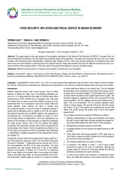
Research articles
Evidence for the transmission of Salmonella from
reptiles to children in Germany, July 2010 to October
2011
M Pees ([email protected])1, W Rabsch2, B Plenz1, A Fruth2, R Prager2, S Simon2, V Schmidt1, S Münch2, P G Braun3
1. Clinic for Birds and Reptiles, University of Leipzig, An den Tierkliniken, Leipzig, Germany
2. National Reference Centre for Salmonella and other bacterial Enterics, Robert Koch Institute, Wernigerode, Germany
3. Institute of Food Hygiene, An den Tierkliniken, Leipzig, Germany
Citation style for this article:
Pees M, Rabsch W, Plenz B, Fruth A, Prager R, Simon S, Schmidt V, Münch S, Braun PG. Evidence for the transmission of Salmonella from reptiles to children in
Germany, July 2010 to October 2011. Euro Surveill. 2013;18(46):pii=20634. Available online: http://www.eurosurveillance.org/ViewArticle.aspx?ArticleId=20634
Article submitted on 21 December 2012 / published on 14 November 2013
This study examines the Salmonella status in reptiles
kept in households with children suffering from gastroenteritis due to an exotic Salmonella serovar, to
obtain information on possible transmission paths. A
number of affected households (n=79) were contacted,
and almost half (34/79) comprised at least one reptile
in the home. Of the households, 19 were further studied, whereby a total of 36 reptiles were investigated.
Samples were taken from the reptiles including the
oral cavity, the cloaca, the skin and, in the case of lizards, the stomach, and isolation of Salmonella strains
was performed using repeated enrichment and typing.
Where the Salmonella serovars of the infected child
and the reptile were identical, typing was followed
by pulsed-field gel electrophoresis (PFGE). Bearded
dragons (Pogona vitticeps) constituted 19 of 36 examined reptiles. Altogether 319 Salmonella isolates were
investigated and 24 different serovars identified in the
reptiles. In 15 of 19 households, an identical serovar
to the human case was confirmed in at least one reptile (including 16 of all 19 bearded dragons examined).
The results demonstrate that reptiles and especially
bearded dragons shed various Salmonella serovars
including those isolated from infected children in the
respective households. Hygiene protocols and parents’
education are therefore highly necessary to reduce
the risk of transmission. From a terminological point
of view, we propose to call such infections ‘ReptileExotic-Pet-Associated-Salmonellosis’ (REPAS).
Introduction
According to Thomas et al. [1] the potential of captive
and wild animals to transmit salmonellae to humans
should not be underestimated, and epidemiological
studies on sources of human salmonellosis should
simultaneously investigate both the human cases and
the wild and domestic animals in contact with them. A
study in 1997 from Canada also estimated that three to
five per cent of human Salmonella cases were associated with exposure to exotic pets (including reptiles,
www.eurosurveillance.org
sugar gliders, and hedgehogs) and involved a great
variety of Salmonella serovars with for example S.
Stanley, S. Poona, S. Jangwani, S. Pomona, S. subsp.
IV 48:g,z51:- (former S. Marina) [2].
Numerous reports exist on the prevalence of Salmonella
enterica in captive reptiles, with recent publications
demonstrating a higher prevalence in lizards (up to
76%) compared to tortoises and turtles [3,4]. Geue and
Löschner [5] showed that reptile collections with purchased animals had a significantly higher prevalence
of Salmonella than collections from pure breeders.
Furthermore, animals from pet shops were more frequently affected (89%) than wild caught animals (59%).
Beside the reptiles per se, the reptile feed, in the form
of rodents, can also cause infections in humans. In
England, a S. Typhimurium definite phage-type was
identified as a source of salmonellosis in humans with
a strong association to those keeping reptiles, and was
also confirmed in frozen feeding-mice originating from
a specific rodent breeding facility [6]. In a further outbreak of S. Typhimurium related to snakes in the United
States (US), pulsed-field gel electrophoresis (PFGE)patterns identical to the human isolates were confirmed for isolates from mice used to feed the snakes
as well as the snakes and the environment [7].
Among reptiles, turtles have been reported to be the
most common source of Salmonella in the 1970s [8].
However, later studies and surveys indicate that other
reptile species, especially lizards, may play a more
important role [9-13]. Measures targeted at the prevention of turtle-associated salmonellosis in the US and
Canada in the 1970s temporary helped to reduce its
occurrence, however reptile-associated salmonellosis
is suspected to be a resurgent problem and estimated
to cause three to 11% of all human salmonellosis cases
in these countries [2,14,15].
1
Most reports of reptile-associated salmonellosis concern babies (less than one year of age) and young
children (up to six years-old). However, adults can be
affected, especially immunocompromised hosts, and
patients with impaired gastric acid production [16].
Fatal outcomes following reptile-associated salmonellosis (RAS) in babies have been observed [17,18]. In
Germany, a recent study [19] reported an increasing
number of salmonellosis related to reptiles, with most
patients being less than one year-old. The aim of this
study was therefore to obtain data on possible links
between captive reptiles and salmonellosis in children
by examining both the children with salmonellosis and
all reptiles kept in the respective households.
Methods
The study was conducted from July 2010 to October
2011. Within this period, the National Reference Centre
(NRC) for Salmonella and other Bacterial Enterics at
the Robert Koch Institute (Wernigerode, Germany)
examined samples from 206 households with salmonellosis in children not older than three years. Of the
Salmonella isolates, 65% (134/206) did not belong to
S. Typhimurium and S. Enteritidis and were therefore of
interest for this study. The responsible federal health
institutes were asked to contact these households. A
total of 79 parents, corresponding to 79 households,
were successfully contacted by mail and asked about
the presence of reptiles in the respective households.
Thirty-five of 79 parents confirmed having reptiles and
were contacted by phone to attend this study. As inclusion criteria for participation, parents had to agree that
all reptiles in the respective household could be sampled and their health status assessed, and the time
period between detection of clinical salmonellosis in
the child and the sampling of the reptiles was not more
than three weeks.
Species and health status of the reptiles were assessed
and sampling was conducted following established
guidelines using sterile cotton swabs (Heinz Herenz,
Hamburg, Germany). Swabs were taken from the oral
cavity, the cloaca, and the skin on the ventral region
of the reptile. For lizards, an additional swab sample
was taken from the stomach. Bacterial isolation and
identification were conducted with repeated enrichment and examination of several colonies in each
sample, in order to find as many different Salmonella
serovars as possible: All samples were immediately
placed into a tube containing Rappaport-Vassiliadis
(RV) medium (Oxoid, Wesel, Germany). This medium
was cultured aerobically for 24h at 39°C. A sample was
then plated onto sheep blood agar as well as XLT-4Agar (Oxoid, Wesel, Germany) and cultured aerobically
for further 24h at 39°C. From the RV bouillon culture,
1 ml was transferred to a new tube containing RV, and
enrichment as well as culture was repeated. This procedure was repeated again, so that in total isolation of
Salmonella spp. was attempted from three enrichment
cycles. From Salmonella-suspicious colonies, at least
five different colonies were subcultured on Brilliant
2
Green agar (Sifin, Berlin, Germany) for confirmatory
testing with biochemical methods. Confirmed colonies
were agglutinated against Salmonella surface antigens
(sera from Sifin, Berlin, Germany) and all strains were
typed at the NRC according to the White-Kauffmann-Le
Minor scheme [20].
The isolates of identical serovars confirmed in the
infected child and the reptile were compared using
PFGE. PFGE was carried out according to the standardised protocol for subtyping Salmonella [21]. 40 strains
(human and animal) were investigated by PFGE. In case
of minor pattern differences between the reptile and
the human isolate of the respective household, the
PFGE was repeated. Interpretation of the PFGE results
followed recent recommendations from the literature
[22-24]: Identical PFGE patterns were considered to represent the same epidemiological type. Depending on
the time the outbreak has been going on and if person
to person spread is the prominent feature, two to three
[23] or up to four differences [22] in PFGE restriction
fragment pattern are considered to be the result of a
single genetic event, and isolates can be designated as
’subpatterns’ or related patterns. Given the maximum
time period (several weeks between initial infection
of the child and final sampling of the reptile) for this
study, more than two differences were considered to
represent an epidemiologically-significant difference.
Results
Nineteen households met the inclusion criteria for
participation in the study and reptiles were thus
investigated. Altogether, 36 reptiles were kept in the
households and included in this study (per household
between 1 and 7, on average 1.9). Details are listed in
Table 1. In these 19 households, Salmonella serovars
isolated from the children belonged mainly to subspecies I (12 households), but also subspecies II (1 household), subspecies IIIa (1 household), subspecies IIIb (2
households) and subspecies IV (3 households) (Table
1). One S. Newport strain (6,8:e,h:1,2:z67, d-tartrate-,
malonate+) was investigated in Paris (Institut Pasteur)
because of its unusual biochemical properties. The
strain exhibited two atypical characteristics: D-tartrate
negative and malonate positive. The ‘e,h’ and the
‘1,2’ flagellar phases have been confirmed by fliC
and fljB sequencing. The multilocus sequence typing
(MLST) profile (ST118) was identical to those of serovar
Newport lineage II [25].
Investigated households were spread well over
Germany, including nine (of 16 possible) federal states
(Bavaria, Baden-Württemberg, Berlin, North RhineWestphalia, Rhineland-Palatinate, Saxony, SaxonyAnhalt, Schleswig-Holstein, Thuringia). From the
investigated households, all children except one were
less than 15 months of age, most (11/19) were less than
six months. All children showed clinical symptoms
of gastroenteritis including fever, with some being
critically ill. Except for three serovars (S. Eastbourne
9,12:e,h:1,5; S. subspec. IV 44:z4,z23:-; S. Monschaui
www.eurosurveillance.org
www.eurosurveillance.org
3
2
6
35
2
3
7
13
5
5
3
4
5
6
7
8
9
10
11
12
S. subspec. IIIb 48:r:z
S. Monschaui 35:m,t:-
S. Kandla 17:z29:-
S. Newport 6,8:e,h:1,2:z67
(d-tartrate-,malonate+)
S. subspec. IIIb 14,24:z10:z
S. subspec. IIIa 41:z4,z23:-
S. Monschaui 35:m,t:-
S. Tennessee 6,7:z29:-
S. subspec. IV 44:z4,z23:-
S. Cotham 28:i:1,5
S. Ealing 35:g,m,s:-
S. Eastbourne 9,12:e,h:1,5
Salmonella serovar
2
2
1
Pogona vitticeps (l)
Pogona vitticeps (l)
Chameleo calyptratus (l)
1
2
1
1
Python sp. (s)
Python sp. (s)
Python sp. (s)
Python sp. (s)
1
2
Pogona vitticeps (l)
0
2
Pogona vitticeps (l)
Python sp. (s)
1
Pantherophis guttatus (s)
Chameleo calyptratus (l)
1
2
Pantherophis guttatus (s)
Pantherophis guttatus (s)
1
4
Pantherophis guttatus (s)
Chameleo calyptratus (l)
2
Pogona vitticeps (l)
1
1
Pantherophis guttatus (s)
3
Pogona vitticeps (l)
3
3
Pogona vitticeps (l)
Pogona vitticeps (l)
Pogona vitticeps (l)
1
1
Pogona vitticeps (l)
3
Pogona vitticeps (l)
N
Pogona vitticeps (l)
Reptile species (type of
reptilea)
S. subspec. IIIb serol. rough (ch)
S. subspec. IIIb serol. rough (cl)
S. subspec. IIIb serol. rough (cl), S. subspec. IIIb 50r:z (cl)
S. subspec. IIIb serol. rough (ch)
a
–
S. Enteritidis 9,12:g,m:-, phage type 8/7 (cl)
S. Enteritidis 9,12:g,m:-, phage type 8/7 (cl)
S. Tennessee 6,7:z29:- (ch, cl); S. Carmel 17:l,v:e,n,x (sk)
S. Tennessee 6,7:z29:- (sk); S. Carmel 17:l,v:e,n,x (ch, sk, st)
S. Kisarawe 11:k:e,n,x (cl)
S. Kisarawe 11:k:e,n,x (ch, cl)
–
S. subspec. IIIb 14,24:z10:z (cl)
S. subspec. IIIb 42:k:z35 (cl)
S. Pomona 28:y:1,7 (cl)
–
–
–
–
–
–
–
–
–
Cl
Cl
Cl
Cl
–
–
–
Cl
S. subspec. IIIb 42:k:z35 (cl); S. Paratyphi B Var.dT+ Java 4,5,12:b:1,2,
phage type Worksop (cl); S. Florida 1,6,14,25:d:1,7 (cl)
S. subspec. IIIb 42:k:z35 (cl)
Cl
Cl
Sk
St
Sk
–
Cl
Ch, cl, sk
Localisation on
reptileb of serovar
identical to child
S. Pomona 28:y:1,7 (cl)
–
S. Enteritidis 9,12:g,m:-, phage type 8/7 (cl); S. Kisarawe 11:k:e,n,x (cl)
S. Eastbourne 9,12:e,h:1,5 (cl); S. Tennessee 6,7:z29:- (cl)
S. Tennessee 6,7:z29:- (cl); S. subspec. I serol. rough (cl)
S. Eastbourne 9,12:e,h:1,5 (cl)
–
S. subspec. I serol. rough (sc, ch); S. subspec. IIIb 50:r:z (cl)
Non-identical isolates to the child (localisation on the reptileb)
Serovars identified in the reptile
PFGE: pulsed-field gel electrophoresis; serol.: serological specificity; sp.:species; subspec.: subspecies;
l: lizard; s: snake; t: tortoise/turtle.
b
ch: choana; cl: cloaca; sk: skin; st: stomach.
c
Where the serovars from the child and the reptile were identical, PFGE was performed; i: identical pattern; s: similar pattern.
3
8
2
11
Age
(months)
1
Household
Child
–
–
i
s (2)
–
i
s (1)
i
i
s (2)
–
s (2)
PGFE resultc
(number
of pattern
differences)
Table 1a
Comparison of Salmonella serovars in reptiles and salmonellosis-affected children living in the same households, with results of pulsed-field gel electrophoresis when the serovars
were identical, Germany, July 2010–October 2011
4
www.eurosurveillance.org
5
5
3
3
16
17
18
19
S. Eastbourne 9,12:e,h:1,5
S. subspec. IV 44:z4,z23:-
S. subspec. IV 48:g,z51:-
S. Kumasi 30:z10:e,n,z15
S. Jangwani 17:a:1,5
1
1
Pogona vitticeps (l)
Mauremys reevesii (t)
1
2
Pogona vitticeps (l)
Pogona vitticeps (l)
2
1
Physignathus cocincinus
(l)
Pogona vitticeps (l)
1
Pogona vitticeps (l)
1
2
Pogona vitticeps (l)
Physignathus cocincinus
(l)
1
2
N
Testudo hermanni (t)
Pogona vitticeps (l)
Reptile species (type of
reptilea)
a
–
–
–
S.Kisarawe 11:k:e,n,x (ch, cl, st)
S. Eastbourne 9,12:e,h:1,5 (cl, sk)
–
–
–
S. Kumasi 30:z10:e,n,z15 (ch, cl)
–
S. Kisarawe 11:k:e,n,x (ch, cl)
Non-identical isolates to the child (localisation on the reptileb)
Serovars identified in the reptile
PFGE: pulsed-field gel electrophoresis; serol.: serological specificity; sp.:species; subspec.: subspecies;
l: lizard; s: snake; t: tortoise/turtle.
b
ch: choana; cl: cloaca; sk: skin; st: stomach.
c
Where the serovars from the child and the reptile were identical, PFGE was performed; i: identical pattern; s: similar pattern.
7
15
S. Potsdam 6,7:l,v:e,n,z15
S. subspec. II 42:r:-
3
11
13
Salmonella serovar
Age
(months)
14
Household
Child
Ch, cl
Cl, sk
Cl, sk
Sk
Ch
Cl
Ch
Ch, cl
Cl
Cl
Ch, st, cl, sk
Localisation
on reptileb
of serovar
identical to
child
i
i
s (2)
i
i
s (1)
i
PGFE resultc
(number
of pattern
differences)
Table 1B
Comparison of Salmonella serovars in reptiles and salmonellosis-affected children living in the same households, with results of pulsed-field gel electrophoresis when the serovars
were identical, Germany, July 2010–October 2011
Table 2
Number of reptile species and representative animals (n=36) in households (n=19) with ‘Reptile-Exotic-Pet-AssociatedSalmonellosis’ (REPAS) human cases and reptile distribution among the households, Germany, July 2010–October 2011
Reptile species
Number of animals
Identical serovars found
in the salmonellosis-affected child
and the reptile
Number of
households
with this
species
Number of households
Number of animals
Bearded dragon
(Pogona vitticeps)
19
13
11
16
Corn snake
(Pantherophis guttatus)
5
3
2
3
Other snakes
5
1
0
0
Chameleon spp.
3
2
0
0
Chinese water dragon (Physignathus cocincinus)
2
1
1
2
Chinese pond turtle (Mauremys reevesii)
1
1
1
1
Hermann’s tortoise (Testudo hermanni)
1
1
1
1
36
19a
15a
23
Total
Spp.: species.
a
The total does not equate the sum of the numerical values in the remaining rows of the column because a given household could comprise
more than one reptile species.
35:m,t:-) found to have caused infections of two children each, the serovars isolated from the children differed from each other.
A total of 319 Salmonella isolates were investigated
and 24 different serovars were identified in the reptiles. The number of serovars within individual reptiles
varied between one and four. Only from one snake was
no Salmonella isolated. In ten reptiles with Salmonella
serovar identical to the salmonellosis-affected child,
no other Salmonella serovar but that identical to the
child was isolated (Table 1).
In 15 of the 19 examined households, an identical
serovar to that of the salmonellosis-affected child was
found in at least one reptile. In six reptiles, the respective identical Salmonella serovar was isolated from the
oral cavity and in two lizards from the stomach. In 17
reptiles with the same serovar as the child, Salmonella
was detected in the cloaca, and in seven the skin swab
sample was positive. The identical serovars found in
the infected children as well as in the reptiles in the
respective households belonged to the following subspecies: I (10), II (1), IIIa (1), IV(3) (details for individual
reptiles and also non-identical serovars are presented
in Table 1).
Bearded dragons (Pogona vitticeps) were the most
common species kept in households with salmonellosis in children (13 of 19 households). Furthermore 16 of
19 of the examined bearded dragons carried an identical Salmonella serovar to the human isolate in the
respective household. In corn snakes (Pantherophis
guttatus), an identical serovar in the snake and the
www.eurosurveillance.org
salmonellosis-affected child was found in three of five
examined animals from two different households. Only
one tortoise and one turtle were kept in households
investigated in this study. These animals, with each an
identical serovar to the salmonellosis-affected child,
were respectively located in two households. Details
are listed in Table 2.
In nine households, the isolates from the infected child
and from the reptile had an identical PFGE profile (PFGE
data not shown). In six households the human and reptile isolates presented related PFGE patterns (repeated
PFGE, Figure 1). Details are also given in Table 1.
Discussion
During the fifteen months period of this study, 65%
(134/206) of all Salmonella infections in children
detected at the NRC were not due to the serovars
S. Typhimurium and S. Enteritidis, which are normally
transmitted via food ingestion. It is therefore possible
that other sources of infection than food, i.e. reptiles in
households, could be of relevance. Nearly half (34/79)
of the parents who were contacted and questioned,
answered that reptiles were present in the respective households. Even though only limited information
exists on the presence of reptiles in Germany in general, this percentage by far exceeds recent estimates
that reptiles were only kept in 1.2% of all German
households in 2010 [26]. This highlights that the presence of reptiles in households appears to coincide with
the occurrence of salmonellosis due to exotic serovars in children. This assumption is also supported
by a study in the US demonstrating that children with
5
Isolate source
(household number)
Salmonella serovar
H (1)
Eastbourne
R (1)
Eastbourne
H (3)
Cotham
R (3)
Cotham
H (6)
Monschaui
R (6)
Monschaui
H (9)
Newport
R (9)
Newport
H (14)
IV 48:g,z51:-
R (14)
IV 48:g,z51:-
R (17)
Potsdam
H (17)
Potsdam
20
40
60
100
150
200
250
300
350
600
500
800
1 ,000
kbp
1,500
2,000
Figure 1
Patterns obtained by XbaI restriction and pulsed-field gel electrophoresis (PFGE) for isolates from children with ‘ReptileExotic-Pet-Associated-Salmonellosis’ (REPAS) (n=6) and reptiles (n=6) living in the same respective households
H: human isolate; R: reptile isolate.
Minor PFGE pattern differences between human and reptile isolates from the same households are shown and indicated by arrows.
confirmed Salmonella infections had more contact to
reptiles and cats in comparison to a control group [27].
In most published case reports on RAS, only reptile faecal samples or faecal swabs were used for Salmonella
detection. In contrast, this study was designed to
obtain as much information as possible on Salmonella
serovars shed by the reptiles. Sampling of the oral
cavity (and stomach) as well as the cloaca should provide information on shedding via both orifices and if
Salmonella are present within the whole digestive system, whereas sampling from the skin should demonstrate whether Salmonella may also be transmitted via
direct contact with the animal. Repeated enrichment
and culture was necessary to provide reliable information on the Salmonella status in the reptiles examined.
Combining the molecular typing using PFGE with other
available data, such as serological typing as in this
study, is highly recommended for accurate analysis
and comparison of samples [24].
The extensive sampling and testing protocol used in
this study is a possible explanation for the high prevalence of Salmonella (in 35 of 36 reptiles) found among
6
the reptiles investigated. Intermittent shedding of
Salmonella in reptiles and the wide array of collection
and sampling techniques have been proposed to be the
main reason for the variability in detection rates [28].
None of the reptiles examined showed clinical signs
indicative for salmonellosis. In consequence, a high
prevalence of Salmonella in reptiles should generally
be assumed, and reptiles should be considered positive for Salmonella until the contrary has been proven,
as reported previously [29].
Up to four different serovars were found within one reptile. Reptiles were frequently colonised with the same
serovars within a given household. This indicates that
Salmonella as a part of the normal flora can spread
amongst individuals within captive reptile collections
and therefore probably shed over long periods of time.
These results are in accordance with observations that
if one reptile carries Salmonella, nearly all other reptiles of the respective owner are also affected [5].
Most Salmonella isolates were found in cloacal swabs.
However, since in some reptiles the identical serovar
to the salmonellosis-affected child was only found on
www.eurosurveillance.org
the skin or the stomach, it can be assumed that shedding via the cloaca is intermittent. A negative cloacal or
faecal sample will therefore not prove that the animal
does not harbour Salmonella. Given the anatomy and
behaviour of most reptiles, the presence of Salmonella
on skin samples was not surprising but underlines the
general transmission risk if handling the reptiles is not
followed by appropriate hand washing. In conclusion,
considering the various shedding sites, several sampling points should be used to increase the detection
rate of Salmonella in reptiles.
In the majority (15/19) of the examined households, an
identical serovar was found in the infected child and
at least one reptile from the respective household. The
epidemiological association between these isolates
was first confirmed using biochemical typing. PFGE
confirmed all reptiles, with either completely identical
patterns (nine households) or only minor differences
in up to two fragments indicating single genetic variations (six households, Figure 1), as described in disease outbreaks [24]. Most serovars found belonged to
subspecies I and some have already been described
as part of the reptile flora or even as potential human
pathogens [2,8,28]. One serovar, S. Tennessee was isolated from four reptiles in three households. However,
so far there seems to be no specific serovar that is of
special zoonotic importance. In consequence, at the
moment all Salmonella strains found in reptiles need to
be considered to be potentially infectious for children.
An important finding of this study was that the reptile
species involved could play an important role in transmission of Salmonella to children. Little data exists
on the proportion of reptile species kept as pets in
Germany. In a post-mortem survey, about 17% were
water turtles, 34% tortoises, 22% snakes and 27%
lizards (including 1.8% bearded dragons) [30]. In one
of the authors’ clinic specialising in reptile medicine,
about 12% of more than 1,000 reptiles presented in
the period of the running study were bearded dragons.
However, in this study, the majority (13/19) of all households studied kept bearded dragons (Pogona vitticeps),
and identical Salmonella strains to the salmonellosisaffected child were confirmed in 16 of 19 of these reptiles. In contrast, only one turtle and one tortoise were
kept in the affected households studied. This result is
in strong contrast to early reports of possible RAS in
humans, where mainly turtles were suspected to be
a source of infection. Interestingly, more recent case
reports also indicate that bearded dragons present a
risk for transmission of salmonellosis to young children [31,32].
Weiss et al. [12] reported a marked increase in infections in infants caused by S. Tennessee in Germany in
2008 and indicated the possibility of bearded dragons being a natural reservoir for this serovar, as this
species was kept in seven of 16 households affected
and S. Tennessee was confirmed in three households.
Furthermore, probable transmission from bearded
www.eurosurveillance.org
dragons has also recently been described in adult
humans [33]. One explanation for the increasing number of reports on bearded dragons as a source of
Salmonella infection in humans in recent years might
be that this species is becoming more popular as a pet,
and that bearded dragons are more often handled and
petted due to their peaceful nature compared to several
other reptile species. Since nearly all children included
in this study were too young to handle reptiles themselves, it is likely that vectors, such as the parents or
the environment may have played a role. Furthermore it
is conceivable that bearded dragons naturally harbour
a variety of potentially zoonotic Salmonella serovars.
The fact that more than one serovar of Salmonella was
isolated in 13 of a total of 19 bearded dragons supports
this assumption.
Husbandry conditions and the households’ hygiene
status were not assessed according to a protocol, as
this was not agreed upon with the owners. However,
the overall impression was that most of the households visited demonstrated at least some deficits that
needed corrections and advice, e.g. the reptile terrarium placed in the kitchen or in the same room as the
child’s bed. Further investigations in the environment
of the reptile and possible interactions between the
reptile and the persons in the households could give
important information on possible transmission paths.
None of the owners was aware of any risk of infection
due to reptiles before, and all were therefore either
willing to improve the situation or decided not to keep
reptiles any longer.
All publications cited above indicate that although
infections attributed to exposure to reptiles and other
exotic pets represent only a small proportion of all
human salmonellosis cases, it is likely an underestimated and growing problem in Europe [34] and in the
US [35] that deserves closer attention. In 2007 more
than 500,000 reptiles were imported to Germany only
via the Frankfurt/Main airport [36]. According to the
European Surveillance System (TESSy) database, the
number of infections affecting children younger than
three years with serovars associated to reptiles also
increased in the last years (Figure 2). Beside the focus
on exotic serovars as in this study, it should be kept in
mind that there is also a potential risk for the transmission of S. Typhimurium and S. Enteriditis due to reptiles
or as already described, via the reptile feed [6,7]. Also
these common serotypes can be shedded by reptiles
and therefore have a potential to cause salmonellosis
in humans. For example, in this study S. Paratyphi B
variant java was isolated from a corn snake. Although
the identical serovar has not been found in the child
from the respective household, this isolate belongs to
phage type Worksop and is therefore of relevance for
human infections.
From a terminological point of view, in addition to
an earlier suggestion (RAS [37]), we propose that
Salmonella infections related to reptiles as observed
7
Figure 2
Occurrence of ‘Reptile-Exotic-Pet-Associated-Salmonellosis’ (REPAS) serovars in children less than three years-old in the
European Union, 2007–2010
100
2007
90
2008
2009
2010
80
70
Number of cases
60
50
40
30
20
Subsp. IV
Subsp. IIIb
Subsp. IIIa
Arizona
Tennessee
Poona
Pomona
Oranienburg
Monschaui
Eastbourne
Ealing
Carmel
0
Apapa
10
Salmonella serovar
Salmonella Arizona includes subspecies IIIa and subspecies IIIb. Arrows indicate where REPAS cases have increased in 2010 compared to
previous years.
Data from the European Surveillance System (TESSy) (29 May 2012).
in this study be called ‘Reptile-Exotic-Pet-AssociatedSalmonellosis’ (REPAS) to give an indication of a possible source of exposure. The main argument for this
proposal is that over the last years, the way of trading
reptiles has changed considerably and this will probably continue in the future. The main risk of Salmonella
transmission from reptiles to humans is not due to
European wild species, but to ‘exotic’ (non-native) reptile species, as the results of this study also demonstrate. Furthermore, following recent examinations,
Salmonella shedding is higher in reptiles kept in captivity in comparison to wild reptiles [5,28] and ‘pet’ reptiles are obviously in closer contact to humans. These
arguments justify the inclusion of ‘exotic pet’ into the
term describing the problem. The risk to human health
connected to the reptile pet market has been highlighted recently [38] and the accurate description of
the problem using REPAS might be important to convey
the problem in education and assist risk managers in
giving recommendations to harmonise animal welfare
and public health.
with human health, should be responsible to inform
more about this problem. Given the fact that most owners in this study were unaware of the risk of Salmonella
transmission from reptiles, we recommend that, in
pet shops, any new exotic pet owner be provided
with appropriate information sheets on Salmonella
prevention. In the authors’ opinion the risk of REPAS
can easily be minimised, using a reasonable management protocol without the need to remove reptiles from
households with young children.
Acknowledgements
The authors thank François-Xavier Weill, Centre for Reference
and Research on Salmonella, Enteric Bacterial Pathogens
Unit, Institut Pasteur, Paris, for the confirmation of the unusual S. Newport. We thank the Member States in the FWD
network for submitting detailed serovar data, and Johanna
Takkinen, European Center for Disease Prevention and
Control (ECDC) for the European data. Furthermore, we thank
Marita Wahnfried, Susanne Kulbe, Dagmar Busse for skillful
technical assistance, and Rachel Marschang, University of
Hohenheim, for linguistic support.
Concluding, emphasis on educational measures will be
key to reduce the risk of Salmonella transmission from
reptiles to children. Professionals dealing with reptiles
(pet shop owners, veterinarians, breeders) should be
aware of such a risk and, together with those dealing
8
www.eurosurveillance.org
References
1. Thomas AD, Forbes-Faulkner JC, Speare R, Murray C.
Salmonellosis in wildlife from Queensland. J Wildl Dis.
2001;37(2):229–38.
http://dx.doi.org/10.7589/0090-3558-37.2.229
PMid:11310872
2. Woodward DL, Khakhria R, Johnson WM. Human salmonellosis
associated with exotic pets. J. Clin Microbiol. 1997;35(11):278690.
PMid:9350734 PMCid:PMC230062
3. Pasmans F, Martel A, Boyen F, Vandekerchove D, Wybo I, Van
Immerseel F, et al. Characterization of Salmonella isolates from
captive lizards. Vet Microbiol. 2005;110(3-4):285–91.
http://dx.doi.org/10.1016/j.vetmic.2005.07.008
PMid:16153787
4. Kikillus KH, Gartrell BD, Motion E. Prevalence of Salmonella
spp., and serovars isolated from captive exotic reptiles in New
Zealand. N Z Vet. J. 2011;59(4):174-8.
http://dx.doi.org/10.1080/00480169.2011.579246
PMid:21660846
5. Geue L, Löschner U. Salmonella enterica in reptiles of German
and Austrian origin. Vet Microbiol. 2002;84 (1-2):79-91.
http://dx.doi.org/10.1016/S0378-1135(01)00437-0
6. Harker KS, Lane C, De Pinna E, Adak GK. An outbreak of
Salmonella Typhimurium DT191a associated with reptile feeder
mice. Epidemiol Infect. 2011;139(8):1254-61.
http://dx.doi.org/10.1017/S0950268810002281
PMid:20943001
7. Fuller CC, Jawahir SL, Leano FT, Bidol SA, Signs K, Davis C, et
al. A multi-state Salmonella Typhimurium outbreak associated
with frozen vacuum-packed rodents used to feed snakes.
Zoonoses Public Health. 2008;55(8-10):481-7.
PMid:18833597
8. Chiodini RJ, Sundberg JP. Salmonellosis in reptiles: a review.
Am J Epidemiol. 1981;113(5): 494–9.
PMid:7013476
9. Willis C, Wilson T, Greenwood M, Ward L. Pet reptiles
associated with a case of salmonellosis in an infant were
carrying multiple strains of Salmonella. J Clin Microbiol.
2002;40(12):4802–3.
http://dx.doi.org/10.1128/JCM.40.12.4802-4803.2002
PMid:12454202 PMCid:PMC154656
10. Center for Disease Control and Prevention (CDC). Reptileassociated salmonellosis – selected states, 1998-2002. MMWR
Morb Mort Wkly Rep. 2003;52(49):1206-9.
PMid:14668712
11. Kaibu H, Iida K, Ueki S, Ehara H, Simasaki Y, Anzai H, et
al. Salmonellosis of infants presumably originating from
an infected turtle in Nagasaki, Japan. Jpn J Infect Dis.
2006;59(4):281.
PMid:16936357
12. Weiss B, Rabsch W, Prager R, Tietze E, Koch J, Mutschmann F,
et al. Babies and bearded dragons: sudden increase in reptileassociated Salmonella enterica serovar Tennessee infections,
Germany 2008. Vector Borne Zoonotic Dis. 2011;11(9):1299–
301.
http://dx.doi.org/10.1089/vbz.2010.0239
PMid:21612527
13. Hernández E, Rodriguez JL, Herrera-León S, García I, de
Castro V, Muniozguren N. Salmonella Paratyphi B var Java
infections associated with exposure to turtles in Bizkaia,
Spain, September 2010 to October 2011. Euro Surveill.
2012;17(25):pii= 20201. Available from: http://www.
eurosurveillance.org/ViewArticle.aspx?ArticleId=20201
PMid:22748004
14. Warwick C, Lambiris AJ, Westwood D, Steedman C. Reptilerelated salmonellosis. J R Soc Med. 2001;94(3):124-6.
PMid:11285792 PMCid:PMC1297927
15. Mermin J, Hutwagner L, Vugia D, Shallow S, Daily P, Bender J,
et al. Reptiles, amphibians, and human Salmonella infection: a
population-based, case-control study. Clin Infect Dis. 2004;38
Suppl 3:S253-61.
http://dx.doi.org/10.1086/381594
PMid:15095197
16. Stam F, Römkens TE, Hekker TAM, Smulders YM. Turtleassociated human salmonellosis. Clin Infect Dis.
2003;37(11):e167-9.
http://dx.doi.org/10.1086/379612
PMid:14614690
17. Baby dies of Salmonella poona infection linked to pet reptile.
Commun Dis Rep CDR Wkly. 2000;10(18):161.
18. Wisconsin Division of Public Health (DPH). Disease fact sheet
series: Reptile associated salmonellosis. Madison: Wisconsin
Department of Health Services; 2004. P-42082.
www.eurosurveillance.org
19. Böhme H, Fruth A, Rabsch W. Reptilien-assoziierte
Salmonelleninfektionen bei Säuglingen und Kleinkindern in
Deutschland. [Reptile-associated Salmonellosis in Infants in
Germany]. Klin Padiatr. 2009;221(2):60-4. German.
http://dx.doi.org/10.1055/s-0028-1112156
PMid:19263324
20. Grimont PA, Weill FX. Antigenic formulae of the Salmonella
serovars. Paris: WHO Collaborating Centre for Reference and
Research on Salmonella, Institut Pasteur; 2007.
21. Ribot EM, Fair MA, Gautom R, Cameron DN, Hunter SB,
Swaminathan B, et al. Standardization of pulsed-field gel
electrophoresis protocols for the subtyping of Escherichia coli
O157:H7, Salmonella, and Shigella for PulseNet. Foodborne
Pathog Dis. 2006;3(1):59-67.
http://dx.doi.org/10.1089/fpd.2006.3.59
PMid:16602980
22. Van Belkum A, Tassios PT, Dijkshoorn L, Haeggman S, Cookson
B, Fry NK, et al. Guidelines for the validation and application
of typing methods for use in bacterial epidemiology. Clin
Microbiol Infect. 2007;13 Supp 3:1–46.
http://dx.doi.org/10.1111/j.1469-0691.2007.01786.x
PMid:17716294
23. Goering RV. Pulsed field gel electrophoresis: A review of
application and interpretation in the molecular epidemiology
of infectious disease. Infect Genet Evol. 2010;10(7):866–75.
http://dx.doi.org/10.1016/j.meegid.2010.07.023
PMid:20692376
24.Barrett TJ, Gerner-Smidt P, Swaminathan B. Interpretation of
pulsed-field gel electrophoresis patterns in foodborne disease
investigations and surveillance. Foodborne Pathog Dis.
2006;3(1):20-31.
http://dx.doi.org/10.1089/fpd.2006.3.20
PMid:16602976
25. Sangal V, Harbottle H, Mazzoni CJ, Helmuth R, Guerra
B, Didelot X, et al. Evolution and population structure
of Salmonella enterica serovar Newport. J Bacteriol.
2010;92(24):6465-76.
http://dx.doi.org/10.1128/JB.00969-10
PMid:20935094 PMCid:PMC3008538
26. Statistica. Anteil der Haushalte in Deutschland mit Haustieren
nach Tierarten in den Jahren 2008 bis 2012. Statistica.
[Accessed 19 Sep 2012]. German. Available from: http://
de.statista.com/statistik/daten/studie/30153/umfrage/
haustiere-heimtierhaltung-in-deutschen-haushalten-seit-2008/
27. Younus M, Wilkins MJ, Davies HD, Rahbar MH, Funk J, Nguyen
C, et al. The role of exposures to animals and other risk factors
in sporadic, non-typhoidal Salmonella infections in Michigan
children. Zoonoses Public Health. 2010;57(7-8):e170-6.
http://dx.doi.org/10.1111/j.1863-2378.2010.01324.x
PMid:20202185
28.Scheelings TF, Lightfoot D, Holz P. Prevalence of Salmonella in
Australian reptiles. J Wildlife Dis. 2011;47(1):1–11.
http://dx.doi.org/10.7589/0090-3558-47.1.1
PMid:21269991
29. Pasmans F, Blahak S, Martel A, Pantchev N. Introducing
reptiles into a captive collection: The role of the veterinarian.
Vet J. 2008;175(1):53–68.
http://dx.doi.org/10.1016/j.tvjl.2006.12.009
PMid:17346998
30. Sinn AD. Pathologie der Reptilien - eine retrospektive Studie
[PhD thesis]. Munich: Ludwig-Maximilian-University; 2004.
German.
31. Tabarani CM, Bennett NJ, Kiska DL, Riddell SW, Botash
AS, Domachowske JB. Empyema of preexisting subdural
hemorrhage caused by a rare Salmonella species after
exposure to bearded dragons in a foster home. J Pediatr.
2010;156(2):322–3.
http://dx.doi.org/10.1016/j.jpeds.2009.07.050
PMid:20105642
32. Haase R, Beier T, Bernstadt M, Merkel N, Bartnicki J.
Neugeborenensepsis durch Salmonella Apapa nach
Reptilienkontakt im Haushalt. [Neonatal infection with
Salmonella apapa after contact with a reptile in the home]. Z.
Geburtshilfe Neonatol. 2011; 215(2):86–8. German.
http://dx.doi.org/10.1055/s-0031-1275308
PMid:21541908
33. Lowther SA, Medus C, Scheftel J, Leano F, Jawahir S, Smith K.
Foodborne outbreak of Salmonella subspecies IV infections
associated with contamination from bearded dragons.
Zoonoses Public Health. 2011;58(8):560–6.
http://dx.doi.org/10.1111/j.1863-2378.2011.01403.x
PMid:21824356
34. Editorial team, Bertrand S, Rimhanen-Finne R, Weill FX,
Rabsch W, Thornton L, et al. Salmonella infections associated
with reptiles: The current situation in Europe. Euro Surveill.
2008;13(24):pii=18902. Available from: http://www.
eurosurveillance.org/ViewArticle.aspx?ArticleId=18902
9
35. Center for Disease Control and Prevention (CDC). Notes from
the field: outbreak of salmonellosis associated with pet turtle
exposures - United States, 2011. MMWR Morb.Mort Wkly Rep.
2012;61(4):79.
PMid:22298304
36. Hatt JM, Fruth A, Rabsch W. Reptilienassoziierte Salmonellosen
- aktuelle Informationen für Tierärztinnen und Tierärzte.
[Reptile-associated salmonellosis –information update for
veterinarians]. Tierärztliche Praxis. 2009;37(3):188-93.
German.
37. De Jong B, Andersson Y, Ekdahl K. Effect of Regulation and
Education on Reptile-associated Salmonellosis. Emerg Infect
Dis. 2005;11(3):398–403.
http://dx.doi.org/10.3201/eid1103.040694
PMid:15757554 PMCid:PMC3298264
38. Arena PC, Steedman C, Warwick C. Amphibian and reptile
pet markets in the EU: An investigation and Assessment.
Düsseldorf: Animal public e.V.; 2012. [Accessed 15 Dec
2012]. Available from: http://animal-public.de/wp-content/
uploads/2012/04/ARPM2012_v131.pdf
10
www.eurosurveillance.org


![This article was downloaded by: [Khalifa, Sherif] On: 8 March 2011](http://cdn1.abcdocz.com/store/data/000032200_2-5d0635dd9933f87aa2c8ba900272991f-250x500.png)

© Copyright 2026