
Committee on Medical Aspects of Radiation in the Environment (COMARE)
Committee on Medical
Aspects of Radiation in
the Environment
(COMARE)
TWELFTH REPORT
The impact of personally initiated X-ray computed tomography scanning for
the health assessment of asymptomatic individuals.
Chairman: Professor A Elliott
© Crown Copyright 2007
Produced by the Health Protection Agency for the
Committee on Medical Aspects of Radiation in the Environment
ISBN 978-0-85951-611-2
CONTENTS
Page
Foreword
5
Chapter 1
Introduction
7
Chapter 2
Rationale
10
Chapter 3
CT scanning of the whole body
20
Chapter 4
CT scanning of the lung
26
Chapter 5
Cardiac CT scanning
32
Chapter 6
CT scanning of the colon
38
Chapter 7
CT scanning for other conditions
45
Chapter 8
Conclusions
49
Chapter 9
Recommendations
51
References
54
Acknowledgements
65
Appendix A
Glossary
69
Appendix B
List of COMARE Members, Secretariat and Assessors
76
Appendix C
Declarations of Interests
80
3
FOREWORD
i
The Committee on Medical Aspects of Radiation in the Environment
(COMARE) was established in November 1985 in response to the final
recommendation of the report of the Independent Advisory Group chaired by
Sir Douglas Black (Black, 1984). The terms of reference for COMARE are:
“to assess and advise Government and the Devolved Authorities on
the health effects of natural and man-made radiation and to assess the
adequacy of the available data and the need for further research”
In over 20 years of providing advice to Government and the Devolved
Authorities, COMARE has produced 11 major reports and many other
statements and documents related mainly to exposure to naturally occurring
radionuclides such as radon and its progeny or to man-made radiation, usually
that emitted by major nuclear installations.
ii
Medical exposures account for over 97% of the annual dose to the UK
population from artificial sources of radiation, and constitute approximately
15% of the average annual dose to the UK population, although obviously there
will be marked variations in the doses received by individuals in the UK
depending on their unique exposure characteristics. While technical
innovations promise greater diagnostic accuracy and an increased range of
clinical applications, there is also the potential for greater radiation doses to
individuals, from interventional techniques and from changes of practice within
X-ray computed tomography (CT). This potential increase is a marked
difference to the trends in radiation dose to patients observed during the 1990s,
where doses to patients were seen to be falling consistently.
iii
The Department of Health’s radiation protection concerns increasingly
centre on medical exposures. As a consequence of this concern, the Department
of Health in 2005 instructed COMARE to include within its sphere of interest,
radiation protection from medical practices. It was decided that the use of CT
scanning of the asymptomatic individual was to be addressed as a first priority.
To achieve an appropriate review of this topic, COMARE established its
Medical Practices Subcommittee. The subcommittee incorporates members
from COMARE, the Royal College of Radiologists, the Royal College of
Physicians, the independent sector and also a patient representative.
iv
The subcommittee’s terms of reference are:
“to advise COMARE on the health effects arising from medical and
similar practices involving the use of ionising and non-ionising
radiation through assessment of the available data and to inform
COMARE of further research priorities”
When the subcommittee had finished its deliberations they were passed to
COMARE for consideration by the full committee with the aim that in due
5
course COMARE would present its advice to the Department of Health. That
advice is contained in this, our Twelfth Report.
v
The aim of this report has been to provide advice for the Department of
Health on the practice of personally initiated CT scanning of asymptomatic
individuals. Consequently, this report does not consider the relevance of such
scanning techniques to a population screening programme. It focuses solely on
the context of scanning the individual.
6
CHAPTER 1
INTRODUCTION
1.1
X-ray computed tomography (CT), originally known as computed axial
tomography (CAT), uses specialised X-ray equipment to obtain image data
from different angles around the body. Digital processing of the information
results in detailed cross-sectional images of body tissues and organs in either a
two- or three-dimensional format.
1.2
The idea of CT was conceived in 1967 in England by
Godfrey Newbold Hounsfield at the THORN EMI Central Research
Laboratories, being publicly announced in 1972. The system was
independently developed by Allan McLeod Cormack at Tufts University and
they shared a Nobel Prize in medicine in 1979. The original prototype,
developed in 1971, used americium as the gamma source, and took 160 parallel
readings over 180 angles, each 1° apart. It took 9 days to collect sufficient
information about the object being scanned and a further 2.5 hours to reconstruct
the data into an image. Later the gamma source was replaced by a more
powerful X-ray source, which reduced the scanning time (Hounsfield, 1973).
1.3
The first commercial CT system (the EMI scanner) was installed in
Atkinson Morley’s Hospital in Wimbledon. It was limited to making
tomographic sections of the brain and the first patient brainscan using the
machine was obtained in 1972. To reduce the dynamic range of the radiation
reaching the detectors, the machine required the use of a water-filled Perspex
tank, with a pre-shaped rubber head cap enclosing the patient’s head. However,
the machine did acquire the image data, taking between 4.5 and 20 minutes per
180° scan, with 7 minutes required to process each image (Beckmann, 2006).
1.4
CT scanners have gone through multiple phases of technological
developments from single slice static machines to single slice spiral/helical
machines. In the last six years, there have been further significant advances
in CT technology. In particular, the recent development of multidetector or
multislice CT has increased the speed of scanning and enabled high resolution
reconstruction of images in all planes. Other recent developments allow
consideration of volume CT acquisition. A chest examination that previously
required 10 separate breath-holds of 10 seconds each can now be performed
with a single 10-second breath-hold. Software packages have been developed
to utilise these benefits, with modern scanners being able to reconstruct a study
of 1000 images in less than 30 seconds. In addition, as with most digital
technologies, these developments have been achieved with a decrease in the
real costs of equipment. The result has been increased patient throughput at
lower costs per capita, which has consolidated CT as a major first-line
diagnostic modality and established its potential as an imaging modality
for screening.
7
1.5
Radiation exposure from medical practices accounts for the largest UK
population dose from artificial sources of radiation and, within the UK, the use
of CT has doubled in the last 10 years. Of the dose received from medical
X-rays, more than 47% is contributed by CT scans (Hart and Wall, 2004). The
average annual dose from both natural and artificial radiation is 2.7 mSv, of
which medical radiation exposure accounts for around 15% (Hughes et al,
2005). Currently in the UK, over 90% of CT scans are undertaken on NHS
patients. However, there appears to be an increasing trend for commercial CT
scans to be offered to the general population. CT scanning now has the
potential to contribute to preventative healthcare through its use in the scanning
of asymptomatic individuals, either with whole body scanning or through
imaging of specific anatomical regions.
1.6
CT scanning of the asymptomatic individual is marketed directly to the
public as a form of preventative medicine to give individuals peace of mind.
Under this premise, scanning is currently offered for several anatomical
regions:
(i)
whole body (from the neck to the pubic symphysis bone),
(ii)
cardiac,
(iii)
lung,
(iv)
colon.
CT scans can reveal the early stages of deformity, deterioration and disease.
While there may be benefits from this approach, there are also detriments to be
considered, such as those from the radiation dose involved and the
psychological impact on the individual. It is important to balance the medical
science, patient care, ethics and economics of such tests (Council on Scientific
Affairs, 2003).
1.7
Clinically, CT screening is currently performed for people thought to
be at risk for specific diseases. Lung cancer is the primary cause of cancerrelated deaths worldwide, due to the difficulty of early detection at a surgically
curable stage. Indeed CT has been used to screen for lung cancer in Japan since
1993 (Kaneko et al, 2000). There is, however, dispute over the efficacy of
using CT screening for lung cancer identification and whether mortality rates
are reduced. Atherosclerotic disease is one of the leading causes of death in the
western world, and coronary calcification measured by CT is considered to be
an early indicator in asymptomatic patients believed to be at risk. Virtual CT
colonography has been used to examine the colon and rectum to detect polyps
and cancers in asymptomatic individuals who are considered to be at risk of
developing a colorectal cancer.
1.8
Dependent on research findings and technological advancements, some
pathologies could, ultimately, be considered for the introduction of a screening
programme for the general population. However, a screening programme
should be developed as part of an integrated system that incorporates the
provision of adequate information for the individual being tested as well as the
facility for further investigations and treatments (BMA, 2005). In this report,
only the scanning of asymptomatic individuals is considered and not the use of
these techniques for a population screening programme.
1.9
While CT scanning has advanced the accuracy of diagnostic radiology
and its benefits are clear, there are potential detriments also. The level of
radiation dose received by the individual may be significant. CT scanning of
asymptomatic individuals can result in detection of a range of pathologies
8
including conditions of no clinical significance and conditions that will not
influence an individual’s outcome, both situations that can be considered as
examples of pseudodisease. Such findings could potentially result in needless
further investigations, which themselves carry additional risks and cost
implications, increase the individual’s anxiety levels and affect their quality of
life. These adverse features are discussed further in Chapter 2. There are also
important issues concerning sensitivity and specificity. Scans of specific
anatomical regions should be optimised for this purpose and may not be able to
detect conditions other than those targeted. It may not be possible to give an
asymptomatic individual a complete ‘all clear’ after a scan, even though such
reassurance is the expectation of the scanned individual. It is also not clear
whether CT imaging detects some cancers (eg lung) that are not as clinically
aggressive as those identified following presentation with symptoms. It is
possible that some tumours detected are ones that might be present at the
individual’s death and would not have been life threatening, skewing the
apparent benefit of detection by scanning. In this report, the term
‘overdiagnosis’ refers to the detection of malignant disease that would never
present clinically during the lifetime of the individual.
1.10
The level of radiation received by the individual is an additional
concern, particularly with whole body CT scanning. For an asymptomatic
individual the potential risk may outweigh the benefits. The dose received by
an individual can vary substantially depending on the type of scan employed
and the machine and protocol used.
1.11
This report looks at the justification of these scanning procedures,
including CT scans of the whole body and of key anatomical areas, the latter
being designed to allay the concerns of asymptomatic individuals with regard
to defined diseases.
1.12
It has taken into consideration published peer-reviewed data and
evidence regarding CT scanning of asymptomatic individuals and information
provided by a number of independent sector organisations offering such
services. The latter included commercially sensitive information which has not
been reproduced within the report.
1.13
The report consists of nine chapters, which include this introduction, a
chapter on the rationale behind CT scanning of the asymptomatic individual,
five chapters on scanning anatomical regions – whole body, lung, heart and
colon and more general scanning. The final two chapters provide conclusions
and recommendations. A glossary and references are provided.
9
CHAPTER 2
RATIONALE
Background – radiation
protection principles and
regulatory factors
2.1
In 1990 the International Commission on Radiological Protection
reaffirmed three key principles of radiation protection (ICRP, 1991):
(i)
justification – exposure to radiation must produce sufficient
benefit to the exposed individuals, or to society, to offset the potential
radiation detriment,
(ii) optimisation – implementing procedures and techniques to keep
exposures as low as reasonably practicable, economic and social
factors being taken into account,
(iii) dose limitation – keeping radiation doses received within specified
limits.
2.2
In the context of medical exposures, justification and optimisation
apply. Dose limitation is not used for medical exposures but the concept of
diagnostic reference levels has been introduced to support the control and
periodic reduction of radiation doses from diagnostic procedures.
2.3
The hazards associated with ionising radiation are addressed in a
number of European Council Directives and subsequently in legislation in
Great Britain and Northern Ireland. For CT scanning, and the protection of the
asymptomatic individual, two sets of regulations are of particular importance:
(i)
The Justification of Practices Involving Ionising Radiation
Regulations (2004),
(ii)
The Ionising Radiation (Medical Exposure) Regulations (2000).
2.4
The guidance on the application and administration of The Justification
of Practices Involving Ionising Radiation Regulations (2004) indicates a range
of types of practice that existed prior to 13 May 2000, the implementation date
for the EC Basic Safety Standards Directive 96/29/Euratom. This Directive
establishes the need for the justification of new classes or types of practice
involving ionising radiation. Medical exposures using CT for diagnosis are
included as an existing type of practice within this guidance. CT scanning of
the asymptomatic individual is considered to be early diagnosis and is therefore
incorporated as an existing practice.
2.5
The Ionising Radiation (Medical Exposure) Regulations 2000 require
that all individual medical exposures are referred, justified and optimised. The
Regulations require an identified person to take legal responsibility for
deciding whether an individual medical exposure is justified. In the UK, this
person is known as the ‘practitioner’. Such persons must be adequately trained
to carry out the task of justification and be entitled to do so by their employer.
Justification of exposures must take into account medical information about the
individual provided by a referrer (also a duty holder under the Regulations) and
should be based on the available scientific evidence. Both the referrer and the
practitioner must be registered healthcare professionals.
10
2.6
Justification cannot be retrospective. Since all persons exposed suffer
radiation detriment, the practitioner’s decision as to whether an individual
medical exposure is justified must be made prior to the exposure, and must be
valid whether the test result is subsequently positive or negative. Procedures
can only be justified if the individual for whom the exposure is proposed will
receive a predictable benefit that outweighs the detriment, or if there is an
overall net benefit to society.
2.7
Optimisation of every medical exposure is the responsibility of the
practitioner and of the operator who undertakes the practical aspects of a
medical exposure, to the extent of their respective involvement. To assist in
optimisation, the employer must ensure that written protocols are in place for
all standard procedures which must be specific to each piece of equipment.
Such protocols should include exposure factors for each routine examination.
Potential benefits and
detriments
2.8
Depending on the initial clinical suspicion as to the presence of disease,
the potential benefits from a diagnostic medical exposure are:
(i)
evidence in favour of, or against, a suspected diagnosis,
(ii)
monitoring the progress of known disease,
(iii)
identification of an unsuspected pathology.
2.9
In all cases these benefits must be balanced against the risks, not only
from the radiation detriment but also from potentially misleading or inaccurate
results. The magnitude of the latter risks depends strongly on the disease
prevalence, and the most appropriate diagnostic investigations are likely to be
different in groups with suspected, as opposed to unsuspected, disease. A test
which is considered justifiable for patients presenting with certain symptoms
may not necessarily be justifiable or clinically valuable when applied to an
asymptomatic individual.
Testing a potential
diagnosis
2.10
Guidance on the balance of risks and benefits for medical exposures
has been produced by international bodies (EC, 2001) and UK professional
bodies (RCR, 2003). These guidelines are intended primarily to assist the
person referring a patient for a medical exposure, and provide clear
recommendations stating which procedures are likely to be useful, and which
are not, for a wide range of clinical conditions and suspected diagnoses. The
guidelines also state the relative radiation detriment for each procedure,
assisting the practitioner and others to assess whether the likely benefit justifies
the risk.
Monitoring known disease
2.11
In these cases the individual disease status and clinical condition of the
patient will primarily determine the justifiable level of risk. Nevertheless,
practitioners still have a duty to ensure that the benefit to be gained in terms of
improved or altered treatment is sufficient to outweigh the risk, especially in
the case of chronic minor disease.
Discovering unsuspected
pathology
2.12
There may be benefits from detecting wholly unexpected findings, and
there are numerous reported cases where serious pathology has been revealed
during an examination undertaken for another purpose. However, the overall
likelihood of discovering wholly unsuspected significant disease is low, and the
justification for the examination cannot have been based on the benefits that
may have been conferred by the unsuspected finding, since by definition the
practitioner was not aware of this potential outcome when the procedure was
justified. It follows that justification for scanning asymptomatic individuals can
be based only on the first potential benefit listed above, which in turn requires a
suspected diagnosis.
11
Risk factors and
suspected diagnoses
2.13
The likelihood that an asymptomatic individual has a suspected
diagnosis will vary according to the presence or absence of associated risk
factors. In some cases, these factors will be sufficiently well correlated with the
disease in question as to raise the likelihood to a level at which a higher risk
investigation may be justifiable. A practitioner using such factors to justify a
medical exposure would require an evidence base indicating probability of
disease in relevant populations.
2.14
Earlier diagnosis does not always improve prognosis, and there may be
no benefit from diagnosing some indolent pathologies which would have
remained asymptomatic. The term pseudodisease is used to describe disease
that does not affect the length or quality of a patient’s life. A large amount of
pseudodisease within a population may render a screening programme
ineffective and, for an individual, may result in additional testing with potential
morbidity and mortality. Two types of pseudodisease occur. Type I is disease
detected on screening that does not necessarily progress to symptomatic
disease. Type II is indolent disease that occurs in patients dying from other
causes. An example might be prostatic carcinoma, with the vast majority of
men diagnosed with clinically localised prostatic carcinoma dying with rather
than of their disease (Albertsen et al, 2005; Potosky et al, 2000).
2.15
Excess cancer risk has not been demonstrated by epidemiological studies
at doses below 100 mSv. The estimated risks from diagnostic medical exposures
are based primarily on extrapolation from the observed increased incidence of
cancer in exposed populations, generally at higher doses. These groups include
the Japanese atomic-bomb survivors (Preston et al, 2004), patients treated with
radiotherapy for benign conditions (Weiss et al, 1994), children irradiated
in utero (Doll and Wakeford, 1997), and workers occupationally exposed
(Cardis et al, 1995). The internationally recommended risk factor for fatal
cancer induction is 5% per Sv for an adult population or 0.005% per mSv using
the linear non-threshold hypothesis (ICRP, 1991). Risks are generally higher
in younger patients and are slightly higher in females than in males, as
demonstrated in recently published estimates of age-specific death rates (BEIR
Committee, 2006 – see Table 12 D-1, D-2 for all cancers).
Number of deaths per 100,000 population
Radiation detriment
Figure 2.1 Deaths per 100,000 persons exposed to a single whole body dose of
10 mSv
12
2.16
A typical CT scan with an effective dose of 10 mSv is associated with
a predicted average risk of fatal cancer induction of 1 in 2000 over a lifetime
(Figure 2.1). Although this is low compared to the spontaneous fatal cancer risk
of approximately 1 in 4 (Quinn et al, 2000), the public health impact in terms
of numbers of excess cancer cases is not negligible. If 100,000 people undergo
a CT scan every 5 years from age 40 to 70 years, receiving an effective dose of
10 mSv from each scan, then the estimated impact is approximately 240 excess
fatalities using the age-specific death rates shown above. For scanning at higher
frequencies (every two years or annually) this increases to 600 and
1200 fatalities, respectively. These estimated risks are proportional to dose.
Clinical benefits,
detriments and test
outcomes
2.17
No diagnostic test is correct in every case. Imaging procedures often
have a high sensitivity (eg 95%), but that still means that 1 in every 20 abnormal
cases would be missed (false negatives). If a procedure is being used and
optimised at an early stage in the diagnostic process then it is likely that the
specificity will be slightly lower (eg 85%), meaning that 3 in every 20 normal
cases would be wrongly diagnosed (false positives). Sensitivity and specificity
values for CT scanning will vary according to the condition being investigated,
but these values are representative of published data for major pathologies
(Beinfeld et al, 2005). When making judgements about the overall benefits to
be gained from a medical exposure, the practitioner must keep in mind all
potential outcomes. The principles can be illustrated using the simplest model
of this process, in which a diagnostic test is used to detect the presence or
absence of a single disease condition. The four possible outcomes are shown in
Table 2.1.
Table 2.1 Benefit and detriment from positive and negative scan results in people
that have disease or are free from disease
Test positive
Test negative
Person has disease
‘True positive’
Benefit, if the condition is
treatable, and early diagnosis
improves the outcome
‘False negative’
Detriment, if the condition is
progressive and diagnosis is
not made by another means
Person free from disease
‘False positive’
Detriment, from additional
tests and/or unnecessary
treatment, as well as being
unnecessarily alarming
‘True negative’
Benefit, in terms of
reassurance and also
eliminating an incorrect
diagnosis
2.18
In the case of medical exposures the exposed person will always suffer
a radiation detriment in the form of a predicted increased cancer risk. It is
therefore essential that the clinical benefits from true positive and true negative
results outweigh the detriments from false positive and false negative results.
Effect of sensitivity,
specificity and disease
prevalence
2.19
The diagnostic value of a test depends on the disease prevalence
as well as on the inherent ability of the test to identify or exclude disease.
Patients who report symptoms, and who are referred to secondary care and
undergo investigation for a specific suspected condition, will have a high
disease prevalence. For a disease prevalence of 30%, and assuming a test with
95% sensitivity and 85% specificity, the distribution of test outcomes is shown
in Table 2.2.
2.20
Even in this group, the benefits gained by the 28.5% of patients with
true positive results may be significantly offset by the 12% of patients with false
positive or false negative results.
13
Table 2.2 Distribution of test outcomes in people who have disease or are free
from disease where the disease prevalence is 30%
Test positive (%)
Test negative (%)
Patient has disease
28.5
1.5
Patient free from disease
10.5
59.5
Table 2.3 Distribution of test outcomes in people who have disease or are free
from disease where the disease prevalence is 3%
Test positive (%)
Person has disease
Person free from disease
Test negative (%)
2.8
0.2
14.6
82.4
2.21
Individuals who are asymptomatic, in whom the index of suspicion is
low and who have not undergone any primary or secondary care assessment,
will have a much lower disease prevalence. The corresponding distribution of
outcomes for the same test when the disease prevalence falls to 3% is shown in
Table 2.3.
2.22
In this group of individuals, those with the greatest benefit (2.8% true
positives) are considerably outnumbered by those for whom the test itself has
produced detriment (14.8% false positive or false negative). Moreover, the benefits
gained by the 82.4% of individuals with true negative results are significantly
less in this case. It is true that they do not have the disease in question, but this
was true for 97% of these individuals before the test was performed.
Ratio of false results to true positive results
2.23
For low disease prevalence (2% or less), there are at least twice as
many individuals suffering some detriment as those gaining clear benefit, even
for high quality tests combining 95% sensitivity and specificity (Figure 2.2).
For lower levels of specificity, which are more likely in clinical practice, there
are between five and ten times as many individuals suffering detriment when the
disease prevalence is less than 2%.
Figure 2.2 Ratio of individuals with detriment (all false test results) to the
individuals with the greatest benefit (true positives only) as the disease prevalence
alters for specificities in the range 80% to 95%
14
Imaging for multiple
pathologies
2.24
The simple model above is based on a single test applied to a single
potential diagnosis. CT imaging is capable of diagnosing a wide range of
pathologies, and it may be possible to design imaging protocols which are
capable of, if not optimal for, detecting any of several possible conditions. If
a true positive result is defined as a positive (correct) test result for any of
these, then the fraction of persons benefiting from the examination increases.
Unfortunately false positive results increase in a similar manner, so that the
ratio of those receiving benefit to those experiencing detriment remains similar.
If the test has an independent 85% specificity for each of four potential
diagnoses, then the probability that a normal individual will receive at least one
false positive result is almost 0.5 (ie almost 50% of normal individuals will
receive a false positive result).
Psychological impact –
benefit and detriment
2.25
The potential psychological impact of receiving test results from
examinations of asymptomatic individuals can be considered on two levels:
firstly, how the individual decision-making process might be altered by the
receipt of test results, particularly where error exists in the test result, and
secondly, the wider social impact of test results. There is little direct research
on the potential psychological responses to CT scan results, hence the
following information has been generalised from studies in other medical
contexts, including other uses of medical radiation.
2.26
Psychological outcomes at an individual level will depend on the
motivations of the subjects undergoing investigation. The psychological
outcome will be influenced by the positioning put forward by those promoting
the procedures. A desire for a healthier lifestyle, as well as increased anxiety
about personal health, have been found to be factors that increase demand for
personal health screening (Michie et al, 1995).
2.27
Public health campaigns are often based on a fear–reassurance model,
creating fear through identifying the negative health consequences of certain
behaviours, such as smoking, and reassurance by providing a route for further
action to be taken. Research has shown that messages that inspire fear or guilt
can be persuasive in making individuals take positive action on their health,
provided there is clear information given to act on the fear (Robberson and
Rogers, 1988).
2.28
In order to generate demand for personal health screening, the
communications models adopted by private firms might employ a similar
approach. It is essential therefore that in the case of an adverse test result from
an investigation, very clear processes should be in place through which the
individual can receive further confirmation and treatment as appropriate. This
planning is important because, when the stakes become higher, psychological
factors take a more important role in decision making. For example, when
given medical results on relatively minor illnesses, patients are able to take in
many of the details and evaluate treatment options. However, when given a
diagnosis of cancer, patients may respond by remembering only the bad news,
and forgetting treatment details. Thus, the problem becomes not the
investigation, but the inability of the individuals to deal with the information
they receive (Press and Browner, 1994). Additionally, recent studies have
found that people are more likely to seek treatment for a disease they see as
severe and treatable, than for one that is severe and untreatable (Dawson et al,
2006), which suggests that understanding treatment options can affect the
decision to have an investigation in the first place.
2.29
For an individual living in fear of a particular disease, a normal result
from an examination can reduce stress and leave a more positive feeling than
15
before the examination took place. Similarly, and perhaps counter intuitively,
an adverse result can reduce uncertainty which may result in greater peace
of mind for the individual and an increased likelihood of taking appropriate
action to deal with any condition. Research into ovarian cancer screening
programmes found that false negative results caused distress (Andrykowski
et al, 2004). However, this distress was short term and typically localised
around the condition; levels of distress were found to return to baseline levels
within four months. These findings suggest that where an individual carries a
belief that they are at risk, then personal health screening can provide some
positive psychological benefits.
2.30
In the case of somatoform disorders, such as hypochondria, similar
benefits of personal health screening may exist, by providing relief from
anxiety over health. Yet, if a side effect of the promotion of personal health
screening is to increase the general level of health anxiety in the population,
then there are some serious potential consequences as a result of the relatively
higher false positive and false negative rates that occur when scanning
asymptomatic individuals.
2.31
The lower incidence of the illness for which the subject is being
investigated means there is an alteration of the ‘benefit–detriment’ outcome
away from benefit and towards detriment. Research has demonstrated that
public responses to radiation risks are not uniform. Individual perceptions are
highly dependent on both the source of radiation, and the individual context in
which the radiation exposure has been received (Slovic, 1996). Although there
are few studies to date on risk perception relating to CT scans, a number of
more general studies have found that individuals perceive exposure to medical
radiation as being very low risk, compared to other sources of radiation such as
nuclear power. Studies on radiation risk have shown that public acceptance of
radiation in a medical context is a function of the high levels of trust in which
medical professionals are held (Slovic, 1996). Trust is based on the capability
of the medical professional to remove uncertainty, and the information
provided can thus move this uncertainty in a particular direction – for example,
by providing greater certainty through a correct diagnosis. This level of trust
has translated into an expectation that the results of medical tests will be
largely accurate, and conclusive. Even if, by personally initiating CT scans,
individuals are contributing to the increase in incorrect results, the responsibility
or perception of blame for any incorrect results is likely to lie with the medical
professional. At an individual level, receipt of an incorrect result may result in
a reduced trust towards the medical professional, and a greater likelihood of
requesting many tests in order to achieve certainty in diagnosis.
2.32
Such individual-level impacts can also create group-level effects.
Public perception of risks is likely to be amplified or attenuated by a range of
social factors. The impacts of incorrect diagnoses, either adverse or reassuring,
are not restricted to the individual. The social amplification of risk is a welldocumented phenomenon in psychology (Kasperson et al, 1988), with the
King’s Cross station fire in 1987 and the Three Mile Island radiation incident
in 1979, and the future of genetically modified food, being examples where the
interpretation of the danger from the risk has been substantially greater than the
physical harm. On the other hand, the risks of, for example, naturally occurring
radon gas and smoking are socially attenuated, and interpreted as being of
lower risk than is actually the case.
2.33
Where there is a large difference between the perceived public benefits
of investigation of asymptomatic individuals, as created by promotional
activities or the media, and the actual efficacy of the tests then there might be
16
a reduction in the trust held in medical radiation as a whole. If the trust in
the technology, or the way it is applied, fails then the perception of the risk
of having a test may be amplified and the public could question the value of
medical radiation as a whole, and avoid treatment. This phenomenon is
comparable to that seen in the case of the measles, mumps and rubella (MMR)
immunisation. Given the current public acceptance of risks associated with
medical radiation technology, the psychological impact of unregulated health
screening is potentially much wider than that of the individuals receiving their
test results. An example of this is a woman who underwent genetic testing,
which indicated a likelihood of breast and ovarian cancers. After an operation
to remove the ovaries, it was found that the test results were incorrect – having
been mistaken for another woman’s (Peres, 1999). Although this could be
dismissed as an isolated case, it raises questions over how the high level of
media coverage may have led to public uncertainty about the accuracy and
reliability of the testing procedures (Brashers, 2001).
Specific issues in CT
scanning of asymptomatic
individuals
2.34
In CT scanning of the asymptomatic individual, as discussed above, a
clinical benefit may be the discovery of an unknown or unsuspected pathology,
possibly enabling quicker treatment where appropriate, with the potential for an
improved outcome for the individual. Where the CT scan demonstrates that
there is no untoward pathology present, there may be a psychological benefit to
the individual from the reassurance that ‘one is well’ (Hillman, 2003).
2.35
The most obvious detriment associated with CT scanning is the
radiation dose itself, which will be significant, especially if the test is repeated
at regular intervals. The dose received by an individual can vary substantially
depending on the type of scan and protocol used as well as on the machine
itself. The potential risk associated with the radiation dose may outweigh the
benefits for an asymptomatic individual.
2.36
CT scans are usually optimised for a specific purpose and therefore may
not be able to detect other conditions. Consequently the reassurance provided
by a negative or ‘all clear’ report following a CT scan might be misleading.
2.37
As discussed, a number of CT examinations will result in the detection
of lesions that may be clinically unimportant or non-life-threatening. These
findings may result in further investigations, which may themselves carry
additional risks to the individual (Furtado et al, 2005).
2.38
Further investigations may involve ionising radiation and thus carry an
additional associated detriment. They may involve more invasive procedures
with associated morbidity and in extreme cases mortality. The risks associated
with these investigations may be difficult to quantify, particularly before the
CT scan takes place, as their magnitude will depend heavily on initial findings.
Nevertheless the individual should be made aware of the potential maximum
detriments. Review of commercial websites and client information literature
indicates that this is not currently provided. Without comprehensive information,
individuals cannot make an informed choice regarding the procedures offered.
It is essential that asymptomatic individuals, although starting from a different
position, should have similar opportunities for discussion on the benefits and
detriments as relevant to their situation.
2.39
Moreover there may be resource and financial implications from the
additional investigations (Beinfeld et al, 2005). It may not be possible for these
to be carried out at the unit that performed the CT scan, resulting in the
individual having to be referred elsewhere. The decision to carry out further,
possibly needless, investigations may place additional financial or psychological
17
burdens on the individual (Hillman, 2003). Furthermore, if the individual is
referred to the NHS for the investigations, there may be resource implications
for the service.
2.40
This situation contrasts starkly with a national screening programme,
which is a whole system, not just a single test. Screening programmes take
into account the need to provide the patient with adequate information about
the initial and possible subsequent investigations, as well as building in the
provision for further investigation and treatment (National Screening
Committee, 2003). Such programmes are properly managed and monitored,
with effective quality assurance in place to ensure that more good than harm
results (BMA, 2005).
2.41
Electing to have a CT scan, even in the absence of symptoms, may
have implications for life assurance applications or premiums. When applying
for an insurance policy, customers must answer insurance questions accurately
and to the best of their knowledge and belief. The results of personally initiated
and asymptomatic CT scans must be disclosed when answering the insurer’s
questions. Customers must inform life assurance companies if they have had
CT scans for screening purposes in the absence of symptoms just as they must
for any other elective investigations or medical consultations (Personal
communication, Association of British Insurers, 2007).
Medical responsibility
2.42
For symptomatic patients who require CT scanning, the established
approach within healthcare services in the UK would be a referral after seeing
their general practitioner (GP) or hospital specialist. This means that they will
be progressing along a care pathway and should receive considerable
information and have occasion for discussion. The discussions should include
information about risks and potential errors as well as potential benefits.
Sufficient information must be provided to enable individuals to make
informed decisions about consent to medical exposures.
2.43
For asymptomatic patients, whether or not the CT scan demonstrates
any pathology, responsibility for the medical management of the individuals
still needs to be considered. This responsibility would normally fall to the
individual’s GP. However, when an asymptomatic individual has a CT scan
performed at an independent centre, that person may elect for the results of the
CT scan not to be passed on to their GP. This failure to share information may
have implications for the future care of the individual. Coordination of scan
results may also be complicated by the increasing use of multiple providers for
the diagnosis and treatment of disease.
Summary
2.44
All medical exposures, including CT scans on asymptomatic
individuals, are subject to legislative control to ensure that risks and benefits
are properly considered. Duty holders have specific and personal
responsibilities to ensure that adequate medical information about the
individual is provided as part of the referral, that this information is sufficient
to justify the proposed procedure in an individual showing no symptoms of
disease, and that CT scan parameters are optimised for that specific procedure.
2.45
The radiation dose from a CT scan is significant, with a consequent
predicted increase in cancer risk. Current estimates of age-related radiation
risks indicate approximately 240 radiation-induced fatalities in a population of
100,000 individuals undergoing CT scans every 5 years from age 40 to 70 years.
The personal risk is small compared to the natural incidence of cancer, but the
population health impact of frequent scanning would be measurable.
18
2.46
CT scans, as all diagnostic tests, are not perfect. A population of
asymptomatic individuals will have a relatively low prevalence of serious
disease, and hence will yield a large fraction of false positive results. Even for
excellent quality tests (95% sensitivity and specificity) the number of false
positive results will exceed the number of true positive results whenever the
prevalence of disease is less than 5%. In addition to diagnostic errors, scanning
a low prevalence population will also produce pseudodisease, either by detecting
precursor conditions of unknown clinical significance (Type 1) or by correctly
diagnosing indolent disease which would not have progressed (Type 2).
2.47
At an individual level there can be a number of benefits of
investigation, such as a reduction in anxiety provided through reassurance.
Such benefits are likely to be greatest where the individual has a close
emotional connection to a disease, such as through a family history. However,
at a broader level, the number of false positives and false negatives present
with investigation of asymptomatic individuals could result in an overall
reduction in the trust held in medical professionals and uncertainty over the
efficacy of medical radiation technology, an impact that could be amplified to a
wider mistrust in the general use of radiation for medical purposes.
2.48
Asymptomatic individuals undergoing CT scanning will require
information on radiation risks, the potential for diagnostic error, the likelihood
of further investigations being required, and any risks associated with
subsequent scans. They will also require advice on how any follow-up should
be undertaken, and how to integrate any findings from the examination with
existing care pathways. If follow-up procedures are undertaken in NHS
facilities, then a considerable additional burden can be anticipated in order to
resolve uncertainties arising from the original investigation.
19
CHAPTER 3
CT SCANNING OF THE WHOLE BODY
Introduction
3.1
At present, full or whole body CT scanning is only performed in the
USA, where a whole body scan refers to a scan of the entire body. In the UK,
whole body CT scanning is the term generally used to refer to scanning of the
torso – ie chest, abdomen and pelvis – and may include multiple scans of
different anatomical regions to investigate the ‘whole body’. It usually does not
include the head, neck or limbs. This chapter presents the evidence for potential
benefit and detriment from the use of such scans in asymptomatic adults, who
may be encouraged to seek them without medical indication in response to their
recent availability in the commercial sector in the UK, where they are marketed
as having the potential to confer significant health benefit.
Whole body CT scanning:
rationale and scope
3.2
The torso includes major vital organs such as heart, lungs, kidney,
liver, pancreas, spleen and ovaries, also the breasts, digestive tract and major
blood vessels (in particular, the aorta and coronary arteries). The potential
health benefit of CT scans targeted at these organs would include the detection
of early or pre-cancerous lesions, aneurysms and atheroma. Such scans would
have no role to play in the detection of other common illnesses for which
screening for early detection, using other specific methodologies, has been
demonstrated to have clear health benefit – for example, in breast cancer
(mammography), cervical cancer (Pap screening), diabetes, hypertension,
obesity and osteoporosis.
Health benefits of whole
body CT scanning
3.3
There are two mechanisms by which whole body CT scanning could
conceivably lead to health benefits to the individual:
(i)
early detection of disease leading to earlier and more effective
intervention with concomitant increase in survival and decrease in
morbidity,
(ii) enhanced peace-of-mind and hence well-being due to the
positive psychological benefits of a normal CT scan demonstrating
absence of disease.
3.4
Early detection of disease
and reduced mortality
(true positives)
The evidence for each of these potential benefits is discussed below.
3.5
To contribute to the avoidance of future ill-health in otherwise
asymptomatic individuals any screening programme must fulfil the following
criteria.
(i)
It must lead to the earlier diagnosis of disease for which there is
effective treatment.
(ii) This treatment is more effective in the earlier rather than later
stages of the disease.
(iii) The disease progresses at a rate sufficiently slow for there to be a
suitable interval in which to implement effective treatment.
20
(iv) The disease so identified must also be one which would progress
during the lifetime of the individual.
(v) The disease must contribute to severe ill-health or the death of
the individual, ie a condition that the individual would be likely to die
from rather than die with.
3.6
Examples around this last issue abound. For example, in the current
controversy over screening for prostate cancer by measuring the blood-borne
prostate specific antigen (PSA), the use of this test may lead to the detection of
slowly developing prostate cancer at very early stages in men in whom there
might not be symptomatic disease in their lifetime.
3.7
Generic to all cancer screening programmes are the issues of lead and
length time bias, of which the prostate cancer controversy is a good example.
3.8
Whilst there are one or two anecdotal reports of early detection of
malignant tumours (Henschke et al, 2006), there are no population based data
reporting the efficacy of whole body CT scanning as a screening tool for
detecting early disease in asymptomatic adults.
3.9
The radiation dose from any medical exposure should be optimised.
The ‘low dose’ whole body settings fail to optimise the images of a specific
organ, reducing the sensitivity for potential early detection of any specific
condition or anomaly within a specific organ. For example, protocols for
settings of the scanner, dosage of contrast agent, sequence and timing of
images would all be specific to individual organs. Those for lung masses would
be very different from those for liver tumours and different again for
colonic masses.
Positive health benefits
from a negative scan
(true and false negatives)
3.10
There have been no studies reported in the literature in which the
perceived benefit to individuals of a negative whole body CT scan has been
evaluated. There are a number of high-profile celebrity reports of such benefit
(including Oprah Winfrey in the USA and Richard Madeley and Judy Finnegan
in the UK) and these individuals support these procedures. Such support is
likely to be highly influential on public opinion. However, there are three
important issues to be considered in this respect.
(i)
There is the potential for those with negative scans (true
negatives) to interpret this as a ‘clean bill of health’ thus reinforcing
reluctance to change an ‘unhealthy’ lifestyle.
(ii) It may lead to a failure to seek appropriate medical advice with
regard to symptoms of conditions which would be outside the scope
of the whole body CT scan in the mistaken belief that no abnormality
was present.
(iii) It may deter individuals from engaging in other screening
programmes of demonstrated effectiveness – for example, for
hypertension or diabetes.
3.11
Inevitably, false negatives may be reported in individuals who have
malignant disease (for which they believe they have been scanned) but the
malignancy has not been detected due to the suboptimal quality and lack of
specific targeting as above. This lack of detection may lead to delayed
diagnosis and treatment with potentially dire consequences. It may, however,
result in inappropriate psychological reassurance.
21
3.12
Because of the high rate of false positive findings with this technology, a
substantial proportion of individuals who undergo a whole body CT will require
further investigation to evaluate suspected pathology which has a high likelihood
of being insignificant in terms of morbidity and mortality. The frequency of
findings increases dramatically with age and is not related to the detection of
early disease and, where applicable, earlier treatment and longer survival.
3.13
Thus many of those undergoing whole body CT scanning will not
receive the comfort of an ‘all clear’ result, which is one of the main planks of
the commercial marketing of this procedure.
Potential harm
3.14
There are a number of serious potential risks associated with whole
body CT scanning and these are currently unavoidable.
Radiation exposure
3.15
The radiation exposure from a whole body CT scan is between 4 and
24 mSv (biologically effective dose). An effective dose of 10 mSv (equivalent
to 500 chest radiographs – Hart and Wall, 2004) results in a risk of cancer
death of 1 in 2000.
3.16
Of particular concern with regard to radiation exposure from whole
body CT scans are the following issues.
(i)
The dose varies substantially between scans depending on
technical and anatomical characteristics. The dose to individuals for
similar scans may vary by a factor of ten. This variation may be due to
differing size of the individual being scanned, and the type of CT
procedure, system and operating technique. Thus, in extreme cases, an
individual may be exposed to up to 100 mSv during a whole body CT
scan, conferring a substantially higher risk of malignant disease to
some (Anderiesz et al, 2004). Further, newer and increasingly
sophisticated CT scanners tend to lead to higher rather than lower
exposures (Allan and Williams, 2003) unless every effort is made to
optimise the protocol.
(ii) The high rate of anomalies detected leads to a high rate of
investigations for what are identified eventually as benign conditions
of no clinical significance. Many of these further investigations will
involve further CT scans with intravenous contrast with further
radiation exposure of the individual. As discussed later in this section,
it is likely that these further investigations will be performed within the
NHS rather than in the private sector and the potential cost and
resource implications of this are profound.
(iii) Some commercial sector marketing of whole body CT scans
recommends repeating the procedure either annually or every
five years from the age of 40 years onwards. This practice would imply
a very high cumulative radiation exposure for any individual
complying with these recommendations, which could lead to 1 in 50 of
those individuals so exposed dying from a malignancy induced by the
CT scanning (Brenner and Elliston, 2004).
(iv) It is estimated that over a recent three year period 15,000,000 men
and women elected for whole body CT scanning in the USA (Margo,
2003), which would represent a cumulative exposure to ionising
radiation of around 200,000,000 mSv. This level of cumulative
population dosage is comparable to that seen in the Japanese survivors of
the atomic bombings at Hiroshima and Nagasaki, who are known to
have significant excess mortality from cancer (BEIR Committee, 2006).
22
3.17
In conclusion, the radiation exposure to an individual from one whole
body CT scan is high. Together with the high rate of false positive findings
leading to further investigation and the implication that whole body CT scans
should be repeated at frequent intervals, there is a very high potential for
cumulative radiation exposure at individual and population level. This exposure
does not at the present time appear to be balanced by tangible benefit to
the individual.
Overdiagnosis and
findings of unknown
clinical significance
(false positives)
3.18
Overdiagnosis is used here to denote the identification of a lesion –
lump, bump or irregularity – which appears on scan and on pathological
invasive biopsy to be a malignant tumour. However, these tumours would
never have presented as clinical disease during the lifetime of the individual.
Failure to present as symptomatic disease could be because the tumour would
have undergone spontaneous regression – as is the case with many screeningdetected neuroblastoma tumours in infants (Katzenstein et al, 1998; Nickerson
et al, 2000) – or would grow so slowly that the individual would die from other
causes before they had overt disease from that tumour. Such overdiagnosis is a
manifestation of all cancer screening programmes, but it has not been
quantified in the context of whole body CT scanning. In all programmes,
however, overdiagnosis leads to unnecessary investigations and invasive and
potentially hazardous interventions which may include surgery and drug
therapy, exposing the individual to significant health risk.
3.19
The major challenge in whole body CT scanning is findings of
unknown clinical significance. They refer to the identification, from a whole
body CT scan, of an anatomical anomaly the clinical significance of which is
unknown. The vast majority of such anomalies are of no importance to the
individual but they will lead to uncertainty on behalf of both the health
professionals and the individual. Unnecessary investigations will result, which
may themselves be hazardous and may cause distress and anxiety in
the individual.
3.20
One of the few studies reporting follow-up of a large number of
individuals undergoing whole body CT scanning encompassed 1192 patients
who received whole body CT scans at an outpatient imaging centre in Southern
California during January–June 2000 (Furtado et al, 2005). The majority (76%)
of patients had personally initiated the procedure and only 4% of patients had
significant medical history. Only 14% of patients had no findings, while 11%
had six or more findings (see Table 3.1).
3.21
The probability of a finding increased with age: in those under 40 years,
43% had at least one finding but for those over 70 this rose to 99%. A variable
number of these findings were followed by recommendations for referral
(see Table 3.2). Overall, 37% of individuals screened were recommended for
follow-up investigations. This was age dependent, 11% of those referred
for follow-up investigations were aged under 40 years old and 56% were over
70 years old. Of the total recommendations, 69% of findings required further
imaging investigation, the majority of which involved a further CT scan with
additional radiation exposure (see Table 3.3). The authors of this paper expressed
additional concern over the general inconsistency in referral patterns. There was
little difference in the findings and referral patterns for either men or women.
3.22
The vast majority of these findings were harmless and it is estimated
that less than 20% of abnormalities detected by whole body CT scanning are
of clinical importance (Johns Hopkins Medicine, 2002). The ‘hit rate’ for
successive scans would be lower as they would be predominantly of conditions
arising rapidly in the time interval since the previous scan.
23
Table 3.1 Number of findings per scan (Furtado et al, 2005)
Number of findings per scan
Percentage of patients scanned (%)
0
14
1
19
2
18
3
15
4
14
5
9
6+
11
Table 3.2 Number of recommendations for investigation (Furtado et al, 2005)
Number of recommendations for
investigation
Percentage of patients scanned (%)
0
63
1
28
2
7.2
3
1.3
4
0.42
5
0.08
Table 3.3 Type of imaging employed in further investigations (Furtado et al, 2005)
Type of follow-up imaging
Percentage of patients (%)
CT
67
Ultrasound
14
MRI
10
Non-specific
6
Mammogram
2
Upper GI tract
1
Inappropriate
reassurance
3.23
A negative scan – even a true negative scan – may lead to a belief by the
individual that they are ‘healthy’; this idea is certainly implied by the advertising
of many private companies operating in this area. There is a wide range of lifelimiting conditions, however, which are not addressed by asymptomatic whole
body CT scanning, eg obesity, hypertension, hyperlipidemia, diabetes and some
forms of cancer. A misconception of ‘normality’ in this respect could lead to the
perpetuation or adoption of an unhealthy lifestyle and delayed referral in the face
of symptoms, if the individual believed that no disease was present.
Economic costs
3.24
The findings of economic modelling of the cost-effectiveness of whole
body CT scanning have been investigated (Beinfeld et al, 2005). In particular,
comparisons of cost estimates for scanning, follow-up investigations and
treatment of disease have been made in hypothetical screened and unscreened
populations. Beinfeld et al estimated that whole body CT scanning provided a
gain in life expectancy of around six days at an average cost of $2513 per
patient or $151,000 per life-year gained (equivalent to £1250 and £75,000,
respectively). They concluded that this expenditure was not cost effective
and would add a substantial burden to the healthcare system. However, the
24
findings of this study are likely to overestimate the benefit in life-years gained
and underestimate the costs as the authors used sensitivity and specificity
estimates from organ-specific imaging which are likely to be superior to those
of whole body CT scanning for the reasons discussed elsewhere in this report.
Furthermore, the cost of whole body CT scanning is higher in the UK than in
the USA and is nearer to $1500 than $1000 per scan.
International opinion
3.25
There is a general consensus from the international radiology community
that whole body CT scanning is not to be recommended. In particular, the
following organisations have issued statements advising against the use of whole
body CT scanning:
American Medical Association (2005)
American College of Radiology (2002)
American College of Cardiology/American Heart Association (2000)
American Association of Physicists in Medicine (AAPM) (2002)
US Agency for Healthcare Research (2005)
US Health Physics Society (2003)
US Food and Drug Administration (2002)
NSW Environment Protection Authority (2003)
Australia and New Zealand Health and Safety Advisory Council (2002)
Radiation Advisory Council of Australia (2003)
Royal Australian and New Zealand College of Radiologists (2002)
College of Radiology, Academy of Medicine of Malaysia (2005)
3.26
In New South Wales it is illegal to perform whole body CT scans
without a written request from an independent medical practitioner.
Summary
3.27
There is little evidence of benefit from whole body CT scanning either in
its ability to identify disease at a more treatable stage or in its ability to reassure.
3.28
Conversely there is substantial evidence of significant health cost to
individuals undergoing this procedure such that in excess of a third will undergo
investigations for findings which will turn out to be of no health consequence
but which themselves carry risk. There is a risk of induced malignancy from
the associated exposure to ionising radiation – while for an individual
undergoing a single scan the risk may be small, the risk increases substantially
with repeated scans and collectively the population burden could be substantial.
Additionally, during referral and subsequent further investigations, there is
increased anxiety for the individual, which may be entirely unwarranted.
3.29
Follow-up investigations and repeated imaging will put a substantial
burden on the NHS without any evidence of benefit.
3.30
In the absence of tangible benefit from whole body CT scanning, the
potential for a substantial risk of malignancy at the population level, which
would follow wide-scale adoption of this procedure, results in an unavoidable,
substantive drain on NHS resources.
3.31
The apparent lack of regulation of the procedure and the ability of
asymptomatic individuals to personally initiate is inappropriate and both should
be addressed urgently.
25
CHAPTER 4
CT SCANNING OF THE LUNG
Introduction
4.1
Lung cancer is responsible for approximately 1,300,000 deaths per year
worldwide, with smokers making up to 80% of these (IARC, 2005). There are a
number of predisposing factors other than smoking that substantially increase
the risk of developing lung cancer, such as the presence of underlying
pulmonary fibrosis, or prior exposure to asbestos (Alberg and Samet, 2003).
Surgical resection is currently the only hope of cure, but is of benefit only to
patients with early stage disease, and in patients with adequate lung function
to withstand pulmonary resection. Unfortunately, even in Stage I disease, the
five-year survival after surgery is 70% (Coleman et al, 2004) and more
disappointingly only 20% of lung cancers are currently diagnosed at Stage I.
The advanced stage of disease at presentation for the majority of patients
results in an overall survival of 10% at five years (Coleman et al, 2004).
4.2
Given the poor outlook for the majority of patients presenting with
lung cancer, a number of investigators have focused on the use of imaging as a
method of detecting disease prior to clinical presentation, and in this way detect
earlier resectable disease.
4.3
Initial attempts at lung cancer screening with imaging began shortly after
its association with smoking became known, and led to at least ten screening
trials using the chest X-ray (CXR), of which four were prospective and
randomised. The most analysed study, the Mayo Lung Project, at both initial
analysis and 20 year follow-up, not only failed to show a benefit from screening,
but showed an increase in mortality in the screened patients (Fontana et al,
1984, 1986; Marcus et al, 2000).
4.4
More recently published work in lung cancer screening reported on the
use of single slice spiral CT (Gray, 1997) and multidetector CT (MDCT) to
detect earlier stage, smaller lung cancers that, if screening is effective, should
result in patient benefit from resection (Lee and Sutedja, 2007).
Basic principles of
screening including its
application to individuals
Prevalence of disease
Low incidence of
pseudodisease
26
4.5
The prevalence of lung cancer is sufficiently high, and can be increased
by selection of a high risk population (for instance, smokers over the age of
60 years) that it is possible to fulfil Wilson and Jeung’s criteria of high disease
prevalence for a screening test (Gray, 1997). In the context of a non-selected
population, these criteria are unlikely to be met, such that screening is not of
value. It has been suggested that those most suitable for lung cancer screening
are unlikely to volunteer on an individual basis, and that the ‘worried well’, for
whom there is no proven benefit and who have a low prevalence of disease, are
likely to personally initiate screening (Silvestri et al, 2007).
4.6
Pseudodisease has been described in Chapter 2. This is disease that
does not affect the length or quality of a patient’s life. A large amount of
pseudodisease within a population may render the screening programme
ineffective and, for the individual, may result in additional testing with
potential morbidity and mortality.
4.7
In lung cancer CT scanning, Type I disease is seen as atypical
adenomatous hyperplasia (AAH) in patients screened for lung cancer. For
individuals undergoing lung cancer screening, the prevalence of Type I
pseudodisease is dependent on age and smoking history, with an increase
occurring from both. Most radiologists were unaware of the existence of AAH
prior to the advent of CT screening for lung cancer, let alone that it is found in
2–3% of patients at autopsy and in 8–10% of patients undergoing resection for
lung cancer (Kayser et al, 2003). It is typically a focal lesion often 5 mm or less
in diameter and may be reported variably by pathologists. There is, as yet, not
enough information available to be confident on the incidence of AAH detected
in lung cancer screening programmes.
4.8
The concept of Type II pseudodisease is controversial in lung cancer,
but by definition exists. Screening for lung cancer in any individual with reduced
life expectancy from other causes will result, in some, in the detection of disease
that will not affect their life expectancy. An example might be screening in a
patient with markedly reduced lung function from pulmonary fibrosis. The
reduced life expectancy in a patient with severe pulmonary fibrosis (Gribbin
et al, 2006) is such that detection of an early malignancy would not result in an
improvement in their survival.
4.9
The report analysing data from a 20 year follow-up of patients from the
original Mayo Lung Project (Marcus et al, 2000) raises concerns voiced almost
20 years ago; namely that screening may detect lung cancers that are not of
importance. The screened group, even after a 20 year follow-up, had a higher
incidence of lung cancer but unchanged mortality compared with the control
group. This would suggest that some cancers detected at screening behave
differently from cancers presenting symptomatically. It is known that
adenocarcinomas make up a larger proportion of cancers detected at screening
than that in clinically presenting lung cancers, suggesting that some screendetected adenocarcinomas may be a different biological entity. At one extreme,
in a series of screen-detected lung cancers, 31% of the tumours were welldifferentiated adenocarcinomas: all were Stage I and their mean volume
doubling time was 831 days (Hasegawa et al, 2000). Screening may detect
biologically benign lung cancers leading to unnecessary biopsy, thoracotomy
and resection in some patients.
4.10
Will the problem of identifying cancers of little importance be made
more complex by utilising even more sensitive MDCT technology? T1
adenocarcinomas in the lung of less than 2 cm in size can been subdivided into
six histological subgroups (A–F, where F is the most advanced), which are
associated with different prognoses (Aoki et al, 2001). In one series the
five-year survival of types A and B was 100%. Modern high resolution CT of
adenocarcinomas can help to differentiate between histological subtypes,
particularly with reference to the amount of ‘ground glass’ opacification
present. In one study, 94% of small peripheral lung adenocarcinomas detected
by screening were type A, ie pure ‘ground glass’ opacities. In another study,
the presence of a higher percentage of ‘ground glass’ appearance was
confirmed as a useful prognostic marker, with these lesions having a
significantly improved survival. This is further evidence of the variable
biological nature of lung cancers, fuelling the argument for the presence of
significant pseudodisease.
Detection in the
pre-clinical phase
of disease
4.11
For lung cancer screening to be successful, the disease has to be
detectable pre-clinically. MDCT has the ability to detect disease prior to
symptom development in some patients and incidental detection of early stage
disease can result in improved survival. For an individual, this incidental
27
disease detection – for instance, on a pre-operative CXR or on a CT pulmonary
angiogram (CTPA) – may result in an increase in life expectancy, but this
fortuitous disease detection has as yet not been shown to be transferable to
screening programmes, nor to personally initiated screening by individuals.
4.12
Unfortunately there is no correlation between tumour size and the
presence of metastatic disease that would make early detection of small tumours
definitely beneficial. There is a wide variation in biological behaviour in lung
cancers and one reported study has shown no correlation in T1 Stage 1A
carcinoma size and survival (Patz et al, 2000), while another study reported on
the presence of tumour cells in the bone marrow of 55% of T1 and T2 tumours
undergoing resection (Cote et al, 1995). These studies suggest that simply
detecting smaller cancers may not result in improved survival for a screened
population and therefore that detecting smaller cancers may not be of benefit
for an individual patient.
Effective treatment
4.13
Surgical resection remains an effective treatment for early stage disease
but, if screening is to be effective in a personally initiated non-screening
programme population, these individuals must be both suitable for and willing
to undergo resection. They must, as in a screening programme, be made aware
of the risks of thoracotomy and lobectomy.
Lung cancer screening
trials
4.14
There is at present no UK lung cancer screening trial from which to
draw information. There are a number of trials in progress around the world.
These trials are predominantly prospective, single arm and non-randomised
outcome studies, although there is a single, very large, prospective randomised
trial running at present in the USA, the NLST trial (NCI, 2002). This trial has
recruited 50,000 participants, is powered to detect a 20% reduction in mortality
and has randomised patients to either low dose spiral CT or a CXR. The nonrandomised trials that have reported so far include: the Mayo Lung Cancer
Screening Trial (Swensen et al, 2002), a German Lung Cancer Screening Trial
(Diederich et al, 2002), a Canadian Lung Cancer Screening Trial (McWilliams
et al, 2003), the Early Lung Cancer Action Project (ELCAP) (Henschke et al,
1999), the Japanese Anti-Lung Cancer Association (ALCA) (Sobue et al, 2002),
the Irish Lung Cancer Screening Trial (PALCAD) (MacRedmond et al, 2004)
and the Italian Lung Cancer Screening Trial (Pastorino et al, 2003). The last of
these included the use of PET scanning as part of the trial. A trial in smokers in
the USA suggests that screening for lung cancer with low dose CT may increase
the rate of lung cancer diagnosis and treatment, but may not meaningfully
reduce the risk of advanced lung cancer or death from lung cancer (Bach et al,
2007a). In addition, the American College of Chest Physicians (ACCP) has
recommended against the use of low dose CT in screening for lung cancer in
the general population, including smokers and others at high risk, except in the
context of a well-designed clinical trial (Bach et al, 2007b).
4.15
Clinical trials have shown that spiral CT has the ability to detect
non-calcified pulmonary nodules, some of which are malignant, and that it is
superior to CXR in detection of both non-calcified nodules and lung cancer.
None of the trials has shown benefit in regard to improved survival, although
a non-randomised trial design makes this a difficult, if not impossible, trial
objective. The five-year report from the Mayo Clinic trial has compared
survival and mortality data to historical data, and has suggested that there is no
benefit (Swensen et al, 2005). The PALCAD trial suggests that in the Republic
of Ireland the identification rate of incidence cancers is lower than that reported
in other trials, and that there may be significant morbidity associated with
investigation following on from screening (MacRedmond et al, 2004, 2006).
Indeed, the Mayo Clinic trial resulted in a number of unnecessary pulmonary
28
resections for benign disease and has also suggested that screening will result
in a large number of investigations for incidentally detected and clinically
irrelevant disease (Swensen et al, 2003).
Detection of unimportant
incidental disease
4.16
In the context of lung cancer screening unimportant incidental disease
predominantly relates to the detection of small benign pulmonary nodules,
usually granulomas. The detection of clinically irrelevant pulmonary nodules has
been reported by most of the screening trials, and these nodules have required
further investigations to confirm that they do not represent malignant disease.
4.17
The prevalence of non-calcified pulmonary nodules in smokers is high
and there are no data on non-smokers. In ELCAP, 23% of the screened
population had non-calcified nodules (Henschke et al, 1999) and this figure
was even higher in the Mayo Clinic study (Swensen et al, 2002), with 69% of
the screened patients having at least one non-calcified nodule at three years of
screening. The prevalence of non-calcified malignant nodules is much lower,
being less than 3% in most of the screening studies. Unfortunately, further
investigations are required to investigate the detected non-calcified nodules to
determine whether they are benign or malignant. For the majority of patients,
this is a repeat CT scan at specified intervals, dependent on the trial protocol.
Some nodules will be of sufficient size that other investigations may be
performed, again according to trial protocol. These investigations may be a
dynamic contrast enhanced CT, a PET scan, biopsy or resection. If a follow-up
scan is performed, this is usually used to detect growth, a surrogate marker for
possible malignancy and, if demonstrated, the patient may then be referred for
further investigation or resection.
4.18
Clearly the detection of a non-calcified nodule results in a significant
workload and may result in further radiation exposure for the patient together
with possible interventional procedures such as image guided biopsy or
even thoracotomy.
4.19
Although the initial report from ELCAP (Henschke et al, 1999) raised
the possibility of being able to exclude all patients with benign disease from
undergoing unnecessary biopsy or thoracotomy, other groups have not been so
successful. The Mayo Clinic group had five patients who underwent
thoracotomy (21% of surgical procedures resulting from lung cancer screening)
for benign disease (Swensen et al, 2002). In a study from Vancouver three
patients – 20% of those undergoing lung resections – had thoracic surgery for
benign disease (McWilliams et al, 2003).
4.20
The use of contrast enhancement as part of a protocol for lung nodule
assessment, as performed by Pastorino et al (2003), may reduce the incidence
for unnecessary surgery, but is in itself not a perfect test. The large multicentre
study assessing nodule enhancement reported by Swensen et al also included
false positive results, with a sensitivity of 98% and specificity of 58% (using a
threshold of 15 Hounsfield Units, HU, as significant enhancement) (Swensen
et al, 2000). To try to exclude these false positives, Pastorino et al (2003) raised
the threshold for calling a test positive. PET scanning as reported in the same
cancer screening trial may also be of value but is yet another test with reporting
failings, particularly in the assessment of small nodules and indolent disease.
4.21
Assuming that a particular nodule has been labelled as suspicious as
a result of the above investigations, percutaneous biopsy is usually the next
step in order to obtain a histological diagnosis. Unfortunately, the majority of
nodules that require biopsy will be 1 cm or less in size. To biopsy these lesions
will be more technically difficult than other biopsies in most radiologists’
29
current practice and the expected consequence of this will be a lower
sensitivity, despite the outstanding results reported from ELCAP (Henschke
et al, 1999).
4.22
Even if the detected nodule is malignant there will be a morbidity and
mortality risk from thoracotomy. A recent review reported the average
operative mortality for patients undergoing all forms of pulmonary resection
was 3.5%, with a mortality of 3% for lobectomy (Smythe, 2003). One critique
of lung cancer screening suggests that the consequent slow but accelerated
decline in lung function secondary to pulmonary resection in the screened
patients may be a further cause of death (Reich, 2002).
Detection of
non-pulmonary
incidental disease
4.23
Screening with MDCT results in the detection of incidental disease
both within and outwith the chest. The Mayo Clinic study resulted in almost
700 additional abnormalities detected in 1500 patients. These included
114 abdominal aortic aneurysms, 4 renal cell carcinomas, 63 indeterminate
renal masses, 56 adrenal masses, 21 hepatic masses and 28 breast nodules
(Swensen et al, 2002). All of these required further investigation. The frequent
finding of incidental disease is further confirmed by the recent report of the
NELSON study group, the lung cancer screening trial in the Netherlands. The
group reported that of 1929 participants in the study, 1410 had incidental
findings, of which only one was malignant, and this was incurable and so
without benefit to the patient (van de Wiel et al, 2007).
Failure to detect
important screened
disease
4.24
The initial trials in lung cancer screening make sobering reading for
radiologists. In the original Mayo Lung Project, even with triple reading of
CXRs (with the sole purpose of detecting malignancy), up to 75% of peripheral
and 90% of central lung cancers were visible in retrospect on review of
previous films, ie ‘missed’ (Fontana et al, 1986). Recent reports suggest that
a lesser number of lung cancers are ‘missed’ using CT. Nevertheless up to a
third of CT screen-detected cancers are visible in retrospect: in the Mayo Clinic
study, four out of eleven cancers detected on repeat screening had been
present in retrospect on the previous scan, including one Stage IIIA tumour
(Swensen et al, 2002). Characteristic misses include cancers that are
predominantly ‘ground glass’ in appearance or associated with scars. Help may
be at hand via computer aided detection (CAD) software programmes, although
these will have their own disadvantages, such as cost, reliability and increased
time requirements.
Interval cancers
4.25
Even in an intensive screening programme such as ELCAP, some lung
cancer patients present symptomatically between the screening rounds
(so-called interval cancers) (Henschke et al, 2001). Prior to the first interval
scan following initial screening, two patients presented symptomatically, both
with endobronchial abnormalities on CT: one of these patients had a limited
small cell carcinoma, and the other had Stage IIB non-small cell lung cancer
(NSCLC) resected successfully. These were two of nine carcinomas diagnosed
by the end of the first interval screening round.
4.26
The Mayo Clinic study also documented interval cancers (Swensen et al,
2002). Within the screening period there were ten NSCLCs and one small cell
cancer detected by CT, but there were two interval cancers in the same period:
one of these was Stage IV NSCLC and the other was a small cell cancer.
4.27
These data raise concerns as the proportion of interval cancers is high
compared to the screen-detected cancers, and although expected for a lung
cancer presenting symptomatically, they are at a comparatively more advanced
stage. This appears to be further evidence of the biological variability in
30
disease and it would seem unlikely that the outcome of such highly aggressive
cancers will be altered, even by an intensive screening programme.
Opportunity costs
4.28
Most actions in medicine have a consequence and this is obviously the
case in lung cancer screening. The limited resources available for healthcare
suggest that, until a lung cancer screening programme is proved to be successful
in reducing disease-specific mortality, any such strategy might be a net user of
resources. Indeed it is possible that even if it successfully reduced lung cancer
mortality it would continue to consume resources. After all, most symptomatic
patients with lung cancer only survive for a limited period, whereas the
screened population are likely to require screening for life, along with the other
identified additional costs such as PET scans, biopsies, and the investigation of
incidental disease. Probably, at least in the UK, the screening programme
would consume resources by taking them from elsewhere within the healthcare
environment. It may be that a greater patient benefit would occur if the funding
of a screening programme were to be utilised in an alternative manner.
Summary
4.29
The pitfalls in CT scanning of the lung are comparable to those in
screening programmes already in place in medicine. These include the
identification of unimportant disease, the failure to identify important disease
successfully, the consequence of investigating and treating disease identified,
and the expenditure of money that may be better utilised elsewhere. All of
these issues would be best assessed following a prospective randomised trial of
MDCT, when true efficacy and cost benefit could be assessed.
4.30
For a personally initiated, self-funded asymptomatic individual, the cost
to the taxpayer relates to the consequences of disease detected on the scan, and
whether the individual continues to fund the further investigations and possible
surgery. The costs differ whether the disease is benign or malignant, but occur
in both instances. Furthermore both the individual and taxpayer may be paying
for investigations and treatment without proven benefit, and with potential harm.
31
CHAPTER 5
CARDIAC CT SCANNING
Background
5.1
Coronary heart disease is the commonest cause of death in industrialised
countries including the UK. In 2004, it was estimated over 230,000 people had
a heart attack and more than 105,000 were known to have died from coronary
heart disease in the UK (Allender et al, 2006). There are many recognised risk
factors for coronary heart disease which include increasing age, smoking,
hypertension, diabetes, hyperlipidaemia (increased low density lipid protein
cholesterol and decreased high density lipid protein cholesterol), obesity,
physical inactivity and family history of premature coronary artery disease.
5.2
These risk factors only explain about 60% of the variability seen in the
prevalence of coronary heart disease. More importantly approximately 50% of
incident presentations are with sudden cardiac death or myocardial infarction
(Tunstall-Pedoe et al, 1996) which results in irreversible heart muscle damage
and leads to heart failure in later life. The total cost of heart failure care
accounts for 2% of all health care costs (Department of Health, 2000).
5.3
The UK government has set targets to reduce mortality from coronary
heart disease by 40% by 2010. To improve the outcome from coronary heart
disease significantly, two strategies have been highlighted: lifestyle changes to
improve primary prevention and the identification of high risk asymptomatic
individuals who will benefit from proven therapies that reduce mortality
(Department of Health, 2000).
CT scanning to detect
coronary artery
calcification
5.4
CT scanning of the heart can be performed to assess coronary artery
calcification or arterial patency (coronary angiography). Coronary artery calcification occurs as part of the atherosclerotic process which leads to coronary
heart disease. It can be detected easily by either ultrafast electron beam CT
(EBCT) or multidetector CT (MDCT), see Figure 5.1. Calcification is defined
as attenuation greater than 130 HU in three or more consecutive pixels.
5.5
The coronary artery calcium score can be calculated by adding all areas
of calcification from the base of the heart down to the apex (Agatston et al,
1990). Protocols for EBCT have been standardised usually using 40 slices that
are 3 mm thick from the base to the apex and are acquired in one-two breathholds. Prospective electrocardiogram (ECG) gating is always used with data
acquisition limited to less than 100 ms in diastole, at 60–80% of the RR
(time between successive heartbeats) interval to limit cardiac motion (Schoepf
et al, 2004).
5.6
Effective doses using EBCT and specific protocols for coronary artery
calcification using EBCT are usually less than 1.5 mSv. In MDCT, ECG gating
can be performed either retrospectively when scanning is carried out
throughout the cardiac cycle or prospectively where data acquisition is limited
to short periods in diastole. The latter approach significantly reduces radiation
exposure, usually to less than 2 mSv. The extent of coronary artery calcification
is related to the number of cardiovascular risk factors, age and sex (Figure 5.2);
32
Calcium score
Calcium score
Figure 5.1 Coronary artery calcification detected by four-slice MDCT. The arrow
indicates a calcified plaque in the left anterior descending artery (Schmermund
et al, 2002)
Age group, y
Figure 5.2 Age-related calcium score normal ranges for EBCT and four-slice
MDCT (Schmermund et al, 2002) in men and women. Comparison of the
75th percentile value for EBCT taken from two independent studies (Raggi et al,
2000, and Hoff et al, 2001)
33
the calcium scores calculated by EBCT and MDCT are closely correlated but
not identical, at least with four-slice MDCT (Schmermund et al, 2002; Stanford
et al, 2004).
5.7
The extent of inter-study and inter-observer variability is around 10%
and may be improved by calculating total calcium volume or mass compared to
the traditional Agatston method (Schoepf et al, 2004). All the major CT
manufacturers have automatic calcium scoring software which generates
scores, but trained operators should confirm that the area of calcification lies
directly over a coronary artery. Inter-study variation is greater at lower calcium
scores. The calcium score is correlated to the total atherosclerotic burden but
underestimates this by approximately 80% and is not related to stenosis
severity (Rumberger et al, 1995; Sangiorgi et al, 1998). Significant plaques
may be present which do not contain calcium. Myocardial perfusion defects are
rarely seen with calcium scores less than 100, but are frequently seen when the
score is greater than 400 (Berman et al, 2004; He et al, 2000).
Calcium score in
asymptomatic patients
5.8
Several early studies using EBCT have shown that higher calcium
scores were associated with adverse cardiovascular events. However, it was
unclear from these early studies whether the calcium score gave additional
information to that of traditional risk factors (O’Rourke et al, 2000) and indeed
one study in high risk subjects showed that the calcium score did not predict
short-term outcome (Detrano et al, 1999). More recently, however, multiple wellcontrolled studies involving tens of thousands of asymptomatic subjects have
demonstrated that EBCT calcium scoring is an independent predictor of outcome
even when considered in a multivariant model with other risk factors (Arad
et al, 2000, 2005a; Greenland et al, 2004; Kondos et al, 2003; Raggi et al, 2000,
2001; Shaw et al, 2003; Taylor et al, 2005; Wayhs et al, 2002; Wong et al, 2000).
5.9
The calcium score confers modest additional information to that of
conventional risk estimates using Framingham calculations, but in those
deemed to have a low ten-year risk (less than 10%) after initial assessment
there have been contradictory results reported (Greenland et al, 2004). The
Framingham risk score gives estimates for ‘hard coronary heart disease’ which
includes myocardial infarction and coronary death. The risk factors included in
the Framingham calculation are age, total cholesterol, HDL cholesterol, systolic
blood pressure, treatment for hypertension, and cigarette smoking. The absolute
and relative risks are incrementally related to age- and sex-standardised calcium
scores (Raggi et al, 2001; Shaw et al, 2003; Taylor et al, 2005) – see Table 5.1.
Concern about the prediction of soft endpoints such as revascularisation, which
Table 5.1 Absolute and relative risk for fatal or non-fatal myocardial infarction by
age- and sex-specific calcium scores in centiles. (Adapted from Raggi et al, 2001)
Calcium score (%)
34
Annual absolute risk (%)
Odds ratio (95% CI)
0
0.4
1.0 (1.0–1.0)
10
0.5
1.4 (1.2–1.6)
20
0.7
2.0 (1.6–2.5)
30
1.0
2.8 (1.9–4.0)
40
1.4
3.9 (2.4–6.4)
50
1.9
5.5 (3.0–10.1)
60
2.6
7.8 (3.8–1.60)
70
3.6
10.9 (4.7–25.4)
80
4.9
15.4 (5.8–40.4)
90
6.5
21.6 (7.3–64.1)
may be driven by the result of calcium scoring (O’Rourke et al, 2000), has been
unequivocally laid to rest.
5.10
Several authors have suggested that cardiovascular risk can be best
predicted using conventional Framingham risk factors and adjusting biological
age according to the calcium score (Nasir et al, 2006; Shaw et al, 2006).
Recently an influential group in the USA has gone one step further and
recommended complete population screening of intermediate risk groups with
calcium scoring or carotid ultrasound to more accurately quantify risk and
tailor preventative therapies accordingly (Naghavi et al, 2006).
Randomised trial of
treatment by calcium
scoring
5.11
There have been no large-scale population studies which have
randomised patients to receive therapy on the basis of the calcium score and
shown significant improvement in medium-term outcome. One report
randomised 1005 asymptomatic patients to a combination of atorvastatin
20 mg, vitamin C and E or matching placebos and demonstrated a trend for
reduced cardiovascular events (p=0.08) at four years of follow-up (Arad et al,
2005b). The low overall event rate in this study may be partly explained by the
fact that patients in the active and placebo arms were prescribed aspirin. The
trial was therefore underpowered to detect a treatment effect, although there
was a statistically significant reduction in those with the highest calcium scores
(over 400) who had the highest event rates.
CT coronary angiography
5.12
MDCT coronary angiography is a technique which should be regarded
separately from calcium scoring. For coronary angiography, an ECG-gated
MDCT is performed with the addition of contrast. The radiation exposure is at
least equal to, and often double (5–10 mSv), that of conventional invasive
coronary angiography and also carries inherent risks of contrast administration.
5.13
It is a rapidly emerging clinical tool in assessment of patients with
coronary artery bypass grafts, or anomalous coronary arteries and may be
useful in the assessment of patients presenting with chest pain (Schoenhagen
et al, 2004). Satisfactory results are generally only obtained with heart rates
less than 60 beats per minute and intravenous beta-blockers may have to be
administered. MDCT coronary angiography has excellent negative predictive
value in the detection of obstructive coronary heart disease but is still limited
by un-interpretable segments and low specificity due to heavy calcification or
motion artefacts (Ropers et al, 2006).
5.14
There are no outcome studies on the use of MDCT coronary
angiography in asymptomatic subjects and this technique will not discussed
further, although clinical applications will continue to develop with technological
advances (Schoenhagen et al, 2004).
Coronary artery
calcification versus
other predictors of
cardiovascular risk
5.15
There is a lack of long-term comparative data on calcium scoring versus
tests of reversible ischaemia such as exercise ECG, myocardial perfusion
scanning and stress echocardiography which also confer prognostic information
(O’Rourke et al, 2000). Other tests which look at subclinical atherosclerosis
such as carotid intima-media wall thickness, left ventricular hypertrophy, ECG
abnormalities, and traditional risk factors will be compared in the Multi-Ethnic
Study of Atherosclerosis (MESA) in different ethnic populations in the USA
(Bild et al, 2002).
5.16
Cardiac MRI, which does not involve ionising radiation, is very useful
in the assessment of patients with ischaemic heart disease but there is general
agreement that the technology will not advance rapidly enough to allow
accurate assessment of coronary artery plaque burden or coronary angiography
in the short to medium term (Pennell et al, 2004).
35
Safety
5.17
CT coronary artery calcification scoring is a completely non-invasive
test when performed without the use of intravenous beta-blockers. There is no
injection of contrast and there are no short-term adverse effects. The major
consideration is radiation protection of the patient and the long-term risk of
radiation-induced cancer.
5.18
There is general agreement that cardiac CT scanning is not justified in
subjects with low risk cardiovascular disease, such as young adults (under
30 years) and in very high risk patients (age over 75 years) or in those with
established coronary heart disease (Naghavi et al, 2006; O’Rourke et al, 2000).
Optimisation of the examination for the individual should include a weightadjusted protocol with prospective gating and complete coverage of the heart
but not the whole chest or torso.
5.19
The radiation dose (1–2 mSv) is approximately 50 to 100 times that
of a standard chest radiograph but just less than annual background radiation.
The lifetime risk of a radiation-induced cancer depends on the age at screening
and in a 45 year old male undergoing calcium scoring every five years to
the age of 75 years gives a total risk of radiation-induced cancer of less
than 0.1%. This radiation risk may be justified in individuals deemed to be at
intermediate risk of cardiovascular events after careful assessment by a cardiac
specialist to allow appropriately tailored medical therapy (Naghavi et al, 2006;
O’Rourke et al, 2000). The risks from further investigation of individuals with
high calcium scores, using myocardial perfusion scanning or invasive coronary
angiography, are difficult to determine at this time due to the lack of randomised
controlled trials.
Training issues
5.20
EBCT is scarcely available in the UK, although MDCT scanners are
in most large hospitals. There are no outcome data for coronary artery
calcification by MDCT and agreed standardised protocols and normal ranges
for different MDCT scanners have yet to be established, particularly scanners
with 16, 64 and potentially 128 row MDCT on which calcium scoring in this
country is likely to be performed. Although calcium scores are calculated
automatically by computer software programmes, manual checking of calcific
lesions should be performed by a specialist familiar with the coronary anatomy
but there is a shortage of trained cardiac radiologists in the UK.
Detection of incidental
findings
5.21
Although the field of view is limited in calcium scoring, large areas of
the lungs, mediastinum, liver, vertebrae and ribs are visualised. Incidental
findings are detected in 8% of patients undergoing calcium scoring with EBCT,
the majority of which are pulmonary nodules (Elgin et al, 2002; Horton et al,
2002). Many of these findings will require clinical follow-up which, as
discussed in Chapter 4, may entail yet further CT scanning.
Impact on NHS resources
5.22
Asymptomatic individuals who require further cardiac investigation are
likely to be referred to the NHS, probably to a cardiologist in the outpatient
clinic to determine which test(s) is(are) most appropriate. A greater number of
individuals may make appointments to discuss their results with their GP who
is extremely unlikely to be familiar with calcium scoring and will probably
refer on to secondary care even though the individual may not need any further
investigation or treatment. Incidental findings are also likely to be investigated
in the NHS.
Patient information
5.23
Individuals should be informed that, although the test is non-invasive,
there is a small long-term increase in the risk of fatal cancer developing.
This risk is less than 0.01% for adults in the potential screening age with
a single examination. The risk will rise correspondingly with repeated
scans. The results of calcium scoring are most useful when combined with a
36
comprehensive cardiovascular risk assessment; the test should not be routinely
performed in asymptomatic individuals unless requested by the patient’s own
GP or cardiac specialist.
5.24
The finding of coronary artery calcification on cardiac CT is common
and increases with age. The test does not accurately reflect the presence of
narrowed arteries and further testing may be recommended by the patient’s GP
or cardiac specialist, particularly for high age-adjusted scores which may involve
further radiation exposure. A completely normal scan does not mean that there
is no risk of suffering from a heart attack in the next few (three to five) years,
although the risk is very low. On the basis of test results, a GP or cardiac
specialist may recommend lifestyle changes or medication for conditions such
as high blood pressure or high cholesterol.
Summary
5.25
Coronary artery calcium scoring by EBCT is a well-validated technique
for predicting cardiovascular risk in asymptomatic populations. However, there
are only a few EBCT scanners in the UK and so calcium scoring is most likely
to be performed by MDCT. Normal age- and sex-specific calcium scores for 16
and 64 row MDCT are likely to be similar to those of EBCT but they have not
been validated for predicting cardiovascular outcome. Calcium scoring provides
moderate improvement over conventional risk factors in the prediction of
cardiovascular adverse events, particularly in patients with intermediate
ten-year risk. No large-scale screening trials have shown improvement in
cardiovascular outcome in patients undergoing therapy following calcium
scoring. It should not be performed in subjects deemed to be at high or low risk
of cardiovascular disease since it is extremely unlikely to alter treatment and
calcium scoring is of no proven benefit in patients with established coronary
heart disease (Greenland et al, 2007).
5.26
Individuals considering calcium scoring should be informed of the
radiation exposure and the possibility of requiring further investigation, which
may involve more radiation exposure, as a result of the test. A significant
proportion of people undergoing calcium scoring are likely to be referred to the
NHS for discussion of results and/or further investigation.
37
CHAPTER 6
CT SCANNING OF THE COLON
Diagnostic pathway for
colorectal cancer
6.1
Symptomatic patients would include individuals over the age of 50 years
with a history of rectal bleeding or mucous, or a sustained change in bowel habit.
The criteria for access to the diagnostic team, and the pathway of investigations
undertaken, are defined by the agreed cancer standards for the relevant area of the
UK. The first investigation may be by flexible sigmoidoscopy, or by colonoscopy,
depending on symptoms (for example, whether they suggest a lesion in the
distal colorectum or the ascending/transverse colon). These procedures allow
for biopsy of any abnormal lesion found, and subsequent management can be
determined in the light of a firm diagnosis. Barium enema may be used in the
assessment of a colorectal lesion, but is less often performed than previously,
and is unlikely to be the sole investigation in the diagnostic workup. Virtual
colonoscopy is not yet routinely performed in the UK, and is only likely to be
part of the diagnostic workup in centres with a particular interest in it.
6.2
Asymptomatic patients are usually individuals with colorectal cancer,
diagnosed via one of the NHS screening programmes. In general terms, the first
investigation will be with faecal occult blood testing, those individuals who test
positively being further investigated, with flexible sigmoidoscopy and/or
colonoscopy as appropriate. Individuals who are found to have an adenomatous
polyp will generally undergo subsequent screening by colonoscopy at two- or
three-year intervals. In England, the pilot bowel cancer screening programme,
based on faecal occult blood testing at two-year intervals in individuals
between 60 and 69 years, finished in March 2007 (NHS Cancer Screening
Programmes, 2007). The programme is currently being rolled out across
England and Scotland, although the age range in Scotland is between 50 and
74 years. In Wales, the programme is being planned to roll out from 2008/09,
screening individuals, also between the ages of 50 and 74 years, every two years.
Planning is also underway in Northern Ireland, with a view to beginning bowel
cancer screening in 2009, although the age range is still under discussion.
Colorectal cancer
screening
Tests available
6.3
There are a number of investigations which are options for population
based screening for colorectal cancer.
(i)
Faecal occult blood testing has been available for many years.
Despite its reputation for high false positive rates, recent trials have
suggested that it can perform well enough to be used in a screening
setting, with or without enhancements such as immunohistochemistry
to improve detection rates. More recently, studies have addressed
whether faecal DNA analysis (Wu et al, 2006), focusing on the
molecular abnormalities associated with colorectal cancer, or on the
detection of epithelial cells, might further refine faecal screening and
this may become more widely available in the future.
(ii) Flexible sigmoidoscopy enables visualisation of the rectum,
sigmoid colon, and descending colon, as far as the splenic flexure, and
is clearly superior to rigid sigmoidoscopy. It can be performed on an
out-patient basis without sedation.
38
(iii) Colonoscopy enables visualisation of the entire colon, as far as
the caecum. It can be performed also as a day-case procedure, but is
generally done under sedation, making it somewhat more cumbersome
than flexible sigmoidoscopy in the general population (Lieberman et al,
2000).
(iv) CT colonography is the topic of this chapter. It is possible that it
will find a role in population screening, but large-scale population
based studies have not yet been performed in this context.
(v) Barium enema, once the standard investigation for large bowel
pathology, is not as popular as an option in a population screening
context, although some studies using it are underway (Kung et al, 2006).
Effects on mortality
6.4
There are now a number of published studies providing evidence that
mortality can be reduced by screening for colorectal cancer (Cotterchio et al,
2005). Studies have generally focused on faecal occult blood testing, flexible
sigmoidoscopy and colonoscopy. Results from randomised trials and largescale studies suggest, in addition to the effects on mortality:
(i)
that there is generally a high enough take-up of tests to justify
population screening (Segnan et al, 2005),
(ii) that any of the three methods of screening above have acceptable
sensitivity and specificity (Cotterchio et al, 2005; Lieberman et al,
2000; Weissfeld et al, 2005).
There have been few direct comparisons between tests, although there have
been, for example, comparisons of biennial faecal occult blood testing and oneoff flexible sigmoidoscopies. As yet, no trials have demonstrated a reduction in
mortality as a consequence of population screening using CT colonography.
However, given the performance of CT colonography, which is now becoming
very close to colonoscopy (see below), in the setting of the investigation of
symptomatic patients, it seems reasonable to expect that it, too, would reduce
mortality from colorectal cancer in a population screening setting.
What is CT
colonography?
6.5
CT colonography (also known as ‘virtual colonoscopy’ or ‘CT
colonoscopy’) is a technique whereby three-dimensional reconstruction of the
large intestine from conventional CT images (acquired using a thin-section
helical technique) can be used to generate high resolution images of diagnostic
quality. The images obtained show strikingly similar morphology to the
appearances of conventional colonoscopy, as seen in Figure 6.1 (Macari and
Bini, 2005).
6.6
The quality and elegance of the images obtained has led to a huge
upsurge of interest in the technique, mainly from research institutions.
However, before this procedure can be recommended for more widespread
use, data on its sensitivity/specificity profile, safety, reproducibility and costeffectiveness will need to be evaluated. The emphasis of this technique would
be on the detection of colonic polyps (which in some cases are pre-cancerous
lesions, whose removal would prevent their development into overt cancer) and
of occult cancers of the colon and rectum.
6.7
The natural history of colorectal cancer is relatively well understood. It
is accepted that all such cancers are potentially life threatening (in contrast to,
say, prostate cancer), and that the removal of an early cancer or pre-cancerous
lesion is curative. Such treatment does not, however, prevent the development
of further, separate cancers elsewhere in the colon or rectum.
39
(a)
(b)
(c)
Figure 6.1 A 4.5 mm splenic flexure polyp in a 53 year old man. (a) Transverse CT image shows small polypoid lesion
(arrow) in the splenic flexure. (b) Endoluminal CT colonographic image shows polypoid morphology (arrow) of the
lesion. (c) Image from conventional colonoscopy performed one week later shows identical morphology (arrow).
Histological evaluation indicated an inflammatory polyp
Sensitivity and specificity
analyses
6.8
Two systematic reviews have been performed, which have included
evaluation of sensitivity and specificity. In the first analysis (Halligan et al,
2005) on 24 studies and 4181 patients, selected for key methodological criteria
including a verification colonoscopy, sensitivity for the detection of colonic
polyps was reported to be high (93%), and specificity was also high for large
polyps (97%). However, both sensitivity and specificity fell when the analysis
was broadened to include medium-sized polyps (86% and 86%, respectively),
and a very wide range was noted when polyps of any size were included
(45–97% for sensitivity and 26–97% for specificity). It was noted that studies
were often poorly reported. The second study (Mulhall et al, 2005) reviewed
data on 33 studies and 6393 patients. The review was restricted to those studies
comparing CT colonography to standard colonoscopy for verification,
employing state-of-the-art (according to the authors’ definition) technology,
but there was still substantial heterogeneity between studies. Once again, polyp
size emerged as a significant determinant, with sensitivity falling from 85%
to 70% to 48% for the detection of polyps of size >9, 6–9 and <6 mm,
respectively. In this study, the specificity was more homogeneous, ranging
from 97% to 93% to 92% for the detection of polyps of size >9, 6–9 and
<6 mm, respectively. The authors also commented that CT colonography was
particularly sensitive for detecting cancer in symptomatic patients – a different
population from those ‘well’ individuals who might take part in a screening
exercise. It should also be noted that some clinicians regard polyps less than
4 mm in size as being ‘clinically unimportant’, although the evidence for this
assertion needs to be critically evaluated (Barish et al, 2005). The low and
often variable specificity seems to be a major limitation of this technique. It
could be related to a number of issues – equipment, technique, training, and so
on, but it remains a significant variable and a barrier to the widespread
introduction of CT colonography for population based screening.
6.9
A wider question arises as to the effects of combined screening
methods (eg CT colonography, plus colonoscopy, plus faecal occult blood
detection), but there do not appear to be any studies addressing this. There
would, inevitably, be questions about the acceptability to patients of these
combined methods, in addition to health economic evaluation.
40
Technique and patient
information
6.10
Conventionally, CT colonography requires full bowel preparation in
the same way as is required for colonoscopy or for barium enema. Full
bowel preparation is the practice by over 90% of specialists in the UK (Burling
et al, 2004), although there are several different protocols by which it may
be performed.
6.11
Some investigators, as well as using different purgatives (Forbes et al,
2005; Ginnerup et al, 2002; Macari et al, 2001; Taylor et al, 2003a), the use of
bowel contrast (Ginnerup et al, 2002; Nagata et al, 2007), intravenous contrast
(Morrin et al, 2000), or intravenous hyoscine (to achieve bowel distension)
(Taylor et al, 2003b), have advocated the use of minimal bowel preparation
prior to CT colonography (Bielen et al, 2003). Minimal bowel preparation has
been used in frail patients (Kealey et al, 2004; Robinson et al, 2002). Of some
concern, however, is the possibility that residual stool is responsible for the
misinterpretation of abnormalities on CT colonography (Arnesen et al, 2005).
6.12
The need for colonic distension has already been referred to, and is
achieved either manually or by automated insufflation, which may be superior
(Burling et al, 2006b). Also already described, detection rates may be higher
when the examination is performed in both supine and prone positions (Barish
et al, 2005; Chen et al, 1999), and occasionally others (Gryspeerdt et al, 2004).
6.13
There are, thus, a number of points purely related to technique, about
which patients need to be informed prior to CT colonography, namely:
(i)
possible need for bowel preparation (which itself carries some
morbidity),
(ii)
possible need for intravenous contrast and/or intravenous hyoscine,
(iii)
need for bowel insufflation,
(iv)
need for the examination to be repeated in (usually) two positions.
6.14
In addition, current practice would indicate that the finding of a
suspicious lesion on CT colonography would require a conventional
colonoscopy, both for verification and also to obtain a histological diagnosis.
Complications and safety
– perforation
6.15
In general, the technique of CT colonography is regarded as being safe,
with few complications reported in large groups of patients. The most common
complication appears to be bowel perforation, related to the need to insufflate
the colo-rectum with CO2 in order to distend it.
6.16
In a UK review of 17,067 examinations from 50 centres, a total of
13 patients suffered a potentially serious adverse event (0.08%), nine of which
were perforations (Burling et al, 2006a). In a second study of 11,870
examinations from 11 Israeli centres, the risk of perforation was 0.06% (Sosna
et al, 2006). Six of the seven cases of perforation occurred in symptomatic
patients in the latter study, and in five out of the nine cases in the UK study.
In the US Virtual Colonoscopy Working Group review of 21,923 studies there
were no perforations in 11,707 screening examinations, and two cases in
10,216 diagnostic examinations in symptomatic patients, yielding an overall
complication rate of 0.02%, which included two cases of renal failure
secondary to bowel preparation and one patient with chest pain (Pickhardt,
2006). In this study the overall perforation rate in asymptomatic cases was
0.009%, substantially lower than in the other studies. It would appear that
complication rates might be minimised if CT colonography were restricted to
asymptomatic patients without known bowel pathology. In addition, at least
41
some perforations appear to be associated with manual insufflation of the
colon, as opposed to using automated CO2 delivery (Pickhardt, 2006).
Safety – radiation doses
6.17
One major difference between CT colonography and standard
colonoscopy is that the former involves a dose of radiation in a healthy person,
who would not otherwise receive that dose. A systematic review of practice
in 36 institutions indicated that in 2004, the median effective radiation dose
for CT colonography was 5.1 mSv per scan, and 10.2 mSv per complete
examination, with a range of 1.2–11.7 mSv per examination position (prone
and supine) (Jensch et al, 2006). The median effective dose appeared to be
relatively constant between 1996 and 2004. The lifetime risks of exposurerelated death (principally from a radiation-induced cancer), associated with CT
colonography with a dose of 10 mSv per examination, have been estimated
as 0.4% for males and 0.6% for females who begin screening at the age
of 40 years with a three-year screening interval (Wise, 2003). This would
obviously fall in subjects beginning screening at an older age, or with a longer
screening interval. A second estimate gives a lifetime risk of 0.14% in subjects
beginning screening at the age of 50 years (Brenner and Georgsson, 2005).
Research is underway to reduce further the dose of radiation associated with
this technique.
6.18
Image quality deteriorates as the dose is lowered; the deterioration
becomes more noticeable below an effective dose of 3.6 mSv per position with
standard techniques (van Gelder et al, 2002). However, preliminary data
suggest that, using noise reduction techniques, it might be possible to perform
CT colonography with doses as low as 1 mSv per examination (Cohnen et al,
2004); this goal appears achievable when using modern equipment (Iannaccone
et al, 2003).
Training issues
6.19
The competence and experience of the radiologist are important
determinants of the sensitivity and specificity of any procedure, and CT
colonography will be no exception. Nonetheless, there are no specific
professional guidelines in the UK, and there is no regulatory mechanism to
ensure that supervising radiologists have a minimum level of competence.
6.20
The need to optimise training has been cited in several studies as a
factor where CT colonography performed less well than colonoscopy (Cotton
et al, 2004), and considerable variation in investigators’ abilities has been seen
in a prospective study (Taylor et al, 2004). Furthermore, this study also
indicated that prior expertise in gastrointestinal radiology was an advantage
and, importantly, that competence could not be assumed, even after the
completion of a formal training process (Taylor et al, 2004).
6.21
Studies that yield good results tend to come from single-centre
institutions, involving a small number of highly dedicated investigators.
Results appear to be less good when the technique is taken into the general
medical community (Soto et al, 2005). Indeed, some recent prospective studies
have mandated a specific course of training prior to investigators being allowed
to participate, and for the US ACRIN II trial this has been made compulsory
for any radiologist who has reported fewer than 500 colonographies with
endoscopic correlation (Soto et al, 2005).
Detection of extra-colonic
lesions
42
6.22
There is no doubt that the use of CT colonography will result in the
detection of extra-colonic lesions in a significant proportion of cases. Indeed, a
proposed system to code and track such lesions has been published (Zalis
et al, 2005).
6.23
In a small study of 75 patients under surveillance for previous cancer
or polyps, 68 instances of extra-colonic findings were reported in 49 patients
(65%), requiring additional investigations in eight patients, including surgery
in two (Ginnerup et al, 2003). The prevalence of extra-colonic findings is
variable, but ranges from modest rates of 24% (Ng et al, 2004) to very high
rates of 85% (Gluecker et al, 2003) or 89% (Spreng et al, 2005), which have
also been reported. It seems reasonable to conclude that additional findings will
be detected in the majority or a substantial proportion of patients undergoing
CT colonography, especially if this is being done as part of a whole body
CT examination.
6.24
Extra-colonic findings are often classified as being of ‘little’ and
‘variable’ significance (Pickhardt and Taylor, 2006), or as clinically ‘relevant’
or ‘irrelevant’ (Chin et al, 2005). In the latter study from Australia, the cost of
following up relevant extra-colonic findings was estimated as $24.37AUD per
case (around £10), which is modest (Chin et al, 2005). However, there are no
studies that demonstrate a reduction in mortality arising from this practice and
in some instances – for example, in very small renal carcinomas – the natural
history of even ‘relevant’ lesions is unclear. Furthermore, no studies have
quantified the additional psychological morbidity, or effects on quality of life
of the detection, further investigation, and interventional procedures for
incidentally-found extra-colonic lesions as a result of CT colonography.
CT colonography in
context
6.25
Even as CT colonography continues to evolve, other techniques are
becoming available, most notably magnetic resonance colonography (Hart and
Wall, 2004; Hartmann et al, 2006; Lauenstein, 2006; Purkayastha et al, 2005),
which may supersede CT within or shortly after the timescale that would be
envisaged to collect and evaluate enough mature data to indicate the potential
routine use of the latter. Although consideration of magnetic resonance
colonography is beyond the scope of this report, its development is of
relevance to the future of CT colonography.
6.26
The risk of developing colorectal cancer in adults increases by around
20 per 100,000 per year with every five-year increase in age range from
40–44 years. Thus, in 40–44 year olds, the annual risk of developing colorectal
cancer is around 1 in 10,000, increasing to 1 in 4,000 for ages 45–49, 1 in
2,000 for ages 50–54, 1 in 1,250 for ages 54–59 and 1 in 800 for those aged
60–64 years.
6.27
There would appear to be little justification in screening those under
the age of 50 years since such a small proportion of cases will occur in this age
group, and even for the 50–54 year olds, the likely rate of diagnosis is very
low. The colorectal cancer screening programme in England will be based
around faecal occult blood testing, and will be restricted to the 60–69 year olds
(NHS Cancer Screening Programmes, 2007). In Scotland, the screening age
range is 55–69 years. In Wales, screening may be extended to a younger age
group, but there are no plans in the UK to screen patients below the age of
50 years. Screening patients younger than this using CT colonography is not,
therefore, in line with the UK screening programme. In its 2005 guidance, the
National Institute for Health and Clinical Excellence (NICE) recognised that
CT colonography may be used in asymptomatic patients with a high risk of
developing colorectal cancer (NICE, 2005).
Summary
6.28
CT colonography has variable sensitivity and (especially) variable
specificity in the screen detection of colonic polyps, with better results in large,
specialist centres. It is best for detecting lesions over 6 mm in diameter. The
technique requires an experienced, specialised, and highly trained radiologist
43
for the accurate interpretation of images. CT colonography has a high
likelihood of generating further investigations and/or interventions for extracolonic findings, an outcome that is especially likely in the context of whole
body CT scanning.
6.29
The procedures involved with CT colonography are generally safe, with
very low rates of complications, the principal one being bowel perforation.
There is an associated estimated lifetime risk of causing cancer of between 0.1%
and 0.4%, depending on the age of onset of screening and screening interval.
6.30
CT colonography may be associated with the need for bowel
preparation, intravenous agents and bowel insufflation, with their attendant
discomfort. If a lesion is found, an additional colonoscopy would be required.
6.31
There may be a place for CT colonography as an investigation for
colorectal cancer. However, the risk–benefit ratio depends on the age at which
it is done, with relatively less benefit and more risk in patients under the age of
50 years compared with patients aged over 60 years.
6.32
Individuals identified as having a high risk of developing colorectal
cancer (eg those with a family history of colorectal cancer or of polyposis coli)
should be managed as part of a comprehensive programme. This should be
undertaken in conjunction with specialist units, where there is full access
to expertise in medical genetics, a colorectal cancer multidisciplinary team,
and a dedicated screening programme targeted to such high risk groups. It is
inappropriate for such patients to be investigated in commercial scanning centres.
6.33
Radiologists interpreting CT colonography images should be trained
appropriately and should have had adequate experience in doing so.
44
CHAPTER 7
CT SCANNING FOR OTHER CONDITIONS
7.1
As indicated in Chapter 3, whole body CT scanning is the term
generally used in the UK to refer to scanning of the torso – ie chest, abdomen
and pelvis. The torso includes major vital organs such as heart, lungs, kidney,
liver, pancreas, spleen and ovaries. Other areas of the torso including the
digestive tract and major blood vessels (in particular, the aorta) may be scanned
as part of a whole body CT scan or independently (eg coronary arteries). It
should be noted that imaging to detect some abnormalities may require
special preparation and it is unlikely that a general whole body CT scan could
detect a range of possible conditions (eg scanning of the gallbladder to detect
gall stones).
7.2
This chapter considers the evidence for potential benefit and detriment
from scanning other parts of the body or types of tissue in asymptomatic
individuals.
7.3
The basis of any scanning of asymptomatic individuals should be
proven value of the modality from its diagnostic use. CT has established
diagnostic applications throughout the body, and this chapter considers a
number of these that have been or might be offered as part of commercial
services addressed towards the asymptomatic individual, but that are not
specifically identified in Chapter 3.
7.4
Spinal problems
Three specific areas have been identified:
(i)
spinal problems,
(ii)
osteoporosis,
(iii)
body fat assessment.
7.5
Spinal problems affect a large proportion of the population but usually
have associated symptoms such as aches and acute pain. Nevertheless, CT
scanning services have indicated that such conditions may be detected with CT.
7.6
Degenerative changes are present in most people from middle age
onwards. The value of any imaging investigation for asymptomatic spinal
conditions is questionable but MRI or radiography, in specific circumstances,
would be the modalities of choice.
7.7
CT in particular is considered to be the diagnostic investigation of
choice only when a high degree of bony detail is required, or as a specialised
investigation to demonstrate sequestra, for diagnosis of some tumours and as
part of biopsy techniques. Patients with the former conditions would be
expected to present with symptoms.
Osteoporosis
7.8
Osteoporosis is a common condition in the elderly that has been linked
to poor diet and hormonal changes in women. Approximately 29% of women
45
and 18% of men aged over 45 years exhibit some degree of osteoporosis, the
differential widening markedly as women reach the menopause. Treatments
using hormone replacements can be offered which may influence the onset
and development of osteoporosis. CT scanning is offered commercially for
this purpose.
7.9
Quantitative CT can provide objective measurement of bone mineral
content when considering metabolic bone disease and specific software
packages have been available for many years from CT equipment
manufacturers. CT is the only modality which can produce three-dimensional
volumetric bone density assessment.
7.10
The most commonly used modality for assessment of osteoporosis is
purpose-designed Dual Energy X-ray Absorptiometry (DEXA). These systems
provide an areal projection of the three-dimensional structure. DEXA has a
large body of calibration data and is widely regarded as the method of choice
for serial measurement of bone density. A typical effective dose for a DEXA
study can be 2.5 μSv compared to 300 μSv – 1 mSv for single and dual energy
CT techniques, respectively (Huda and Morin, 1996).
7.11
Ultrasound methods, particularly calcaneal (heel bone) measurement,
have been used to obtain bone mineral density information without any
radiation dose, which could be regarded as a significant advantage when
used with asymptomatic individuals, although it should be noted that the
radiation dose associated with DEXA is low. Calcaneal ultrasound
measurements have been regarded as too inaccurate for screening in low risk
populations (Fenton and Deyo, 2003). Sim et al (2005) studied the possibility
of using ultrasonic measurement as a pre-screen for DEXA but found it not to
be cost-effective.
7.12
There are no randomised clinical trials relating CT to selection of
patients for drug therapy or to reducing the risk of fracture, although a 36%
difference in fracture rate between (non-randomised) groups which did and did
not undergo bone mineral density testing has been reported (Kern et al, 2005).
This surprisingly large difference is thought to be due largely to confounding
factors between the groups. Because of its sensitivity, CT appears to overreport and mis-categorise osteoporosis (Damilakis et al, 2007). It is unlikely
that clinical trials with fracture outcomes would be carried out on the basis of
CT inclusion criteria.
7.13
International opinion on the utility of bone mineral measurement
varies. The US Preventive Services Task Force (Nelson et al, 2002) and other
organisations have recommended that measurement of bone mineral density is
cost-effective in women over 65 years of age. Guidelines also recommend
measurement in younger, post-menopausal women who have at least one
strong risk factor for fracture. The Swedish Council on Technology
Assessment in Health Care reviewed the literature and reached the conclusion
that there is no scientific evidence to support the use of bone density
measurement as a screening method in healthy, middle-aged individuals
(Swedish Council on Technology Assessment in Health Care, 2003).
7.14
Comparison of DEXA and CT for measurement of bone mineral
density of the lumbar spine of thalassaemia patients has shown that these
methods cannot be used interchangeably; it was not possible to determine
which technique measured the overall vertebral strength more exactly
(Angelopoulos et al, 2006).
46
7.15
DEXA and calcaneal ultrasound are considered to be the best methods
for predicting fracture risk (Placide and Martens, 2003). For the longer-term
study of patients undergoing therapy, these authors considered that DEXA was
the optimal technique.
Body fat assessment
7.16
Body fat is a major factor in a range of clinical conditions, although the
conditions themselves (eg diabetes) are not generally assessed through imaging.
7.17
Body fat is also the subject of a multi-million pound industry that
encourages weight loss and body change in the interests of fashion. Liposuction
is becoming one of the more common cosmetic surgery procedures.
7.18
CT demonstrates body fat well, in terms of position and amount and
indeed the imaging of some organs is greatly enhanced through structural
delineation by fat. CT has been used to demonstrate fat deposition in a number
of research studies. Although not currently offered commercially, it might
be possible to use CT to identify fat deposits prior to cosmetic procedures, or
following such procedures to demonstrate removal or redistribution of body fat
as required.
7.19
Accurate determination of body fat can be achieved through the use
of other modalities. Recently, software modifications have been introduced
which permit the use of DEXA. There is a long history of fat measurement
by anthropometric (skinfold) techniques also, and, within the last 20 years,
bioelectrical impedance analysis techniques have been developed.
7.20
Measurements of percentage body fat using DEXA and abdominal CT
showed that the former gave consistently lower values (34% versus 54%),
although the coefficient of variation was around 2% (Lane et al, 2005). Similarly,
DEXA measurements of leg fat were significantly lower than multislice CT
values for leg adipose tissue volume, although they were highly correlated; the
differences were consistent with the 10–15% non-fat components of adipose
tissue (Levine et al, 2000). This study found poor correlations for DEXA with
single slice CT.
7.21
In contrast, the differences in fat mass estimated by DEXA and CT
were small, although significantly different; DEXA was felt to be sufficiently
accurate (Salamone et al, 2000). The same group had previously compared body
fat-free mass estimation by DEXA and a four-compartment body composition
model, and found a high correlation (Visser et al, 1999).
7.22
Bioelectrical impedance analysis measures the resistance of the body to
a small alternating electric current, typically at several frequencies. All such
techniques employ some form of predictive modelling (Chumlea, 2004).
Although widely used, bioelectrical impedance analysis appears to underestimate
fat-free mass when compared to DEXA (Lerario et al, 2006). Chumlea (2004)
recommended that, while useful for group measurements, it should not be used
for individual measurements due to the large predictive error. However,
skinfold measurements have greater precision than bioelectrical impedance
analysis and should be used instead of bioelectrical impedance analysis wherever
possible, in the presence of trained staff (Utter et al, 2005).
7.23
In HIV/AIDS patients, skinfold measurements of central subcutaneous
fat correlated with both DEXA and abdominal CT, suggesting that the simpler,
non-radiation methodology is adequate for both research and healthcare
purposes (Florindo et al, 2004).
47
7.24
The cost of the CT study is higher than the alternative assessment
techniques, eg the cost of CT for the determination of osteoporosis is some five
times that for DEXA.
Summary
48
7.25
There is a range of established approaches for assessing spinal
problems, osteoporosis and body fat. These approaches involve very small doses
of ionising radiation or use non-ionising radiation, and consequently have
lower or no radiological risk consequences. CT is the diagnostic investigation
of choice in only a very few circumstances and should be considered a
specialised investigation, none of which is applicable to the investigation of the
asymptomatic individual.
CHAPTER 8
CONCLUSIONS
8.1
This, our Twelfth Report, follows the Department of Health’s request
that radiation protection aspects of medical practices should be included within
our sphere of interest. This is a significant development in the work programme
of COMARE.
8.2
CT scanning of the asymptomatic individual may provide benefits to
that person. However, these benefits will not be the same as those in the use
of diagnostic CT where the patient presents with symptoms. Therefore the
justification of CT scanning of the asymptomatic individual cannot be considered
in the same way as justification for patients.
8.3
Scanning of the asymptomatic individual by using CT is a practice that
has implications for public health. There are limited resources available for
state provided healthcare. Where CT scanning of the asymptomatic individual
is proved to result in reducing disease-specific mortality, there is a basis for
consideration of increasing or diverting resources into investigations and
treatment following this CT scanning. If, however, CT scanning of the individual
results in additional procedures without high expectation of benefits, then the
likely impact on NHS resources provides a basis for controlling such practice.
8.4
Care needs to be taken when comparing the benefits of CT for
diagnosis of patients with symptoms, national CT screening programmes (of
which there is none at present in the UK) and the use of CT in the assessment
of asymptomatic individuals. While this report addresses only the last of these,
it must in part draw on the experience of the other two scenarios.
8.5
It is recognised that CT is a fast evolving modality. The capability of
CT in terms of its spatial and contrast resolution and the time in which scans
are performed will continue to evolve. This may improve sensitivity and
specificity (ie reduce false positive and false negative rates).
8.6
Within the context of this report, other factors must be taken into
account such as the time taken for a scan, the relative costs of scanners and the
subsequent cost per scan. These will all influence the availability of CT
services offered for asymptomatic individuals.
8.7
The expected increased use of dose reduction technology within CT
over the next few years will significantly influence its use in asymptomatic
individuals. This might be provided by hardware changes or software control
but both can affect the benefit to detriment ratio.
8.8
Improvements in some or all of these factors may influence advice on
the appropriateness of CT scanning for asymptomatic individuals. These
factors will need to result in new research evidence demonstrating improved
benefit, such as impact on outcome, before the recommendations within this
report are reviewed. The adoption of CT scanning in national screening
49
programmes will also depend on these factors and will require a different
evaluation, but the mechanisms to do so are already in place within the NHS.
8.9
In considering this topic, we have reviewed the evidence available
from a wide range of sources. In contrast to the data on specificity and
sensitivity of CT in the diagnostic symptomatic arena, reliable data are not
abundant on its use for assessment of asymptomatic individuals outside, or
even within, a screening programme.
8.10
We have also considered, in producing this report, the amount and type
of information that is made available to individuals who take advantage of
commercially available services.
8.11
We have noted that the available data, such as they exist, are not
generally made available to individuals. The amount of information given
regarding radiation dose and risk of the procedure itself is not always
consistent or well presented. Information relating to follow-on procedures
that will be necessary to confirm initial findings and how these are provided
is not clear. Commercial companies do not tend to explain that a percentage
of results will be false positives and that these may have physical and
psychological implications.
8.12
The ownership and availability of results to inform subsequent care of
the individual need to be made clearer and in many cases more care needs to be
given to the integration of such services into a clinical care pathway, if such
services are to have real benefit.
8.13
Finally, we have considered the regulation of these services themselves
rather than the regulatory framework that addresses medical exposures in all
circumstances. These and the points highlighted above are addressed in the
recommendations of this report.
50
CHAPTER 9
RECOMMENDATIONS
In this report we have reviewed the literature regarding the benefit and detriment
associated with X-ray computed tomography (CT) scanning in the health
assessment of asymptomatic individuals. We have not restricted our evaluation
to the detriment caused by radiation alone. Indeed, the detriment associated
with a single targeted CT scan, when expressed in terms of the risk of cancer
induction alone, is usually below that considered to be unacceptable. Instead
we have considered the total potential detriment from the first and subsequent
scans, that from other investigations which might be necessary to confirm a
diagnosis, and balanced this against the benefit to the individual of the first CT
scan. While reviewing this type of practice, we have also considered alternative
techniques using lower doses of ionising radiation or non-ionising radiation.
Recommendation 1
Medical exposures using ionising radiation and the equipment used to
undertake these exposures are controlled by a number of regulations, including
the Ionising Radiation (Medical Exposure) Regulations 2000 and Ionising
Radiations Regulations 1999. These regulations apply to exposures undertaken
both in the NHS and in the commercial sector. Commercial CT services
themselves, however, are not subject to additional regulation as they do not
involve interventions or treatment. We recommend that the Department of
Health should review this situation and consider regulating these services
against agreed standards. Any regulation should address and provide guidelines
on appropriate referral processes, justification and optimisation of CT scans. It
should also require that providers of CT services should submit agreed datasets
to the regulator regarding the rate of reported findings.
Recommendation 2
The information supplied to asymptomatic clients attending commercial CT
services is inconsistent and incomplete. We recommend that all such services
should provide comprehensive information regarding eligibility criteria and the
dose and risk of the initial CT scan. The rates of false negative and false
positive findings associated with CT scanning of asymptomatic individuals
should be independently audited and explained. In particular, the range of
further investigations that may be required to confirm initial findings and the
risks associated with subsequent scans if recommended, should be discussed.
The provision of these investigations will need to be clarified. An outline of
this information should be made available to individuals before they present for
scanning, as part of websites, advertising literature, etc.
Recommendation 3
Any medical intervention will be most effective when part of a locally agreed
and coordinated clinical care pathway that is under the supervision of a
multidisciplinary team. We recommend that commercial CT services should
have well-developed, robust and confidential mechanisms for integrating the
results of their examinations into an established care pathway, including the
availability of scans and data relating to any individual scanned in formats
consistent with NHS information technology programmes. This intended
transfer of medical data must be discussed with and agreed by patients prior to
medical exposures taking place.
51
Recommendation 4
Any individual with symptoms relevant to conditions likely to be identifiable
by CT scanning, should be entered into an appropriate care pathway as soon as
possible. The customary process is for this to be initiated by a referral from a
general practitioner (GP). Therefore commercial CT services, which may not
be able to provide a full range of diagnostic capabilities, should in most
circumstances refer personally initiating symptomatic individuals back to their
GP without delay. This will, of course, not apply where a patient has been
referred for a CT scan by their GP or a relevant NHS hospital-based medical
specialist who is responsible for the individual’s care.
Recommendation 5
There is a regulatory requirement that all medical exposures using ionising
radiation should be referred, justified and optimised. Referral and justification
must be carried out by registered healthcare professionals. Justification of any
medical exposure should be based on the scientific evidence available. There
is little evidence that demonstrates, for whole body CT scanning, the benefit
outweighs the detriment. We recommend therefore that services offering
whole body CT scanning of asymptomatic individuals should stop doing so
immediately. Where scans are offered for a number of discrete anatomical
regions within a single scanning procedure, the advertising should clearly state
which regions are examined and for which conditions the scan is optimised. In
CT scanning it is not possible to optimise exposure parameters for scans of the
whole of the body.
Recommendation 6
Investigation of a number of clinical conditions can be better undertaken using
modalities other than CT. We recommend that where there is evidence that CT
is not the modality of choice for diagnostic purposes, then it should not be
made available for the assessment of asymptomatic individuals. In particular,
CT scanning primarily for spinal conditions, osteoporosis and body fat
assessment should cease, since there are more appropriate methods available
and which have lower radiological risk consequences. If analysis of data
available from a scan intended for other purposes provides clinically useful and
reliable information on, for example, osteoporosis, it would be permissible to
include these data in the results.
Recommendation 7
Current evidence suggests that there is no benefit to be derived from CT
scanning of the lung in asymptomatic individuals. Further research is required
in this area but, until this is available, CT scanning of the asymptomatic
individual cannot be justified for the lung and should not be made available.
Recommendation 8
Electron beam CT scanning to determine coronary artery calcification is
valuable for predicting cardiovascular risk in asymptomatic individuals.
Further studies with multidetector CT are expected to have similar results. We
recommend that CT scanning should only be undertaken on individuals with
intermediate risk identified by a comprehensive cardiovascular Framingham
risk score assessment, unless the referral is by a cardiac specialist. Research
will be required to determine the feasibility and efficacy of a combined
coronary artery calcification score/conventional risk score approach in reducing
coronary heart disease events in this population. It is recommended that scans
should not be performed routinely more frequently than once every three years.
Recommendation 9
CT colonography has the potential to detect small lesions in asymptomatic
individuals, although the finding of a suspicious lesion on CT colonography
would require a conventional colonoscopy for histological diagnosis or
treatment. Despite this, CT colonography may find a place in routine diagnostic
and screening practice. We recommend that screening for colorectal cancer
outside of the NHS screening programmes should only be undertaken in
individuals in the appropriate age group, and not, therefore, under the age of
52
50 years, unless they have been referred by an appropriate medical specialist.
In keeping with the NHS screening programmes, scans should not be
performed routinely more frequently than once every two or three years.
Individuals at high risk of developing colorectal cancer (eg with familial
adenomatous polyposis, or those with a family history of colorectal cancer)
should be assessed in a specialist unit that includes access to medical genetics,
and specialist services in surgery, histopathology and oncology. Screening of
high risk individuals by CT colonography should only be performed as part of
a multidisciplinary care package with input from an appropriate specialist unit.
53
REFERENCES
Agatston A S, Janowitz W R, Hildner F J, Zusmer N R, Viamonte M, Jr, and Detrano R
(1990). Quantification of coronary artery calcium using ultrafast computed
tomography. J Am Coll Cardiol 15, 827–832.
Alberg A J and Samet J M (2003). Epidemiology of lung cancer. Chest 123, 21S–49S.
Albertsen P C, Hanley J A, Barrows G H, Penson D F, Kowalczyk P D, Sanders M M
and Fine J (2005). Prostate cancer and the Will Rogers phenomenon. J Natl Cancer
Inst 97, 1248–1253.
Allan P L and Williams J R (2003). ‘Full-body’ CT scans: are they worth the cost in
money and radiation exposure? Behind the Medical Headlines. Royal College of
Physicians of Edinburgh and Royal College of Physicians and Surgeons of Glasgow.
Available at www.behindthemedicalheadlines.com.
Allender S, Peto V, Scarborough P, Boxer A and Rayner M (2006). Coronary Heart
Disease Statistics (14th Edition). London, British Heart Foundation.
Anderiesz C, Elwood J M, McAvoy B R and Kenny L M (2004). Whole-body
computed tomography screening: looking for trouble? Med J Aust 181, 295–296.
Andrykowski M A, Boerner L M, Salsman J M and Pavlik E (2004). Psychological
response to test results in an ovarian cancer screening program: a prospective,
longitudinal study. Health Psychol 23, 622–630.
Angelopoulos N G, Katounda E, Rombopoulos G, Goula A, Kaltzidou V, Kaltsas D,
Ioannis P and Tolis G (2006). Evaluation of bone mineral density of the lumbar spine
in patients with beta-thalassemia major with dual-energy X-ray absorptiometry and
quantitative computed tomography: a comparison study. J Pediatr Hematol Oncol 28,
73–78.
Aoki T, Tomoda Y, Watanabe H, Nakata H, Kasai T, Hashimoto H, Kodate M,
Osaki T and Yasumoto K (2001). Peripheral lung adenocarcinoma: correlation of thinsection CT findings with histologic prognostic factors and survival. Radiology 220,
803–809.
Arad Y, Spadaro L A, Goodman K, Newstein D and Guerci A D (2000). Prediction of
coronary events with electron beam computed tomography. J Am Coll Cardiol 36,
1253–1260.
Arad Y, Goodman K J, Roth M, Newstein D and Guerci A D (2005a). Coronary
calcification, coronary disease risk factors, C-reactive protein, and atherosclerotic
cardiovascular disease events: the St Francis Heart Study. J Am Coll Cardiol 46,
158–165.
Arad Y, Spadaro L A, Roth M, Newstein D and Guerci A D (2005b). Treatment of
asymptomatic adults with elevated coronary calcium scores with atorvastatin, vitamin C,
and vitamin E: the St Francis Heart Study randomized clinical trial. J Am Coll Cardiol
46, 166–172.
Arnesen R B, Adamsen S, Svendsen L B, Raaschou H O, von Benzon E and Hansen O H
(2005). Missed lesions and false-positive findings on computed-tomographic
colonography: a controlled prospective analysis. Endoscopy 37, 937–944.
54
Bach P B, Jett J R, Pastorino U, Tockman M S, Swensen S J and Begg C B (2007a).
Computed tomography screening and lung cancer outcomes. JAMA 297, 953–961.
Bach P B, Silvestri G A, Hanger M and Jett J R (2007b). Screening for lung cancer:
ACCP evidence-based clinical practice guidelines (2nd edition). Chest 132(3 Suppl),
69S–77S.
Barish M A, Soto J A and Ferrucci J T (2005). Consensus on current clinical practice
of virtual colonoscopy. AJR Am J Roentgenol 184, 786–792.
Beckmann E C (2006). CT scanning the early days. Br J Radiol 79, 5–8.
Beinfeld M T, Wittenberg E and Gazelle G S (2005). Cost-effectiveness of whole-body
CT screening. Radiology 234, 415–422.
BEIR Committee (2006). Committee to Assess Health Risks from Exposure to Low
Levels of Ionizing Radiation. Health Risks from Exposure to Low Levels of Ionizing
Radiation: BEIR VII Phase 2. Washington DC, National Academy of Sciences/
National Research Council.
Berman D S, Wong N D, Gransar H, Miranda-Peats R, Dahlbeck J, Hayes S W,
Friedman J D, Kang X, Polk D, Hachamovitch R, Shaw L and Rozanski A (2004).
Relationship between stress-induced myocardial ischemia and atherosclerosis measured
by coronary calcium tomography. J Am Coll Cardiol 44, 923–930.
Bielen D, Thomeer M, Vanbeckevoort D, Kiss G, Maes F, Marchal G and Rutgeerts P
(2003). Dry preparation for virtual CT colonography with fecal tagging using watersoluble contrast medium: initial results. Eur Radiol 13, 453–458.
Bild D E, Bluemke D A, Burke G L, Detrano R, Diez Roux A V, Folsom A R,
Greenland P, Jacob D R, Jr, Kronmal R, Liu K, Nelson J C, O’Leary D, Saad M F,
Shea S, Szklo M and Tracy R P (2002). Multi-ethnic study of atherosclerosis:
objectives and design. Am J Epidemiol 156, 871–881.
Black D (1984). Investigation of the Possible Increased Incidence of Cancer in West
Cumbria. Report of the Independent Advisory Group. HMSO, London.
BMA (2005). Population Screening and Genetic Testing: A Briefing on Current
Programmes and Technologies. London, British Medical Association.
Brashers D E (2001). Communication and uncertainty management. J Commun 51,
477–497.
Brenner D J and Elliston C D (2004). Estimated radiation risks potentially associated
with full-body CT screening. Radiology 232, 735–738.
Brenner D J and Georgsson M A (2005). Mass screening with CT colonography:
should the radiation exposure be of concern? Gastroenterology 129, 328–337.
Burling D, Halligan S, Taylor S A, Usiskin S and Bartram C I (2004). CT
colonography practice in the UK: a national survey. Clin Radiol 59, 39–43.
Burling D, Halligan S, Slater A, Noakes M J and Taylor S A (2006a). Potentially
serious adverse events at CT colonography in symptomatic patients: national survey of
the United Kingdom. Radiology 239, 464–471.
Burling D, Taylor S A, Halligan S, Gartner L, Paliwalla M, Peiris C, Singh L, Bassett P
and Bartram C (2006b). Automated insufflation of carbon dioxide for MDCT
colonography: distension and patient experience compared with manual insufflation.
AJR Am J Roentgenol 186, 96–103.
Cardis E, Gilbert E S, Carpenter L, Howe G, Kato I, Armstrong B K, Beral V, Cowper G,
Douglas A, Fix J et al. (1995). Effects of low doses and low dose rates of external
ionizing radiation: cancer mortality among nuclear industry workers in three countries.
Radiat Res 142, 117–132.
Chen S C, Lu D S, Hecht J R and Kadell B M (1999). CT colonography: value of
scanning in both the supine and prone positions. AJR Am J Roentgenol 172, 595–599.
55
Chin M, Mendelson R, Edwards J, Foster N and Forbes G (2005). Computed tomographic
colonography: prevalence, nature, and clinical significance of extracolonic findings in a
community screening program. Am J Gastroenterol 100, 2771–2776.
Chumlea W C (2004). Anthropometric and body composition assessment in dialysis
patients. Semin Dial 17, 466–470.
Cohnen M, Vogt C, Beck A, Andersen K, Heinen W, vom D S, Aurich V, Haeussinger D
and Moedder U (2004). Feasibility of MDCT colonography in ultra-low-dose technique
in the detection of colorectal lesions: comparison with high-resolution video
colonoscopy. AJR Am J Roentgenol 183, 1355–1359.
Coleman M P, Rachet B, Woods L M, Mitry E, Riga M, Cooper N, Quinn M J,
Brenner H and Esteve J (2004). Trends and socioeconomic inequalities in cancer
survival in England and Wales up to 2001. Br J Cancer 90, 1367–1373.
Cote R J, Beattie E J, Chaiwun B, Shi S R, Harvey J, Chen S C, Sherrod A E, Groshen S
and Taylor C R (1995). Detection of occult bone marrow micrometastases in patients
with operable lung carcinoma. Ann Surg 222, 415–423.
Cotterchio M, Manno M, Klar N, McLaughlin J and Gallinger S (2005). Colorectal
screening is associated with reduced colorectal cancer risk: a case–control study within
the population-based Ontario Familial Colorectal Cancer Registry. Cancer Causes
Control 16, 865–875.
Cotton P B, Durkalski V L, Pineau B C, Palesch Y Y, Mauldin P D, Hoffman B,
Vining D J, Small W C, Affronti J, Rex D, Kopecky K K, Ackerman S, Burdick J S,
Brewington C, Turner M A, Zfass A, Wright A R, Iyer R B, Lynch P, Sivak M V and
Butler H (2004). Computed tomographic colonography (virtual colonoscopy): a multicenter comparison with standard colonoscopy for detection of colorectal neoplasia.
JAMA 291, 1713–1719.
Council on Scientific Affairs (2003). Commercialized Medical Screening. American
Medical Association. Available at www.ama-assn.org/ama/pub/category/13628.html.
Damilakis J, Maris T G and Karantanas A H (2007). An update on the assessment of
osteoporosis using radiologic techniques. Eur Radiol 17(6), 1591–1602.
Dawson E, Savitsky K and Dunning D (2006). ‘Don’t tell me, I don’t want to know’:
understanding people’s reluctance to obtain medical diagnostic information 1. J Appl
Soc Psychol 36, 751–768.
Department of Health (2000). Coronary Heart Disease: National Service Framework
for Coronary Heart Disease – Modern Standards and Service Models. London,
Department of Health.
Detrano R C, Wong N D, Doherty T M, Shavelle R M, Tang W, Ginzton L E,
Budoff M J and Narahara K A (1999). Coronary calcium does not accurately predict
near-term future coronary events in high-risk adults. Circulation 99, 2633–2638.
Diederich S, Wormanns D, Semik M, Thomas M, Lenzen H, Roos N and Heindel W
(2002). Screening for early lung cancer with low-dose spiral CT: prevalence in
817 asymptomatic smokers. Radiology 222, 773–781.
Doll R and Wakeford R (1997). Risk of childhood cancer from fetal irradiation. Br J
Radiol 70, 130–139.
Elgin E E, O’Malley P G, Feuerstein I and Taylor A J (2002). Frequency and severity
of ‘incidentalomas’ encountered during electron beam computed tomography for
coronary calcium in middle-aged army personnel. Am J Cardiol 90, 543–545.
EC (2001). Referral Guidelines for Imaging. Luxembourg, European Commission.
Radiation Protection 118.
Fenton J J and Deyo R A (2003). Patient self-referral for radiologic screening tests:
clinical and ethical concerns. J Am Board Fam Pract 16, 494–501.
56
Florindo A A, Latorre M R, Santos E C, Borelli A, Rocha M S and Segurado A A
(2004). Validation of methods for estimating HIV/AIDS patients’ body fat. Rev Saude
Publica 38, 643–649.
Fontana R S, Sanderson D R, Taylor W F, Woolner L B, Miller W E, Muhm J R and
Uhlenhopp M A (1984). Early lung cancer detection: results of the initial (prevalence)
radiologic and cytologic screening in the Mayo Clinic study. Am Rev Respir Dis 130,
561–565.
Fontana R S, Sanderson D R, Woolner L B, Taylor W F, Miller W E and Muhm J R
(1986). Lung cancer screening: the Mayo program. J Occup Med 28, 746–750.
Forbes G M, Edwards J T, Foster N M, Wood C J and Mendelson R M (2005).
Randomized single blind trial of two low-volume bowel preparations for screening
computed tomographic colonography. Abdom Imag 30, 48–52.
Furtado C D, Aguirre D A, Sirlin C B, Dang D, Stamato S K, Lee P, Sani F, Brown M A,
Levin D L and Casola G (2005). Whole-body CT screening: spectrum of findings and
recommendations in 1192 patients. Radiology 237, 385–394.
Ginnerup P B, Moller Christiansen T E, Viborg M F, Christensen H and Laurberg S
(2002). Bowel cleansing methods prior to CT colonography. Acta Radiol 43, 306–311.
Ginnerup P B, Rosenkilde M, Christiansen T E and Laurberg S (2003). Extracolonic
findings at computed tomography colonography are a challenge. Gut 52, 1744–1747.
Gluecker T M, Johnson C D, Wilson L A, Maccarty R L, Welch T J, Vanness D J and
Ahlquist D A (2003). Extracolonic findings at CT colonography: evaluation of
prevalence and cost in a screening population. Gastroenterology 124, 911–916.
Gray J A M (1997). In: Evidence-based Health Care: How to Make Health Policy and
Management Decisions. New York, NY, Churchill Livingstone.
Greenland P, LaBree L, Azen S P, Doherty T M and Detrano R C (2004). Coronary
artery calcium score combined with Framingham score for risk prediction in
asymptomatic individuals. JAMA 291, 210–215.
Greenland P, Bonow R O, Brundage B H, Budoff M J, Eisenberg M J, Grundy S M,
Lauer M S, Post W S, Raggi P, Redberg R F, Rodgers G P, Shaw L J, Taylor A J and
Weintraub W S (2007). ACCF/AHA 2007 clinical expert consensus document on
coronary artery calcium scoring by computed tomography in global cardiovascular risk
assessment and in evaluation of patients with chest pain. A report of the American
College of Cardiology Foundation Clinical Expert Consensus Task Force (ACCF/AHA
Writing Committee to Update the 2000 Expert Consensus Document on Electron Beam
Computed Tomography) developed in collaboration with the Society of Atherosclerosis
Imaging and Prevention and the Society of Cardiovascular Computed Tomography.
J Am Coll Cardiol 49, 378–402.
Gribbin J, Hubbard R B, Le J, I, Smith C J, West J and Tata L J (2006). Incidence
and mortality of idiopathic pulmonary fibrosis and sarcoidosis in the UK. Thorax 61,
980–985.
Gryspeerdt S S, Herman M J, Baekelandt M A, van Holsbeeck B G and Lefere P A
(2004). Supine/left decubitus scanning: a valuable alternative to supine/prone scanning
in CT colonography. Eur Radiol 14, 768–777.
Halligan S, Altman D G, Taylor S A, Mallett S, Deeks J J, Bartram C I and Atkin W
(2005). CT colonography in the detection of colorectal polyps and cancer: systematic
review, meta-analysis, and proposed minimum data set for study level reporting.
Radiology 237, 893–904.
Hart D and Wall B F (2004). UK population dose from medical X-ray examinations.
Eur J Radiol 50, 285–291.
Hartmann D, Bassler B, Schilling D, Adamek H E, Jakobs R, Pfeifer B, Eickhoff A,
Zindel C, Riemann J F and Layer G (2006). Colorectal polyps: detection with darklumen MR colonography versus conventional colonoscopy. Radiology 238, 143–149.
57
Hasegawa M, Sone S, Takashima S, Li F, Yang Z G, Maruyama Y and Watanabe T
(2000). Growth rate of small lung cancers detected on mass CT screening. Br J Radiol
73, 1252–1259.
He Z X, Hedrick T D, Pratt C M, Verani M S, Aquino V, Roberts R and Mahmarian J J
(2000). Severity of coronary artery calcification by electron beam computed
tomography predicts silent myocardial ischemia. Circulation 101, 244–251.
Henschke C I, McCauley D I, Yankelevitz D F, Naidich D P, McGuinness G, Miettinen
O S, Libby D M, Pasmantier M W, Koizumi J, Altorki N K and Smith J P (1999). Early
Lung Cancer Action Project: overall design and findings from baseline screening.
Lancet 354, 99–105.
Henschke C I, Naidich D P, Yankelevitz D F, McGuinness G, McCauley D I, Smith J P,
Libby D, Pasmantier M, Vazquez M, Koizumi J, Flieder D, Altorki N and Miettinen O S
(2001). Early Lung Cancer Action Project: initial findings on repeat screenings. Cancer
92, 153–159.
Henschke C I, Yankelevitz D F, Libby D M, Pasmantier M W, Smith J P and Miettinen
O S (2006). Survival of patients with stage I lung cancer detected on CT screening.
N Engl J Med 355, 1763–1771.
Hillman B J (2003). CT screening: who benefits and who pays. Radiology 228, 26–28.
Hoff J A, Chomka E V, Krainik A J, Daviglus M, Rich S and Kondos G T (2001). Age
and gender distributions of coronary artery calcium detected by electron beam
tomography in 35,246 adults. Am J Cardiol 87, 1335–1339.
Horton K M, Post W S, Blumenthal R S and Fishman E K (2002). Prevalence of
significant noncardiac findings on electron-beam computed tomography coronary
artery calcium screening examinations. Circulation 106, 532–534.
Hounsfield G N (1973). Computerized transverse axial scanning (tomography). 1.
Description of system. Br J Radiol 46, 1016–1022.
Huda W and Morin R L (1996). Patient doses in bone mineral densitometry. Br J
Radiol 69, 422–425.
Hughes J S, Watson S J, Jones A L and Oatway W B (2005). Review of the radiation
exposure of the UK population. J Radiol Prot 25, 493–496.
Iannaccone R, Laghi A, Catalano C, Brink J A, Mangiapane F, Trenna S, Piacentini F
and Passariello R (2003). Detection of colorectal lesions: lower-dose multi-detector
row helical CT colonography compared with conventional colonoscopy. Radiology
229, 775–781.
IARC (2005). GLOBOCAN 2002: Cancer Incidence, Mortality and Prevalence
Worldwide (2002 Estimates). Lyon, International Agency for Research on Cancer.
ICRP (1991). 1990 recommendations of the International Commission on Radiological
Protection. ICRP Publication 60. Ann ICRP 21, 1–201.
Jensch S, van Gelder R E, Venema H W, Reitsma J B, Bossuyt P M, Lameris J S and
Stoker J (2006). Effective radiation doses in CT colonography: results of an inventory
among research institutions. Eur Radiol 16, 981–987.
Johns Hopkins Medicine (2002). A full body scan for everyone? Johns Hopkins Med
Lett Health After 50 14, 1–2, 7.
Kaneko M, Kusumoto M, Kobayashi T, Moriyama N, Naruke T, Ohmatsu H,
Kakinuma R, Eguchi K, Nishiyama H and Matsui E (2000). Computed tomography
screening for lung carcinoma in Japan. Cancer 89, 2485–2488.
Kasperson R E, Renn O, Slovic P, Brown M A, Emel J, Goble R, Kasperson J X,
Ratick S and Atkin W (1988). The social amplification of risk: a conceptual
framework. Risk Analysis 8, 177–187.
58
Katzenstein H M, Bowman L C, Brodeur G M, Thorner P S, Joshi V V, Smith E I,
Look A T, Rowe S T, Nash M B, Holbrook T, Alvarado C, Rao P V, Castleberry R P
and Cohn S L (1998). Prognostic significance of age, MYCN oncogene amplification,
tumor cell ploidy, and histology in 110 infants with stage D(S) neuroblastoma: the
pediatric oncology group experience – a pediatric oncology group study. J Clin Oncol
16, 2007–2017.
Kayser K, Nwoye J O, Kosjerina Z, Goldmann T, Vollmer E, Kaltner H, Andre S and
Gabius H J (2003). Atypical adenomatous hyperplasia of lung: its incidence and
analysis of clinical, glycohistochemical and structural features including newly defined
growth regulators and vascularization. Lung Cancer 42, 171–182.
Kealey S M, Dodd J D, MacEneaney P M, Gibney R G and Malone D E (2004).
Minimal preparation computed tomography instead of barium enema/colonoscopy for
suspected colon cancer in frail elderly patients: an outcome analysis study. Clin Radiol
59, 44–52.
Kern L M, Powe N R, Levine M A, Fitzpatrick A L, Harris T B, Robbins J and Fried L P
(2005). Association between screening for osteoporosis and the incidence of hip
fracture. Ann Intern Med 142, 173–181.
Kondos G T, Hoff J A, Sevrukov A, Daviglus M L, Garside D B, Devries S S, Chomka
E V and Liu K (2003). Electron-beam tomography coronary artery calcium and cardiac
events: a 37-month follow-up of 5635 initially asymptomatic low- to intermediate-risk
adults. Circulation 107, 2571–2576.
Kung J W, Levine M S, Glick S N, Lakhani P, Rubesin S E and Laufer I (2006).
Colorectal cancer: screening double-contrast barium enema examination in averagerisk adults older than 50 years. Radiology 240, 725–735.
Lane J T, Mack-Shipman L R, Anderson J C, Moore T E, Erickson J M, Ford T C,
Stoner J A and Larsen J L (2005). Comparison of CT and dual-energy DEXA using
a modified trunk compartment in the measurement of abdominal fat. Endocrine 27,
295–299.
Lauenstein T C (2006). MR colonography: current status. Eur Radiol 16, 1519–1526.
Lee P and Sutedja T G (2007). Lung cancer screening: has there been any progress?
Computed tomography and autofluorescence bronchoscopy. Curr Opin Pulm Med 13,
243–248.
Lerario M C, Sachs A, Lazaretti-Castro M, Saraiva L G and Jardim J R (2006). Body
composition in patients with chronic obstructive pulmonary disease: which method to
use in clinical practice? Br J Nutr 96, 86–92.
Levine J A, Abboud L, Barry M, Reed J E, Sheedy P F and Jensen M D (2000).
Measuring leg muscle and fat mass in humans: comparison of CT and dual-energy
X-ray absorptiometry. J Appl Physiol 88, 452–456.
Lieberman D A, Weiss D G, Bond J H, Ahnen D J, Garewal H and Chejfec G (2000).
Use of colonoscopy to screen asymptomatic adults for colorectal cancer. Veterans
Affairs Cooperative Study Group 380. N Engl J Med 343, 162–168.
Macari M and Bini E J (2005). CT colonography: where have we been and where are
we going? Radiology 237, 819–833.
Macari M, Lavelle M, Pedrosa I, Milano A, Dicker M, Megibow A J and Xue X
(2001). Effect of different bowel preparations on residual fluid at CT colonography.
Radiology 218, 274–277.
MacRedmond R, Logan P M, Lee M, Kenny D, Foley C and Costello R W (2004).
Screening for lung cancer using low dose CT scanning. Thorax 59, 237–241.
MacRedmond R, McVey G, Lee M, Costello R W, Kenny D, Foley C and Logan P M
(2006). Screening for lung cancer using low dose CT scanning: results of 2 year follow
up. Thorax 61, 54–56.
59
Marcus P M, Bergstralh E J, Fagerstrom R M, Williams D E, Fontana R, Taylor W F
and Prorok P C (2000). Lung cancer mortality in the Mayo Lung Project: impact of
extended follow-up. J Natl Cancer Inst 92, 1308–1316.
Margo J (2003). Full-body scans fail to reveal the whole picture. Australian Financial
Review, 14 August, 59.
McWilliams A, Mayo J, MacDonald S, leRiche J C, Palcic B, Szabo E and Lam S
(2003). Lung cancer screening: a different paradigm. Am J Respir Crit Care Med 168,
1167–1173.
Michie S, Johnston M, Cockcroft A, Ellinghouse C and Gooch C (1995). Methods and
impact of health screening for hospital staff. J Organizational Behav 16, 85–92.
Morrin M M, Farrell R J, Kruskal J B, Reynolds K, McGee J B and Raptopoulos V
(2000). Utility of intravenously administered contrast material at CT colonography.
Radiology 217, 765–771.
Mulhall B P, Veerappan G R and Jackson J L (2005). Meta-analysis: computed
tomographic colonography. Ann Intern Med 142, 635–650.
Nagata K, Endo S, Ichikawa T, Dasai K, Moriya K, Kushihashi T and Kudo S E
(2007). Polyethylene glycol solution (PEG) plus contrast medium vs PEG alone
preparation for CT colonography and conventional colonoscopy in preoperative
colorectal cancer staging. Int J Colorectal Dis 22(1), 69–76.
Naghavi M, Falk E, Hecht H S, Jamieson M J, Kaul S, Berman D, Fayad Z, Budoff
M J, Rumberger J, Naqvi T Z, Shaw L J, Faergeman O, Cohn J, Bahr R, Koenig W,
Demirovic J, Arking D, Herrera V L, Badimon J, Goldstein J A, Rudy Y, Airaksinen J,
Schwartz R S, Riley W A, Mendes R A, Douglas P and Shah P K (2006). From
vulnerable plaque to vulnerable patient – Part III: Executive summary of the Screening
for Heart Attack Prevention and Education (SHAPE) Task Force report. Am J Cardiol
98, 2H–15H.
Nasir K, Santos R D, Roguin A, Carvalho J A, Meneghello R and Blumenthal R S
(2006). Relationship of subclinical coronary atherosclerosis and National Cholesterol
Education Panel guidelines in asymptomatic Brazilian men. Int J Cardiol 108, 68–75.
National Cancer Institute (2002). National Lung Screening Trial. www.cancer.gov/nlst.
National Screening Committee (2003). Criteria for Appraising the Viability,
Effectiveness and Appropriateness of a Screening Programme. www.nsc.nhs.uk.
Nelson H D, Helfand M, Woolf S H and Allan J D (2002). Screening for postmenopausal
osteoporosis: a review of the evidence for the US Preventive Services Task Force. Ann
Intern Med 137, 529–541.
Ng C S, Doyle T C, Courtney H M, Campbell G A, Freeman A H and Dixon A K
(2004). Extracolonic findings in patients undergoing abdomino-pelvic CT for suspected
colorectal carcinoma in the frail and disabled patient. Clin Radiol 59, 421–430.
NHS Cancer Screening Programmes (2007). Bowel Cancer Screening Programme.
www.cancerscreening.nhs.uk.
NICE (2005). Computed Tomographic Colonography (Virtual Colonoscopy) –
Guidance. IPG129. London, National Institute for Health and Clinical Excellence.
www.nice.org.uk.
Nickerson H J, Matthay K K, Seeger R C, Brodeur G M, Shimada H, Perez C,
Atkinson J B, Selch M, Gerbing R B, Stram D O and Lukens J (2000). Favorable
biology and outcome of stage IV-S neuroblastoma with supportive care or minimal
therapy: a Children’s Cancer Group study. J Clin Oncol 18, 477–486.
O’Rourke R A, Brundage B H, Froelicher V F, Greenland P, Grundy S M,
Hachamovitch R, Pohost G M, Shaw L J, Weintraub W S and Winters W L, Jr (2000).
American College of Cardiology/American Heart Association Expert Consensus
Document on electron-beam computed tomography for the diagnosis and prognosis of
coronary artery disease. J Am Coll Cardiol 36, 326–340.
60
Pastorino U, Bellomi M, Landoni C, De F E, Arnaldi P, Picchio M, Pelosi G, Boyle P
and Fazio F (2003). Early lung-cancer detection with spiral CT and positron emission
tomography in heavy smokers: 2-year results. Lancet 362, 593–597.
Patz E F, Jr, Rossi S, Harpole D H, Jr, Herndon J E and Goodman P C (2000).
Correlation of tumor size and survival in patients with stage IA non-small cell lung
cancer. Chest 117, 1568–1571.
Pennell D J, Sechtem U P, Higgins C B, Manning W J, Pohost G M, Rademakers F E,
van Rossum A C, Shaw L J and Yucel E K (2004). Clinical indications for cardiovascular
magnetic resonance (CMR): Consensus Panel report. Eur Heart J 25, 1940–1965.
Peres J (1999). Gene tests shed light on illness, but also hold dark side. Chicago
Tribune 1, 11.
Pickhardt P J (2006). Incidence of colonic perforation at CT colonography: review of
existing data and implications for screening of asymptomatic adults. Radiology 239,
313–316.
Pickhardt P J and Taylor A J (2006). Extracolonic findings identified in asymptomatic
adults at screening CT colonography. AJR Am J Roentgenol 186, 718–728.
Placide J and Martens M G (2003). Comparing screening methods for osteoporosis.
Curr Womens Health Rep 3, 207–210.
Potosky A L, Legler J, Albertsen P C, Stanford J L, Gilliland F D, Hamilton A S, Eley
J W, Stephenson R A and Harlan L C (2000). Health outcomes after prostatectomy or
radiotherapy for prostate cancer: results from the Prostate Cancer Outcomes Study.
J Natl Cancer Inst 92, 1582–1592.
Press N A and Browner C H (1994). Collective silences, collective fictions: how
prenatal diagnostic testing became part of routine prenatal care. In Women and
Prenatal Testing Facing the Challenges of Genetic Technology (K H Rothenburg and
E J Thomson, eds). Columbus, Ohio State University Press, pp 201–218.
Preston D L, Pierce D A, Shimizu Y, Cullings H M, Fujita S, Funamoto S and Kodama K
(2004). Effect of recent changes in atomic bomb survivor dosimetry on cancer
mortality risk estimates. Radiat Res 162, 377–389.
Purkayastha S, Tekkis P P, Athanasiou T, Aziz O, Negus R, Gedroyc W and Darzi A W
(2005). Magnetic resonance colonography versus colonoscopy as a diagnostic
investigation for colorectal cancer: a meta-analysis. Clin Radiol 60, 980–989.
Quinn M J, Babb P J, Kirby E A and Brock A (2000). Registrations of cancer
diagnosed in 1994–1997, England and Wales. Health Statistics Quarterly. Office for
National Statistics.
Raggi P, Callister T Q, Cooil B, He Z X, Lippolis N J, Russo D J, Zelinger A and
Mahmarian J J (2000). Identification of patients at increased risk of first unheralded
acute myocardial infarction by electron-beam computed tomography. Circulation 101,
850–855.
Raggi P, Cooil B and Callister T Q (2001). Use of electron beam tomography data to
develop models for prediction of hard coronary events. Am Heart J 141, 375–382.
Reich J M (2002). Improved survival and higher mortality: the conundrum of lung
cancer screening. Chest 122, 329–337.
Robberson M R and Rogers R W (1988). Beyond fear appeals: negative and positive
persuasive appeals to health and self-esteem. J Appl Soc Psychol 18, 277–287.
Robinson P, Burnett H and Nicholson D A (2002). The use of minimal preparation
computed tomography for the primary investigation of colon cancer in frail or elderly
patients. Clin Radiol 57, 389–392.
61
Ropers D, Rixe J, Anders K, Kuttner A, Baum U, Bautz W, Daniel W G and
Achenbach S (2006). Usefulness of multidetector row spiral computed tomography
with 64- x 0.6-mm collimation and 330-ms rotation for the noninvasive detection of
significant coronary artery stenoses. Am J Cardiol 97, 343–348.
RCR (2003). Making the Best Use of a Department of Clinical Radiology – Guidelines
for Doctors. London, Royal College of Radiologists.
Rumberger J A, Simons D B, Fitzpatrick L A, Sheedy P F and Schwartz R S (1995).
Coronary artery calcium area by electron-beam computed tomography and coronary
atherosclerotic plaque area. A histopathologic correlative study. Circulation 92,
2157–2162.
Salamone L M, Fuerst T, Visser M, Kern M, Lang T, Dockrell M, Cauley J A, Nevitt
M, Tylavsky F and Lohman T G (2000). Measurement of fat mass using DEXA: a
validation study in elderly adults. J Appl Physiol 89, 345–352.
Sangiorgi G, Rumberger J A, Severson A, Edwards W D, Gregoire J, Fitzpatrick L A
and Schwartz R S (1998). Arterial calcification and not lumen stenosis is highly
correlated with atherosclerotic plaque burden in humans: a histologic study of
723 coronary artery segments using nondecalcifying methodology. J Am Coll Cardiol
31, 126–133.
Schmermund A, Erbel R and Silber S (2002). Age and gender distribution of coronary
artery calcium measured by four-slice computed tomography in 2,030 persons with no
symptoms of coronary artery disease. Am J Cardiol 90, 168–173.
Schoenhagen P, Halliburton S S, Stillman A E, Kuzmiak S A, Nissen S E, Tuzcu E M
and White R D (2004). Noninvasive imaging of coronary arteries: current and future
role of multi-detector row CT. Radiology 232, 7–17.
Schoepf U J, Becker C R, Ohnesorge B M and Yucel E K (2004). CT of coronary
artery disease. Radiology 232, 18–37.
Segnan N, Senore C, Andreoni B, Arrigoni A, Bisanti L, Cardelli A, Castiglione G,
Crosta C, DiPlacido R, Ferrari A, Ferraris R, Ferrero F, Fracchia M, Gasperoni S,
Malfitana G, Recchia S, Risio M, Rizzetto M, Saracco G, Spandre M, Turco D, Turco
P and Zappa M (2005). Randomized trial of different screening strategies for colorectal
cancer: patient response and detection rates. J Natl Cancer Inst 97, 347–357.
Shaw L J, Raggi P, Schisterman E, Berman D S and Callister T Q (2003). Prognostic
value of cardiac risk factors and coronary artery calcium screening for all-cause
mortality. Radiology 228, 826–833.
Shaw L J, Raggi P, Berman D S and Callister T Q (2006). Coronary artery calcium as a
measure of biologic age. Atherosclerosis 188, 112–119.
Silvestri G, Nietert P, Zoller J, Carter C and Bradford D (2007). Attitudes towards
screening for lung cancer among smokers and their non-smoking counterparts. Thorax
62(2), 126–130.
Sim M F, Stone M D, Phillips C J, Cheung W Y, Johansen A, Vasishta S, Pettit R J and
Evans W D (2005). Cost effectiveness analysis of using quantitative ultrasound as a
selective pre-screen for bone densitometry. Technol Health Care 13, 75–85.
Slovic P (1996). Perception of risk from radiation. Radiat Prot Dosim 68, 165–180.
Smythe W R (2003). Treatment of stage I non-small cell lung carcinoma. Chest 123,
181S–187S.
Sobue T, Moriyama N, Kaneko M, Kusumoto M, Kobayashi T, Tsuchiya R, Kakinuma
R, Ohmatsu H, Nagai K, Nishiyama H, Matsui E and Eguchi K (2002). Screening for
lung cancer with low-dose helical computed tomography: Anti-Lung Cancer Association
Project. J Clin Oncol 20(4), 911–920.
Sosna J, Blachar A, Amitai M, Barmeir E, Peled N, Goldberg S N and Bar-Ziv J
(2006). Colonic perforation at CT colonography: assessment of risk in a multicenter
large cohort. Radiology 239, 457–463.
62
Soto J A, Barish M A and Yee J (2005). Reader training in CT colonography: how
much is enough? Radiology 237, 26–27.
Spreng A, Netzer P, Mattich J, Dinkel H P, Vock P and Hoppe H (2005). Importance of
extracolonic findings at IV contrast medium-enhanced CT colonography versus those
at non-enhanced CT colonography. Eur Radiol 15, 2088–2095.
Stanford W, Thompson B H, Burns T L, Heery S D and Burr M C (2004). Coronary
artery calcium quantification at multi-detector row helical CT versus electron-beam
CT. Radiology 230, 397–402.
Swedish Council on Technology Assessment in Health Care (2003). Osteoporosis –
prevention, diagnosis and treatment. A systematic literature review. SBU conclusions
and summary. Lakartidningen 100, 3590–3595.
Swensen S J, Viggiano R W, Midthun D E, Muller N L, Sherrick A, Yamashita K,
Naidich D P, Patz E F, Hartman T E, Muhm J R and Weaver A L (2000). Lung nodule
enhancement at CT: multicenter study. Radiology 214, 73–80.
Swensen S J, Jett J R, Sloan J A, Midthun D E, Hartman T E, Sykes A M,
Aughenbaugh G L, Zink F E, Hillman S L, Noetzel G R, Marks R S, Clayton A C and
Pairolero P C (2002). Screening for lung cancer with low-dose spiral computed
tomography. Am J Respir Crit Care Med 165, 508–513.
Swensen S J, Jett J R, Hartman T E, Midthun D E, Sloan J A, Sykes A M,
Aughenbaugh G L and Clemens M A (2003). Lung cancer screening with CT: Mayo
Clinic experience. Radiology 226, 756–761.
Swensen S J, Jett J R, Hartman T E, Midthun D E, Mandrekar S J, Hillman S L, Sykes
A M, Aughenbaugh G L, Bungum A O and Allen K L (2005). CT screening for lung
cancer: five-year prospective experience. Radiology 235, 259–265.
Taylor S A, Halligan S, Goh V, Morley S, Atkin W and Bartram C I (2003a).
Optimizing bowel preparation for multidetector row CT colonography: effect of
Citramag and Picolax. Clin Radiol 58, 723–732.
Taylor S A, Halligan S, Goh V, Morley S, Bassett P, Atkin W and Bartram C I
(2003b). Optimizing colonic distention for multi-detector row CT colonography: effect
of hyoscine butylbromide and rectal balloon catheter. Radiology 229, 99–108.
Taylor S A, Halligan S, Burling D, Morley S, Bassett P, Atkin W and Bartram C I
(2004). CT colonography: effect of experience and training on reader performance. Eur
Radiol 14, 1025–1033.
Taylor A J, Bindeman J, Feuerstein I, Cao F, Brazaitis M and O’Malley P G (2005).
Coronary calcium independently predicts incident premature coronary heart disease
over measured cardiovascular risk factors: mean three-year outcomes in the
Prospective Army Coronary Calcium (PACC) project. J Am Coll Cardiol 46, 807–814.
Tunstall-Pedoe H, Morrison C, Woodward M, Fitzpatrick B and Watt G (1996). Sex
differences in myocardial infarction and coronary deaths in the Scottish MONICA
population of Glasgow 1985 to 1991. Presentation, diagnosis, treatment, and 28-day case
fatality of 3991 events in men and 1551 events in women. Circulation 93, 1981–1992.
Utter A C, Nieman D C, Mulford G J, Tobin R, Schumm S, McInnis T and Monk J R
(2005). Evaluation of leg-to-leg BIA in assessing body composition of high-school
wrestlers. Med Sci Sports Exerc 37, 1395–1400.
van de Wiel J C, Wang Y, Xu D M, van der Zaag-Loonen HJ, van der Jagt E J, van
Klaveren R J and Oudkerk M (2007). Neglectable benefit of searching for incidental
findings in the Dutch–Belgian lung cancer screening trial (NELSON) using low-dose
multidetector CT. Eur Radiol 17(6), 1474–1482.
van Gelder R E, Venema H W, Serlie I W, Nio C Y, Determann R M, Tipker C A, Vos
F M, Glas A S, Bartelsman J F, Bossuyt P M, Lameris J S and Stoker J (2002). CT
colonography at different radiation dose levels: feasibility of dose reduction. Radiology
224, 25–33.
63
Visser M, Fuerst T, Lang T, Salamone L and Harris T B (1999). Validity of fan-beam
dual-energy X-ray absorptiometry for measuring fat-free mass and leg muscle mass.
Health, Aging, and Body Composition Study – Dual-Energy X-ray Absorptiometry and
Body Composition Working Group. J Appl Physiol 87, 1513–1520.
Wayhs R, Zelinger A and Raggi P (2002). High coronary artery calcium scores pose an
extremely elevated risk for hard events. J Am Coll Cardiol 39, 225–230.
Weiss H A, Darby S C and Doll R (1994). Cancer mortality following X-ray treatment
for ankylosing spondylitis. Int J Cancer 59, 327–338.
Weissfeld J L, Schoen R E, Pinsky P F, Bresalier R S, Church T, Yurgalevitch S,
Austin J H, Prorok P C and Gohagan J K (2005). Flexible sigmoidoscopy in the PLCO
cancer screening trial: results from the baseline screening examination of a randomized
trial. J Natl Cancer Inst 97, 989–997.
Wise K N (2003). Solid cancer risks from radiation exposure for the Australian
population. Australas Phys Eng Sci Med 26, 53–62.
Wong N D, Hsu J C, Detrano R C, Diamond G, Eisenberg H and Gardin J M (2000).
Coronary artery calcium evaluation by electron beam computed tomography and its
relation to new cardiovascular events. Am J Cardiol 86, 495–498.
Wu G H, Wang Y M, Yen A M, Wong J M, Lai H C, Warwick J and Chen T H (2006).
Cost-effectiveness analysis of colorectal cancer screening with stool DNA testing in
intermediate-incidence countries. BMC Cancer 6, 136.
Zalis M E, Barish M A, Choi J R, Dachman A H, Fenlon H M, Ferrucci J T, Glick S N,
Laghi A, Macari M, McFarland E G, Morrin M M, Pickhardt P J, Soto J and Yee J
(2005). CT colonography reporting and data system: a consensus proposal. Radiology
236, 3–9.
64
ACKNOWLEDGEMENTS
Michael Macari MD for supplying composites for Figure 6.1 (Department of
Radiology, Division of Abdominal Imaging, NYU Medical Center, NYU
School of Medicine, 560 First Avenue, Suite HW 207, New York, NY 10016).
Elsevier Publishing and the Radiological Society of North America for
permission to use figures.
65
THE APPENDICES
67
APPENDIX A
GLOSSARY
ABSORBED DOSE
The quantity of energy imparted by ionising radiation to a unit mass of matter such as
tissue. Absorbed dose has the units J kg-1 and the specific name gray (Gy), where 1 Gy
= 1 joule per kg.
ADENOCARCINOMA
A carcinoma formed from glandular tissue.
ADIPOSE TISSUE
Body tissue that provides insulation and serves as an energy reserve, consisting of large
spherical cells specialised for the storage of fat and oil.
AGATSTON SCORING
Agatston scoring, introduced in 1990, is the traditional method for quantifying
coronary calcium with EBCT. The method is based on the maximum X-ray attenuation
coefficient, or CT number (measured in Hounsfield Units), and the area of calcium
deposits.
ANEURYSM
A localised widening (dilatation) of an artery, vein or the heart. At the area of an
aneurysm, there is typically a bulge and the wall is weakened and may rupture.
ANGIOGRAM
An X-ray image of the blood vessels, with a radiocontrast agent added to the blood via
a catheter to make visualisation possible.
ANOMALY
A marked deviation from the usual; something different, peculiar or abnormal.
ASYMPTOMATIC
Without obvious symptoms of disease.
ATHEROMA
A fatty deposit in the intima (inner lining) of an artery, resulting from atherosclerosis.
Also called an atherosclerotic plaque.
ATHEROSCLEROSIS
A process of progressive thickening and hardening of the walls of medium-sized and
large arteries as a result of fat deposits on their inner lining. This build-up of fat may
slow down or stop blood flow. This is known to occur to some degree with ageing, but
other risk factors that accelerate this process have been identified. These factors include
high cholesterol, high blood pressure, smoking, diabetes and family history for
atherosclerotic disease. Atherosclerosis is responsible for much coronary artery disease
(angina and heart attacks) and many strokes.
ATORVASTATIN
Atorvastatin is a drug used to lower levels of cholesterol and fats in the blood.
ATYPICAL
ADENOMATOUS
HYPERPLASIA (AAH)
A putative precursor lesion of pulmonary adenocarcinoma, according to many
immunohistochemical and genetical studies.
BENIGN
Non-cancerous or non-malignant. A benign tumour may grow but it does not invade
surrounding tissue or spread to other parts of the body.
BETA-BLOCKERS
A large group of medications that act to block specific receptors in the nervous system.
The effect of beta-blockade results in slowing of the heart rate, and reduction in blood
pressure. Beta-blockers are used in the treatment of high blood pressure and other
heart conditions.
BIOPSY
A medical test involving the removal of cells or tissues for examination.
69
BODY FAT ASSESSMENT
See obesity.
CARCINOMA
A malignant growth. Carcinomas invade surrounding tissues and organs, and may
spread to lymph nodes and distal sites (metastasis).
CARDIAC GATING
Using an electrical signal from the contraction of the heart to trigger the imaging of
separate phases of the cardiac cycle. The ECG comprises various waves, of which the
R wave is the most prominent. The RR interval is the time between successive heart
contractions.
COLONOSCOPY
A procedure in which a long flexible viewing tube (a colonoscope) is threaded up
through the rectum for the purpose of inspecting the entire colon and rectum and, if
there is an abnormality, taking a biopsy of it or removing it. The colonoscopy
procedure requires a thorough bowel cleansing to assure a clear view of the lining.
COLORECTAL
Pertaining to the colon and the rectum, or the entire large bowel.
COMPUTED
TOMOGRAPHY (CT)
A special radiographical technique that uses a computer to assimilate multiple X-ray
images into a two-dimensional cross-sectional image.
CONTRAST AGENT
A substance that is introduced into or around a structure and, because of the difference
in absorption of X-rays by the contrast medium and the surrounding tissues, allows
radiographical visualisation of the structure.
CORONARY
CALCIFICATION
Coronary calcification is an organised, regulated process similar to bone formation that
occurs only when other aspects of atherosclerosis are also present. Calcification is
found more frequently in advanced lesions, but it may also occur in small amounts
in earlier lesions that appear earlier in life. Plaques with microscopic evidence of
calcification are larger and associated with larger coronary arteries than are plaques or
arteries without calcification. The relation of arterial calcification to the probability
of plaque rupture is unknown, and further research is needed to better elucidate the
relation of calcification to the pathogenesis of both atherosclerosis and plaque rupture.
DIABETES
Refers to diabetes mellitus or, less often, to diabetes insipidus. Diabetes mellitus and
diabetes insipidus share the name ‘diabetes’ because they are both conditions
characterised by excessive urination (polyuria).
When ‘diabetes’ is used alone, it generally refers to diabetes mellitus. The two main
types of diabetes mellitus – insulin-requiring type 1 diabetes and adult-onset type 2
diabetes – are distinct and different diseases in themselves.
DIASTOLE
The time, in between ventricular contractions of the heart, at which ventricular filling
occurs.
DISTAL
In anatomy, the farthest from the point of attachment.
DISTENSION
Stretched or enlarged, as in distended bowel.
DOSE
A measure of the amount of radiation received. More strictly it is related to the energy
absorbed per unit mass of tissue (see absorbed dose). Doses can be estimated for
individual organs or for the body as a whole.
EFFECTIVE DOSE
Effective dose is the sum of the weighted equivalent doses in all the tissues and organs
of the body. It takes into account the biological effectiveness of different types of
radiation and variation in the susceptibility of different organs and tissues to radiation
damage. Thus it provides a common basis for comparing exposures from different
sources. It has the unit of sievert (Sv).
EQUIVALENT DOSE
The quantity obtained by multiplying the absorbed dose by a factor to allow for the
different effectiveness of the various ionising radiations in causing harm to tissue. It
has the unit of sievert (Sv).
ELECTROCARDIOGRAM
A recording of the electrical activity of the heart.
70
ELECTRON BEAM
COMPUTED
TOMOGRAPHY (EBCT)
A high resolution volume X-ray scanning method used to identify calcification in and
around the coronary arteries.
FALSE NEGATIVE
This is an erroneous test result which occurs when the test result is negative but the
individual does have the condition under test.
FALSE POSITIVE
This is an erroneous test result which occurs when the test result is positive but the
individual does not have the condition under test.
FRAMINGHAM
CORONARY RISK
CALCUATION
The Framingham risk score gives estimates for ‘hard coronary heart disease’ which
includes myocardial infarction and coronary death. The risk factors included in the
Framingham calculation are age, total cholesterol, HDL cholesterol, systolic blood
pressure, treatment for hypertension and cigarette smoking.
GRANULOMA
Granulomas are small nodules that are seen in a variety of diseases such as Crohn's
disease, tuberculosis, sarcoidosis, berylliosis and syphilis, or as a reaction to a foreign
body in the tissues. They are often seen in the lungs and typically cause no signs or
symptoms. Although they are benign, they may resemble cancer on an X-ray.
GRAY (Gy)
The international (SI) unit of absorbed dose. 1 gray is equivalent to 1 joule of energy
absorbed per kilogram of matter such as body tissue.
HETEROGENEITY
The quality of being made of many different elements, forms, kinds or individuals,
each distinct from each other.
HISTOLOGY
The microscopic study of tissue sectioned as a thin slice. It can also be described as
microscopic anatomy and is an important tool of anatomical pathology, eg in the
accurate diagnosis of cancer.
HOMOGENEITY
The opposite of heterogeneity.
HOUNSFIELD UNIT
A normalised index of X-ray attenuation based on a scale of –1000 (air) to +1000
(bone), with water being 0.
HYOSCINE
An alkaloid drug that acts on the autonomic nervous system to prevent muscle spasm
and nausea.
HYPERLIPIDAEMIA
A disorder of lipoprotein metabolism, including lipoprotein overproduction or
deficiency. Hyperlipidaemias may be manifested by elevation of the total cholesterol,
the ‘bad’ low density lipoprotein (LDL) cholesterol and the triglyceride concentrations,
and a decrease in the ‘good’ high density lipoprotein (HDL) cholesterol concentration
in the blood.
Hyperlipidaemia comes under consideration in many situations including diabetes, a
common cause of lipidemia. For adults with diabetes, it has been recommended that the
levels of LDL, HDL, and total cholesterol, and triglyceride be measured every year.
Optimal LDL cholesterol levels for adults with diabetes are less than 100 mg dL–1
(2.60 mmol L–1), optimal HDL cholesterol levels are equal to or greater than 40 mg dL–1
(1.02 mmol L–1), and desirable triglyceride levels are less than 150 mg dL–1
(1.7 mmol L–1).
HYPERTENSION
This is a condition of a consistently high arterial blood pressure. Hypertension can
cause blood vessel changes in the back of the eye (retina), abnormal thickening of the
heart muscle, kidney failure and brain damage. There may be no known cause or it may
be associated with other primary diseases (secondary hypertension). The condition may
be treated with regular aerobic exercise, weight loss, salt restriction, and medications.
Hypertension is considered a risk factor for the development of heart disease,
peripheral vascular disease, stroke and kidney disease.
IMMUNOHISTOCHEMISTRY
Immunohistochemistry is a method of analysing and identifying cell types based on the
binding of antibodies to specific components of the cell.
71
INCIDENCE
This is the number of new cases of a disease arising in a population over a specific
period of time, usually one year.
INDOLENT
An indolent condition is such that it is recognised as not rapidly spreading, in contrast
to an ‘aggressive’ condition.
INSUFFLATE
Injection of a gas (such as carbon dioxide) or powder into the body cavity.
INTRAVENOUS
CONTRAST
A dye injected into the vein used to provide contrast between blood vessels and other
tissues, or to enhance the visibility of tumours on an image.
IONISING RADIATION
Radiation that is sufficiently energetic to remove electrons from atoms in its path. In
human or animal exposures ionising radiation can result in the formation of highly
reactive particles in the body which can cause damage to individual components of
living cells and tissues.
ISCHAEMIA
A low oxygen state usually due to obstruction of the arterial blood supply or inadequate
blood flow leading to hypoxia in the tissue.
LEAD TIME
Lead time is the interval by which the time of diagnosis is brought forward by
screening.
LEAD TIME BIAS
This is the apparent prolongation in survival for screened individuals following early
diagnosis by screening due to lead time.
LENGTH TIME
This is the interval during which a tumour may be detected by screening before it is of
sufficient size to be detected clinically or cause symptoms.
LENGTH TIME BIAS
Tumours which are slow growing are generally associated with more favourable
outcome. These tumours have a longer period during which they may be detected preclinically by screening and so are more likely to be detected by pre-clinical screening.
Hence, length time bias is the apparent improved outcome in screen-detected patients
due to the fact that slow growing tumours are more likely to be detected.
LIPOSUCTION
A type of cosmetic surgery in which localised areas of fat are removed from beneath
the skin using a suction-pump device inserted through a small incision.
LOBECTOMY
Surgical removal of one lobe of the lung.
MALIGNANT
Cancerous growth, a mass of cells showing uncontrolled growth, a tendency to invade
and damage surrounding tissues and an ability to seed daughter growths to sites remote
from the primary growth.
MAMMOGRAPHY
Mammography is a specific type of imaging that uses an X-ray system for the
examination of breasts. The technique is used as a screening tool to detect early breast
cancer in asymptomatic women. A mammogram is able to detect early breast cancer
when a lump is less than 2 cm in size. Currently, it is believed that routine
mammography is life saving in women over the age of 50 years, useful between 40 and
50 years and not normally recommended as a routine test for women under 40 years,
and useful to detect and diagnose breast disease in women with symptoms.
METASTATIC DISEASE
A disease which is able to spread from the organ or tissue of origin to another part of
the body.
MORBIDITY
Morbidity is the presence of symptomatic disease or illness in a population.
MORTALITY
This is the rate of death in a population.
MYOCARDIAL
PERFUSION
Blood flow through the heart.
MULTIDETECTOR CT
(MDCT)
A form of CT technology used in diagnostic imaging, where a two-dimensional array
of detector elements replaces the linear array typically used in conventional and helical
CT scanners. This arrangement allows the acquisition of multiple slices or sections
simultaneously and therefore greatly increases the speed of image acquisition.
72
NEUROBLASTOMA
A leading childhood form of cancer that arises in the adrenal gland or in tissue of the
nervous system relating to the adrenal gland. It is often present at birth but usually is
not detected until later in infancy or childhood.
NODULES
A small aggregation of cells, which are usually benign and often painless. They may,
however, affect the function of the organ.
OBESITY
An increase in body weight beyond the limitation of skeletal and physical requirement,
as the result of an excessive accumulation of fat in the body. A person is considered
obese if he or she has 20 per cent (or more) extra body fat for his/her age, height, sex
and bone structure. Extra body fat is thought to be a risk factor for many conditions,
including diabetes, stroke and coronary artery disease.
OPACIFICATION
The process of becoming cloudy or opaque.
OSTEOPOROSIS
This is a condition where thinning of the bones occurs with a reduction in bone mass
brought about by a depletion of calcium and bone protein. Osteoporosis predisposes a
person to fractures, which are often slow to heal and fail to heal properly. It is more
common in older adults, particularly post-menopausal women, in patients on steroids,
and in those who take steroidal drugs. Unchecked osteoporosis can lead to changes in
posture, physical abnormality, and decreased mobility. Osteoporosis can be detected
by using tests that measure bone density. Treatment of osteoporosis includes ensuring
that the diet contains adequate calcium and other minerals needed to promote new
bone growth, and for post-menopausal women, oestrogen or combination hormone
supplements.
OVERDIAGNOSIS
The identification during a screening examination of a lesion, which appears both on a
scan and on biopsy to be a malignant tumour, but would not have presented as clinical
disease during the lifetime of the individual.
PATENCY
The state of being freely open or expanded or unblocked.
PERCUTANEOUS
A surgical procedure where access to inner organs or other tissue is gained via needlepuncture of the skin, instead of using an ‘open’ approach where inner organs or tissues
are exposed.
PIXEL
Picture element (pix for picture, el for element). A single, finite-sized element of a
digitised video picture. A pixel is defined by its X and Y coordinates and its grey level
or colour, commonly expressed by binary numbers.
PLACEBO
A substance that is administered as a drug but has no medicinal content, either given to
a patient for its reassuring and therefore beneficial effect, or used in a clinical trial of a
real drug, in which participants who have been given a placebo (though believing that it
is the real drug) serve as untreated control subjects for comparison with those actually
given the drug.
PNEUMONECTOMY
Surgical removal of a whole lung.
POLYPS
A mass of tissue that develops on the mucosal wall of a hollow organ, such as in the
colon or rectum. Polyps are usually benign, but some have the potential to become
cancerous.
POSITRON EMISSION
TOMOGRAPHY (PET)
SCAN
A diagnostic examination involving the acquisition of physiological images based on
the detection of radiation through the emission of positrons. The positrons are emitted
from a short-lived radioactive isotope incorporated into a metabolically active substance
administered to the patient prior to the examination.
PREVALENCE
This is the number of cases of disease present in a population at any one time.
PRONE
With the front surface downwards; an individual lying prone has their face downwards.
PROGNOSTIC
A sign or symptom indicating the course and termination of a disease.
73
PROSTATE CANCER
(PROSTATIC
CARCINOMA)
Cancer that begins in the prostate. Cells in the prostate start to divide and grow out of
their normal pattern, and grow into lumpy bundles of cells called tumours. Tumours
disrupt the normal function of the prostate, and cells that come free from the tumour
can travel elsewhere in the body, and begin to grow tumours there.
PSEUDODISEASE
A disease that does not affect the quality or the length of a patient’s life.
PUBIC SYMPHYSIS
The join between the pubic bones at the front of the pelvis.
PULMONARY FIBROSIS
The formation or development of excess fibrous connective tissue in the lung as a
reparative or reactive process.
PURGATIVE
An agent that stimulates evacuation of the bowel.
RADIOLOGIST
A medically qualified doctor who specialises in the use of imaging techniques (X-rays,
ultrasound, CT, MR, fine needle biopsy, etc) for diagnosis (diagnostic radiologist) or
one who specialises in the use of imaging techniques in assisting treatment (in inserting
catheters into blood vessels, in choking the blood supply of a tumour by injection of a
type of glue, etc) (interventional radiologist).
RECONSTRUCTION
The computerised creation of images from a series of X-ray projections in computed
tomography.
REGRESSION
A subsidence of symptoms or of a disease process to an earlier state, particularly that of
a tumour.
RESECTION
A surgical procedure to remove part of an organ or structure.
REVASCULARISATION
The regrowth of blood vessels.
RISK
The probability that an event will occur, eg that an individual will become ill or die
before a stated period of time or age. This is also a non-technical term encompassing a
variety of measures of the probability of a (generally) unfavourable outcome.
SCREENING
PROGRAMME
This is the performance of a test or examination in a population aimed at early detection
of disease. Screening may be targeted at a high risk group, or be population based and
therefore include symptomatic and asymptomatic individuals, or include asymptomatic
individuals only.
SENSITIVITY
A measure for assessing the results of diagnostic and screening tests. Sensitivity is the
proportion of diseased persons who are identified as being diseased by the test. It is the
probability of correctly diagnosing a condition in a person who has that disease.
SEQUESTRUM
A fragment of dead tissue, usually bone, that separates from surrounding living tissue.
SIEVERT (Sv)
The international (SI) unit of effective dose obtained by weighting the equivalent dose
in each tissue in the body with the ICRP recommended tissue weighting factors and
summing over all tissues. Because the sievert is a large unit, effective dose is
commonly expressed in millisieverts (mSv) – ie one-thousandth of one sievert. The
average annual radiation dose received by members of the public in the UK is 2.7 mSv.
SIGMOIDOSCOPY
A procedure in which an endoscope is used to inspect the sigmoid section of the colon.
SOMATOFORM
A group of disorders in which there are physical symptoms suggesting physical
disorders for which there are no demonstrable organic findings or known physiological
mechanisms, and for which there is positive evidence, or a strong presumption, that the
symptoms are linked to psychological factors (eg hysteria, conversion disorder,
hypochondriasis and pain disorder).
SPECIFICITY
A measure for assessing the results of diagnostic and screening tests. Specificity is the
proportion of normal individuals who are so identified by the screening test. It is the
probability of correctly excluding a disease in a normal individual.
STENOSIS
The narrowing of an artery or vessel.
74
SUPINE
With the back surface downwards; an individual lying supine has their face upwards.
SYMPTOMATIC
A person with symptoms of disease.
T1 AND T2 TUMOURS
Tumours can be classified using the TNM (Tumour Node Metastases) system. The
T classifies the extent of the primary tumour, and is normally given as T0 through T4.
T0 represents a tumour that has not even started to invade the local tissues. T4,
however, represents a large primary tumour that has probably invaded other organs by
direct extension, and which is usually inoperable.
THORACOTOMY
A surgical incision to the chest to allow a surgeon access to the thoracic organs, eg the
heart or the lungs.
TORSO
The main part of the human body, without the limbs and head; the trunk.
TRANSVERSE
In anatomy, lying in a crosswise direction.
TRUE NEGATIVE
This is a correct test result which occurs when the test result is negative and the
individual does not have the condition under test.
TRUE POSITIVE
This is a correct test result which occurs when the test result is positive and the
individual does have the condition under test.
TUMOUR
Mass of tissue formed by unregulated growth of cells; can be benign or malignant.
X-RAY
An image obtained using high energy radiation with waves shorter than those of visible
light. X-rays possess the properties of penetrating most substances (to varying extents),
of acting on a photographic film or plate (permitting radiography), and of causing a
fluorescent screen to give off light (permitting fluoroscopy). In low doses X-rays are
used for making images that help to diagnose disease, and in high doses to treat cancer.
75
APPENDIX B
COMMITTEE ON MEDICAL ASPECTS OF
RADIATION IN THE ENVIRONMENT
CHAIRMAN
Professor A Elliott BA PhD DSc CPhys FInstP FIPEM
Western Infirmary, Glasgow
DEPUTY CHAIRMAN
Professor R Waters BSc PhD DSc
Pathology Department
University of Wales College of Medicine, Cardiff
PRESENT MEMBERS
Professor T C Atkinson BSc PhD
Department of Geological Sciences
University College London
Dr H R Baillie-Johnson MB BS FRCR FRCP
Department of Oncology
Norfolk and Norwich University Hospital
Professor R Dale MSc PhD FInstP FIPEM FRCR(Hon)
Radiation Physics and Radiobiology
Charing Cross Hospital
Dr C J Gibson BA MSc PhD FIPEM
Medical Physics and Clinical Engineering
Oxford
Professor S V Hodgson BM BCh DM FRCP
Department of Clinical Development Sciences
St George's University of London
Professor P A Jeggo BSc PhD
Genome Damage and Stability Centre
University of Sussex
Professor G McKenna BSc MD PhD FRCR FMedSci
Department of Radiation Oncology and Biology
Churchill Hospital, Oxford
76
Professor P McKinney BSc PhD MFPHM(Hon)
Paediatric Epidemiology Group
University of Leeds
Dr G Maskell MA MRCP FRCR
Department of Radiology
Royal Cornwall Hospital, Truro
Professor M D Mason MD FRCP FRCR
Oncology and Palliative Medicine
University of Wales College of Medicine
Dr C D Mitchell PhD FRCP
Paediatric Haematology/Oncology Unit
John Radcliffe Hospital, Oxford
Dr M Murphy BA MB BChir MSc FFPH
Childhood Cancer Research Group
University of Oxford
Dr R A Shields MA MSc PhD FIPEM
Medical Physics Department
Manchester Royal Infirmary
Professor I Stratford BSc PhD
School of Pharmacy and Pharmaceutical Sciences
University of Manchester
Dr J Verne BSc MSc MB BS PhD FFPH
Regional Public Health Group
Government Office for the South West (Bristol)
FORMER MEMBERS WHO SERVED DURING THE PREPARATION
OF THIS REPORT
Dr J Mackay MA MD FRCP CRCPE
Clinical Genetics Unit
Great Ormond St Hospital NHS Trust, London (until March 2007)
Professor L Parker BSc PhD FRCPH FFPM(Hon)
Sir James Spence Institute of Child Health
Newcastle University (until August 2006)
Professor J Thacker BSc PhD
MRC Radiation and Genome Stability Unit
Oxfordshire (until June 2006)
Professor E Wright HNC BSc PhD CBiol MIBiol MRCPath FRCPath
Department of Molecular and Cellular Pathology
University of Dundee (until March 2007)
77
SECRETARIAT
Dr R Hamlet BSc PhD CBiol MIBiol (Scientific)
Mr S Ebdon-Jackson BSc MSc HonMRCP (Scientific)
Dr E Petty BSc PhD (Minutes)
Dr K Broom BSc DPhil CBiol MIBiol (Minutes)
Miss J Kedward (Administrative)
ASSESSORS IN ATTENDANCE REPRESENTING THE FOLLOWING
ORGANISATIONS
Department of Communities and Local Government
Department for the Environment, Food and Rural Affairs
Department of Health
Department of Health, Social Services and Public Safety (Northern Ireland)
Department of Trade and Industry
Environment Agency
Food Standards Agency
Health Protection Agency – Radiation Protection Division (formerly NRPB)
Health and Safety Executive
Information and Statistics Division, Common Services Agency, NHS Scotland
Medical Research Council
Ministry of Defence
Office for National Statistics
Scottish Environment Protection Agency
Scottish Executive
Welsh Assembly Government
MEDICAL PRACTICES SUBCOMMITTEE
CHAIRMAN
Professor A Elliott BA PhD DSc CPhys FInstP FIPEM
Western Infirmary, Glasgow
MEMBERS
Mr L Gabriel DCR
DMS (Health) Imaging Department
Wellington Hospital, London
Dr C J Gibson BA MSc PhD FIPEM
Medical Physics and Clinical Engineering, Oxford
78
Dr F V Gleeson FRCP FRCR
Department of Radiology, Churchill Hospital
Oxford Radcliffe Hospitals NHS Trust
Ms J Lockhart
Patient Representative
Dr C G Markham MB ChB DObstRCOG FRCR
Royal College of Radiologists, London
Professor M D Mason MD FRCP FRCR
Oncology and Palliative Medicine
University of Wales College of Medicine
Dr G McCann BSc MB ChB MRCP MD
Glenfield General Hospital, Leicester
Professor L Parker BSc PhD FRCPH FFPM(Hon)
Sir James Spence Institute of Child Health
Newcastle University
ASSESSORS
Mr I Chell
Department of Health
Dr E O Crawley
Welsh Assembly Government
Dr A Johnston
Scottish Executive
Dr G Mock
Department of Health, Social Services and Public Safety (Northern Ireland)
SECRETARIAT
Dr K Broom
Mr S Ebdon-Jackson
Dr R Hamlet
Dr E Petty
Mrs K Slack
79
APPENDIX C
DECLARATION OF MEMBERS’ INTERESTS
CODE OF PRACTICE
Introduction
1
This code of practice guides members of COMARE as to the
circumstances in which they should declare an interest in the course of the
Committee’s work.
2
To avoid any public concern that commercial interests of members
might affect their advice to Government, Ministers have decided that
information on significant and relevant interests of members of its advisory
committees should be on the public record. The advice of the Committee
frequently relates to matters which are connected with the nuclear industry
generally and, less frequently, to commercial interests involving radioactivity
and it is therefore desirable that members should comply with the Code of
Practice which is set out below.
Scope and definitions
3
This code applies to members of COMARE and sub-groups or working
groups of COMARE which may be formed.
4
For the purposes of this code of practice, the ‘radiation industry’ means:
(a)
companies, partnerships or individuals who are involved with
the manufacture, sale or supply of products processes or services which
are the subject of the Committee’s business. This will include nuclear
power generation, the nuclear fuel reprocessing industry and associated
isotope producing industries, both military and civil;
(b)
trade associations representing companies involved with such
products;
(c)
companies, partnerships or individuals who are directly
concerned with research or development in related areas;
(d)
interest groups or environmental organisations with a known
interest in radiation matters.
It is recognised that an interest in a particular company or group may, because
of the course of the Committee’s work, become relevant when the member
had no prior expectation this would be the case. In such cases, the member
should declare that interest to the Chairman of the meeting and thereafter to
the Secretariat.
5
In this code, ‘the Department’ means the Department of Health, and
‘the Secretariat’ means the secretariat of COMARE.
Different types of interest
– definitions
80
6
The following is intended as a guide to the kinds of interests which
should be declared. Where a member is uncertain as to whether an interest
should be declared he or she should seek guidance from the Secretariat or,
where it may concern a particular subject which is to be considered at a
meeting, from the Chairman at that meeting. Neither members nor the
Department are under an obligation to search out links between one company
and another, for example where a company with which a member is connected
has a relevant interest of which the member is not aware and could not
reasonably be expected to be aware.
If members have interests not specified in these notes but which they believe
could be regarded as influencing their advice they should declare them to the
Secretariat in writing and to the Chairman at the time the issue arises at
a meeting.
Personal interests
6.1
A personal interest involves payment to the member personally. The
main examples are:
(a)
Consultancies or employment: any consultancy, directorship,
position in or work for the radiation industries which attracts regular or
occasional payments in cash or kind.
(b)
Fee-paid work: any work commissioned by those industries for
which the member is paid in cash or kind.
(c)
Shareholdings: any shareholding in or other beneficial interest
in shares of those industries. This does not include shareholdings
through unit trusts or similar arrangements where the member has no
influence on financial management.
Non-personal interests
6.2
A non-personal interest involves payment which benefits a department
for which a member is responsible, but is not received by the member
personally. The main examples are:
(a)
Fellowships: the holding of a fellowship endowed by the
radiation industry.
(b)
Support by industry: any payment, other support or
sponsorship by the radiation industry which does not convey any
pecuniary or material benefit to a member personally but which does
benefit their position or department, eg:
(i)
a grant from a company for the running of a unit or
department for which a member is responsible;
(ii)
a grant or fellowship or other payment to sponsor a
post or a member of staff in the unit for which a member is
responsible. This does not include financial assistance for
students, but does include work carried out by postgraduate
students and non-scientific staff, including administrative and
general support staff;
(iii)
the commissioning of research or work by, or advice
from, staff who work in a unit for which the member is
responsible.
(c)
Support by charities and charitable consortia: any payment,
other support or sponsorship from these sources towards which the
radiation industry has made a specific and readily identifiable
contribution. This does not include unqualified support from the
radiation industry towards the generality of the charitable resource.
Trusteeships: where a member is trustee of a fund with investments in the
radiation industry, the member may wish to consult the Secretariat about the
form of declaration which would be appropriate.
81
Members are under no obligation to seek out knowledge of work done for or on
behalf of the radiation industry within departments for which they are
responsible if they would not reasonably expect to be informed.
Declaration of interests
Declaration of interests to
the department
Declaration of interests at
meetings and participation
by members
7
Members should inform the Department in writing when they are
appointed of their current personal and non-personal interests and annually in
response to a Secretariat request. Only the name of the company (or other
body) and the nature of the interest is required; the amount of any salary, fees,
share-holding, grant, etc, need not be disclosed to the Department. An interest
is current if the member has a continuing financial involvement with the
industry, eg if he or she holds shares in a radiation company, has a consultancy
contract, or if the member or the department for which he or she is responsible
is in the process of carrying out work for the radiation industry. Members are
asked to inform the Department at the time of any change in their personal
interests, and will be invited to complete a form of declaration once a year. It
would be sufficient if changes in non-personal interests are reported at the next
annual declaration following the change. (Non-personal interests involving less
than £1000 from a particular company in the previous year need not be
declared to the Department.)
8
Members are required to declare relevant interests at Committee
meetings and to state whether they are personal or non personal interests. The
declaration should include an indication of the nature of the interest.
(a)
If a member has a current (personal or non-personal) interest in
the business under discussion, he or she will not automatically be
debarred from contributing to the discussion subject to the Chairman’s
discretion. The Chairman will consider the nature of the business under
discussion and of the interest declared (including whether it is personal
or non-personal) in deciding whether it would be appropriate for the
relevant member to participate in the item.
(b)
If a member has an interest which is not current in the business
under discussion, this need not be declared unless not to do so might be
seen as concealing a relevant interest. The intention should always be
that the Chairman and other members of the Committee are fully aware
of relevant circumstances.
9
A member who is in any doubt as to whether he or she has an interest
which should be declared, or whether to take part in the proceedings, should
ask the Chairman for guidance. The Chairman has the power to determine
whether or not a member with an interest shall take part in the proceedings.
10
If a member is aware that a matter under consideration is or may
become a competitor of a product process or service in which the member has a
current personal interest, he or she should declare the interest in the company
marketing the rival product. The member should seek the Chairman’s guidance
on whether to take part in the proceedings.
11
If the Chairman should declare a current interest of any kind, he or she
should stand down from the chair for that item and the meeting should be
conducted by the Deputy Chairman or other nominee if he or she is not there.
12
Some members of the Committee may, at the time of adoption of this
note, or (in the case of new members) of their joining the Committee, be bound
by the terms of a contract which requires them to keep the fact of the contractual
arrangement confidential. As a transitional measure, any member so affected
should seek to agree an entry for the public record (see paragraph 14) with the
82
other party. If such agreement does not prove possible, the members shall seek
a waiver permitting them to disclose their interest, in confidence, to the
Chairman and the Secretariat. The Secretariat will maintain a confidential
register of such disclosures which will not form part of the public record.
13
On adoption of this note members shall not enter into new contractual
obligations which would inhibit their ability to declare a relevant interest.
Record of interests
14
A record will be kept in the Department of the names of members who
have declared interests to the Department on appointment, as the interest first
arises or through an annual declaration, and the nature of the interest.
15
Information from the record will be made available by the Secretariat
to bona-fide enquirers and published by any other means as and where the
Department deems appropriate.
Members’ declarations of
interests – 2006
Personal
interest
Company
Non-personal
interest
Prof T C Atkinson
None
UKAEA
Consultancy
Dr H R Baillie-Johnson
None
None
Prof R Dale
None
None
Prof A Elliott
None
None
Dr C J Gibson
None
None
Prof S V Hodgson
None
Prof P A Jeggo
None
None
Prof G McKenna
None
None
Prof P McKinney
None
None
Dr G Maskell
None
None
Prof M D Mason
None
None
Dr C D Mitchell
None
None
Dr M Murphy
None
None
Dr R A Shields
None
None
Shares
Grants and
consultancy
Grants
None
Dr J Verne
None
None
Prof R Waters
None
None
Member
Prof I Stratford
Company
Oxford Biomedica
AstraZeneca
UCB/Celltech
CR-UK
Support for
research
83

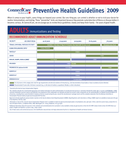


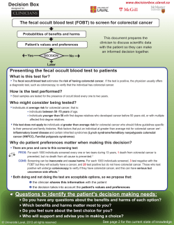

© Copyright 2026