
Introduction
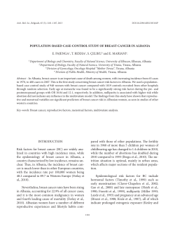
ISSN: 2347-3215 Volume 1 Number 1 (2013) pp. 106-111
www.ijcrar.com
Cytotoxic properties of Acorus calamus in MCF -7 breast cancer cells
S.B. Sreejaya and K.S. Santhy*
Department of Zoology, Avinashilingam Institute for Home Science and Higher Education for
Women, Coimbatore -641 043, Tamil Nadu, India
*Corresponding author e-mail: [email protected]
KEYWORDS
A B S T R A C T
Acorus calamus;
Cytotoxicity;
IC50;
MTT assay.
In this study cytotoxic effect of Acorus calamus extract was evaluated on
breast carcinoma (MCF-7) cell lines. MCF-7 cells were cultured in DMEM
medium and incubated with different concentrations (18.75, 37.5, 75, 150 and
300 g/ml) of Acorus calamus methanol extract. Cell viability was assessed
by MTT assay. Acorus calamus decreased cell viability in malignant cells in a
concentration dependent manner. The IC50 values in MCF-7 cells were
determined as 52.07 g/ml. It may be concluded that Acorus calamus can
cause cell death in MCF-7 cancer cells which can be considered as a
promising chemotherapeutic agent in breast cancer treatment.
.
Introduction
Cancer has been thought to be a
preventable disease due to its slow
development and progression, taking many
years to become invasive in a step by- step
manner (Yao et al., 2011). Such property
provides a great opportunity not only for
early detection, but also for prevention of
the disease progression. Despite this, breast
cancer is the most frequently diagnosed
cancer in women worldwide and ranks
second as a cause of cancer death
(Am.Can.Soc: Cancer Facts and Figures
2012). Over the past several decades, there
has been a particular interest in the role of
medicinal plant extracts in cancer
prevention. Plants are rich sources of
chemically diverse compounds, many with
beneficial properties to human health.
Consequently, about 50% of the anticancer
therapeutic agents known are derived from
plants (Balunas and Kinghorn, 2005). For
example, compounds such as Taxol and
vinca alkaloids act to destabilize the
microtubules of cancer cells, preventing the
rapid proliferation of tumors (Prasain and
Barnus, 2007).
Acorus calamus (known as calamus or
sweet flag) (A. calamus) is a perennial plant
with flavoring scent that grows in aquatic
106
environments. It has a long history of
medical, cultural, and ritual use and hence
was spread outside its indigenous areas in
Asia and is now found across Australia,
Europe, and North America (Ratsch.,2005).
In India, traditional use of A. calamus in
ayurvedic medicine is documented for
treatment of insomnia, neurosis, and
remittent fevers. Extractives of different
parts of Acorus calamus and calamus oil
are widely used now in pharmaceuticals,
traditional systems of medicines for a
number of ailments and in perfumes. Here
it has generated a lot of interest amongst
the scientist to evaluate the activity by
using modern parameters too.
The solvent was removed by evaporation
and extract was concentrated by using
vacuum rotator evaporator.
Chemicals
3-(4, 5-dimethyl thiazol-2-yl)-5-diphenyl
tetrazolium bromide (MTT), Fetal Bovine
Serum (FBS), Phosphate Buffered Saline
(PBS), Eagle s minimum essential medium
(DMEM) and Trypsin were obtained from
sigma Aldrich Co, St. Louis, USA. EDTA,
glucose, Trichloroacetic acid (TCA), Acetic
acid, Tris base and antibiotics from Himedia
Laboratories
Ltd.,
Mumbai.
Dimethyl Sulfoxide (DMSO) and Propanol
from E. Merck Ltd., Mumbai, India.
Cytotoxicity has been defined as the cell
killing property of a chemical compound
independent from the mechanism of death
(Graham-Evans et al., 2003). Cytotoxicity
assay is an appropriate method for
screening new substances within a short
time in order to determine cytotoxicity on
cancer cells (Alley et al., 1988). Usually in
oncology research and clinical practices, in
vitro testing is preferred prior to in vivo
testing. In vitro cultures can be cultivated
under a controlled environment (pH,
temperature, humidity, oxygen / CO2
balance etc.) resulting in a homogenous
batches of cells and thus minimizing
experimental errors. MTT assay has been
described as rapid, simple and reproducible
method, widely used in the screening of
anticancer drugs and to measure the
cytotoxic properties. Hence in the current
study, the cytotoxic properties of
A.calamus was evaluated using this assay.
Cell Lines and Culture Medium
The human Breast cancer cell line (MCF-7)
was obtained from National Centre for Cell
Sciences (NCCS), Pune and grown in
Eagles minimum essential medium
(EMEM) containing 10% Fetal Bovine
Serum (FBS). All cells were maintained at
37 C, 5% CO2, 95% air and 100% relative
humidity. Maintenance cultures were
passage weekly, and the culture medium
was changed twice a week.
Cell Treatment Procedure
The monolayer cells were detached with
trypsin ethylenediamine tetra acetic acid
(EDTA) to make single cell suspensions
and viable cells were counted using a
hemocytometer and diluted with medium
containing 5% FBS to give final density of
1 x 10⁵ cells / ml. One hundred micro litres
per well of cell suspension were seeded in
to 96-well plates at plating density of
10,000 cells / well and incubated to allow
for cell attachment at 37 C, 5% CO2, 95%
air and 100% relative humidity. After 24 h
Materials and Methods
A. calamus extract
Dried and powered plant was extracted
with methanol by using soxhlet apparatus.
107
Table.1 In vitro Cytotoxicity of Acorus calamus on MCF-7 cell lines
Plant Extract
Concentration ( g / ml)
18.75
37.5
Acorus calamus 75
150
300
% inhibition IC50 g / ml R2
17.04
45.23
52.07
0.97
58.64
77.86
94.50
Fig.1 Percentage growth inhibition of Acorus calamus against MCF-7 cell line
% Growth Inhibition
100
80
60
40
20
0
0
1
2
3
Log10 concentration (µg/ml)
the cells were treated with serial
concentration of the test samples. They
were initially dissolved in Dimethyl
sulfoxide (DMSO) and diluted to twice the
desired final maximum test concentration
with serum free medium. Additional four, 2
fold serial dilutions were made to provide a
total of five sample concentration. Aliquots
of 100 l of these different sample dilutions
were added to the appropriate wells already
containing 100 l of medium, resulted the
required final sample concentrations.
MTT Assay
After 48 h of incubation, 15 l of MTT (5
g / ml) in phosphate buffered saline (PBS)
was added to each well and incubated at
37 C for 4 h. The medium with MTT was
then flicked off and the formed formazan
crystals were solubilized in 100 l of
DMSO and then measured the absorbance
at 570 nm using microplate reader. The %
cell inhibition was determined using the
following formula: Percentage cell
inhibition = 100
Abs (Sample) / Abs
(Control) x 100
Following treatment with methanolic
extract of Acorus calamus, the plates were
incubated for an additional 48 h at 37 C,
5% CO2, 95% air and 100% relative
humidity. The medium without samples
were served as control and triplicate was
maintained for all concentrations.
Statistical analysis
Non linear regression graph was plotted
between % cell inhibition and log10
concentration and IC50 was determined
using graph pad prism software.
108
Fig.2 Proliferation of MCF-7 cells treated with Acorus calamus
a
b
c
d
e
a
f
Control ; b
18.75 g ; c
37.5
g;d
75
109
g;e
150
g ; f 300
g;
compared with control. On the other hand
the percentage of non-viable cells on cell
lines increased with the increasing period
of treatment. These results were in
concordance with the studies investigated
the cytotoxic effect of Goniothalamin
towards human breast cancer cells by AlQubaisi et al., (2011).
Result and Discussion
The results for cell growth inhibition by the
methanol extract of A.calamus on MCF-7
cell lines for various concentrations is
shown in Table I. In the present study MCF
cells showed growth inhibition in a dose
dependent manner when treated with
A.calamus at concentrations ranging from
18.75 g
300 g (Figs.1 & 2). The
percentage of dead cells for each
concentration was found to be 17.04, 45.23,
58.64, 77.86 and 94.50. The 50% cytotoxic
effect (IC50) of A.calamus was found to be
52.07 g / ml.
Several mechanisms of action were
detected in MCF-7 cells. In the present
study, methanol extract of A.calamus was
found to be cytotoxic towards human MCF7 in MTT assay and the concentration
required for 50% cell death was found to be
52.07 g / ml. Hence present study shows
the efficacy of A.calamus for the
cytotoxicity towards MCF-7 cells thus
suggesting protection against breast cancer.
In the last few decades, human cancer cell
lines have aggregated an accessible, easily
usable set of biological models to examine
cancer biology (Green, 2003). The utility of
cell lines acquired from tumor allows the
investigation of tumor cells in a simplified
and controlled environment (Arya et al.,
2011). MTT cytotoxicity assay used to
measure the cytotoxic effect of A.calamus
on breast carcinoma (MCF-7) cells. In
screening result, A.calamus has shown
broad spectrum cytotoxicity and it had most
active cytotoxic activity on breast
carcinoma (MCF-7) cells.
References
Alley, M. C., D.A. Scudiro, A. Monks,
M.L. Hursey. M.J. Czerwinski and
Fine, D. L. 1988. Feasibility of drug
screening with panels of human tumor
cell lines using a microculture
tetrazolium assay. Cancer Res. 48, 589601.
Al-Qubaisi, M., R. Rozita, S.K. Yeap, et al.
2011.
Selective
cytotoxicity
of
Goniothalamin against Hepatoblastoma
HepG2 Cells. Molecules, 16, 2944-59
American Cancer Society: Cancer Facts &
Figures. 2012. In Atlanta, GA:
American Cancer Society; 2012.
Arya, V., C.P. Kashyap, T. Bandana, S.
Shiksha, K. Sweta, P. Verma and Swati
Sharma, 2011. Human cancer cell lines
A brief communication, J. Chem.
Pharm. Res. 3(6) : 514-520.
Balunas, M.J., and Kinghorn, A.D. 2005.
Drug discovery from medicinal plants.
Life Sci. 78(5):431-441.
Ferrari, M., M.C. Fornasiero and Isetta,
A.M. 1990. MTT colorimetric assay for
MTT proliferation assay was carried out to
determine the growth rate of cells. In this
study, methanol extract of A.calamus have
indicated significant growth inhibition in
MCF-7 cell line at low concentration of
IC50 values. The IC50 of extract on cell line
less than 100 g / ml is categorized as a
potential cytotoxic substance (Spavieri et
al., 2010). A linear relationship between
the formazan generated and the number of
viable cells was demonstrated, together
with time-dependent growth characteristics
for MCF-7 cells by Ferrari et al., (1990).
A.calamus treatment on MCF-7 cell lines
showed significant decrease in growth rate
110
testing macrophage cytotoxic activity in
vitro. J. Immunological Methods. 131:
165-70.
Graham-Evans, B., P.B. Tchounwou and
Cohly, H.H. 2003. Cytotoxicity and
proliferation studies with arsenic in
established
human
cell
lines:
keratinocytes, melanocytes, dendritic
cells, dermal fibroblasts, microvascular
endothelial cells, monocytes and Tcells. Int. J. Mol. Sci. 4: 13-21.
Green, J.E., 2003.Mouse models of human
breast cancer: evolution or convolution.
Breast Cancer Res. 5(1):1.
Prasain, J.K., and Barnes, S. 2007.
Metabolism and bioavailability of
flavonoids in chemoprevention: current
analytical
strategies
and
future
prospectus. Mol. Pharm. 4(6):846-864
Rätsch, C., 2005. The Encyclopedia of
Psychoactive Plants. Park Street Press,
Rochester, VT.
Spavieri, J., A. Allmendinger, M. Kaiser,
R. Casey, S. Hingley-Wilson and
Lalvani, A. 2010. Antimycobacterial,
antiprotozoan and cytotoxic potential of
twenty-one
brown
algae
(Phaeophyceae) from British and Irish
waters. Phytother. Res. 24 : 1724-29.
Yao, H., W. Xu, X. Shi, and Zhang, Z.
2011. Dietary flavonoids as cancer
prevention agents. J .Environ. Sci.
Health. 29(1):1-31.
111


© Copyright 2026