
3 T P REATMENT OF
WHO/CDS/CSR/EDC/99.2 Plague Manual Epidemiology, Distribution, Surveillance and Control 3 TREATMENT OF PLAGUE Dr Jack D. Poland and Dr D. T. Dennis Case management: therapy and prevention of spread When a diagnosis of human plague is suspected on clinical and epidemiological grounds, appropriate specimens for diagnosis should be obtained immediately and the patient should be started on specific antimicrobial therapy without waiting for a definitive answer from the laboratory (Table 2). Suspect plague patients with evidence of pneumonia should be placed in isolation, and managed under respiratory droplet precautions (1) . Specific therapy Aminoglycosides: streptomycin and gentamicin Streptomycin is the most effective antibiotic against Y. pestis and the drug of choice for treatment of plague, particularly the pneumonic form (2-6). Therapeutic effect may be expected with 30 mg/kg/day (up to a total of 2 g/day) in divided doses given intramuscularly, to be continued for a full course of 10 days of therapy or until 3 days after the temperature has returned to normal. Gentamicin has been found to be effective in animal studies, and is used to treat human plague patients (7-10). Chloramphenicol Chloramphenicol is a suitable alternative to aminoglycosides in the treatment of bubonic or septicaemic plague and is the drug of choice for treatment of patients with Y. pestis invasion of tissue spaces into which other drugs pass poorly or not at all (such as plague meningitis, pleuritis, or endophthalmitis) (3,4,11,12). Dosage should be 50 mg/kg/day administered in divided doses either parenterally or, if tolerated, orally for 10 days. Chloramphenicol may be used adjunctively with aminoglycosides. 55 WHO/CDS/CSR/EDC/99.2 Plague Manual Epidemiology, Distribution, Surveillance and Control Tetracyclines This group of antibiotics is bacteriostatic but effective in the primary treatment of patients with uncomplicated plague (3-5). An oral loading dose of 15 mg/kg tetracycline (not to exceed 1 g total) should be followed by 25-50 mg/kg/day (up to a total of 2 g/day) for 10 days. Tetracyclines may also be used adjunctively with other antibiotics. Sulfonamides Sulfonamides have been used extensively in plague treatment and prevention; however, some studies have shown higher mortality, increased complications, and longer duration of fever as compared with the use of streptomycin, chloramphenicol or tetracycline antibiotics (3-6,13). Sulfadiazine is given as a loading dose of 2-4 g followed by a dose of 1 g every 4-6 hours for a period of 10 days. In children, the oral loading dose is 75 mg/kg, followed by 150 mg/kg/day orally in six divided doses. The combination drug trimethoprim-sulfamethoxazole has been used both in treatment and prevention of plague (6,14,15). Fluoroquinolones Fluoroquinolones, such as ciprofloxacin, have been shown to have good effect against Y. pestis in both in vitro and animal studies (16,17). Ciprofloxacin is bacteriocidal and has broad spectrum activity against most Gram-negative aerobic bacteria, including Enterobacteriaceae and Pseudomonas aeruginosa, as well as against many Gram-positive bacteria. Although it has been used successfully to treat humans with Francisella tularensis infection (18,19), no studies have been published on its use in treating human plague. Other classes of antibiotics (penicillins, cephalosporins, macrolides) These classes of antibiotics have been shown to be ineffective or of variable effect in treatment of plague and they should not be used for this purpose. Supportive therapy The clinician must prepare for intense supportive management of plague complications, utilizing the latest developments for dealing with Gram-negative sepsis (20). Aggressive monitoring and management of possible septic shock, multiple organ failure, adult respiratory distress 56 WHO/CDS/CSR/EDC/99.2 Plague Manual Epidemiology, Distribution, Surveillance and Control syndrome (ARDS) and disseminated intravascular coagulopathy should be instituted. Treatment of plague during pregnancy and in children With correct and early therapy, complications of plague in pregnancy can be prevented. The choice of antibiotics during pregnancy is confounded by the potential adverse effects of three of the most effective drugs. Streptomycin may be ototoxic and nephrotoxic to the foetus. Tetracycline has an adverse effect on developing teeth and bones of the foetus. Chloramphenicol carries a low risk of "grey baby" syndrome or bone-marrow suppression. Experience has shown that an aminoglycoside judiciously administered is effective and safe for both mother and foetus, and in children. Because of its safety, intravenous or intramuscular administration, and ability to have blood concentrations monitored (21), gentamicin is the preferred antibiotic for treating plague in pregnancy (22). Prophylactic therapy Persons in close contact with pneumonic plague patients, or persons likely to have been exposed to Y. pestis-infected fleas, to have had direct contact with body fluids or tissues of a Y. pestis-infected mammal, or exposed during a laboratory accident to known infectious materials should receive antibiotic preventive therapy, if the exposure was in the previous six days (23). The preferred antimicrobials for preventive or abortive therapy are the tetracyclines, chloramphenicol, or one of the effective sulfonamides (Table 3). True prophylaxis, i.e. the administration of an antibiotic prior to exposure, may be indicated when persons must be present for short periods in plague-active areas under circumstances in which exposure to plague sources (fleas, pneumonic cases) is difficult or impossible to prevent (23). Hospital precautions Standard patient-care precautions should be applied to management of all suspected plague patients. These include prescribed procedures for handwashing, wearing of latex gloves, gowns, and protective devices to protect mucous membranes of the eye, nose and mouth during those 57 WHO/CDS/CSR/EDC/99.2 Plague Manual Epidemiology, Distribution, Surveillance and Control procedures and patient-care activities likely to generate splashes or sprays of blood, body fluids, secretions and excretions (1). Additionally, a patient with suspected respiratory plague infection should be specifically managed under respiratory droplet precautions (1), including management in an individual room, restriction of movement of the patient outside the room, and masking of the patient as well as persons caring for the patient until the patient is no longer infectious. Vaccination Worldwide, live attenuated and formalin-killed Y. pestis vaccines are variously available for human use. The vaccines are variably immunogenic and moderately to highly reactogenic. They do not protect against primary pneumonic plague. In general, vaccinating communities against epizootic and enzootic exposures is not feasible; further, vaccination is of little use during human plague outbreaks, since a month or more is required to develop a protective immune response. The vaccine is indicated for persons whose work routinely brings them into close contact with Y. pestis, such as laboratory technicians in plague reference and research laboratories and persons studying infected rodent colonies (23). 58 WHO/CDS/CSR/EDC/99.2 Plague Manual Epidemiology, Distribution, Surveillance and Control Table 2 Plague treatment guidelines Drug Dosage Interval (hours) Route of administration Streptomycin Adults Children 2 g/day 30 mg/kg/day 12 12 IM IM 3 mg/kg/day 6.0-7.5 mg/kg/day 7.5 mg/kg/day 8 8 8 IM or IV IM or IV IM or IV 2 g/day 25-50 mg/kg/day 6 6 PO PO 50 mg/kg/day 50 mg/kg/day 6 6 PO or IV PO or IV Adults 200 mg/day 12 or 24 PO Children $ 9 years 200 mg/day 12 or 24 PO 250-300 mg/day 250 mg/day 8,12 or 24 8,12, or 24 PO or IM PO or IM Gentamicin Adults Children Infants/neonates Tetracycline Adults Children $ 9 years Chloramphenicol Adults Children $ 1 year Doxycycline Oxytetracycline Adults Children $ 9 years IM=Intramuscular; IV=Intravascular; PO=Orally source: Adapted with permission from DT Dennis, Plague, in Conn’ s current therapy 1996, RE Rakel (ed). Philadelphia, WB Saunders, 1996, p 124. 59 WHO/CDS/CSR/EDC/99.2 Plague Manual Epidemiology, Distribution, Surveillance and Control Table 3 Plague prophylaxis guidelines Drug Interval (hours) Route of administation 6 or 12 6 or 12 PO PO 100-200 mg/day 100-200 mg/day 12 or 24 12 or 24 PO PO 1.6 g//day * 40 mg/kg/day * 12 12 PO PO Dosage Tetracycline Adult Children $ 9 years 1-2 g/day 25-50 mg/kg/day Doxycycline Adults Children $ 9 years Sulfamethoxazole/ trimethoprim Adults Children $ 2 months * Sulfamethoxazole component PO=Orally source: Adapted with permission from DT Dennis, Plague, in Conn’ s current therapy 1996, RE Rakel (ed). Philadelphia, WB Saunders, 1996, p 124. 60 WHO/CDS/CSR/EDC/99.2 Plague Manual Epidemiology, Distribution, Surveillance and Control References 1. Garner JS, Hospital Infection Control Practices Advisory Committee. Guideline for isolation precautions in hospitals. Infection Control Hospital Epidemiology, 1996;17:53-80. 2. Campbell GL, Dennis DT. Plague and other Yersinia infections. Fauci AS, Braunwald E, Isselbacher KJ et al. (eds). Harrison’ s principles of internal medicine. New York, McGraw-Hill, 1998:975-980. 3. Smadel JE, Woodward TE, Amies CR, Goodner K. Antibiotics in the treatment of bubonic and pneumonic plague in man. Annals of the New York Academy of Sciences, 1952;55:1275-1285. 4. Meyer KF, Quan SF, McCrumb FR, Larson A. Effective treatment of plague. Annals of the New York Academy of Sciences, 1952;55:1228-1274. 5. Pollitzer R. Plague. Geneva, World Health Organization, 1954 (Monograph series). 6. Butler T, Levin J, Linh NN, Chau DM, Adickman M, Arnold K. Yersinia pestis infection in Vietnam. II. Quantitative blood cultures and detection of endotoxin in the cerebrospinal fluid of patients with meningitis. Journal of Infectious Diseases, 1976;133:493-499. 7. Byrne WR, Welkos SL, Pitt ML, Davis KJ et al. Antibiotic treatment of experimental pneumonic plague in mice. Antimicrobial Agents and Chemotherapy, 1998;42:675-681. 8. Welty TK. Plague. In: Conn HF (ed.). Current therapy. Philadelphia, WB Saunders, 1984:44-45. 9. Hull HF, Montes JM, Mann JM. Plague masquerading as gastrointestinal illness. Western Journal of Medicine, 1986;145:485-487. 10. Wong TW. Plague in a pregnant patient. Tropical Doctor, 1986;16:187-189. 11. McCrumb FR, Mercier S, Robic J, Bouillat M et al. Chloramphenicol and terramycin in the treatment of pneumonic plague. American Journal of Medicine, 1953;14:284-293. 12. Becker TM, Poland JD, Quan TJ, White ME et al. Plague meningitis B a retrospective analysis of cases reported in the United States, 19701979. Western Journal of Medicine, 1987; 147:554-557. 13. Meyer KF. Modern therapy of plague. Journal of the American Medical Association, 1950;144:982-985. 14. Ai NV, Hanh ND, Dien PV, Le NV. Co-trimoxazole in bubonic plague. British Medical Journal, 1973;4:108-109. 15. Butler T, Bell WR, Linh NN, et al. Yersinia pestis infection in Vietnam. I. Clinical and hematological aspects. Journal of Infectious Diseases, 1974;129(suppl):S78-S84. 61 WHO/CDS/CSR/EDC/99.2 Plague Manual Epidemiology, Distribution, Surveillance and Control 16. Russell P, Eley SM, Green M, Stagg AJ et al. Efficacy of doxycycline and ciprofloxacin against experimental Yersinia pestis infection. Journal of Antimicrobial Chemotherapy, 1998;301-305. 17. Frean JA, Arntzen L, Capper T, Bryskier A, Klugman KP. In vitro activities of 14 antibiotics against 100 human isolates of Yersinia pestis from a southern African plague focus. Antimicrobial Agents and Chemotherapy, 1996;40:2646-2647. 18. Syrjala H, Schildt R, Raisainen. In vitro susceptibility of Francisella tularensis to fluoroquinolones and treatment of tularemia with norfloxacin and ciprofloxacin. European Journal of Clinical Microbiology and Infectious Diseases, 1991;10:68-70. 19. Enderlin G, Morales L, Jacobs RF, Cross JT. Streptomycin and alternative agents for the treatment of tularemia: review of the literature. Clinical Infectious Diseases, 1994;19:42-47. 20. Wheeler AP, Bernard GR. Treating patients with severe sepsis. New England Journal of Medicine, 1999; 340:207-214. 21. AHFS drug information 1999. Litvak K, Welsh OH, and Snow EK, (eds) American Society of Health System Pharmacists, Bethesda, MD, 1999, 64-71. 22. Inglesby TV, Henderson DA, Bartlett JG, Dennis DT et al. Plague as a biological weapon: medical and public health management. Journal of the American Medical Association, 1999; submitted for publication. 23. Centers for Disease Control and Prevention. Prevention of plague. Morbidity and Mortality Weekly Report, 1996;45:1-15. 62 WHO/CDS/CSR/EDC/99.2 Plague Manual Epidemiology, Distribution, Surveillance and Control 4 RODENT RESERVOIRS & FLEA VECTORS OF NATURAL FOCI OF PLAGUE Dr Norman Gratz Rodent reservoirs Plague is primarily a disease of rodents. The infection is maintained in natural foci of the disease in wild rodent colonies through transmission between rodents by their flea ectoparasites. For the most part, the sylvatic rodent reservoirs are species that are susceptible to the infection but resistant to the disease. While upwards of 200 species of rodents and lagomorphs have been implicated in the epidemiological cycle of plague in one geographical area or another, the true number of rodent species important as more than accidental reservoirs of plague is uncertain. Many species of rodents and other small mammals are susceptible to infection but are only occasionally infected and are not necessarily important reservoirs of infection. The animal hosts of plague are classified as enzootic (maintenance) hosts and epizootic (amplification) hosts (1). The first group includes rodents from genera that are relatively resistant to plague. In this group mortality from plague infection is low, although antibody surveys of field populations may show a positivity rate as high as 100%. Dieoffs commonly seen among more susceptible rodent species are rare in this group. The plague organism is occasionally introduced into colonies or areas of more susceptible species. This occurs in nature by an overlap of individuals or populations of two species. When this happens in a species that is highly susceptible to plague, an epizootic B sometimes of considerable magnitude B may occur, and high mortality (rodents positive for plague) is seen in sylvatic and peridomestic areas or even in villages or cities. It is difficult to group the many different species of rodents, lagomorphs and other small mammals involved as common or occasional reservoirs or hosts of plague to fit the above classification. The susceptibility to plague infection of a given species may vary even within 63 WHO/CDS/CSR/EDC/99.2 Plague Manual Epidemiology, Distribution, Surveillance and Control the geographical limits of a foci. Furthermore, susceptibility may vary temporally with variations in the density of the host populations or in the density of their flea ectoparasite vectors. The virulence of the particular strain of the plague bacterium involved in the epizootic may also vary over a period of time. As most of the natural foci of plague have existed for long periods of time, it is clear that a portion of any reservoir population must survive infection. In some species the infection can continue to circulate with relatively little mortality (2). Flea vectors About a dozen cosmopolitan species are implicated in the transmission of domiciliary plague (3). However, many more species of the order Siphonaptera have been implicated in the transmission of sylvatic plague (4). To understand the epidemiology and transmission of the infection from rodent reservoirs to human hosts, it is essential to determine the flea species involved in plague transmission in a given area. Information on the bionomics of the flea vectors is basic to their control and control of transmission of the infective agent. The following section provides information on the most important flea vectors of plague in the various endemic foci. If this information is not already available for an area in which plague is suspected or known to be endemic, surveys of flea ectoparasites should be done. Survey methods are described elsewhere in this publication. Entomological expertise is needed for the design, implementation and (particularly) identification of the flea species taken and evaluation of their importance in relation to plague transmission. Cosmopolitan vectors of plague The majority of the flea species described below are ectoparasites of commensal or peridomestic rodents. Because of their close proximity to humans and their dwellings, these fleas are often found on livestock and household animals. Most of these species have a wide distribution, although their percentage in the flea population varies from place to place as does their role as vectors of plague. All, however, readily feed on humans. The commensal rodent fleas are classed as follows (5): 64 WHO/CDS/CSR/EDC/99.2 Plague Manual Epidemiology, Distribution, Surveillance and Control (1) Fleas specific to commensal rodents which show a wide distribution and are found in several plagueendemic areas. Xenopsylla cheopis (Oriental rat flea) has a wide distribution, while the distribution of X. brasiliensis and Nosopsylla fasciatus is more limited. (2) Species specific to commensal rodents which show a limited or even restricted geographical distribution, such as X. astia. (3) Wild rodent fleas which frequently infest commensal rodent species. (4) Flea species which, because they are common in the environment of commensal rodents, are often found in limited numbers on these rodents although they are not specific for them. Echidnophaga gallinacea and Pulex irritans, both of which have a cosmopolitan distribution, and the cat flea, Ctenocephalides feli, are examples of this latter group. To act as an efficient plague vector, the flea must be able to ingest the plague organism with its blood meal. Second, it must live long enough for the pathogen to multiply sufficiently. Third, it must be able to transfer the pathogen to an animal or human host in sufficient concentrations to cause an infection and last, it must be present in large enough numbers to maintain the infection in the local rodent hosts (6). There are a number of other characteristics but these are the most important. When a flea sucks blood from an infected rodent or other host, some of the bacteria settle on the flea=s proventriculus. This spined structure shuts off the stomach while the flea is sucking but opens to allow ingested blood to enter the stomach. Plague bacteria that have settled on the spines of the proventriculus multiply and eventually block the passage of blood into the stomach. Although the flea continues to feed (with increasing avidity as time passes) blood cannot continue to enter its stomach and instead remains in the oesophagus. When the flea stops sucking, the oesophagus recoils and the accumulated blood is driven into the bite wound, bringing Y. pestis with it. A flea in this condition is known as a Ablocked@ flea. Those species of fleas most subject to blocking are the most efficient vectors of plague, providing that the other requirements of transmission are met and that the flea survives long enough to transmit the infection. Xenopsylla cheopis is the most important vector of plague and the rickettsial infection murine typhus. The species is thought to have originated in Egypt but during the 19th century spread to all parts of the 65 WHO/CDS/CSR/EDC/99.2 Plague Manual Epidemiology, Distribution, Surveillance and Control world as parasites of rats infesting ships= cargos. A high incidence of plagueinfected X.cheopis in a given focus, greatly increases the risk of transmission to humans. X. cheopis most commonly parasitizes Rattus species but is frequently found on other rodent species in and around houses. Xenopsylla astia is a parasite of both gerbils and rats. It ranges from the Arabian peninsula through Iran to southeast Asia and to Korea (7) and has been found on the east coast of Africa. It is a less efficient vector than X. cheopis. Xenopsylla brasiliensis is native to all Africa south of the Sahara where it is the most common vector in some areas (8), often more common than X. cheopis. It has spread to other parts of the world such as Brazil and India. It is an effective plague vector, especially in rural environments. It is less tolerant of high temperatures than X. cheopis but is more resistant to drought conditions. Nosopsyllus fasciatus, the Northern rat flea, is one of the most prevalent fleas in Europe on commensal rats (9). Its distribution is virtually global and it is found from the United States to China (10) and Korea (11). Its numbers appear to be increasing in Japan (12). It is also found on mammals and rodents other than Rattus and feeds freely on humans. It is relatively unimportant as a vector of plague. Monopsyllus anisus is the common rat flea of temperate east Asia, extending from China and Transbaikala Russia to Japan. It has been found in ports in San Francisco and Vancouver and in the United Kingdom. Leptopsylla segnis, the mouse flea, probably originated in western Asia on Mus or Apodemus. It is generally abundant on rats than on mice. It is widely distributed, particularly in temperate areas, but is only a weak vector of plague and an uncertain vector of murine typhus (13). Pulex irritans, the human flea, was considered to have originated as an Old World species (3) but a more recent review (14) observes that the species probably originated in South or Central America as an ectoparasite of the guinea pig or peccary. P. irritans is now worldwide in its distribution. Despite its common name it has a wide range of hosts: it is found in the wild on foxes, badgers, ground squirrels, guinea pigs and rats as well as domestically on pigs, goats, dogs, cats and humans. It is often found in high densities in habitations. P. irritans has been considered as a 66 WHO/CDS/CSR/EDC/99.2 Plague Manual Epidemiology, Distribution, Surveillance and Control possible or probable vector of plague in Angola (15), Brazil (16), Burundi (17), the Democratic Republic of the Congo (21), Iran (2), Iraq (18), Nepal (19), and Tanzania (20). Ctenocephalides felis, the cat flea, has become completely cosmopolitan in its distribution. It is frequently found not only on cats but also on a large number of other hosts, including dogs, humans, other mammals and birds (22). There appears to be a gradual northern extension of this species (12). It may be a vector of murine typhus and is also an intermediate host of some cestodes. Both the cat flea and the dog flea (Ctenocephalides canis) are able to transmit plague to humans from pet animals. The following section considers the main rodent reservoirs and flea vectors of plague in most of the betterknown endemic foci. Some foci are large and contiguoussuch as those in the western United States, the Russian Federation, China and Mongoliaand extend across borders to more than one country. In foci such as these, reservoir and flea vector species may differ considerably from one part of the focus to another. Plague reservoirs and vectors in Africa Plague foci of southern Africa (23,24,25,26,27,28,29,30) This area includes foci in South Africa, Lesotho, Namibia and Zimbabwe. Although the number of plague outbreaks in this subregion have declined considerably in recent years, the infection persists in many areas where human plague has not been apparent for years. It is therefore important to understand the mechanism and the rodent species responsible for persistence in the natural foci. The main reservoir in many parts of this geographical region was long thought to be the gerbil, Tatera brantsi. The passage of plague infection in Orange Free State, South Africa, has been traced from gerbils as the reservoir to other wild rodents, Otomys irroatus to Mastomys natalensis to Rattus rattus and to humans. M. natalensis is now understood to be a species complex: early studies have separated it into species A and B. The distribution of human plague in southern Africa is apparently linked to the distribution of species B of the Mastomys(Praomys) natalensis species complex. Studies have been done to determine if the sibling species of M.natalensis, Aethomys chrysophilus, Mastomys coucha, Tatera leucogaster and 67 WHO/CDS/CSR/EDC/99.2 Plague Manual Epidemiology, Distribution, Surveillance and Control A. namaquensis differed in their potential as reservoirs of plague in southern Africa. M. natalensis with 32 diploid chromosomes was significantly more resistant to experimental plague infections with high level inoculations of Y. pestis than M. coucha with 36 diploid chromosomes. The geographic distribution of human plague in southern Africa corresponds closely with that of the plaguesusceptible species, M. coucha, while the plagueresistant species M. natalensis predominates in areas where human plague has not been recorded. A. namaquensis is extremely plaguesensitive, much more so than A. chrysophilus, and they may play different roles in the plague cycle. In an outbreak of plague in Coega in the Cape Province of South Africa in 1982 plague antibody was found in two rodent species: the four striped mouse, Rhabdomys pumilo and the vlei rat, Otomys irroratus. Sera from 3012 rodents of 24 species captured in South Africa were tested for antibody to the Fraction 1 antigen of Y. pestis by passive haemagglutination. Of 24 species investigated, antibodies were found in seven (0.23%) rodents of three species, Desmodillus auricularis and Tatera brantsii in the northern Cape Province and in R. pumilo in the eastern Cape Province. The gerbils Tatera brantsi, T.leucogastor and T.afra play an important role in southern African plague epidemiology. Rhabdomys pumilio and Otomys irroratus were found infected in Cape Province in studies carried out in 1982 (29) . The fleas most frequently found on the rodent reservoirs of plague are X philoxera, X. brasiliensis and Dinopsyllus ellobius. However, in ports and coastal towns X. cheopis is the dominant flea species on Rattus species and is the dominant flea vector of plague. In Zimbabwe, T. leucogaster and M. coucha are highly susceptible to plague and die soon after infection, making it unlikely that they act as reservoir hosts. Because they are relatively resistant to plague, Aethomys chrysophilus and M. natalensis are the more likely reservoirs. In Zimbabwe both M. coucha and M. natalensis are semidomestic and probably act as a link between humans and the true sylvatic foci of plague (30). Plague foci of East Africa This area includes plagueendemic regions of Kenya, Tanzania, Mozambique and Madagascar. Plague is widely endemic in the four countries. 68 WHO/CDS/CSR/EDC/99.2 Plague Manual Epidemiology, Distribution, Surveillance and Control Kenya (31,32,33,34) In an early survey of rodents for plague in a plague focus near Rongai, north of Nakaru, plague was isolated from five species of wild rodents: Otomys angoniensis, Arvicanthis abyssinicus, M. natalensis, Lemniscomys striatus and Rhabdomys pumilo. The reservoirs of plague have been extensively studied in Kenya. Sera from 8,860 rodents and other small mammals were examined for antibodies to Y. pestis in one survey, where it was noted that enzootic plague in Kenya is much more widely distributed than the human cases reported. A. niloticus, M. natalensis and R. rattus are probably the most important and widespread reservoirs of plague in Kenya. Ten percent of all R. rattus tested were found to be positive, as compared to 12% of the Arvicanthis. Tatera robustis has also been found positive at a low level. The high prevalence of plague antibodies in R. rattus is significant, in that the species readily lives both as a commensal and wild species and thus can serve to introduce plague from its sylvatic reservoirs into a commensal cycle. That plague in Kenya can be more widespread than previously thought was shown by a survey in the Tana River area prior to the construction of a dam at that site. Four of the seven species of rodents captured (T. robusta, A. niloticus, L. striatus and Pterodromus tetradactylus) were positive for plague. Xenopsylla cheopis, X. braziliensis and Dinopsyllus lypusus are abundant on the most important rodent reservoirs of plague in Kenya and, as elsewhere in East Africa, are the main vectors of the infection. Tanzania (35,36,37) In Tanzania the most important commensal and peridomestic rodents involved in the transmission of plague are R. rattus and M. natalensis. Cricetomys gambianus, Lophuromys flavopunctatus, Tatera robusta, Otomys angoniensis, Arvicanthis niloticus and A. abyssinicus are also involved where human cases occur. In most of the plagueendemic areas of the country, the majority of the rodents are A. abyssinicus and M. natalensis. Lemniscomys striatus has been found positive for plague in the Mbulu focus. Lophuromys flavopunctus, L. sikapusi, Otomys angoniensis, Pelomys fallax, O. denti and Gramomys dolichurus are among other rodent species found positive for plague in a serological survey in the western Usumbara mountains. Once surveyed, plague will probably be found to be endemic in still other areas of the country and in other species of rodents. Reservoir species are widespread and human cases of plague occur in the country nearly every year. 69 WHO/CDS/CSR/EDC/99.2 Plague Manual Epidemiology, Distribution, Surveillance and Control Xenopsylla cheopis and X. brasilensis are common on both Rattus and T. robusta. P. irritans has also been found frequently in the plagueendemic area of Lushoto (38). X. brasiliensis and D. lypusus are more common than X. cheopis on rodents in the country (39). X. humilis and X. nilotica are found on Tatera and Gerbillus species (40). Mozambique (41,42,43) Mastomys natalensis is widespread in Mozambique as well as in neighbouring countries and is probably the main sylvatic reservoir of plague. In the cities, population densities of R. norvegicus and R. rattus are high and plague may have spread from M. natalensis to R. rattus in the 1976 outbreak. Madagascar (41,44,45) An estimated 15% of the island of Madagascar is endemic for plague and there is some evidence that strains of Y. pestis have become more virulent. The infection established itself on the high plateau of central Madagascar in 1921, remaining endemic and spreading over the years with the occurrence of sporadic cases. There are two large foci in the country: the first from the central province of Tananarive to the south in Fianarantsoa; the second in the north near the region of Balanana. The only apparent reservoir of plague in Madagascar is R. rattus. The number of rodent species on the island is relatively small, with only three muroid rodents: R. norvegicus, the only species found in the ports and the most common species in the city of Antananarivo; Mus musculus, which is found everywhere but appears to have no role in the epidemiology of plague; and R. rattus, whose density is often high and is widely distributed in rural areas, rice fields, villages and urban areas. The flea vector is mainly X. cheopis but R. rattus is frequently parasitized by Synopsyllus fonquerniei. Plague foci of central Africa In central and southwest Africa, plague is endemic in Angola, Equatorial Guinea and the Democratic Republic of the Congo. Little information is available on the reservoirs and vectors in Angola or Equatorial Guinea. Democratic Republic of the Congo (46,47) Extensive studies have been carried out on the rodent reservoirs of the two plague foci in the Democratic Republic of the Congo. The areas have a rich rodent fauna and the main species involved in the 70 WHO/CDS/CSR/EDC/99.2 Plague Manual Epidemiology, Distribution, Surveillance and Control epidemiology of plague are Arvicanthis abyssinicus, M. natalensis, Lemniscomys striatus, R. rattus and Leggada minutodies, which continue to maintain plague transmission in the northeastern part of the country. A. abyssinicus is a peridomestic species which serves as an intermediary between the wild or sylvatic reservoirs and domestic species. M. natalensis is frequently found nesting in thatched roofs. P. irritans is a possible vector of plague in the Democratic Republic of the Congo (46) and Angola, at least in domestic transmission (17). The fleas Dinopsyllus lypusus and Ctenophthalmus cabirus and C. phyris are common on Arvicanthis and Lophuromys and have been found plague positive, especially in the Blukwa plague focus. In the Lake Edward focus in the Democratic Republic of the Congo, R. rattus and M. natalensis are the principal commensal and peridomestic rodents and Xenopsylla brasiliensis the most important flea vector. The plague focus of northwest Africa Mauritania (48,49) A focus of plague exists in the northern part of western Mauritania. The rodent populations of the area, particularly the gerbils, Gerbillus gerbillus and G. nanus, the jerboa Juculus jaculus and Psammomys obesus are important desert or semidesert rodent species. The gerbils are the principal reservoirs of plague in the area. Xenopsylla ramesis is the vector among the Psammomys populations. X. nubica is common on gerboas Jaculus jaculus. Synosternus cleopatrae is the most common flea on Gerbillus species and is the vector of plague among gerbil populations. X. cheopis is found only in seaside towns. All these species feed readily on humans and can transmit Y. pestis from rodent reservoirs to domestic animals and humans. The plague focus of North Africa Libya (50,51) Libya appears to be the only country in North Africa still endemic for plague. Though the focus was silent for some thirty years, cases appeared in the Nofila area in 1972. Surveys of rodents in the area indicate that G. gerbillus and Meriones shawi are the most common species of rodents in areas where human cases of plague have been reported. The former were captured inside the tents of nomads and may serve as maintenance host for the infection. M. libycus is an even more widespread 71 WHO/CDS/CSR/EDC/99.2 Plague Manual Epidemiology, Distribution, Surveillance and Control species and is comparatively resistant to plague; it was also found to be seropositive for plague in Libya. Other animals, including camels, may also be involved in the epidemiology of plague. Further investigation is necessary for a better understanding of the reservoirs maintaining plague in this longstanding focus. Flea densities are low in the Libyan plague foci. In the northern plague foci, M. libycus, M. caudatus, M. shawi and P. obesus are present. The flea ectoparasites are X. ramesis, X. cheopis, X. taractes and Nosopsylla henleyi. The plague focus of the Arabian Peninsula Yemen (52) A small outbreak of plague occurred in Yemen in 1969 in a focus in which earlier outbreaks had occurred at the beginning of the century and in 1951 and 1952. Epidemiological investigation following the 1969 outbreak showed R. rattus present in houses, and Meriones rex and gerbils (Gerbillus species) in the fields surrounding the infected village, although none were found infected with Y. pestis. No information is available on the flea vectors in this focus nor on its current status. Plague foci of southwestern Asia Islamic Republic of Iran (2,53) Though no human cases have been reported for many years, there are three active areas of endemic plague still known to exist. These are Kordestan (Kurdistan) and Hamadan in the west, and a focus in East Azerbaijan (including the Sarab desert) in the northwest. Prior to its discovery in 1980 plague had never been reported from this area. The other foci have been known for a long time and are well studied. The most important rodent reservoirs in the area are the gerbils M. libycus and M. persicus, both of which are highly resistant to plague infection, and M. tristrami and M. vinogradovi which are highly susceptible to both infection and the disease. Tatera indica has also been associated with transmission of Y. pestis in the country. The flea vectors among the gerbils are Xenopsylla buxtoni and Stenoponia tripectinata. Flea densities are often high on M. persicus. Past epidemics of bubonic plague may have been due to humantohuman transmission by P. irritans. 72 WHO/CDS/CSR/EDC/99.2 Plague Manual Epidemiology, Distribution, Surveillance and Control Plague foci of the Russian Federation and the CIS Republics (54,55,56,57,58,59,60) The endemic foci of plague cover vast areas and their ecology, reservoirs and vectors differ considerably from one another. They will therefore be considered separately based on a report by B.K. Fenjuk and V.P. Kozakevic to WHO, 1968 (unpublished report). An extensive review of the plague literature in the former USSR was made by Pollitzer in 1966 (54). The classification of these foci are taken from that report. A large natural focus of plague remains active in the Asian part of the Russian Federation and in the Asian republics. In the preCaspian region, the main rodent reservoir of plague is the suslik, Citellus pygmaeus. In sandy areas, Meriones meridanus (a species rather resistant to plague infection) and M. tamoriscinus may also be reservoirs. In the central Asian plague focus, the main rodent reservoirs in the desert lowlands are Rhombomys opimus and Meriones erythrourus and in the high mountain areas of this large focus, the marmots Marmota baibacina and M. caudata. In the transcaucasian area, gerbils (M. libycus and others) are important reservoirs, while Marmota siberica and Citellus dauricus are involved in the epidemiology of plague in the Transbaikalian focus. Commensal rodent species have rarely been involved in plague transmission in these foci. The northwest Caspian focus The focus covers an area lying to the west of the lower source of the Volga and the northern shores of the Caspian Sea. The western boundary of the focus is the River Don. Enzootic plague is reported to have disappeared from a large portion of this focus. The main reservoir of plague is the small or lesser suslik, Citellus pygmaeus. Two species of voles, Microtus arvalis and laagers may have been involved as reservoirs in the focus (61). The most important flea vectors are Ceratophyllus tesquorum and Neopsylla setosa. The focus between the Rivers Volga and the Ural Mountains Two types of landscape are found in this area: rocky steppes in the north, west and east; and sandy semidesert (the VolgaUral sands). The main reservoir of plague in the steppes is the small suslik, C. pygmaeus. In the sandy areas it is the gerbil Meriones meridianus and to a lesser extent M. tamariscinus. 73 WHO/CDS/CSR/EDC/99.2 Plague Manual Epidemiology, Distribution, Surveillance and Control The most important flea vectors in the steppe regions are Ceratophyllus tesquorum and Neopsylla setosa and in the sandy semideserts, Xenopsylla conformis, Ceratophyllus laeviceps and Rhadinopsylla cedestis. The focus on the left bank of the Ural River The reservoirs in this area are also C. pygmaeus and M. tamariscinus. The flea vectors are the same as those mentioned above. The focus in the Transcaucasian lowlands This focus in Azerbaijan may be linked with the natural focus in Iranian Kurdistan. The main plague reservoir in this area is the gerbil, Meriones libycus erythrourus. The flea vectors are X. conformis and C. laeviceps. The focus in the high mountain areas of Transcaucasia This focus of plague is located at an altitude of 2000 to 3000m and covers areas in Armenia and Azerbaijan. The main reservoir species is the vole Microtus arvalis; infected vole fleas Ctenophthalmus teres, C. wladimiri and Ceratophyllus caspius have been found in nature. The identity of the main rodent reservoir in the lower altitudes and plains of this focus remains uncertain. The central Asian desert focus This focus covers a large area of central Asia and southern Kazakhstan Republic to the borders with China in the east and with Afghanistan and Iran in the south. The most important reservoir is the gerbil, Rhombomys opimus. The flea vectors are Xenopsylla skrjabini, X. hirtipes, X. gerbilli gerbilli, X. gerbilli minax, X. gerbilli caspica, X. nuttali and X. conformis. The TianShan focus This focus is situated in a mountainous area of Kazakhstan and Kirgasia. The main reservoir is Marmota baibacina and the flea vectors are Oropsylla silantiewi and Rhadinopsylla ventricosa. The PamirAlai focus This is a focus of limited size in the Alai valley. The reservoir is the Altai marmot, Marmota caudata. The flea vectors are R. ventricosa and possibly O. sillantiewi and Ceratophyllus lebedwi. 74 WHO/CDS/CSR/EDC/99.2 Plague Manual Epidemiology, Distribution, Surveillance and Control The Transbaikalian focus This is a focus on the northeast edge of the extensive Mongolian focus of plague. The rodent reservoirs are Marmota sibirica and Citellus dauricus. The main vector flea is Oropsylla silantiewi. Isolations of Y. pestis have also been made from the flea Frontopsylla luculenta. The High Altai and Tuva Autonomous Region focus In this area, also adjacent to Mongolia, the weasel Putorius eversmannni and the suslik Citellus undulatus have been found plague positive. The fleas on the suslik species are Ceratophyllus tesquorum. Plague foci of southeast Asia and the western Pacific India (62,63,64,65,66,67) A large number of rodent species are known from the Indian subcontinent, including some 46 genera, 135 species and many subspecies. The diverse ecological conditions in different parts of this large country has also resulted in a diverse rodent and flea ectoparasite fauna. Rodents cause serious agricultural and stored food losses and are important reservoirs of a number of diseases including plague, leptospirosis and murine typhus. Many species of rodents have been reported as actual or potential reservoirs of plague. Depending on the region, the more important species are Bandicota bengalensis, Tatera indica, Rattus norvegicus, R. rattus and R. rattus diardii, among others. The species shown to be important as reservoirs of plague at one time or another include the urban rats, R. rattus, R. norvegicus and B. bengalensis; the latter is also an important agricultural pest. The gerbil Tatera indica, the Indian field mouse Mus budooga, and the squirrels Funambulus pennanti and F. palmarum have all been found positive for plague in various foci. Until the recent outbreak of plague in Maharashtra and Gujurat States of India in 1994, no human cases of the disease had been reported since the cases in Karnataka State in 1966. However, there have been a number of suspected outbreaks reported including in Himachal Pradesh in 1983, similar to pneumonic plague (22 cases, 17 deaths). From the 1960s to 1989, a total of 188,025 rodent sera were examined in India. Only 12 sera from Tatera indica were found positive for Y. pestis antibody in 1979 and three from the same species found positive in 1989. Only two R. rattus were reported as serologically positive for 75 WHO/CDS/CSR/EDC/99.2 Plague Manual Epidemiology, Distribution, Surveillance and Control Y. pestis in 1988 despite many reports of rat falls from the country. Population densities of rats including B. bangalensis, R. norvegicus and R. rattus in most urban areas are generally high. In rural areas agricultural development, including large irrigation projects, is changing ecological patterns and the composition of rodent populations. As of 1973, 76 species of fleas have been recorded in India (68). The most important rat flea vector of Y. pestis in urban or domestic situations (found on wild rodents) is X. cheopis, while X. astia predominates on wild rodents. X. brasiliensis is also frequently found on rodents. Nosopsyllus faciatus has also been found infected by Y. pestis. Nepal (69) Only a few cases of plague have been reported from Nepal and little information is available on the reservoirs. During a small outbreak in 1971, P. irritans was reported to be the vector in the affected village. Myanmar (4,70,71,72,73,74,75,76) Zoonotic plague is endemic over large areas of the country. The rat species with the highest plague antibody rates in Yangon (Rangoon) among 1,620 animals tested in 1976 was the bandicoot B. bengalensis, the most common rodent species in the city. Its rate of positivity was 15.4%. R. norvegicus showed 11.1% positivity, R. rattus 7.6%, and the insectivore Suncus murinus 3.35%. Plague antibody in B. bengalensis is transient in nature and when found indicates recent infection. Little is known, however, about the epidemiology, maintenance cycle or reservoirs of plague in the rural or sylvatic areas of the country. Xenopsylla cheopis and X. astia have been recovered from the three species of Rattus as well as from B. bengalensis and the shrew S. murinus in Yangon (Rangoon). X. astia is most abundant on the bandicoot and Norway rat while X. cheopis is more common on R.exulans and S. murinus. Both species of Xenopsylla are found in almost equal numbers on R. rattus. The two species of Xenopsylla are probably the most important vectors of both plague and murine typhus (75). R. rattus has been considered the most important reservoir of plague in the foci in the country and X. cheopis the most important vector with X. astia also a vector (76). Indonesia (77,78,79,80) A focus of plague was active until recently in the Boyolali area of central Java. There have been no recent reports of plague activity in this focus despite an active surveillance programme. The two rodent species 76 WHO/CDS/CSR/EDC/99.2 Plague Manual Epidemiology, Distribution, Surveillance and Control from which Y. pestis was detected in this area are R. rattus diardii and R. exulans ephippium. R. r. diardii is the predominant species inside houses and R. exulans is the most common species in the fields. The most common flea species and vectors of plague in the Boyolali focus are X. cheopis and Stivalis cognatus. R. rattus and X. cheopis have been collected most often from buildings, where contact with humans occurs readily. R. exulans and S. cognatus have generally been taken in field and forest habitats. Viet Nam (81,82,83,84,85,86,87,88,89,90,91,92) In urban areas, the reservoirs of plague are the domestic rats R. norvegicus and R. rattus and the insectivore S. murinus. Sylvatic plague was first found in Viet Nam in 1968 when specimens of the large bandicoot B. indica and the fleas (X. cheopis) infesting it collected near a plague focus were found positive for plague. Recent studies indicate that plague is probably maintained by these species in a domestic or peridomestic cycle and it is doubtful that there is a true sylvatic cycle in the country (90). Only X. cheopis was collected on all four species of small mammals trapped in the Pleiku plagueendemic area: R. rattus, R. norvegicus, B. bengalensis and S. murinus. The species R. rattus, R. norvegicus and S. murinus are most closely associated with plague transmission. Of the fleas collected on four small mammal species in Pleiku, 94% were on R. rattus (91). X. cheopis was the most common flea species collected on small mammals in a plague focus; X. vexabilis was found in much smaller numbers (92). It thus seems likely that the most important flea vector of plague in the country is the Oriental rat flea, X. chopis. B. indica has also been found plaguepositive in Viet Nam, infested with X. cheopis (85). China (74,93,94,95) China is the only country of the western Pacific region aside from Viet Nam where plague remains endemic. There are ten geographical foci of plague in China. The following review of the status of plague in these foci is taken from a report provided by Xu Rongman ( 94). Foci are classified according to rodent reservoir species. (1) The plague focus of the commensal rat Rattus flavipectus. This species is found in southern Yunnan and the coastal areas of Zenjiang, Fujian, Taiwan, Guangdong and Guangxi in southern China, an area of over 20 000 sq. km which includes 77 WHO/CDS/CSR/EDC/99.2 Plague Manual Epidemiology, Distribution, Surveillance and Control 56 counties. Other hosts infected with Y. pestis in these regions have been R. norvegicus, M. musculus and Suncus murinus. The only part of this area where human plague cases have been reported since 1953 is southern Yunnan. (2) The plague focus characterized by Eothenomys miletus is located in the mountains of northwestern Yunnan over an area of 600 sq. km. The main vectors are Ctenophthalmus quadratus and, to a lesser degree, Neopsylla specialis. The main reservoir host is Eothenomys miletus. Apodemus chevrieri, Apodemus speciosus and Rattus nitidus have also been found infected in the focus. While enzootic plague has been reported on many occasions, no human cases have been reported. (3) The Marmota himalayana plague focus. This large focus is found mainly in Tibet and Qinghai, south to the Himalaya Mountains, north to the Qilian mountains in Xinjiang and east to southern Gansu, covering nearly 1 000 000 sq. km of land and 54 counties. The principal flea vectors are Callopsylla dolabris and Oropsylla silantiewi. Other fleas and hosts found infected are Rhadinopsylla li and Pulex irritans, Ochotona daurica annectens, O. curzoniae, Lepus oiostolus, Vulpes ferrilata, Procarpra picticauda, Mus musculus, Cricetulus migratorius, Microtus oeconomus and Pitymys leucurus. This stable enzootic focus is active from April to September. It is the most important focus of plague in China and the majority of human cases in the country arise from this focus. (4) The Marmota caudata plague focus is in southwestern Xinjiang. It is part of the Pamir Plateau plague focus in Middle Asia and covers 600 sq. km in two counties. The main vectors are Oropsylla silanteiwi and Rhadinopsylla li. Citellophilus lebedewi priceps has also been found infected, as has the rodent Pitymys juldaschi. There have been no human cases of plague recorded in this zoonotic focus. (5) The Marmota baibacina and Spermophilus undulatus focus. Located in the Tianshan Mountains of Xinjiang Province, the focus covers an area of 7,000 sq. km over 10 counties, extending into Kazakhstan and Kyrgyzstan. The main flea vector is Oropsylla silantiewi. Callopsylla dolabis,Citellophilus tesquorum altaicus and the widespread Clethrionomys glareolus are other rodents that have been reported as infected in the focus. Epizootic plague occurs from May to September. No human cases have been reported in this focus since 1973. (6) The Spermophilus alaschanicus plague focus in GansuNigxia covers eastern Gansu and southern Ningxia in northern China, 78 WHO/CDS/CSR/EDC/99.2 Plague Manual Epidemiology, Distribution, Surveillance and Control an area of 3,000 sq km over five counties. The main flea vector is Citellophilus tesquorum mongolicus. Neopsylla abagatui, Frontopsylla elata and Ophtalmopsylla praefecta are also found infected with Y. pestis. Other mammal species infected are Myospalax fontanieri, Meriones meridianus, Cricetulus triton, Allactaga siberica and Ochotona daurica. Epizootic plague occurs from April to October. No human cases have been reported since 1978. (7) The Meriones ungiculatus plague focus in the Inner Mongolian plateau covers the Inner Mongolian plateau and the three nearby Provinces of Ningxia, Shaanxi and Herbei, an area of 100 000 sq km. The principal flea vectors are Nosopsyllus laeviceps and Xenopsylla conformis. Neopsylla pleskei, Citellophilus tesquorum mongolicus, Paradoxopsyllus kalabukovi, Rhadinopsylla insolita and Rhadinopsylla tenella have also been found infected. Other mammals found infected in the focus are Spermophilus dauricus, Spermophilus erythrogenys, Meriones meridianus, Dipus sagitta and Mus musculus. Epizootic plague occurs from April to November; there have been no human cases reported since 1973. (8) The Spermophilus dauricus plague focus in the plains of the SonghuajiangLiaohe Rivers includes parts of Inner Mongolia, Liaoning, Jilin and Heilongjang provinces over an area of 120 000 sq. km. The main vector is Citellophilus sungaris; Neopsylla bidentaformis and Xenopsylla cheopis are also involved. Rodent reservoirs are R. norvegicus and M. musculus. No human cases have been reported since 1959. (9) The Microtus brandti focus on the Xilin Gol Plateau covers 60,000 sq. km. in northern Inner Mongolia. The main vectors in this purely zoonotic focus are Amphipsylla primaris and Neopsylla pleskei along with Frontopsylla luculenta, Neopsylla bidentaformis, Citellophilus tesquorum mongolicus and Nosopyllus laeviceps. Meriones ungiculatus, Spermophilus dauricus, Ochotona daurica, Allactga siberica and Mus musculus are rodent species found infected. (10) The Marmota bobac siberica focus in the Hulum Buir Plateau. This epizootic focus covers 40 000 sq. km. in northeastern Inner Mongolia and is part of a focus with the same reservoir in the Russian Federation and Mongolia. No isolation of plague has been made from marmots or their fleas for many decades. 79 WHO/CDS/CSR/EDC/99.2 Plague Manual Epidemiology, Distribution, Surveillance and Control Plague reservoirs of North America United States of America 96,97,98,99,100,101,102,103,104,105,106,107,108,109) Plague infection has been found in many different animal species in North America. During a period of active surveillance in 19701980, evidence of plague infection was found in 76 species of five mammalian orders. Most of the wildrodentassociated plague cases in the United States are reported in the south west, including most of New Mexico, northeastern Arizona, southern Colorado and southern Utah. The major hosts of Y. pestis in this area are the prairie dog Cynomys gunnisoni and the rock squirrel Spermophilus variegatus. Devastating plague epizootics are common among prairie dog populations in the large colonies formed by these species. Epizootics among C. gunnisoni may kill 99% of the colony and it may take four to five years for the affected colony to recover. Despite the heavy mortality, survivors are found with antibody to plague. Human cases acquired from prairie dog sources are relatively few. Similar epizootics have been observed among C. ludovicianus, C. leucurus, and C. parvidens. More than 80% of the cases of wild rodent associated human plague in the United States occur in this area and are associated with these hostflea complexes. Despite the size of epizootics, human cases are relatively few and generally result from contact with an infected animal rather than from the bite of the Opisocrostis species, which do not readily bite humans. On the Pacific coast the reservoirs are Spermophilus beecheyi (the most important rodent species in the epidemiology of plague on the Pacific coast), and the chipmunks Eutamias species, Microtus californicus and S. lateralis. There has been a single report of an epizootic in the domestic fox squirrel, Sciurus niger, in Colorado state. In the northern foci of plague ground squirrels, including S. beldingi, are important reservoirs. Other rodent species are frequently infected and there has been a report of the black footed ferret Mustela nigripes found infected with plague in Wyoming which endangers the only known colony of this species. Cats have frequently been a source of commensal infection in the southwestern United States. Several cases of plague have been contracted directly from domestic cats, Felis catus, infected after contact with plagueinfected rodents. The flea vectors of plague in the southeast are Opisocrostis hirsutus and O. tuberculatus on the prairie dog C. gunnisoni, and Diamanus montanus and Hoplopsyllus anomalus on the rock squirrel S. varieqatus. 80 WHO/CDS/CSR/EDC/99.2 Plague Manual Epidemiology, Distribution, Surveillance and Control Rapid human population growth and rural development have increased the densities of Spermophilus variegatus populations by providing additional habitats. Plague cases in California generally originate from two primary epizootic complexes: S. beecheyi and its fleas D. montanus and H. anomalus, and a less welldefined complex involving several species of chipmunks, Eutamias species and the goldenmanteled squirrel S. lateralis. The hostflea complexes involved in the transmission of Y. pestis both in zoonotic and reservoirtohuman transmission are summarized in Table 4. Plague foci of South America Bolivia (3,110) Since the first reports of plague in Bolivia in the early 1920s, plague has spread widely throughout the country. Today there are two widely separated foci, one in the northwest near La Paz, the other in south central Bolivia. When plague outbreaks occur in settled areas the rodent involved is usually R. rattus and the vector flea X. cheopis. In sylvatic areas in Vallegrande Province, Graomys griseoflavus and Galea musteloides have both been found infected with plague. G. griseoflavus is particularly important, as it frequently infests domestic areas and transmits plague to purely sylvatic rodent populations. Other rodents found infected with plague in Bolivia are Dasyprocta variegata boliviae, Hesperomys fecundus, H. venustus, Oryzomys flavescens, Oxymycterus paramensis, Phyllotis wolhhsohni, Rhipidomys lecucodatylus and Sylvilagus braziliensis gibsoni. More research is needed to clarify the relative importance of each of these species in the sylvatic foci. Brazil (3,111,112,113) Plague apparently entered Brazil by sea route in 1899, infecting first Santos and then Sao Paulo. Plague has spread to other ports and to rural areas of Brazil; while the infection has disappeared from Sao Paulo several natural foci have become established in the country. Of the commensal reservoirs of plague, R. rattus is the most important. In the plague foci which persist in northeastern Brazil, the most important wild rodent reservoir is Zygodontomys lasiurus pixuna. The cavia species Galea spixii, Cercomys inermis, Holochilus sciureus, Kerodon rupestris and Cavia aperea are among the species that have been found naturally infected with plague. Plagueinfected fleas have been found on Calomys callosus and Oryzomys subflavus. 81 WHO/CDS/CSR/EDC/99.2 Plague Manual Epidemiology, Distribution, Surveillance and Control Table 4 List of hostflea complexes found involved in epizootic plague amplification in western North America by geographic regions States & regions Rodent species Flea vectors Arizona, New Mexico so. Colorado, so. Utah Spermophilus variegatus Diamanus montanus Hoplopsyllus Arizona, New Mexico Colorado, Utah, Rocky Mts and west Cynomys gunnisoni Opisocrostis hirsutus O. tuberculatus cynomuris Colorado (east of Rocky Mts) western Texas, Oklahoma, Kansas C. ludovicanus O. hirsutus O. tuberculatus cynomuris Wyoming, northwestern S. richardsoni Colorado, northeastern Utah (high plains grasslands) O. labis, Oropsylla idahoensis (Rocky mts) O. t. tuberculatus, Thrassis bacchi California, Oregon, northern Nevada, Southeastern Idaho (montane meadows, great Basin sagebrushgrasslands) S. beldingi Thrassis francisi, T. pandorae, T. petiolatus Opisocrostis t. tuberculatus Southern Idaho, eastern Oregon, Nevada, Utah, (Great Basin, sagebrush) S. townsendi T. francisi Idaho, Utah, Wyoming (Great Basin & mountain 40008000 elevation) S. armatus T. pandorae T. francisi California, Oregon, western Nevada (valleys, foothill savanna, open pine forest to temperate rain forest edge) S. beecheyi D. montanus H. anomalus Arizona, California, Colorado S. lateralis Idaho, Montana Nevada, New Mexico, Oregon (mountain areas, open pine forest) Oropsylla, idahoensis D. montanus (SierraCascade, O. labis (Rocky mountains) Western United States from Rocky mts westward M.eutamiadis, Eutamias sppa 16 species Monopsyllusm eumolpi, M. ciliatus, M. fornacis (last 3 from Pacific states only) Western USA from Texas to the Pacific States (desert to high Montana shrubby habitats) Neotoma spp b 8 species Orchopeas sexdentatus O. neotomae Anomiopsyllus spp Colorado, Wyoming Sciurus nigerc California (urban residential and rural enironments) States. Orchopeas howardii a Individuals of nine species found plague infected or carrying plagueinfected fle as b Individuals of five species were found to have been plagueinfected or carried plaguepositive fleas c This peridomestic species introduced in western cities as a park squirrel with O. howardi source: Barnes, A.M. (1982) Surveillance and Control of Bubonic Plague in the United States. Symp.Zool. Soc. London, 50:237270. 82 WHO/CDS/CSR/EDC/99.2 Plague Manual Epidemiology, Distribution, Surveillance and Control Several species of fleas found on wild hosts in northeastern Brazil and the State of Bahia may be involved in the maintenance and transmission of plague, particularly fleas of the genus Polygenis. Of these P. bohlsi jordani has perhaps the widest distribution, highest density and greatest contact with domestic rats followed by P. tripus. Further to the south in the plagueendemic area of Goias 14% of the O. elurus and Calomys callosus have been found infested with P. bohlsi. The infestation rate for Zygodontomys sp. has been reported at 42%. Still further south in the focus of Minas Gerais, the infestation rates of P. tripus were 50% on O. subflavus, 47% on Z. lasiurus and 30% on R. norvegicus. Ecuador (115,116) Rattus norvegicus, R. rattus and their common flea ectoparasite X. cheopis are found in most of the towns of the coast of Ecuador. However, the Rattus species appear to have little role in the transmission of plague in the country. Domesticated guinea pigs are frequently infected and pass the infection on to humans. The specific flea of the guinea pig, Tiamastus cavicola, has been found naturally infected with plague (116). Guinea pigs are often infested with P. irritans though their vectorial role is uncertain. The most common wild rodents in some areas of plague outbreaks are Akodon mollis and Oryzomys xanthaeolus. These species have been found infected with plague inside houses. Sigmodon peranus and S. puna have also been found naturally infected with plague. The squirrel Sciurus stramineus nebouxi is considered a reservoir in Loja province as it is comparatively resistant to plague and is responsible for acute epizootics in the highly plaguesusceptible A. mollis and O. xanthaeolus. Polygenis litargus is one of the most important flea vectors of plague on wild rodents in Ecuador. The fleas P. litargus, P. bohlsi bohlsi, and P. brachimus infest the important reservoirs Oryzomys xanthaeolus and Akodon mollis in Loja province where Sciurus stramineus may be one of the wildrodent plague reservoirs in this province. There is little information on the principal rodent reservoirs or flea vectors of plague in Tungurahua and Canar provinces, which also have foci of plague. Peru (110,115,116,117) At the beginning of the century X. cheopistransmitted plague was introduced into populations of R. rattus and R. norvegicus and subsequently wild rodent foci and epizootics developed on the PeruEcuador border and in the Andean district of Huancabamba. The principal reservoir in the PeruEcuador border focus is the tree squirrel Sciurus stramineus, parasitized by the flea Polygenis litargus. In the Huancabamba district, the infection is carried mainly by 83 WHO/CDS/CSR/EDC/99.2 Plague Manual Epidemiology, Distribution, Surveillance and Control the mountain field mouse Akodon mollis and a oricetine rat, Oryzomys andinus. Other species of Oryzomys are associated with plague in the area as are the cavy Cavia tschudii and the cottontail rabbits Sylvilagus andinus and S. ecaudatus. The progenitor of the guineapig, Cavia porcellus, is frequently kept in houses in the area and is often infected by plague. C. porcellus and C. tschudii are parasitized by Hectopsylla species and Tiamastus cavicola, all of which have been found infected by plague in nature. In urban areas and the coastal cities, R. norvegicus and R. rattus are common and are parasitized by X. cheopis; this is the only important vector species when Rattus species are involved in plague transmission in settlements. While it appears that A.mollis and Oryzomys xanthaeolus are the most common sylvatic rodents and most frequently found infected with plague, many aspects remain to be clarified regarding the epidemiology of plague transmission in Peru, particularly those related to the wild rodent reservoirs. 84 WHO/CDS/CSR/EDC/99.2 Plague Manual Epidemiology, Distribution, Surveillance and Control References 1. Poland JD, Barnes AN. In Stoenner H, Kaplan W, Torten M (eds). Plague, CRC Handbook Series in Zoonoses, A, Bacterial, Rickettsial and Mycotic Diseases, Boca Raton Fla., CRC Press, 1979, 1:515556. 2. Baltazard M, Bahmanyar M, Mostachfi P, Eftekhari M, Mofidi C. Recherches sur la peste en Iran. Bulletin Organisation Mondiale de la Santé, 1960, (23):141155. 3. Gratz NG, Brown AWA. Flea biology and control. Geneva, World Health Organization, 1983 (unpublished document VBC/83.874). 4. Brown AWA. Flea fauna of known and probable foci of plague. Geneva, World Health Organization, 1983 (unpublished document, VBC/83.2). 5. Pollitzer R. Plague. Geneva, World Health Organization, 1954 (Monograph series). 6. Christie AB. Plague: Review of ecology. Ecology of Disease, 1982, 1(2/3):111115. 7. Hass GE, Walton DW. Fleas (Siphonoptera) infesting small mammals from the western Oriental region. Korean Journal of Parasitology, 1973, 2(2):102107. 8. Kilonzo BS. DDT resistance in Xenopsylla brasiliensis, the common plague vector in Tanzania. Insect Science Application, 1985, 6(1):111 114. 9. Dobec M, Hrabar A. Murine typhus on the northern Damatian islands, Yugoslavia. Journal of Hygiene, Epidemiology, Microbiology and Immunology, 1990, 34(2):175181. 10. Ye SM. Investigation on rat fleas in the Luda District. Acta Entomologica Sinica, 1983, 26(4):403408. 11. Ahn YK, Soh CT. Flea fauna of rodents in the coastal region of Korea. Yonsei Report on Tropical Medicine, 1973, 4(1):4149. 12. Ikeda O, Abe H. Studies on rats and fleas in the harbour areas of Yokahama and Kawasaki. 1. On the harbor side areas. Japanese Journal of Sanitary Zoology, 1967, 18(4):279283. 13. FarhangAzad A, Traub R. Transmission of murine typhus rickettsiae by Leptopsylla segnis. Journal of Medical Entomology, 1987, 24(6):689693. 14. Buckland PC, Sadler JP. A biogeography of the human flea Pulex irritans. Journal of Biogeography, 1989, 16(2):115120. 87 WHO/CDS/CSR/EDC/99.2 Plague Manual Epidemiology, Distribution, Surveillance and Control 15. Beaucournu JC, Le Piver M, Guiguen C. Actualité de la conquête de l=Afrique intertropicale par Pulex irritans Linné. Bulletin Société Pathologie Exotique, 1993, 86:290294. 16. Karimi Y, Eftekhari M, Almeida CR. Sur l=Ecologie des puces impliquées dans l=epidémiologie de la peste et le rôle eventuel de certains insectes hematophages dans son processus au nordest du Brésil. Bulletin Société Pathologie Exotique, 1974, 67:583591. 17. Beaucournu JC, Guiguen C. Presence de Pulex irritans (L. siphanoptera) au Burundi, région à risque pesteux. Bulletin Société Pathologie Exotique, 1979, 72(56):481486. 18. Baltazard M, Seydian B. Enquête sur les conditions de la peste au MoyenOrient. Bulletin Mondiale de la Santé, 1960, 23:157167. 19. Kilonzo BS. Studies on determining the involvement of domestic animals in plague epidemiology in Tanzania. Tanzania Veterinary Bulletin, 1980, 2(2):3744. 20. Karimi Y, Farhang Azad A. Sur Pulex irritans, puce humaine dans le foyer de la peste au lac du général Mobutu (ancien lac Albert): Déduction épidemiologique. Bulletin of the World Health Organization, 1974, 50:564565. 21. Wen Jer Wu Meng Haur Shyu, Ting Ching Hsu. Ecology and control of the cat flea, Ctenocephalides felis. Chinese Journal of Entomology, 1991, 5:4965. 22. Hallett AF, McNeil D, Meyer KF. A serological survey of the small mammals for plague in southern Africa. South African Medical Journal, 1970, 44:831837. 23. Taylor P, Gordon DH, Isaacson M. The status of plague in Zimbabwe. Annals of Tropical Medicine and Parasitology, 1981, 75(2):165173. 24. Isaacson M. A review of some recent developments in human plague with special reference to southern Africa. Ecology of Disease, 1983, 2(3):161171. 25. Isaacson M, Taylor P, Arntzen L. Ecology of plague in Africa: Response of indigenous wild rodents to experimental plague infection. Bulletin of the World Health Organization, 1983, 61(2):339 344. 26. Taylor P, Gordon DH, Shepherd AJ, Hummitzsch DE, Leman PA, Hartwig EK. Studies on plague in the eastern Cape Province of South Africa. Transactions of the Royal Society of Tropical Medecine and Hygiene, 1983, 77(6):800808. 88 WHO/CDS/CSR/EDC/99.2 Plague Manual Epidemiology, Distribution, Surveillance and Control 27. Shepherd AK, Leman PA. Plague in South African rodents 1972 1981. Transactions of the Royal Society of Tropical Medecine and Hygiene, 1983, 77(2):208211. 28. Shepherd AJ, Leman PA, Hummitzsch DE, Hartwig K. Studies on plague in the eastern Cape Province of South Africa. Transactions of the Royal Tropical Medecine and Hygiene, 1983, 77(6):800808. 29. Plague surveillance: Epidemiology of plague in southern Africa. WHO Weekly Epidemiological Record, 1983, 58:141148. 30. Heisch RB. Rodents as reservoirs of arthropodborne diseases in Kenya. East African Medical Journal, 1961, 38(5):256261. 31. Davis DHS, Heisch RB, McNeill D, Meyer KF. Serological survey of plague in rodents and other small mammals in Kenya. Transactions of the Royal Society of Tropical Medicine and Hygiene, 1968, 62(2):838861. 32. Siongok TKS, Njagi AM, Masaba S. Another focus of sylvatic plague in Kenya. East African Medical Journal, 1977, 54(12):694 698. 33. Schwan TG. Seasonal abundance of fleas on grassland rodents in Lake Nakuru National Park, Kenya and potential for plague transmission. Bulletin of Entomology Research, 1986, 76:633648. 34. Kilonzo BS, Mtoi RS. Entomological, bacteriological and serological observations after the 1977 plague outbreak in Mbulu district, Tanzania. East African Medical Journal, 1983, 60(2):9197. 35. Njunwa KJ, Mwaiko GL, Kilonzo BS, Mhina JIK. Seasonal patterns of rodents, fleas and plague status in the western Usambara mountains, Tanzania. Medical and Veterinary Entomology, 1989, 3:1722. 36. Telford SR Jr. Population biology of the multimammate rat, Pramoys (Mastomys) natalensis at Morogoro, Tanzania 19811985. Bulletin of the Florida State Museum, 1989, 34(6):249287. 37. Makundi RH, Kilonzo BS. Seasonal dynamics of rodent fleas and its implication on control strategies in Lushoto district, north eastern Tanzania. Journal of Applied Entomology, 1994, 118(2):165 171. 38. Kilonzo BS. Observations on the epidemiology of plague in Tanzania during the period 19741988. East African Medical Journal, 1992, 69(9):494499. 39. Lewis RE. Notes on the geographical distribution and host preferences in the order Siphonaptera. Part I: Pulicidae. Journal of Medical Entomology, 1972, 9:511520. 89 WHO/CDS/CSR/EDC/99.2 Plague Manual Epidemiology, Distribution, Surveillance and Control 40. Ravaoalimalala VE, Coulanges P. Report Service de la peste. Archives Institut Pasteur II, 1991, 59:6267. 41. Wulff H, McIntosh BM, Hammer D B, Johnson KM. Isolation of an arenavirus closely related to Lassa virus from Mastomys natalensis in southeast Africa. Bulletin of the World Health Organization, 1977, 55(4):441444. 42. Reyes de la Maza MC, Munoz Garcia C. Fluctuações populacionais em duas espécies de ratos domésticos na cidade de Maputo Moçambique. Revista Médica de Moçambique, 1984, 2(2):5359. 43. Brygoo E. Epidémiologie de la peste à Madagascar. Archives Institut Pasteur Madagascar, 1966, 35(9):9147. 44. Botton A, Queinnec J, Nedelec G. Neuf cas épidémiques de peste bubonique à Tananarive (Madagascar). Médecine Tropicale, 1982, 42(5):491495. 45. Misonne X. Les rongeurs des foyers de peste Congolais. Annales Société Belge Médecine Tropicale, 1959, 39(4):436494. 46. Pirlot PL. Rongeurs nuisibles aux cultures des environs du lac Kivu (Congo Belge). Bulletin Agricole du Congo Belge, 1957, 48(3):703 730. 47. Klein JM, Alonso JM, Baranton G, Poulet AR, Mollaret HH. La Peste en Maurtitanie. Re. Médecine. Maladies Infectieuses, 1975, 5(4):198207. 48. Klein JM, Poulet AR, Simonkovich E. Observations écologiques dans une zone enzootique de peste en Mauritanie. 1. Les rongeurs, et en particulier Gerbillus gerbillus Olivier, 1801 (Rodentia: Gerbillinae). Cahiers O.R.S.T.O.M. Entomologie Médicale et Parasitologie, 1975, 13(1):128. 49. Misonne X. Un foyer naturel de peste in Libye. Annales Société Belge Médicine Tropicale, 1977, 57(3):163168. 50. Fedorov Y, Abgarian G. Plague control project in Libya. Geneva, World Health Organization, 1983 (unpublished report EM/PLA/5). 51. Bahmanyar M. Human plague episode in the district of Khawlan, Yemen. American Journal of Tropical Medicine and Hygiene, 1971, 20(5):123128. 52. Karimi Y. Decouverte d'un nouveau mesofoyer de peste sauvage dans l'Azerbaidjan oriental de l'Iran. Bulletin Société Pathologie Exotique,1980, 1:2835. 90 WHO/CDS/CSR/EDC/99.2 Plague Manual Epidemiology, Distribution, Surveillance and Control 53. Pollitzer R. Plague and plague control in the Soviet Union: History and bibliography through 1964. New York, Institute of Contemporay Russian Studies, Fordham University, 1966. 54. Fenyuk BK. Experience in the eradication of enzootic plague in the northwest part of the Caspian region of the USSR. Bulletin of the World Health Organization, 1960, 23:263273. 55. Bykov LT, Tsoi DC, Rakhimov KR. Results of using the serological method of epidemiological investigation of plague foci in the Muyunkum and eastern Kyzylkum deserts in 19781982. Journal of Hygiene, Epidemiology, Microbiology and Immunology, 1985, 29(4):369 376. 56. Levi MI. Hypotheses explaining the epizootic process in plague. Journal of Medical Parasitology and Parasitic Diseases.,1985, 1:3642. 57. Shilov MN, Varshavski SN. Population numbers of Rhombomys opimus (Rodentia: Cricetidae) in the PredUstyurt and northern Ustyurt as related to plague epizootics. Journal of Zoology, 1987, 66(10):15521560. 58. Varshavski SN, Shilov MN, Popov NV, Survillo AVC, Tarasov MA, Kozakevitch VP, Denisov PS, Varshavski BS, Sorokina ZS, Adaamyan AO, Golubev PD. [Distribution of Rattus norvegicus in the southeastern European part of the USSR and tasks of the anti plague service]. Journal of Zoology, 1989, 68(10):8594. 59. Burdelov LA, Zhubanzarov IZh, Rudenchik NF, Koshenov VA. [Search for links between fleas and indices of plague infection in small mammals] Parazitologiya, 1989, 23(2):98103. 60. Fenyuk BK. Influence of human activities, fluctuation in carrier numbers and limit lines of their distribution areas upon the boundaries of natural foci of plague. Czechoslovak Academy of Science: Symposium, Theoretical questions of natural foci of disease. 1965, 255 265. 61. Datta KK.(ed) Plague. Epidemiology, prevention and control. Delhi, National Institute of Communicable Diseases, 1994. 62. Krishnaswami AK, Ray SN, Chandrahas RK. Serological survey of small mammals in the south Indian plague focus. Indian Journal of Medical Research, 1970, 58:14071412. 63. Renapurkar DM, Sant MV. Changing ecology and plague. Bulletin Hafkine Institute, 1974, 2(1):4043. 64. Renapurkar DM. Serological investigations of urban and rural commensal rodent plague in Maharashtra. Journal of Communicable Diseases, 1981, 13(2):110114. 91 WHO/CDS/CSR/EDC/99.2 Plague Manual Epidemiology, Distribution, Surveillance and Control 65. Prakash I. Changing patterns of rodent populations in India. In: Prakash I. (ed). Rodent Pest Management, Boca Raton, CRC Press, 1988, 179190. 66. Renapurkar DM. Plague surveillance studies: Summary of serological investigations. Pestology, 1989, 13(10):46. 67. Iyengar, R. The Siphonoptera of the Indian subregion. Oriental Insects (Suppl. no. 3), 1973. 68. Laforce FM, Achatya IL, Stott G, Brachman PS, Kaufman AF, Clapp RF, Shah NK. Clinical and epidemiological observations on an outbreak of plague in Nepal. Bulletin of the World Health Organization, 1971, 45:693706. 69. U Ko Ko, Thaung U, Min, UT, Myint UMM, Myat UA. Plague epidemic in Mandalay, 196667. Burma Medical Journal, 1967, 4:185191. 70. Ming CK, Kyi KM, Thaung U, Myint S. Outbreaks of plague in nonendemic areas in Burma in 1976. Burma Medical Journal, 1976, 22(3/4):3743. 71. Brooks JE, Naing H, Walton DW, Myint DS, Tun MM, Thaung U, Kyi DO. Plague in small mammals and humans in Rangoon, Burma. Southeast Asian Journal of Tropical Medecine and Public Health, 1977, 8(3):335344. 72. Thaung U, Kyi KM, Sein MM, Myint DS, Win US, Hein R, Ming CK. An outbreak of plague in Hlegu, Burma in 1977. Southeast Asian Journal of Tropical Medecine and Public Health. 1978, 9(3):390397. 73. WHO Consultation on plague, New Delhi, 1989. World Health Organization, 1990 (unpublished document WHO/MIM/PLA 90.1). 74. Walton DW, Tun MM. Fleas on small mammals from Rangoon, Burma. Southeast Asian Journal of Tropical Medecine and Public Health, 1978, 9(3):369377. 75. U Ko Ko. Epidemiology of plague in Burma. Union of Burma Journal of Life Sciences, 1968, 1:8895. 76. Baltazard M, Bahmanyar M. Research studies on plague in Java. Bulletin of the World Health Organization, 1960, 23:217246. 77. Turner RW, Supalin Martoprawiro, Soeharto Arimbi Padmowiryono. Dynamics of plague transmission cycle in Central Java (ecology of potential flea vectors). Bulletin of Health Studies, 1974, 11(2):1537. 92 WHO/CDS/CSR/EDC/99.2 Plague Manual Epidemiology, Distribution, Surveillance and Control 78. Kusharyono C, Udayati Sustriayu N, Lim BL. Surveillance of small mammals and their flea indices in plague endemic area at Boyolali, Central Java, Indonesia. International Journal of Zoonoses, 1980, 7:114. 79. Williams JE, Hudson BW, Turner RW, Saroso JS, Cavanaugh DC. Plague in Central Java, Indonesia. Bulletin of the World Health Organization, 1980, 58(3):459468. 80. Marshall JD, Joy RJT, Ai NV, Quy DV, Stockard JL, Gibson FL. Plague in Viet Nam, 19651966. American Journal of Epidemiology, 1967, 86(2):603616. 81. Marshall JD, Quy DV, Gibson TC, Dung TC, Cavanaugh DC. Ecology of plague in Viet Nam: Commensal rodents and their fleas. Military Medicine, 1967, 132(11):896903. 82. Marshall JD, Quy DV, Gibson TC, Dung TC, Cavanaugh DC. Ecology of plague in Viet Nam I. Role of Suncus murinus. Proceedings of the Society for Experimental Biology and Medicine,1967, 124:1083 1086. 83. Cavanaugh DC, Dangerfield H, Hunter DH, Joy JT, Marshall JD, Quy DV, Vivona S, Winter, PE. Some observations on the current plague outbreak in the Republic of Viet Nam. American Journal of Public Health, 1968, 58(4):742752. 84. Cavanaugh D, Hunter DH, Nguyen van Ba, Dung TC, Ryan PF, Marshall JD. Ecology of plague in Vietnam III. Sylvatic plague: Bandicota indica transitional species. Transactions of Royal Society of Tropical Medicine and Hygiene, 1968, 62(3):456. 85. Van Peenen PFD, Marshall JD, Cavanaugh DC, Rust JH. Mammals of South Viet Nam II: Disease implications. Miltary Medicine, 1970, 135(5):391397. 86. Velimirovic B. 1974. Investigations on the epidemiology and control of plague in South Viet Nam. Part I. Zentralblatt für Bakteriologie Originale, 1(abt. orig. A 228):482508. 87. Velimirovic B. 1974. Investigations on the epidemiology and control of plague in South Viet Nam. Part I I. Zentralblatt für Bakteriolgie Originale, 1(abt. orig. A 228):509532. 88. Lee Hyeung Kyoo. Serological tests of rodent sera for the detection of plague foci in Viet Nam,Yonsei Republic. Tropical Medicine, 1975, 6(1):4850. 89. Suntsov V, Li Tkhi Vi Khoung, Suntsova NI, Gratz NG. Plague foci in Viet Nam: Zoological and parasitological aspects. Bulletin of the World Health Organization, 1997, 74(2):117123. 93 WHO/CDS/CSR/EDC/99.2 Plague Manual Epidemiology, Distribution, Surveillance and Control 90. Olson WP. Ratflea indices, rainfall and plague outbreaks in Viet Nam, with emphasis on the Pleiku area. American Journal of Tropical Medicine and Hygiene, 1969, 18(4):621628. 91. Suntsov VV, Li Tkhi Vi Khoung, Suntsova NI. [Some aspects of the fauna of fleas from small mammals in Viet Nam]. Zoologicheskii Zharnal, 1992, 71(9):8894. 92. Zhu JQ, Wu WL, Li YZ, Li G, Wang GM. A study of the epidemic patterns and control measures of human plague in Qinghai Province. Endemic Diseases Bulletin, 1993, 8(1):18. 93. Xirao Ruodeng, Ciren Dunzhu. Study on epidemiological characteristics of plague in Tibet and its control strategy. Endemic Diseases Bulletin, 1995, 10(3):2026. 94. Rongman Xu. Plague: Geographical foci situation in China. Vector Ecology Newsletter, 1997, 28(1):45. 95. Hubbert WT, Goldenberg MI, Kartman L, Prince FM. Public health potential of sylvatic plague. Journal of the American Veterinary Medecine Association, 1966, 149(12):16511654. 96. Hudson BW, Goldenburgh MI, McCluskie JD, Larson HE, McGuire CD, Barnes AM, Poland JD. Serologic and bacteriological investigations of an outbreak of plague in an urban tree squirrel population. American Journal of Tropical Medicine and Hygiene, 1971, 20(2):255263. 97. Rutledge LC, Moussa MA, Zeller BL, Lawson MA. Field studies of reservoirs and vectors of sylvatic plague at Fort Hunter, Liggett, California. Journal of Medical Entomology, 1979, 15(5/6):452458. 98. Barnes AM. Surveillance and control of bubonic plague in the United States. Symposia of the Zoological Society of London,1982, 50:237270. 99. Centers for Disease Control. Human Plague: United States, 1988. Morbidity and Mortality Weekly Report, 1988, 37(42):653656. 100. Ubico SR, Maupin GO, Fagerstone KA, McLean RG. A plague epizootic in the whitetailed prairie dogs ( Cynomys leucurus) of Meeteetse, Wyoming. Journal of Wildlife Diseases, 1988, 24(3):399 406. 101. Barnes AM, Quan TJ, Beard ML, Maupin GO. Plague in American Indians. Morbidity and Mortality Weekly Report, Surveillance Summaries 37/SS3, 1988. 94 WHO/CDS/CSR/EDC/99.2 Plague Manual Epidemiology, Distribution, Surveillance and Control 102. Thomas RE, Barnes AM, Quan TJ, Beard ML, Carter LG, Hopla CE. Susceptibility to Yersinia pestis in the northern grasshopper mouse (Onychomys leucogaster). Journal of Wildlife Diseases, 1988, 24(2):327333. 103. Clover JR, Hofstra TD, Kjuluris BG, Schroeder MT, Nelson BC, Barnes AM, Botzler RG. Serologic evidence of Yersinia pestis infection in small mammals and bears from a temperate rainforest of north coastal California. Journal of Wildlife Diseases, 1989, 25(1):5260. 104. Lang JD, Wills W. Ecology of plague in the San Jacinto mountains of southern Califonia. Bulletin Society of Vector Ecology, 1991, 16(1):183189. 105. Gage KL, Lance SE, Dennis DT, Montenieri JA. Human plague in the United States: A review of cases from 19881992 with comments on the likelihood of increased plague activity. Border Epidemiological Bulletin, 1992, 6:114. 106. Craven RB, Maupin GO, Beard ML, Quan TJ, Barnes AM. Reported cases of human plague infections in the United States, 19701971. Journal of Medical Entomology, 1993, 30(4):758761. 107. Centers for Disease Control. Human plague: United States, 1993 1994. Morbidity and Mortality Weekly Report, 1994, 43(13):242246. 108. Williams ES, Mills K, Kwiatkowski DR, Thorne ET, BoergerFields A. Plague in a blackfooted ferret ( Mustela nigripes). Journal of Wildlife Diseases, 1994, 30:581585. 109. Plague in the Americas. Pan American Health Organization, 1965. PAHO Scientific Publication No. 115. 110. Almeida CR, Almeida AR, Baptiste Vieira J, Guida U, Butler T. Plague in Brazil during two years of bacteriological and serological surveillance. Bulletin of the World Health Organization, 1981, 59(4):591597. 111. VeigaBorgeaud T. Epidémiologie de la peste au NordEst du Brésil: Facteurs phytogrographiques et climatiques responsables de la dynamique de population des rongeurs. Mammalia, 1981, 45(3):289298. 112. Guimarães LR. Contribuição à epidemiologia da peste endémica no nordeste do Brasil e estado da Bahia Estudo das pulgas encontradas nessa região. Revista Brasileira de malariologia e doenças Tropicais, 1972, 24(1/4):95164. 95 WHO/CDS/CSR/EDC/99.2 Plague Manual Epidemiology, Distribution, Surveillance and Control 113. Macchiavello A. Estudios sobre peste selvatica en America de sud. V Peste selvatica en Bolivia. Consideraciones generales sobre la geografica e historia de la peste. Boletin de la Oficina Sanitarria Panamericana, 1959, 46: 509524. 114. Machiavello A. A focus of sylvatic plague on the Peruvian Ecuadorian frontier. Science, 1946, 104:522. 115. Jervis Alarcon O. La peste bubonica: Problema de urgente resolucion. Revista Eucatoriana Hiyene y Médicina Tropicale, 1958, 15(3):105137. 116. Gratz NG. Rodents and human disease: A global appreciation. In: Pakash I. (ed). Rodent Pest Management,Boca Raton Fla., CRC Press, 1988:101169. 96 WHO/CDS/CSR/EDC/99.2 Plague Manual Epidemiology, Distribution, Surveillance and Control 5 CONTROL OF PLAGUE TRANSMISSION Dr Norman G. Gratz Plague is primarily a disease of wild rodents, transmitted from one wild rodent to another or from wild rodents to commensal rodents B and to humans B through fleas. Control of transmission is directed at controlling the rodent reservoirs and flea vectors of the disease. As will be discussed below, during outbreaks immediate control of flea vectors should precede any measures against rodent hosts. As a first step in ensuring preparedness for plague outbreaks, known endemic foci should be identified and essential information accumulated on the epidemiology and epizoology of the infection. Such information should include the seasonality of past outbreaks and the identity of rodent reservoirs and flea vectors. If it is anticipated that plague control measures may have to be carried out at some time in the focus, baseline data should be gathered on factors likely to affect control. These include the insecticide susceptibility status of the most important flea vectors to insecticides likely to be used, seasonal variations in flea population densities and indices on their most important hosts. Information on normal seasonal variations in population density of rodent reservoirs is essential for detecting any abnormal changes such as a sudden decline or increase in the populations, which may indicate an epizootic. In addition to the above measures, plague=s endemic cycle in the focus must be understood, by gathering information on the species and degree of immunity of small mammal reservoirs, and the species and vectorial capacity of the flea vectors. The most important measure thereafter will be to establish a surveillance system adequate to detect unusual plague activity in a focus (see Surveillance). A natural focus of plague may be dormant for many years, during which time no human cases are reported. Subsequently, for reasons which may include ecological changes, human population movements into the focus, occurrence of an epizootic and others, the focus may flare up and cases of human plague occur. Thus, from the viewpoint of anticipating the appearance of plague, knowledge of the location of existing natural foci is as important as 97 WHO/CDS/CSR/EDC/99.2 Plague Manual Epidemiology, Distribution, Surveillance and Control knowing where cases have appeared in a given period. The known, and in some cases, the suspected foci are shown on the map compiled from published literature and government reports. The foci have been described in the first section of this manual. Principles of control Control of plague transmission, from one reservoir animal to another or from animals to humans, can be most rapidly effected by control of the flea vector. The question of whether to give priority to control of the rodent reservoir or the flea vector was considered by Gordon and Knies, who concluded that the flea is the primary objective, the rat (diseased or harboring fleas) is secondary, and that the principle of focal disinfection applies (1). Certain principles they recommended remain valid, although their insecticide of choice B DDT B would not probably be the one now selected: The first consideration in control of human plague is direct attack on reported foci of infection. This involves diagnosis and recognition of the disease, which is essential to establish firmly the existence of plague, isolation of the patient and of the immediate contacts, focal attack on the area invaded by plague through disinfestation of premises and persons with insecticide DDT (1). This approach was first developed by Simond in 1898 (2) and is still followed in the sense that plague control measures should start with the control of the vector flea rather than the reservoir rodent. Although it might be feasible to achieve a high level of rodent control in a plague focus (whether rural or urban), the death of a large number of plague–infected rodents is likely to introduce large numbers of flea ectoparasites of the killed rodents, (many of which might be infected with plague) into the environment. These fleas, particularly Ablocked@ fleas, will avidly seek another host, thus spreading the disease to a greater extent than would have been likely had the rodent hosts not been killed. Thus the first step in controlling an outbreak of plague and interrupting its transmission remains that of control of the vector flea. Control of flea vectors The literature on control of the flea vectors through the use of insecticides is extensive (3). Every large–scale rodent control action, especially in an urban area or in a rural area in or close to human 98 WHO/CDS/CSR/EDC/99.2 Plague Manual Epidemiology, Distribution, Surveillance and Control habitations, should be preceded by or (at the very least) accompanied by flea control, the objective of which is to reduce the density of the rodent– flea vectors as quickly and as completely as possible. Although residual sprays as applied for the control of malaria vectors may effectively reduce indoor flea populations, they will have relatively little effect on fleas on rodents or in rodent burrows, and would thus have little or no effect on interrupting plague transmission occurring outside dwellings (4). Dusts applied to rodent runways and burrows (commensal rodents) or into rodent burrows (wild rodents) is effective in controlling flea vectors. Rodents crossing dust patches on runways or when exiting burrows pick up the insecticidal dust on their fur and spread it over themselves when grooming, killing the flea ectoparasites. Dusts are the formulation of choice but may not be readily available. When flea control is urgent a liquid insecticide spray can be used to control flea ectoparasites on indoor rodent populations. If a residual spray formulation is applied, greater attention will have to be placed on spraying floors and rodent holes than would normally be done when carrying out a residual spray application for malaria vector control. Flea control on commensal rodents In most towns or urban areas endemic for plague the flea vector is likely to be X. cheopis, X. astia or X. brasiliensis. Their rodent hosts, often R. rattus or R. exulans, usually nest in dwellings or buildings. R. norvegicus and B. bengalensis usually nest in burrows around houses, warehouses and other structures. No matter what the species of rodent host, control staff must learn to recognize and seek out rodent runways and burrows which must be treated. The insecticidal dust should be blown into the mouth of a burrow and a patch of dust approximately 1cm thick left around it. Indoors, patches of dust should be applied to rat runways, which are usually found along walls. Patches 15–30cm wide should be placed at several points along each runway. A shaker can attached to a long pole can be used to reach runways along rafters or the wall–roof junction. As much as possible, the dust patches should be left where they will not be swept away or disturbed by human activity. Care must be taken not to contaminate foodstuffs or cooking utensils. Special care should be taken when dusting food warehouses or storage rooms, which are often heavily infested by rodents. An alternative is to use bait boxes, which contain both a slow–acting rodenticide in an attractive bait and insecticidal dust at the openings. In tropical countries 99 WHO/CDS/CSR/EDC/99.2 Plague Manual Epidemiology, Distribution, Surveillance and Control bait boxes can be rapidly and cheaply constructed of sections of bamboo tubes approximately 40cm long and 7–10cm in diameter. Some 30gm of bait B with or without a rodenticide B is placed in the centre of the tube and 5–6gm of the insecticide dust placed at each opening. The tube is fastened to the earth or floor by a long nail (5). This method is labour– intensive but has several advantages, including the protection of dust by placement inside the tubes. The use of bait boxes for rural areas is described below. The use of dust patches is advantageous in that application can be carried out rapidly with a minimum of training and the patches can easily be checked for rodent tracks, indicating that they have been crossed. The extent of an area to be dusted in a city or town where plague has appeared is determined by the location of plague cases, whether human or rodents were found bacteriologically positive, and the size of the area to be protected. The risk can probably best be judged by the extent of rodent activity in and around the focus. In any event, insecticidal dusting should begin as soon as possible after the verification of human cases or rodents positive for plague. The dusting operations should be announced in schools, on the radio and in the local press to ensure that teams carrying out the work are allowed free access to all structures and that dust deposits are not swept up but left undisturbed as long as possible. Actions to be taken in towns or villages are similar but great attention must be given to avoid contaminating stored foodstuffs in houses and farm areas. In areas at high risk for plague periodic surveys should be made of flea densities, their seasonal variation and their susceptibility to insecticides in stock or to those which may be procured should a dusting programme be required. Flea control on wild rodents Wild rodents and their flea ectoparasites are more difficult to control than commensal species, due to difficulties in locating burrows and runways, wide population dispersion and the difficulties of deciding on the limits of the area to be treated. Before the appearance of DDT and in some areas of the world to this day, flea and rodent control were carried out in conjunction by fumigating burrows with cyanide gas through insufflation of HCN dusts or granules. While the results of fumigation are often dramatic, this method has several shortcomings. First, in large burrow systems the fumigant is often too light to reach all parts of the burrow system and rodents can often escape its effect. Second, there is no 100 WHO/CDS/CSR/EDC/99.2 Plague Manual Epidemiology, Distribution, Surveillance and Control persistence of action and rodents or fleas which have not been controlled by the fumigation will not be affected when the gas has dissipated. Last, toxic fumigants carry considerable risk to applicators and to people living in houses where fumigants are applied. In as much as fumigants are easily and rapidly applied and results are seen to be immediate (dead rodents free of living fleas in their burrows) directly after the application, their use was and is still popular. However, fumigants––whether cyanide or others––have little persistence of action and the appearance of DDT and other organochlorine insecticides created immediate interest in their use for plague flea vector control. Indeed, some of the earliest uses of DDT on a large scale in the mid–1940s were in large–scale dusting programmes to halt plague epidemics (6,7,8). Wild rodent fleas have since been controlled by a variety of different methods of insecticide application, including broadcast from aircraft and application in and around burrows with power and hand dusters. With the growing concern about the introduction of insecticides into the environment, increasing use has been made in the United States of bait boxes (referred to above). Such boxes, whatever their shape and construction, include a food bait attractive to rodents in the interior and insecticidal dusts at the box entrances. Rodents entering the boxes cross the dust, picking up insecticide onto their fur and carrying it back to their nests, killing the fleas on their bodies and those in the nests (9,10,11). Bait boxes have been found to be quite effective, reducing flea populations over a considerable radius from the boxes as the rodents bring the insecticide back to their nests. As has been observed above, the method is labour–intensive and the stations require rebaiting and replenishment of the dusts until the threat of plague abates. Because of these limitations most countries will probably use insufflation of dusts in and around rodent burrows as the approach of choice. If this is assiduously carried out little else need be done except to evaluate periodically the effect of the dusting and repeat, if necessary, when the effect of the insecticide begins to wane. Insecticides used in rodent flea control Prior to selecting an insecticide for use in a plague–flea vector control programme, susceptibility tests must be done to determine the status of resistance of the flea populations to the insecticides which may be used (discussed under Flea resistance to insecticides). If possible, field trials should be done to determine the efficacy of candidate insecticides against flea vector populations under local conditions. 101 WHO/CDS/CSR/EDC/99.2 Plague Manual Epidemiology, Distribution, Surveillance and Control In the past, 10% DDT dust was one of the most common and effective compounds used in rodent–flea control programmes. However, due to the widespread development of insecticide–resistant populations among several important vector species, including X. cheopis, and the increased concern over environmental contamination, alternative compounds are now used. Most of these compounds, are effective against both adult and larval fleas. Use should be made of alternative insecticides among the organo–phosphorus, carbamate, pyrethroid and insect–growth– regulator compounds shown to be effective in field trials. Table 5 lists those compounds readily available and commonly employed in flea control. Table 5 Insecticide dusts commonly employed in flea control Insecticide class Concentration (%) rats (mg/kg oral) Oral LD50 to bendiocarb carbamate 1.00 55.00 carbaryl carbamate 2.0 –5.0 3,000.00 deltamethrin pyrethroid 0.005 135.00 diazinon OP 2.00 300.00 diflubenzuron IGR 5.00 fenitrothion OP 2.00 503.00 jofenphos OP 5.00 2,100.00 lambdacyhalothin pyrethroid lindane Org.chl 3.00 100.00 malathion OP 5.00 2,100.00 methoprene IGR 0.50 430.00 permethrin pyrethroid propetamphos OP pirimiphos– OP 2.00 2,018.00 carbamate 1.00 95.00 methyl propoxur Source: Gratz, N.G. & Brown, A.W.A.: 1983 102 WHO/CDS/CSR/EDC/99.2 Plague Manual Epidemiology, Distribution, Surveillance and Control Other insecticides now available, among them fipronil, imidacloprid, lufenuron and pyriproxyfen, are very effective in the control of fleas. They should undergo field trials against populations of flea vectors of plague to determine their efficacy and best manner of application under local, field conditions. Field trials have demonstrated the potential of systemic insecticides, including phoxim, chlorphoxim and dimethoate incorporated in rodent baits for controlling flea ectoparasites (11, 13, 14). Little use appears to have been made of these compounds. It is unlikely that insect growth regulators would be applicable under plague epidemic conditions. They are considered here inasmuch as they are highly effective (though not rapid) in their action. Field trials carried out with the insect growth regulator methoprene for flea control in domestic situations as well as against the flea ectoparasite of ground– squirrel wild reservoirs of plague in Texas (15) have shown good results. Application to rodent burrows in the fall at a rate of 0.05g of a.i./ burrow resulted in a complete disappearance of adult fleas from mid–June to late fall. Field trials have also been carried out with Bacillus thuringiensis preparations; while some of these containing beta–endotoxin were larvicidal against X. cheopis, they were more effective against first–instar larvae than later instars which required a 15–fold greater dose for effective control (16). Vector flea resistance to insecticides As noted above, flea resistance to insecticides can be a serious impediment to control. Therefore the susceptibility of target flea populations to locally–used insecticides should be determined periodically. DDT resistance was first confirmed in X.cheopis in the Poona District of India (17). Insecticide resistance has since spread widely in other flea vectors of plague (Table 6). Where flea control programmes are planned or there is a threat of flea–borne diseases which may make the application of insecticides necessary, surveys of the prevalent flea species and their seasonal variations in population densities should be accompanied by tests to determine their susceptibility status. This is especially important in areas where extensive applications of residual insecticides have been made to houses, as in malaria or Chagas disease vector control programmes. 103 WHO/CDS/CSR/EDC/99.2 Plague Manual Epidemiology, Distribution, Surveillance and Control The test for the determination of insecticide susceptibility or resistance in fleas can be carried out on adult fleas using a WHO Susceptibility test kit. The test kit, along with instructions for use (18), may be ordered from the WHO Regional Offices or from the Division of Control of Tropical Diseases, WHO (Address: 20 avenue Appia, CH–1211 Geneva 27, Switzerland). Table 6 Insecticide resistance reported in flea populations Species Ceratophyllus fasciatus Ctenocephalides felis Pulex irritans Insecticides DDT OP compounds USSR — Colombia, Guyana, USA USA, Tanzania Brazil, Czechoslovakia, — Others USA — Ecuador, Egypt, Greece, Peru, Turkey Stivalius cognatus Synopsyllus fonquerniei Xenopsylla astia Xenopsylla brasiliensis Xenopsylla cheopis Indonesia Indonesia Madagascar Madagascar Burma, India — — Tanzania Brazil,Burma,China, Ecuador, Egypt India India Tanzania Madagascar Madagascar Indonesia, Israel, Madagascar,Philiipines, Tanzania, Thailand, Vietnam Source: "Vector Resistance to Pesticides" Tech.Rpt.Ser. 818 (1992) WHO, Geneva. Control of rodent reservoirs As emphasized above, during an outbreak of plague in a human population or an epizootic among either commensal or sylvatic rodent populations, the first step is to control flea vectors on the rodents. In areas where flea populations are high and plague infections intense, killing rodent hosts may result in the release of large numbers of avid fleas carrying plague organisms seeking new hosts. If the rodent population has been decimated by an epizootic, many flea species, including efficient vectors of plague, will seek an alternative host which in the absence of rodent hosts might well be humans, resulting in spread of infection to humans. 104 WHO/CDS/CSR/EDC/99.2 Plague Manual Epidemiology, Distribution, Surveillance and Control Once flea indices have been reduced, control of rodent reservoirs can be undertaken. In areas where plague is not endemic or during periods when plague is not circulating in a sylvatic or commensal rodent population, rodent control measures can be carried out independently of flea control. Knowledge of the species present in a plague focus or an area into which plague has been introduced as well as of the bionomics of the reservoir or potential reservoir rodent species is essential as a base for rodent control. For target control areas, the extent of infestations, population densities, seasonal fluctuations, rodent movements and the status of susceptibility to the anticoagulant rodenticides must be known. Effective rodent control is a complex undertaking and the following provides only basic information on methods and materials used to control reservoir populations of plague. Readily available publications are listed at the end of the section. Target commensal species: bionomics and reservoir importance The material in this section is based on the WHO Vector Biology and Control Training and Information Guide, Rodents, 1987 (unpublished document No.VBC/87.949). Copies can be requested from the Control of Tropical Diseases Programme, WHO (Address: 20 Avenue Appia, CH– 1211 Geneva 27, Switzerland). Three species of commensal rodents with a global distribution are the Norway rat R. norvegicus, the roof rat R. rattus and the house mouse M. musculus (Table 7). Although it is a reservoir and vector of other diseases of humans, the house mouse has little role in plague epidemiology. The Norway rat Norway rats are stocky, medium–to large–sized rodents; the tail is shorter than the head and body. Under favorable conditions colonies of several hundred Norway rats may develop. It is primarily a burrowing species and is commonly found living near sources of food and water, such as refuse and drainage ditches, streams or sewers. While mainly a temperate climate species with a patchy distribution in the tropics, its range appears to be continually expanding. The Norway rat is more 105 WHO/CDS/CSR/EDC/99.2 Plague Manual Epidemiology, Distribution, Surveillance and Control abundant in the northern than the southern hemisphere and is the predominant species of commensal rat in Europe, North America and parts of the Mediterranean basin (Map 2). In temperate areas it is commonly found in both urban and rural areas. The Norway rat is omnivorous, consuming food waste, stored food such as cereal grains and seeds and growing crops. Poor disposal of garbage and other types of organic refuse offers a ready supply of foodstuffs to this species. Warehouses or other areas with stored foodstuffs can be readily infested if not rodent–proofed. Reproduction is rapid with a gestation period of 22–24 days with large litters. In warmer areas, reproduction may continue throughout the year. In temperate areas, there are litters in the spring and autumn. There is generally a high mortality among the young and few rats live longer than a year in the wild. An abundance of food and harbourage will result in better survival rates. R. norvegicus is often heavily infested by X. cheopis and is readily susceptible to plague, though some individuals in a population may survive the infection. Because of its proximity to human populations, an epizootic of plague in R. norvegicus populations represents an immediate danger to humans. The roof rat The roof rat is a moderate–sized, slender agile rat. The snout is slender, ears are large and thin and the eyes are prominent. The tail is generally longer than the head and body. The species has been displaced to some extent by R. norvegicus in many urban areas but still finds ecological niches adequate in most areas to maintains its presence. In Asia a number of rat species are closely related to R. rattus, including R. jalorensis, R. argentiventer, R. diardii and R. exulans. The roof rat exists in small family groups in smaller colonies than the Norway rat. It is found both indoors and outdoors depending on the climate. It is a semi–arboreal species, climbing shrubs, vines and trees, and nests outdoors in warmer areas. In temperate areas it inhabits a wide range of buildings, from dwellings to food stores and warehouses. It is the most frequent rat found on vessels and is also known as the "ship rat". It is a more skilful climber than the heavier Norway rat, and more extensively distributed (Map 3) in both the northern and southern hemispheres. 106 WHO/CDS/CSR/EDC/99.2 Plague Manual Epidemiology, Distribution, Surveillance and Control In general the roof rat prefers grains, seeds, nuts and fruits but will readily change to insects and herbivorous foods if necessary. They can live on cereals for relatively long periods without access to free water. Reproduction is slightly faster than the Norway rat with a gestation period of 20–22 days but with fewer embryos and young per year. The roof rat appears to be as susceptible to infection by Y. pestis as the Norway rat and suffers considerable mortality when exposed to infection. Its flea load is often lighter than that of the Norway rat but their propensity for living inside dwellings makes them an effective reservoir and source of infection to fleas and humans. The Polynesian rat R. exulans is a small species of rat rarely weighing more than 110g in the wild. It usually lives in close association with humans throughout its range in southeast Asia and Indonesia but can be found in fields and ricefields as well. It has been found infected with plague in several endemic countries. The lesser bandicoot rat The lesser bandicoot B. bengalensis is a medium–to large–sized rat. It is a burrowing species, creating large burrow systems in urban areas and in fields in rural areas. It does not readily climb. It has become the main urban species of rat in many cities of southeast Asia including Bombay, Calcutta, Madras, Dhaka, Yangon (Rangoon) and Bangkok. It has been frequently found infected with plague in India, Myanmar (Burma) and Viet Nam and can serve as an important reservoir, as in some areas it is susceptible to infection but relatively resistant to the disease. The multimammate rat M. natalensis, or the multimammate rat, occurs over large areas of Africa south of the Sahara and can reach high population densities. Though frequently found in fields and forest clearings, it is a peri– domestic species living in close association with humans and readily inhabiting houses or granaries. It is mainly granivovorus, eating wild grasses, millet, maize and rice as well as stored foodstuffs in houses and stores. This rat is the most economically important of all rodent species in Africa, although it is being replaced in some areas by the roof rat. 107 WHO/CDS/CSR/EDC/99.2 Plague Manual Epidemiology, Distribution, Surveillance and Control The species reproduces rapidly: females breed at approximately 3 months with a gestation period of 23 days. Litter size is from 9.5 to 12.1. M. natalensis is highly–susceptible to plague infection. It is the main link in many parts of Africa between peridomestic and wild rodents and is the main reservoir of plague in many parts of the continent. Commensal rodent control There are different approaches to control utilizing chemical rodenticides, traps or environmental measures, including rodent exclusion. Environmental measures, while more effective in reducing rodent population densities, are slow to take effect and it may be more important in a plague–threatened area to immediately reduce the rodent reservoir populations. Rodenticides Most measures to control commensal rodents depend on the application of rodenticides, incorporated in either bait, dust or water formulations (1). Rodenticides are classified as chronic (multiple dose, slow–acting) or acute (single dose, quick–acting) compounds. The most widely used are the anticoagulants: these slow–acting compounds are now regarded as first–choice rodenticides against commensal rodents in most control operations. Acute rodenticides are principally and most effectively employed in situations demanding a rapid reduction of high–density populations. As will be seen, some of the most recently developed anticoagulants are effective in a single feeding and the distinction between the two groups is somewhat blurred. A comparison is given in Table 7. Anticoagulants The anticoagulant rodenticides disrupt the mechanism that controls blood–clotting and cause fatal internal haemorrhages (2). Their action is cumulative and most must be ingested over a period of several days to be effective. Anticoagulants have two main advantages over acute rodenticides. First, they are readily accepted by commensal rodents when they are included in bait at low concentration so that sublethal dosing and bait–shyness problems do not normally arise. Second, primary and secondary poisoning hazards to non–target species are generally low and, if accidental poisoning of humans or animals does occur, an effective 108 WHO/CDS/CSR/EDC/99.2 Plague Manual Epidemiology, Distribution, Surveillance and Control antidote (phytomenadione––vitamin K) is available. Even so, their use can present a danger to non–target species and the utmost care should be taken in their application. Table 7 Comparison of acute and chronic rodenticides Acute Chronic Advantages in use 1. Fast kill 1. Do not cause bait shyness 2. Bodies seen by user 2. Good control by inexpert user 3. Effective where anticoagulant resistance is a problem 3. Multidosing decreases possibility of accidental poisoning 4. Relatively small amounts of bait rodent kill 4. Palatable because of low required per concentrations 5. Very low concentration means active ingredient cost per kg of formulation is low 6. Antidote very effective and practical (except bromethalin and calciferol) Disadvantages in use 1. Require prebaiting to achieve practical control 1. Bodies generally not seen (die under cover) 2. Cause bait shyness 2. Tend to be non–selective 3. Even where a few antidotes exist, time to give them is short 3. Slow to act; dominant rodents may eat several lethal doses; wasteful and may increase secondary poisoning hazard 4. Relatively high concentrations making active ingredient cost per kg of formulation high 4. Relatively large quantities of bait required per rodent kill can lead to underbaiting 5. High concentrations required can lead to unpalatability 5. Anticoagulant resistance 6. Poor selectivity –high hazard to non–target species 7. Formulation options restricted almost entirely to food baits The anticoagulants have been particularly successful in controlling Norway rats. The roof rat is less susceptible and house mice can be highly variable in their response. Recommended dosage levels for anticoagulant rodenticides are given in Table 8. In the non–target species, pigs are about as susceptible to anticoagulants as are rats; cats and dogs are moderately susceptible; and chickens, rabbits and horses are the least susceptible to poisoning. 109 WHO/CDS/CSR/EDC/99.2 Plague Manual Epidemiology, Distribution, Surveillance and Control Table 8 Relative potencies, recommended concentrations to give a LD50 dose of several anticoagulant rodenticides to Norway rats Anticoagulant LD50 mg/kg Norway rat Bait conc. ppm. LD50 dose g bait/250g rat Brodifacoum 0.3 50 1.5 Flocoumafen 0.4 50 2.0 Bromadiolone 1.3 50 6.5 Difenacoum 1.6 50 9.03 Coumatetralyl 16.5 375 11.0 Diphacinone 3.0 50 15.0 Warfarin 58.0 250 58.0 Pival 50.0 250 50.00 Chlorphacinone 20.5 50 102.5 All anticoagulant compounds are virtually insoluble in water, although the sodium or calcium salts of most are water–soluble and available for the preparation of liquid baits. Chlorophacinone and bromadiolone are available as mineral oil–soluble concentrates. All are chemically stable either in concentrate or in prepared bait form. There are 12 anticoagulants in use throughout the world. Most of these are considered here, including the so–called "second–generation" anticoagulants, difenacoum, brodifacoum bromadiolone and, most recently, flocoumafen, which appears from preliminary data to be almost as toxic as brodifacoum (3). As the availability of different anticoagulant rodenticides varies considerably from country to country, the following section reviews the characteristics of those used to any extent. Some are no longer readily available, though stocks may still be found. First–generation anticoagulants Warfarin. Warfarin [3–a–acetonylbenzyl)–4–hydroxycoumarin] was the first major anticoagulant to be developed in 1950 as a rodenticide. It has had widespread use. Warfarin was the most effective of the early anticoagulants against Norway rats. In many countries warfarin use has been declining, since the introduction of the newer, more potent anticoagulants, the development of physiological resistance (4). The sodium salt is available as a 0.5% concentrate; this is dissolved in water to make a final concentration of 0.05%mg/ml. In contrast to highly–purified warfarin incorporated in bait, sodium warfarin solution can be detected by rats and sugar is usually added to mask the taste. There appears to be some unacceptability in baits at the 0–05% level or higher. 110 WHO/CDS/CSR/EDC/99.2 Plague Manual Epidemiology, Distribution, Surveillance and Control Fumarin. Fumarin, or coumafuryl [3–(a–cetonylfurfuryl)–4– hydroxycoumarin], is a whitish or cream–coloured compound supplied as a 0.5% concentrate in cornstarch. It has been shown to be equally as effective and palatable as warfarin and a water–soluble salt is used in preparing liquid baits. Coumachlor. Coumachlor [3–(l–p–chlorophenyl–2–acettylethyl)–4– hydroxycoumarin], also known as Tomorin, was one of the first anticoagulants. While it is similar to warfarin it is the least toxic of the first generation anticoagulants and is somewhat less useful against R. norvegicus. It has been applied successfully in dust formulations. Coumatetralyl. Coumatetralyl [3–(a–tetralyl–4–hydroxycoumarin], also known as Racumin, has been widely used against all three commensal species. It has been reported that coumatetralyl at 0.03% and 0.05% is extremely well–accepted by Norway rats, better than warfarin at 0.025% At 0.05% it is about as toxic to warfarin–resistant Norway rats as 0.005% warfarin is to normally–susceptible individuals (5). Coumatetralyl was not effective against warfarin–resistant rats in the field in Denmark (6), but in other field trials it was found to be more toxic than warfarin against the house mouse. A high degree of resistance to coumatetralyl and many other anticoagulants has been reported in Germany (7). Coumatetralyl is still widely used throughout the world and, next to the second–generation anticoagulants, remains one of the most important of the earlier anticoagulant rodenticides. Pival. Pival [2–pivalyl–1, 3–indandione], also known as pindone, is a fluffy yellow powder with a slightly acrid odour. The sodium salt (Pivalyn) is a grainy powder with only a trace of odour. Pival is only slightly soluble in water; the sodium derivative is soluble up to 0.1 mg/ml, but nevertheless it precipitates unless a suitable agent is added when it is used with many natural waters. Pival is available as a 2.0% concentrate and a 0.5% concentrate in cornstarch. The sodium salt is available in sachets, dosed for a litre of water. Pival has a good record of performance against all three species of commensal rodents. It was found to be as effective as warfarin against roof rats and house mice, but less so against Norway rats (8). Diphacinone. Diphacinone [2–diphenylacetyl–1, 3–indandionel] is a pale yellow, odourless crystalline material, nearly insoluble in water (the sodium salt is soluble). Diphacinone is supplied as a 0.1% concentrate in 111 WHO/CDS/CSR/EDC/99.2 Plague Manual Epidemiology, Distribution, Surveillance and Control cornstarch and the sodium salt as a 0.106% concentrate mixed with sugar for use in either cereal or water bait. The concentrate is added to bait (1:19) to give a final concentration of 0.005% of diphacinone. Diphacinone is reported to be considerably more toxic to rats, mice, dogs and cats than warfarin. Diphacinone at a concentration of 0.0125%, was reported as the most effective of the anticoagulants against roof rats. Resistance has been reported from Denmark where the compound had no effect on bromadiolone–resistant Norway rats (9). Chlorphacinone. Chlorophacinone, [2(2–p–chlorophenyl–a– phenylacetyl)–l, 3–indandionel], also known as Kozol, has been found to be more toxic to Norway rats and house mice than warfarin. It is available as a 0.28'4 concentrate in mineral oil, for dilution in bait to give a 0.005% concentration. A 0.2% formulated dust for use against Norway rats and house mice is also marketed. Resistance to chlorphacinone has been reported in R. rattus diardii in Malaysia (10) and Germany (8). Second–generation anticoagulants Difenacoum. Difencoum [3–(3–p–diphenyl–1,2,3,4–tetrahydronaph–1–yl)– 4–hydroxycoumarin] is a close relative of coumatetralyl. It was discovered as a result of the search for alternative rodenticides to overcome anticoagulant–resistant rat problems in the United Kingdom. Probably because of the novel structure of the molecule, difenacoum was toxic to Norway rats resistant to warfarin or other anticoagulants. Laboratory and field reports on the efficacy of difenacoum showed it to be an excellent rodenticide against Norway rats, including warfarin– resistant populations (11). It is also highly toxic to R. rattus and M. musculus. In trials against confined colonies of warfarin–resistant wild mice, difenacoum resulted in 88.9% and 97.0% mortality when offered in bait at 0.005% and 0.01% respectively for 21 days in the presence of unpoisoned food (12). Initial field trials of difenacoum (3) on farms in England and Wales gave excellent control of warfarin–resistant Norway rat populations when used at 0.005–001%. No difference in effectiveness was evident and the lower concentration was recommended for field use. The first reports of resistance to difenacoum came in 1976 and by 1980 resistant Norway rat populations were established in Hampshire, England. Other reports 112 WHO/CDS/CSR/EDC/99.2 Plague Manual Epidemiology, Distribution, Surveillance and Control indicate the occasional occurrence of difenacoum–resistance in the roof rat in France and England and in house mice in the United Kingdom (13). Brodifacoum. Brodifacoum 3–(3–[4'–bromobiphenyl–4–yl]–1,2,3,4– tetrahydronaphth–1–yl)–4 hydroxycoumarin] is closely related to but more toxic to rodents than difenacoum (14). Brodifacoum even in small doses is highly toxic, more so than most acute rodenticides. Thus it is more hazardous to non–target species than the previously–described anticoagulants. Its extreme toxicity has suggested that brodifacoum be used as a "one shot" poison; that is, used in the same way as acute rodenticides. Its use in conventional anticoagulant treatments (baiting until feeding ceased) resulted in complete control when it was included at either 0.002, 0.001 or 0.005% (15). Brodifacoum is recommended at a field concentration of 0.005% against Norway rats. Brodifacoum gave complete kills of both warfarin–resistant and nonresistant Norway rats in the laboratory at a concentration of 0.0005% in bait for two days, or at 0.001% for one day. At 0.005% complete kills of warfarin–resistant R. rattus were obtained in two–day feeding tests and resistant house mice were found to be similarly susceptible. In pen trials, using warfarin–resistant mice given alternative food, brodifacoum at 0.002, 0.005 and 0.01% in cereal bait gave kills of 98.6, 98.4 and 100% respectively and it performed slightly better than difenacoum. It has now been widely tested against different species in many countries and is generally effective against most rodent pest and reservoir species (16). Bromadiolone. Bromadiolone, 3–[3–(4'–bromo[l,l'biphenyl]–4–yl)–3– hydroxy–l–phenylpropyl]–4–hydroxy–2H–1–benzopyran–2–one], is another potent hydroxycoumarin derivative. It is a white powder, insoluble in water but soluble in acetone, ethanol and dimethylsulfoxide. Bromadiolone is highly toxic to rats and mice. It is well accepted by Norway rats at a concentration of 0.005% in bait and extremely effective against this species (LD50 less than 1.2 mg/kg). House mice are also susceptible to bromadiolone. Bromadiolone at 0.005% in bait for one night only gave 100% mortality in test groups of wild Norway rats and house mice. Its potency, and that of brodifacoum and flocoumafen, has led to the experimental use of each of these anticoagulant poisons in restricted amounts of bait, minimal or Apulsed@ baitings at intervals of five to seven days over a several–week period. In numerous field trials indoors and outdoors in the United States and Europe, it has given 70–100% control of Norway rats, 113 WHO/CDS/CSR/EDC/99.2 Plague Manual Epidemiology, Distribution, Surveillance and Control 85–100% control of roof rats and 75% to near 100% reduction of house mouse populations (17). In 1982, Norway rat populations in the United Kingdom were reported to be slightly resistant to this compound in spite of its being effective against difenacoum–resistant strains. Field tests resulted in only 51% mortality after 14 days of baiting and 83% after 35 days, values that compare unfavourably with the results obtained in trials on susceptible populations (3). Laboratory tests on mice surviving brodifacoum treatment in farm buildings showed that some individuals were resistant to bromadiolone. Similar evidence of increased tolerance to bromadiolone has been found in house mice in Canada. Bromadiolone and difenacoum resistance in Norway rats has been detected in Denmark and in house mice in Sweden. Flocoumafen. Flocoumafen is chemically related to brodifacoum; it is – [3= (4'–trifluoromethylbenzyl–oxyphenyl–4–yl)–1,2,3,4–tetrahydro–l– naphthyl–4–hydroxycoumarin], an off–white powder, almost insoluble in water, slightly soluble in alcohols and soluble in acetone. It is recommended for use at 0.005% in loose grain baits and wax–bound cereal blocks. The acute oral LD50 values have been determined to be 0.4 mg/kg for male laboratory R. norvegicus and 0.8 mg/kg for male laboratory M. musculus. The LD50 for male rats compares favourably with that for brodifacoum of 0.3 mg/kg, making flocoumafen the second most toxic anticoagulant to R. norvegicus. "No–choice" tests on a homozygous Welsh strain of warfarin–resistant R. norvegicus and resistant house mice killed all animals after only one day of feeding at 50 ppm active ingredient. Field trials in England using flocoumafen at 0.005% against M. musculus showed no further bait consumption 16 days after the bait was first laid and no further activity at the end of 24 days. Resistance has already been reported to flocoumafen in a Norway rat population in the United Kingdom (18). 114 WHO/CDS/CSR/EDC/99.2 Plague Manual Epidemiology, Distribution, Surveillance and Control Acute rodenticides Acute–acting rodenticides used in commensal rodent control are grouped in three hazard–in–use categories: (1) Compounds that are highly toxic and extremely hazardous to humans and non–target animals; (2) Compounds that are both moderately toxic and hazardous to humans and non–target animals, requiring considerable care in use; and (3) Compounds of relatively lower toxicity that are the least hazardous to humans and animals. The main characteristics of the compounds reviewed are outlined in Table 9. Apart from zinc phosphide and Calciferol, few are now used to any marked extent in rodent control. All of the compounds described have some disadvantage or another, either in relation to toxicity, acceptability, safe usage or secondary poisoning hazards. Regulations governing their use vary among countries and it is mainly for this reason and for historical reference purposes that some of the better–known compounds which are not now recommended as rodenticides are described. Some of these are still stocked in certain countries and every effort should be made to safely dispose of those likely to be toxic to humans and non–target animals. Table 9 Characteristics of acute and subacute rodenticides Compound Lethal dose % used mg/kg in baits Species efficacy Rn Rr Mm Hazard to man Recommended? Arsenic trioxide 13–25 1.5 x x x extreme Bromethalin Cimidin Fluroacetamide Sodium flouroacetate Strychnine Thallium sulfate Alpha–chloralose Alpha–chlorohydrin ANTU Calciferol Zinc phosphide Red squill 2.5 1–5 13–16 5–10 0.005 0.5 2.0 0.25 x x x x x x x x x moderate extreme extreme extreme 6–8 25 300 165 6–8 40 40 500 0.6 1.5 4.0 1.0 1.5 0.1 1.0 10.0 x x x x x x x x x x x x x x x x x extreme extreme moderate moderate extreme moderate moderate low no no no a. LD50 for R. norvegicus b. Rn=R. norvegicus Rr=R.rattus Mm=M. musculus c. Recommendation of WHO Expert Committee (19) 115 WHO/CDS/CSR/EDC/99.2 Plague Manual Epidemiology, Distribution, Surveillance and Control Extremely hazardous rodenticides Arsenic trioxide. Arsenic trioxide, AS203, when chemically pure, is a fine, white powder, practically insoluble in water and chemically stable in air. The impure compound has a bitter acid taste. Early field trial reports indicated that 85–100% kills of Norway rats could be expected in poison treatments carried out after adequate prebaiting. Arsenic–treated bait is also relatively effective against roof rats but not against house mice. Arsenic trioxide is a slow–acting poison. Death occurs in rats from a few hours to several days after poisoning when corrosion of the gastrointestinal lining results in haemorrhage and shock. Arsenic trioxide is also toxic to humans, domestic animals and birds. There is a slight degree of safety, particularly in cats and dogs, because arsenic poisoning can cause vomiting. Since arsenic can be absorbed through cuts or breaks in the skin, gloves must be worn in preparing or handling baits. The use of arsenic trioxide as a rodenticide is not recommended by a 1973 WHO Expert Committee (19) nor is there any advantage in its use. It should not be used in plague reservoir control programme. Bromethalin. Bromethalin [N–methyl–2, 4–dinitro–N–(2,4,6– tribromo–phenyl)–6–(trifluoromethyl) benzenamine] is one of a class of toxic diphenylamines developed as a possible replacement for anticoagulant rodenticides. Bromethalin is a highly–toxic, single–or multi– dose rodenticide. Death follows a lethal dose (at initial feeding) by two to five days. It has been shown to be effective against all three species of commensal rodents. Technical bromethalin is a pale yellow, odourless, crystalline solid. It is soluble in many organic solvents but insoluble in water. Bromethalin is supplied as a 0.5% concentrate to be mixed as a final concentration of 0.005% in ready–to–use bait. Bromethalin in levels as low as 10 ppm has given 100% kills of laboratory Norway rats after feeding for one night. Bromethalin apparently does not cause bait shyness in rodents. The LD50 for male and female Norway rats is 2.46 and 2.01 mg/kg, respectively. House mice require between 5.25 to 8.13 mg/kg and roof rats 6.6 mg/kg to give an LD50 dose. On free–choice feeding tests, bromethalin was well accepted by Norway rats, house mice and roof rats at 50 ppm. Bromethalin has 116 WHO/CDS/CSR/EDC/99.2 Plague Manual Epidemiology, Distribution, Surveillance and Control been found to be effective against anticoagulant–resistant Norway rats and house mice (20). Field trial data indicate that bromethalin is exceptionally effective against Norway rats and house mice in a variety of habitats. Bromethalin treatments ranged from 7 to 30 days= duration and averaged 14 and 16 days for Norway rats and house mice, respectively. The long treatment duration is due in part to the delay in time of death after feeding. A greater–than–90% reduction in rodent numbers was obtained in most field trials. Crimidin. Crimidin (2, chloro–4, dimethylamino–6, methydlpyrimidine), also called Castrix, was developed in Germany in the 1940s and further evaluated in the United States. Partly due to its extreme toxicity (oral LD50 of 1–5 mg/kg for Norway rats), but more importantly because of the availability of sodium fluoroacetate and warfarin, it was never accepted commercially. It has had rather limited use outside the Federal Republic of Germany and Denmark (21). Crimidin is a fast–acting poison. The symptoms shown are typical of central nervous stimulation. Following oral ingestion and a latent period of 15–45 minutes, seizures occur intermittently, terminating in death––or in complete recovery in the case of sublethal dosing. This rodenticide is toxic to dogs and cats as well as to rodents. It has been reported to be acceptable to rats at concentrations of 0.25–1.0% in bait. The 1% concentration killed all Norway rats in two hours and the lower concentrations were lethal in less than 12 hours. Vitamin B6 is an effective antidote against crimidin poisoning in rats and dogs, even when given after convulsions have started. The availability of this antidote places crimidin, along with phosacetim, in a unique class among the highly–toxic rodenticides. Fluoroacetamide. Fluoroacetamide was first proposed as a rodenticide on the grounds that it was safer to manufacture and handle than sodium fluoroacetate. The onset of effect was also found to be slower than sodium fluoroacetate, resulting in ingestion of many times the lethal dose before poisoning symptoms appear. In field trials against Norway rats in sewers, fluoracetamide at 2% in bait proved to be more successful than sodium fluoroacetate at 0.25%. 117 WHO/CDS/CSR/EDC/99.2 Plague Manual Epidemiology, Distribution, Surveillance and Control Fluoroacetamide is effective against all three commensal rodent species. However, its use has been largely confined to treating rats living in sewers (22). Fluoroacetamiden at 1% in bait gave excellent control (99% and 100%) in two trials against R. rattus in sewers. The poison was incorporated in paraffin wax blocks containing rolled oats and 5% sucrose. It was reported that the application of fluoroacetamide–treated bait on several farms in the Netherlands resulted in the eradication of anticoagulant–resistant Norway rat populations. Although fluoroacetamide is slightly less toxic than sodium fluoroacetate, it is used at a higher concentration in bait; hence, it is just as hazardous to domestic animals and humans, and subject to the same restrictions in use. Where still available, it should only be used by well– trained licensed personnel under conditions where there is no access to the baits by non–target animals. It should not be made available for general use. Sodium fluroacetate. This compound is also known as 1080. Early work on the monofluoroacetate compounds was done in Poland and one of the compounds discovered, sodium fluoracetate, was assigned the laboratory code number 1080 in the United States. Sodium fluoroacetate is a white odourless powdery salt which is essentially tasteless and highly soluble in water. It is chemically stable in air but has some instability in water with solutions becoming less toxic in time. Sodium fluoroacetate is highly toxic to rats, mice, domestic animals, birds and primates. It is fast–acting, producing symptoms in rats in 30 minutes or less and causing death in one to eight hours. Rats do not detect sodium fluoroacetate in bait and by the time poisoning symptoms occur, a lethal dose has usually been consumed. In surface treatments sodium fluoroacetate is preferably used in water, since cereal or other highly–toxic baits may be displaced by rats and prove difficult to recover. It has been mainly used at a concentration of 0.025% in water or solid bait. The use of sodium fluoroacetate should be restricted to sewers, ships and other structures where the operator can completely control the rodenticide and the environment (23). It has been used, for example, in feed mills during weekends, where the treated premises were locked, patrolled, and all bait stations accounted for. Excess poison bait, bait containers and rat carcasses should be disposed of by incineration or deep burial. 118 WHO/CDS/CSR/EDC/99.2 Plague Manual Epidemiology, Distribution, Surveillance and Control It should be applied only by well–trained personnel under conditions where there is no access to the baits by non–target animals, and should not be made available for general use. Strychnine. Strychnine, an alkaloid, is a white, crystalline compound insoluble in water. The sulfate is slightly soluble in water. Both the alkaloid and the sulfate have a bitter taste. Strychnine and its salts are highly toxic to all mammals. An LD50 of 6–8 mg/kg is given for wild R. norvegicus. Strychnine produces violent muscular spasms, symptoms often appearing within a few minutes. Death due to paralysis of the central nervous system generally occurs in half an hour or less. Strychnine is not effective against Norway rats which find its bitter taste objectionable, but it has been used for the control of house mice (applied to oats or canary seed). Its use is not recommended owing to its high toxicity (rapid and violent death it causes) and its stability, which can cause secondary poisoning problems in other animals. Even available, it should not be used in any plague reservoir control programme. Thallium sulfate. Thallium sulfate, T12SO4, is a white crystalline material, stable in air and baits and soluble in water. It is odourless and tasteless when chemically pure and rodents readily accept it in bait. Thallium sulfate has both advantages and disadvantages as a rodenticide. Its ready acceptance in bait and its slow action are distinctly advantageous attributes. However, treated bait, being odourless and tasteless, can easily be eaten accidentally by birds and mammals, including humans. Other disadvantages concern its solubility, cumulative effect and hazards associated with secondary poisoning. It is readily absorbed through cuts and wounds on the skin and rubber gloves should be worn during handling and mixing in bait or water. Thallium sulfate is highly toxic to Norway rats and most other mammals. It is slow–acting in relation to the other rodenticides and although death can occur in 36 hours it may be delayed up to six days. Thallium sulfate has been used at a 0.5–2% concentration in food or water bait. Despite its proven efficacy and acceptability to rodents the use of thallium sulfate is prohibited on safety grounds, in many countries. A WHO Expert Committee has recommended against its use: it should not be used in any plague reservoir control programme (19). 119 WHO/CDS/CSR/EDC/99.2 Plague Manual Epidemiology, Distribution, Surveillance and Control Moderately hazardous rodenticides Alpha–chloralose. Alpha–chloralose is a narcotic drug used for mice control. It acts by retarding metabolic processes, causing death from hypothermia. It is most effectively employed when outside temperatures are below 16C. Poisoning symptoms occur in mice within 5–10 minutes, and feeding usually ceases after 20 minutes, sometimes leading to inadequate intake of bait and sublethal poisoning. It is most effective in cool conditions against small rodents, such as mice, which have a high surface–to–volume ratio (24). Alpha–chloralose is not recommended for use against rats. It is recommended for use in indoor environments only against house mice at 2–4% in baits. It has no role in plague reservoir control programmes. Alpha–chlorohydrin. Alpha–chlorohydrin (3–chloro–1.2–propanediol), also known as U–5897 and EPIBLOC, is a single–dose toxicant/ chemosterilant. The technical material is a light straw–coloured liquid, miscible with water and most organic solvents. It is supplied as a 1% concentration in a ground cereal grain bait mixture. Alpha–chlorohydrin is generally effective against Norway rats, less so against roof rats and with no permanent effect against house mice and Polynesian rats. In the Norway rat, the margin between the sterilizing dose and the lethal dose is small and only the sexually–mature male rat is sterilized. It is poorly accepted by both laboratory and wild Norway rats when given a choice of baits. Field trials of alpha–chlorohydrin have given conflicting results. Several trials reported moderate–to–high kills (70–90%), with a high percentage of the adult males made sterile and a continued population decline. In other studies, even a high level of sterility among adult male rats did not decrease female pregnancies significantly and population growth was unaffected. It is difficult to see a role for this chemosterilant in a plague control programme. ANTU. Alpha–naphthyl–thiourea (ANTU) is a greyish–white fine powder; its bitter taste is not discernible to all people. Insoluble in water, it is highly toxic to adult wild Norway rats, dogs and pigs. ANTU is a slow–acting compound, rats dying up to 48 hours after ingestion. Death results from drowning or pulmonary oedema. 120 WHO/CDS/CSR/EDC/99.2 Plague Manual Epidemiology, Distribution, Surveillance and Control ANTU is effective against adult Norway rats; young R. norvegicus, roof rats and house mice are much less affected. Rats ingesting a sublethal dose can develop tolerance to subsequent doses as high as 50 times the normal lethal dose. This tolerance can persist for up to six months. For this reason ANTU should not be used against the same rat population more than once every 6 months. ANTU has been used at a 1–2% concentration in cereal, fish or ground meat baits and incorporated in dust (20% ANTU and 80% pyrophyllite). Field trials have been done using directly laid poison bait; in other tests the dust has been placed in burrow openings and on runways with good results. WHO Expert Committee, noting the potential induction of bladder tumours in humans by 2–naphthylamine (a 2% impurity in ANTU), has recommended against the use of ANTU (19). Where it is still available it should not be used in plague rodent reservoir control. Calciferol. Calciferol (Vitamin D2, activated ergosterol) has been used to control both susceptible and anticoagulant–resistant house mice and Norway rats. It is a white crystalline material, slightly soluble in vegetable oils and soluble in organic solvents such as acetone, chloroform and ether. Calciferol is unstable and degrades into less toxic products in the presence of sunlight, air or moisture. Calciferol is a common dietary supplement in homogenized milk, infants' diets, animal feed and vitamins. When taken in toxic amounts it promotes the absorption of calcium from the gut and from bone tissue. This results in a high level of calcium in the blood which is deposited in the lungs, cardiovascular system and kidneys. Death occurs in rats four to eight days following feeding on calciferol baits. The acute oral toxicity of calciferol for M. musculus 15.7 mg/kg and for R. norvegicus about 40 mg/kg. The chronic oral toxicity over three days for each species is 8 mg/kg and 11.5 mg/kg, respectively. Calciferol is palatable to both rats and mice at a 0.1% concentration in bait. Treated bait is generally well–accepted only for the first two or three days, as poisoning symptoms then occur and feeding and drinking virtually stop. Calciferol treatments are similar to anticoalugant treatments. Field trials with 0.1% calciferol bait against Norway rats on farms in a warfarin– resistant area in Denmark were reported successful in most cases, even though alternative foods were abundant. In a control trial against R. norvegicus on farms in Hampshire, 20–50% of the rats survived despite repeated access to the poison (25). In six field trials against house mice infesting farm buildings up to 97–100% mortality was obtained (12). 121 WHO/CDS/CSR/EDC/99.2 Plague Manual Epidemiology, Distribution, Surveillance and Control Calciferol is toxic to many mammals, including humans, but its slow action allows adequate time for antidotal measures (with cortisone and procaine calcitonin). There may be a primary poisoning hazard to birds. Calciferol can be used against single anticoagulant–resistant Norway rat or house mouse populations, but its high cost tends to preclude its use in large–scale rat poisoning operations. Because of its subacute action, there is a possibility that sublethal dosing and consequent bait shyness may develop; prebaiting is recommended in situations where alternative foods are abundant. This rodenticide is not recommended for use in rodent reservoir control. Zinc phosphide. Zinc phosphide is a fine–greyish black powder with a definite garlic–like odour and strong taste. It is a good general rodenticide that has been widely used for several decades to control a number of rodent species. Although fairly stable in air and water, it degrades in the presence of dilute acids, liberating highly toxic phosphine gas. Zinc phosphide is moderately fast–acting: death may occur in less than an hour, most rats dying from heart failure accompanied by liver and kidney damage. It is generally used at 1–2.5% in cereal, fish, meat, vegetable or fruit baits; sometimes a fat or oil is used as a binder. The characteristics that make zinc phosphide attractive to domestic rodents (odour, taste and colour) apparently make it unattractive to other mammalian species. It has a good record of safety in use, although it is toxic to humans and domestic animals, especially chickens (26). Primary and secondary poisoning of domestic animals and wildlife has been reported. A dust mask should be worn when mixing bait to avoid inhalation of the technical powder; gloves should also be worn when applying fresh baits. The shelf life of ready–made zinc phosohide baits in the tropics may be greatly reduced due to extreme heat and humidity, so baits should be used as fresh as possible. Zinc phosphide may still be considered for large–scale use as an acute poison against commensal rodents (23). Minimally–hazardous acute rodenticides Red squill. Red squill is derived from the bulb of the onion–like plant, Urginea maritima, which grows near the Mediterranean. The bulbs of the squill plant are sliced, dried and ground to a fine reddish powder. 122 WHO/CDS/CSR/EDC/99.2 Plague Manual Epidemiology, Distribution, Surveillance and Control Squill keeps well if stored in a tightly–capped can or bottle, but slowly loses its toxicity when exposed to air. A method of stabilizing the powder has been developed whereby squill is formulated to give a minimum LD50 of 500 mg/kg for Norway rats. Squill has been used as a rat poison since the Middle Ages, its toxicity depending on the presence of a glycoside (scilliroside). It kills by a digitalis–like action which causes heart paralysis and is moderately slow–acting, death occurring within 24 hours (23). Red squill powder has a bitter taste and severe vomiting occurs after ingestion. Despite its taste, squill is fairly well accepted in bait by Norway rats, at least initially, but should not be used at concentrations exceeding 10%. Red squill is not effective against roof rats but has been incorporated in dust for house mouse control. It exhibits a differential toxicity to male and female Norway rats, with females twice as susceptible. Rats consuming a sublethal dose of the poison become bait–shy, which lasts for a long period. Field trials showed that only about 75% of rat populations were killed when squill was used in damp bait. Laboratory and field trials showed that stabilized scilliroside is a highly–effective rodenticide against Norway rats when used at a concentration of 0.015% in cereal bait (27). While considered generally safe for use because it acts as its own emetic in animals capable of vomiting, it is extremely irritating to the skin and must be handled with rubber gloves. Its use has been banned in some countries as a cruel poison and, due to the problems associated with its use, it is not recommended as a rodenticide for use in plague rodent reservoir control. The use of anticoagulants When anticoagulants are used against rats or mice there is no need to prebait. It is essential to survey the infested area and record the sites to be baited. Baits should be set out under cover and protected from the weather and other animals. Adequate protection can usually be devised from materials at hand, such as bricks and planks, but bait containers are sometimes required or preferred. If it is necessary to use bait containers, they should be put down for 4–10 days before baiting begins, thereby allowing their thorough investigation by rodents. It is extremely important to maintain surplus anticoagulant bait throughout the entire operation. When a large enough amount is used initially (25–50g for mice and 200g or more for rats at each baiting point) and quantities are replenished as necessary, the intervals between visits 123 WHO/CDS/CSR/EDC/99.2 Plague Manual Epidemiology, Distribution, Surveillance and Control can be lengthened. If the infestation is large, the baits should be checked every one to two days, at least during the early stages of a treatment, and more bait added as necessary. When no more bait is being consumed, generally after about two or three weeks, the excess bait should be removed. Dead rats or mice recovered are burned or buried. All obvious rodent traces should be removed and a survey made for fresh traces a few days later. If new traces are found, a different palatable bait should be tried. With rats it is not normally necessary to change the anticoagulant at the same time, although this can be done if another one is at hand. In the case of surviving mice, it is best to adopt another control method, either an acute rodenticide in a different bait or traps. Typically, a treatment against rats involves surveying the infested areas and leaving about 200g of anticoagulant bait at or near sites where rat traces are found. Each site is then revisited on the second, fourth and seventh days of each seven–day cycle. The baiting sites where feeding is active are recorded on work sheets and the schedule of visits is continued until no more bait is consumed. The second generation anticoagulants have proved so lethal to susceptible rats and mice on one feeding that an alternative baiting strategy has been developed, known as Apulsed@ or Aminimal@ baiting. The strategy is to use a large number of small baits (5–15g) in a once every 5–7 days baiting schedule, placing the small baits at all sites where large quantities of first–generation anticoagulants normally would have been laid. The purpose is to minimize the possibility of excessive bait consumption by any one rodent. This also exploits the extreme toxicity of the newer rodenticides by using minimum amounts of bait to achieve a satisfactory kill, instead of the saturation amounts (200 to 500g) laid when using first generation anticoagulants. The effect of this baiting strategy is that after one baiting up to 75% of the initial population should be dead or dying after one week: a second "pulse" or baiting reduces the surviving population again by 75% and a third "pulse" after 14 days gives a final mortality leading to near–extinction (98.5–100% mortality). Field trials using "pulsed" baiting methods have shown its effectiveness in a variety of habitats. Its advantages are that there is a considerable saving in both labour and bait costs to achieve the same level of control as saturation baiting. The safety for primary and secondary non–target species in laying much less bait per unit area is another consideration. 124 WHO/CDS/CSR/EDC/99.2 Plague Manual Epidemiology, Distribution, Surveillance and Control The application of acute rodenticides When using an acute rodenticide it is essential to first survey the infested area and number the baiting points to be used. Poison bait is generally better accepted and an improved kill obtained by laying prebait for a few days beforehand. The prebait should be the same as that used later in the poison treatment. Small amounts of prebait, about 50–100g for rats and 10g for mice, should be placed wherever traces of rodents are found––close to burrows, nests and runways––to encourage feeding on the bait before other food sources are reached. Baits should be set out under cover, using containers where necessary, in a manner similar to that employed with anticoagulants. While prebaiting may not be practical in a plague reservoir control programme, if an effective flea vector control has been carried out then time may be available for prebaiting. Prebaiting usually achieves its purpose in four to eight days; at the appropriate time all uneaten prebait should be removed and the acute poison bait laid. Generally, only one–fourth to half as much poison bait is needed at each site as was eaten on the last day of prebaiting. The poison baits should be maintained for one or two nights. During the poison treatment, particularly during the first night, the area should be disturbed as little as possible. At the end of the treatment period, the uneaten poison baits and any dead rodents should be collected and disposed of by incineration or deep burial. Burrows should be filled in, all obvious traces of rodents removed and, a few days later, the area re–inspected for fresh traces. Where rodents still appear to be active a different prebait should be laid down and if any is eaten in a day or two a second poison treatment should be applied, using a different poison. The use of rodenticidal dusts, gels and grease The use of rodenticides in dusts or other contact formulations in rodent control is an alternative approach to toxic baits. Their main use is in cases where poison acceptance or other baiting problems arise. This control method relies upon rodents coming (inadvertently) into contact with the poison in the form of a dust, as a liquid on a wick or in a gel or grease formulation. The poison sticks to the rodent=s fur and feet and is ingested during normal grooming. Advantages of this method of control are that affected rodents do not suspect the source of illness resulting from ingestion of the poison, nor do they avoid normal travel routes or change their feeding habits. 125 WHO/CDS/CSR/EDC/99.2 Plague Manual Epidemiology, Distribution, Surveillance and Control Rodenticidal dusts usually contain a considerably higher concentration of the toxicant than that used in food baits because contaminated rats or mice consume considerably less poison during grooming than eating. This makes the use of dusts uneconomical since excess dust must be laid although only a small amount will be consumed. Dusts must be used with great care to avoid contaminating food supplies and killing other non–target species. Dusts can be applied as patches on runways or other areas frequented by rodents, around the openings and on the floors of bait containers, or blown into burrows, between walls or into other spaces occupied by rodents. They can also be applied inside plastic or cardboard tubes, placed on runways or along walls. It is usual to lay poisonous dust in isolated patches about 5cm wide, 0.5m long and 3mm thick B inside buildings B along walls, in corners and in areas well away from food. Further applications should be made as necessary during the course of a treatment. The patches should be examined and smoothed every few days to determine whether they are still being crossed by rodents. Although DDT dust was extensively used at one time for the control of mice its use in most countries is now banned. In Europe anticoagulant dusts have been used extensively, even against rats in refuse dumps. Dusts surrounding poisoned water bait have been used successfully against mice. Fumigants Fumigants can be used to kill rodents and their ectoparasites living in inaccessible areas in buildings, ships and in burrows in the soil. They are generally fast–acting but their use can be quite dangerous both to the person applying them and to other persons and animals in the immediate area. They should only be applied by persons well–trained and experienced in their use. Fumigants with a molecular weight of less than 29 tend to rise to the top of the burrow systems when used in soil. Factors which can be important in burrow fumigation are the moisture content of the soil and its particle size. Table 10 gives characteristics of some commonly used and available fumigants. 126 WHO/CDS/CSR/EDC/99.2 Plague Manual Epidemiology, Distribution, Surveillance and Control Table 10 Characteristics of rodent fumigants Fumigant Molecular weight Action mg/litre LD50 (rat) Flammable Hydrogen cyanide** 27 C. A. 0.4 yes Carbon monoxide 28 C. A. (0.35% conc) no Hydrogen phosphide 34 I. 0.8 yes Carbon dioxide 44 S. A. (20–30% conc) no Sulfur dioxide 64 I. 1.6 no Methyl bromide 95 I. 3.6 no Chloropicrin 164 I. 2.0 no * C.A.=chemical asphyxiant; S.A.=simple asphyxiant; I=irritant ** Produced from Calcium cyanide Calcium cyanide. Ca(CN) is available in granular and powdered form and when blown or placed into a burrow, releases hydrogen cyanide gas (HCN). It should only be used outdoors. As the gas is lighter than air, it gathers in the upper part of the burrow system and thus all burrows into which the calcium cyanide has been placed must be sealed quickly. It has frequently been used at quarantine stations for the deratization of vessels. It should only be applied by specially–trained personnel who are aware of the precautions that must be taken in its use. Due to its very high toxicity to humans and all other non–target animals it should not be made available to untrained personnel. Fumigation with cyanide should always be done by more than one operator, as a person working alone could be exposed and die without assistance. Ampoules of amyl–nitrate should be carried during use, in case of accidental poisoning. Cyanide fumigation should not be used in plague reservoir control programmes. Hydrogen phosphide. This fumigant, also known as phosphine, is sometimes used to fumigate burrows of R. norvegicus, B. bengalensis and Nesokia indica in parts of Asia and elsewhere. One or two tablets are placed into each burrow entrance and the openings are then closed with soil. The speed of liberation of the gas in burrow systems depends upon both soil moisture and temperature levels but it normally takes several hours to fumigate a burrow. Tablets containing this rodenticide must be handled with gloves. Carbon monoxide. (CO) from petrol engine exhaust fumes can be used to kill rats in outdoor burrows. A hose is attached to the exhaust pipe and the other end is inserted inside the burrow. All of the burrow openings are then sealed and the engine run for about five minutes. Precautions must 127 WHO/CDS/CSR/EDC/99.2 Plague Manual Epidemiology, Distribution, Surveillance and Control be taken to ensure good ventilation of the vehicle since carbon monoxide might be forced back along the exhaust system and leak into it. Control by CO is usually not very efficient and should not be encouraged as a rodent control method in general, nor in plague reservoir control programmes. Sulfur dioxide. (SO2) is a colourless, non–flammable gas with a strong suffocating odour. It is intensely irritating to the eyes and to the respiratory tract. Sulfur dioxide was formerly used to fumigate rat–infested ships but now it is mainly used in the preservation of fruits and vegetables. Sulfur mixed with potassium nitrate (saltpetre) and a small amount of tallow constitutes the so–called Asmoke ferrets@; the smoke produced on burning has been used to bolt rats from their burrows when they can be killed by force. The use of S02 as a general burrow fumigant is not recommended for use in plague reservoir control programmes. Village rodent control Control of rodent populations in villages is complicated by the constant infestation by native or commensal rodents from surrounding fields or adjacent vegetable gardens. Large–scale reduction of the rodents living in and around the village structures frequently leads to invasion of the village habitat by field rodents. Invasion may also occur on a seasonal basis when crops are harvested. Thus, control methods in villages must consider potential immigrant rodents and may have to be scheduled according to a community=s cropping and harvesting practices. For plague reservoir control, a high degree of control of rodent populations in and around structures is required. Once this has been accomplished villagers should be encouraged to carry out rodent–proofing to prevent or reduce re–entry. There is no effective way to rodent–proof the open houses common to many areas in the tropics, so it is virtually impossible to keep rats and mice from seeking harbourage in residences and shops. In Africa, southern Asia and the Pacific, village structures are infested by one or more species of commensal rodent. Under these conditions it important to at least provide rodent–proof containers for stored foods. 128 WHO/CDS/CSR/EDC/99.2 Plague Manual Epidemiology, Distribution, Surveillance and Control In carrying out treatments to eliminate rodents, it is essential to survey the entire village area for signs of rodents. Plots of vacant land, outhouses, latrines and refuse heaps as well as houses and stores must be checked. Records of the survey and of each treatment (amount of poison bait used, length of treatment, labour and transport costs and so on) should be kept to evaluate the success and cost. In addition to poisoning, traps can be used to deal with small infestations, especially in areas subject to repeated invasion. Traps should be used in adequate numbers and maintained in good operating condition. All buildings and places frequented by rodents should be trapped, paying particular attention to latrines, cooking houses, food stores, nearby undergrowth and rubbish piles. Conclusions It must be emphasized that the efficient and safe control of plague rodent reservoirs requires well–trained personnel and an efficient organization. Most countries have rodent control organizations. Their personnel should receive additional training in the control of rodent reservoirs of plague before they must take the responsibility of carrying out reservoir and vector control measures. They should receive specific training in methods to protect against exposure to infection, and in the safe disposal of the bodies of rats poisoned in plague–endemic areas. Professional supervision of plague reservoir control is essential. The control of rodents in rural areas is a more difficult undertaking. In areas where plague is endemic, surveys should be carried out to ascertain the most important rodent species, their importance as reservoirs and the best methods to control them well before it becomes necessary because of an outbreak of the disease. 129 WHO/CDS/CSR/EDC/99.2 Plague Manual Epidemiology, Distribution, Surveillance and Control References 1. Gordon JE, Knies PT. Flea versus rat control in human plague. American Journal of Medical Science, 1947, 213:362–376. 2. Simond PL. La propagation de la peste. Annals Institut Pasteur, 1898, 12:625–687. 3. Gratz NG, Brown AWA. Fleas: Biology and Control. Geneva, World Health Organization, 1983 (unpublished document No.WHO/VBC/83.874). 4. Gratz NG. Problems and developments in the control of flea vectors of disease. In Traub R, Starcke H. (eds.) Fleas, A.A. Balkema Rotterdam, 1980. 5. Brooks JE, Walton DW, Tun UMM, Naing UH, Htun UPT. Field trials of insecticidal dusts for the control of fleas on small mammals in Rangoon, Burma. Geneva, World Health Organization, 1978 (unpublished document No.WHO/VBC/78.697). 6. Kartman L. On the DDT control of Synosternus pallidus Taschenberg (Siphonoptera, Pulicidae) in Dakar, Senegal, French West Africa. American Journal of Tropical Medicine, 1946, 26(6):841–848. 7. Macchiavello A. Plague control with DDT and 1080. American Journal of Public Health, 1946, 16(8):842–854. 8. Simeons ATW, Chhatre KD. Further observations on plague. Indian Medical Gazette, 1947, 82(8):447–451. 9. Kartman L. An insecticide–bait–box method for the control of sylvatic plague vectors. Journal of Hygiene, 1958, 56(4):455–465. 10. Barnes AM, Kartman L. Control of plague vectors on diurnal rodents in the Sierra Nevada of California by use of insecticide bait–boxes. Journal of Hygiene Cambridge, 1960, 58:347–355. 11. Barnes AM, Ogden LJ, Archibald WS, Campos E. Control of plague vectors on Peromyscus maniculatus by use of 2% carbaryl dust in bait stations. Journal of Medical Entomology, 1974, 11(1):83–87. 12. Clark PH, Cole MM. Oriental rat fleas:evaluation of three systemic insecticides in baits for control on cotton rats in outdoor pens. Journal of Economic Entomology, 1974, 67:235–236. 13. Miller BE, Bennett WC, Graves GN, Wheeler JR. Field studies of systemic insecticides II: Evaluation of chlorphoxim for control of fleas on five rodent species. Journal of Medical Entomology, 1977, 14(2):161–166. 131 WHO/CDS/CSR/EDC/99.2 Plague Manual Epidemiology, Distribution, Surveillance and Control 14. Miller BE, Graves GN, Bennett WC, Wheeler JR. Field studies of systemic insecticides V: Evaluation of seven organophosphate compounds for flea control on native rodents in SW New Mexico. Journal of Medical Entomology, 1978, 14(6):651–661. 15. Lang JT, Chamberlain WF. Methoprene dust for flea (Siphonoptera: Ceratophyllidae) suppression on ground squirrels (Rodentia: Sciuridae). Journal of Medical Entomology, 1986, 23(2):141–145. 16. Maciejewska J, Chamberlain WF, Temeyer KB. Toxic and morphologic effects of Bacillus thuringiensis preparations on larval stages of the Oriental Rat flea (Siphonoptera: Pulicidae). Journal of Economic Entomology, 1988, 81(6):1657–1661. 17. Patel TB, Bhatia SC, Deobhanker RB. A confirmed case of DDT– resistance in Xenopsylla cheopis in India. Bulletin of the World Health Organization, 1960, 23:301–312. 18. Instructions for determining the susceptibility or resistance of fleas to insecticides. Geneva, WHO, 1981 (unpublished document No. WHO/VBC/81.815). 19. Pratt H. Rodenticides: What to use where,when and how. Pest Control, 1983, 51(10):19–26. 20. Bentley EW. A review of anticoagulant rodenticides in current use. Bulletin of the World Health Organization, 1972, 47:275–280. 21. Graves JH, Shepherd DS, Quy R. Field trials of second–generation anticoagulants against difenacoum–resistant Norway rat populations. Journal of Hygiene, 1982, 89:295–301. 22. Graves JH. Managing resistance to anticoagulant rodenticides: An appraisal. Pesticide Science, 1995, 43:79–82. 23. Graves JH, Ayres P. Some rodenticidal properties of coumatetralyl. Journal of Hygiene Cambridge, 1969, 67:311–315. 24. Lund M. Rodent resistance to the anticoagulant rodenticides, with particular reference to Denmark. Bulletin of the World Health Organization, 1972, 47:611–618. 25. Pelz HJ, Hanisch D, Laurenstein G. Resistance to anticoagulant rodenticides in Germany and strategies to control Rattus norvegicus. Pesticide Science, 1995, 43:61–67. 26. Hayes WJ, Gaines TB. Laboratory studies of five anticoagulant rodenticides. Public Health Reports, 1959, 74:105–113. 27. Danish Pest Infestation Laboratory. Resistance to anticoagulants in the brown rat. Annual Report, 1992. 132 WHO/CDS/CSR/EDC/99.2 Plague Manual Epidemiology, Distribution, Surveillance and Control 28. Lam YM. Responses of three Malaysian rat species to regular intermittent feedings of first generation anticoagulant rodenticides. Tropical Pest Management, 1986, 32(Suppl):155–169. 29. Bull JO. Laboratory and field investigations with difenacoum, a promising new rodenticide. Proceedings of the Seventh Vertebrate Pest Conference; Monterey, California, 1976. 30. Rowe FP, Bradfield A. The use of confined colonies of wild mice (Mus musculus L.) in the evaluation of rodenticides. European and Mediterranean Plant Protection Bulletin, 1977, 7:473–477. 31. Myllymaki A. Anticoagulant resistance in Europe: Apprasial of the data from the 1992 EPPO questionnaire. Pesticide Science, 1995, 43,69–72. 32. Dubock AC, Kaukelnen DE. Brodifacoum (Talon rodenticide), a novel concept. Howard WE Marsh RE (eds) Proceedings of the 8th Vertebrate Pest Conference, Sacramento, California, 1974, 1227–137. 33. Rennison BD, Dubock AC. Field trials of WBA 8119 (PP581 brodifacoum) against warfarin resistant infestations of Rattus norvegicus. Journal of Hygiene Cambridge, 1978, 80:77–82. 34. Kaukelnen DE, Rampaud M. A review of brodufacoum efficacy in the US and worldwide. Marsh RE Beadle DE (eds). Proceedings of the 12th Vertebrate Pest Conference, San Diego, California, 1986, 16–50. 35. Richards CGJ. Field trials of bromadiolone against infestation of warfarin resistant Rattus norvegicus. Journal of Hygiene Cambridge, 1981, 86:363–367. 36. Quy RJ, Cowan D, Prescott CV, Gill E, Kerins G, Dunsford G, Jones A. Control of a population of Norway rats resistant to anticoagulant rodenticides. Pesticide Science, 1995, 45:247–256. 37. Safe use of pesticides. WHO 20th Report of the Expert Committee on Insecticides. Technical Report Series No. 513, Geneva, 1973. 38. Spaulding SR. Bromethalin: An alternative to anticoagulants. British Crop Protection Council Monograph No.37, Stored products Pest Control, 1987. 39. Brooks JE, Rowe FP. Commensal rodent control. Geneva, WHO, 1987 (unpublished document No. WHO/VBC/87.949). 40. Bentley EW, Hammond LE, Bathard AH, Greaves JH. Sodium fluoracetate and fluoroacetamide as direct poisons for the control of rats in sewers. Journal of Hygiene Cambridge, 1961, 59:135–149. 133 WHO/CDS/CSR/EDC/99.2 Plague Manual Epidemiology, Distribution, Surveillance and Control 41. Gratz NG. A critical review of currently used single–dose rodenticides. Bulletin of the World Health Organization, 1973, 48:469–477. 42. Buckle AP, Smith RD (eds). Rodent control methods: Chemical. Rodent pests and their control. Wallingford, CAB International, 1994. 43. Brunton CFA, Macdonald DW, Buckle AP. Behavioural resistance towards poison baits in brown rats, Rattus norvegicus. Applied Animal Behavioral Science, 1993, 38(2):159–174. 44. Hood GA. Zinc phosphide: A new look at an old rodenticide for field rodents. Marsh RE (ed). Proceedings of the 5th Vertebrate Pest Conference, Fresno, California, 1972, 85–92. 45. Maddock DR, Schoof HF. New red squill derivative: laboratory and field trials on stabilized scilliroside against Norway rats. Pest Control, 1970, 38(8): 32–35, 45–46. 134


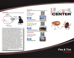


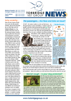
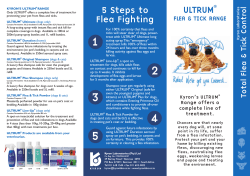
© Copyright 2026