
2014 CANDIDATE MANUAL PATIENT BASED American Board of Dental Examiners
American Board of Dental Examiners Dental Examination PATIENT BASED Periodontal and Restorative Sections 2014 CANDIDATE Administered by: MANUAL Administered by: Council of Interstate Testing Agencies, Inc. 1003 High House Road, Suite 101 Cary, NC 27513 www.citaexam.com North East Regional Board of Dental Examiners, Inc. 1304 Concourse Drive, Suite 100 Linthicum, MD 21090 www.nerb.org Southern Regional Testing Agency, Inc. 4698 Honeygrove Road, Suite 2 Virginia Beach, VA 23455 www.srta.org Please read this manual in detail prior to attending the examination Copyright © 2014 Council of Interstate Testing Agencies, Inc. Final VIII ATTENTION DENTAL CANDIDATES The ADEX Dental Examination Series is administered on behalf of a number of state dental boards and in accordance with state licensing requirements. This examination should be valid in any state accepting the ADEX Dental Examination. However, to be certain, candidates should check with the state dental board of any state in which they wish to be licensed to determine whether this examination will qualify them for licensure in that state. Currently there are three testing agencies which administer the ADEX examination series. Although the content, scoring systems and basic administrative features are uniform, each agency may have some administrative elements which are unique to that agency. Therefore candidates should obtain and thoroughly read the manual published by the agency which will be administering the examination at the date and site the candidate plans to attend. This manual is published by the Council of Interstate Testing Agencies, Inc. and is specific to the administration of that examination. For information about examination sites, dates and fees, visit the CITA website at www.citaexam.com, the NERB website: www.nerb.org. or the SRTA website: www.srta.org. The examination sites and dates will be found under the Examination Calendar drop-down list. Occasionally examinations are interrupted or postponed because of hurricanes, blizzards, other severe weather, power outages, or similar occurrences. CITA, NERB and SRTA reserve the right in its sole discretion, to delay, halt, postpone, or cancel an examination because of unforeseen and serious events. In the event of predicted severe weather events, candidates should monitor the testing agency website for site specific candidate information. Council of Interstate Testing Agencies, Inc. 1003 High House Road, Suite 101 Cary, NC 27513 www.citaexam.com 919-460-7750 2 Table of Contents I. Examination Overview ............................................................................................ 7 Curriculum Integrated Format ...................................................................................... 8 Examination Schedules................................................................................................ 8 Dates and Sites ............................................................................................................ 8 Timely Arrival ............................................................................................................... 8 Retest Schedules ....................................................................................................... 10 Examination Schedule Changes ................................................................................ 12 Registration for Part IV and V .................................................................................... 13 Score Release ........................................................................................................... 13 Examination Scoring System and Content ................................................................. 14 Scoring System for Patient-Based Examinations ....................................................... 14 Scoring Discussion..................................................................................................... 15 Content ...................................................................................................................... 17 Penalties .................................................................................................................... 19 Pulpal Exposure ......................................................................................................... 21 Critical Lack of Diagnostic or Clinical Skills ................................................................ 21 Maintaining Professional Standards ........................................................................... 21 18-Month Completion Rule ........................................................................................ 23 3-Time Failure Rule.................................................................................................... 23 Electronic Scoring ...................................................................................................... 23 II. Policies and Procedures ..................................................................................... 24 Standards of Conduct ................................................................................................ 25 Dismissal from Examination ....................................................................................... 26 Infection Control Requirements .................................................................................. 27 III. The Examination ................................................................................................. 29 Time Management ..................................................................................................... 30 ADEX Status .............................................................................................................. 30 General Administrative Flow ...................................................................................... 30 Section IV: Periodontal Clinical Examination Section ................................................ 32 Section V: Restorative Clinical Examination Section ................................................. 32 Assignments .............................................................................................................. 32 Candidates Assigned First to Restorative Treatment ................................................. 32 Candidates Assigned First to Periodontal Treatment ................................................. 33 Exam Progression ...................................................................................................... 33 Patient Selection ........................................................................................................ 35 Patient’s Medical History ............................................................................................ 35 Health Qualifications and Eligibility ............................................................................ 36 Medical Clearance ..................................................................................................... 38 Patient Rejection ........................................................................................................ 38 Interpreters................................................................................................................. 39 3 Documentation ........................................................................................................... 40 Radiographs............................................................................................................... 41 Procedures................................................................................................................. 41 Communication from Examiners ................................................................................ 41 Infection Control ......................................................................................................... 41 Clinic Attire ................................................................................................................. 42 Retakes of Either the Periodontal or Restorative Examination Alone ........................ 42 Check-Out Procedure for All Exam Sections ............................................................. 42 Check-Out Procedure for Retakes ............................................................................. 43 IV. Periodontal Scaling Examination ...................................................................... 44 General Guidelines .................................................................................................... 55 Periodontal Section Requirements ............................................................................. 48 Radiographic Requirements ....................................................................................... 48 Instruments and Equipment ....................................................................................... 49 Treatment Selection Requirements ............................................................................ 50 Qualifications ............................................................................................................. 50 Exclusions .................................................................................................................. 50 Performance Requirements ....................................................................................... 51 Electronic Submission of Treatment Selection ........................................................... 51 Periodontal Examination Criteria ................................................................................ 52 V. Restorative Examination ..................................................................................... 55 Restorative Examination Requirements ..................................................................... 56 Radiographs............................................................................................................... 56 Forms ......................................................................................................................... 58 Bases, Liners, and Varnishes .................................................................................... 59 Blood Pressure and Anesthetic Record ..................................................................... 59 Treatment Selection and Patient Check-In................................................................. 59 Other Recommendations ........................................................................................... 61 Treatment Exclusions................................................................................................. 61 Patient Check-In ........................................................................................................ 61 Instruments ................................................................................................................ 62 Restorative Treatment Guidelines .............................................................................. 63 Isolation Dam ............................................................................................................. 63 Cavity Sealers/liners/bases ........................................................................................ 63 Caries Detector .......................................................................................................... 63 Pulpal Exposure ......................................................................................................... 64 Recontouring.............................................................................................................. 64 Instructions to Candidate ........................................................................................... 64 Modifications from the Ideal ...................................................................................... 64 Returning from Evaluation of a Modification Request ................................................ 66 Exposure Processing ................................................................................................. 66 Restorative Procedure and Patient Management Guidelines ..................................... 67 Examiner Evaluation of the Class II/Class III Preparation Procedure ........................ 67 Final Evaluation of the Class II Restoration Procedure .............................................. 68 Returning from Evaluation of the Class II/Class III Preparation Procedure ................ 68 4 Examination Completion Following Examiner Evaluation .......................................... 69 Restorative Check-Out Procedure ............................................................................. 70 Separation of Scoring for the Anterior and Posterior Restorations ............................. 70 Requirements for the Class III Composite Preparation and Restoration .................... 71 Requirements for the Class II Amalgam Preparation and Restoration ....................... 71 Requirements for the Class II Composite Preparation and Restoration ..................... 72 Requirements for the Class II Composite (slot) Preparation and Restoration ............ 73 Restorative Criteria ................................................................................................... 74 VI. Examination Forms .......................................................................................... 100 Forms Completed Before the Examination .............................................................. 101 Medical History Form ............................................................................................... 101 Patient Consent Form .............................................................................................. 101 Periodontal Treatment Selection Worksheet ............................................................ 101 Electronic Periodontal Evaluation Form ................................................................... 102 Forms Completed at the Examination ...................................................................... 102 Progress Forms ....................................................................................................... 102 Modification Request Form ...................................................................................... 103 Instruction to Candidate Form .................................................................................. 103 Follow-Up Care Form ............................................................................................... 103 APPENDIX A GLOSSARY OF WORDS, TERMS AND PHRASES ......................... 104 APPENDIX B POLICY FORMS ............................................................................... 113 Patient Notification ................................................................................................... 115 Certification of Status as Junior Student of Record .................................................. 116 Certification of Status as Senior Student of Record ................................................. 117 Certification of Status as Graduate Student of Record............................................. 118 Special Accommodations Request Form ................................................................. 119 Dental Assisting Form .............................................................................................. 123 Interpreter Badge Form ............................................................................................ 124 Certification of Examination Preparation and Orientation Form ............................... 125 Limitation of Liability and Indemnity Agreement ....................................................... 126 APPENDIX C: MODIFICATION REQUEST .......................................................... 127 APPENDIX D: CRITERIA POINTS ....................................................................... 133 APPENDIX E: TREATMENT SELECTION WORKSHEET ................................... 138 APPENDIX F: LESION APPROVAL FORM .......................................................... 140 APPENDIX F: EXPOSURE PROCESSING FORM............................................... 142 5 IMPORTANT NOTICE Beginning in 2014 CITA will be administering the ADEX examination, and thus this manual will reflect the examination content and scoring systems as developed by ADEX. As a member of ADEX all agencies administering the ADEX examination are allowed latitude in administrative elements of the examination. Thus this manual has been developed to provide the candidate with the information required for the candidate to hopefully be successful on the CITA administration of the ADEX examination process. Every effort has been made to ensure that this manual is accurate, comprehensive, clear and up-to-date. In the rare instances when examination related instructions need to be updated or clarified during the examination year those changes will be posted on CITA’s website (http://www.citaexam.com “Examination Manuals”). There may also be other test related material sent to candidates directly by the CITA office should the need arise. All candidates who participate in the CITA administration of the ADEX examination are responsible for reading and understanding the CITA examination manual(s), any website documented changes to the published CITA manual(s) and for reviewing and understanding all other material provided by CITA. If, in reviewing any CITA provided material, questions arise, it is the candidate’s responsibility to resolve those questions by directing them to the CITA office at the earliest possible date. For candidate’s convenience, CITA’s address, e-mail address, telephone number, website and facsimile number appear below. Prior to taking an ADEX examination, each candidate will be required to sign a statement certifying that he/she has reviewed the CITA manual, viewed the on-line candidate tutorials, read other material provided by CITA and that he/she has addressed and resolved all questions prior to the date of the examination. It is also extremely important that candidates maintain a current mailing address with CITA by updating their online profile. This is the only way to ensure that there will be a timely receipt of important material. Address Council of Interstate Testing Agencies 1003 High House Road, Suite 101 Cary, North Carolina 27513 Telephone: (919) 460-7750 Fax (919) 460-7715 [email protected] http://www.citaexam.com 6 The ADEX Dental Examination Series PATIENT BASED I. Examination Overview 7 Examination Overview Curriculum Integrated Format The Curriculum Integrated Format (CIF) is the pre-graduation format of the ADEX Dental Examination Series for junior and senior dental students of record. Both the Curriculum Integrated Format and the Traditional Format examinations are identical in content, criteria and scoring. The major difference between the two formats is in the sequencing of examination sections. In the Traditional or Pre-Graduation Format, the manikin and patient-based examination sections are administered in their entirety at each site over the course of two consecutive days. Senior dental students may sit for all Sections of the Examination. In the Curriculum Integrated Format, examination sections are administered in segments over the course of up to 18 months to eligible dental students or students in a graduate program at a dental school where this examination is offered. The spaced sequencing of this format provides the opportunity for candidates to remediate when necessary within the dental school curriculum and provides for the timely issuance of licenses upon graduation. Examination Schedules Dates and Sites Specific examination dates for a participating dental school can be found on the CITA website. Please refer to the supplement to this manual for policies specific to the CITA testing agency. The Restorative and Periodontal/Scaling Clinical Examination Sections will typically be administered at the candidate’s dental school on a specified date(s) at the beginning of the new year as determined by the Dean of the dental school and agreed upon by the respective testing agency. Candidates may take these examination sections up to three times. Please refer to the supplement for specific policies on retake and the available timeframes. Timely Arrival It is the candidate’s responsibility to determine their travel and time schedules to ensure they can meet all CITA’s time requirements. The candidate and his/her auxiliary are expected to arrive at the examination site at the designated time stipulated in the published schedule for that particular examination. Failure to follow this guideline may result in failure of the examination currently being taken by the candidate. Candidates will be informed as to the date on which they are to take each section of the examination. Candidates should note that the Patient Based Examination has specific time restraints for each section, and all procedures for each examination must be completed within the allotted time for that examination. The following schedule is an example of the administration of these examinations; however, examination schedules are not finalized until after the examination application deadline. Candidate’s actual schedule will be emailed at least three (3) weeks prior to the examination date. 8 A. Full Perio - Restorative (Three Procedures) Time 6:30 AM – 6:45 AM Full Restorative and Periodontal Scaling Schedule Registration (Candidates must register at designated room within facility) 6:45 AM Announcements-Questions/Answer Session 7:00 AM Patient Set-up and Lesion Approval (Candidates allowed into clinic) 8:30 AM Restorative and Periodontal Examinations Begin Final Procedure Restorative 4:00 PM Case (Lesion) Acceptance Guideline 4:30 PM Checked In for Preparation Evaluation 5:30 PM Checked in For Final Evaluation 5:30 PM Examination Ends Final Procedure Periodontal Scaling 4:15 PM Case Acceptance Guideline Finish Times Assigned 4:45 PM Pre Treatment Evaluation Completed- All Candidates 5:30 PM Candidates Must be Checked In for Calculus Removal Evaluation 5:30 PM Examination Ends In scheduling patients and planning the time utilization, candidate should consider the fact that the time allowed for completion of the Restorative Examination INCLUDES THE TIME DURING WHICH PATIENTS WILL BE AT THE GRADING STATION and thus should plan their time accordingly. As such, this time may vary according to the procedure being evaluated, the testing site, and the number of candidates. Candidates taking both Restorative procedures and the Periodontal Scaling Examination will begin set up in the clinic at 7:00 a.m. Candidates will obtain their registration materials and registration packet during registration at 6:30 a.m. the morning of the examination. Candidates should consult the Policies and Procedures Manual for all items required for registration. After completing the initial examination procedure, candidates may continue to the remaining procedures in whichever order they choose; however, all procedures must be completed by 5:30 p.m. 9 If the final procedure is the periodontal procedure, the case acceptance should be completed by 4:15 and pre-treatment evaluation must be completed by 4:45 p.m. in order to allow 45 minutes for treatment time. If the final procedure is a restorative procedure, the case acceptance/lesion approval for the second restoration should be completed by 3:45 p.m., the candidate must be in line at the Check In Station for Paperwork Acceptance for the preparation evaluation by 4:30 p.m. and the candidate must be in line at the Check In Station for Paperwork Acceptance for final evaluation by 5:30 p.m. If candidates are not in line for Paperwork Acceptance for the final evaluation by 5:30 p.m., the candidate must temporize any prepared teeth, and restorations already in place will not be graded Retest Schedules Candidates which are required to retake portions of the exam will register for retest through the online registration process. Retest candidates will typically be retesting for both restorative procedures, one restorative procedure, the periodontal scaling procedure or a combination of one restorative procedure and the periodontal scaling procedure. Sample exam schedules for procedure retest and retest involving multi section examinations are depicted below. If a candidate needs to retake only one procedure (the Periodontal procedure or only one of the Restorative Examination procedures), he/she will be assigned either a morning or an afternoon assignment. Candidates will have three hours total (90 minutes actual treatment time) for the Periodontal procedure or four hours for one Restorative Examination procedure according to the following examination schedule tables. Candidates taking two procedures across two sections (multi section) will in essence be taking one procedure from the restorative section and one from the periodontal section of the examination. Those candidates shall perform the periodontal scaling procedure first. When the periodontal scaling procedure is completed they may transition to the restorative procedure. The candidate will then have four (4) hours to complete the restorative procedure. B. Multi Section Perio-Restorative (Two Procedures) Time 6:30 AM - 6:45 AM Two Procedure Schedule- Periodontal First Registration 6:45 AM Announcements – Questions/Answer Session 7:00 AM Patient Set-up and Approval (Candidates allowed into clinic) 8:30 AM Periodontal Scaling Examination Begins (90 Minutes Assigned Treatment Time) Candidates Transition to Restorative (4 hours treatment time) 2:30 PM Deadline for Check In for the Final Restorative Preparation Evaluation 3:30 PM Examination Ends (Perio and One Restoration - 7 hours) 10 C. Restorative (Two Procedures) Time 6:30 AM – 6:45 AM Restorative Section Retake Registration 6:45 AM Announcements 7:00 AM Patient Set-up and Lesion Approval 8:30 AM Restorative Examination Begins 2:00 PM Final Lesion Acceptance 2:30 PM Checked In for Final Preparation Evaluation 3:30 PM Checked In for Final Evaluation 3:30 PM Examination Ends (Two Restorations - 7 Hours) D. Restorative (One Procedure) Time 6:30 AM – 6:45 AM Restorative Retake Morning Schedule Registration 6:45 AM Announcements 7:00 AM Patient Set-up and Lesion Approval 8:30 AM Restorative Examination Begins 11:30 PM Checked In for Final Preparation Evaluation 12:30 PM Checked in for Final Evaluation 12:30 PM Examination Ends (One Restoration - 4 Hours) 11 Time 11:30 AM - 11:45 AM Restorative Retake Afternoon Schedule Registration 11:45 AM Announcements 12:00 PM Patient Set-up and Lesion Approval 1:30 AM Restorative Examination Begins 4:30 PM Checked In for Final Preparation Evaluation 5:30 PM Checked in for Final Evaluation 5:30 PM Examination Ends (One Restoration - 4 Hours) E. Periodontal Scaling Examination Time 6:30 AM – 6:45 AM Periodontal Morning Schedule Registration 6:45 AM Announcements 7:00 AM Patient Set-up and Approval 8:30 AM Periodontal Scaling Examination Begins Finish Time Assigned 10:45 AM Pre Treatment Evaluation Completed- All Candidates 11:30 AM Candidates Must be Checked In for Calculus Removal Evaluation 11:30 AM Periodontal Scaling Examination Ends (3 hours) 12 Time 11:30 AM – 11:45 AM Periodontal Afternoon Schedule Registration 11:45 AM Announcements 1:00 PM Patient Set Up and Approval 2:30 PM Periodontal Scaling Examination Begins Finish Times Assigned 4:45 PM Pre Treatment Evaluation Completed- All Candidates 5:30 PM Candidates Must be Checked In for Calculus Removal Evaluation 5:30 PM Periodontal Scaling Examination Ends Candidates taking only the Periodontal procedure may be assigned to take that procedure in either the morning or the afternoon schedule. Assignments are based on the site related considerations as well as the composition of site test takers. All assignments are final. Candidates are considered to have met required end time deadlines i.e. “Candidates Must be Checked In for Calculus Removal Evaluation” when they have either completed the Check In procedure for Calculus Removal Evaluation or the candidate is in line for Paperwork Acceptance at the Check In Station for Calculus Removal Evaluation. Examination Schedule Changes Requests for a change in assignment time will not be considered or made once the schedule has been distributed. Dental school personnel do not have the authority to accept a candidate for an examination at their site or to make any assignment changes within an examination series. Such arrangements between dental school personnel and a candidate may preclude the candidate from being admitted to the examination, as well as result in forfeiture of all fees. CITA’s Chief Examiner is the only authorized individual who may consider a request for a schedule change. If unusual circumstances warrant such a change and space is available, it is the decision of CITA’s Chief Examiner whether to approve such a request. This decision is made on-site on the day of examination, and prior requests are neither accepted nor considered. Registration for Part IV and V It is the sole responsibility of each candidate to know and understand the examination(s) for which they have registered and the time they have been assigned for each of those examinations. If the candidate is taking the Restorative and Periodontal Examinations in their entirety, the Restorative only, the Periodontal Examination only, or a combination of the periodontal scaling and one of the restorative procedures, those candidates will present their credentials and identification at an assigned registration period prior to the Set Up Period. The candidate’s identification will be confirmed, the candidate will receive their registration materials and the candidate will be allowed at the stipulated time to begin their Set Up process. 13 CANDIDATES SHOULD NOT BEGIN THE SET UP PROCESS UNTIL THEY HAVE REGISTERED. FAILURE TO OBSERVE AND FOLLOW THIS GUIDELINE WILL RESULT IN DISMISSAL FROM THE EXAMINATION. In order to receive an examination packet and be admitted to the examination, the candidate MUST present a government-issued photo identification (e.g., valid driver’s license, military identification card, passport) or a school-issued identification card and completed Radiograph Verification/Follow-Up Care Form. Candidates are required to bring to registration a completed Examination Preparation and Orientation Form and Limited Liability Form. Candidates who do not have the required identification and completed forms WILL NOT be admitted to the examination. In the event there are extenuating circumstances such as weather, acts of God, or other unforeseen circumstances which may impact or alter the schedule and administration of the examination(s), candidates should consult the CITA web site (www.citaexam.com) for notifications as to the conduct of the examinations and any alterations in times, rooms and/or schedules. Score Release Because the opportunity for remediation within the dental school is intended to be a significant feature of the Curriculum Integrated Format, as well as Pre-Graduation Examinations, the candidate’s individual scores will be released electronically to the candidate’s dental school, in addition to the candidate him/herself, to facilitate the remediation. Scores are not released to candidates or their representatives by telephone or fax. Scores are not released to anyone other than the candidate, the candidate’s dental school and the participating jurisdictions, unless a request for a Score Report is received and a Confidentiality Agreement has been executed with CITA. (See the section titled Score Report Request below). Beginning with the CIF Class of 2014 examinations, scores will be listed as “Pass, score 75 or above” for a passing score and “Fail, score below 75” for a failing score. Scores are automatically sent for each individual ADEX Dental Examination to the participating licensing jurisdictions. A Line Item Level (LIL) report or critique of the candidate’s deficiencies in their clinical performance for all failing candidates is furnished to the candidate and the candidate’s school along with the examination score. In order to maintain the security of the examinations, this report is issued in lieu of a review of actual examination papers or clinical paper or Electronic Evaluation Forms. In order to obtain “ADEX status” it is not necessary to take or pass Section IV, the Periodontal/Scaling exercise. However, some states will require this for licensure. Check with the state dental board in the state where you wish to practice before applying to take the ADEX examination. Examination Scoring System and Content The scoring system is criterion referenced and is based on an analytical model. The examination is conjunctive, in that its content is divided into five separate sections containing related skill sets, and each section scored independently. Candidates must demonstrate competence in each of the five sections. A compensatory scoring system is used within each examination section to compute the final score for each section as explained below. 14 To pass the ADEX Dental Examination Series and achieve “ADEX Status,” the candidate must score 75 or better on each exam section. While only state boards of dentistry can legally determine the standards of competency for licensure in their states, ADEX has recommended a score of 75 to be a demonstration of sufficient competency, and the participating state dental boards have agreed to accept this standard. Scoring System for Patient-Based Examinations Testing agencies throughout the U.S. have worked together through ADEX to draft and refine the performance criteria for each procedure in this examination. For the majority of those criteria, gradations of competence are described across a 3-level rating scale. Those criteria appear in the manual and are the basis for the scoring system. The three rating levels may be generally described as follows: Acceptable: The treatment is of acceptable quality, demonstrating competence in clinical judgment, knowledge and skill; however, slight deviations from the mechanical and physiological principles of the satisfactory level may exist which do not damage the patient nor significantly shorten the expected life of the restoration. Marginally Substandard: The treatment is of poor quality, demonstrating less than desirable clinical judgment, knowledge of or skill in the mechanical and physiological principles of restorative dentistry, which if left unmodified, will substantially shorten the life of the restoration. Critically Deficient: The treatment is of unacceptable quality, demonstrating critical areas of incompetence in clinical judgment, knowledge or skill of the mechanical and physiological principles of restorative dentistry. The tooth may or may not be temporized, or the treatment plan must be altered and additional care provided in order to sustain the function of the tooth and the patient’s oral health and well-being. There are some criterion which are not scored across the three ratings but who only have defined criteria to meet two of the ratings. Caries Remaining is scored as either Acceptable or as Critically Deficient. The Treatment Management criteria, which scored across the three rating levels of Acceptable, Marginally Substandard and Critically Deficient, are weighted and assigned points values of 2, 1 and 0 respectively. Scoring Discussion Subject matter experts (SMEs) have determined that there are specific performance criteria for each procedure which shall be evaluated in determining whether the performance demonstrates a predetermined level of acceptable clinical competency. This manual contains the specific line item criteria or criterion which are used by the graders in the rating of the candidate’s performance in each of those scored points/criterion. As previously stated they are typically separated into three categories as described in the section above, as Acceptable, Marginally Substandard, and Critically Deficient and are typically assigned five (5) points, three (3) points and zero (0) points respectively. A candidate’s score for each line item criteria is determined by there being confirmation of one of those scoring points by at least two of the three graders for any given line item criteria. In the absence of confirmation of one of the scoring points by the grades rendered by the three examiners, the median score is assigned to that line item criteria. However, if a criterion is assigned a rating of critically deficient by two or more examiners, no points are awarded for that procedure or for the examination section. 15 Based on the number of scorable criteria for each procedure, there will be a given number of points which are possible to attain. By way of explanation, if a procedure has eleven (11) criteria which are scored at five (5) points each, and four (4) which are scored as 2 points each, the total available points for that procedure are sixty three (63) points. This is the situation for the Class II Amalgam preparation. The Class II Amalgam Restoration has the potential for 36 possible points to be awarded the candidate for a total of ninety nine possible points across the section.. See Appendix D. The score for the candidate is calculated by the application of the formula: C/T x 100 – (M) where C= Total points the candidate earned T= Total points possible for the procedure(s) M= Total penalty points or denied Modification Request The compensatory nature of the currently employed scoring rubric is effected in the manner in which the points available are totaled. The candidate is not required to pass each procedure with a score of 75 of higher, as the points are first totaled across the procedures and then the calculation of final score is made at the sectional level. Thus, points lost in one procedure the preparation, which would potentially result in a score of less than 75 for an individual procedure, are offset by the fact that the candidate may have scored better in another procedure, the restoration. In effect, marginally substandard performances in one procedure are off set by acceptable performances in the other procedures. Let’s see how this works in practice. For the restorative section there are four procedures which are scored. However for this year the Class II and the Class III will be scored as free standing sections. Thus the total number of possible points earned for the Class II section will be sixty three (63) for the preparation, and 36 for the restoration or ninety-nine (99) total points. The points that the candidate has been awarded for the two procedures is totaled, and then those points are compared to the total number of points available for the two scorable items, which in this instance is ninety-nine (99). This ratio of point awarded versus the total of available points is then scaled to one hundred (100) and the final tabulated score for the section is thus attained. A hypothetical candidate receives a Marginally Substandard confirmed score for the criterion of Proximal Clearance, Pulpal Floor on the preparation, and a confirmed Marginally Substandard score of Marginally Substandard for the Surface Finish of the restoration. The candidate would be awarded 93 of the possible 99 points. The hypothetical candidate did not incur any penalty deductions. 93/99 x 100 – (M) = 93.9 M in this case equals zero. The candidate’s tabulated score is 94. Regardless of the tabulated score for the section, beginning with the 2014 testing cycle scores will be posted on the website, and disseminated to state boards as either: “Pass, score 75 or above” for a passing score or “Fail, score below 75” for a failing score. The CITA office WILL NOT provide tabulated scores for candidates, state boards or other agencies. 16 Content Section IV: Periodontal/Scaling Clinical Examination – 100 points PERIODONTAL SCALING CONTENT Assignment 1. Case acceptance 2. Pocket depth qualification 3. Subgingival calculus detection FORMAT Performed on a patient Time: 3 hours or less at the candidate’s discretion Treatment 4. Subgingival calculus removal 5. Supragingival plaque/stain removal 6. Tissue and treatment management Treatment Time: 1.5 hours (after case acceptance) (Optional for ADEX Status, but may be required for licensure depending on state licensing requirements) Section V: Restorative Clinical Examination – 100 points RESTORATIVE CONTENT FORMAT 1. Anterior restoration: Class III composite - cavity preparation and restoration are graded separately 2. Posterior restoration: candidate’s choice of either: Class II amalgam - cavity preparation and restoration Class II composite - cavity preparation and restoration Class II posterior proximal occlusal composite (slot) cavity preparation and restoration Performed on a patient Time: 7 hours or less treatment time at the candidate’s discretion (Two Restorations) Time: 4 Hours or less treatment time (One Restoration) Description of Sections IV- V and the number of criteria that are evaluated for the procedures in each section appear below: 17 Sections IV: Periodontal/Scaling Examination Content – 100 Points The Periodontal/Scaling Examination is a patient-based examination consisting of four parts. 1. Treatment Selection – Penalties are assessed for those areas that do not meet the described criteria for case acceptance. 2. Calculus Detection and Removal. – 90 points total with 7.5 points for each surface of subgingival calculus correctly detected and removed. 3. Subgingival Deposit Removal – 6 points total with one point for each one of the first 6 teeth selected in ascending order. 4. Treatment Management – 4 points total for pain control and tissue management that meets the written criteria. Sections V: Restorative Examination Content – 100 Points The Restorative Examination is a patient-based examination which consists of one anterior composite preparation and restoration which are graded separately, as well as one posterior preparation and restoration that may be either an amalgam, a traditional composite or a proximal occlusal (slot) composite. The preparation and restoration are graded separately as well. Anterior Restoration 12 Criteria 10 Criteria Class III Composite Preparation Class III Composite Restoration Posterior Restoration Class II Amalgam Preparation Class II Amalgam Finished Restoration 15 Criteria 9 Criteria Class II Composite Preparation Class II Composite Finished Restoration 15 Criteria 11 Criteria Posterior Proximal-Occlusal Composite Preparation Posterior Proximal-Occlusal Composite Finished Restoration 14 Criteria 11 Criteria 18 Penalties Throughout the examination, the conduct and clinical performance of the candidate will be observed and evaluated. A number of considerations are weighed in determining the final scores. Penalties are assessed for violation of the examination standards for certain procedural errors as described below: Any of the following may result in a deduction of points from the score of the entire examination part or dismissal from the examination: o Violation of universal precautions, infection control or disease barrier technique or failure to dispose of potentially infectious materials and clean the operatory after individual examination sections o Unprofessional demeanor: unkempt, unclean or unprofessional appearance; inconsiderate or uncooperative behavior with other candidates, examiners or testing site personnel o Poor patient management, disregard for patient welfare or comfort o Improper management of significant history or pathosis o Request or repeated requests to modify/extend the approved treatment plan without clinical justification (i.e., attempting to have the examiner “coach” the candidate) o Unsatisfactory completion of required modifications o Improper operator/patient/manikin position o Improper record keeping o Improper treatment selection o Improper liner/base placement o Inadequate isolation o Administration of anesthetic before approval of tooth selection or periodontal assignment by examiners The following will result in the loss of all points for an individual examination: o Temporization or failure to complete a finished restoration o Violation of examination standards, rules or guidelines o Treatment of teeth other than those approved or assigned by examiners o Gross damage to adjacent teeth or tissue o Unrecognized exposure o Unavoidable mechanical exposure that is poorly managed or irreparable o Avoidable mechanical pulpal exposure o Failure to complete treatment within the stated time guidelines This listing is not exhaustive, and penalties may be applied for errors not specifically listed, since some procedures will be classified as unsatisfactory for other reasons, or for a combination of several deficiencies. DE D 19 DEDON Penalty Deductions are depicted in the following table format along with their deduction value. PATIENT MANAGEMENT DEDUCTION Temporization or failure to complete an examination procedure 100 Points Violation of examination standards, rules or guidelines or time schedule 100 points Improper management of significant medical history or pathological condition 100 Points Treatment of teeth other than those approved or assigned by examiners 100 Points Gross damage to adjacent tooth structure – teeth or tissue 100 Points Unrecognized exposure 100 Points Unavoidable mechanical exposure that is poorly managed or irreparable 100 Points Unjustified or avoidable mechanical exposure 100 Points Failure to complete treatment within the stated guidelines of the examination 100 Points Administration of anesthetic before approval of tooth selection assignment by examiners 100 Points rd Non-diagnostic radiographs(s): 3 Time 100 Points Gross Infection control violation: Items by report 10 Points Poor patient management and/or disregard for the patient’s welfare or comfort 10 Points Improper management of significant history or pathology 10 Points Request to remove caries or decalcification without clinical justification 10 Points Inappropriate Modification Request 10 Points Unsatisfactory completion of each required modification sent to the candidate from the Evaluation Station, includes liner a or base placement 10 Points Non-diagnostic radiographs(s) : 2 nd 10 Points time Any Denied Modification request 1 Point Appearance unprofessional. unkempt, unclean 1 Point Violation of universal precautions, either candidate or assistant 1 Point Improper/Incomplete recordkeeping 1 Point Inadequate Isolation 1 Point Attitude rude, inconsiderate/uncooperative with examiners/other personnel 1 Point Improper operator/patient position 1 Point 20 Pulpal Exposure Pulpal exposures, whether avoidable mechanical or inappropriately managed, will result in the following point deductions: PULPAL EXPOSURE INFRACTION POINT DEDUCTION Unjustified mechanical pulpal exposure 100 Points Unrecognized pulpal exposure 100 Points Unavoidable pulpal exposure which is inappropriately managed 100 Points Critical Lack of Diagnostic or Clinical Skills In addition the candidate’s performance may be terminated during the course of the examination if the candidate has demonstrated a critical lack of diagnostic or clinical judgment skills This penalty may only be assigned by the Chief or Restorative Captain(s) and would be applied when the candidate’s lack of clinical judgment or clinical skills seriously jeopardizes the prognosis of the treatment and/or the patient’s well-being. For example, The candidate requests a modification anticipating pulpal exposure, but the preparation is still in enamel. The candidate requests a modification to extend the preparation, but an unauthorized extension already exists. The candidate tells the Clinic Floor Examiner (CFE) that an exposure exists. The CFE finds an exposure that is determined to be unjustified, and there has been no prior approved request for modification in anticipation of the exposure. The candidate tells the CFE that an exposure exists. The CFE finds no exposure, nor do the examiners at the Express Chair. Maintaining Professional Standards As a participant in an examination to assess professional competency, each candidate is expected to maintain professional standards before, during and after the examination. The candidate’s conduct and treatment standards will be observed during the examination and failure to maintain appropriate conduct and/or standards may result in point penalties, failure, and/or dismissal from the examination. Each candidate will be expected to conduct himself/herself in an ethical, professional manner and maintain a professional appearance at all times. Candidates are prohibited from using any study or reference materials during the examination except for CITA approved materials. Any substantiated evidence of falsification or intentional misrepresentation of application requirements, collusion, dishonesty, and use of unauthorized assistance or intentional misrepresentation during registration, pre-examination, or during the course of the examination, SHALL AUTOMATICALLY RESULT IN DISMISSAL FROM AND FAILURE OF THE ENTIRE EXAMINATION, as well as forfeiture of all examination fees for the current examination. 21 Furthermore, the candidate cannot apply for re-examination for one (1) full year from the time of the infraction. Additionally, all state dental boards will be notified of any candidate cited for dishonesty during the examination process. In some states, candidates failed for dishonesty may be permanently ineligible for licensure. Therefore, candidates who have been cited for dishonesty should address this matter with the state(s) wherein they desire licensure prior to examination retesting. The standards itemized in this section apply to all relevant portions of the examination. Failure to adhere to these standards may result in failure of the examination procedure in progress, failure of the entire examination currently being taken by the candidate (e.g., Part I, Part II, Part III, Part IV, or Part V), point deductions from the candidate’s overall score on the examination currently being taken by the candidate, forfeiture of examination fees, or withholding of final examination results by CITA until the candidate complies with the examination requirements set forth in this manual. In addition, a candidate’s conduct prior to and after an examination which does not reflect the level of professionalism expected of a licensed dentist can constitute just cause for CITA providing a summary of relevant facts to a state licensure board or boards. Examples of situations where such an action might be appropriate include a candidate making inappropriate comments about classmates, instructors, school personnel or others associated with the educational or testing environment or a candidate’s misrepresentation of information about why the candidate was unsuccessful in taking the examination. All substantiated evidence of falsification or intentional misrepresentation of registration requirements, collusion, dishonesty or use of unwarranted assistance during the course of the examination will result in automatic failure of the entire examination series. In addition, there will be no refund of examination fees and the candidate will not be allowed to reapply for reexamination for one full year from the time of the infraction. Any of the following infractions will result in failure of the entire examination series: Falsification or intentional misrepresentation of registration requirements Cheating (Candidate will be dismissed immediately) Demonstrating complete disregard for the oral structures or welfare of the patient Demonstrating a complete lack of skill and dexterity to perform the required clinical procedures Misappropriation of equipment (theft) Receiving unauthorized assistance either prior to, or during the examination Alteration of examination records and/or radiographs 22 18-Month Completion Rule All required (4 or 5) sections must be completed successfully within 18 months after the first section is initiated. If any section is not successfully completed within 18 months, regardless of reason, all required (4 or 5) sections must be retaken utilizing the Traditional Format. See the Testing Agency Supplement for how to reapply for one or more sections. 3-Time Failure Rule A candidate may apply to retake each failed or incomplete section of the exam during the following available examination period. A candidate may attempt each examination section up to 3 times during the 18 months after the date he/she took the first section. After three failures of any one section, the entire exam must be retaken. See the Testing Agency Supplement for how to reapply for the entire examination. Electronic Scoring Candidate scoring will typically occur utilizing an electronic scoring process brought to each testing site by the test administering agency. Candidates will not be allowed to access any networks established by the testing agency during the conduct of the examination. All results will be posted online by the agency within the published time frame. CITA reserves the right to conduct its examinations in a non- electronic format if in the sole determination of CITA it is in the best interest of the examination process. Reporting of test scores may therefore vary based on the methodology of scoring employed at any given test site. 23 The ADEX Dental Examination Series PATIENT BASED II. Policies and Procedures 24 Policies and Procedures Standards of Conduct The ADEX examination strives to evaluate the candidate’s clinical judgment and skills in a fair manner. In addition, conduct, decorum and professional demeanor are evaluated. The candidate is required to adhere to the rules, regulations and standards of conduct for the ADEX Dental Examination Series. Throughout the examination, the candidate’s professional conduct and clinical performance will be evaluated. A number of considerations will weigh in determining the candidate’s final score, and penalties may be assessed for violation of examination standards and/or for certain procedural errors, as defined and further described within this manual. 1. Unethical personal/professional conduct: Any substantiated evidence of collusion, dishonesty, use of unauthorized assistance or intentional misrepresentation during registration or during the course of the examinations or failure of the candidate to carry out a directive of the chief examiner shall automatically result in failure of all five examination sections. The candidate and assisting auxiliary must behave in an ethical and proper manner. Patients shall be treated with proper concern for their safety and comfort. Improper behavior is cause for dismissal from the examination at the discretion of the chief examiner and will result in failure of the examination. Additionally, the candidate shall be denied reexamination for one full year from the time of the infraction. 2. Termination of the examinations: The right is reserved to terminate or delay the examinations at any time if 1) that action becomes necessary to safeguard the health, safety or comfort of the patient, 2) the candidate or examiners are threatened in any manner or 3) other interfering events occur that are not under the control of the administering testing agency. 3. Completion of the examinations: All five examination sections (or four, if the candidate is not taking the Periodontal/Scaling Section) of the ADEX Dental Examination Series must be completed within the specified time frame in order to be considered for “ADEX Status.” Examination procedures performed outside the assigned time will be considered incomplete, and the candidate will fail the examination section. If all specified materials and required documentation are not turned in at the end of an examination section that section will be considered incomplete, and the candidate will fail the section. 4. Misappropriation and/or damage of equipment: No equipment, instruments or materials shall be removed from the examination site without written permission of the owner. Willful or careless damage of dental equipment, typodonts, manikins or shrouds may result in failure. All resulting repair or replacement costs will be charged to the candidate and must be paid to the host site before the candidate’s examination results will be released. 5. Submission of examination records: All required records and radiographs must be turned in before the examination is considered complete. If all required documentation is not turned in at the end of the examination, the examination will be considered incomplete, and the candidate will fail all exam sections involved. 6. Assigned procedures: Only the treatment and/or procedures assigned may be performed. (In the Periodontal/Scaling Clinical Examination Section, all surfaces of the selected teeth may be scaled and polished at the discretion of the candidate, but only the selected surfaces will be evaluated.) Performing other treatment or procedures may result in failure of the examination. 25 7. Electronic recording devices and cameras: The use of electronic recording devices or cameras by the candidate, an auxiliary or a patient during any part of the examination is a violation of examination guidelines and may result in failure of the entire ADEX Dental Examination Series. However, intra-oral photographs may be taken by authorized examiners or school personnel during the course of the examination for the purpose of future examiner standardization and calibration. 8. Electronic equipment: The use of pagers, cell phones, computers, DVDs, CDs, PDAs, Blackberries, radios (including walkie-talkies with or without earphones) and any other electronic equipment is not permitted on the clinic floor by candidates, auxiliaries or patients during the examination. Any such use will be considered unprofessional conduct and may result in dismissal from the examination. Dismissal from Examination In addition to the standards of conduct listed in the previous section, the following list is provided as a quick reference for candidates. While the following is not an all-inclusive listing, it does provide examples of behaviors that may result in dismissal/failure of the examination: Using unauthorized equipment at any time during the examination time Altering patient records or radiographs Performing required examination procedures outside the allotted examination time Failure to follow the published time limits and/or complete the examination within the allotted time Receiving assistance from another practitioner, including another candidate, dentist, school representative(s), etc. Exhibiting dishonesty Failure to recognize or respond to systemic conditions that potentially jeopardize the health of the patient, and/or total disregard for patient welfare, comfort and safety Unprofessional, rude, abusive, uncooperative or disruptive behavior to other candidates, patient and/or exam personnel Misappropriation or thievery during the examination Noncompliance with anonymity requirements Noncompliance with established guidelines for asepsis and/or infection control Charging patients for services performed Use of cellular telephones, pagers or other electronic equipment in patient care areas Use of electronic recording devices or cameras by the candidate, auxiliary or patient during any part of the examination 26 Infection Control Requirements Candidates must follow the current recommended infection control procedures as published by the Centers for Disease Control and Prevention for the Restorative, Periodontal/Scaling, Fixed Prosthodontics and Endodontics Examination Sections. These infection control procedures must begin with the initial set-up of the unit, continue throughout the Restorative, Periodontal/Scaling, Fixed Prosthodontics and Endodontics Clinical Examination Sections and include the final cleanup of the operatory. It is the candidate’s responsibility to ensure that both the candidate and his/her auxiliary fully comply with these procedures. Failure to comply will result in loss of points, and any violation that could lead to direct patient harm will result in failure of the examination. As much as possible, dental professionals must help prevent the spread of infectious diseases. Because many infectious patients are asymptomatic, all patients shall be treated as if they are, in fact, contagious. Use of barrier techniques, disposables whenever possible, and proper disinfection and sterilization are essential. Candidates must adhere to the following infection control procedures: 1. Barrier Protection • Gloves must be worn when setting up or performing any intra-oral procedures and when cleaning up after any treatment. If rips or tears occur, don new gloves. Do not wear gloves outside the operatory. Patients with known allergies to latex will not be allowed to sit for the examination at those sites where latex may still be used or present. • Wash and dry hands between patients and whenever gloves are changed. Do not wear hand jewelry that can tear or puncture gloves. • Wear clean, long-sleeved uniforms, gowns or laboratory coats, and change them if they become visibly soiled. Remove gowns or laboratory coats before leaving the clinic area. Wear facemasks and protective eyewear during all procedures in which splashing of any body fluids is likely to occur. Discard masks after each patient, or sooner if the masks become damp or soiled. • Do not wear sandals or open-toed shoes. • Cover surfaces that may become contaminated with impervious-backed paper, aluminum foil or plastic wrap. Remove these coverings (while gloved), discarded them and replace them between patients (after removing gloves). • The patient must wear a clean patient napkin when he/she goes to the Evaluation Station. • Patients must wear protective eyewear during all clinical procedures and are required to bring protective eyewear with them to the Evaluation Station for use during the evaluation of clinical procedures. 27 2. Sterilization and Disinfection • Instruments that become contaminated must be placed in an appropriate receptacle and identified as contaminated. • Any instrument that penetrates soft or hard tissue shall be disposed of or sterilized before and after each use. Instruments that do not penetrate hard or soft tissues but do come in contact with oral tissues shall be single-use disposable items and must be properly discarded. • If not barrier wrapped, surfaces and counter tops shall be pre-cleaned and disinfected with a site-approved tuberculocidal hospital-level disinfectant. • Handpieces, prophy angles and air/water syringes shall be sterilized before and after use or properly disposed of after use. • Used sharps are to be placed in a spill-proof, puncture-resistant container. Needles are to be recapped with a one-handed method or with special devices designed to prevent needle-stick injuries and disposed of properly. • All waste and disposable items shall be considered potentially infectious and shall be disposed of in accordance with federal, state and local regulations. • Resuscitation equipment (sterilizable or disposable), pocket masks, resuscitation bags or other ventilation devices will be provided by the school in strategic locations to minimize the need for any emergency mouth-to-mouth contact. Candidates should be familiar with their use. 3. Exposure to blood borne pathogens An exposure incident is defined as contact with blood or other potentially infectious materials (PIMS) through • Needle stick, sharp or other percutaneous exposure • Non-intact skin exposure, such as an open cut, burn or abrasion • Contact with a mucous membrane (e.g., inside nose, eye or mouth) Since maximum benefit of therapy is most likely to occur with prompt treatment, the following policy has been established: • Immediately following the exposure incident, puncture wounds or other percutaneous exposures should be cleaned with soap and water. Mucous membrane exposed to blood or other PIMS should be extensively rinsed with water or sterile saline. • All percutaneous exposures and other exposures to blood and PIMS should be reported immediately to the Chief Examiner and the person in authority at the examination site so that appropriate measures can be initiated and the exposure incident documented. • If possible, post-exposure prophylactic treatment should be initiated at the examination site if appropriate, as determined by the U.S. Department of Health and Human Services recommendations, or an appropriate referral should be made. • At the completion of all clinical examinations performed in operatories, it is the responsibility of candidates to clean the operatory thoroughly utilizing accepted infection control procedures. 28 The ADEX Dental Examination Series PATIENT BASED III. The Examination 29 The Examination Time Management The Patient Based Sections of the ADEX examination consist of the Section IV, the Periodontal Scaling Examination and Section V, the Restorative Examination. Candidates may take either or both sections the same examination day. When taking both sections the candidate must plan their time to accommodate all procedural considerations such as patient approval, modification request and grading of the candidate performance. Time management is considered an aspect of the examination and the assessment of the candidate’s competency. In scheduling patients and planning the utilization of time, the candidate should be aware that the time allowed for the examination includes the time during which the patient(s) will be at the Evaluation Station for assignment and evaluation. The minimum time patients will be in the Evaluation Station is approximately 30 minutes – possibly longer, depending on the time of day. Times may vary according to the procedure being evaluated, the testing site and the number of candidates being processed. Additionally, when a candidate submits a Modification Request, his/her patient must be sent to the Express/Modification Chair in the Evaluation Station. Candidates are allowed to submit multiple requests for modification simultaneously in order to save time. Patient selection is also an important aspect of the examination and should be taken into consideration in selecting an individual who will serve as the candidate’s patient for the intended procedure. Lastly, there is no substitute for preparation. Candidates are strongly advised to read the candidate manual for the procedure(s) that they may be taking, and to bring the candidate manual with them to the exam site as a reference if needed. Candidates should come to the examination understanding the process and what will occur during the course of the day’s activities. ADEX Status The Periodontal and Restorative Clinical Examination Sections consist of three patient-based procedures performed on the same day. For the acquisition of ADEX Status, the Periodontal Section is optional, and it may not be required for licensure depending on the licensing requirements of the state in which the candidate seeks to practice. Candidates should contact the appropriate state board of dentistry directly to determine state-specific requirements. General Administrative Flow During the course of the examination candidates will have their patients sent to the Evaluation Station for both assessment of the candidate’s performance as well as to meet other procedural aspects of the examinations such as Modification Requests. There will be a table(s) designated in the clinic which will serve as the Check In Station. These tables will be denoted as the Check In Station and will have CITA administrative staff present at all times. 30 The candidate will begin their clinical day by identifying their operatory number and placing their instruments and supplies, along with their patient in the operatory assigned. During the Set Up Period Clinic Floor Examiners (CFEs) will be available in the clinic area to answer candidate questions regarding procedural issues. When the candidate is ready to present their patient and the associated documents and forms for approval, the candidate should summons a CFE who will, at the candidate’s operatory begin the patient/paperwork and Medical History approval process. Should the review uncover an error or deficiency in candidate patient presentation, the candidate, if appropriate, may be allowed to correct such deficiency and re-submit the patient for approval. Candidates will not be allowed to proceed with treatment until their patient and documents have been approved. When the candidate is ready to send their patient for lesion approval, evaluation, or a Modification Request the candidate will gather all required forms, including a completed Evaluation Station Request Form, radiographs, and records and place them in a candidate folder which will be provided at the test site. The folder with all required materials will be presented to the Paperwork Review Station where they will be reviewed by CITA staff. This is a review to ascertain that the required documents are present and does not substitute for the approval process which has been conducted by a Clinic Floor Examiners on the clinic floor. If the documents and radiographs are complete and meet the requirements for the evaluation or process being requested, the candidate will be issued an Evaluation Station Request Card. The Grading Area Request Card will be placed in the front pocket of the candidate folder so that it is easily visible. This folder will also be utilized for Modification Request. Upon receipt of the Grading Area Request Card the candidate should take the Grading Area Request Card and folder to the adjacent Desk Coordinator at the Check In Station who will enter the procedure the candidate is wishing to have evaluated into the examination management software program. If an operatory is available in the Evaluation Station, an assistant/runner will take the folder and paperwork to the operatory and escort the patient to the Evaluation Station. If an operatory is not available the candidate should then return to their operatory and have the folder and the required instruments ready for the arrival of an assistant who will escort the patient to the Evaluation Station when an operatory is available. Candidates should NOT bring their patients to the Check In Station for Paperwork Acceptance. Failure to have the required instruments may result in a penalty being assessed to the candidate. Candidates should consult their manual and/or the candidate tutorial for a list and description of the instruments required for each visit to the Evaluation Station. Once the patient is returned from the Evaluation Station, the candidate should check the paperwork to see that all forms have been completed and to note the presence or absence of an Instructions to Candidate Form which must, if present, be reviewed with the candidate by a Clinic Floor Examiner. 31 Section IV: Periodontal Clinical Examination Section Section IV: Periodontal Clinical Examination – 100 points (Optional, depending on state licensing requirements) CONTENT FORMAT Assignment 1. Case acceptance 2. Pocket depth qualification 3. Subgingival calculus detection Performed on a patient Treatment 4. Subgingival calculus removal 5. Supragingival plaque/stain removal 6. Tissue and treatment management Treatment Time: 1.5 hours (after case acceptance) Time: 3 hours or less at the candidate’s discretion Section V: Restorative Clinical Examination Section Section V: Restorative Clinical Examination – 100 points CONTENT FORMAT 1. Anterior restoration: Class III composite - cavity preparation and restoration 2. Posterior restoration: candidate’s choice: Class II amalgam - cavity preparation and restoration Class II composite - cavity preparation and restoration Posterior proximal occlusal (slot) composite – cavity preparation and restoration Performed on a patient Time: 7 hours or less at the candidate’s discretion (Two Restorations) Time: 4 Hours or less (One Restoration) Assignments Candidates Assigned First to Restorative Treatment Candidates should enter the clinic and begin setting up at 7:00 AM. For candidates assigned to this group, case acceptance for restorative procedure only takes place between 7:00 AM and 8:30 AM, prior to the treatment start time. Clinic Floor Examiners (CFEs) will be available in the clinic beginning at 7:00 AM to assist candidates, whenever appropriate, with examination protocol. Beginning at 7:00 AM, candidates assigned to do a restorative procedure first, may call over a CFE to review and approve the patient presentation (medical history, treatment consent, radiographs, and all other required forms and documentation) and to verify the taking of the patient’s blood pressure. Subsequent to patient presentation and approval by the Clinic Floor Examiner the candidate should present the required forms, radiographs and materials to the 32 Paperwork Review Station for paperwork review and the issuance of a Grading Area Request Card prior to the patient being presented to the Evaluation Station. Once a Grading Area Request Card has been issued, the candidate will return to their operatory and an assistant will be notified that the candidate’s patient is cleared to be taken to the Evaluation Station for lesion approval. The patient is then escorted by the assistant to the Evaluation Station for approval of the lesion(s) which have been selected by the candidate for treatment. When the patient is returned from the Evaluation Station and a lesion has been approved for treatment, the candidate may continue pre-treatment preparations but no treatment (including administration of anesthetic) may be performed until the start of the examination is announced at 8:30 AM, to begin treatment. When cavity preparation has been completed, the candidate again presents to the Paperwork Review Station for paperwork review and issuance of a Grading Area Request Card. When a chair is available in the Evaluation Station the patient is escorted to the Evaluation Station by an assistant for grading of the preparation (procedure) to be evaluated. When the restoration is completed, the candidate again submits the required forms and materials to the Paperwork Review Station and the process proceeds per the previous procedures. Thus a minimum of two patient visits to the Evaluations Station during candidate treatment time (each taking approximately one half hour) are required. Candidates Assigned First to Periodontal Treatment Candidates should enter the clinic and begin setting up at 7:00 AM. Case acceptance for the periodontal examination takes place at the candidate’s cubicle between 7:00 AM and 8:30 AM. Beginning at 7:00 AM, candidates assigned to do the Periodontal procedure first, may call over a CFE for patient presentation which includes a review of the Teeth Selection, Required Paperwork and Forms, and Blood Pressure recording. At 8:30 AM the examination begins and the patient may be submitted to the Evaluation Station for Treatment Selection Evaluation (Perio Pre-Treatment Evaluation). All candidates will be given ninety (90) minutes of treatment time, beginning when the Pre-Treatment Evaluation has been completed and the patient is returned from the Evaluation Station. By the end of that ninety (90) minute time period (which will be recorded on the candidate’s Periodontal Progress Form), the candidate must be in line for issuance of an Grading Area Request Card at the Check In Station for return to the Evaluation Station for Treatment Evaluation (Post Treatment Evaluation). Thus two patient visits to the Evaluation Station (each taking about one half hour) are required for the Periodontal Examination. Exam Progression Assuming that the candidate is taking both the restorative and the periodontal sections, after the initial procedure, either restorative or periodontal, is completed, the candidate may proceed to the other required procedures in whichever order they choose as long as all parts of the examination are completed by 5:30 PM on that day – with the following stipulations: If the final procedure of the day is the periodontal procedure, the Pre-treatment Evaluation must be completed by 4:45 PM so the candidate will have at least 45 minutes to treat the patient. All candidates must be in line for Paperwork Acceptance at the Check In Station no later than 5:30 PM. Candidates who are not in line at the Check In Station by 5:30 PM will not have their patients evaluated. 33 If the final procedure is a restorative procedure, the case acceptance must be completed by 4:00 p.m., the candidate must be in line for Paperwork Acceptance for the preparation check by 4:30 p.m. and the candidate must be in line for Paperwork Acceptance for the final evaluation by 5:30 p.m. If candidates are not in line for Paperwork Acceptance for final evaluation by 5:30 p.m., any prepared teeth will be temporized and any teeth with restorations already in place will not be graded. Note: If in the afternoon, the candidate finds that restorative treatment begun that morning, is running late, the candidate may not, at that time, elect not to take the optional Periodontal Examination, in order to extend the time for the Restorative Examination. The time allowed for treatment in the Restorative Examination remains at no more than 7 hours for two procedures and no more than 4 hours for one procedure. Testing Schedule Overview – Restorative and Periodontal Scaling Candidates will be assigned to begin the day with either a restorative or periodontal procedure. Restorative Procedure First TIME 6:30-6:45 a.m. 7:00 a.m. Periodontal Procedure First ACTIVITY Candidate Registration TIME 6:30-6:45 a.m. Set Up and Approval 7:00 a.m. Candidate will have patient acceptance completed at their operatory. Treatment Time Set Up and Approval Candidate request a CFE to review and approve Medical History, Patient Consent Form, anesthetic record on the Progress Form and radiographs. Treatment time begins. Candidates work to completion of the first restorative treatment. Candidate Registration Case acceptance takes place in the candidate’s operatory. Candidates will send their patient to the Evaluation Station to have the lesion approved. 8:30 a.m. ACTIVITY 8:30 a.m. Treatment Time (Note that candidates must complete two restorative treatments and one periodontal treatment before 5:30 p.m.) Examination begins. Candidates send patients to the Evaluation Station for Treatment Selection (Pre-Treatment) evaluation. Candidates have 90 minutes to complete the periodontal treatment, starting after the patient returns from the Evaluation Station for the Treatment Selection (Pre-Treatment) evaluation. Candidates must be in line for Paperwork Acceptance line for per stated time lines Candidates must send their patients to the Evaluation Station after completing the preparation and again after restoration for both procedures Thus, patients must visit the Evaluation Station twice during the Periodontal Section – once before the treatment and once afterward. Each visit will take approximately 30 minutes. Candidates will send their patient to the Evaluation Station for all Modification Request 34 Patient Selection Candidates must furnish their own patients for Sections IV and V. Patient selection and management is an important part of the examination and should be completed independently, without the help or assistance of faculty or colleagues. Patient Management.- The candidate and assisting auxiliary must behave in an ethical and proper manner towards all patients. Patients shall be treated with proper concern for their safety and comfort. The candidate shall accurately complete the appropriate Medical History Form and establish a diagnosis and treatment plan as required for each selected patient. The patient’s health status must be acceptable for clinical treatment and the lengthy examination process. For the Restorative Examination Section, candidates may present a backup patient if their firstchoice patient is not accepted by the examiners. However, only one patient may be submitted for the Periodontal Examination. Due to the natural stress of an examination, candidates should avoid selecting patients who are apprehensive, hypersensitive, have physical limitations that could hinder the examination process or aren’t able to stay for the duration of the examination. However, at the candidate’s discretion, an individual who has a physical disability may, in most cases, be a patient in the examination. Candidates must contact the testing agency a minimum of 60 days prior to the examination for authorization for patients with special requirements. Patient’s Medical History Medical History Form - A Medical History Form must be completed independently by the candidate (without help of faculty or colleagues) for each clinical patient prior to the examination. This form may be completed prior to the examination date; however, the form must reflect the patient’s current health at the time of the examination. Follow up Questions - The Medical History Form includes questions pertaining to medical conditions that might affect the patient’s suitability for treatment. If the patient gives a positive response to one of these questions, the candidate must explore the nature of the condition and provide an adequate explanation on the Medical History Form. Blood Pressure - A screening blood pressure reading should be taken when the patient is selected and must be retaken on the day of the examination, during the Set Up Period and recorded on the Medical History Form. The examination-day reading must be documented by a CFE. If the patient is sitting for more than one examination section on the same day, his/her blood pressure must be taken and recorded prior to each section. Failure to take or falsification of the blood pressure reading will result in dismissal of the candidate form the examination. Medications - On the day of the examination, the candidate must document on the Medical History Form all medications or supplements taken by the patient within the last 24 hours. Candidates should document antibiotic premedication on the appropriate Progress Form, as well as on the Medical History Form. 35 Health Qualifications and Eligibility In selecting a patient, candidates should remember that in the clinical examination setting it is necessary that both the candidate and examining personnel be able to monitor the patient at all times. Therefore, CITA will not accept a patient whose face, neck, temples and ears are not fully visible at all times. Such visibility is necessary (1) for detection of acute conditions which might be identified on observation, (2) to monitor possible allergic reactions and (3) for other similar purposes generally recognized in the profession. Unacceptable patients will be dismissed, and to continue with the examination, the case acceptance criteria must be corrected on the previously submitted patient or an acceptable back-up patient must be approved. Candidates must advise their patients of the time required to participate in this examination. No extra time will be given in the event a patient is deemed unacceptable. The minimum patient age is sixteen (16) years, and a parent or guardian must be available in the waiting area during treatment who must provide written consent for minors under the age of eighteen (18). For Alabama written consent is required for minors under the age of nineteen (19) and for Puerto Rico written consent is required for minors under the age of twenty one (21). Proof of legal guardianship is required. Dentists, dental hygienists, third, fourth or final year dental students, and dental hygiene students are NOT acceptable as patients for this examination. Dental Assistants may be used as patients for this examination. In order to participate in the examination, patients must meet the following criteria: 1. Patients must have a blood pressure reading of 159/94 or below to proceed without medical clearance. Patients with a blood pressure reading between 160/95 and 179/109 are accepted only with a written medical clearance from the patient’s physician. Patients with a blood pressure reading 180/110 or greater will not be accepted for this examination, even if a physician authorizes treatment. 2. Candidates who are sharing a patient requiring antibiotic prophylaxis must treat the patient the same day. Treatment of the same patient on subsequent clinical days will not be permitted. 3. Patient must have no history of heart attack (myocardial infarction), stroke, or cardiac surgery within the last six months. 4. Patients may not have active tuberculosis. A patient who has tested positive for tuberculosis or who is being treated for tuberculosis but does not have clinical symptoms is acceptable 5. Patients may not have undergone chemotherapy for cancer within the last six months. 6. Patients participating in the Periodontal Examination Section may not have a history of taking IV or orally-administered bisphosphonate medications. Patients participating in the Restorative Examination Section may not have a history of taking IV-administered bisphosphonate medications (except an annual IV dosage for osteoporosis). However, patients who have taken oral bisphosphonates may participate in the Restorative Section. 7. Patients may not have an active incidence of bisphosphonate osteonecrosis of the jaw (BON) also known as osteochemonecrosis or osteonecrosis of the jaw (ONJ). 36 8. Patients may not have any condition or medication/drug history that might be adversely affected by the length or nature of the examination process. 9. Patients with latex allergies may not participate in the examination. 10. If the patient answers “yes” to any of the questions on the Medical History Form, the candidate must explore the item further and determine whether a medical clearance from a licensed physician would be appropriate. A medical clearance is required if the finding could affect the patient’s suitability for elective dental treatment during the examination. 11. Candidates must obtain written medical clearance for patients reporting a disease, condition, or problem not listed on the Medical Health History Form that would pose a significant risk to their own health or safety or others during the performance of dental procedures. If this clearance and/or verification of premedication is not available, the patient will not be accepted for treatment. Furthermore, the medical clearance MUST NOT contain the candidate’s name anywhere in the document. 12. The patient’s medical clearance, if necessary, must include: a) A legible statement from a physician written within thirty (30) days of the examination clearly stating the medical concern, b) A positive statement of how the patient should be managed, and c) The physician’s signature, printed or typed name, address and telephone number. 13. Candidates must obtain written medical clearance for patients who have taken Dexfluramine, Fenfluramine, Adipex, Pondimin or Redux. 14. Patients must obtain premedication with a written statement from their physician in the case of any significant medical problems that the American Heart Association classifies as moderate-to-high risk. Written clearance and/or antibiotic premedication from a physician or dentist are specifically required for the conditions listed in the table below. The medical clearance must indicate the specific medical concern and must be attached to the Medical Health History Form on the day of the examination. 15. Candidates must follow the current American Heart Association antibiotic premedication recommendations when treating patients at potential risk of infective endocarditis following dental treatment. A medical clearance may be indicated to determine the patient’s potential risk of infective endocarditis. 16. Candidates must obtain written medical clearance and/or antibiotic prophylaxis, if necessary, for all patients who respond “yes” to question numbers 5.C., 5.D., 5.E., 5.G., 5.H., 5.J., 5.R., and 5.S. on their Medical Health History Form. These items are summarized in the table listed below: 5.C.: Heart Condition 5.D.:Heart Valves—Damaged or Replaced 5.E.: Heart Attack 5.G.: Inborn Heart Defects 5.H.: Infective Endocarditis 5.J.: Unshielded Pacemaker 5.R.: Joint Replacement 5.S.: Stroke 37 17. For the purposes of this examination, CITA has adopted the current American Heart Association guidelines for antibiotic coverage. Antibiotic Prophylaxis is recommended for the prevention of infective endocarditis in the conditions listed below: a. Prosthetic cardiac valve or prosthetic material used for cardiac valve repair b. Previous infective endocarditis c. Congenital heart disease (CHD) d. Unrepaired cyanotic CHD, including palliative shunts and conduits; e. Completely repaired congenital heart defect with prosthetic material or device, whether placed by surgery or by catheter intervention during the first 6 months after the procedure; f. Repaired CHD with residual defects at the site or adjacent to the site of a prosthetic patch or prosthetic device (which inhibit endothelialization). g. Cardiac transplantation recipients who develop cardiac valvulopathy Except for the conditions listed, antibiotic prophylaxis is no longer recommended for any other form of congenital heart disease: Medical Clearance If the patient indicates a medical history that could affect his/her suitability for treatment, the candidate must receive written medical clearance from a licensed physician indicating that the patient may participate in the examination. The medical clearance, if necessary, must include: A clearly legible statement from a licensed physician written within 30 days prior to the examination on official letterhead A positive statement of how the patient should be medically managed The physician’s clearly legible name, address and phone number A telephone number where the physician may be reached on the day of the examination if a question arises regarding the patient’s health The Medical History Form and medical clearance will be reviewed by a CFE for the Restorative and Periodontal Clinical Examinations and must accompany the patient when the treatment selection is submitted for evaluation (patient check-in/case acceptance). If the patient sits for more than one candidate, a separate Medical History Form and Patient Consent Form must be completed for each examination. Patient Rejection Patients who fall into these categories will NOT be accepted: Patients who are under 16 years of age Patents who are unable give legal consent Minors, as defined by the host state, who are unaccompanied by legal parent or guardian Dentists (licensed or unlicensed), third or fourth-year (final year) dental students Dental hygienists (licensed or unlicensed) and final-year dental hygiene students (Periodontal Section only) 38 Interpreters Candidates can employ the services of an interpreter for their patients who do not speak English or who are hearing impaired with a hearing loss which cannot be corrected. (This is particularly important when the patient has a history of medical problems or is on medications.) Interpreters may be related to a patient, but in all cases an interpreter must be at least eighteen (18) years old (nineteen (19) years old in Alabama and twenty one (21) years old in Puerto Rico). Candidates may not share an interpreter during the examination. All interpreters that are utilized by a candidate during the course of the examination will be required to wear a photo identification badge. Three (3) week prior to the scheduled examination, candidates will be required to submit to the CITA office an “Interpreter Form”, that states the candidate’s name, interpreter’s name and contact information and the candidate’s patient’s name along with two (2) passport size photographs taken within the last six (6) months at a local post office, drug store or similar venue. Interpreters will be required to wear the identification badge at all times while on the clinic floor and assisting the patient in the grading station. An interpreter will be not be permitted to assist a candidate and his/her patient if he/she does not have a CITA issued photo identification badge. Candidates are responsible for the conduct of their interpreter during the examination. While there is no strict dress code for interpreters, candidates must be mindful of the fact that the examination site is a professional setting and all personnel should be appropriately dressed. Inappropriate dress would include short shorts, tank tops and/or halter-tops. Candidates should also be mindful of the fact that CITA is committed to providing a safe and secure examination site. Therefore, CITA requires that: All interpreters must appear for the examination with full facial exposure. Mustaches and beards are acceptable for male interpreters as long as the photograph is reflective of the interpreter’s facial condition at the time of the examination. Cosmetics are acceptable for female interpreters on both the photograph and at the examination as long as the photograph readily permits identification of the interpreter at the examination. Dark sunglasses will not be permitted at the examination. permitted. Transitional lenses are Coats, jackets, and other bulky clothing will not be permitted in the clinic area. Interpreters may be asked to stand outside the grading area operatory during Examiner grading The Interpreter Form can be downloaded from the CITA website at (http://www.citaexam.com). All interpreters will be required to have a CITA issued identification badge and will be required to wear the badge at all times while on the clinic floor. The badge will be provided in the candidate examination packet on the day of the scheduled examination. Failure to timely provide or comply with the items listed above will result in the interpreter being prohibited from participating in the examination. 39 An interpreter may NOT be; Under 18 years of age A faculty member, dentist or dental hygienist (licensed or unlicensed) A third, fourth or final year dental student A final-year dental hygiene student The chairside assistant On the day of the examination and prior to the start of the examination, the candidate must complete an Interpreter Disclosure Statement and Interpreter ID Form available at the Coordinator Desk. The candidate must have two 2” x 2” photographs of the interpreter to affix to the Interpreter ID Form, and the interpreter must wear the ID at all times when on the clinic floor. Misinformation or missing information that would endanger the patient, candidate, auxiliary personnel or examiners is considered cause for dismissal from the examination. Documentation Patient Consent Form - A Patient Consent Form must be completed and signed by each patient prior to any treatment being rendered. Initially, only the candidate’s initials and date should be recorded on the Consent Form; the candidate’s name must be added after the examination is completed and before the records are turned in. Premedication Record - A record must be kept for each patient who requires premedication prior to or during the course of the examination. For each procedure, there is a place on the Progress Form to record the type(s) and dosage(s) of medication(s) administered. Candidates who are sharing a patient requiring antibiotic prophylaxis must treat the patient the same clinical day. Treatment of the same patient on subsequent days will not be permitted. Anesthetic Record - At the time of the examination and prior to the start-check for each restorative or periodontal clinical procedure, the following anesthetic information must be indicated on the appropriate Progress Form: Type(s) of Injection (specific block or infiltration to be administered) Anesthetic(s) (generic or brand name and percent used) Vasoconstrictor (type and concentration) Quantity (volume) If more than two dental anesthetic carpules (approximately 3.6 cc) of local anesthetic are needed during any clinical procedure, the candidate must request approval from a CFE, who will document and initial the request. This protocol must be followed for each subsequent carpule. Additional anesthetic solution may be administered only with approval by the CFE. The total quantity of anesthetic solution used must also be documented on the Progress Form. An aspirating syringe and proper aspirating technique must be used for the administration of local anesthetic solutions. The administration of inhalation or parenteral analgesia or sedation is not permitted for any clinical procedures. If the patient has already received anesthesia earlier on the same day, the candidate must enter on the Progress Form the record of the previous anesthesia, including type and amount, to the CFE before administering additional anesthesia. 40 Radiographs For the Restorative Section the radiograph(s) submitted for lesion approval must demonstrate sufficient contrast and resolution to reveal clearly the extent of caries and other pathoses. If the candidate submits poor quality radiographs (film or digital prints), examiners will take the following action: First offense – examiners will request a new set. Second offense – examiners will deduct points and request a new set. Third offense – candidate may be dismissed form the exam Additional radiographs may be required by the examiner during the course of the examination. The radiographic films or digital prints or images used in the examination may be collected at the end of the examination and become the property of the testing agency. Post-operative radiographs or digital prints or images are not routinely required. However, a post-operative radiograph may be requested at any time at the discretion of an examiner. Altering or failing to provide radiographs or digital prints or images will result in failure of the examination. Procedures Sequence of Treatment - Candidates will be assigned to start with either the periodontal procedure or one of the two restorative procedures. Once the initial procedure is completed, the candidate may begin the remaining two procedures in whichever order he/she desires, unless both lesions were approved on the same patient at the same lesion approval submission. In that case the candidate must complete both restorative lesions prior to beginning the periodontal procedure. Instruments submitted with the patient to the Evaluation Station must be fully functional. Mirrors that are clouded, tinted or unclear and explorers that are not fine and sharp will be rejected, and the candidate will be required to submit new instruments. Communication From Examiners Candidates may receive written instructions (Instruction to Candidate Form) from the examiners in the Evaluation Station to resubmit a treatment selection or to modify their treatment. A CFE should deliver this instruction and will check to see that the candidate understands its contents. Candidates who receive an Instruction to Candidate Form should not assume that they have failed. It is possible to pass the examination after being instructed to modify a procedure. Conversely, candidates who receive no instructions to modify procedures should not necessarily assume that their performance is totally satisfactory or will result in a passing grade. In every instance, each procedure is evaluated as it is presented rather than as it may be modified. The examiner ratings are not converted to scores until after the examination is completed and all records are processed by computer. Examiners at the examination site do not know and cannot provide information on whether each candidate has passed or failed a specific examination. Infection Control Candidates must follow all infection control guidelines required by the state where the examination is taking place and must follow the current CDC guidelines for infection control in dental healthcare settings. It is the candidate’s responsibility to ensure that both the candidate and his/her auxiliary fully comply with these protocols. Failure to comply will result in loss of points, and any violation that could lead to direct patient harm will result in termination of the examination and loss of all points. 41 Clinic Attire Clinic attire that meets CDC and OSHA standards must be worn in clinic areas. No bare arms or legs or open-toed shoes are allowed in the clinic areas. Laboratory coats, laboratory jackets, and/or long-sleeve protective garments are all acceptable. Color and style are not restricted. There must be no personal or school identifying information on any clinic attire. The only acceptable identification is the candidate identification badge. Retakes of Either the Periodontal or Restorative Examination Alone If a candidate needs to retake only the Periodontal or the Restorative Examination Section, he/she will have three hours total (90 minutes actual treatment time) for the Periodontal Section or a total of six hours to complete the Restorative Section (both required restorative procedures). Check-Out Procedure for ALL Examination Sections Upon completion of all examinations, candidates must personally submit all examination packets and any unaccounted for typodonts, teeth and/or modules to a central location determined by the chief examiner. Usually there will be a Check Out Station set up in an easily accessible and prominent area either in or adjacent to the clinic where the candidates will be working. The Check Out Station will be manned by CITA Staff. These staff persons will help guide the candidate through the examination check out process. In addition there will be a form in the candidate’s white envelope which will list the forms, badges and other items required for the candidate to successfully check out from the procedure. Candidate MUST check out to have their performance scores released. The following items must be submitted in the provided white envelope and accounted for prior to dismissal from the examination site: Pre-operative and post-operative (if requested during the examination) radiographs of teeth restored during the examination must be submitted and clearly marked for identification. The complete mouth series of radiographs for the Periodontal Clinical Examination Section need not be submitted unless requested by an examiner. (If the testing site requires that radiographs be retained in the patient record, the candidate may submit duplicates of restorative or periodontal radiographs.) At sites where digital images are displayed on a monitor, an electric copy of the digital images used must be submitted on disk to the testing agency. Completed Progress Forms. The Fixed Prosthodontic and Endodontic Clinical Examination Progress Forms are submitted with the typodont to the CFE. Photo ID card for candidate and chairside assistant Patient Consent Form(s) Medical History Form(s) ID cards 42 Check-Out Procedure for Retakes Check-out for those retaking a single examination section is on the day the clinical examination is completed. Those taking examination sections on both days will check out the second day. All required examination records must be submitted to the check-out station before the examination is considered complete. 43 IV. PERIODONTAL SCALING EXAMINATION 44 Patient Management Guidelines General Guidelines 1. The patient must be informed that he/she will be participating in an examination and that additional treatment may be required to meet his/her oral health needs. 2. Only one patient may be presented for the Periodontal Scaling Clinical Examination Section. Once a patient has been submitted to the Clinic Floor Examiner for patient check in and approval, a back-up patient may not be presented if that patient is found not be acceptable due to examination protocols, guidelines or requirements. If the patient is otherwise acceptable but there has been a correctable paperwork or radiographic requirement error, the candidate may be allowed to correct those errors and re-submit that patient for approval. In all circumstances the candidate must have their patients presented and approved for treatment BEFORE proceeding further with the examination. Treatment on a patient without documented approval by a Clinic Floor Examine is a violation of examination protocol and may subject the candidate to dismissal from the examination. 3. The Treatment Selection Worksheet, a practice form provided in Appendix E, may be completed prior to the day of the examination to help the candidate identify the selection of teeth he/she will present for evaluation. Candidates are responsible for independently (without the help of faculty and/or colleagues) selecting and documenting teeth and surfaces for treatment that fulfill the published criteria. Prior to the day of the examination the information on the Treatment Selection Worksheet should be accurately transferred to the Electronic Periodontal Evaluation Form, which is the official form used by examiners. 4. The candidate must accurately transfer the information from the Treatment Selection Worksheet to the Electronic Periodontal Evaluation Form (available on the CITA website up to 48 hours prior to the exam) to indicate his/her treatment selection. The teeth should be listed in ascending order, and the surfaces to be treated should be indicated in the smaller box to the right. 5. The Periodontal Progress Form will be provided at the examination site. When the candidate receives the Progress Form, he/she should place a candidate identification label on the form and enter his/her cubicle number. 6. The procedures, instruments and materials used are the choice of the candidate, as long as they are currently accepted and taught by accredited dental schools and the candidate has been trained in their use. It is the responsibility of the candidate to provide the instruments used in this examination and listed in this Candidate Manual, unless such instruments are furnished by the school. 7. If the candidate is scheduled to perform the periodontal procedure as the first procedure of the day, he/she may call over a CFE beginning at 7:00 a.m. to check the Medical History Form, Patient Consent Form (including the anesthetic record section) and confirm the patient’s blood pressure was taken that day during the Set Up Period. At 8:30 a.m. the patient may be sent to the Evaluation Station for patient check-in/case acceptance. If the Periodontal Examination Section is not the candidate’s first procedure of the day, the candidate may begin the periodontal procedure at any time after the first restorative procedure is completed unless both lesions were approved simultaneously. 45 8. If any problems arise during the examination, the candidate should immediately notify a CFE. The CFE is also present to aid in any emergencies that may occur. 9. Candidates must complete the anesthesia portion on the Progress Form whether or not anesthesia is to be used. If the patient is too sensitive to withstand the use of a periodontal probe or explorer during patient check-in at the Evaluation Station, the candidate may request authorization from a CFE to anesthetize the patient prior to patient check-in. 10. When the patient is sent to the Evaluation Station for patient check-in, he/she will first sign in with the Paperwork Review Station and then the Desk Coordinator. Patients will be evaluated for case acceptance in the order in which they are signed in by the Desk Coordinator. Patients must take the required forms and instruments with them to the Evaluation Station. Only the patient may carry the instrument tray to the Evaluation Station. The following items must be presented on the instrument tray for patient checkin: Completed Medical History Form Signed Patient Consent Form Completed Periodontal Progress Form Perio Pre-Treatment Grading Area Request Form Radiographs Color-coded cubicle ID card Instruments (no scalers or curettes): o Clear mirror (unscratched, untinted, non-disposable) o 11/12 explorer o Probe with Williams markings (1, 2, 3, 5, 7, 8, 9, 10 mm) o Air/water syringe tip Note: The instruments must be placed in a Tupperware container, with the top closed. The Progress Form, Medical History Form, Patient Consent Form, radiographs and color-coded cubicle ID card must be placed on top. DO NOT turn in the Treatment Selection Worksheet. A candidate Sequential ID label must be placed on the right hand side of the patient napkin. 11. The examiners will evaluate the three teeth with 4 mm or deeper pockets and the six to eight teeth with the 12 surfaces of subgingival calculus charted. 12. The Desk Coordinator will indicate a Start and Finish Treatment Time on the Periodontal Progress Form. The approximate total time for the Periodontal Examination Section is 3 hours. The patient treatment time is 1 ½ hours. Candidates must receive a start time 45 minutes prior to the end of the examination day if they are beginning the Periodontal Section after completing the Restorative Section. When the patient returns from the Evaluation Station, treatment should begin. Treatment continues until it is completed or until the Finish Time, as noted on the Periodontal Progress Form. If candidates finish the patient treatment before their assigned Finish Time, they may sign-in the patient with the Desk Coordinator for evaluation. The candidate must scale all subgingival surfaces on the six to eight selected teeth, but only the 12 selected surfaces selected by the candidate will be evaluated. 46 Supragingival calculus, plaque and stain must be removed from all surfaces of the selected teeth. No other teeth may be scaled or polished during the examination, and once the examination is completed, the patient must be dismissed. 13. By the stated Finish Time, each candidate should have completed subgingival calculus removal on the 12 selected surfaces and removed all supragingival calculus, plaque and stain from the entire crown of the selected teeth. The patient must be signed in with the Desk Coordinator for evaluation at the Evaluation Station by the recorded Finish Time. 14. For the treatment evaluation, the candidate must send the patient, wearing a clean napkin with ID Label affixed on the right upper quadrant of the napkin, to the Evaluation Station. The patient should carry the following items to the Evaluation Station on an instrument tray. Instruments must be covered with a napkin and forms and radiographs placed on top of the napkin. Completed Medical History Form Patient Consent Form Completed Periodontal Progress Form Radiographs Color-coded cubicle ID card Required instruments (no scalers or curettes): o Clear mirror (unscratched, untinted, non-disposable) o 11/12 explorer o Probe with Williams markings (1, 2, 3, 5, 7, 8, 9, 10 mm) o Air/water syringe tip Napkin 15. The examiners will evaluate tissue management and subgingival calculus removal from the selected tooth surfaces and evaluate supragingival calculus, stain and plaque removal from all surfaces on the selected teeth. 16. When the patient returns from the Evaluation Station, the candidate may dismiss the patient, unless directed to do otherwise. The candidate must clean the clinic area following accepted infection control procedures . 47 Periodontal Section Requirements Radiographic Requirements Radiographs: Radiographs for the Periodontal Scaling Examination Section must meet the following criteria: Candidates must submit a diagnostic panoramic radiograph or complete (full) mouth radiographic series exposed within the last three years. If a full mouth series is presented, films must be mounted according to ADA procedures (convexity up). Both the options must indicate the exposure date, patient’s name, right and left side and candidate identification number. Candidates must also submit four bitewing radiographs exposed within the previous year (twelve months). If utilizing a full mouth series, this must be mounted separately from the bitewings, unless the complete mouth series were taken within the previous year. Copies are acceptable for the Periodontal/Scaling Examination. Digital images or digital prints are permitted. Candidates from outside the host school will need to submit digital prints as the school will not up load images from outside the facility. If digital prints are to be used, the radiographs must be printed and submitted on photo quality paper or acetate (preferably blue). If the school name is normally incorporated into the digital image, this should be removed or masked, if possible, before printing out the image on photo quality paper or the CFE should be asked to cover such a school identifier on the day of the examination. Alternatively, images may be displayed on monitors if they are available from the school’s database. Candidates from outside the school will need to submit digital prints, when using digital images, as the school will not up load images from an outside facility. Radiographs must not be retaken simply to produce a “perfect” image. Radiographs that have minor errors such as minor cone cutting, not showing all of a third molar or a slightly off center panoramic film, will not result in any loss of points and should not be retaken. Radiographic technique is not being evaluated in this part of the examination. 48 Instruments and Equipment All necessary materials and instruments for the clinical procedures, other than the operating chair, light, and dental unit must be provided by the candidate. Arrangements for rental hand pieces and/or other equipment may be made through the testing site, if such equipment is available. Sonic/ultrasonic instruments are permissible, but they must be furnished by the candidate along with the appropriate connection mechanisms. Airabrasive polishers are NOT permissible. It is the responsibility of the candidate to arrange for his/her own handpiece, sonic/ultrasonic, and all other equipment necessary to complete the clinical examination. The following instruments and equipment are specifically REQUIRED and must be provided by the candidate for this examination: Unscratched, untinted # 4 or #5 front-surface, non-disposable mouth mirror Probe with Williams Markings (1, 2, 3, 5, 7, 8, 9, 10 mm) ODU #11/12 explorer Patient eye protection (personal eyewear is acceptable) Patient napkin holder (chain, self-adhesives, clips, etc.) 2 x 2 gauze Sealed container (such as Tupperware), no larger than 10” x 6” x 3.5”, for transporting instruments. The candidate should be aware that mouth mirrors which are clouded, tinted, or unclean will be rejected. Furthermore, a candidate’s performance will not be evaluated without the proper instruments. Candidates are not limited to the items outlined above but all instruments must be properly sterilized in order to be used. 49 Treatment Selection Requirements Qualifications The candidate’s treatment selection must include the proper number of teeth, adequate deposits of calculus and appropriate pocket depths as defined below: Teeth - There must be at least six and not more than eight permanent teeth selected, at least three of which are molars or premolars, including at least one molar. All posterior teeth must have at least one approximating tooth surface within 2 mm distance. Each of the selected teeth must have at least one surface of subgingival calculus selected for removal. The numbers of the teeth must be listed in ascending order. The teeth selected for treatment will be entered electronically prior to the day of the examination by going to the CITA website at www.citaexam.com Pocket depths - There must be three pockets of 4 mm or greater in depth, each on a separate tooth from among the six to eight teeth selected for treatment. There is a penalty if one of these pockets is less than 4 mm and a severe penalty if two or more are less than 4 mm. (Examiners allow a ± 1 mm leeway in measurement.) It is recommended that pocket depths greater than 6 mm not be included; however, the patient will not be rejected if pockets are this deep. Although the three pockets of 4 mm or more must be on the teeth within the treatment selection, it is not necessary that those surfaces be selected for calculus removal. Calculus - There must be exactly 12 surfaces of explorer-detectable subgingival calculus identified on the selected teeth, and no more than four surfaces may be on the incisors. Three of the 12 identified surfaces of calculus must be on interproximal surfaces of posterior teeth, i.e., on molars and/or premolars. o Explorer detectable subgingival calculus is defined as a distinct deposit of calculus that can be felt with an #11/12 explorer as it passes over the calculus. Qualified deposits may exhibit such characteristics as: o A definite “jump” or “bump” felt by the explorer, with the rough surface characteristic of calculus Ledges or ring formations Spiny or nodular formations Qualified deposits must be apical to the gingival margin and may occur with or without associated supragingival deposits. Exclusions Patients with full-banded orthodontics are not acceptable. Implants or teeth with any fixed appliance – banded, bonded or splinted, either orthodontically or periodontally – may not be included in the treatment selection. No retained primary teeth may be included in the treatment selection. . 50 Performance Requirements Scaling - After the candidate performs the periodontal procedure, the subgingival surfaces of the assigned teeth must be smooth, with no deposits detectable with an 11/12 explorer. Air may be used to deflect the tissue to locate areas for tactile confirmation. (All subgingival surfaces on an assigned tooth must be scaled, but only the selected surface will be evaluated.) Supragingival Deposits (polishing) - All supragingival calculus, plaque and stain must be removed from all coronal surfaces of the assigned teeth so that all surfaces are visually clean when air-dried and tactilely smooth upon examination with an 11/12 explorer. The use of disclosing solution is not permitted. Electronic Submission of Treatment Selection Candidates may use the Treatment Selection Worksheet (available in Appendix B) to identify and document a selection of the patient’s teeth that meet the stated criteria. The candidate should indicate the presence of subgingival calculus on the Treatment Selection Worksheet by marking the appropriate letter for the surface in the box next to the number of the tooth selected for treatment. If subgingival calculus is present on the line angles of the tooth, it must be marked on the interproximal surface, e.g., a deposit on the distofacial line angle would be marked on the distal surface. The numbers of the selected teeth must be listed in ascending order. Prior to the examination, the candidate who is taking the ADEX exam administered by CITA must transfer the information about his/her treatment selection from the Worksheet to the Electronic Periodontal Evaluation Form, which will be reviewed by examiners. Visit the CITA website to access the Electronic Periodontal Evaluation and enter the teeth selected for the treatment Selection. The candidate should note that there is a deadline before each exam for teeth selection to be accomplished prior to the examination. Should a candidate need to enter their teeth selection at the examination site a computer will be available for that purpose, either in or accessible to the clinic areas. 51 Periodontal Criteria PERIODONTAL EXAMINATION PATIENT SELECTION TREATMENT GOALS 1. The Patient Consent Form, Medical History, Progress Form and Periodontal Evaluation Form are complete, accurate and current. Both systolic and diastolic blood pressure are less than or equal to 159/94, or systolic and diastolic blood pressure are between 160/95 and 179/109 with a written medical clearance from a physician authorizing treatment during the examination. Radiographs are of diagnostic quality and reflect the current clinical condition of the mouth. Periapicals have been exposed within the past three years, and bitewings have been exposed within the past twelve months. Radiographs are properly mounted and labeled with exposure date and patient’s name. The Calculus Detection portion of the Periodontal Evaluation Form is properly completed, indicating - Six to eight teeth selected, each with at least one surface of calculus charted - At least three posteriors (molars, premolars), including at least one molar, in the selection. All posterior teeth must have at least one approximating tooth within 2 mm distance. - Exactly 12 surfaces of subgingival calculus charted, including at least three surfaces of interproximal calculus on molars/premolars - At least eight of the surfaces on canines, premolars or molars (no more than four surfaces on incisors) - Three pockets of 4 mm or greater in depth, each on a different tooth within the selection 2. 3. 4. ACCEPTABLE 1. 2. The Patient Consent Form is incorrect or not signed by patient.* The Medical History is incomplete*, is missing candidate initials* or patient signature* or has slight inaccuracies that do not endanger the patient or change the treatment. The Progress Form has inaccuracies or is incomplete or missing.* Blood pressure has not been taken or is not recorded* but, upon correction, meets Satisfactory criteria. Radiographs are available but were not submitted with the patient for initial evaluation.*** The Calculus Detection portion of the Evaluation Form has not been filled out or is filled out incorrectly, e.g., the form demonstrates - Fewer than six or more than eight selected teeth - Fewer than three molars or premolars and/or no approximating tooth within 2 mm of one or more of the selected posterior teeth - One or more selected teeth without any surfaces of calculus charted - More or fewer than 12 surfaces of subgingival calculus charted - Fewer than three surfaces of interproximal calculus on molars and/or premolars more than four surfaces of subgingival calculus on incisors** - Three separate teeth and/or surfaces are not indicated for Pocket Depth Qualification and/or one or more of the teeth are outside the treatment selection.** 3. 4. 5. 6. * ** *** Records and patient will be sent back to the candidate with an Instruction to Candidate Form requesting correction. (If the Periodontal Evaluation Form is completed correctly, it will be retained in the Evaluation Station.) Records and patient will be sent back to the candidate with an Instruction to Candidate Form requesting correction. The candidate will receive an Instruction to Candidate Form requesting radiographs. 52 MARGINALLY SUBSTANDARD 1. Medical History has inaccuracies that do not endanger the patient but do change the treatment or require further explanation by candidate. The candidate submits an incomplete or incorrect Periodontal Progress Form or Evaluation Form for the second time. Radiographs are of poor diagnostic quality and/or do not meet all of the criteria to be considered Satisfactory. Of the three teeth indicated with pocket measurements of 4 mm or more in depth, only two teeth are found to have measurements of 4 mm or more and/or one or more of these teeth are outside the treatment selection on the second submission. 2. 3. * Records and patient are sent back to the candidate with an Instruction to Candidate Form Requesting corrections. CRITICAL DEFICIENCY 1. The Medical History has inaccuracies or indicates the presence of conditions that do endanger the patient, candidate and/or examiners (in this situation, the Periodontal Examination Section will be stopped). The candidate submits an incomplete and/or incorrect Patient Consent Form or Medical History for the second time. 2. The patient’s systolic and/or diastolic blood pressure is between 160/95 and 179/109 without a written medical clearance from a physician authorizing treatment, or blood pressure is 180/110 or greater even with a written medical clearance from a physician authorizing treatment. 3. Radiographs are of unacceptable diagnostic quality and/or are missing and not available on request. (In this situation, the Periodontal Examination Section will be stopped). 4. Of the three teeth indicated with sulcus/pocket measurements of 4 mm or more in depth, fewer than two teeth are found to have pockets of 4 mm or more. 53 PERIODONTAL EXAMINATION TREATMENT AND TISSUE MANAGEMENT TREATMENT GOALS 1. 2. The patient has adequate anesthesia for pain control, is comfortable and demonstrates no evidence of distress or pain. Instruments, polishing cups or brushes and dental floss are effectively utilized so that no unwarranted soft or hard tissue trauma occurs as a result of the scaling and polishing procedures ACCEPTABLE 1. There is slight soft tissue trauma that is consistent with the procedure. MARGINALLY SUBSTANDARD 1. 2. 3. There is inadequate anesthesia for pain control. (The patient is in obvious distress or pain.) There is minor soft tissue trauma that is inconsistent with the procedure. Soft tissue trauma may include, but is not limited to, abrasions, lacerations or ultrasonic burns. There is minor hard tissue trauma that is inconsistent with the procedure. Hard tissue trauma may include root surface abrasions that do not require additional definitive treatment. CRITICAL DEFICIENCY 1. There is major damage to the soft and/or hard tissue that is inconsistent with the procedure and preexisting condition. This damage may include, but is not limited to, such trauma as - Amputated papillae - Exposure of the alveolar process - A laceration or damage that requires suturing and/or periodontal packing - One or more ultrasonic burns that require follow up treatment - A broken instrument tip in the sulcus or soft tissue - Root surface abrasions that require additional definitive treatment. 54 V. RESTORATIVE EXAMINATION 55 Restorative Examination Requirements Radiographs For the purposes of the ADEX examination process, examiners are instructed to evaluate each proposed restoration from many perspectives, but in the final analysis, to decide if a restoration of the tooth in question is clinically defensible. Radiographic presentation is one component, but examiners are challenged to review the visual and tactile evidence, as well as consideration of the overall clinical condition of the accompanying dentition. In the final analysis, the examiner is asked to make a determination as to whether or not there is sufficient clinical evidence to support the restoration of the tooth being presented by the candidate. As a candidate preparing to take the examination, it is advisable to present a patient with a proposed restoration which from a radiographic perspective would demonstrate support for a restorative diagnosis. Typically, that radiographic appearance may be a lesion which appears to have penetrated at least to the DEJ. What is ill advised for the candidate is to present a proposed restoration which demonstrates questionable radiographic support for a diagnosis of disease, and which also lacks radiographic, visual or tactile supporting evidence for a restorative diagnosis. a. Class II Pre-Operative Radiographs A periapical and a bitewing radiograph of the tooth selected for the Class II restoration must be presented when that procedure is presented for acceptance. b. Class III Pre-Operative Radiographs A periapical radiograph of the tooth selected for a Class III restoration, must be submitted at the time the treatment selection is presented for acceptance. Both the Class II and Class III pre-operative radiographic films must be of diagnostic quality, cannot be more than one year old (twelve months), and must depict the CURRENT CLINICAL CONDITION OF THE TOOTH to be treated as well as the surrounding teeth. In other words, there must have been no treatment between the time of taking the radiograph and CITA’s examination that would alter the situation depicted on the radiograph. Duplicate radiographs of diagnostic quality are acceptable. The radiographic films must be mounted according to ADA guidelines (convexity up). These radiographs must be submitted at the conclusion of the examination, and they become the property of CITA. If the dental school stipulates that radiographs must be returned to the school as part of the patient’s examination records, candidates must submit duplicates of the required radiographs to the dental school. c. Post-Operative or Additional Radiographs Post-operative radiographs are NOT required. However, additional or post-operative radiographs may be requested at any time during the conduct of the examination and at the dis56 cretion of any examiner. All such requested radiographs should be mounted, meet the same criteria as previously specified for pre-operative radiographs, and returned to the requesting examiner for evaluation. d. Digital Radiographs Although conventional radiographs are CITA’s preferred radiographic method for this examination, digital radiographs may be accepted if they are presented in a standardized format as specified below: The films or digital images must be of diagnostic quality. For the patient-based restorative examinations, digital periapicals and bitewing images must each be a 4” x 6” image with both images printed on one sheet of 8½” x 11” premium quality photographic paper. Digital prints on photo quality paper may be utilized. The candidate must provide the following information on the back of the print: o Patient’s name o Date the radiographs were taken o Candidate’s Barcode or Identification Label If the school name is normally incorporated into the digital image, this should be removed or masked, if possible, before printing out the image on photo quality paper or the CFE should be asked to cover such a school identifier on the day of the examination. Candidates are advised that with high speed radiographic film (such as F-speed film) the lesion is likely to be larger clinically than what is seen on the radiograph. Alternatively, digital images may be displayed on monitors if they are approved as original and unaltered by the school where the examination takes place. Candidates from outside the school will need to submit digital prints, if using digital images, as the school will not up load images from outside the facility. For the lesion to be acceptable the lesion must penetrate at least to the dento-enamel junction (or have evidence of equivalent depth clinically). If the candidate submits poor quality or non-diagnostic radiographs (film or digital prints), examiners will take the following action: First offense – examiners will request a new set. Second offense – examiners will deduct points and request a new set. Third offense – candidate will be dismissed from the exam 57 Forms All forms are available for review and download on the CITA website http://www.citaexam.com. a. Patient Medical Health History Form This form is part of the Restoration Progress Forms and is used by the candidate to record the health and medical history of the patient. All notations regarding the patient’s medical history must be made on the Medical History Form. The patient’s current blood pressure must be taken by the candidate during the Set Up Period, and the required records should be reviewed for accuracy and completeness. b. Patient Consent Form This form is part of the Progress Forms and is reviewed and signed by the patient. c. Instructions to Candidate Form This form is used by the Examiners to notify the candidate of the need for any additional action or requirement. d. Dental Notification Form This form is reviewed by the patient and signed by the patient should there be a need for patient follow up care. e. Progress Form The candidate should fill out the Progress Forms and refer to this form for patient approval and patient rejection. f. Grading Station Request Forms The candidate will need to fill out the appropriate Grading Station Request Form each time his/her patient is sent to the grading station for evaluation. g. Radiograph Verification Form This form is provided in the confirmation e-mail and candidates are required to complete the form and turn it in during registration the day of the examination. This form verifies the radiographs presented during the examination are of the patient being presented for approval. h. Follow-Up Care Form This form is provided in the confirmation e-mail and is part of the Radiograph Verification Form and candidates are required to complete the form and turn it in during registration the day of the examination. The candidate must indicate who or what facility will provide follow-up care for the patient, in the event that follow-up care is required. i. Modification Forms This form is used by a candidate to request a modification from the ideal preparation. 58 Bases, Liners, and Varnishes The candidate must decide whether a treatment LINER is indicated by checking the liner/base request box on the Class II or Class III Grading Room Request Form, PRIOR TO sending the patient to the Grading Station for the preparation check. a. Cavity Sealers Cavity Sealers provide a protective coating for the freshly cut tooth structure of the prepared cavity. Varnish is a natural gum, such as copal rosin, or a synthetic resin dissolved in an organic solvent, such as acetone, chloroform, or ether. Examples include Copalite, Plastodent Varnish, and Barrier. Resin Bonding Agents include the primers and adhesives of dentinal and all-purpose bonding agents. Examples include All-Bond 2, Scotchbond MP+, Optibond, ProBond, Amalgambond, etc. b. Cavity Liners Cavity Liners are typically a material placed between dentin and a dental restoration to provide protection to the dentin and/or pulp. Examples include Gluma, Vitrebond, Dycal and Cavitec. c. Cavity Bases Cavity Bases are typically a replacement material for missing dentinal tooth structure, used for bulk buildup and/or for blocking out undercuts. Examples include ZOE B&T, IRM, glass ionomer cement and zinc-phosphate cement. NOTE: For the purposes of the CITA examination, the terms base and liner may be used synonymously; however, preparations will not typically be required to be based to anatomical preparation form prior to final restoration placement. Blood Pressure and Anesthetic Record During the Set-Up Period, the patient may be seated and his/her blood pressure MUST be taken by the candidate, and the required records must be reviewed for accuracy and completeness. Candidates must complete the anesthetic record portion of the Progress Form WHETHER OR NOT anesthesia is to be used. Treatment Selection and Patient Check-in Patient selection is very important. If the candidate is unable to complete a procedure due to patient management problems, the procedure cannot be evaluated and no credit will be assigned. No more than two treatment selections may be submitted for a procedure. If a second treatment selection is rejected or a second treatment selection is not presented after the first lesion is rejected, a candidate may not continue with that procedure and will receive a “0” for that portion of the examination. Careful clinical judgments should be used if planning approximating lesions. 59 Treating all lesions Class III (Anterior teeth) Candidates may elect to treat the medial or the distal of an anterior tooth without being required to treat both the mesial and the distal of the tooth, so long as the lesion is not contiguous. Class II (Posterior teeth) Each tooth selected for treatment will need all existing lesions on that tooth treated by the end of the examination day. If a tooth selected for treatment has other lesions then the submission will be denied. Alternatively, lesions other than the one required for the examination may be treated prior to the examinations day. The candidate must enter on the appropriate Progress Form, all proposed treatment for the selected tooth. This includes the primary lesion and any other lesions requiring treatment, except Class V lesions. Class V lesions must be treated prior to the examination. The primary lesion is circled at the top left of the Progress Form and any additional lesions that will be treated are written in ink below. The same restorative material must be utilized for all restorations on the same tooth. All lesions/preparations will be considered together as one submission. The same general criteria as used for the standard preparations will be used for any additional treatments. Any confirmed findings on any of the lesions/preparations will be graded and reported the same as for the required lesion. Sharing patients Any Class II (posterior) tooth selected for treatment must have all lesions on that tooth treated before the end of the examination day. For the posterior restoration, one tooth may not be shared by two candidates for treatment during the examination. If the tooth has a mesial and distal lesion when presented for evaluation, the candidate must treat both lesions by the end of the examination. Any other carious lesions on the tooth must have been previously treated or the submission will be rejected. For the anterior restoration, the candidate may treat either the mesial or the distal, or both if the lesion is contiguous; however, if the lesions are not contiguous the candidate may treat just one aspect of the tooth. Candidates are not required to treat both lesions if they are NOT contiguous. Therefore an anterior tooth may be treated by one candidate in a morning session and subsequently treated by another candidate in an afternoon session. The decision of candidates to share patients during the examination process comes with certain inherent risks, certainly one of which is the inability of another candidate to perform their procedures within speculated time frames. In addition, there are occurrences which impact the timelines of the examination which are beyond the control of either candidates or the test administering body. While CITA will attempt to administer the examination in an expeditious manner, there are circumstances which occur at exam sites which may change the time lines of exam administration and candidates should be aware that changes in administrative timelines may affect their ability to share patients with other candidates. Candidates should make their decision as to whether sharing of a patient is appropriate with these understandings. 60 Other Recommendations Lesions on the distal surface of mandibular first premolars are acceptable for Class II amalgam, Class II composite or proximal occlusal box (slot) restoration but they not recommended due to pulpal anatomy. If there are proximal but no occlusal caries on a posterior tooth, the candidate may elect to do a Class II amalgam or Class II composite restoration but a proximal occlusal box (slot) preparation is recommended. Lesions on the distal surface of cuspids are allowed for Class III composite only, not Class II amalgam. Avoid potential pulpal involvement (too large a lesion) or cuspal replacement. contiguous with the lesion or proposed restoration is discouraged Treatment Exclusions The following will not be accepted for the Restorative Examination Section: Non-vital teeth, and/or teeth with pulpal pathology or endodontic treatment Teeth with facial veneers Mobility of Class III or greater . Patient Check-In The candidate should arrive at the designated clinic and begin set up at the designated time for the Set Up Period and have all materials prepared to send patients to the Evaluation Station for patient check-in, where the treatment selection will be evaluated by the examiners. On the clinic floor, the CFE verifies the blood pressure and that the appropriate and required forms have been completed; then, in the Evaluation Station, all treatment selections (lesion approval) are approved or rejected by at least two of three examiners evaluating anonymously. If a patient meets the requirements for both the posterior and anterior restorations, both may be approved in the Evaluation Station at the same time, but the first restoration must be completed before the second restoration may be begun. Only one patient may be submitted for a patient check-in at a time. If the candidate is utilizing two patents for the Restorative Examination Section, only one may be submitted to start the examination. The second may not be submitted until the first is finished. The local anesthesia request portion of the Restorative Progress Form must be filled out prior to submitting the patient to the Evaluation Station for patient check-in/case acceptance. In the event that the first lesion submitted is not approved, a second lesion may be submitted to the Evaluation Station. If a candidate is scheduled to do the Periodontal examination first and elects not to do the Periodontal examination, the first Restorative patient is submitted to the Evaluation Station between 9:00 and 10:00 AM and treatment begins at 10:00 AM. The second case assignment, evaluation of the second preparation and evaluation of the second restoration takes place at the 61 same time as the candidates taking both the Periodontal and Restorative exams are completing the second restorative lesion. For patient check-in, the candidate will present: Completed Medical History Form Signed Patient Consent Form Progress Form, including completed anesthetic record and indicating tooth number and type of restoration Radiographs (bitewing and periapical) that depict the tooth and surrounding structures, properly mounted and attached with paper clip to the Progress Form- no more than six months old, is required for the Class II restoration A periapical radiograph that depicts the tooth and surrounding structures, properly mounted and attached with paper clip to the Progress Form – no more than six months old, is required for the Class III restoration. Anesthetic solution CANNOT be administered prior to patient check-in. Instruments Candidates must provide the following materials for use during the Restorative Examination Section: Clear mirror (unscratched, untinted, non-disposable) Explorer (fine and sharp) Cotton pliers Probe with Williams markings (1, 2, 3, 5, 7, 8, 9, 10 mm) 62 Restorative Treatment Guidelines Isolation dam During the Restorative Examination Section, cavity preparations may be instrumented with or without an isolation dam. If the extension of the lesion extends beyond ideal preparation form, the candidate MUST place a rubber dam for any additional preparation. An isolation dam that is intact (not torn or leaking) must be in place when the patient is sent for evaluation of the amalgam and composite preparations, as well as for all requests for modification and for the placement of restorative materials. An isolation dam must be in place if a pulpal exposure is anticipated or occurs. The isolation dam must be removed when the patient is sent for evaluation of the finished amalgam and composite restorations. Bite blocks may be used during treatment, but the patient may not travel to the Evaluation Station with a bite block in place under an isolation dam. The isolation dam must be placed by the candidate and not the assistant. Cavity Sealers/liners/bases The candidate must decide if a treatment liner or base is indicated, and if so, check the liner/base request box on the appropriate Progress Form prior to sending the patient in to the Evaluation Station for evaluation of the preparation. Liners will be required only in very deep preparations to cover areas immediately impacting pulpal health and integrity. Failure to request a liner in this circumstance will result in penalties. The examiners in the Evaluation Station will either approve or disapprove the request for placement of the liner or base as part of the evaluation of the preparation. The examiner will indicate his/her decision by placing his/her examiner number in either the Granted or Not Granted box next to the request. If the liner/base is granted, the candidate should place it and then summon the CFE to check its placement before continuing with the final restoration. If the examiners in the Evaluation Station, during grading of the final preparation, make a determination that a liner/base is indicated and has not been requested by the candidate, an Instruction to Candidate Form will be issued to ask that the candidate place the liner/base. In this instance, the liner/base must also be checked by the CFE. In either case, if the CFE finds the insertion of the liner/base to be defective, the patient must be sent to the Grading Station/Evaluation Station evaluators for assessment along with the Progress Form and the Grading Area Request Form prior to any alteration, and before the candidate proceeds with the insertion of the restoration. 1. The liner must be placed only in those pulpal and/or axial wall areas that deviate from the established ideal depth. 2. The liner must not be placed on enamel or within 1 mm of any cavosurface margin. 3. The liner must not compromise the internal retentive and resistance features of the cavity preparation. 4. The liner must not be subject to dislodgement during placement of the permanent restoration. 5. Placement must reflect consideration of limitations of the materials used. 63 Caries Detector Caries detector liquid may be used by the candidate. If used, it must be completely removed prior to the submission of the preparation for evaluation. Pulpal Exposure The candidate MUST immediately call a CFE, who will consult with the Chief examiner to determine the appropriate course of treatment. See section on Exposure Processing. Recontouring Recontouring of adjacent teeth or restorations is allowed only after the preparation has been evaluated and only with the approval of a CFE. Candidates must enter the request to recontour the adjacent tooth in the Additional Comments section of the Progress Form. A CFE must then review the situation and will place his/her examiner number and the time next to the request. The candidate may then restore the tooth after the CFE checks the adjacent tooth recontouring. Instructions to Candidate Evaluators may provide written instructions to candidates if they believe a treatment should be modified during the course of the examination. When the patient returns from the Evaluation Station, if the candidate does not receive an Instruction to Candidate Form, the candidate should continue to the next step of the treatment. If the candidate does receive an Instruction to Candidate Form, it should be delivered by a CFE or an assistant. The CFE will review the instructions with the candidate, and both the candidate and CFE will enter their identification numbers on the form to indicate that the candidate understands the instructions. The corrections must be completed as stated on the form and checked by a CFE. It is a violation of examination protocol to proceed with treatment without CFE interaction and review of the Instructions to Candidate Form. Modifications from the Ideal If, during the preparation, the tooth indicates a need for a significant change from the ideal, the candidate will need to complete a Modification Request Form explaining the proposed modification(s) PRIOR TO PERFORMING THEM. The request to modify should include: Type (external outline, internal form) Where (gingival axial line angle, mesial box) Why (due to caries, decalcification) How much (reference back to either ideal or to the start) All requests for modifications will be sent to the Grading Station. If the candidate feels a finger extension is appropriate and/or necessary to eliminate marginal decalcification, such a modification also should be submitted for approval. 64 IF THE CANDIDATE ANTICIPATES OR ACTUALLY EXPERIENCES A PULPAL EXPOSURE, THE CLINIC FLOOR EXAMINER SHOULD BE NOTIFIED AT ONCE. In the event the lesion, that is originally presented and approved for treatment, needs to be extended, and the extension extends or would extend onto and/or involve a pre-existing restoration, the candidate must submit a Modification Request to extend the lesion. If the Modification Request is approved, the candidate must then remove ALL preexisting restorative material prior to submitting the preparation for another Modification Request or for evaluation of the Class II/Class III Preparation. DO NOT SEND ANY GRADE SHEETS ALONG WITH THE MODIFICATION REQUEST FORMS. NOTE: Excessive use of modification requests MAY result in the candidate failing the examination for failure to complete the procedure(s) in the allotted time period. a. Presenting the Patient to the Grading Station for a Modification Request If the candidate desires to submit a modification request, the candidate will need to submit to the check-in station the required paperwork and materials. Candidates are NOT to bring their patients to the check-in station. Candidates are encouraged to refer to the Candidate Tutorial Booklet received in the confirmation packet for instructions on submitting paperwork. A CITA staff representative will check all paperwork and material and will give the candidate a “Paperwork Acceptance” card and a “Modification Request” card. The candidate will then be instructed to return to their operatory and a CITA staff representative and/or assistant will retrieve the patient from the operatory, along with the paperwork and instruments and will escort the patient to the grading station. With the rubber dam in place, the patient is sent to the Grading Station for approval of the modification request. The following materials will need to be sent with the patient to the Grading Station: Required Instruments in a sealable container no larger than 10” x 6” x 3.5” Modification Request Form (including any prior requests approved or rejected) Progress Form Radiographs Terminology to be used when requesting modifications 65 Returning from Evaluation of a Modification Request When the patient returns from the Grading Station, if the candidate does not receive an Instructions to Candidate Form the candidate should continue with treatment. If a Modification Request is rejected, a red “Modification Request Denied” card will be returned to the candidate with the paperwork and Instructions to Candidate Form. If the candidate receives an Instructions to Candidate Form, THE CANDIDATE MUST INFORM THE CLINIC FLOOR EXAMINER BEFORE PROCEEDING and follow the instructions that have been issued by the examiners. Exposure Processing If the candidate anticipates or actually experiences a pulpal exposure, a clinic floor examiner should be notified at once. The candidate should inform the examiner that either an exposure is anticipated, or that there is an exposure and the basis for making that observation. The clinic floor examiner will not clinically evaluate the patient or the preparation, but will summons the Chief or a Co-Chief Examiner who will instruct the candidate to complete an Exposure Processing Form. The Exposure Processing Form will require the candidate to note the exact location of the exposure within the preparation outline and the approximate dimensions. The candidate will then describe the precise procedure for management of the exposure, including all medicaments and instructions to the patient. Lastly, the candidate should describe any additional extensions or removal of tooth structure which would be required prior to the preparation being submitted to the grading station. If the candidate has finished all cavity preparations and desires to have the preparation graded while the patient is in the grading area, the candidate should also complete a Grading Area Request Form and check those boxes which are applicable to the candidate’s situation. In total, the candidate will need to submit to the Check-In Station the Exposure Processing Form, the Progress Form, any Modification Request Forms, and a Grading Area request Form if the candidate wishes to have the preparation graded while the patient is in the Grading Station for the processing of the exposure. Please note, that the preparation will not be graded unless the candidate specifically designates that grading of the preparation should occur. When all paperwork has been completed, the Chief, or Co-Chief, or CITA staff will escort the patient to the grading station where it will be evaluated. 66 Restorative Procedure and Patient Management Guidelines Examiner Evaluation of the Class II/Class III Preparation Procedure Once the candidate is ready to have their patient evaluated for the Class II/Class III preparation procedure, the candidate will need to submit to the Check-In station the required paperwork and materials. Candidates are NOT to bring their patients to the Check-in station. Candidates are encouraged to refer to the Candidate Tutorial Booklet received in the confirmation packet for instructions on submitting paperwork. A CITA staff representative will check all paperwork and material and will give the candidate a “Grading Area Request Card”. The candidate will then be instructed to return to their operatory and a CITA staff representative and/or assistant will retrieve the patient from the operatory, along with the paperwork and instruments and will escort the patient to the Evaluation Station. With the rubber dam in place, the patient is sent to the Evaluation Station for assessment of the Class II/Class III Preparation procedure. To be properly isolated, at least one tooth on either side of the prepared tooth must be included under the isolation dam unless it is the most posterior tooth.) Candidate requests for a liner or base must be filled out on the appropriate Progress Form The candidate should send the following items: Completed Medical History Form Progress Form (includes signed Patient Consent Form) Required Instruments in a sealable container no larger than 10” x 6” x 3.5” Preparation Grading Room Request Form All Modification Request Forms (Approved or Rejected) Pre-operative radiographs Patient Napkin Cotton Plier The preparation should be presented in sufficient time for the patient to be evaluated (which may involve waiting delays) and for the finished restoration if amalgam, to be condensed, carved, and set up enough to withstand flossing during evaluation Note: The instruments must be placed in the sealable container. The Progress Form, Medical History Form, radiographs and color-coded cubicle ID card must be placed on top. The patient’s napkin must have a candidate ID label affixed on the upper right quadrant of the napkin. Three independent examiners will evaluate the prepared cavity. If modifications of the preparation are required prior to restoration, the examiners will complete an Instruction to Candidate Form and return it to the candidate with the patient. If a candidate receives an Instruction to Candidate Form, he/she must follow the instructions. The candidate must not request an opinion from CFEs concerning the instructions on the Instruction to Candidate Form. If the instructions are to temporize the tooth, the chief examiner must be notified and a Follow-Up Form completed. 67 Final Evaluation of the Class II Restoration Procedure For the Class II amalgam restoration, the amalgam must be sufficiently set to allow a check of the occlusion. For the Class II Composite restoration, any of the composite restorations must be presented without any surface glaze/sealer on the restoration. After removing the isolation dam and any wedges placed during treatment, the candidate should send the patient to the Evaluation Station with a tray containing; Completed Medical History Form Treatment Patient Consent Form Amalgam or Composite Progress Form with properly mounted radiographs Color-coded cubicle ID card Instruments/Materials o Fine and sharp traditional explorer (17-23, or 11/12 explorer) o #4 or #5 front-surface mirror (unscratched, untinted, non-disposable) o Cotton plier o Articulating paper In addition the patient’s napkin must have a candidate ID label affixed on the upper right quadrant of the napkin. Please note that the second restorative preparation may not be started by the candidate until the first restorative patient is dismissed ( that is, after the completed restoration has been evaluated and any required modifications have been completed by the candidate and approved by a CFE). Returning from Evaluation of the Class II/Class III Preparation Procedure When the patient returns from the Grading Station, if the candidate does not receive an Instructions to Candidate Form, the candidate should continue with treatment. If the candidate receives an Instructions to Candidate Form, THE CANDIDATE MUST INFORM THE CLINIC FLOOR EXAMINER BEFORE PROCEEDING and follow the instructions that have been issued by the examiners. A treatment liner is neither required nor evaluated; however, if the candidate determines that a treatment liner is indicated, or should he/she have been directed to place one by the examiners, the placement of the liner must be checked at the Grading Station AFTER THE PREPARATION HAS BEEN EVALUATED. If amalgam, the condensed and carved amalgam surface should NOT be polished or altered by abrasive rotary instrumentation except for purposes of adjusting occlusion. Proximal contact is a critical part of the evaluation, and the candidate should be aware that the examiners will be checking the contact with WAXED dental floss. Field trials have indicated most amalgams can withstand floss being passed through the contact within thirty (30) minutes AFTER THE MATRIX BAND HAS BEEN REMOVED. The candidate should be familiar with the properties of the amalgam being used and should allow sufficient time for the amalgam to set before sending the finished restoration to the Grading Station. A developed and mounted postoperative bitewing MAY be requested at any time at the discretion of a restorative examiner or the Clinic Floor Examiner. If composite, the restorative material does not need to be polished; however, it should be free from void or defect, must be cured to sufficient hardness to retain interproximal contact, withstand forces of mastication, and not dislodge within the cavity walls. 68 Examination Completion Following Examiner Evaluation If the candidate receives no communication from the Grading Station the patient may be dismissed after a clinic floor examiner has approved the patient for dismissal and recorded his/her number in the appropriate space on the Progress Form and given the candidate a green “Patient Dismissal” card. Once the candidate has received the green Dismissal Card, if they have completed all procedures for the Restorative examination, they should compile the necessary documents for Check Out, and present those documents in their proper order to the Check Out station. There will be a Check Out Form which is disseminated to candidates at registration which contains the required documents for Check Out and specifies the order those documents should be arranged. If the final restoration is unacceptable, the candidate will receive an Instruction to Candidate Form and will be instructed to remove the restoration and temporize the tooth. In such cases, the Clinic Floor Examiner must be contacted, and a Dental Patient Notification Form is completed by the candidate and Chief Examiner to ensure the responsibility for further treatment is understood and that the patient will receive the proper care. All post-treatment required as a result of treatment rendered during the examination is the responsibility of the candidate and handled at the expense of the candidate. A Follow-Up Care Form will be issued to the candidate and signed by the patient. When treatment is completed, the CFE will be called to check the provisional restoration before the patient is dismissed. At the discretion of the Chief examiner, the restoration although critically deficient and unacceptable for the purposes of the examination, may be allowed to remain and to serve as a temporary until the patient can have it evaluated and removed. Any restoration left in place at the discretion of the Chief examiner does not indicate an acceptable restoration for the purposed of the examination. If temporization occurs on the first restorative procedure, the candidate will be dismissed from the examination before attempting the second restorative procedure and will fail the Restorative Section. Any post-examination treatment required as a result of treatment rendered during the examination process is the responsibility of the candidate and will be completed at the expense of the candidate. A Follow-Up Care Form must be completed to indicate the follow-up treatment required and clarify responsibility for the treatment. CFE should be notified before the patient is dismissed. 69 Restorative Check-Out Procedure Candidates are encouraged to consult the Candidate Tutorial Booklet regarding the check out process. The items specified below should be enclosed in the original Candidate packet and turned-in at the Check-Out Station in the following order: 1. Identification Badge without the badge holder (only if it the candidate is NOT taking the Periodontal and/or Manikin Examinations) 2. Assistant Badge without the badge holder (only if it is the candidate is NOT taking the Periodontal Examination or utilizing the same assistant for the Periodontal Examination) 3. Class II/Class III Restorative Progress Forms 4. Class II/Class III Restorative Preparation Grading Room Request Forms 5. Class II/Class III Restorative Restoration Grading Room Request Forms 6. Class II/Class III Restorative Radiographs 7. Class II/Class III Modification Request Forms 8. Any Class II/Class III Restorative Procedure Grade Sheets not used 9. Any Progress Forms from patient rejections 10. Any yellow Instructions to Candidate Forms (if applicable) 11. Any extra barcode labels (only if candidate is NOT taking the Periodontal examination) Separation of Scoring for the Anterior and Posterior Restorations Beginning with the CIF Class of 2014 Examinations, the anterior and posterior restorations are graded separately. If the first restorative procedure, which is the choice of the candidate, receives a passing grade, that grade for that singular procedure will stand even if the grade on the second preparation/restoration is below 75. In that case the candidate may apply and retake the second restorative procedure alone at a subsequent examination. However, if the grade on the first restorative procedure is not passing, the candidate will not be permitted to proceed to the second restoration for reasons of patient safety and the candidate will need to apply to retake both restorative procedures at a later examination. 70 Requirements for the Class III Composite Preparation and Restoration 1. The tooth selected for the Class III composite restoration must be a permanent anterior tooth that meets the following requirements: At least one proximal primary carious lesion that shows no signs of previous excavation and appears, radiographically or clinically to extend to the DEJ. OR A defective restoration, defined as one that exhibits recurrent caries or a defective cavosurface margin that, even though it may not yet be carious, can be penetrated with an explorer. (A mismatched shade is not an acceptable indication.) Existing defective restorations must be completely removed before submitting the patient to the Evaluation Station for a modification request or evaluation of the completed preparation. There must be visually closed contact with the adjacent tooth on the proximal surface to be restored, although the area to be restored may or may not be in contact. The approximating contact of the adjacent tooth must be natural tooth structure or a permanent restoration. There may be a lesion on the proximal surface of the adjacent tooth, provided that there is no breakdown of the contact before or during the preparation that would jeopardize proximal contour or contact of the restoration. Occlusion may or may not be present 2. Lesions that may initially be described as Class IV will not be not be accepted. However, Class II lesions that may require modifications resulting in Class IV restorations are acceptable. 3. Lingual dovetails are acceptable when appropriately used. 4. Surface sealants must not be place on the finished composite restoration. Requirements for the Class II Amalgam Preparation and Restoration 1. The amalgam must be a Class II restoration, and the tooth selected for the amalgam restoration must be a permanent posterior tooth that meets these requirements: At least one proximal surface being restored must have a primary carious lesion that shows no signs of being previously excavated and appears, radiographically or clinically, to extend at least to the DEJ. The tooth must be in contact with a sound enamel surface or a permanently restored surface of an adjacent tooth. There may be a lesion on the proximal surface of the adjacent tooth, provided that there is no breakdown of the contact before or during the preparation that would jeopardize proximal contour or contact of the finished restoration. 2. Other surfaces of the selected tooth may have an existing occlusal or proximal restoration, as long as there is a qualified surface with primary caries. Preexisting restorations and any underlying liner must be entirely removed, and the preparation must demonstrate acceptable principles of cavity preparation. An MOD treatment selection must have at least one proximal contact to be restored. In the event of a defect 71 that would qualify as an acceptable lesion on the proximal surface opposite from the surface with primary caries, the treatment plan must be an MOD unless there is an intact transverse or oblique ridge. 3. The condensed and carved amalgam surface should not be polished or altered by abrasive rotary instrumentation except for the purpose of adjusting occlusion. Proximal contact is a critical part of the evaluation, and the candidate should be aware that the examiners will be checking the contact with floss. Please note that, for this examination, proximal contacts must be visibly closed. Some resistance to the passage of floss is not sufficient for judging a contact to be closed. Also, contacts must not prevent floss from passing through. Proximal contacts that are not visibly closed or that do not permit the passage of floss are evaluated as Critical Deficiencies. The candidate must be familiar with the properties of the amalgam being used and should be sure to allow sufficient time for the amalgam to set before sending the finished restoration to the Evaluation Station. A developed and mounted post-operative bitewing may be requested at any time at the discretion of the examiners. Requirements for the Class II Composite Preparation and Restoration 1. The tooth selected for the Class II composite restoration must be a permanent posterior tooth that meets the following requirements: At least one proximal surface being restored must have a primary carious lesion that shows no signs of being previously excavated and appears, radiographically or clinically, to extend at least to the DEJ. The tooth must be in contact with a sound enamel surface or a permanently restored surface of an adjacent tooth. There should be evidence of caries and/or an existing occlusal restoration on the occlusal surface of the tooth that warrants extending the preparation across the occlusal surface. There may be a lesion on the proximal surface of the adjacent tooth, provided that there is no breakdown of the contact before or during the preparation that would jeopardize proximal contour or contact of the finished restoration. 2. Other surfaces of the selected tooth may have an existing occlusal or proximal restoration, as long as there is a qualified surface with primary caries. Preexisting restorations and any underlying liner must be entirely removed, and the preparation must demonstrate acceptable principles of cavity preparation. An MOD treatment selection must have at least one proximal contact to be restored. In the event of a defect that would qualify as an acceptable lesion on the proximal surface opposite from the surface with primary caries, the treatment plan must be an MOD unless there is an intact transverse or oblique ridge. 72 Requirements for the Class II Composite (slot) Preparation and Restoration The tooth selected for the posterior proximal occlusal (slot) composite restoration must be a permanent posterior tooth that meets the following requirements: At least one proximal surface being restored must have a primary carious lesion that shows no signs of being previously excavated and appears, radiographically or clinically, to extend at least to the DEJ. The tooth must be in contact with a sound enamel surface or a permanently restored surface of an adjacent tooth. If there is both occlusal caries and proximal caries, an occlusal restoration and a separate proximal occlusal (slot) restoration is permitted, if there is at least 1 mm of sound tooth structure between the two preparations. Otherwise, a Class II restoration is required. There may be a lesion on the proximal surface of the adjacent tooth, provided that there is no breakdown of the contact before or during the preparation that would jeopardize proximal contour or contact of the finished restoration. When in centric occlusion, the selected tooth must be in cusp/fossa occlusion with an opposing tooth or teeth. Those opposing tooth/teeth may be natural dentition, a fixed bridge or any permanent artificial replacement thereof. 73 Restorative Criteria ANTERIOR CLASS lll COMPOSITE PREPARATION EXTERNAL OUTLINE FORM TREATMENT GOALS 1. Outline form provides adequate access for complete removal of caries and/or previous restorative material and for insertion of composite resin. Access entry is appropriate to the location of caries and tooth position. 2. The gingival contact must be broken. The incisal contact need not be broken, unless indicated by the location of the caries. If a lingual approach is initiated, facial contact may or may not be broken as long as the margin terminates in sound tooth structure. 3. Cavosurface margins form a smooth, continuous curve with no sharp angles. 4. Cavosurface margins terminate in sound natural tooth structure. There is no previous restorative material, including sealants, at the cavosurface margin. All unsupported enamel is removed unless it compromises facial esthetics. 5. Enamel cavosurface margins may be beveled. ACCEPTABLE 1. The wall opposite the access, if broken, may extend no more than 1 mm beyond the contact area. 2. The gingival clearance does not exceed 1.5 mm. 3. The outline form may be over-extended mesiodistally 0.5-1 mm beyond what is necessary for complete removal of caries and/or previous restorative material. 4. The cavosurface margins may be slightly irregular. 5. There may be a small area of unsupported enamel, which is not necessary to preserve facial esthetics. 6. Enamel cavosurface margin bevels, if present, do not exceed 1 mm in width. MARGINALLY SUBSTANDARD 1. The outline form is under-extended, making caries removal or insertion of restorative material questionable. 2. The outline form is over-extended mesiodistally by more than 1 mm but no more than 2 mm beyond what is necessary for complete removal of caries and/or previous restorative material. 3. The incisal cavosurface margin is over-extended so that the integrity of the incisal angle is compromised. 4. The wall opposite the access opening extends more than 1 mm beyond the contact area. 5. The gingival clearance is greater than 1.5 mm. 6. Gingival contact is not visually broken. 7. The cavosurface margin is rough and severely irregular. 8. The cavosurface margin does not terminate in sound natural tooth structure, or there is explorerpenetrable decalcification or previous restorative material remaining on the cavosurface margins. 9. There are large or multiple areas of unsupported enamel that are not necessary to preserve facial esthetics. 10. Enamel cavosurface margin bevels, if present, exceed 1 mm in width, are not uniform or are inappropriate for the size of the restoration. 74 CRITICAL DEFICIENCY 1. The outline form is under-extended, making it impossible to manipulate and finish the restorative material. 2. The outline form is over-extended mesiodistally by more than 2 mm beyond what is necessary for complete removal of caries and/or previous restorative material. 3. The incisal cavosurface margin is over-extended so that the incisal angle is removed or fractured. A Class IV restoration is now necessary without prior justification. 4. The gingival clearance is greater than 2 mm. 5. The wall opposite the access opening extends more than 2.5 mm beyond the contact area. 6. There is caries remaining. 75 ANTERIOR CLASS lll COMPOSITE PREPARATION INTERNAL FORM TREATMENT GOALS 1. The axial wall follows the external contours of the tooth, and the depth should not exceed 0.5 mm beyond the DEJ. 2. All prepared surfaces are smooth and well defined. 3. If used, rounded internal retention is placed in the dentin of the gingival and incisal walls just axial to the DEJ as dictated by cavity form. Retention is observed tactilely and visually. 4. All carious tooth structure and/or previous restorative materials are removed. ACCEPTABLE 1. The depth of the axial wall is no more than 1.5 mm beyond the DEJ. 2. The internal walls may be slightly rough and irregular. MARGINALLY SUBSTANDARD 1. The depth of the axial wall is deeper than 1.5 mm beyond the DEJ. 2. When used, retention is excessive and undermines enamel, jeopardizes the incisal angle or encroaches on the pulp. 3. The internal walls are rough and irregular. CRITICAL DEFICIENCY 1. Caries or previous restorative material remains. 2. The axial wall is more than 2.5 mm beyond the DEJ. 76 ANTERIOR CLASS lll COMPOSITE PREPARATION TREATMENT MANAGEMENT TREATMENT GOALS 1. The isolation dam is adequate to isolate sufficient teeth for visibility and accessibility with no debris, saliva or hemorrhagic leakage into the preparation. Ideally, the treated tooth and both proximal adjacent teeth should be isolated, if possible. 2. The patient has adequate anesthesia for pain control. 3. The adjacent teeth and/or restorations are free from damage. 4. The soft tissue is free from damage, or there is tissue damage that is consistent with the procedure. ACCEPTABLE 1. Any damage to adjacent tooth/teeth can be removed with polishing without adversely altering the shape of the contour and/or contact. MARGINALLY SUBSTANDARD 1. The isolation dam is inappropriately applied, torn and/or leaking, rendering the preparation unsuitable for evaluation or the subsequent manipulation of the restorative material. 2. There is inadequate anesthesia for pain control. 3. Damage to adjacent tooth/teeth requires recontouring that changes the shape and/or contour and/or contact. 4. There is iatrogenic soft tissue damage that is inconsistent with the procedure. CRITICAL DEFICIENCY 1. There is gross damage to adjacent tooth/teeth, requiring a restoration. 2. There is gross iatrogenic damage to the soft tissue that is inconsistent with the procedure and preexisting condition of the soft tissue. 77 ANTERIOR CLASS lll COMPOSITE FINISHED RESTORATION MARGIN INTEGRITY AND SURFACE FINISH TREATMENT GOALS 1. There is no marginal excess (overhang) or deficiency. No marginal excess is detectable, either visually or with the tine of an explorer, at the restoration-tooth interface. There is no evidence of voids or open margins. 2. The surface of the restoration is uniformly smooth and free of pits and voids. 3. There is no evidence of unwarranted or unnecessary removal, modification or recontouring of tooth structure adjacent to the restoration. 4. The restoration is bonded to the prepared tooth structure. 5. The shade of the restoration blends with the surrounding tooth structure. ACCEPTABLE 1. There may be a marginal excess or deficiency at the restoration-tooth interface, detectable either visually or with the tine of an explorer, but it is no greater than 0.5 mm. There is no evidence of pits and voids at the cavosurface margin. 2. The surface of the restoration may be slightly grainy or rough, but it is free of significant pits and voids. 3. There may be minimal evidence of unwarranted or unnecessary removal, modification or recontouring of tooth structure adjacent to the restoration. MARGINALLY SUBSTANDARD 1. The restoration-tooth interface is detectable visually or with the tine of an explorer. There is evidence of marginal excess or deficiency of more than 0.5 mm and up to 1 mm, including pits and voids at the cavosurface margin. 2. The surface of the restoration is rough and exhibits significant surface irregularities, pits or voids. 3. There is evidence of unwarranted or unnecessary removal, modification or recontouring of tooth structure adjacent to the restoration. 4. There is flash with contamination, but the contamination is not internal to the cavosurface margin and could be removed by polishing or finishing. 5. The shade of the restoration contrasts markedly with the surrounding tooth structure. CRITICAL DEFICIENCY 1. There is evidence of marginal excess or deficiency of more than 1 mm, including pits and voids at the cavosurface margin, or there is an open margin. 2. There is internal contamination at the interface between the restoration and the tooth. 3. The restoration is debonded and/or movable in the preparation. 4. There is gross enameloplasty resulting in the exposure of dentin. 5. The restoration is fractured. 78 ANTERIOR CLASS lll COMPOSITE FINISHED RESTORATION CONTOUR, CONTACT AND OCCLUSION TREATMENT GOALS 1. Interproximal contact is present, the contact is visually closed and is properly shaped and positioned and there is definite, but not excessive, resistance to dental floss when passed through the interproximal contact area. 2. When checked with articulating ribbon or paper, all centric and excursive contacts on the restoration are consistent in size, shape and intensity with such contacts on other teeth in that quadrant. 3. The restoration reproduces the normal physiological proximal contours of the tooth, lingual anatomy and marginal ridge anatomy. ACCEPTABLE 1. Interproximal contact is visually closed, and the contact is adequate in size, shape or position but may demonstrate little resistance to dental floss. 2. The restoration may not reproduce the normal lingual anatomy, proximal contours of the tooth or marginal ridge anatomy but would not be expected to adversely affect the tissue health. MARGINALLY SUBSTANDARD 1. Interproximal contact is visually closed, but the contact is deficient in size, shape or position and demonstrates little resistance to dental floss or shreds the floss. 2. Interproximal contact is visually open, but the tooth already lacked proximal contact at the time of assignment. The final restoration demonstrates physiologic contour. 3. When checked with articulating ribbon or paper, the restoration is in hyper-occlusion inconsistent in size, shape and intensity with the contacts on surrounding teeth. The restoration requires adjustment. 4. The restoration does not reproduce the normal lingual anatomy, proximal contours of the tooth or marginal ridge anatomy and would be expected to adversely affect the tissue health. CRITICAL DEFICIENCY 1. The interproximal contact is visually open or will not allow floss to pass through the contact area. 2. There is gross hyper-occlusion so that the restoration is the only point of occlusion in that quadrant. 79 ANTERIOR CLASS lll COMPOSITE FINISHED RESTORATION TREATMENT MANAGEMENT TREATMENT GOALS 1. The patient demonstrates no post-operative discomfort that is inconsistent with the procedure. 2. The adjacent and/or opposing hard tissue is free from evidence of damage and/or alteration. 3. The soft tissue is free from damage, or there is soft tissue damage consistent with the procedure. ACCEPTABLE 1. Any damage to adjacent tooth/teeth can be removed with polishing without adversely altering the shape of the contour and/or contact. MARGINALLY SUBSTANDARD 1. The patient demonstrates discomfort inconsistent with the procedure. 2. Adjacent and/or opposing hard tissue shows evidence of damage and/or alteration inconsistent with the procedure. 3. There is iatrogenic damage to the soft tissue inconsistent with the procedure. CRITICAL DEFICIENCY 1. There is evidence of gross damage and/or alteration to adjacent and/or opposing hard tissue inconsistent with the procedure. 2. There is gross iatrogenic trauma to the soft tissue inconsistent with the procedure and preexisting condition of the soft tissue. 80 CLASS ll AMALGAM PREPARATION EXTERNAL OUTLINE FORM TREATMENT GOALS 1. 2. 3. 4. Contact is visibly open up to 0.5 mm proximally and gingivally. The proximal gingival point angles may be rounded or sharp. The isthmus must be 1-2 mm wide, but not more than one-fourth the intercuspal width of the tooth. The external cavosurface margin meets the enamel at 90°. There are no gingival bevels. The gingival floor is flat, smooth and perpendicular to the long axis of the tooth. 5. The outline form includes all carious and non-coalesced fissures and is smooth, rounded and flowing. 6. The cavosurface margin terminates in sound natural tooth structure. There is no previous restorative material, including sealants, at the cavosurface margin. There is no decalcification on the gingival margin. ACCEPTABLE 1. Contact is visibly open when viewed proximally, and proximal clearance at the height of contour extends not more than 1.5 mm on either one or both proximal walls. 2. The gingival clearance is up to 0.5 mm but not greater than 2 mm. 3. The isthmus is from 1 to 2 mm wide to not more than one-third of the intercuspal width. 4. The proximal cavosurface margin may deviate from 90° but is unlikely to jeopardize the longevity of the tooth or restoration; this includes small areas of unsupported enamel. MARGINALLY SUBSTANDARD 1. The gingival floor and/or proximal contact is not visually open, or proximal clearance at the height of contour extends beyond 1.5 mm but not more than 2.5 mm on either one or both proximal walls. 2. The gingival clearance is greater than 2 mm but not more than 3 mm or is not visually open. 3. The outline form is inappropriately over-extended so that it compromises the remaining marginal ridge and/or cusp(s). The outline form is under-extended, and remaining non-coalesced fissure(s) extend to the DEJ and are contiguous with the outline form. 4. The isthmus is less than 1 mm or greater than ⅓ the intercuspal width. 5. The proximal cavosurface margin deviates from 90° and is likely to jeopardize the longevity of the tooth or restoration. This includes unsupported enamel and/or excessive bevel(s). 6. The cavosurface margin does not terminate in sound natural tooth structure, or there is explorer penetrable decalcification remaining on the cavosurface margin, or the cavosurface margin terminates in previous restorative material. 7. There is explorer-penetrable decalcification remaining on the gingival floor. 81 CRITICAL DEFICIENCY 1. The proximal clearance at the height of contour extends beyond 3 mm on either one or both proximal walls. 2. The gingival clearance is greater than 3 mm. 3. The isthmus is greater than one-half the intercuspal width. 4. The outline form is over-extended so that it compromises, undermines and leaves unsupported the remaining marginal ridge to the extent that the pulpal-occlusal wall is unsupported by dentin or the width of the marginal ridge is 1 mm or less. 82 CLASS ll AMALGAM PREPARATION INTERNAL FORM TREATMENT GOALS 1. The axial wall is 0.5 mm from the DEJ, follows the external contours of the tooth and is entirely in dentin. 2. The pulpal floor depth should be 0.5 mm beyond the DEJ in all areas. 3. The pulpal-axial line angle is rounded. 4. All caries and/or previous restorative material are removed. 5. When used, retention is well defined, in dentin and does not undermine enamel. 6. The walls of the proximal box should be convergent occlusally and meet the external surface at a 90° angle. ACCEPTABLE 1. The pulpal floor depth is 0.5 mm to 1.5 mm beyond the DEJ. 2. The depth of the axial wall is 0.5 mm to 1.5 mm beyond the DEJ. The walls of the proximal box should be convergent but may be parallel, but appropriate internal retention is present. MARGINALLY SUBSTANDARD 1. Enamel remains on the axial wall. 2. The axial wall is more than 1.5mm beyond the DEJ. 3. The pulpal floor is not entirely in dentin and island(s) of enamel are evident. The pulpal floor is more than 1.5 mm beyond the DEJ. 4. Retention, when used, undermines the enamel or may compromise the tooth or restoration. The walls of the proximal box diverge occlusally, which is likely to jeopardize the longevity of the tooth or restoration. CRITICAL DEFICIENCY 1. 2. 3. 4. 5. The axial wall is more than 2.5 mm beyond the DEJ. The pulpal floor is more than 2.5 mm beyond the DEJ or entirely in enamel. Caries or previous restorative material remains in the preparation. Retention, when used, grossly compromises the tooth or restoration. The walls of the proximal box diverge occlusally, offering no retention and jeopardizing the longevity of the tooth or restoration. 83 CLASS ll AMALGAM PREPARATION TREATMENT MANAGEMENT TREATMENT GOALS 1. The isolation dam is adequate to isolate sufficient teeth for visibility and accessibility and has no debris, salivary or hemorrhagic leakage into the preparation. Ideally, the treated tooth and both proximal adjacent teeth should be isolated, if possible. 2. The patient has received adequate anesthesia for pain control. 3. The adjacent teeth and/or restorations are free from damage. 4. The soft tissue is free from damage, or there is tissue damage that is consistent with the procedure. ACCEPTABLE 1. Any damage to adjacent tooth/teeth can be removed with polishing without adversely altering the shape of the contour and/or contact. MARGINALLY SUBSTANDARD 1. The isolation dam is inappropriately applied, torn and/or leaking, resulting in debris, saliva and/or hemorrhage leakage, rendering the preparation unsuitable for evaluation or the subsequent manipulation of the restorative material. 2. There is inadequate anesthesia for pain control. 3. Damage to adjacent tooth/teeth requires recontouring that changes the shape and/or contour and/or contact. 4. There is iatrogenic soft tissue damage that is inconsistent with the procedure. CRITICAL DEFICIENCY 1. There is gross damage to adjacent tooth/teeth, requiring a restoration. 2. There is gross iatrogenic damage to the soft tissue inconsistent with the procedure and pre- existing condition of the soft tissue. 84 CLASS ll AMALGAM FINISHED RESTORATION MARGIN INTEGRITY AND SURFACE FINISH TREATMENT GOALS 1. No marginal excess or deficiency is detectable, either visually or with the tine of an explorer, at the restoration-tooth interface.. There is no evidence of voids or open margins. 2. The surface of the restoration is uniformly smooth and free of pits and voids. There is no evidence of unwarranted or unnecessary removal, modification or recontouring of tooth structure adjacent to the restoration. ACCEPTABLE 1. There is a marginal excess or deficiency may be detectable, either visually or with the tine of an explorer, at the restoration-tooth interface, but it is no greater than 0.5 mm. There is no evidence of pits and voids at the cavosurface margin. 2. The surface of the restoration may be slightly grainy or rough, but it is free of significant pits and voids. 3. There is no or minimal evidence of unwarranted or unnecessary removal, modification or recontouring of tooth structure adjacent to the restoration (enameloplasty). MARGINALLY SUBSTANDARD 1. A marginal excess or deficiency is detectable visually or with the tine of an explorer, and the discrepancy is greater than 0.5 mm and up to 1 mm, including pits and voids at the cavosurface margin. 2. The surface of the restoration is rough and exhibits significant surface irregularities, pits or voids. 3. There is evidence of unwarranted or unnecessary removal, modification or recontouring of tooth structure adjacent to the restoration (enameloplasty). CRITICAL DEFICIENCY 1. There is evidence of marginal excess or deficiency of more than 1 mm, including pits and voids at the cavosurface margin, and/or there is an open margin. 2. The restoration is fractured. 3. There is gross enameloplasty resulting in the exposure of dentin. 85 CLASS ll AMALGAM FINISHED RESTORATION CONTOUR, CONTACT AND OCCLUSION TREATMENT GOALS 1. Interproximal contact is present. The contact is visually closed and properly shaped and positioned. There is definite, but not excessive, resistance to dental floss when passed through the interproximal contact area. 2. When checked with articulating ribbon or paper, all centric and excursive contacts on the restoration are consistent in size, shape and intensity with such contacts on other teeth in that quadrant. 3. The restoration reproduces the normal physiological proximal contours of the tooth, occlusal anatomy and marginal ridge anatomy. ACCEPTABLE 1. Interproximal contact is visually closed, and the contact is adequate in size, shape or position but may demonstrate little resistance to dental floss. 2. The restoration may not reproduce the normal occlusal anatomy, proximal contours of the tooth or marginal ridge anatomy but would not be expected to adversely affect the tissue health. MARGINALLY SUBSTANDARD 1. Interproximal contact is visually closed, but the contact is deficient in size, shape or position and demonstrates little resistance to dental floss or shreds the floss. 2. When checked with articulating ribbon or paper, the restoration is in hyper-occlusion inconsistent in size, shape and intensity with the occlusal contacts on surrounding teeth. The restoration requires adjustment. 3. The restoration does not reproduce the normal occlusal anatomy, proximal contours of the tooth or marginal ridge anatomy, and would be expected to adversely affect the tissue health. CRITICAL DEFICIENCY 1. The interproximal contact is visually open or will not allow floss to pass through the contact area. 2. There is gross hyper-occlusion so that the restoration is the only point of occlusion in that quadrant. 86 CLASS ll AMALGAM FINISHED RESTORATION TREATMENT MANAGEMENT TREATMENT GOALS 1. The patient demonstrates no post-operative discomfort that is inconsistent with the procedure. 2. The adjacent and/or opposing hard tissue is free from evidence of damage and/or alteration. 3. The soft tissue is free from damage, or there is soft tissue damage consistent with the procedure. ACCEPTABLE 1. Any damage to adjacent tooth/teeth can be removed with polishing without adversely altering the shape of the contour and/or contact. MARGINALLY SUBSTANDARD 1. The patient demonstrates discomfort inconsistent with the procedure. 2. Adjacent and/or opposing hard tissue shows evidence of damage and/or alteration inconsistent with the procedure. 3. There is iatrogenic trauma to the soft tissue inconsistent with the procedure. CRITICAL DEFICIENCY 1. There is evidence of gross damage and/or alteration to adjacent and/or opposing hard tissue inconsistent with the procedure. 2. There is gross iatrogenic trauma to the soft tissue inconsistent with the procedure and preexisting condition of the soft issue. 87 POSTERIOR COMPOSITE RESTORATION EXTERNAL FORM TREATMENT GOALS 1. Proximal contact is either closed or visibly open up to 0.5 mm. Gingival contact is visibly open up to 0.5 mm. 2. The outline form includes all carious and non-coalesced fissures and must be smooth, flowing and rounded with no sharp curves or angles. 3. The isthmus must be 1-2 mm in width, not to exceed one-fourth the intercuspal width of the tooth. o 4. The external cavosurface margin should meet the enamel at 90 . The gingival floor is flat, smooth and perpendicular to the long axis of the tooth. 5. The cavosurface margin terminates in sound tooth structure. There is no previous restorative material, including sealants, at the cavosurface margin. ACCEPTABLE 1. Proximal contact is either closed or visibly open, and proximal clearance at the height of contour may extend not more than 1 mm on either one or both proximal walls. 2. The gingival clearance is not greater than 1 mm. 3. The outline form may be sharp and irregular. 4. The isthmus is may be 1 to 2 mm in width to not more than one-third the intercuspal width. MARGINALLY SUBSTANDARD 1. The gingival floor is not visually open or proximal clearance at the height of contour extends beyond 1 mm but not more than 2.5 mm on either one or both proximal walls. 2. The gingival clearance is greater than 1 mm but not more than 2 mm. 3. The isthmus is less than 1 mm or greater than one-third the intercuspal width, up to one-half the intercuspal width. 4. The outline form is inappropriately over-extended so that it compromises the remaining marginal ridge and/or cusp(s). 5. The cavosurface margin does not terminate in sound natural tooth structure, there is explorerpenetrable decalcification remaining on the cavosurface margin or the cavosurface margin terminates in previous restorative material. 6. There are remaining non-coalesced fissure(s) that extend to the DEJ and are contiguous with the outline form. o 7. The proximal cavosurface margin deviates from 90 and is likely to jeopardize the longevity of the tooth or restoration. This includes unsupported enamel and/or excessive bevel(s). CRITICAL DEFICIENCY 1. The proximal clearance at the height of contour extends beyond 2.5 mm on either one or both proximal walls. 2. The gingival clearance is greater than 2 mm. 3. The isthmus is greater than one-half the intercuspal width. 4. The outline form is grossly over-extended so that it compromises and undermines the remaining marginal ridge to the extent that the cavosurface margin is unsupported by dentin or the width of the marginal ridge is 0.5 mm or less. 88 POSTERIOR COMPOSITE RESTORATION INTERNAL FORM TREATMENT GOALS 1. 2. 3. 4. 5. 6. The axial wall follows the external contours of the tooth and includes the DEJ but does not exceed 0.5 mm beyond the DEJ. The pulpal floor depth must be at least 1.5-2 mm in all areas; there may be remaining enamel. All caries and/or previous restorative material are removed. All prepared surfaces are smooth, rounded and well defined. When used, retention is well defined, placed in dentin and does not undermine enamel. The walls of the proximal box should be parallel or convergent occlusally. ACCEPTABLE 1. 2. 3. The pulpal floor depth is between 1.5 and 3 mm in all areas; there may be remaining enamel. The depth of the axial wall is no more than 1.5 mm beyond the DEJ. The walls of the proximal box may be slightly divergent, but not likely to jeopardize the longevity of the tooth or restoration. MARGINALLY SUBSTANDARD 1. 2. 3. 4. 5. The pulpal floor depth is less than 1.5 mm or greater than 3 mm, up to 4 mm. The axial wall is more than 1.5 mm, but no more than 2.5 mm beyond the DEJ. The walls of the proximal box are too divergent or too convergent (resulting in excessively undermined enamel). Prepared surfaces are rough, sharp and irregular. Retention, when used, undermines the enamel. CRITICAL DEFICIENCY 1. 2. 3. The pulpal floor depth is 4 mm or greater from the cavosurface margin or is less than 0.5 mm. The axial wall is more than 2.5 mm beyond the DEJ or is still in enamel and does not include the DEJ. Caries or previous restorative material remains in the preparation. 89 POSTERIOR COMPOSITE RESTORATION TREATMENT MANAGEMENT TREATMENT GOALS 1. The preparation is adequately isolated with no debris, salivary or hemorrhagic leakage in the preparation. 2. The patient has received adequate anesthesia for pain control. 3. The isolation dam is adequate to isolate sufficient teeth for visibility, accessibility and a dry preparation. Ideally, the treated tooth and both proximal adjacent teeth should be isolated, if possible. 4. The adjacent teeth and/or restorations are free from damage. 5. The soft tissue is free from damage, or there is tissue damage that is consistent with the procedure. ACCEPTABLE 1. Any damage to adjacent tooth/teeth can be removed with polishing without adversely altering the shape of the contour and/or contact. MARGINALLY SUBSTANDARD 1. Damage to adjacent tooth/teeth requires recontouring that changes the shape and/or contour and/or contact. 2. The isolation dam is inappropriately applied, torn and/or leaking, rendering the preparation unsuitable for evaluation or the subsequent manipulation of the restorative material. 3. There is inadequate anesthesia for pain control. 4. There is iatrogenic soft tissue damage that is inconsistent with the procedure . CRITICAL DEFICIENCY 1. There is gross damage to adjacent tooth/teeth, requiring a restoration. 2. There is gross iatrogenic damage to the soft tissue inconsistent with the procedure and pre-existing condition of the soft tissue. 90 POSTERIOR COMPOSITE FINISHED RESTORATION MARGIN INTEGRITY AND SURFACE FINISH TREATMENT GOALS 1. There is no marginal excess (overhang) or deficiency. No marginal excess is detectable, either visually or with the tine of an explorer, at the restoration-tooth interface. There is no evidence of voids or open margins. 2. The surface of the restoration is uniformly smooth and free of pits and voids. 3. There is no evidence of unwarranted or unnecessary removal, modification or recontouring of tooth structure adjacent to the restoration. 4. The restoration is bonded to the prepared tooth structure. 5. Shade selection matches surrounding tooth structure. ACCEPTABLE 1. There is any marginal excess or deficiency is detectable, either visually or with the tine of an explorer, at the restoration-tooth interface, but it is no greater than 0.5 mm. There is no evidence of pits and voids at the cavosurface margin. 2. The surface of the restoration may be slightly grainy or rough, but it is free of significant pits and voids. 3. There is no or minimal evidence of unwarranted or unnecessary removal, modification or recontouring of tooth structure adjacent to the restoration. MARGINALLY SUBSTANDARD 1. The restoration-tooth interface is detectable visually or with the tine of an explorer. There is evidence of marginal excess or deficiency of greater than 0.5 mm and up to 1 mm, including pits and voids at the cavosurface margin. 2. The surface of the restoration is rough and exhibits significant surface irregularities, pits or voids. 3. There is significant evidence of unwarranted or unnecessary removal, modification or recontouring of tooth structure adjacent to the restoration. 4. Shade selection does not match surrounding tooth structure. CRITICAL DEFICIENCY 1. There is evidence of marginal excess or deficiency of more than 1 mm, including pits and voids at the cavosurface margin, and/or there is an open margin. 2. There is gross enameloplasty resulting in the exposure of dentin. 3. The restoration is debonded and/or movable in the preparation. 4. The restoration is fractured. 91 POSTERIOR COMPOSITE FINISHED RESTORATION CONTOUR, CONTACT AND OCCLUSION TREATMENT GOALS 1. Interproximal contact is present. The contact is visually closed and is properly shaped and positioned. There is definite, but not excessive, resistance to dental floss when passed through the interproximal contact area. 2. When checked with articulating paper, all centric and excursive contacts on the restoration are consistent in size, shape and intensity with such contacts on other teeth in that quadrant. 3. The restoration reproduces the normal physiological proximal contours of the tooth, occlusal anatomy and marginal ridge anatomy. ACCEPTABLE 1. Interproximal contact is visually closed, and the contact appears adequate in size, shape or position but may demonstrate little resistance to dental floss. 2. The restoration may not reproduce the normal occlusal anatomy, proximal contours of the tooth or marginal ridge anatomy but would not be expected to adversely affect the tissue health. MARGINALLY SUBSTANDARD 1. Interproximal contact is visually closed, but the contact is deficient in size, shape or position and demonstrates little resistance to dental floss or shreds the floss. 2. When checked with articulating paper, the restoration is in hyper-occlusion inconsistent in size, shape and intensity with the occlusal contacts on surrounding teeth. The restoration requires adjustment. 3. The restoration does not reproduce the normal occlusal anatomy, proximal contours of the tooth or marginal ridge anatomy and would be expected to adversely affect the tissue health. CRITICAL DEFICIENCY 1. The interproximal contact is visually open or will not allow floss to pass through the contact area. 2. There is gross hyper-occlusion, such that the restoration is the only point of occlusion in that quadrant. 92 POSTERIOR COMPOSITE FINISHED RESTORATION TREATMENT MANAGEMENT TREATMENT GOALS 1. The adjacent and/or opposing hard tissue is free from evidence of damage and/or alteration. 2. The patient demonstrates no post-operative discomfort that is inconsistent with the procedure. 3. The soft tissue is free from damage, or there is soft tissue damage consistent with the procedure. ACCEPTABLE 1. Any minimal damage to adjacent tooth/teeth can be removed with polishing without adversely altering the shape of the contour and/or contact. MARGINALLY SUBSTANDARD 1. There is iatrogenic damage to the soft tissue inconsistent with the procedure. 2. Adjacent and/or opposing hard tissue shows evidence of damage and/or alteration inconsistent with the procedure. 3. The patient demonstrates discomfort inconsistent with the procedure. CRITICAL DEFICIENCY 1. There is gross iatrogenic damage to the soft tissue inconsistent with the procedure. 2. There is evidence of gross damage and/or alteration to adjacent and/or opposing hard tissue inconsistent with the procedure. 93 POSTERIOR PROXIMAL OCCLUSAL COMPOSITE PREPARATION EXTERNAL FORM TREATMENT GOALS 1. Proximal contact is either closed or visibly open up to 0.5 mm. Gingival contact is visibly open up to 0.5mm. 2. The outline form must be smooth, flowing and rounded with no sharp curves or angles. o 3. The external cavosurface margin meets the enamel at 90 . The gingival floor is flat, smooth and perpendicular to the long axis of the tooth. 4. The cavosurface margin terminates in sound tooth structure. There is no previous restorative material, including sealants, at the cavosurface margin. ACCEPTABLE 1. Proximal clearance at the height of contour is closed or visually open not more than 1 mm on either one or both proximal walls. 2. The gingival clearance is not greater than 1 mm. 3. Outline form may be irregular and sharp. MARGINALLY SUBSTANDARD 1. The gingival floor is not visually open or proximal clearance at the height of contour extends beyond 1 mm but not more than 2.5 mm on either one or both proximal walls. 2. The gingival clearance is greater than 1 mm but not more than 2 mm. 3. The outline form is inappropriately over-extended so that it compromises the cusp(s). 4. The cavosurface margin does not terminate in sound natural tooth structure, or there is explorerpenetrable decalcification remaining on the cavosurface margin, or the cavosurface margin terminates in previous restorative material. o 5. The proximal cavosurface margin deviates from 90 and is likely to jeopardize the longevity of the tooth or restoration. This includes unsupported enamel and/or excessive bevel(s). CRITICAL DEFICIENCY 1. The proximal clearance at the height of contour extends beyond 2.5 mm on either one or both proximal walls. 2. The gingival clearance is greater than 2 mm. The outline form is grossly over-extended so that it compromises and undermines the remaining cusp(s) to the extent that the cavosurface margin is unsupported by dentin. 94 POSTERIOR PROXIMAL OCCLUSAL COMPOSITE PREPARATION INTERNAL FORM TREATMENT GOALS 1. The axial wall follows the external contours of the tooth and includes the DEJ but should not exceed 0.5 mm beyond the DEJ. 2. The proximal walls should be parallel or convergent occlusally. 3. All caries and/or previous restorative material are removed. 4. All prepared surfaces are smooth, rounded and well defined. 5. When used, retention is well defined, placed in dentin and does not undermine enamel. ACCEPTABLE 1. The depth of the axial wall is from 0.5 mm to 1.5 mm from the DEJ. 2. The proximal walls are parallel or convergent occlusally but may be slightly divergent and are not likely to jeopardize the longevity of the tooth or restoration. MARGINALLY SUBSTANDARD 1. The axial wall is more than 1.5 mm up to 2.5 mm beyond the DEJ. 2. The proximal walls are too divergent or too convergent (resulting in excessively undermined enamel). 3. Prepared surfaces are rough, sharp and irregular. 4. Retention, when used, undermines the enamel. 5. The internal retentive features may be inadequate and may compromise the tooth or restoration. CRITICAL DEFICIENCY 1. The axial wall is more than 2.5 mm beyond the DEJ or is still in enamel and does not include the DEJ. 2. Caries or previous restorative material remains in the preparation. 95 POSTERIOR PROXIMAL OCCLUSAL COMPOSITE PREPARATION TREATMENT MANAGEMENT TREATMENT GOALS 1. The preparation is adequately isolated with no debris, salivary or hemorrhagic leakage in the preparation. 2. The patient has adequate anesthesia for pain control. 3. The isolation dam is adequate to isolate sufficient teeth for visibility, accessibility and a dry preparation. Ideally, the treated tooth and both proximal adjacent teeth should be isolated, if possible. 4. The adjacent teeth and/or restorations are free from damage. 5. The soft tissue is free from damage, or there is tissue damage that is consistent with the procedure. ACCEPTABLE 1. Any damage to adjacent tooth/teeth can be removed with polishing without adversely altering the shape of the contour and/or contact. MARGINALLY SUBSTANDARD 1. Damage to adjacent tooth/teeth requires recontouring that changes the shape and/or contour and/or contact. 2. The isolation dam is inappropriately applied, torn and/or leaking, rendering the preparation unsuitable for evaluation or the subsequent manipulation of the restorative material. 3. There is inadequate anesthesia for pain control. 4. There is iatrogenic soft tissue damage that is inconsistent with the procedure. CRITICAL DEFICIENCY 1. There is gross damage to adjacent tooth/teeth, requiring a restoration. 2. There is gross iatrogenic damage to the soft tissue inconsistent with the procedure and pre-existing condition of the soft tissue. 96 POSTERIOR PROXIMAL OCCLUSAL COMPOSITE FINISHED RESTORATION MARGIN INTEGRITY AND SURFACE FINISH TREATMENT GOALS 1. There is no marginal excess (overhang) or deficiency. There is no detectable marginal excess at the restoration-tooth interface, either visually or with the tine of an explorer. There is no evidence of voids or open margins. 2. The surface of the restoration is uniformly smooth and free of pits and voids. 3. There is no evidence of unwarranted or unnecessary removal, modification or recontouring of tooth structure adjacent to the restoration. 4. The restoration is bonded to the prepared tooth structure. 5. Shade selection matches surrounding tooth structure. ACCEPTABLE 1. There may be a marginal excess or deficiency at the restoration-tooth interface detectable either visually or with the tine of an explorer, but it is no greater than 0.5 mm. There is no evidence of pits and voids at the cavosurface margin. 2. The surface of the restoration may be slightly grainy or rough, but it is free of significant pits and voids. 3. There is minimal or no evidence of unwarranted or unnecessary removal, modification or recontouring of tooth structure adjacent to the restoration. MARGINALLY SUBSTANDARD 1. The restoration-tooth interface is detectable visually or with the tine of an explorer. There is evidence of marginal excess or deficiency of greater than 0.5 mm and up to 1 mm, including pits and voids at the cavosurface margin. 2. The surface of the restoration is rough and exhibits significant surface irregularities, pits or voids. 3. There is evidence of unwarranted or unnecessary removal, modification or recontouring of tooth structure adjacent to the restoration. 4. Shade selection does not match surrounding tooth structure. CRITICAL DEFICIENCY 1. There is evidence of marginal excess or deficiency of more than 1 mm, including pits and voids at the cavosurface margin and/or there is an open margin. 2. There is gross enameloplasty resulting in the exposure of dentin. 3. The restoration is debonded and/or movable in the preparation. 4. The restoration is fractured. 97 POSTERIOR PROXIMAL OCCLUSAL COMPOSITE FINISHED RESTORATION CONTOUR, CONTACT AND OCCLUSION TREATMENT GOALS 1. Interproximal contact is present. The contact is visually closed and is properly shaped and positioned. There is definite, but not excessive, resistance to dental floss when passed through the interproximal contact area. 2. When checked with articulating paper, all centric and excursive contacts on the restoration are consistent in size, shape and intensity with such contacts on other teeth in that quadrant. 3. The restoration reproduces the normal physiological proximal contours of the tooth, occlusal anatomy and marginal ridge anatomy. ACCEPTABLE 1. Interproximal contact is visually closed, and the contact is adequate in size, shape or position but may demonstrate little resistance to dental floss. 2. The restoration may not reproduce the normal occlusal anatomy, proximal contours of the tooth or marginal ridge anatomy but would not be expected to adversely affect the tissue health. MARGINALLY SUBSTANDARD 1. Interproximal contact is visually closed, but the contact is deficient in size, shape or position and demonstrates little resistance to dental floss or shreds the floss. 2. The restoration does not reproduce the normal occlusal anatomy, proximal contours of the tooth or marginal ridge anatomy and would be expected to adversely affect the tissue health. 3. When checked with articulating paper, the restoration is in hyperocclusion inconsistent in size, shape and intensity with the occlusal contacts on surrounding teeth. The restoration requires adjustment. CRITICAL DEFICIENCY 1. The interproximal contact is visually open or will not allow floss to pass through the contact area. 2. There is gross hyperocclusion, such that the restoration is the only point of occlusion in that quadrant. 98 POSTERIOR PROXIMAL OCCLUSAL COMPOSITE FINISHED RESTORATION TREATMENT MANAGEMENT TREATMENT GOALS 1. The adjacent and/or opposing hard tissue is free from evidence of damage and/or alteration. 2. The patient demonstrates no post-operative discomfort that is inconsistent with the procedure. 3. The soft tissue is free from damage, or there is soft tissue damage consistent with the procedure. ACCEPTABLE 1. Any damage to adjacent tooth/teeth can be removed with polishing without adversely altering the shape of the contour and/or contact. MARGINALLY SUBSTANDARD 1. There is iatrogenic damage to the soft tissue inconsistent with the procedure. 2. Adjacent and/or opposing hard tissue shows evidence of damage and/or alteration inconsistent with the procedure. 3. The patient demonstrates discomfort inconsistent with the procedure. CRITICAL DEFICIENCY 1. There is gross iatrogenic damage to the soft tissue inconsistent with the procedure. 2. There is evidence of gross damage and/or alteration to adjacent and/or opposing hard tissue inconsistent with the procedure. 99 The ADEX Dental Examination Series PATIENT BASED VI. Examination Forms 100 Examination Forms Forms Completed Before the Examination Medical History Form The candidate must complete a Medical History Form for each patient participating in the examination. This form is available on the CITA website at www.citaexam.com. The Medical History Form may be completed prior to the examination and will be reviewed at patient checkin. If the patient will be treated by more than one candidate, each candidate must submit a separate Medical History Form. The patient’s blood pressure must be taken on the day of the examination and documented by a clinic floor examiner (CFE). If the patient has a medical condition that could affect his/her suitability for treatment, the candidate must obtain a written medical clearance from the patient’s physician to indicate that the patient is healthy enough to participate in the examination. The medical clearance must also be submitted on the day of the examination and should meet the following criteria: • Clearly legible statement from a licensed physician • Written within 30 days prior to the examination on official letterhead • Containing a positive statement of how the patient should be medically managed • Containing the physician’s clearly legible name, address and phone number • Containing a telephone number where the physician may be reached on the day of the examination if a question arises regarding the patient’s health Patient Consent Form (Patient Consent, Disclosure and Assumption of Liability) Candidates must review the Patient Consent Form with their patients and submit a signed copy on the day of the examination. This form is available on the CITA website at www.citaexam.com. Because this form will be reviewed by examiners during the procedure, candidates should initial – but not sign – the form before beginning treatment, in order to preserve anonymity. (Patients should sign with their full signature.) After the examination is completed and before submitting all records during check-out, candidates should complete the form with their full signature. Periodontal Treatment Selection Worksheet The Periodontal Treatment Selection Worksheet is a practice form candidates may use to identify the teeth they will treat during the Periodontal Clinical Examination Section. This form is available on the CITA website at www.citaexam.com. It is also attached as Appendix E. To earn a Satisfactory rating for patient selection on the Periodontal Examination Section, the candidate must identify a selection of teeth that meet these criteria: • Six to eight teeth selected, each with at least one surface of calculus charted • At least three posteriors (molars, premolars), including at least one molar, in the selection • All posterior teeth must have at least one approximating tooth within 2 mm distance 101 • Exactly 12 surfaces of subgingival calculus charted, including at least three surfaces of interproximal calculus on molars/premolars • At least eight of the surfaces on canines, premolars or molars (no more than four surfaces on incisors) • Three pockets of 4 mm or greater in depth, each on a different tooth within the selection Electronic Periodontal Evaluation Form Electronic Evaluation Forms are used by examiners to score the candidate’s performance. In most cases, candidates will not have access to these forms, with one exception: prior to the Periodontal Clinical Examination Section, candidates must enter their treatment selection into the Electronic Periodontal Evaluation Form to indicate to examiners which teeth are to be evaluated. Typically, candidates use the Periodontal Treatment Selection Worksheet to identify and chart the selected teeth, and then transfer their responses from the Worksheet onto the Electronic Periodontal Evaluation Form. To enter the treatment selection on the Electronic Periodontal Evaluation Form, candidates can log-in to their profile for a CITA administered examination on the CITA website at www.citaexam.com. Access to the Electronic Periodontal Evaluation Form is closed beginning 48 hours prior to the first exam day at a given exam site, to allow uploading of the information prior to the examination. However, a computer will be available at the Coordinator’s Desk to enter the periodontal treatment selection on the day of the examination. In order to reduce lost time on the day of the examination, it is highly recommended that this step prior to the day of the examination. Forms Completed at the Examination Once the examination begins, examination materials distributed by the testing agency may not be removed from the examining area. Forms may not be reviewed by unauthorized personnel. Progress Forms Color-coded Progress Forms are utilized to track the candidate’s progress through each procedure, document anesthesia administered and treatment provided, collect examiner signatures for all completed portions of the examination and provide appropriate progress notes from the candidate to examiners during the course of treatment. Candidates will be provided with identification labels to place on each procedure’s Progress Form, as indicated on the form. The appropriate Progress Forms must be presented to the examiners at the time of patient check-in. Original, pre-operative radiographs must be mounted with transparent tape on the appropriate Progress Forms for the Restorative Clinical Examination Section. The Fixed Prosthodontic and Endodontic Examination Section Progress Form will remain with the candidate throughout the examination. It must be filled out at the beginning of the examination and turned in as directed on the day of the examination. 102 Modification Request Form Modification Request Forms are utilized to request permission to deviate from a Satisfactorylevel restorative preparation. The form requires the candidate to provide the following information: • What is the candidate requesting to do? (Type of modification) • Where? (e.g., gingival axial line angle, mesial box) • How Much is to be removed? (e.g., gingival axial line angle, mesial box) • Why is the modification needed? (e.g., due to caries, decalcification) Candidates who need to request a modification should place an identification label on the Modification Request Form and indicate their cubicle number, procedure, day and time. See Appendix C for additional instructions/illustrations on Modification Request submission. Instruction to Candidate Form Candidates may receive written instructions from examiners on an Instruction to Candidate Form if the examiners believe the treatment should be modified. The Instruction to Candidate Form is generated electronically by the examiners in the Evaluation Station, printed out at the Coordinator’s Desk and delivered to the candidate by a clinic floor examiner, in order to preserve anonymity. The candidate must initial on the Instruction to Candidate Form that he/she understands the instructions. Follow-Up Care Form The Follow-Up Form is utilized to advise the patient and candidate of additional treatment needs or whenever the treatment started by the candidate is incomplete or the final treatment is unacceptable. Like the Instruction to Candidate Form, the Follow-Up Form is sent electronically to the Coordinator Desk for delivery to the candidate. The Follow-Up Form identifies the problem and establishes responsibility for further treatment. The patient is informed that followup care is necessary, financial responsibility is clarified and the candidate and chief examiner sign the form. 103 APPENDIX A GLOSSARY 104 APPENDIX A GLOSSARY OF WORDS, TERMS AND PHRASES The following information provides definitions and/or descriptions of words, terms or phrases used by CITA for purposes of examining and evaluating candidates for dental licensure. Furthermore, this information should assist not only candidates with their understanding of the criteria and procedures for this examination, but also examiners in making consistent evaluations of candidate performance. The words, terms or phrases have been collected from many sources, including, but not limited to ADEX‘s evaluation criteria, various evaluation forms, and information appearing elsewhere this manual. Other similar items not found in the foregoing sources have been included, inasmuch as they also may be used by examiners or candidates during the course of the examination. The definitions or descriptions for the words, terms or phases were derived from dictionaries, dental dictionaries, operative dentistry textbooks, glossaries from dental schools, operative dentistry technique or procedure manuals. The periodontal terms were taken from the “Glossary of Periodontic Terms” published by the American Academy of Periodontology. TERM Abfraction Abrasion Abutment Acrylic Resin Adjustment Angle Apical Attached Gingiva Attrition Axial Wall DEFINITION The deep V-shaped groove usually noted at the CEJ which is caused by bruxism. This may be visible or below the gingival margin. Abnormal wearing of tooth substance or restoration by mechanical factors other than tooth contact. A tooth used to provide support or anchorage for a fixed or removable prosthesis. Synthetic resin derived from acrylic acid used to manufacture dentures/denture teeth and provisional restorations Selective grinding of teeth or restorations to alter shape, contour, and establish stable occlusion A corner; cavosurface angle: angle formed between the cavity wall and surface of the tooth; line angle: angle formed between two cavity walls or tooth surfaces. The tip, or apex, of a root of a tooth and its immediate surroundings. The portion of the gingiva that extends apically from the base of the sulcus to the mucogingival junction. Loss of tooth substance or restoration caused by mastication or tooth contact. An internal cavity surface parallel to the long axis of the tooth. 105 TERM Base Bevel Bonding Agent Bridge Build Up Calculus Caries Cavity Preparation Cavosurface Margin Cementoenamel Junction Centric Occlusion Centric Relation Contact Area Convenience Form Convergence Core DEFINITION Typically a replacement material for missing dentinal tooth structure, used for bulk buildup and/or for blocking out undercuts. Examples include ZOE B&T, IRM, glass ionomer cement and zincphosphate cement. A plane sloping from the horizontal or vertical that creates a cavosurface angle which is greater than 90°. See “Sealers.” Permanently fixed restoration that replaces one or more missing natural teeth. A restoration associated with a cast restoration, which replaces some, but not all, of the missing tooth structure coronal to the cementoenamel junction; the buildup provides resistance and retention form for the subsequent cast restoration; also called Pin Amalgam Build Up (PABU) or Foundation. A hard deposit attached to the teeth, usually consisting of mineralized bacterial plaque. An infectious microbiological disease that results in localized dissolution and destruction of the calcified tissues of the teeth. The diagnosis of dentinal caries is made by tactile sensation with light pressure on an explorer described as (1) a defect with a soft, sticky base, or (2) a defect that can be penetrated and exhibits definite resistance upon withdrawal of the explorer. Removal and shaping of diseased or weakened tooth tissue to allow placement of a restoration. The line angle formed by the prepared cavity wall with the unprepared tooth surface; the margin is a continuous entity enclosing the entire external outline of the prepared cavity; also called the cavosurface line angle. Line formed by the junction of the enamel and cementum of a tooth. That vertical and horizontal position of the jaws in which the cusps of the maxillary and mandibular teeth interdigitate maximally. That operator guided position of the jaws in which the condyles are in a rearmost and uppermost position in the fossae of the temporomandibular joint. The area where two adjacent teeth approximate. The shape or form of a cavity preparation that allows adequate observation, accessibility, and ease of operation in preparing and restoring the cavity. The angle of opposing cavity walls which, when projected in a gingival to occlusal direction, would meet at a point some distance occlusal to the occlusal or incisal surface. A restoration associated with a cast restoration which replaces ALL coronal tooth structure and is usually associated with a post of one type or another; the core provides resistance and retention form for the subsequent cast restoration. 106 TERM Crown Cusp (Functional) Cusp (Non-Functional) Debris Defective Restoration Dentin DEFINITION Cast-metal restoration or porcelain restoration covering most of the surfaces of an anatomical crown. Those cusps of teeth which by their present occlusion provide a centric stop which interdigitates with a fossa or marginal ridge of an opposing tooth/teeth. Those cusps of teeth which by their present occlusion DO NOT provide a centric stop which interdigitates with a fossa or marginal ridge of an opposing tooth/teeth. Scattered or fragmented remains of the cavity preparation procedure; all debris should be thoroughly removed from the preparation before the restoration is placed. Any dental restoration which is judged to be causing or is likely to cause damage to the remaining tooth structure if not modified or replaced. Calcified tissue surrounding the pulp and forming the bulk of the tooth. Deposits--Subgingival Deposits which are apical to the gingival margin. Deposits--Supragingival Deposits which are coronal to the gingival margin. Divergence Embrasure Enameloplasty Erosion Exposure Fissure Flash Foundation Gingival Recession Gingival Wall Gingivitis The angle of opposing cavity walls which, when projected in an occlusal to gingival direction, would meet at a point some distance gingival to the crown of the tooth. A “V” shaped space continuous with an interproximal space formed by the point of contact and the subsequent divergence of these contacting surfaces in an occlusal (incisal), gingival, facial or lingual direction. The selected reshaping of the convolutions of the enamel surface (fissures and ridges) to form a more rounded or “saucer” shape to make these areas more clean able, finish able, and allow more conservative cavity preparation external outline forms. Abnormal dissolution of tooth substance by chemical substances; typically involves exposed cementum at the CEJ. See “Pulp Exposure.” A developmental linear fault in the occlusal, buccal or lingual surface of a tooth, commonly the result of the imperfect fusion of adjoining enamel lobes. Excess restorative material extruded from the cavity preparation extending onto the unprepared surface of the tooth. See “Build Up.” The visible apical migration of the gingival margin, which exposes the CE junction and root surface. An internal cavity surface perpendicular to the long axis of the tooth near the apical or cervical end of the crown of the tooth or cavity preparation. Inflammation of the gingiva. 107 TERM Glass Ionomer Grainy Ill-Defined Infra-Occlusion Interproximal Contact Isthmus Keratinized Gingiva Line Angle Line of Draw Liner Liner - Treatment Long Axis Marginal Deficiencies Marginal Excess DEFINITION Material containing polyacrylic acid and aluminosilicate glass that that can be used as restorative, lining or luting material. The rough, perhaps porous, poorly detailed surface of a material. A cavity preparation which, while demonstrating the fundamentals of proper design, lacks detail and refinement in that design. A tooth or restoration which lacks opposing tooth contact in centric when such contact should be present. The area of contact between two adjacent teeth; also called proximal contact. A narrow connection between two areas or parts of a cavity preparation. In healthy mouths, this includes both the free marginal and attached gingiva which are covered with a protective layer of keratin; it is the masticatory oral mucosa which withstands the frictional stresses of mastication and tooth brushing; and provides a solid base for the movable alveolar mucosa for the action of the cheeks, lips and tongue. The angle formed by the junction of two surfaces; in cavity preparations there can be internal and external line angles which are formed at the junction of two cavity walls. The path or direction of withdrawal or seating of a removable or cast restoration. Typically, a material placed between dentin and a dental restoration to provide protection to the dentin and/or pulp. Examples of liners include Gluma, Vitrebond, Dycal and Cavitec. An appropriate dental material placed in deep portions of a cavity preparation to produce desired effects on the pulp such as insulation, sedation, stimulation of odontoblasts, bacterial reduction, etc.; also called therapeutic liner. An imaginary straight line passing through the center of the whole tooth occluso-apically. Failure of the restorative material to properly and completely meet the cut surface of the cavity preparation; the marginal discrepancy does not exceed 0.5 mm, and the margin is sealed; may be either voids or under-contour. Restorative material which extends beyond the cavosurface margin of the cavity walls; marginal excess may or may not extend onto the unprepared surface(s) of the tooth; see also “over-contoured,” “flash,” “over-extension.” Mobility The degree of looseness of a tooth. Occluso-Axial Line Angle In a casting preparation, the angle formed by the junction of the prepared occlusal and axial (lingual, facial, mesial, distal) surfaces. A cavity margin or section of margin at which the restorative material is not tightly adapted to the cavity preparation wall(s); margins are generally determined to be open when they can be penetrated by the tine of a sharp dental explorer Open Margin 108 TERM Outline Form (External) Outline Form (Internal) Over-Contoured Over-Extension (Preparation) Over-Extension (Restoration) Overhang (Restoration) Path of Insertion Periapical Periodontitis Pits (Surface) Polishing (Restoration) Pontic Porous (Restoration) Provisional Restoration Pulp Cap (Direct) DEFINITION The external boundary or perimeter of the area of the tooth surface to be included within the outline or enamel margins of the finished cavity preparation. The internal details and dimensions of the finished cavity preparation. Excessive shaping of the surface of a restoration so as to cause it to extend beyond the normal physiologic contours of the tooth when in health. The placement of final cavity preparation walls beyond the position required to properly restore the tooth as determined by the factors which necessitated the treatment. Restorative material which extends beyond the cavosurface margin of the cavity walls; marginal excess may or may not extend onto the unprepared surface(s) of the tooth; see also “Over-Contoured,” “Flash,” “Marginal Excess.” The projection of restorative material beyond the cavosurface margin of the cavity preparation but which does not extend on to the unprepared surface of the tooth; also, the projection of a restoration outward from the nominal tooth surface; see also “Flash.” The path or direction of withdrawal or seating of a removable or cast restoration; see “Line of Draw.” Area around the root end of a tooth. Inflammation of the supporting tissues of the teeth; usually a progressively destructive change leading to loss of bone and periodontal ligament; an extension of inflammation from gingiva into the adjacent bone and ligament. Small voids on the polished surface (but not at the margins) of a restoration. The act or procedure of imparting a smooth, lustrous, and shiny character to the surface of the restoration The suspended portion of a fixed bridge that replaces the lost tooth or teeth. To have minute orifices or openings in the surface of a restoration which allow fluids or light to pass through. Any restoration, which by its intent, is placed for a reduced period of time or until some event occurs; any restorative material can be placed as a provisional restoration; it is only the intent or the restoration and not the material which determines the provisional status. The technique of placing a base (usually a calcium hydroxide material) over the exposed pulp to promote reparative dentin formation and the formation of a dentinal bridge across the exposure; the decision to perform a pulp cap or endodontics and the success of the procedure is determined by the conditions under which the pulp was exposed. 109 TERM DEFINITION Pulp Cap (Indirect) The technique of deliberate incomplete caries removal in deep excavation to prevent frank pulp exposure followed by basing of the area with a calcium hydroxide material to promote reparative dentin formation; the tooth may or may not be re-entered in 6-8 weeks to remove the remaining dentinal caries. Pulp Exposure (Carious) The frank exposure of the pulp through clinically carious dentin. Pulp Exposure (General) Pulp Exposure (Irreparable) Pulp Exposure (Mechanical) (Unwarranted) Pulp Exposure (Reparable) Pulpal Wall Pulpoaxial Line Angle Pulpotomy Resistance Form Retention Form Root Planing Scaling Surface Sealant Composite Resin Restoration Coating The exposure of the pulp chamber or former pulp chamber of a tooth with or without evidence of pulp hemorrhage. Generally, a pulp exposure in which most or all of the following conditions apply: the exposure is greater than 0.5 mm; the tooth had been symptomatic; the pulp hemorrhage is not easily controlled; the exposure occurred in a contaminated field; the exposure was relatively traumatic. The frank exposure of the pulp through non-carious dentin caused by operator error, misjudgment, pulp chamber aberration, etc. Generally, a pulp exposure in which most or all of the following conditions apply: the exposure is less than 0.5 mm; the tooth had been asymptomatic; the pulp hemorrhage is easily controlled; the exposure occurred in a clean, uncontaminated field; the exposure was relatively atraumatic. An internal cavity surface perpendicular to the long axis of the tooth; also pulpal floor. The line angle formed by the junction of the pulpal wall and axial wall of a prepared cavity. The surgical amputation of the vital dental pulp coronal to the cement-enamel junction in an effort to retain the radicular pulp in a healthy, vital state. The features of a tooth preparation that enhance the stability of a restoration and resist dislodgement along an axis other than the path of placement. The feature of a tooth preparation that resists dislodgment of a crown in a vertical direction or along the path of placement. A definitive treatment procedure designed to remove cementum or surface dentin that is rough, impregnated with calculus, or contaminated with toxins or microorganisms. Instrumentation of the crown and root surfaces of the teeth to remove plaque, calculus, and stains from these surfaces. After polishing, the application of the unfilled resin (bonding agent) of the composite resin system to the surface of the restoration to fill porosities or voids in the body of the restoration or at the margins or to provide a smooth surface to the restoration followed by curing. 110 TERM DEFINITION a. Cavity sealers provide a protective coating for freshly cut tooth Sealers Shade (Restoration) Shoulder Preparation Sound Tooth Structure Stain - Extrinsic Stain - Intrinsic Sonic Scaler structure of the prepared cavity; Varnish: A natural gum, such as copal rosin, or a synthetic resin dissolved in an organic solvent, such as acetone, chloroform, or ether; examples include Copalite, Plastodent, Varnish, and Barrier; Resin Bonding Agents: Include the primers and adhesives of dentinal and all-purpose bonding agents; examples include All-Bond 2, Scotchbond MP+, Optibond, ProBond, Amalgambond, etc. The color of a restoration, as defined by hue, value, and chroma which is selected to match as closely as possible the natural color of the tooth being restored. A shelf cut around the tooth as for a porcelain jacket crown. Enamel that has not been demineralized or eroded; it may include proximal decalcification that does not exceed ½ the thickness of the enamel and cannot be penetrated by an explorer Stain which forms on and can become incorporated into the surface of a tooth after development and eruption; these stains can be caused by a number of developmental and environmental factors. Stain which becomes incorporated into the internal surfaces of the developing tooth; these stains can be caused by a number of developmental and environmental factors. An instrument tip attached to a transducer through which high frequency current causes sonic vibrations (approximately 6,000 cps). These vibrations, usually accompanied by the use of a stream of water, produce a turbulence which in turn removes adherent deposits from the teeth. Sterilization A heat or chemical process to destroy microorganisms. Supra-Occlusion A tooth or restoration which has excessive or singular opposing tooth contact in centric or excursions when such contact should not be present and should be balanced with the other contacts in the quadrant or arch. Taper To gradually become more narrow in one direction Temporary Restoration See “Provisional Restoration.” Tissue Trauma – Gross Hard Tissue Trauma – Gross Soft Unwarranted iatrogenic damage to the intraoral hard tissues resulting in injury to the teeth and/or bone which would require that reparative measures are taken to correct the injury to the patient and restoration of the patient’s hard tissues. Unwarranted iatrogenic damage to extra/intraoral soft tissues resulting in significant injury to the patient such as lacerations greater than 3.0 mm, burns, amputated papilla, or large tissue tags. 111 TERM Tissue Trauma - Hard Tissue Trauma - Soft Ultrasonic Scaler Uncoalesced Under-Contoured Undercut Under-Extension (Preparation) Under-Extension (Restoration) Undermined Enamel Unsound Marginal Enamel DEFINITION Abrasions, alterations, and/or disruptions to the natural cellular composition and/ or morphology of the teeth and/or supporting alveolar bone which are the result of chemical or mechanical contact with agents, instruments, and/or armamentarium employed by the candidate during the examination evaluation process. Breaks, alterations, abrasions, disruptions or other changes to the natural cellular composition or morphology of that area, which are the result of chemical or mechanical contact with agents, instruments, and/or armamentarium employed by the candidate during the examination evaluation process. An instrument tip attached to a transducer through which high frequency current causes ultrasonic vibrations (approximately 30,000 cps); these vibrations, usually accompanied by the use of a stream of water, produce a turbulence which in turn removes adherent deposits from the teeth. The failure of surfaces to fuse or blend together such as the lobes of enamel resulting in a tooth fissure. Excessive removal of the surface of a restoration so as to cause it to be reduced beyond the normal physiologic contours of the tooth when in health. a. Feature of tooth preparation that retains the intra-coronal restorative material; an undesirable feature of tooth preparation for an extra-coronal restoration. Failure to place the final cavity preparation walls at the position required to properly restore the tooth as determined by the factors which necessitated the treatment. Restorative material which fails to extend to the cavosurface margin of the cavity walls thereby causing exposure of the prepared cavity wall. During cavity preparation procedures, an enamel tooth surface (particularly enamel rods) which lacks dentinal support; also called unsupported enamel. Loose or fragile cavosurface enamel that is usually discolored or demineralized, which can be easily removed with hand instruments when mild to moderate pressure is applied. Varnish See “Sealers.” Void(s) An unfilled space within the BODY of a restoration or at the restoration margin which may or may not be present at the external surface and therefore may or may not be visible to the naked eye. 112 APPENDIX B POLICY FORMS 113 PATIENT NOTIFICATION INFORMATION TO BE SUPPLIED TO ALL PATIENTS WHO SIT FOR THE EXAMINATION IN DENTISTRY You are sitting as a patient for a qualifying examination for licensure in dentistry. This is a most important day for the dentist who is a candidate for licensure in the states and jurisdictions which participate in this examination. Everything you can do to cooperate with him/her is greatly appreciated. Your promptness and understanding are most important. A successful result of this examination for your dentist means he/she will be able to enter practice and render a valuable service of oral health care to many people. As a patient of this licensure candidate, any continuing care which you may require as a result of the procedures performed on this examination is the responsibility of the candidate who performed the service for you. Please be sure that your name, address and telephone number are supplied to the candidate and are recorded on the Progress Form. Conversely, be sure you receive the same information concerning your dentist. Qualified examiners are always present during this examination to evaluate the performance of the candidate and if necessary to attend to your well being. The examiners are unbiased and professional. Their behavior should not seem to be unfriendly, but to ensure fairness; they are instructed to not fraternize with patients or candidates at any time. While they are expected to be cordial, they will not be allowed to engage in unnecessary conversation with you or the candidate. Patients, candidates and auxiliary personnel will be treated with respect and understanding according to the rules of the examination. Thank you for your cooperation. 114 ADEX Dental Examination Certification of Status as Junior Student of Record This form must be completed by the Dean or designated school official as certification to take the ADEX Manikin examination, and MUST be submitted with the application, for all junior students of record attending a school accredited by the ADA Commission on Dental Accreditation. Student’s Name: Last Name Student’s SS#: First Name - Middle Name - School: This letter certifies that the student listed above is a junior student of record and that the candidate is sufficiently prepared to participate in the ADEX Manikin examination. Signature of Dean or designated school official Date 115 ADEX Dental Examination Certification of Status as Senior Student of Record This form must be completed by the Dean or designated school official as certification to take the ADEX examinations, and MUST be submitted with the application, for all senior students of record attending a school accredited by the ADA Commission on Dental Accreditation. Student’s Name: Last Name Student’s SS#: First Name - Middle Name - School: This letter certifies that the student listed above is a senior student of record and that the candidate is sufficiently prepared to participate in the ADEX examination. Signature of Dean or designated school official Date 116 ADEX Dental Examination Certification of Status as Graduate Student of Record If you are a graduate student, faculty member, etc. this form must be signed by the Dean or other designated official at the school you are currently attending/serving granting you permission to take the examination at that school. Once signed, please scan the form and submit it with your online application, instructions will be provided online. You MUST also submit your transcripts as stated in the manual. Student’s Name: Last Name Student’s SS#: First Name - Middle Name - School: This letter certifies that the candidate listed above is a graduate student, faculty member, etc. of record and is granted permission to take the CIF examination at our examination site. Signature of Dean or designated school official Date 117 Council of Interstate Testing Agencies, Inc. Special Accommodations Request Form Clinical Licensure Examination in Dentistry & Dental Hygiene Section I: Personal Information ______________________________________________________________________________ First Name Middle Initial Last Name Social Security Number: ___________ - __________ - __________ Exam Location: ____________________________ Exam Date: _____________________ Section II: Disability Information (Please check all that apply) _____ Learning Disability _____ Deaf/Hard of Hearing _____ Attention Deficit Disorder Other: ________________________________________________________________________ Date when your disability was first diagnosed: _________________________________________ Date of your recent evaluation: _____________________________________________________ Name of evaluator making the diagnosis: _____________________________________________ Have you ever requested accommodations for taking an exam? _____ Yes _____No If YES, please specify the exam (e.g. Dental-Patient Based, Dental-Manikin Based, Hygiene, other certification exam) or circumstances and the accommodations you requested: ______________________________________________________________________________ ______________________________________________________________________________ Section III: Accommodation Request The accommodations being requested should be supported and recommended by the evaluator filling in Section 5-8 of this form. _____ Extra Time. Please specify how much additional time you are requesting. ______________ _____ Separate Testing Environment Other: ______________________________________ Section IV: Candidate Signature I certify that the information I have given above is true to the best of my knowledge. __________________________________________________________ Signature 118 ________________ Date Section V: Evaluator Certification The evaluator who has conducted the most recent evaluation should fill out this page. Name: ________________________________________________________________________ License Type: _____________________________State: ________ License #: _________ Diagnosis: ______________________________________________________________ Diagnostic instruments used for diagnosis: _____________________________________ ________________________________________________________________________ Section VI: Recommended Accommodations List recommended accommodations for each section of the exam. Attach a separate sheet if you need more room. 1. __________________________________________________________________ Rationale: _________________________________________________________ __________________________________________________________________ 2. __________________________________________________________________ Rationale: _________________________________________________________ __________________________________________________________________ 3. __________________________________________________________________ Rationale: _________________________________________________________ __________________________________________________________________ Section VII: Signature of Evaluator I certify that I am qualified to make the diagnosis and recommendations listed above for this candidate. ________________________________________________ Signature __________________ Date Important: Include with this form a report of your test results that include all test scores and a narrative detailing your findings. The accommodations you recommended must be supported by your findings. Be sure to read and follow the “Disability Documentation Guidelines.” Forms submitted without appropriate supporting documentation will be returned as incomplete. 119 Special Accommodation Information CITA, in accordance with the Americans with Disabilities Act, will provide reasonable and appropriate accommodation for candidates with documented disabilities. CITA will provide reasonable accommodation, auxiliary aids, or services that are necessary to the extent required by law provided the requested accommodation, auxiliary aids, or services would not fundamentally alter the measurement of the skills or knowledge the exam is intended to test. Candidates with disabilities who require accommodation while taking the ADEX Dental exam may apply to CITA for consideration of the accommodation by submitting the Special Accommodation Request Form and documentation no later than 45 days prior to the exam. For an accommodation request to be considered the Special Accommodation Request Form must be completed and submitted to CITA with supporting documentation in accordance with the conditions and guidelines stated. The candidate is responsible for obtaining documentation of disabling conditions that require accommodation. Specific guidelines for acceptable documentation of disability can be found under the “Disability Documentation Guidelines” section. It is recommended that these guidelines be shared with the evaluators providing the documentation for the applicant, as incomplete or inadequate documentation written in support of accommodation may be denied by CITA as insufficient. CITA reserves the right to verify all information submitted by an applicant in support of a request for accommodation, and additional information from evaluators providing the supporting document may be requested. CITA has the right to refuse an accommodation request and/or deny the candidate’s eligibility status if it is found that either the candidate has deliberately misrepresented the information or the profession providing the information. Appeal Process Candidate may appeal a CITA accommodation decision. Appeals must be submitted to CITA in writing within 10 working days from the date the written notification of denial of accommodation is received. The candidate must submit pertinent, additional information for consideration with the written request for appeal. CITA’s decision regarding the appeal is final. Confidentiality All information requested on the following pages and any supporting documentations submitted in support of an accommodation request will be treated as strictly confidential information by CITA and its assignees, except as authorized by the express permission of the candidate. Disability Documentation Guidelines Documentation submitted to CITA in support of an accommodation request must include a diagnosis of the disabling condition and show that the applicant is substantially limited in one or more of life’s activities. The documentation must support the request accommodation, auxiliary aids, or services. 120 Documentation submitted in support of accommodation requested will be evaluated by CITA using the following criteria: Evaluators Must be Qualified Evaluators conducting assessments and providing a diagnosis of any disability must be qualified to do so. Documentation submitted to CITA should include information about the evaluator’s licensure and/or certification. For the diagnosis of learning disabilities, examples of qualified evaluators include licensed school, education and clinical psychologies, neuropsychologists, psychiatrists, learning disability specialists, or medical professionals experienced within the field of learning disabilities. For the diagnosis of Attention Deficit/Hyperactivity Disorder (AD/HD) or other mental disorders that require accommodation, examples of qualified evaluators include licensed school, education and clinical psychologists, neuropsychologists, psychiatrists or other medical evaluators with experience and training in psychological/psychiatric evaluation. Documentation Must be Recent Documentation submitted must be less than three years old. The Evaluation and Assessment Procedures MUST BE Comprehensive and Appropriate for Adult Subjects Documentation must be thorough and comprehensive. AD/HD and learning disability documentation must minimally include appropriately named measures of intellectual ability and/or information processing such as the Wechsler Adult Intelligence Scale, Third Ability, or the Woodcock-Johnson Pscho-Educational Battery-Revised Tests of Cognitive Ability, and appropriately normed measures of academic ability such as Woodcock-Johnson Psycho-Education Battery-Revised Test of Academic Achievement. Screening tests such as the Wide Range Achievement Test are inappropriate as the sole measure of a person’s academic skill development. A narrative describing the test procedures and their instruments used must be included. Documentation submitted without scores of tests administered will be denied, and scores submitted without a narrative report that supports diagnosis will be denied. The Documentation MUST Provide Evidence of Support for the Specific Accommodation Requested by the Candidate and MUST List the Specific Accommodation Required It is the candidate’s responsibility to ensure that the evaluator completing the documentation understands the nature of the exam and specifies the accommodation requested for each section of the exam. Since this is a clinical exam, the accommodation routinely made for written exams may not apply. When a written exam is involved, it is imperative that the recommendation include whether the candidate requires a separate testing environment. Requests for additional time must specify the amount of additional time required. APPLICATIONS WHICH DO NOT COMPORT WITH CRITERIA OR WHICH ARE RECEIVED BY CITA LESS THAN 45 DAYS PRIOR TO THE EXAM MAY BE DENIED 121 ADEX Dental Examination DENTAL ASSISTING FORM Directions: Candidates wishing to use a Dental Assistant for the Patient Section of the CITA administered ADEX exam, required to sign and mail this form with photos to CITA no later than three (3) weeks prior to the examination. MAIL form and two (2) passport-size photos of their assistant, with name on back directly to the CITA office: CITA, 1003 High House Road, Suite 101, Cary, NC 27513. All auxiliary personnel will be required to have a CITA issued identification badge and will be required to wear the badge at all times while on the clinic floor. The badge will be provided in the candidate examination packet on the day of the scheduled examination. Failure to timely provide the items listed above will result in the assistant being prohibited from participating in the examination. (Copies of this form may be made as necessary). Candidate Name: ___________________________________________________________________ Last 4 digits of social security number__________ Exam Site: __________ Exam Date: ___________ I do intend to utilize a dental assistant for one or more portions of the ADEX Examination. o Restorative Examination o Periodontal Examination Dental Assistant Name: ______________________________ Telephone: ( ) __________________ Address: _____________________________________ City: _______________ Zip: _____________ Dental Assistant Place of Employment: ___________________________________________________ I, ____________________________, certify that the dental assistant listed above is not a licensed or (Candidate’s Name) unlicensed dentist/dental hygienist; fourth year dental student; final year dental hygiene student; dental technician; serving as an Interpreter during the exam; employee of the School where the examination is being administered; or an expanded duty auxiliary (if providing services normally done by a dentist). ____________________________________________________________________ Candidate Signature Date 122 Council of Interstate Testing Agencies, Inc. (CITA) INTERPRETER BADGE FORM Directions: Candidates wishing to have an interpreter are required to sign and mail form to CITA no later than three (3) weeks prior to the examination. MAIL form and two (2) passport-size photos of their interpreter, with name on back directly to the CITA office: CITA, 1003 High House Road, Suite 101, Cary, NC 27513 All interpreters will be required to have a CITA issued identification badge and will be required to wear the badge at all times while on the clinic floor. The badge will be provided in the candidate examination packet on the day of the scheduled examination. Failure to timely provide the items listed will result in the interpreter being prohibited from participating in the examination. (Copies of this form may be made as necessary). Candidate Name: ___________________________________________________________ Last 4 digits of social security number_________ Exam Site: _______ Exam Date: _______ Interpreter Name: ___________________________________________________________ Interpreter’s Address: ________________________________________________________ ___________________________________________________________________________ Interpreter’s Telephone: ( ) __________________________________________________ Procedure that the interpreter will be present during: _______Restorative _______Periodontal _______Dental Hygiene I, _______________________________________, certify that the interpreter listed above is not (Candidate’s Name) a licensed or unlicensed dentist/dental hygienist; third or fourth year dental student; final year dental hygiene student; dental technician; serving as an dental assistant during the exam; an employee of the School where the examination is being administered; or an expanded duty auxiliary (if providing services normally done by a dentist). _____________________________________________________________________________ Candidate Signature Date 123 Council of Interstate Testing Agencies, Inc. (CITA) CERTIFICATION OF EXAMINATION PREPARATION AND ORIENTATION FORM Directions: This form must be signed and turned at the exam registration. You will NOT be admitted into the examination without this form completed. I certify that I have thoroughly read the Announcements that pertain to the examination part(s) for which I am taking. Announcements are available to view and/or download from the CITA website: http://www.citaexam.com/presentations.html. I certify that I have thoroughly read the Examination Manual(s) for the Examination(s) and have prepared for the examination to the best of my knowledge. ______________________________________________________________________________ Signature Date ____________________________________________________________________________ Print Name Last 4 Digits of SS # ____________________________________________________________________________ Examination Site Exam Date 124 Council of Interstate Testing Agencies, Inc. (CITA) Dental/Dental Hygiene Examination Disclaimer Directions: This form must be signed and turned at the exam registration. LIMITATION OF LIABILITY AND INDEMNITY AGREEMENT By signing below, I certify that I have read and understand the statements contained in this document and that I assume all risks associated with this examination. I recognize and agree that neither CITA, any CITA member state boards of dentistry, or the School of Dentistry/Dental Hygiene where the CITA administers examinations, are responsible for the prevention or management of any of the incidents listed below. I agree to release and discharge CITA, any CITA member state boards of dentistry and the School of Dentistry/Dental Hygiene for any liability or damage that may occur to my assistant, my patient(s) or myself regarding the incidents listed. I understand that neither CITA, nor any CITA member state boards of dentistry or the School of Dentistry/Dental Hygiene has any responsibility or duty to provide medical evaluation, treatment, counseling, follow-up care nor any type of compensation in connection with any of the incidents listed below. I also agree to indemnify and hold CITA any CITA member state boards of dentistry and the School of Dentistry/Dental Hygiene harmless for any occurrence under this agreement, including CITA, any CITA member state boards of dentistry and the School of Dentistry/ Dental Hygiene attorneys’ fees, costs and expenses, in the event a claim is made against them. This disclosure document pertains to the candidate’s voluntary assumption of risk in the event any of the following incidents occurs during this licensure examination: 1. 2. 3. 4. 5. Untoward events or reactions to anesthetics, antibiotics or other prescribed medications Occurrence of paresthesia Exposure to respiratory or oral secretions Exposure to blood borne infectious agents such as HBV, HIV and other microorganisms Other dental injuries or inadequacies of patient care and/or treatment. As part of the clinical examination, CITA utilizes for licensure purposes, you will be required to perform clinical procedures on patients that you provide expressly for this purpose. In addition, you may be permitted to utilize a dental assistant to assist you with these procedures, which, again, you provide. Neither CITA or any CITA member state boards of dentistry nor the School of Dentistry/Dental Hygiene assumes any responsibility or liability for the health status of you, your assistant or your patient(s). Should an injury or exposure occur during the administration of this examination, there is no responsibility on the part of CITA, any CITA member state boards of dentistry or the School of Dentistry/Dental Hygiene to you, your assistant or your patient(s) to provide serologic testing, counseling, follow-up care or any other service. It is your responsibility to assure that any and all individuals involved see(s) the appropriate licensed health care professional(s) and obtain(s) the appropriate treatment and follow-up care. CANDIDATE NAME: _____________________________________ LAST 4 DIGITS OF SS# __________ (Please Print) I, the undersigned, verify that I have read and understood this Disclaimer and Limitation of Liability and Indemnity Agreement. EXAMINATION SITE: ______________________ EXAMINATION DATE: _____________________ SIGNATURE: ______________________________________ DATE: ______________________ 125 APPENDIX C MODIFICATION REQUEST 126 APPENDIX C MODIFICATION REQUEST The concept of ideal cavity form is basic to the tenants of dental education and as such should be familiar to all candidates for licensure in dentistry The criteria established by CITA for evaluation of cavity preparations in the restorative sections, are based upon the candidate’s preparation of an ideal cavity design for retention and resistance form. In the situation where the candidate contemplates that extension of the cavity preparation beyond ideal is necessary for complete removal of caries, the candidate should first prepare the cavity to ideal form and then submit a Modification Request to the Grading area, BEFORE extending the cavity preparation beyond ideal in any dimension. The Modification Request Form utilized to communicate with the Grading Station must be completed in its entirety. In the “Candidate Identification” box on the Modification Request Form, if a barcode label is not preprinted, the candidate must place a barcode label in the box. On the form, the candidate must denote whether this is the first, or a subsequent Modification Request, and whether it is for the amalgam or composite procedure. The Modification Request must be specific and denote: 1. 2. 3. 4. “Type” of modification, Will it be made to the Internal or External Form ? “Where” the modification of the preparation from ideal will occur, “Why” the modification from ideal is required, ( i.e. caries, undermined enamel ) “How Much” modification from ideal will occur. (Specifically .25mm to 2.0mm) If the Modification Request Form is not properly completed in its entirety, it will be returned to the candidate for completion, and a penalty will be assessed. The candidate must take the preparation to ideal form prior to submission for a Modification request. If the preparation is not taken to ideal form and a modification request is submitted to the Grading Station, the Modification Request will be denied and the Modification Request Form will be returned to the candidate with instructions that “cavity preparation must be taken to ideal before submission for a modification request,” and a penalty will be assessed to the candidate. Should a patient be presented for a Modification Request and the candidate performance or the nature of the Modification Request demonstrates a lack of clinical judgment, critical thinking and/or demonstrates a disregard for patient welfare, the candidate’s participation in the examination may be terminated. For demonstration purposes on the following pages there is one illustration of an incorrect modification request scenario and correct modification request scenario. Candidates should consult the candidate presentations on the CITA website (www.citaexam.com) for further discussion and examples of modification requests. 127 Example One Modification Request Form submitted to the Grading Station requesting a modification from ideal to remove remaining caries present on the gingival floor of the proximal box. External Outline of Gingival Floor of Proximal Box Remaining Caries .5mm Internal Axial Wall and Gingival Floor of the Proximal Box Remaining Caries .5mm External Occlusal Outline Caries remain in fissures 1.0 mm 128 Noncoallsedfissure not removed for ideal outline form Level of gingival box does not break gingival contact Depth of pulpal floor not through enamel. Enamel islands remaining Pulpal floor extension does not include carious fissures Incorrect Modification Request (Not at Ideal Prep Stage) Clinical evaluation of the preparation reveals that while caries is present on the floor of the gingival box, and all defective fissures have not been removed from the outline form, the preparation has not been taken to ideal preparation stage based upon the following: a. b. c. Depth of Pulpal floor is not through the enamel Outline extension and Pulpal floor extension does not include carious fissures Level of gingival box floor does not break gingival contact. This Modification Request would be rejected with a notation made on an Instructions to Candidate Form that the candidate should take the initial cavity preparation to ideal before submission for modification request. 129 Example Two Modification Request Form submitted to the Grading Station requesting a modification from ideal to remove remaining caries present on the axio gingival line angle and the axial wall of the proximal box. Internal Gingivo Axial Line Angle Remaining Caries .25 mm 130 Area Area of decay of decay remaining remaining Correct Modification Request (Ideal Prep Stage) This modification request would be approved and the candidate would proceed with the removal of the remaining caries as indicated on the Modification Request Form. Note, the candidate should not remove more tooth structure than approved in the Modification Request. Should additional removal of tooth structure be indicated, the candidate must submit an additional Modification Request, BEFORE proceeding with the additional removal of tooth structure. 131 APPENDIX D CRITERIA POINTS This appendix is provided as an adjunct to aid the candidate in their understanding of the point additive scoring rubric employed by the ADEX examination. The line item criteria are listed and the point values for each line item criteria follow, depending upon the graders evaluation of that line item as SAT, ACC, SUB or Critically Deficient. These are not penalty points! Candidate should note that in the ADEX scoring rubric, any confirmed Critical Deficiency results in termination of the candidate’s participation for that procedure and a score of zero being assigned. 132 133 134 135 136 APPENDIX E DENTAL TREATMENT SELECTION WORKSHEET 137 138 APPENDIX F LESION APPROVAL FORM 139 140 APPENDIX G EXPOSURE PROCESSING FORM 141 142 PAGE INTENTIONALLY LEFT BLANK 143
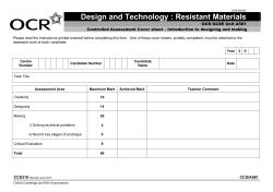
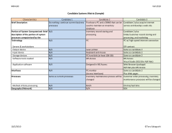

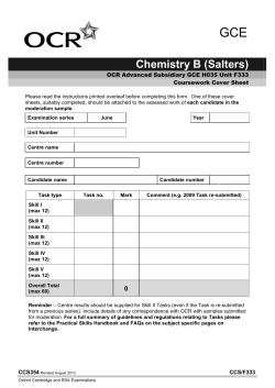


© Copyright 2026