
Paraneoplastic Syndromes Associated with Anti-Hu Antibodies Horacio Senties-Madrid MD
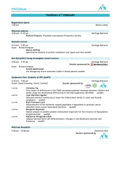
Mexico±Israel Symposium
Paraneoplastic Syndromes Associated with Anti-Hu Antibodies
Horacio Senties-Madrid MD
1
1
and Felipe Vega-Boada MD
1,2
Department of Neurology, National Institute of Medical Sciences and Nutrition Salvador Zubiran, and
2
National Research
Systems, National Council of Science and Technology (CONACYT), Mexico City, Mexico
Key words:
paraneoplastic syndromes, Hu-antigens, anti-Hu antibodies, paraneoplastic sensory neuropathy, paraneoplastic
encephalomyelitis, small cell lung carcinoma
Abstract
Paraneoplastic syndromes are disorders associated with
cancer but without a direct effect of the tumor mass or its
metastases on the nervous system. Small cell carcinoma of
lung associated with paraneoplastic sensory neuronopathy
and/or paraneoplastic encephalomyelitis with the presence of
anti-Hu antibodies has been termed ``anti-Hu syndrome.'' AntiHu associated PSN-PEM is an immune disorder in which both
cell-mediated and humoral mechanisms are involved. Patients
are considered affected by Anti-Hu associated PSN-PEM
when they develop clinical signs and symptoms of CNS
dysfunction and/or sensory neuropathy not caused by metastases or other disorders, and serum or cerebrospinal fluid is
positive for Hu abs. SCLC is found in more than 90% of
patients with cancer and positive Hu abs. Individual patients
with Hu abs associated to SCLC may suffer PSN-PEM, limbic
encephalitis, brainstem encephalopathy, opsoclonus-myoclonus, paraneoplastic cerebellar degeneration or myelopathy.
Hu abs have a specificity of 99% and sensitivity of 82% in
detecting paraneoplastic neurological syndromes. There are
two types of treatment: the first is to treat the cancer, the
second is to suppress the immune reaction with the use of
corticosteroids, cyclophosphamide, azathioprine, plasma exchange, intravenous immunoglobulin and immunoadsorption;
however, treatment of paraneoplastic syndromes is generally
unsatisfactory.
IMAJ 2001;3:94-103
Paraneoplastic syndromes are disorders associated with cancer
but without a direct effect of the tumor mass or its metastases
on the nervous system [1]. Small cell carcinoma of lung
associated with paraneoplastic sensory neuronopathy and/or
paraneoplastic encephalomyelitis with the presence of anti-Hu
antibodies has been termed ``anti-Hu syndrome.'' However,
other neurological manifestations and neoplasms have been
related to these antibodies. The term also includes Huseropositive patients without an apparent underlying neoplastic
PSN = paraneoplastic sensory neuropathy
PEM = paraneoplastic encephalomyelitis
Hu abs = anti-Hu antibodies
SCLC = small cell lung carcinoma
94
H. Senties-Madrid and F. Vega-Boada
disease. During the past few years scientific interest in
paraneoplastic syndromes has been enhanced by the discovery
of antibodies responsible for inducing the pathological process.
Paraneoplastic syndromes are difficult to diagnose because in
the majority of patients the tumor is not evident at the onset of
the neurological disorder. In this article we offer an overview of
paraneoplastic syndromes associated with Hu abs.
Historical considerations
At the end of the nineteenth century Oppenheim reported the
earliest cases of remote effects of cancer on nervous tissues. The
most common clinical manifestation, a sub-acute progressive
sensory neuronopathy, was reported by Weber and Hill in 1933,
when they noted a complete degeneration of the posterior
columns on examination of the spinal cord from a patient with
``chronic polyneuritis'' and oat cell carcinoma of the lung. One
year later, the central neuraxis components of this disorder were
described by Greenfield under the rubric of sub-acute spinocerebellar degeneration.In 1948 Wyburn-Masonrecorded a case of
cancer-related ``polyneuritis'' and the same year Derek DennyBrown published two cases that defined the condition's
anatomic substrate as the destruction of dorsal root ganglion
cells (hence its designation as a neuronopathy). Guichard's
accounts in 1949 of paraneoplastic polyneuritis with neurologic
complaints represent the earliest use of this term. Ten years
later, Brain and Henson [2] suggested the possibility that
immunological disturbances were the cause of neurological
syndromes associated with carcinoma. This point of view was
shared by Dorothy Russell in 1961 when she proposed that the
lymphoid infiltration of carcinomatous encephalomyelitis implied a local immunological reaction that might be provoked by
the tumor itself, rather than an infectious agent. Peter
Wilkinson reported that the sera of four patients suffering
from SCLC and sensory neuronopathy harbored antibodies
active against saline extracts of human brain and DRG by
complement fixation assay. Cerebrospinal fluid samples from
two of these patients were also antibody positive. In 1965 the
first paraneoplastic antineuronal antibody was reported by
Wilkinson and Zeromski [3]. Graus et al. [4] in 1985 described
the anti-Hu immunoglublin G antibody in immunohistochemical preparations and yielded bands in the 35±40 kD region on
DRG = dorsal root ganglion
IMAJ
.
Vol 3
.
February 2001
Mexico±Israel Symposium
Western blot analysis of proteins from cerebrocortical neurons,
serum and CSF of patients with SCLC and sub-acute sensory
polyneuropathy. They designated the antibody by the first two
letters of their index patient's last name, Hu. The presence of
circulating antineuronal antibodies with nuclear immunoreactivity was confirmed in additional investigations. Anderson and
co-workers [5] demonstrated that anti-Hu abs correspond to
IgG in the sera and CSF of patients with SCLC and
encephalomyelitis complex, including bulbar and cerebellar
dysfunction, myelopathy, limbic encephalitis and autonomic
failure.
promote differentiation and maintenance of the neuronal
phenotype. Hu-related antigens can be detected in both the
nucleus and the membranes of most SCLC and in 50±78% of
neuroblastomas [10]. HuD, HuC and Hel-N1 have been found
in patients with anti-Hu-associated PEM-PSN [11].
The Hu antibody, immune response and
neuropathology
Anti-Hu associated PSN-PEM is an immune disorder in which
both cell-mediated and humoral mechanisms are involved
[Figure 1]. While SCLC is more common in men than in
women, with ratios ranging from 1.8:1 to 3:1, the anti-Hu
syndrome is more common in women, as are other autoimmune
Hu antigens are 35±40 kD proteins expressed mainly in the disorders.
nucleus (Hu proteins, Nova) but also in the cytoplasm (CDR,
Tr, Ulip, amphiphysin) of all neurons of the central and
peripheral nervous system as well as in the underlying tumor [6]. Hu abs belong to the IgG family. Anti-Hu is a polyclonal,
Normal non-neural tissues assayed for Hu expression have been complement-fixing IgG, predominantly of the IgG1 subclass
negative, except for isolated cells of probable neuronal lineage [12]. Originally named anti-Hu by Graus et al. [4], these
in the adrenal medulla and bronchial mucosa. They can also be antibodies have also been called type IIa neuronal antibodies
expressed by the testes. The principal Hu antigens in humans and type 1 antineuronal nuclear antibodies [12]. The terminolare: HuD, HuC, and Hel-N1, each containing three RNA ogy for antibodies in paraneoplastic syndromes proposed by
recognition motifs, and 86±90% of them are identical to each Lennon in 1989 was ANNA (antineuronal nuclear antibody)
other. Others are HuR, Hel-N2 and ple21. HuC and ple21 and PCAb (anti-Purkinje cell cytoplasmic antibody). After
cDNAs show a very high degree of identity and may in fact Anti-Ri was characterized, Lennon now proposes the addition
correspond to a single gene. HuD was the first one to be of numbers. Depending on the pattern of reactivity on different
described: it is encoded by a gene mapped to the short arm of animal tissues, ANNA becomes ANNA-1 (anti-Hu) or ANNAchromosome 1 and has a molecular mass of approximately 42 2 (anti-Ri) and PCAb becomes PCA-1 [13] [Table 1]. They
kD and the presence of three RNA-binding domains. HuD, immunoreact with the Hu antigens present in the nervous
HuC and Hel-N1 expression is restricted to neurons. HuR is system and neoplasms [14]. Hu abs also react with cells of the
expressed in extraneuronal tissues.
retina and adrenal cortex, and therefore are
The Hu antigens in humans constitute a family of similar adenohypophysis,
not
exclusively
an
antineuronal
nuclear antibody. ANNA-1 also
proteins that could be analogous to the
protein Elav react with Purkinje cells. According
immunochemical criteria
(embryonic lethal abnormal visual system), which is required for alone, the anti-Hu antibody is also atoPurkinje
cell antibody and
normal development and maintenance of the fly's eye and a paraneoplastic cerebellar degeneration antibody.
nervous system [7,8]. HuD has extensive sequence homology elements (e.g., Schwann cells, vasculature, etc)Supporting
are not
(51% identity) with Elav. Furthermore, mutation of the Elav recognized by the antibody.
gene prevents development of the
nervous system.
Early expression of the Hu antigens occurs in vertebrate
neurogenesis and they bind to the AU-rich (ARE) element The exact role of Hu abs in the pathogenesis of the disease is
resident in the 3'-untranslated region of the mRNA (c-myc, c- unknown. It is thought that the expression of the Hu antigens
fos, Gap43 and GM-CSF) that regulates cell proliferation. by the tumor triggers a humoral immune response and a cellAlthough not present in neuroblasts, Hu proteins first appear in mediated response that cross-react with the cells of the nervous
early lineage neurons that are still capable of proliferation. system. This hypothesis is supported by the fact that the
Marusich and Weston [9] reported that the antigen is the first expression of each Hu antigen is found in all SCLC patients and
identifiable marker of neuronal maturation in quail, appearing in up to 78% of patients with neuroblastoma. Both tumors are
when E2 neuroblasts become committed neurons. Thus they of neuroectodermal origin. Anti-Hu sera react with HuD, HuC
appear to be the earliest marker of neuronal commitment in and Hel-N1, however from the three antigens only HuD is
CNS-stem cells. The exact function of Hu proteins remains expressed in SCLC. Thus, it plays a main role in triggering the
unknown, but it has been postulated that they act as immune response.
transfactors involved in selective mRNA degradation and likely
The Hu antigens
Humoral response
Hu
an tibo d y
Drosophila
Drosophila
I m m un e r es po n se
CSF = cerebrospinal fluid
IMAJ
.
Vol 3
.
February 2001
ANNA-1 = antineuronal nuclear antibodies
PCA-1 = anti-Purkinje cell cytoplasmic antibody
Paraneoplastic Syndromes Associated with Anti-Hu Antibodies
95
Mexico±Israel Symposium
synthesis of Hu abs in six of
seven patients with PEM/PSN.
Plasmapheresis reduced the level of antibodies in serum without affecting that in the CSF.
Vega et al. [17] showed that
patients with PEM have higher
intrathecal synthesis of Hu abs
than patients with isolated sensory neuropathy. Hu abs are
probably synthesized within the
brain and perivascular spaces
by B cells found in those areas.
The presence of elevated intrathecal antibody synthesis
has been suggested as a factor
to explain the lack of clinical
response to plasmapheresis or
other immunosuppressive treatments. It is also possible that
IgG is selectively transported
from the serum into CSF, or
that this transport occurs normally but antibody release from
the CSF is prevented. However,
there is no precedent for selective transport or reabsorption
of normal IgG. The usual and
logical explanation is that
clones of B cells cross the
blood-brain barrier and remain
in the CNS to produce their
antibodies. The presence within
CNS of Hu-cognizant B cell
clones would account for the
fact that anti-Hu activity is
consistently higher in CSF than
in serum.
Immunohistochemical studies with
intracellular staining revealed
that in areas where the bloodbrain barrier is broken the antibody migrates through the neurophil and finds its way into both
glial cells and, to a greater degree, into neurons. Whether the
internalization of IgG is a primary feature or secondary to the
initial lesion of the neuron by other cause is presently unknown.
Also, how antibodies enter the cell is an open question. One
possibility is that antibodies enter at the synapse and are
retrogradely transported to the cell body and finally concentrate
in the nuclei. Another possibility is the internalization of the
IgG via the neuronal cell body. An alternate mechanism is that
the antigen represents a cell surface receptor and when the
antibody binds to it, both antigen and antibody are internalized.
Antibody internalization.
Figure 1.
Neuropathogenesis of Anti-Hu syndrome
IgG1 and IgG3 are the predominant isotypes of
the anti-Hu IgG in the serum, nervous system and tumor
(particularly IgG1). A few patients also have IgG2. There is no
correlation between specific IgG isotype or the reactivity of the
Hu abs with HuD, HuC, Hel-N1 and neurological symptoms
[15].
Hu abs are synthesized intrathecally as well
as peripherally. Furneaux et al. [16] demonstrated intrathecal
Antibody types.
Antibody synthesis.
96
H. Senties-Madrid and F. Vega-Boada
IMAJ
.
Vol 3
.
February 2001
Mexico±Israel Symposium
Paraneoplastic antibodies, their associated cancers and neurological findings
Antibody
Antigen, Western blot molecular mass (kD) Onconeural antigen Associated tumor
Anti-Hu
All neuronal nuclei, adenohypophysis,
HuD, HuR
SCLC, SCGC, lung adenocarcinoma,
ANNA-1
adrenal cortex, retina, testes
HuC/ple21
neuroblastoma, prostate and thymic
Type IIa*
35-40
Hel-N1
carcinoma, HL, rhabdosarcoma, synovial
sarcoma, seminoma and NSTGCT
Anti-Yo
Purkinje's cytoplasm, 34, 62
CDR34
Gynecological, breast, lung (adenocarcinoma)
PCA-1**
CDR62-1, 62-2
Type-1
52
PCD17
58
CZF
Anti-Ri
Neuronal nuclei CNS, retina, 55, 80
Nova1, Nova2
Breast, gynecological, SCLC
ANNA-2
Type IIb
Anti-Ta
Neuronal nuclei and cytoplasm, 40, 50
Ta (Ma2)
Testicular
Table 1.
Syndrome
PSN-PEM
PCD
Cerebellar ataxia,
psoclonus
Limbic encephalitis,
brainstem
dysfunction
Ma1, Ma2
Multiple (colon, breast, lung, parotid gland) Cerebellar, brainstem
dysfunction
Recoverin
SCLC, others
Photoreceptor
degeneration
Amphiphysin
Breast, SCLC
Stiff-man syndrome,
PEM
Alpha1 subunit
SCLC
LEMS
Alpha subunit of Thymoma, SCLC, others
Neuromyotomia
several VGKC
(Isaac's syndrome)
POP66
SCLC, thymoma, others
PEM, PCD
Beta subunit VGCC SCLC
LEMS
In progress
HL, NHL
PCD.
Anti-Ma
Anti-CAR
Anti-amphiphysin
Anti-VGCC
Anti-VGKC
Anti-CV2
Anti-MysB
Anti-Tr
Neuronal nuclei and cytoplasm, 37, 40
Retinal photoreceptor, 23
Synaptic vesicles, 128
Presynaptic VGCC
Several VGKC (peripheral nerves,
central neurons, glia, DRG.
Glia (subset), 66
Presynaptic VGCC
Cytoplasm neurons and Purkinje's
spiny dendrites
* Greenlee et al. [39] use the names ``type IIa antineuronal antibodies'' for ANNA-1 reactivity, ``type I antineuronal antibodies'' for PCA-1, and ``type IIb
antineuronal antibodies'' for ANNA-2.
** Most PCA-1 are probably the anti-Yo antibodies
CAR = cancer-associated retinopathy, CNS = central nervous system, DRG = dorsal root ganglia, HL = Hodgkin's lymphoma, LEMS = Lambert-Eaton myasthenic
syndrome, NHL = non-Hodgkin's lymphoma, NSTGCT = non-seminomatous testicular germ cell tumors, PCD = paraneoplastic cerebellar degeneration, PEM =
paraneoplastic encephalomyelitis, PSN = paraneoplastic sensory neuropathy, SCGC = small cell gallbladder carcinoma, SCLC = small cell lung cancer, VGCC =
voltage-gated calcium, VGKC = voltage-gated potassium channel.
(The above text corresponds to the figure legend)
HuD and Hel-N1 do not contain any hydrophobic stretch
suggestive of a transmembrane domain; in contrast, HuC/ple21
and Hel-N2 contain a hydrophobic stretch that could correspond to a transmembrane domain. Similarly, sequences of Elav
and Rbp-9 contain hydrophobic stretches. The internalization
process has a duration of one hour by SCLC cell lines.
The anti-Hu immune response is directed
towards two separate domains with at least two distinct
epitopes. They bind to the first and second RRM of HuD,
HuC and Hel-N1 but not HuR. For HuD the first and second
RRM are essential for RNA binding. Thus, it has been
suggested that Hu antigen inhibition by Hu abs might lead to
unregulated expression of c-fos and c-myc, which in turn could
result in cell death or apoptosis.
Antibody attack.
RRM = RNA recognition motifs
IMAJ
.
Vol 3
.
February 2001
Although IgG1 and IgG3 activate
complement (they can bind C1q), only weak complement
reactivity is found. Graus and colleagues [11] did not identify
natural killer cells or deposits of complement C3 in the inflamed
sensory ganglia and medulla of a patient with Hu abs-associated
PEM-PSN. Neither did Jean et al. [12] identify NK cells and
small amounts of complement in a few areas of the nervous
system of four patients. After uptake of the antibody by rat
granule cells, a neuronal destruction occurred and complement
was not required, however its presence accelerated the neuronal
damage. This supports the notion that complement-mediated
toxicity antibody-dependent cell cytotoxicity mediated by NK
cells has a minor role in PEM-PSN. An alternative would be
that complement has a role but the fractions C3 or C5b can be
detected immunohistochemically only for a short time.
Complement
response.
NK = natural killer
Paraneoplastic Syndromes Associated with Anti-Hu Antibodies
97
Mexico±Israel Symposium
Cellular response
Cellular immune response has been suggested [18] for the
following reasons:
. Presence of extensive infiltrates of T cells in the nervous
system.
. T cell proliferation in the tumor.
. Most tumors express major histocompatibility complex
proteins, which indicate the need for antigen presentation
to T cells, whereas MHC class 1 proteins are rarely
expressed in SCLCs that are not associated with
paraneoplastic syndromes.
. Passive transfer experiments do not provide evidence in
favor of antibody-mediated disease alone. The transference of Hu abs to animals does not reproduce the disease
[19]. Furthermore, animals immunized with HuD develop
antibodies but not symptoms. Thus, a cytotoxic mechanism may explain the difficulties in creating an animal
model of the disorder by passive transfer of Hu abs.
MHC pr otei ns : an ti ge n pre s en t atio n t o cy t otox ic
Hu is normally restricted to cells that do not express MHC
antigens requisite for the triggering of immunological responses.
Hu may be sequestered from the maturing immune system
during the establishment of self-tolerance. Genetically determined variation in individual immunoresponsiveness, differences in Hu structure among neoplasms, or failure of all but a
few small cell carcinomas to express Hu in association with
MHC class I antigens can determine the different Anti-Hu
responses [20]. Probably there is a greater surface expression of
MHC class I and II by tumors associated with anti-Hu
syndrome. Neuronal damage induced by antibodies or cytokines can result in this greater expression of MHC molecules.
The development of the anti-Hu immune response may depend
on the ability of the tumor to present the antigen to the
immunological system, because there is a correlation between
the expression of both MHC class I and Hu antigens and the
development of this immune response. Co-expression of HuD
and MHC class I proteins can be present and correlate with
PSN-PEM. MHC class I molecule expression appears restricted
to non-neuronal satellite cells, endothelium, perivascular
lymphocytes and process-bearing elements morphologically
consistent with microglia, and exhibits intense labeling with
antibodies to HLA-DR. They are required for the presentation
of viral or tumor neoantigens to cytotoxic T lymphocyes. A
direct attack on the neuron by cytotoxic T lymphocytes
sensitized to Hu would presumably require its surface display
in association with MHC class I antigens. However, membrane
expression is not a prerequisite for T cell recognition. Local
MHC class II expression is greatly amplified and may
contribute to the anti-Hu immune response.
T ce l l s a n d tu m o r l i m i t a t i o n
IL-2 = interleukin 2
IFNg = interferon-gamma
98
H. Senties-Madrid and F. Vega-Boada
Tumor limitation
The immune response limits tumor spread. However, while
most animals immunized with HuD cDNA showed smaller
tumor volumes or rejection of the tumor, they did not develop
neurological symptoms. This model is closest to the phenomenon observed in SCLC patients with low titers of Hu abs and
no neurological deficits. The likely explanation for the tumor
limitation in patients with paraneoplastic syndromes has a
genetic basis that is closely related to MHC molecules.
Neoplasms with low or no expression of MHC class I proteins
have a growth advantage and increased metastatic potential. In
this way, SCLC and neuroblastoma usually have low or no
expression of MHC class I and have widely metastatic properties, with the exception of those patients with a paraneoplastic
syndrome. In the series of Dalmau et al. [21] systemic metastases
were detected in only 5 of 15 autopsied patients; the tumor was
mostly confined to the chest when it was diagnosed (96%), with
metastases outside the chest in only 2 patients (4%). The belief
that paraneoplastic tumors are more indolent may represent an
artifact, since a search for the tumor many months previously
would have revealed it. The frequent neurological death limits
evaluation of the natural history of the tumor and this
hypothesis. A mild immune response is a good tumor
prognostic factor, but an intense immune response is associated
with severe neurological disease that often causes death.
T cell infiltrates can be part of a specific immune response or
can result from a non-specific attraction by a pro-inflammatory
environment. In the latter case one would expect to find all
Vbeta families of T cell receptors with no clear predominance of
any particular one. In the work of Voltz and colleagues [22], the
lack of representation of some Vbeta families in the nervous
system of five patients and over-representation of certain Vbeta
families in three patients indicate that the T cell response is
specific. Thus, T lymphocytes are specifically targeted to
neuronal and tumor antigens, where they recognize this specific
antigen and proliferate. Antigen-specific proliferation in peripheral blood mononuclear cells is much higher in seropositive
patients. The ability of the IgG1 and IgG3 to bind Fc receptors
may play role in the recruitment of monocyte/macrophage cells
since they are the only two subclasses of IgG for which there
exist specific receptors on monocytes/macrophages. However,
aggregated IgG did not inhibit antibody binding to tumor cells,
leaving uncertain the possibility that these interactions result
from Fc-mediated antibody binding. There is an increase of
memory helper T cells in anti-Hu seropositive patients. HuD
protein stimulates peripheral blood mononuclear cells
with a significant increase of memory helper (CD45RO+
CD4+) T cells and increased interferon-gamma/interleukin 4
ratio [22]. CD4+ cells can be Th1 and Th2 subtypes. Th1 cells
secrete IL-2 and IFN-g, activate macrophages and are
responsible for delayed-type hypersensitivity, whereas Th2 cells
produce IL-4, IL-5, IL-10 and IL-13 and thus favor IgE
in vitro
MHC = major histocompatibility complex
IMAJ
.
Vol 3
.
February 2001
Mexico±Israel Symposium
Clinical manifestations of Anti-Hu syndrome
in two studies (%)
Symptom
Dalmau et al. [21]
Voltz et al. [24]
(n=71) (%)
(n=173) (%)
PSN
59
69
PEM/seizures
21
16
Autonomic dysfunction
10
10
Cerebellar dysfunction
13
23
Brainstem dysfunction
11
11
Motor weakness
14
14
Table 2.
production and suppress cell-mediated immunity. The distinction between Th1 and Th2 types relies on the IFNg/IL-4 ratio.
A significant increase in the IFNg/IL-4 ratio suggests that HuD
protein is an antigenic target for autoreactive CD4+ T cells,
presumably of the Th1 type. This explains the predominance of
Ig1 and Ig3 isotypes. Activated CD4 Th1 cells induce a direct
cytotoxic effect, possibly through the induction of apoptosis.
Moreover, an antigen-driven oligoclonal CD8+ cytotoxic T cell
expansion due to an oligoclonal expansion is involved in
pathogenesis.
The tumor may compete with the nervous system for an
essential substrate (glucose, tryptophan) and result in neurological dysfunction. Synthesis of hormone-like substances and
secretion of cytokines (IL-1, IL-6, tumor necrosis factor-alpha)
can participate in pathogenesis.
The term sub-acute sensory neuronopathy is actually a
misnomer since the pathological substrate is a dorsal root
ganglion. Animals injected intraperitoneally with human IgG
showed deposits of IgG in the interstitium of the DRG whereas
the CNS was negative. There were deposits of IgG in the
neurons and some glial cells of medulla and DRG of affected
patients but not in the frontal cortex and medulla of controls.
Thus, DRG was the initial target in 70% of patients [11]. This is
due to the partial absence of the blood-brain barrier in the
normal DRG. There is a distinct infiltration pattern. Inflammatory infiltrate comprises CD19+ (B cells), CD4+ (helper/
inducer) cells in the perivascular spaces, CD8+ CD11(cytotoxic/suppressor) cells in the interstitial spaces surrounding
neurons, and less frequently plasma cells in the perivascular
space. The failure of most CD8-positive cells to express the
CD11 epitope suggests that these are mainly of the cytotoxic,
rather than
suppressor class. Also, there are infiltrates of
EBM11+ (monocyte/macrophage) in the perivascular (macrophage phenotype) and interstitial (microglial) spaces. Associated neuronal loss and gliosis occur, and ganglion cells are
replaced by reactive proliferations of satellite (capsular) cells
known as nodules of Nageotte, resulting in dorsal root
degeneration. Nerve biopsy reveals axonal neuropathy. In
myenteric plexi a comparable inflammatory assault is responsible for gastrointestinal paresis, damage to the sympathetic
ganglia and the intermediolateral cell column, leading to
in situ
O t h er da m ag e m ec h an i sm s
N e u r o p a t h ol o g y
IMAJ
.
Vol 3
.
February 2001
dysautonomia. The central components reflect an inflammatory
insult to the neuron at the level of its perikaryon. The effect has
a predilection for the medial temporal lobes (hippocampi and
amygdeloid nuclei), brainstem and anterior horns of the spinal
cord, that correlate with limbic encephalitis, bulbar dysfunction
and motor neuron disease respectively. Macroscopic evidence of
injury results in brownish discoloration and contraction of the
hippocampal formation, and advanced cerebellar cortical
degeneration with shrinkage folia. The distribution of the more
severe tissue alterations corresponds to the particularly symptomatic regions of the nervous system.
Clinical issues
Patients are considered affected by anti-Hu associated PSNPEM when they develop clinical signs and symptoms of CNS
dysfunction and/or sensory neuropathy not caused by metastases or other disorders, and Hu abs-positive serum or CSF.
SCLC has been found in more than 90% of patients with cancer
and positive Hu abs. However, other malignancies have been
described to cause PEM-PSN and high titer Hu abs; for
example, exceptional cases of carcinoma of the lung, thymic
carcinoma, synovial sarcoma, Hodgkin's disease, and nonseminomatous testicular germ cell tumors. We have encountered a case of a women with a PSN associated with a small-cell
carcinoma of the gallbladder (in press). In their series Dalmau et
al. [21] found SCLC in 77.5% of patients ± one had adrenal
tumor, one had lung adenocarcinoma, one had chondromyxosarcoma, two had prostate carcinomas, and one had neuroblastoma. The presence of low titer antibodies is associated with
limited cancer stage, a complete response to chemotherapy, and
longer survival.
N e o p l a s m s a s s o c i a t e d wi t h an t i - H u s y n d r o m e
Clinical manifestations
Individual patients with Hu abs associated with SCLC may
suffer from PSN-PEM, limbic encephalitis, brainstem encephalopathy, opsoclonus-myoclonus, paraneoplastic cerebellar degeneration or myelopathy according to the classification of
Henson and Urich [23]. The onset of neurological symptoms in
PSN-PEM generally begins between 6 months and 3.5 years
before the detection of the tumor (median time 4 months). On
the other hand, the tumor was diagnosed before the onset of
neurological symptoms in 12% of patients. In Dalmau's series
[21] of 71 serologically confirmed cases the disorder was more
frequent in women (55%) and the median age at onset was 60
years, but the syndrome can also develop in childhood (the
youngest patient was a 4 year old girl with neuroblastoma and
bulbar dysfunction). Neurological manifestations preceded the
detection of tumor in 83% of patients. Multifocal involvement
was more frequent than unifocal (73% vs. 27%). In another
large series of 173 patients with anti-Hu syndrome and
pathologically confirmed SCLC, 31% had more than one
presenting symptom [24] [Table 2].
Paraneoplastic Syndromes Associated with Anti-Hu Antibodies
99
Mexico±Israel Symposium
Sensory neuronopathy is the mostcommon manifestationand is
often the dominant feature (59% in Dalmau's series and 69% in
Voltz's analysis). Symptoms are relatively pure, including the
most common ones like anesthesia, paresthesias and dysesthesias of the distal extremities, proprioceptive and nociceptive
sensory loss. Asymmetry is common. In a minority of patients
the sensory symptoms at onset were restricted to the trunk or
face, while back and/or radicular pain and flexor spasms were
described by others. The sensory symptoms can evolve
suddenly, but more frequently erupt in a sub-acute fashion
that progresses relentlessly, often to total deafferentation and
absence of muscle stretch reflexes in a matter of weeks or
months. Reflexes could be abolished or increased. Severely
affected patients cannot walk, stand or sit unsupported and may
be unable to feed themselves despite unimpaired motor
function. In the Mayo Clinic series the most affected area was
the upper limbs (15 or 27 patients). Sensory ataxia was severe
and disabling. The probable reason for sub-acute sensory
neuropathy being the dominant manifestation is that dorsal
root ganglia are readily accessible to large circulating molecules,
whereas access to the central nervous system is restricted by the
blood-brain barrier.
In order to obtain a precise diagnosis it is important that the
physician follow a logical order when encountering a PSN
patient. The exact kind of neuropathy should be categorized
and differentiated from other causes of ganglionopathy such as
Sjogren's syndrome, the use of cisplatin or vincristine and its
analogues, vitamin B6 toxicity, HIV-related sensory neuronopathy and idiopathic sensory neuronopathy. Although most of
the symptoms are sensory there might be other neurological
manifestations.
In Dalmau's series [21], 41% of patients had extrasensory
abnormalities characteristic of PEM. These researchers reported
symptomatic autonomic dysfunction in 10% of patients,
seizures in 26%, brain stem dysfunction in 11%, myelitis in
14% and ataxia in 13%. In Voltz's analysis, motor incoordination, tremor or ataxia was found in 23%, limbic encephalopathy
in 16%, motor weakness, fasciculations, hypo- or hypereflexia
in 14%, dysphagia, dysarthria, ocular paresis or opsoclonus in
11%, and autonomic dysfunction in 10%. PEM and PSN are
different manifestations of the same nosologic process. Limbic
encephalitis is the second most common clinical manifestation
of anti-Hu syndrome in some series (autonomic dysfunction in
others). This is characterized by confusion, disordered affect,
cognitive decline and short-term memory loss. Some experience
hallucinations, others have symptoms indicative of complex
partial seizure activity. Limbic symptoms were presented in
21% of Dalmau's series [21].
Brainstem encephalitis is manifest as diplopia, oscillopsia,
dysarthria, dysphagia, supra- and internuclear gaze abnormalities, sensorineural deafness and facial numbness or palsy.
PSN
P E M an d l i m b i c e n c e p h a l i t i s
B r ai n st e m en c ep ha l i t i s
100
H. Senties-Madrid and F. Vega-Boada
Evidence of cerebellar dysfunction (25%) includes ataxia,
intention tremor, uncoordination, diminished muscle tone,
nystagmus and scanning speech. Brainstem dysfunction was
present in 32% of patients.
Present in 14% of patients, motor neuron involvement is
reflected by an asymmetric pattern of loss of strength in the
proximal extremities that spreads distally and is associated with
muscle atrophy and fasciculations. Abnormal pseudoathetotic
involuntary movements of the hands and fingers were a
common finding. Denervation of the intercostal muscles may
result in a fatal hypoventilatory syndrome. Thus, motor
weakness should not be considered an exclusion criterion due
to concomitant involvement of the motor neurons [25].
Found in 28% of Dalmau's patients [21], autonomic failure
results in orthostatic hypotension, abnormal pupillary responses, urinary retention, gastrointestinal paresis, hyperhydrosis, impotence, cardiac arrhythmias, and chronic intestinal
pseudo-obstruction due to neuronal destruction of the myenteric plexus neurons. The frequency of autonomic manifestations varies between the different series.
Hersh and co-workers [26] reported a case of opsoclonusmyoclonus and Hu abs. However, in some opsoclonusmyoclonus patients the detection of Hu abs may be coincidental
or part of two paraneoplastic syndromes. There are previous
reports of association between two different paraneoplastic
syndromes (i.e., Lambert-Eaton myasthenic syndrome and antiHu associated PSN-PSM).
The course of the disease is usually not regressive, with the
exception of a few cases of limbic encephalitis. The prognosis is
poor and the median survival is 7 months (range 1.5±34). Death
is due mainly to cardiac arrest or cardiovascular collapse
attributable to the autonomic dysfunction and respiratory
failure secondary to motor neuron disease and bulbar dysfunction, rather than to widespread metastases. A paraneoplastic
etiology should be considered in patients with indolent sensory
neuropathies, particularly when the neuropathies are asymmetric and the arms are involved.
Motor neuron involvement
A u to n o m i c f a i l u r e
Op so c l on us -my oc l on us
P r o gn os i s
Laboratory, electrophysiological and
radiological studies
The most common finding in electrophysiologic studies is
reduced or abolished sensory potentials associated with normal
or mild reduction of nerve conduction, absent H responses with
normal motor conductions, F waves and electromyography
implicating the DRG as the site of pathology. Signs of
denervation such as fibrillation or increased amplitude of
motor unit potentials were found in patients in Dalmau's series
[21].
E l e ct r o p hy s i o l og i c al
s t ud i e s
IMAJ
.
Vol 3
.
February 2001
Mexico±Israel Symposium
PSN-PEM) can be explained by factors related either to the
CSF protein levels are usually elevated, 10±40 white blood cells tumor (such as expression of MHC antigens, changes in the Hu
can be found, oligoclonal bands can be positive, and there is protein antigen itself) or the host (sex or genetic factors that
regulate the immunological makeup of the patient). The titers of
intrathecal synthesis of antibodies
Hu abs also correlate with the differentiation of neoplasms.
Tumors that are poorly differentiated in PSN patients have
Magnetic resonance imaging studies are abnormal in only a few higher titers, whereas patients without antibodies have wellpatients. Limbic encephalopathy can course with increased differentiated tumors. Moreover, recombinant Fab fragment
signal areas on T2 in the medial temporal region, which revert against Hu proteins has been used as an index of neuronal
to normal and probably represent an early inflammatory differentiation in human brain tumors.
component. With progression of the disease, scarring and In the presence of positive Hu abs a search for an underlying
contraction of the temporal lobes eventually occur [27]. SCLC should be undertaken. This should include an initial
Computerized tomography scan is not sufficiently sensitive to bronchoscopy and CT scan or MRI of the chest and
radiological studies every 6 months, as some positive results
detect these abnormalities.
are provoked by cancers that successfully eluded the initial
detection owing to their small size ± only a few millimeters in
or of microscopic proportions. Reports of Hu absThere are two principal assays for the detection of Hu abs: diameter
patients with sensory neuropathy who did not develop
immunohistochemistry [10] (using avidin-biotin peroxidase and positive
immunofluorescence) and Western blot analysis [28] (to identify cancer after a follow-up period of 4 or more years are
proteins of 35±40 kD molecular mass). The use of dilutions exceptional. However, the one patient with sensory neuropathy
greater than 1:500 is preferable because reactivity at lesser and anti-Hu antibodies who developed SCLC 7 years after the
of the neuropathy emphasizes the need for a long followdilutions is sometimes associated with non-specific glial stain- onset
before excluding the possibility of a paraneoplastic origin. It
ing. Hu abs have a specificity of 99% and sensitivity of 82% in upis important
to mention that 17% of patients with no cause for
detecting paraneoplastic neurological syndromes [29]. Some sensory neuropathy
the initial evaluation developed a
antibodies with ANNA-1 reactivity are notAnti-Hu. Lovblad et tumor in spite of theafter
absence of Hu abs. For this reason, the
al. [30] found 10% false-positive anti-Hu sera using immuno- detection of an underlying
tumor in anti-Hu antibody-negative
histochemistry alone. Sera of Sjogren syndrome patients reacted
with 38 kD on Western blots of rat cerebellar homogenate, patients with sensory neuropathy should be similar to that
resembling anti-Hu immunoreactivity,but therewas no reaction recommended for patients with Hu abs and targeted at the
If a tumor other than SCLC is detected but does not
with purified Purkinje cells or purified recombinant HuD lung.
express
antigens, the presence of a second neoplasm
protein. Moll et al. [31] found that antibodies of patients with (probablyHuSCLC)
must be considered. Lucchinetti et al. [32]
Sjogren syndrome had a reactivity similar to Hu abs by identified 14 patients
with positive ANNA-1 associated with
immunostaining but no reactivity with HuD recombinant SCLC and previous diagnosis
of cancer, thus the search for
protein [18]. These findings demonstrate that there are at least SCLC should not end on discovering
a different neoplasm. In
two groups of patients with ANNA-1, one paraneoplastic and
children
the
detection
of
Hu
abs
should
direct the search
the other non-paraneoplastic. Because there are other antiprimarily
for
neuroblastoma.
bodies with identical immunohistochemical characteristics but
no specific tumor association, the presence of ANNA-1 (Hu abs
seropositivity) in serum and CSF ± particularly in patients with
unknown cancer ± must be studied either with a recombinant Patients with PSN occasionally harbor autoantibodies against
Hu protein or the combination of immunohistochemistry and neuronal antigens other than the Hu family, such as amphiWestern blot analysis of HuD protein to detect false-positive physin, a neuronal 66 kD protein related to axon guidance and
results. PSN-PEM are not restricted to the tumors found by a 106 kD neural protein of unknown function. Amphiphysin
Dalmau's team [21]; they were also encountered in other antibodies were positive in 7 of 155 patients with SCLC and
neoplasms previously mentioned. Antibodies associated with paraneoplastic neurological disorders [29]. Systemic autoantithe same neurological syndrome and, conversely, that the same bodies ± antibodies against DNA, centromeres, nRNP, Sn
antibody may be associated with different syndromes led to the antigen, Scl-70, Ro(SS-A), La(SS-B), mitochondria, thyroid
concept of immunological phenotypic diversity. The titers of Hu antigens, brush border antigen, parietal cells and rheumatoid
abs correlate with the presence of paraneoplastic syndromes. factor ± were found in 52% of patients with paraneoplastic
SCLC associated with PSN-PEM harbors higher titers of neurological syndromes, compared with 16% in the cancer-only
antibody (>1:1,000). A low titer was observed in up to 16% group [33]. In 25% of patients two or more antibodies were
of patients with SCLC without paraneoplastic symptoms. identified. In the subgroup of Hu abs-positive patients, 33%
However, even in patients with SCLC and paraneoplastic yielded systemic autoantibodies excluding antinuclear antibosyndromes the Hu abs are not always found. Why some patients dies,or67% includingANA. Four percent of patients withantiwith SCLC have no detectable immune response (Hu abs or Hu associated PSN-PEM also have anti-Ro antibodies in the
L umb ar pu nc tur e
R a di o l o g i ca l s tu di es
H u a bs es s ay s
O t h er an t i bo di e s
IMAJ
.
Vol 3
.
February 2001
Paraneoplastic Syndromes Associated with Anti-Hu Antibodies
101
Mexico±Israel Symposium
serum, characteristic of Sjogren syndrome. On the other hand,
antineuronal antibodies were present in 55% of patients with
Sjogren syndrome and major neurological complications and in
11% without this complication. The presence of antineuronal
antibodies does not correlate with the presence of anti-Ro/SSA, anti-La/SS-B or ANA. Sixteen percent of patients have
positive p53 antibodies, but no correlation between this
antibody and tumor prognosis was found.
Treatment
Two types of treatment are practiced: the first is to treat the
cancer, the second is to suppress the immune reaction with the
use of corticosteroids, cyclophosphamide, azathioprine, plasma
exchange, intravenous immunoglobulin and immunoadsorption. With the exception of myasthenia gravis, LEMS,
neuromyotonia, dermatomyositis and certain peripheral neuropathies associated with myeloma, treatment of paraneoplastic
syndromes is generally unsatisfactory. Only 2 of 71 patients in
Dalmau's series [21] showed a neurological improvement
following steroid treatment. Both patients suffered from limbic
encephalopathy. With vigorous immunosuppressive treatment
(combining high doses of steroids, cyclophosphamide and
immunoglobulin) no patient improved, 56% stabilized and
44% deteriorated in the study of Keime-Guibert et al. [34]. Oh
and Dropcho [35] reported two severely disabled patients with
PSN-PEM and Hu abs who improved after immunosuppressors, but some caution is necessary to interpret this finding
because spontaneous improvement has also been described in
the anti-Hu syndrome. Isolated case reports have shown partial
remission following antineoplastic therapy with or without
plasmapheresis in a few patients with PEM or PCD. On the
other hand, antibody-negative patients with PCD or limbic
encephalitis and SCLC had major remissions after antineoplastic therapy. Most patients with improvement after immunosuppressor treatment had exclusive involvement of the peripheral
nervous system with sensory neuropathy, which leads to the
suggestion that the lack of a blood-brain or blood-nerve barrier
at the level of the DRG could facilitate the entry and action of
immunosuppressors. The second possibility is the presence of an
indolent form of the disease with spontaneous stabilization.
Intravenous immunoglobulin treatment in paraneoplastic CNS
syndromes associated with antineuronal antibodies was not
effective in the study by Uchuya and co-workers [36], but
stabilization occurred in 47.6% of patients.
The results of plasmapheresis are disappointing [37] despite
the few enthusiastic reports of clinical responses. No patient
improved with plasmapheresis (3 to 6 plasma exchanges over 1
or 2 weeks) in the study conducted by Graus's group [38], but 4
of 7 patients remained stable and autoantibody titers in serum
dropped in all. The lack of response is due to irreversible
ANA = antinuclear antobodies
LEMS = Lambert-Eaton myasthenic syndrome
102
H. Senties-Madrid and F. Vega-Boada
neuronal damage, and decreased antibody titer in serum but not
in CSF due to elevated intrathecal antibody synthesis or
antibody may not be responsible for the symptoms but may
only be a marker of the disease. Because of neuronal cell loss, a
beneficial treatment might not lead to clinical improvement but
to stabilization only. For this reason, if the antibody is
identified early one should consider vigorous immunosuppression with plasmapheresis and steroids or cyclophosphamide.
However, patients with mild sensory symptoms of several
months duration at the time of diagnosis should not be included
in treatment protocols unless there has been a clear progression
in the previous month, since it is possible that immune
suppression produces a more rapidly growing tumor. In
conclusion, patients whose condition is very mild and stable
or those who have been severely disabled should not be treated
with immunosuppressive therapy; they should receive, exclusively, antineoplastic treatment. On the other hand, patients
with evidence of progressive disease, particularly those who are
mildly disabled, should be considered for immunosuppressive
therapy.
Finally, membrane Hu antigen expression may provide a
suitable target for the use of monoclonal antibody-based
diagnostic and therapeutic strategies.
References
1.
2.
3.
Posner JB. Neurologic Complications of Cancer. Philadelphia: FA Davis
Company, 1995.
Brain R, Henson RA. Neurological syndromes associated with carcinoma.
1958;2:971±5.
Wilkinson PC, Zeromski J. Immunofluorescent detection of antibodies
against neurons in sensory carcinomatous neuropathy.
1959;
88:529±38.
Graus F, Cordon-Cardo C, Posner JB. Neuronal antinuclear antibody in
sensory neuronopathy from lung cancer.
1985;35:538±43.
Anderson NE, Rosemblum MK, Graus F, Wiley RG, Posner JB. Autoantibodies in paraneoplastic syndromes associated with small-cell lung
cancer.
1988;38:1391±8.
Dalmau J, Furneaux HM, Rosemblum MK, Graus F, Posner JB. Detection
of the anti-Hu antibody in specific regions of the nervous system and tumor
from patients with paraneoplastic encephalomyelitis/sensory neuronopathy.
1991;41:1757±64.
Robinow S, Campos A, Yao K, White K. The elav gene product of
Drosophila, required in neurons, has three RNP consensus motifs.
1988;242:1570±2.
Robinow S, White K. The locus elav of Drosophila melanogaster is expressed
in neurons at all development stages.
1988;126:294±303.
Marusich MF, Weston JA. Identification of early neurogenic cells in the
neural crest lineage.
1992;149:295±306.
Graus F, Ribalta T, Campo E, Monforte R, Urbano A, Rozman C.
Immunohistochemical analysis of the immune reaction in the nervous system
in paraneoplastic encephamyelitis.
1990;40:219±22.
Graus F, Elkon KB, Cordon-Cardo C, Posner JB. Sensory neuronopathy
and small cell lung cancer. Antineuronal antibody that also reacts with the
tumor.
1986;80:45±82.
Jean WC, Dalmau J, Ho A, Posner JB. Analysis of the IgG subclass
distribution and inflammatory infiltrates in patients with anti-Hu associated
paraneoplastic encephalomyelitis.
1994;44:140±7.
Lennon VA. Paraneoplastic autoantibodies: the case for a descriptive generic
nomenclature.
1994;44:2236±40.
Dalmau J, Posner JB. Neurologic paraneoplastic antibodies (anti-Yo; antiLancet
Brain
4.
5.
6.
7.
Neurology
Neurology
Neurology
Science
8.
9.
10.
11.
12.
13.
14.
Dev Biol
Dev Biol
Neurology
Am J Med
Neurology
Neurology
IMAJ
.
Vol 3
.
February 2001
Mexico±Israel Symposium
15.
Hu; anti-Ri): the case for a nomenclature based on antibody and antigen
specificity.
1994;44:2241±6.
Manley GT, Smith PS, Dalmau J, Posner JB. Hu antigens: reactivity with Hu
antibodies, tumor expression, and major immunogenic sites.
1995;38:102±10.
Furneaux HM, Reich L Posner JB. Autoantibody synthesis in the central
nervous system of patients with paraneoplastic syndromes.
1990;40:1085±91.
Vega F, Graus F, Chen QM, Poisson M, Schuller E, Delattre JY. Intrathecal
synthesis of the anti-Hu antibody in patients with paraneoplastic encephalomyelitis or sensory neuronopathy: clinical-immunologic correlation.
1994;44:2145±7.
Benyahia B, Liblau R, Merle-Beral H, Tourani JM, Dalmau J, Delatrre J-Y.
Cell mediated autoimmunity in paraneoplastic neurological syndromes with
Anti-Hu antibodies.
1999;45:162±7.
Sillevis-Smith PA, Manley GT, Posner JB. Immunization with the
paraneoplastic encephalomyelitis antigen HuD does not cause neurologic
disease in mice.
1995;45:1873±8.
Dalmau J, Graus F, Cheung NKV, Rosemblum MK, Ho A, Canete A,
Delattre JY Thompson SJ, Posner JB. Major histocompatibility proteins,
antiHu antibodies, and paraneoplastic encephalomyelitis in neuroblastoma
and small cell lung cancer.
1995;75:99±109.
Dalmau J, Graus F, Rosenblum MK, Posner JB. Anti-Hu-associated
paraneoplastic encephalomyelitis/sensory neuronopathy. A clinical study of
71 patients.
1992;71:59±72.
Voltz R, Dalmau J, Posner JB, Rosenfeld MR. T-cell receptor analysis in
anti-Hu associated paraneoplastic encephalomyelitis.
1998;
51:1146±50.
Henson RA, Urich H. Encephalomyelitis with carcinoma. In: Cancer and the
Nervous System. The Neurological Manifestations of Systemic Malignant
Disease. London: Blackwell Scientific Press, 1982:314±45.
Voltz R, Graus F, Posner JB, Dalmau J. Paraneoplastic encephalomyelitis:
an update of the effects of the anti-Hu immune response on the nervous
system and tumour.
1997;63:133±6.
Forsyth PA, Dalmau J, Graus F, Cwik V, Rosemblum MK, Posner JB.
Motor neuron syndromes in cancer patients.
1997;41:723±30.
Hersh B, Dalmau J, Dangond F, Gultekin S, Geller E, Wen PY.
Paraneoplastic opsoclonus-myoclonus associated with anti-Hu antibody.
1994;44:1754±5.
Dirr LY, Elster AD, Donofrio PD, Smith M. Evolution of brain MRI
abnormalities in limbic encephalitis.
1990;40:1304±6.
Dalmau J, Furneaux HM, Gralla RJ, Kris MG, Posner JB. Detection of the
anti-Hu antibody in the serum of patients with small cell lung cancer ± a
Neurology
Ann Neurol
16.
Neurology
17.
18.
19.
20.
21.
22.
Neurology
Ann Neurol
Neurology
Cancer
Medicine
Neurology
23.
24.
25.
26.
27.
28.
J Neurol Neurosurg Psychiatry
Ann Neurol
Neurology
Neurology
quantitative western blot analysis.
1990;27:544±52.
29. Molinuevo JL, Graus F, Serrano C, Rene R, Guerrero A, Illa I. Utility of
Anti-Hu antibodies in the diagnosis of paraneoplastic sensory neuropathy.
1998;44:976±80.
30. Lovblad KO, Boucraut J, Bourdenet S, Burger D, Bernard D, Regli F Steck
AJ. Sensory neuronopathy and small cell lung cancer: antineuronal antibody
reacting with neuroblastoma cells.
1993;240:327±32.
31. Moll JWB, Markusse HM, Pijnenburg JJJM, Vecht ChJ, Henzen-Logmans
SC. Antineuronal antibodies in patients with neurologic complications of
primary Sjogren's syndrome.
1993;43:2574±81.
32. Lucchinetti CF, Kimmel DW, Lennon VA. Clinical, oncological, and
serological profiles of patients seropositive for type 1 anti-neuronal nuclear
antibody (ANNA-1, aka ``anti-Hu'').
1994;44(Suppl 2):A156.
33. Moll JWB, Hooijkaas H, van Goorbergh BCM, Roos LGE, HenzenLogmans SC, Vecht CJ. Systemic and anti-neuronal auto-antibodies in
patients with paraneoplastic neurological disease.
1996;243:51±6.
34. Keime-Guibert F, Graus F, Fluery A, Rene R; Honnorat J, Broet P, Delattre
J-Y. Treatment of paraneoplastic syndromes with antineuronal antibodies
(Anti-Hu. Anti-Yo) with a combination of immunoglobulins, cyclophosphamide, and methylprednisolone
2000;68:479±82.
35. Oh SJ, Dropcho EJ. Steroid-responsive paraneoplastic encephaloneuropathy.
Neurology 1990;40(Suppl 1):246.
36. Uchuya M, Graus F, Vega F, Rene R, Delattre J-Y. Intravenous
immunoglobulin treatment in paraneoplastic neurological syndromes with
antineuronal autoantibodies
1996;60:388±92.
37. Graus F, Vega F, Delattre JY, Bonaventura I, Rene R, Arbaiza D, Tolosa E.
Plasmapheresis and antineoplastic treatment in CNS paraneoplastic syndromes with antineuronal autoantibodies.
1992;42:536±40.
38. Graus F, Abos J, Roquer J, Mazzara R, Pereira A. Effect of plasmapheresis
on serum and CSF autoantibody levels in CNS paraneoplastic syndromes.
1990;40:1621±3.
39. Greenlee JE, Brashear HR, Rodnitzky RL, Corbett JJ, Digre KB. Fall in
antineuronal antibody titers and improvement of neurologic deficit following
tumor removal in paraneoplastic cerebellar degeneration.
1986;36(Suppl 1):334.
Ann Neurol
Ann Neurol
J Neurol
Neurology
Neurology
J Neurol
.
J
Neurol
Neurosurg
Psychiatry
. J Neurol Neurosurg Psychiatry
Neurology
Neurology
Neurology
Correspondence: Dr. F. Vega Boada, Instituto Nacional de Ciencias,
Medicas y Nutricion, Salvador Zubiran, Vasco de Quiroga 15, Deleg.
Tlalpan 14000, Mexico D.F. Phone: (52-5) 573-1200 Ext 5052,
Fax: (52-5) 424-1155, email: [email protected]
There's no such thing as a free lunch
Milton Friedman, American economist (1912± )
Capsule
Cultural adaptation
Hepatitis C virus (HCV) infection has emerged as a major
public health problem that now affects 170 million people
worldwide. A major barrier to the development of new
therapies has been the absence of a reliable cell-culture system
for studying viral RNA replication.
In a study of a previously described replication system,
Blight et al. discovered a constellation of adaptive mutations
IMAJ
.
Vol 3
.
February 2001
in the HCV non-structural protein NS5A that confer greatly
increased replicative capacity to HCV RNA
. These
mutations have been used as tools to establish a more robust
system for genetic and functional analyses of HCV.
in vitro
Science
2000;290:1972
Paraneoplastic Syndromes Associated with Anti-Hu Antibodies
103


© Copyright 2026