
Document 970
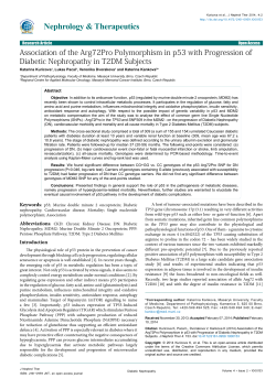
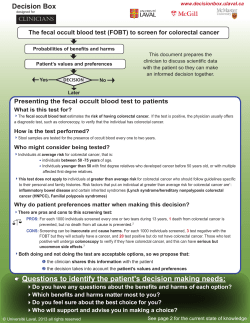
Downloaded from gut.bmj.com on June 9, 2014 - Published by group.bmj.com
Gut 1997; 40: 356-361
356
Detection and monitoring of serum p53 antibodies
in patients with colorectal cancer
P Hammel, B Boissier, M-T Chaumette, P Piedbois, N Rotman, J-C Kouyoumdjian,
R Lubin, J-C Delchier, T Soussi
on a routine clinical basis. Because most
Background-Detection of p53 antibodies mutations modify the confirmation and the
in serum might be an effective indirect stability of the p53 protein and lead to its
procedure to detect alterations of the p53 accumulation in the nucleus of tumour cells,
there has been intensive investigation of
gene.
Aims-To assess the prevalence and the screening p53 alterations by immunohistovariation under treatment of p53 anti- chemical analysis.8 A third approach for the
diagnosis of p53 alterations which has recently
bodies in patients with colorectal cancer.
Patients and methods-Fifty four patients been developed consists of the detection of p53
with colorectal cancer (26 men and 28 antibodies (p53-Abs) in the serum of patients
women, mean age 65, range 33-90 years) affected by a wide variety of cancers.9 16 The
and 24 patients with non-malignant presence of these antibodies usually correlates
digestive disease were tested for p53 with the accumulation of the p53 protein in
antibodies by enzyme linked immuno- tumour cells, but there are some excepsorbent assay (ELISA), and for the car- tions.17 18 p53-Abs belong to IgG, or IgG2
cinoembryonic antigen and carbohydrate classes, suggesting that they correspond to a
antigen 19*9. Immunohistochemical detec- secondary immune response induced by two
tion of p53 protein tumour overexpression immunodominant regions localised in the
carboxyterminus and the aminoterminus of the
was performed in 38 cases.
Results-Fourteen patients (26%) with p53 protein outside the central mutational
colorectal cancer but none of those with hotspot region.19-21 A recent study has clearly
non-malignant disease displayed p53 anti- shown that these antibodies are directed
bodies. Overexpression of p53 was shown toward human p53 either in wild type or
by immunohistochemistry in 22 patients mutant form.22 Serological analysis of p53
(58%), 10 of whom also had p53 anti- alterations has several advantages: (1) it is
bodies. The antibodies were present in relatively easy to perform and to repeat; in
four patients with high carcinoembryonic addition, the stability of p53-Abs allows
Service d'Hepatologie
et de Gastroent6rologie antigen and three patients with high retrospective studies; (2) it does not require
P Hammel
carbohydrate antigen 19*9 concentrations, tumour material; (3) it is of potential interest
J-C Delchier
but also in 10 patients (33.30/6) with normal for monitoring patients with cancer.
Colorectal cancer is the most common
values of these markers. The ratio of p53
Laboratoire de
Biochimie
antibodies decreased in 11 of 13 patients gastrointestinal malignancy worldwide, and
B Boissier
after tumour resection. In two patients alterations of the p53 gene are found in about
J-C Kouyoumdjian
variations in p53 ratio strongly correlated 60% of these cases.1 24 To the best of our
Laboratoire
knowledge, p53-Ab expression in colorectal
with tumour relapse or progression.
d'Anatomie
cancer has not been effectively studied.'5
antifor
serum
p53
Conclusion-Testing
Pathologique et
The purpose of this prospective study was to
for
technique
a
useful
constitutes
bodies
Cytologique
M-T Chaumette
assessing alterations in p53 and may help assess: (1) the prevalence of p53-Abs in
physicians to follow up patients with colorectal cancer and to compare this with the
Service de
widely used tumour markers the carcinoemcolorectal cancer.
Cancerologie
bryonic antigen (CEA) and the carbohydrate
(Gut 1997; 40: 356-361)
P Piedbois
antigen (Ca) 19-9; (2) the specificity of p53-Ab
Service de Chirurgie
Keywords: p53 gene, p53 antibodies, colorectal cancer, testing in patients with various digestive nonDigestive, H6pital
carcinoembryonic antigen, carbohydrate antigen 19-9.
malignant diseases; (3) the potential value of
Henri Mondor, 94010
Creteil, France
p53-Ab monitoring for the management of
N Rotman
Inactivation of the p53 tumour suppressor patients with colorectal cancer.
gene is the most common genetic alteration in
Unite 301 INSERM,
Institut de G6netique
human cancers.' 2 This alteration is usually
Moleculaire, 75010
caused by missense point mutations of the Patients and methods
Paris, France
Sometimes, inactivation of the p53
gene.
R Lubin
T Soussi
protein may occur through complex formation Patients' characteristics
with cellular proteins.3 Alterations in p53 have Fifty four patients, 26 men and 28 women,
Correspondence to:
Dr P Hammel,
prognostic value in colon, breast, and gastric were prospectively studied. The mean age was
Service de Gastroenterologie,
cancers and so their recognition may be 65 (range 33-90). All patients were treated at
Hopital Beaujon, 100
boulevard Leclerc, 92110
important for clinicians.'7 Although the most our institution between March 1994 and
Clichy, France.
accurate procedure for analysis of p53 status is August 1995 for a histologically established
Accepted for publication
DNA sequencing, this technique is not feasible colorectal adenocarcinoma.
31 October 1996
Abstract
Downloaded from gut.bmj.com on June 9, 2014 - Published by group.bmj.com
Antibodies to p53 in colorectal cancer
Tumours
According to the Dukes' classification,25 10
patients were stage A, 19 were stage B, and 25
were stage C (nine with liver metastases). The
site of the primary tumour was the rectum in
14 cases, the left colon in 26 cases, and the
right colon in 14 cases. Patients who had
locally advanced rectal cancer (Dukes' B or C)
received preoperative irradiation (40 Gy). All
patients studied underwent resection of the
colorectal tumour, including those with distant
metastases.
Serum samples
Serum samples of the 54 patients with colorectal cancer were collected after diagnosis
but before treatment during routine blood
sampling. Serum samples of 24 patients with
various non-malignant digestive diseases (benign colonic adenoma, familial adenomatous
polyposis, Peutz-Jeghers syndrome, gastritis,
alcoholic cirrhosis, and chronic viral hepatitis)
were also collected. Patients were informed
and gave their consent for the study. Whole
blood was centrifuged at 3000 rpm for 15
minutes and the supernatant was stored at
-80°C until use.
Enzyme linked immunosorbent assay (ELISA)
Enzyme linked immunosorbent assay (ELISA)
for the detection of p53-Abs in serum has
been previously described.22 Briefly, we have
developed a highly specific ELISA by testing all
samples with a two antigen preparation. The
first preparation contained the relevant antigen
(purified p53 protein) and in the second the
antigen was omitted. All results have been
expressed as the ratio between the number
of wells containing the p53 antigen and the
corresponding wells without. We have previously reported that samples of blood donors
(200) and patients with various carcinomas
(more than 1000) indicated that a ratio higher
than 2-0 confirms the presence of p53-Ab. All
analyses were performed in duplicate.
Carcinoembryonic antigen and carbohydrate
antigen 19 9
The antigens CEA and Ca 19 9 were tested
with commercially available radioimmunoassay kits (Boehringer, Mannheim, Germany).
Cut off concentrations of 5 ng/ml and 37
U/ml respectively, were recommended by the
manufacturers.
Immunohistochemistry
Immunostaining for p53 was performed on 3
,um deparaffinised sections using the monoclonal antibody anti-p53 D07 (Dakopatts A/S,
Glostrup, Denmark) which recognises, on both
wild type and mutant forms of the proteins, an
epitope in the N-terminal part of the human
p53 protein between amino acids 35 and 45.
A preliminary treatment was performed by
incubating the deparaffinised slides twice for
five minutes at 650 W in citrate buffer (pH 7 6)
357
Sections were then left
for 90 minutes before
incubation with the D07 antibody at a
concentration of 1:50 for 30 minutes.
The pathologist who performed the immunohistochemical analysis was not informed of the
patients' p53-Ab status.
in
a
microwave
oven.
at room temperature
Statistical analysis
Comparisons of the patients' clinical status
(age, sex, localisation of tumour, stage) according to p53-Ab status were performed by a
logistic regression test.
Results
Detection ofp53-Ab in serum
Among the 54 patients with colorectal cancer,
14 (26%) had p53-Abs at a ratio varying from
2 1 to 57. By contrast, all 24 patients with nonmalignant digestive disease had a p53/control
ratio lower than 2-0. Figure 1 shows these
results. The Table describes the clinical
characteristics, treatment, and follow up of
patients with p53-Abs.
Patients with p53-Abs were significantly
younger than patients without p53-Abs (54 v
66 years, p<0.03) but other variables (sex,
localisation of tumour, stage) were not associated with the presence of p53-Ab.
Detection ofp53 overexpression by
immunohistochemistry
Overexpression of the p53 protein was detected by immunohistochemistry in 22 out of
38 patients (58%) for whom results were
available. Ten of these 22 patients (45%) had
p53-Abs in serum. The level of p53 expression
by immunohistochemistry was similar in
patients with or without p53-Abs. On the other
hand, one stage B, and one stage C patient
displayed p53-Abs in the absence of any
detectable p53 overexpression in several tumour
samples studied by immunohistochemistry.
CEA and CA 19 9
Values for CEA were above normal in 20 out
4
60 _
0
440
CY
40 _-
20
0.
cv
LO
QL
..VI
5~~~~~~~~
-2 -
I
-------
-----*
*Nm-
Non-malignant Colorectal cancer
digestive disease
Figure 1: p53 Antibodies in 54 patients with colorectal
cancer and in 24 patients with non-malignant digestive
disease.
Downloaded from gut.bmj.com on June 9, 2014 - Published by group.bmj.com
Hammel, Boissier, Chaumette, Piedbois, Rotman, Kouyoumdjian, Lubin, Delchier, Soussi
358
of 54 patients (37%) tested before treatment.
Four of these patients also had high p53-Ab
concentrations in serum. Values for Ca 19 9
were above normal in 15 out of 54 patients
(28%) tested before treatment. Three of these
patients also had high p53-Ab concentrations
in serum. Among the 30 patients with normal
CEA and Ca 19 9 values, 10 (33-3%) had high
p53-Ab concentrations. By contrast, all 24
patients with non-malignant digestive disease
had normal CEA and Ca 19 9 values.
Monitoring ofp53-Abs
Twelve patients with p53-Abs were tested
several times to identify the variation of ratio
within the follow up period. The Table gives
the results. Five of the eight patients (CC1,
CC21, CC24, CC38, CC47), who were tested
within the first month after tumour resection,
showed a 225% drop in the p53-Ab ratio.
Three of the eight patients (CC3, CC34,
CC38) tested one year or more after tumour
resection, returned to normal p53-Ab ratios.
Three other patients (CC6, CC30, CC49) still
had p53-Ab but showed a considerable decrease (of 30%, 79% and 72% respectively) in
concentrations (Table). These patients were
asymptomatic 23, 21, and nine months after
tumour resection, respectively. Patient CC49 is
described in detail.
Patient CC49-This 33 year old woman had a
history of .10 years of ulcerous pancolitis. In
March 1994, multiple biopsies throughout the
colon were performed during a systematic complete colonoscopy. Several foci of severe dysplasia were seen at histological examination of
the left colon samples. The patient underwent
total colectomy with ileo-anal anastomosis.
Examination of the resected colon confirmed
the presence of several scattered areas of high
grade dysplasia. Moreover, a focal (<1 cm2)
cancer with transparietal involvement and a
single juxtatumorous metastatic lymph node 5
mm in diameter was discovered. The patient
was diagnosed with a Dukes' C colon cancer.
Overexpression of p53 was demonstrated not
only at the cancer site but also on the adjacent
non-tumorous mucosa. The p53 ratio decreased
after tumour resection (Table). Concentrations
of p53-Abs are still being monitored.
In two patients, a postoperative increase in
p53-Ab concentrations was seen when tumour
relapse or progression was diagnosed. Both
findings are described in detail below (patients
CC24 and CC37).
Patient CC24-This 44 year old female patient
was operated on in March 1994 for a Dukes'
B adenocarcinoma involving the sigmoid colon.
This patient received chemotherapy consisting
of a combination of 5 fluorouracil and folinic
acid (nine courses) within the 10 months after
surgery. Before treatment, she had high serum
concentrations of p53-Abs (ratio 58 1). Immunohistochemical analysis of the resected
specimen showed overexpression of the p53
protein. Preoperative serum values for CEA
and Ca 19-9 were normal; p53-Abs fell within
the first months after tumour resection and
remained stable at a fairly high concentration
(ratios of between 13 and 18). In June 1995,
the p53-Ab ratio increased to 31 8 but the
patient remained asymptomatic. A colorectal
perianastomosis tumour relapse was suspected
by CT and confirmed by endo-ultrasonography.
The patient underwent laparotomy for complete resection of a tumorous mass measuring
25 X 50 mm in diameter. There were no
peritumorous metastatic nodes nor other
patent tumour localisation in the abdomen.
The p53-Ab ratio decreased once again after
this second operation (Fig 2). Immunohistochemistry of the resected specimen also
disclosed p53 overexpression. In April 1996,
the patient suddenly died of pulmonary
embolism although her p53-Ab concentrations
had been stable (ratio 10 to 12) for seven
months. A postmortem examination was not
done.
Patient CC37-This 37 year old female patient
had a Dukes' C rectal adenocarcinoma which
was diagnosed at the end of October 1994. The
patient had high concentrations of p53-Abs in
serum (ratio 65 7), but CEA and Ca 19-9
concentrations were normal. Overexpression
of p53 was shown by immunohistochemical
analysis performed on biopsy samples obtained
before treatment. Radiotherapy (45 Gy) was
given preoperatively. In January 1995, she
underwent tumour resection followed by distal
colorectal anastomosis and then received
chemotherapy consisting of a combination of
Clinical characteristics, treatment, andfoHlow up of the 14 patients with colorectal cancer and p53 antibodies
p53 Ratio
Patient
No
CC1
CC3
CC6
CC18
CC21
CC24
CC30
CC34
CC37
CC38
CC43
CC47
CC49
CC53
Site!
Before
treatment
26-4
Sex
Age
Dukes' stage
F
F
F
M
M
F
M
F
F
M
F
M
F
F
54
54
52
56
54
44
76
84
36
73
87
62
33
53
LC/C (lm)
LC/C
RC/B
LC/C (1m)
RC/C (lm)
LC/B
LC/B
RC/B
Re/C
LC/C
CD/B
Re/C
LC/C
LC/B
4-0
3-2
39
36-9
58-1
36-4
2-1
65
11-1
3-2
3-4
16-6
2-7
After resection ofprimary
tumour (months)
.12
3
<1
Postoperative
chemotherapy
Follow up time
after tumour resection
11
ND
40
ND
49
36-9
ND
2-8
ND
8-4
3-8
1-4
ND
-
No
Yes
Yes
Yes
Yes
Yes
No
No
Yes
No
No
No
Yes
No
Died at 3 months
8 months/good condition
23 months/good condition
Died at 9 months
Died at 3 months
Died at 24 months (see Fig 2)
21 months/good condition
15 months/good condition
18 months/good condition (see Fig 3)
21 months/good condition
Died at 1 month
Died at 2 months
9 months/good condition
Lost to follow up
RC=right colon; LC=left colon, Re=rectum; ND=not done; lm=liver metastasis.
-
ND
4-7
09
-
15-6
ND
ND
40 7
ND
1.0
2-8
-
11-8
7-5
1-3
20-8
1.0
-
ND
-
-
5-4
-
Downloaded from gut.bmj.com on June 9, 2014 - Published by group.bmj.com
Antibodies to p53 in colorectal cancer
359
°CEA
*p53
Resection of the
primitive tumour
60
A"Ca19.9
Diagnosis of relapse
+ surgical resection
50-
30;
Chemotherapy
.4
>
25K
20
Ca 1!9.9
U/nnl
F-
CEA
ng/ml
15
l
4
O-
10
5
5
0
2
6
4
8
10
12
16
14
20
18
Time (months)
Figure 2: Evolution ofp53 antibodies, CEA, and Ca 19*9 serum values in patient CC24
*p53
o CEA
A Ca
J
19.c
60K
Diagnosis of hepatic
metastasis
+ surgical resection
50o
40
+y
Chemotherapy
.4
-
Radiotherapy
30
asymptomatic eight months after hepatic
resection whereas the p53-Ab concentration is
still high (ratio 17). Concentrations of p53-Abs
are still being monitored.
-
Ca 1!9.9
U/nnI
F-
CEA
ng/ml
20
10 F-
20
5
I
0
2
4
6
8
10
-E
12
14
16
18
20
Time (months)
Figure 3: Evolution ofp53 antibodies, CEA, and Ca 19 9 serum values in patient CC3;7
5-fluorouracil and folinic acid. A pronounc-ed
decrease in p53-Ab concentrations was fouind
after tumour resection (Fig 3). In May 19'95,
the p53 ratio again increased but the patitent
remained asymptomatic. Hepatic ultrasornography showed a single hepatic metastaisis
located in the left lobe (3 cm in diameter) and
the patient underwent segmental hepaltic
resection. There were no other appar ent
abdominal tumours. Immunohistochemis t-ry
on the resected metastasis disclosed p)53
overexpression as in the rectal tumotur.
Concentrations of p53-Abs decreased after 1the
second operation (Fig 3). The patient remaiins
Discussion
This work represents the largest published
series examining p53-Ab response in colorectal
cancer. We found that 26% of the patients with
colorectal cancer had p53-Abs. This is the
highest percentage of p53-Abs reported in
patients with cancer compared with those
reported for lung (24%), pancreatic (19%),
bladder (17%) and breast (13%) cancers.22
As the prevalence of p53-Abs seems to be
clearly proportional to the occurrence of p53
mutations,2022 it is not surprising to find
similar prevalences of p53-Abs in colorectal
and lung cancer, two diseases with a 60% rate
of p53 mutations. In a study published by
Angelopoulou et al, 15 a 15% prevalence of
p53-Abs in patients with colon cancer was
reported. The lower rate of p53-Abs in this
series could be due to patient selection bias or
because of the use of different assays. This
emphasises the importance of standardised
techniques and methods for recording p53
antibody titres for comparative studies. In
the present series, only 10 of the 22 patients
who showed accumulation of the p53 protein
by immunohistochemical analysis developed
p53-Abs. In this study, as in a study by Winter
et al.'3 the level of p53 expression by immunohistochemistry did not seem to correlate with
the p53-Ab production. It is not clear why
many tumours with p53 mutations or protein
overexpression are not immunogenic. It is clear
that the stabilisation and accumulation of
mutant p53 proteins are prerequisites for
p53-Ab production.20 Although it has been
shown that p53 mutations leading to the
formation of complexes between p53 and a 70
kDa heat shock protein are found in antibody
eliciting tumours,"I other studies suggest that
p53-Ab expression is not related to specific
mutations of the p53 gene.'3 26 The influence
of other variables in the development of
p53-Abs such as tumour stage, the immune
status of patients, or major histocompatibility
merits further investigation.'
Two patients in the present series showed
p53-Abs without evidence of immunohistochemical overexpression of p53. This has been
previously described,'7 18 20 and several explanations are possible: (1) immunohistochemistry is highly dependent on sampling when the
tumour is heterogeneous. By contrast, p53-Ab
detection represents a more global approach to
detect p53 alterations and is not dependent on
a sample; (2) frameshift mutations of the p53
gene may lead to an immune response without
p53 protein accumulation through modifications of p53 antigen processing or its
presentation to the immune system'7; (3) the
presence of another occult p53-Ab eliciting
cancer cannot be excluded.
In the present study, p53-Ab detection was
100% specific for diagnosis of cancer as none
of the patients with various non-malignant
Downloaded from gut.bmj.com on June 9, 2014 - Published by group.bmj.com
Hammel, Boissier, Chaumette, Piedbois, Rotman, Kouyoumdjian, Lubin, Delchier, Soussi
360
digestive diseases displayed these antibodies.
This finding is supported by previous reports
which show that p53-Abs were specifically
encountered in patients with cancer.'0 15 In this
series, there was also no false positive CEA or
Ca 19'9 in the control patients. However, it has
been shown that false positive increases in
CEA may occur in smokers, and false positive
increases in Ca 19'9 have been found in
patients with cirrhosis, pancreatitis, or acute
cholestasis.27 Immunisation against the p53
antigen leading to p53-Ab production may
represent an early event in the progression of
colorectal cancer.'2 14 In our series,7 patients
with non-metastatic colorectal cancer exhibited
p53-Abs. It has been shown that p53-Abs can
even precede tumour detection."6 We noted that
they did not appear during disease progression
if they were initially absent (data not shown) as
it was previously reported.'2 22
Interestingly, one patient (CC49) with a
history of ulcerous colitis displayed high
p53-Ab titres. This patient was operated on
because of severe dysplasia foci. However, the
very focal Dukes' C colon cancer was missed
in preoperative colonoscopy and was only
found when the whole resected colon was
examined. It has been previously shown that
mutations in the p53 gene represent an early
marker of neoplastic progression in ulcerative
colitis.28 However, neoplastic areas are
sometimes difficult to identify at endoscopy
and can be missed during random biopsies.
Thus the clinical use of p53-Ab testing for the
early detection of p53 alterations in
preneoplastic digestive diseases (ulcerative
colitis or Barrett's oesophagus) merits further
investigation.
At present,
CEA and Ca 19 9 are the most
extensively used tumour markers for the management of patients with colorectal cancer.27
Thus we thought that it was important to
compare p53-Ab testing with both these
reference tumour markers. There was a
simultaneous increase in CEA and Ca 19-9,
and high p53-Ab titres in only 20% of patients.
Interestingly, 30% of the patients with normal
CEA and Ca 19 9 concentrations exhibited
high p53-Ab concentrations. Despite the fact
that the sensitivities of CEA and Ca 19 9 (37%
and 28% respectively) for the diagnosis of
colorectal cancer were slightly higher than that
of p53-Ab (26%), our results suggest that CEA
measurement and Ca 19'9 and p53-Ab testing
may be complementary methods for the management of patients with colorectal cancer.
Variations of p53-Abs during treatment of
patients with cancer have been poorly studied.
Angelopoulou et al'5 reported p53-Ab monitoring in five patients with ovarian cancer and
one with breast cancer. In our series, the
p53-Ab ratio decreased in 12 out of 14 patients
within the first months after surgery, including
in
those with Dukes'
C
cancer and
hepatic
metastasis who only underwent palliative
resection. Moreover, we currently reported the
results of repeat testing in 12 patietns with
p53-Ab. In four of them, the follow up was too
short to draw any conclusions. Among the
eight patients followed up for 12 months or
more, six showed a decrease in p53-Ab concentrations but only three of them returned to
a normal p53-Ab ratio. In the three patients,
the persistent high p53-Ab ratio without
evidence for cancer progression may be due to
persistent immunisation against p53 protein.
However, a longer follow up is needed to
ensure that this phenomenon is not caused by
undetectable cancer.
The examples of patients CC24 and CC37
clearly show that temporal changes in the
p53-Ab ratio can be closely correlated with
disease progression or regression. Rapid but
incomplete decreases in both patients' p53-Ab
ratio, followed by a period of stability at a fairly
high concentration (p53 ratio> 15) in one
patient, was found before cancer progression.
In patient CC37, it is possible that hepatic
metastases was present at the time of rectal
tumour resection but undetectable. Large scale
studies of p53-Ab positive patients with colorectal cancer are needed to assess whether
some patterns of change in concentrations of
p53-Abs indicate either incomplete resection of
the tumour or early disease progression.
It has been shown that the presence of circulating p53-Abs in patients with breast cancer is
an independent factor of poor prognosis.23 Thus
whether the use of p53-Ab testing might help
physicians to select subgroups of patients with
colorectal cancer for preoperative or postoperative adjuvant treatments, needs to be
investigated.
We conclude that p53-Ab testing is a
convenient method to detect alterations of the
p53 gene in patients with colorectal cancer.
Monitoring of p53-Ab may help in the early
diagnosis and treatment of relapse in
asymptomatic patients.
We thank Dr MJ Boudet, D Cherqui, PL Fagniez, JL Hingot,
M Julien, and B Tantawi for performing the tumour resections,
and D Roche and Y Remvikos for helping in the preparation
of the manuscript. This work was supported by grants from the
Association de Recherche sur le Cancer, Ligue Nationale contre
le Cancer (Comite de Paris) and Ligue Nationale contre le
Cancer (Comite National).
Addendum
During the period of revision of this manuscript,
patient CC37 who had high persistant concentrations of p53-Ab after resection of a hepatic
metastasis (Fig 3) developed pulmonary metastases.
1 Soussi T, Legros Y, Lubin R, Ory K, Schlichtholz B.
Multifactorial analysis of p53 alteration in human cancer:
a review. IntJ Cancer 1994; 57: 1-9.
2 Hollstein M, Sidransky D, Vogelstein B, Harris C. p53
mutations in human cancers. Science 1991; 253: 49-53.
3 Bourdon JC, D'Errico A, Paterlini P, Grigioni W, May E,
Debuire B. p53 protein accumualtion in european
hepatocellular carcinoma is not always dependent on p53
gene mutation. Gastroenterology 1995; 108: 1176-82.
4 Remvikos Y, Tominaga 0, Hammel P, Laurent-Puig P,
Salmon RJ, Dutrillaux B, Thomas G. Increased p53
protein content of colorectal tumours correlates with poor
survival. Bry Cancer 1992; 66: 758-64.
5 Hamelin R, Laurent-Puig P, Olschwang S, Jego N,
Asselain B, Remvikos Y, et al. Association of p53
mutations with short survival in colorectal cancer.
Gastroenterology 1994; 106: 42-8.
6 Thor AD, Moore DH, Edgerton SM, Kawasaki ES,
Reihsaus E, Lynch H, et al. Accumulation of p53 tumour
suppressor gene protein- an independant marker of
prognosis in breast cancers. J Nad Cancer Inst 1992; 84:
845-55.
7 Starzynska T, Bromley M, Ghosh A, Stem PL. Prognostic
significance of p53 overexpression in gastric and
colorectal carcinoma. BrJ Cancer 1992; 66: 558-62.
Downloaded from gut.bmj.com on June 9, 2014 - Published by group.bmj.com
Antibodies to p53 in colorectal cancer
8 Porter PL, Gown AM, Kramp SG, Coltrera MD.
Widespread p53 overexpression in human malignant
tumours. Am 7 Pathol 1992; 140: 145-53.
9 Crawford LV, Pim DC, Bulbrook RD. Detection of
antibodies against the cellular protein p53 in sera from
patients with breast cancer. IntJ Cancer 1982; 30: 403-8.
10 Caron de Fromentel C, May-Levin F, Mouriesse H,
Lemerle J, Chandrasekaran K, May P. Presence of
circulating antibodies against cellular protein p53 in a
notable proportion of children with B-cell lymphoma. Int
J'Cancer 1987; 39: 185-9.
11 Davidoff AM, Iglehart JD, Marks JR. Immune response to
p53 is dependent upon p53/HSP70 complexes in breast
cancers. Proc Natl Acad Sci USA 1992; 89: 3439-42.
12 Schlichtholz B, Legros Y, Gillet D, Gaillard C, Marty M,
Lane D, et al. The immune response to p53 in breast
cancer patients is directed against immunodominant
epitopes unrelated to the mutational hot spot. Cancer Res
1992; 52: 6380-4.
13 Winter SF, Minna JD, Johnson BE, Takahashi T,
Gazdar AF, Carbone DP. Development of antibodies
against p53 in lung cancer patients appears to be
dependent on the type of p53 mutation. Cancer Res 1992;
52: 4168-74.
14 Labrecque S, Naor N, Thomson D, Matlashewski G.
Analysis of the anti-p53 antibody response in cancer
patients. Cancer Res 1993; 53: 3468-71.
15 Angelopoulou K, Diamandis EP, Sutherland DJA,
Kellen JA, Bunting PS. Prevalence of serum antibodies
against the p53 tumour suppressor gene protein in various
cancers. IntJ Cancer 1994; 58: 480-7.
16 Lubin R, Zalcman G, Bouchet L, Tredanel J, Legros Y,
Cazals D, et al. Serum p53 antibodies as early markers of
lung cancer. Nat Med 1995; 1: 701-2.
17 Preudhomme C, Lubin R, Lepelley P, Vanrumbeke M,
Fenaux P. Detection of serum anti-p53 antibodies and
their correlation with p53 mutations in myelodysplastic
syndromes and acute myeloid leukemia. Leukemia 1994;
8:1589-91.
18 Wild CP, Ridanpaa M, Anttila S, Lubin R, Soussi T,
Husgafvel-Fursiainen K, Vanino H. p53 antibodies in the
sera of lung cancer patients: comparison with p53
361
19
20
21
22
23
24
25
26
27
28
mutations in the tumour tissue. Int J Cancer 1995; 64:
176-81.
Legros Y, Lafon C, Soussi T. Linear antigenic sites defined
by the B-cell response to human p53 are localized
predominantly in the amino and carboxy-termini of the
protein. Oncogene 1994; 9: 2071-6.
Lubin R, Schlichtholz B, Bengoufa D, Zalcman G,
Tredaniel J, Hirsch A, et al. Analysis of p53 antibodies in
patients with various cancers define B-cell epitopes of
human p53: distribution on primary structure and
exposure on protein surface. Cancer Res 1993; 53:
5872-6.
Schlichtholz B, Tredaniel J, Lubin R, Zalcman G,
Hirsch A, Soussi T. Analyses of p53 antibodies in sera of
patients with lung carcinoma define immunodominant
regions in the p53 protein. BrJ Cancer 1994; 69: 809-16.
Lubin R, Schlichtholz B, Teillaud JL, Garay E, Bussel A,
Wild CP, Soussi T. p53 antibodies in patients with various
types of cancer: identification and characterization. Clin
Cancer Res 1995; 1: 1463-9.
Peyrat JP, Bonneterre J, Lubin R, Vanlemmens L,
Foumier J, Soussi T. Prognostic significance of circulating
p53 antibodies in patients undergoing surgery for
locoregional breast cancer. Lancet 1995; 345: 621-2.
Schein PS. Gastrointestinal cancer: a global perspective. In:
JD Ahlgren, JS Macdonald, eds. Gastrointestinal oncology.
Philadelphia: JB Lippincott, 1992: 3-11.
Dukes CE. The classification of cancer of the rectum. _7
Pathol Bacteriol 1932; 35: 323-32.
Guinee Jr DG, Travis WD, Trivers GE, De Benedetti VMG,
Cawley H, Welsh JA, et al. Gender comparisons in human
lung cancer: analysis of p53 mutations, anti-p53 serum
antibodies, and C-erb B-2 expression. Carcinogenesis
1995; 16: 993-1002.
Johnson KA, Wolley PV. The role of tumour markers in the
management of gastrointestinal malignancies. In: JD
Ahlgren, JS Macdonald, eds. Gastrointestinal oncology.
Philadelphia: JB Lippincott, 1992: 63-65.
Brentnall TA, Crispin DA, Rabinovitch PS, Haggitt RC,
Rubin CE, Stevens AC, Burmer GC. Mutations in the
p53 gene: an early marker of neoplastic progression in
ulcerative colitis. Gastroenterology 1994; 107: 369-78.
Downloaded from gut.bmj.com on June 9, 2014 - Published by group.bmj.com
Detection and monitoring of serum p53
antibodies in patients with colorectal
cancer.
P Hammel, B Boissier, M T Chaumette, et al.
Gut 1997 40: 356-361
doi: 10.1136/gut.40.3.356
Updated information and services can be found at:
http://gut.bmj.com/content/40/3/356
These include:
References
Article cited in:
http://gut.bmj.com/content/40/3/356#related-urls
Email alerting
service
Receive free email alerts when new articles cite this article. Sign
up in the box at the top right corner of the online article.
Topic
Collections
Articles on similar topics can be found in the following collections
Colon cancer (1308 articles)
Notes
To request permissions go to:
http://group.bmj.com/group/rights-licensing/permissions
To order reprints go to:
http://journals.bmj.com/cgi/reprintform
To subscribe to BMJ go to:
http://group.bmj.com/subscribe/


© Copyright 2026