
Massive Four-Teritories Stroke: Bilateral Middle and Anterior Cerebral Artery Infarctions
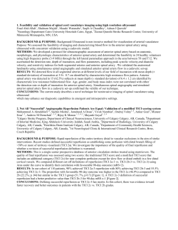
Eur Neurol 2002;47:58–61
Massive Four-Teritories Stroke: Bilateral Middle
and Anterior Cerebral Artery Infarctions
Gerhard F. Hamann a, Ilonka Eisensehr a, Thomas Mayer b
Martin Liebetrau a
Departments of a Neurology and b Neuroradiology, Ludwig
Maximilians University, Klinikum Grosshadern, Munich,
Germany
Introduction
Multiple brain infarction is seen in a limited number of acute
strokes. For example, in the Lausanne stroke register, only 40 of 751
patients with acute first-time stroke of the anterior circulation demonstrated multiple infarctions [1]. Only 10 of the 40 patients with
multiple brain infarction showed bilateral infarctions, related either
to cardioembolism or to large artery disease [1]. Using diffusionweighted sequences (DWI), magnetic resonance imaging (MRI) can
evaluate the age of each lesion, determining whether these multiple
brain infarcts occurred simultaneously or singly [2]. Using this sensitive technique, it is evident that the rate of multiple infarctions could
be higher than previously suspected. In another study, among 142
patients with acute infarcts, 43 had more than one lesion on diffusion-weighted MRI [3]. From these 43 patients, 13 had acute multiple infarctions and 6 had multiple new and old strokes. Thus, about
15% of all patients experienced acute multiple brain infarctions in
this patient group [3]. This rate is substantially higher than previously reported 5% [1]. Here, we report a most striking case of multiple
brain infarctions involving four cerebral artery territories.
Case Report
A woman aged 85 years was admitted to our acute stroke unit
with motor problems and sensory deficit in the left arm. The neurological examination revealed a left-sided paresis of the arm and facial
paresis. A history of atrial fibrillation and coronary heart disease was
noted. The initial MRI examination detected a small lesion on DWI
in the right internal capsule. Less than 1.5 cm in diameter, and
located in the lenticulostriatal territory, this lesion was judged to be a
lacunar infarction (fig. 1b). Multiple older white matter lesions seemingly confirmed this pathophysiological concept. Ultrasonographic
investigations depicted a stenosis of the right internal carotid artery
(ICA) in the carotid siphon area. Magnetic resonance angiography
(MRA) confirmed this discovery, showing a signal reduction in the
C1 and C2 segment of the right ICA. The MRA of the circulus of
Willis revealed the lack of posterior communicating arteries on both
58
Short Reports
Fig. 1. MRI and MRA from the initial investigation (day 1) of our
patient following her admission with slight hemiparesis of the left
arm. a MRA shows the signal loss in the right ICA in the C1 and C2
segment. It is also obvious that both posterior communicating arteries are absent. b MRI shows a small signal hyperdensity in the right
internal capsule at the border zone to the thalamus.
sides (fig. 1a). On day 2 after admission, the neurological deficit had
improved and the patient was apparently symptom free. The neurological examination at this time showed only a slight paresis on the
left side without other symptoms. On day 3, the patient suddenly lost
consciousness. She developed a deep coma without reactivity to
painful stimuli and a progressive loss of brain stem reactivity.
An MRI on the third day detected bilateral signal intensities on
DWI in the territories of the middle cerebral artery (MCA) and anterior cerebral artery (ACA) (fig. 2a). On the MRA, both ICAs were
occluded (fig. 3). The posterior part of the MCA was still partly perfused, which was proven by MR perfusion images. On the fourth day,
the T2 images also showed extensive infarcts in both ACA and MCA
territories, sparing the posterior part of both MCA territories (fig.
2b). The patient never regained consciousness and died on the fourth
day with signs of transtentorial herniation.
The pathological examination showed a small thrombus in the
right ICA. The left ICA was recanalized, and no thrombus in the
heart or aorta could be found.
Short Reports
Discussion
The patient developed a small infarct in the right internal capsule
due to either a lacunar syndrome, large artery disease of the carotid
artery on the right side or atrial fibrillation. The initial MRA showed
a stenosis of the right ICA in the C1 and C2 segment, which ultrasound techniques also proved. Both posterior communicating arteries were aplastic, and most of the carotid artery territories in both
hemispheres were supplied by the left ICA. The initial MRA and
ultrasound investigations demonstrated a normal left ICA without
stenosis. The sudden deterioration of the patient, 3 days after the
initial symptoms, suggested a basilar artery occlusion. The MRI
proved that the basilar artery was open and occlusion of the left ICA
had produced a severe forebrain ischemia in both hemispheres. The
transient occlusion of the left ICA was probably caused by an acute
embolic mechanism on the basis of the underlying atrial fibrillation.
Both the negative MRA and ultrasound investigations before the left
ICA occlusion and the open vessel in the pathological examination
support this hypothesis. After this, the patient experienced a complete forebrain ischemia, which is produced experimentally by ligation of both ICAs in rats and gerbils, causing an ideal situation to
study the effects of global ischemia and the amelioration of that
devastating state [4]. Taking into account the diverse anatomical
structures and physiologies of the animals studied, pathological
examination does suggest striking similarities [5]. The anomalousness of the circulus of Willis with aplasia of both communicating posterior arteries was the anatomic substrate which allowed the development of these extensive bilateral lesions in our patient. The highgrade stenosis of the right ICA was the second prerequisite which
allowed the embolic occlusion of the left ICA to produce a nearly
complete carotid artery territory stroke in both hemispheres. The salvage of the posterior MCA territories on both sides could be
explained by leptomeningeal anastomosis from the posterior cerebral
arteries. Previous reports on bilateral ICA disease refer mainly to
chronic arterial occlusive disease with rather few neurological
changes [6]. In a French group of 19 patients with bilateral ICA
occlusion, only minor neurological symptoms were noted. In young
persons, even acute bilateral ICA occlusion may result in minor neurological symptoms. In one report, a young patient was reported to
have suffered from bilateral ICA occlusion by a blunt trauma with
only a narrow visual field deficit [7]. Just one study from Japan
reports a 68-year-old woman who experienced sudden onset of coma
with bilateral internal carotid occlusion by cardioembolism. This
patient apparently suffered from a similar infarction type to that
reported here, even though the exact lesion location and size was not
detailed [8].
In the opinion of most stroke neurologists, multiple brain infarction is likely caused by cardioembolism; however, recent Korean evidence suggests that mainly large vessel disease causes multiple brain
infarctions in the anterior circulation [9]. In the instances of Yamaguchi et al. [8], our current case and other severely affected patients, a
cardioembolic source plus large vessel disease might contribute to the
pathophysiological background. Various other mechanisms may also
be involved in multiple brain infarctions, such as paraneoplastic
coagulation abnormalities, infections and various diseases [1, 10].
We conclude that total forebrain ischemia in humans is a rare but
devastating condition. Prerequisites can be assumed, such as severe
carotid stenosis, absence of a functional collateral system via the circulus of Willis and the combination of large artery disease and a cardioembolic source. The clinical presentation may mimic the onset of
acute basilar occlusion.
59
Fig. 2. a Diffusion-weighted MRI on the
third day reveals the diffusion deficit in both
ACA and most of both MCA territories.
b The T2 images from the MRI on the fourth
day show a similar distribution to that previously noted on DWI.
Fig. 3. MRA after the onset of coma on day
3 shows a loss of signal in both ICA territories. Note the absence of any carotidal vessels in the intracranial portion.
60
Short Reports
References
1 Bogousslavsky J, Bernasconi A, Kumral E: Acute multiple infarction
involving the anterior circulation. Arch Neurol 1996;53:50–57.
2 Fitzek C, Tintera J, Muller-Forell W, Urban P, Thomke F, Fitzek S, Hopf
HC, Stoeter P: Differentiation of recent and old cerebral infarcts by diffusion-weighted MRI. Neuroradiology 1998;40:778–782.
3 Altieri M, Metz RJ, Muller C, Maeder P, Meuli R, Bogousslavsky J: Multiple brain infarcts: Clinical and neuroimaging patterns using diffusionweighted magnetic resonance. Eur Neurol 1999;42:76–82.
4 Dirnagl U, Thoren P, Villringer A, Sixt G, Them A, Einhaupl KM: Global
forebrain ischaemia in the rat: Controlled reduction of cerebral blood flow
by hypobaric hypotension and two-vessel occlusion. Neurol Res 1993;15:
128–130.
5 Ogata J, Fujishima M, Morotomi Y, Omae T: Cerebral infarction following
bilateral carotid artery ligation in normotensive and spontaneously hypertensive rats: A pathological study. Stroke 1976;7:54–60.
6 Catala M, Rancurel G, Raynaud C, Leder S, Kieffer E, Koskas F: Bilateral
occlusion of the internal carotid arteries. Analysis of a series of 19 patients
(in French). Rev Neurol (Paris) 1995;151:648–656.
7 Sadun AA, Sebag J, Bienfang DC: Complete bilateral internal carotid
artery occlusion in a young man. J Clin Neuroophthalmol 1983;3:63–66.
8 Yamaguchi S, Oki S, Ogasawara H, Sato H, Ito Y, Hibino S: A case of
simultaneous bilateral carotid occlusion (in Japanese). No Shinkei Geka
1997;25:247–251.
9 Roh JK, Kang DW, Lee SH, Yoon BW, Chang KH: Significance of acute
multiple brain infarction on diffusion-weighted imaging. Stroke 2000;31:
688–694.
10 Vassallo R, Remstein ED, Parisi JE, Huston J, Brown RD: Multple cerebral infarctions from nonbacterial thrombotic endocarditis mimicking
cerebral vasculitis. Mayo Clin Proc 1999;74:798–802.
Prof. Dr. Gerhard Hamann, Department of Neurology
Ludwig Maximilians University, Klinikum Grosshadern
Marchioninistrasse 15, D–81377 Munich (Germany)
Tel. +49 89 7095 3670, Fax +49 89 7095 3677
E-Mail [email protected]
Short Reports
61


© Copyright 2026